Article

Therapeutic Effects of Photobiomodulation Combined with Exercise on Patients with Peripheral Artery Disease Plus Diabetic Foot Ulcer: A Pilot and Feasibility Study

1
Division of Physical Therapy, Department of Physical Medicine and Rehabilitation, Ministry of Health and Welfare Feng Yuan Hospital, Taichung 42055, Taiwan
2
Department of Physical Therapy, Graduate Institute of Rehabilitation Science, China Medical University, Taichung 406040, Taiwan
3
Faculty of Health Science, Juntendo University, Tokyo 113-0033, Japan
4
Department of Rehabilitation, China Medical University Hospital, Taichung 404327, Taiwan
*
Author to whom correspondence should be addressed.
Life 2025, 15(9), 1391; https://doi.org/10.3390/life15091391
Submission received: 2 August 2025 / Revised: 28 August 2025 / Accepted: 28 August 2025 / Published: 1 September 2025

Abstract

Background: Diabetic foot ulcers (DFUs) in patients with peripheral artery disease (PAD) are difficult to treat and associated with poor healing outcomes. Photobiomodulation therapy (PBMT) and exercise have shown individual benefits, but evidence on their combined effects is limited. Objective: To evaluate whether PBMT combined with resistance exercise improves wound healing and walking ability in patients with DFU and PAD. Methods: In this pilot randomized trial, 11 patients with DFU and PAD were allocated to either PBMT plus supervised exercise or exercise alone for 4 weeks. Outcome measures included wound size, skin temperature, and 6-min walking distance. Results: PBMT combined with exercise improved wound healing and walking capacity compared with baseline; however, no significant between-group differences were observed. A positive correlation was observed between post-PBMT plantar skin temperature and percentage of wound reduction. Conclusions: PBMT combined with resistance exercise may enhance wound healing and functional mobility in patients with DFU and PAD.

1. Introduction

Diabetic foot ulcers (DFU) are among the most serious complications of diabetes mellitus (DM) and associated with high risk of infection, amputation, and mortality. The global prevalence of diabetic foot ulcers is 6.3% and is higher in patients with type 2 diabetes (6.4%) than in those with type 1 (5.5%) [1]. More than half of these ulcers become infected [2]. About 20% of serious infections lead to some level of lower-extremity amputation [3]. DFUs can lead not only to the loss of the lower extremity, but also to high mortality and morbidity. According to previous studies, about one-third of DFUs fail to heal [4] and progress to lower-extremity amputation.
DM is a major risk factor for developing peripheral artery disease (PAD) due to its association with atherogenesis [5]. Lower-extremity PAD affects >230 million adults worldwide and is associated with an increased risk of various adverse clinical conditions, such as coronary artery disease and stroke [6]. When PAD complicates DFUs, which occurs in up to 50% of cases, it significantly increases the risk of delayed wound healing and lower-extremity amputation. Moreover, PAD also contributes to reduced mobility, impaired quality of life [7,8], and increased cardiovascular risk, making this comorbidity particularly challenging to manage.
Revascularization reduces the risk of major amputation and improves amputation-free survival in patients with severe PAD [9,10]; however, no single revascularization method has been proven superior [11], and more effective adjunctive interventions are needed for DFUs. Photobiomodulation therapy (PBMT) has emerged as a promising approach [12,13,14,15,16], with evidence showing accelerated ulcer healing through reduced inflammation, enhanced granulation tissue formation, fibroblast and keratinocyte proliferation, and collagen synthesis [17,18]. A meta-analysis by Mendes-Costa et al. reported an average wound closure of −5.25 cm2 with PBMT [19], particularly when radiant exposures of 3–7 J/cm2 and wavelengths of 630–660 nm or 850–890 nm were applied [20].
Although exercise was not able to facilitate diabetic ulcer healing [21], exercise may prevent muscle loss in diabetic and PAD patients who experience a faster decline in muscle mass and strength [22,23,24,25]. Patients with diabetic PAD showed less walking distance in exercise testing [26], and diabetic PAD patients presented lower physical activity levels and reduced physical function when compared with non-diabetic PAD patients [27]. Resistance exercise was an effective strategy to counteract the deterioration of muscular performance and walking ability in patients with DM [28,29,30] and patients with PAD [31].
We therefore conducted this study to evaluate whether PBMT combined with exercise improves wound healing and walking ability in patients with DFUs and PAD. To our knowledge, no prior randomized controlled trial has examined this specific combination in this patient population, making this study the first to address this gap.

2. Materials and Methods

2.1. Ethics Statements

This study was conducted in accordance with the Declaration of Helsinki and approved by Feng-Yuan Hospital’s ethical review board (number: 109003). Written informed consent was obtained from all participants.

2.2. Study Design and Participants

Participants were recruited from the patients admitted to the vascular surgery departments at Feng-Yuan Hospital, Taichung, Taiwan. Participants were recruited from the vascular surgery departments or referred by the rehabilitation departments between March 2021 and August 2022. All participants were diagnosed based on the following criteria: (1) diagnosis of Type 2 DM; (2) PAD was diagnosed by an angiogram, duplex ultrasonography, or ankle–brachial blood pressure index [32]; (3) the ulcer was located on a lower extremity; (4) the ulcer had persisted for a minimum of two weeks without any signs of healing or with signs of deterioration. Exclusion criteria were as follows: (1) evidence of acute cellulitis and osteomyelitis in the affected extremity; (2) presence of any of one or more medical conditions, including hepatic, hematologic, or immune disease [33]; (3) presence of malignant disease not in remission for more than 5 years; (4) use of oral or parenteral corticosteroids, immunosuppressive, or cytotoxic agents; (5) pregnancy.
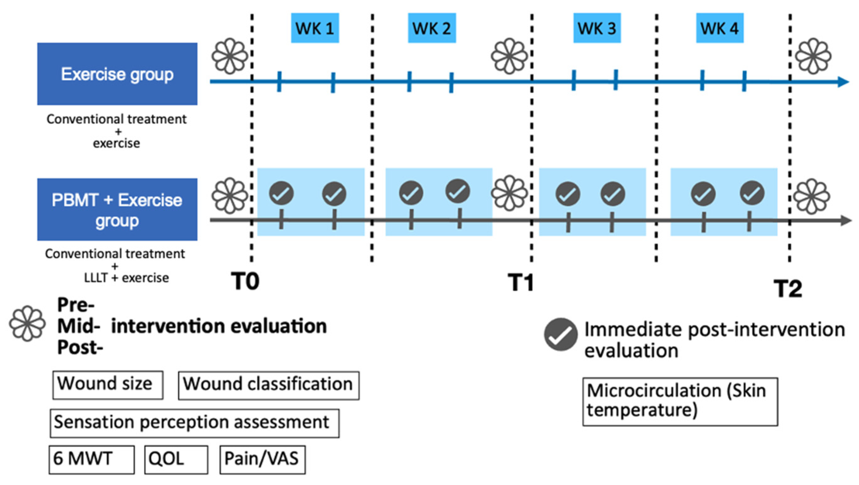
After recruitment and screening for eligibility, the participants were randomized into two groups. Randomization was conducted using a simple, stratified approach. The first participant was assigned to either the intervention or control group using an online random number generator. Subsequently, stratified allocation was performed based on two factors: (1) wound size (≥7 cm2 or <7 cm2) and (2) ambulatory status (ambulatory vs. non-ambulatory) [34,35]. For each stratum, participants were alternately assigned to the PBMT+exercise group or the exercise-only group to ensure balance. Allocation was performed by the principal investigator at the time of enrollment. The experimental group (PBMT+exercise) underwent PBMT combined with exercise therapy, while the exercise group received exercise therapy alone. Both groups received conventional wound treatment. Details are illustrated in Figure 1.

2.3. PBMT Irradiation

Patients were exposed to a light dose of 7.5 mW/cm2 on the wound surface. After the wound was cleaned and washed with normal saline, the wound was dried before being exposed to PBMT. A continuous wave from a gallium–aluminum–arsenide (Ga-Al-As) diode low-level LASER (TI-816-20, Transverse industries, Taiwan) with wavelengths of 660 nm and 830 nm irradiated for 10 min, 2 to 5 times per week. Total treatment time was four weeks. After LASER irradiation, the ulcers were dressed with 1% silver sulfadiazine cream, covered with gauze, and bandaged, or treated according to the physician. The attendance rate was recorded as the percentage of attendance per patient.
PBMT was delivered using a diode laser device equipped with a total of 30 diodes,15 at 660 nm and 15 at 880 nm. The system consisted of five modules (six diodes per module) with a total output ≤1500 mW across a ≤200 cm2 treatment area, yielding an average power density of ~7.5 mW/cm2. Based on device calibration, 10 min of exposure corresponded to ~4.5 J/cm2. The dual-wavelength setting (660 nm + 880 nm) was chosen to combine superficial tissue stimulation (red spectrum, promoting epithelial and dermal repair) with deeper penetration effects (near-infrared spectrum, improving vascular and neural function). The selected energy density (4–7 J/cm2) and wavelength ranges (red: 630–660 nm; near-infrared: 850–890 nm) are consistent with prior systematic reviews and meta-analyses that have identified these parameters as effective for diabetic foot ulcer healing [19].

2.4. Exercise Intervention

The exercise program mainly consisted of resistance exercise and functional activity training. The exercise protocol was individualized because of the participants’ different underlying diseases and different levels of functional abilities. In patients who received bypass surgery, the amount of weight-bearing allowed was determined by the physician. The exercise frequency was twice to 5 times per week and four weeks of treatment.
Resistance exercise: Elastic bands or free weights were used for resistance training, including hip extensor, hip abductor, and knee extensor for lower extremities and lateral pull and biceps curl for upper extremities. The intensity was set at 12–14 RPE (rating of perceived exertion) and each for 1–3 sets of 10–15 repetitions. For participants who were not able to attend the rehabilitation department regularly, home exercise was allowed.
Functional activity training: Functional activity training was implemented if the participants lacked ability. The training followed a progressive sequence of functional activities, starting with rolling, moving to supine to sitting, sitting balance, sitting to standing, standing balance, and stepping, until achieving walking ability.
The exercise programs started at least one week after the participants received bypass surgery and depended on the referral time by the vascular surgery departments. The sequence of PBMT and exercise interventions was randomly assigned for each session.

2.5. Outcome Measures

Outcome measurements were performed before (T0), 2 weeks (T1), and 4 weeks (T2) after the intervention, including wound size and classification, skin temperature (microcirculation), pain intensity, sensation perception assessment, walking ability, and quality of life. Skin temperature was measured immediately after PBMT irradiation in the PBMT + exercise group.
Wound size was measured before PBMT treatment. The wounds were photographed using an iPhone. The ulcer area in each photograph was measured using Image J 1.51 software (US National Institutes of Health, Bethesda, MD, USA). Wound classification was based on the Wagner classification [36]. The percentage of wound reduction was also calculated. If the participants had more than one wound, the mean size and percentage of multiple wounds of the same person were calculated and used to run the statistics.
Skin temperature was measured using, a non-invasive and reliable method for evaluating cutaneous microcirculation [37]. This technique has been validated in patients with diabetes/DFU and used to assess peripheral circulatory status [38,39]. Patients were examined by infrared thermography while maintaining a lying position for 10 min. The room temperature was between 20 °C and 25 °C. The humidity was set constantly. The infrared thermal camera was positioned 1 m away from the foot of the patient. The regions of interest were the dorsal and plantar sides of the foot. Thermal images were carried out with handheld thermography (Sonel KT-80, SONEL S.A., Świdnica, Poland) with a spatial resolution of 80 × 80 pixels and thermal sensitivity of 0.08 °C. The accuracy was ±2 °C or ±2%.
After treatment, each patient was evaluated for pain status. A visual analog scale (VAS) score of wound pain was requested and recorded [40].
Protective sensation was assessed using the 10 g Semmes–Weinstein monofilament (SWMN) at standard plantar sites. This monofilament test is widely recommended for screening diabetic peripheral neuropathy and has demonstrated predictive validity for foot ulcer risk in patients with diabetes [41,42,43]. The buckling force for the 5.07 mono-filament is 10 g, which is also the force felt by the patient when the monofilament bends. Loss of protective sensation leads to the inability to sense the 5.07/10 g SWMN. Ten sites of the foot were tested, including the dorsal surface of the foot between the base of the first and second toes, the first, third, and fifth toes, the first, third, and fifth metatarsal heads, the medial and lateral midfoot, and the heel. If the patient was unable to perceive the monofilament at more than 4 of 10 sites, the sensation was regarded as abnormal [44]. The sensitivity and specificity were 93.1% and 100.0%, respectively.
Walking capacity was measured with the 6-min walking test (6MWT) following standard procedures. The 6MWT is a standardized and validated measure of submaximal functional capacity, and in patients with diabetes, it has been shown to be reliable and associated with cycle ergometer graded exercise test [45]. The 6MWT was carried out in a marked corridor 30 m long. The test involves the patient attempting to cover the maximum distance within 6 min. The patient was allowed to walk at their desired speed but not allowed to trot or run. The patient was allowed to stop and rest at any time, and every such situation was recorded. The maximum walking distance was recorded.
QOL was assessed with the SF-36 (36-item Short-Form Health Survey). The SF-36 has been widely used in patients with DM [46] or PAD [47] for surveying their quality of life. The SF-36 scoring system contains 36 items in 8 domains: physical functioning (PF), physical role functioning (RP), emotional role functioning (RE), vitality (VT), mental health (MH), social functioning (SF), bodily pain (BP), and general health (GH) perceptions. Each domain contained 2–10 items. Moreover, the score for each domain was transformed into a scale from 0 to 100. A higher score indicates higher functioning and better health and QOL.

2.6. Statistical Analysis

Data obtained from the experiments were expressed as mean ± SD. The statistical analysis was performed with the use of GraphPad Prism software (GraphPad Software, Boston, Massachusetts, USA, version 9). The normality of the data was tested with the Shapiro–Wilk test. For intra-group comparison, significant differences of measurement were analyzed using the Friedman test and Wilcoxon signed-rank test, while between-group comparison was analyzed by the Mann–Whitney test. Category data were analyzed by the Chi-square test. Correlations were calculated by nonparametric Spearman correlation to identify the relationships between the covariates. Statistical significance was set at p < 0.05. The missing data of the participants lost to follow-up at T1 were imputed by linear interpolation. These data included the variables of wound size, wound grade, skin temperature, VAS, 6MWT, and scores of the SF-36.

2.7. Ethics

This study was conducted in accordance with the Declaration of Helsinki and approved by the Institutional Review Board of Feng-Yuan Hospital (approval number: 109003; date of approval: 25 March 2020). The trial was registered in the Thai Clinical Trials Registry (TCTR20230810003).

3. Results

3.1. Participants

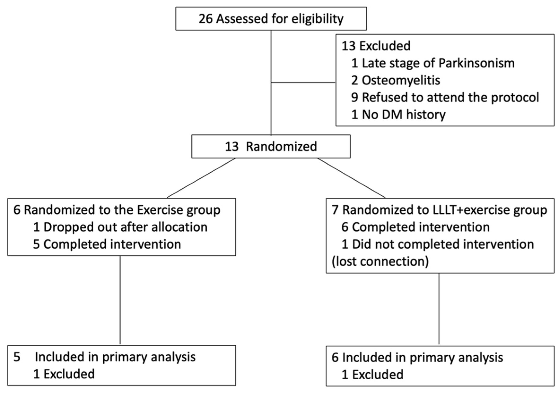
A total of 26 participants were assessed for eligibility, and 13 participants were recruited (Figure 2). Of these patients, one was lost to follow-up and one dropped out after allocation. Finally, the data from 11 participants were analyzed for this study. Of the six patients who completed the PBMT irradiation, an attendance rate of 100% was defined as attending PBMT sessions at least twice per week for four weeks. The average irradiation frequency per week was 2.46 times/week (range of 2–3.75 times/week). Table 1 reports the characteristics of the participants. There were no significant differences in the baseline characteristics between the two groups (p > 0.05).

3.2. Clinical Outcomes Before and After Intervention

Baseline and post-intervention outcome measures for both groups are summarized in Table 2 and Table 3. Within-group analysis showed significant improvements in wound size, percentage of wound reduction, and 6MWT distance in the PBMT + exercise group, whereas no significant changes were observed in the exercise-only group. No significant within-group changes were found in skin temperature, and between-group comparisons revealed no significant differences in any outcome, including the SF-36 scores.

3.3. Between-Group Differences in Clinical and Functional Outcomes

Between-group comparisons of changes from baseline to week 4 are shown in Table 4 (clinical outcomes) and Table 5 (SF-36 scores). No statistically significant differences were observed in any outcome measure. Effect size analysis indicated large effects for wound size, plantar skin temperature, and pain reduction, and a moderate effect for 6MWT, all favoring PBMT + exercise. By contrast, most SF-36 domains showed small effect sizes, except for the body pain subscale, which demonstrated a large effect favoring PBMT + exercise. These findings suggest potentially meaningful clinical benefits despite the lack of statistical significance, which may be related to the small sample size and variability among participants.

3.4. Effects of PBMT on Sensation Perception

Results of the monofilament assessment are shown in Table 6. No significant between-group differences were observed at baseline or after the intervention. Although the number of detectable sites increased in some feet, impaired status persisted in ulcerated legs. The proportion of normal feet increased in both groups by the end of the intervention.

3.5. Correlation Between Skin Temperature and Percentage of Wound Reduction

Although skin temperature in the wounded leg did not change significantly between groups, a Spearman’s correlation analysis was conducted to examine the relationship between skin temperature and the percentage of wound reduction. As shown in Table 7, a strong positive correlation was observed between plantar-side skin temperature and wound reduction, particularly at T1 (rs = 0.8857, p = 0.0333) and the mean of T1 and T2 (rs = 0.9429, p = 0.0167) in the PBMT + exercise group.

4. Discussion

To our knowledge, this is the first study to investigate the combined effects of PBMT and resistance exercise in patients with diabetic PAD and foot ulcers. The 4-week intervention significantly improved wound healing and walking capacity and revealed a strong positive correlation between post-PBMT plantar-side skin temperature and the percentage of wound reduction.
PBMT has been reported to accelerate wound healing in DFUs by reducing inflammation, enhancing granulation tissue, and stimulating fibroblast and keratinocyte proliferation, as well as collagen synthesis [12,13,14,15,16,17,18]. Beyond tissue repair, PBMT also promotes angiogenesis and improves microcirculation, effects that are partly mediated through nitric oxide (NO) signaling [38,48,49,50,51,52,53]. Since impairment of NO signaling and endothelial dysfunction are common in patients with PAD and diabetic neuropathy [54,55], these mechanisms are particularly relevant for this population. Taken together, these biological effects are consistent with the improvements in wound healing and walking capacity observed in the PBMT + exercise group in our study.
Notably, the PBMT + exercise group demonstrated a significant improvement in 6-min walking distance, highlighting the potential of this combined intervention to restore mobility in patients with diabetic PAD. Compared to 18 m in the exercise-only group, the PBMT + exercise group gained 30.67 m on average.
While both groups received resistance and functional training, some participants in the control group appeared to rely more on home-based exercise, which may have limited the effectiveness of the intervention. In contrast, participants in the PBMT + exercise group were required to visit the hospital regularly for PBMT sessions, which also increased the likelihood of receiving exercise under supervision. This likely contributed to better adherence and greater training efficacy, but also constitutes a potential source of bias when interpreting the greater improvements observed in the PBMT + exercise group.
Previous studies have shown that supervised exercise improves walking ability more than unsupervised programs [56,57], and that diabetic PAD patients, who often have reduced physical activity and mobility [27], may respond less to exercise alone. According to McDermott et al. [31], the 6MWT of participants with PAD who did not engage in exercise interventions declined over time. In our study, the exercise-only group maintained but did not significantly improve their walking capacity, suggesting that the exercise protocol was sufficient to prevent further decline. However, only the PBMT + exercise group showed significant improvement, which could be attributable to additive benefits of PBMT—physiologically through improved circulation, and behaviorally through enhanced adherence with hospital-based supervision. These findings support the potential of PBMT as a valuable adjunct to exercise in maintaining or restoring functional mobility in patients with diabetic PAD.
Our findings also provide exploratory insights into potential markers and outcomes for future research. Persistently low plantar temperature after PBMT was associated with less wound healing, consistent with the non-thermal mechanism of PBMT [58] and prior reports linking baseline skin temperature with treatment responsiveness [59]. These findings are consistent with plantar temperature serving as an early biomarker of PBMT effects, and with infrared thermography being considered as a tool for monitoring and patient stratification. In addition, effect size analysis revealed a large effect in the SF-36 Body Pain subscale, favoring PBMT + exercise, indicating possible benefits for pain-related quality of life. While preliminary, these observations highlight potentially meaningful clinical signals that merit confirmation in larger, adequately powered trials.
This study has several limitations. First, the sample size was small, which limits statistical power and increases the risk of type II error. Second, although wounds improved significantly within the PBMT + exercise group, no significant differences were found between the groups. The mean PBMT frequency in our study was 2.46 sessions/week (range 2–5), which falls within the range of previous studies (2 sessions/week to daily) [20]. However, lower frequencies may attenuate between-group effects, and Lopez and Brundage [60] reported that daily laser exposure was most effective for wound healing in animal models. Therefore, the relatively low frequency in our study could partly explain the absence of significant between-group improvements. Third, participants in the PBMT + exercise group were required to attend hospital visits for PBMT sessions, which increased the likelihood of receiving supervised exercise. While this may have contributed to better adherence and training efficacy, it also represents a potential source of bias when interpreting the greater improvements observed in this group. Finally, quality of life was assessed using the SF-36, which has been widely applied in patients with diabetes and PAD to capture overall health-related quality of life. However, disease-specific instruments, such as the Diabetic Foot Ulcer Scale–Short Form, may provide greater sensitivity to ulcer-related changes. The choice of the SF-36 was consistent with the feasibility focus of this pilot trial, but future studies could benefit from incorporating disease-specific QoL tools to better capture patient-reported outcomes.
5. Conclusions

In conclusion, this pilot study provides preliminary evidence that combining PBMT with resistance exercise enhances wound healing and walking ability in patients with diabetic PAD and foot ulcers. A positive correlation was observed between plantar skin temperature and wound reduction after PBMT, which is consistent with an underlying physiological response related to treatment effects. Although the sample size was small, the findings support the potential of PBMT as a valuable adjunct to exercise-based rehabilitation. Further large-scale studies are warranted to confirm these effects and to explore patient-specific factors influencing responsiveness to PBMT. If validated in larger trials, this combined approach could be integrated into multidisciplinary care models to improve patient outcomes.

Author Contributions

S.-Z.C., T.T., H.-J.L., H.-H.S. and Y.-J.C. contributed to the study concept and design. Material preparation, data collection, and data analyses were performed by Y.-J.C. and S.-Z.C. The first draft of the manuscript was written by S.-Z.C. and revised by Y.-J.C. All authors have read and agreed to the published version of the manuscript.

Funding

This study was supported by the Taiwanese Ministry of Science and Technology (grant number: MOST 108-2410-H-039-009-MY2) and China Medical University (grant number: CMU112-MF-90).

Institutional Review Board Statement

The study was conducted in accordance with the Declaration of Helsinki and approved by the Institutional Review Board of Feng-Yuan Hospital (number:, approval number 109003; date of approval: 25 March 2020). The trial was registered in the Thai Clinical Trials Registry (TCTR20230810003).

Informed Consent Statement

Written informed consent was obtained from all participants prior to study enrollment.

Data Availability Statement

The original contributions presented in this study are included in the article. Further inquiries can be directed to the corresponding author.

Acknowledgments

During the preparation of this manuscript, the authors used OpenAI’s ChatGPT (GPT-4, July 2024 version) to assist in drafting and revising sections of the text, including the abstract and discussion. The authors have reviewed and edited the output and take full responsibility for the content of this publication.

Conflicts of Interest

The authors declare no conflicts of interest.

Abbreviations

The following abbreviations are used in this manuscript:
PBMTPhotobiomodulation
DMDiabetes mellitus
PADPeripheral artery disease
DFUDiabetic foot ulcer
6MWT6-min walking test

References

  1. Zhang, P.; Lu, J.; Jing, Y.; Tang, S.; Zhu, D.; Bi, Y. Global epidemiology of diabetic foot ulceration: A systematic review and meta-analysis (dagger). Ann. Med. 2017, 49, 106–116. [Google Scholar] [CrossRef]
  2. Prompers, L.; Huijberts, M.; Apelqvist, J.; Jude, E.; Piaggesi, A.; Bakker, K.; Edmonds, M.; Holstein, P.; Jirkovska, A.; Mauricio, D.; et al. High prevalence of ischaemia, infection and serious comorbidity in patients with diabetic foot disease in Europe. Baseline results from the Eurodiale study. Diabetologia 2007, 50, 18–25. [Google Scholar] [CrossRef]
  3. Lipsky, B.A.; Berendt, A.R.; Cornia, P.B.; Pile, J.C.; Peters, E.J.; Armstrong, D.G.; Deery, H.G.; Embil, J.M.; Joseph, W.S.; Karchmer, A.W.; et al. 2012 Infectious Diseases Society of America clinical practice guideline for the diagnosis and treatment of diabetic foot infections. Clin. Infect. Dis. 2012, 54, e132–e173. [Google Scholar] [CrossRef] [PubMed]
  4. Jeffcoate, W.J.; Chipchase, S.Y.; Ince, P.; Game, F.L. Assessing the outcome of the management of diabetic foot ulcers using ulcer-related and person-related measures. Diabetes Care 2006, 29, 1784–1787. [Google Scholar] [CrossRef] [PubMed]
  5. Gerhard-Herman, M.D.; Gornik, H.L.; Barrett, C.; Barshes, N.R.; Corriere, M.A.; Drachman, D.E.; Fleisher, L.A.; Fowkes, F.G.; Hamburg, N.M.; Kinlay, S.; et al. 2016 AHA/ACC Guideline on the Management of Patients with Lower Extremity Peripheral Artery Disease: Executive Summary: A Report of the American College of Cardiology/American Heart Association Task Force on Clinical Practice Guidelines. Circulation 2017, 135, e686–e725. [Google Scholar] [CrossRef]
  6. Criqui, M.H.; Matsushita, K.; Aboyans, V.; Hess, C.N.; Hicks, C.W.; Kwan, T.W.; McDermott, M.M.; Misra, S.; Ujueta, F.; American Heart Association Council on Epidemiology and Prevention; et al. Lower Extremity Peripheral Artery Disease: Contemporary Epidemiology, Management Gaps, and Future Directions: A Scientific Statement From the American Heart Association. Circulation 2021, 144, e171–e191. [Google Scholar] [CrossRef]
  7. Foley, T.R.; Armstrong, E.J.; Waldo, S.W. Contemporary evaluation and management of lower extremity peripheral artery disease. Heart 2016, 102, 1436–1441. [Google Scholar] [CrossRef]
  8. Maksimovic, M.; Vlajinac, H.; Marinkovic, J.; Kocev, N.; Voskresenski, T.; Radak, D. Health-related quality of life among patients with peripheral arterial disease. Angiology 2014, 65, 501–506. [Google Scholar] [CrossRef]
  9. Wiseman, J.T.; Fernandes-Taylor, S.; Saha, S.; Havlena, J.; Rathouz, P.J.; Smith, M.A.; Kent, K.C. Endovascular Versus Open Revascularization for Peripheral Arterial Disease. Ann. Surg. 2017, 265, 424–430. [Google Scholar] [CrossRef]
  10. Ward, R.; Dunn, J.; Clavijo, L.; Shavelle, D.; Rowe, V.; Woo, K. Outcomes of Critical Limb Ischemia in an Urban, Safety Net Hospital Population with High WIfI Amputation Scores. Ann. Vasc. Surg. 2017, 38, 84–89. [Google Scholar] [CrossRef]
  11. Forsythe, R.O.; Apelqvist, J.; Boyko, E.J.; Fitridge, R.; Hong, J.P.; Katsanos, K.; Mills, J.L.; Nikol, S.; Reekers, J.; Venermo, M.; et al. Effectiveness of revascularisation of the ulcerated foot in patients with diabetes and peripheral artery disease: A systematic review. Diabetes Metab. Res. Rev. 2020, 36 (Suppl. S1), e3279. [Google Scholar] [CrossRef]
  12. Schindl, M.; Kerschan, K.; Schindl, A.; Schön, H.; Heinzl, H.; Schindl, L. Induction of complete wound healing in recalcitrant ulcers by low-intensity laser irradiation depends on ulcer cause and size. Photodermatol. Photoimmunol. Photomed. 1999, 15, 18–21. [Google Scholar] [CrossRef]
  13. Minatel, D.G.; Frade, M.A.; Franca, S.C.; Enwemeka, C.S. Phototherapy promotes healing of chronic diabetic leg ulcers that failed to respond to other therapies. Lasers Surg. Med. 2009, 41, 433–441. [Google Scholar] [CrossRef] [PubMed]
  14. Kajagar, B.M.; Godhi, A.S.; Pandit, A.; Khatri, S. Efficacy of low level laser therapy on wound healing in patients with chronic diabetic foot ulcers-a randomised control trial. Indian J. Surg. 2012, 74, 359–363. [Google Scholar] [CrossRef] [PubMed]
  15. Mathur, R.K.; Sahu, K.; Saraf, S.; Patheja, P.; Khan, F.; Gupta, P.K. Low-level laser therapy as an adjunct to conventional therapy in the treatment of diabetic foot ulcers. Lasers Med. Sci. 2017, 32, 275–282. [Google Scholar] [CrossRef] [PubMed]
  16. Tantawy, S.A.; Abdelbasset, W.K.; Kamel, D.M.; Alrawaili, S.M. A randomized controlled trial comparing helium-neon laser therapy and infrared laser therapy in patients with diabetic foot ulcer. Lasers Med. Sci. 2018, 33, 1901–1906. [Google Scholar] [CrossRef]
  17. Houreld, N.N.; Ayuk, S.M.; Abrahamse, H. Expression of genes in normal fibroblast cells (WS1) in response to irradiation at 660 nm. J. Photochem. Photobiol. B 2014, 130, 146–152. [Google Scholar] [CrossRef]
  18. Silveira, P.C.; Streck, E.L.; Pinho, R.A. Evaluation of mitochondrial respiratory chain activity in wound healing by low-level laser therapy. J. Photochem. Photobiol. B 2007, 86, 279–282. [Google Scholar] [CrossRef]
  19. Dos Santos Mendes-Costa, L.; de Lima, V.G.; Barbosa, M.P.R.; Dos Santos, L.E.; de Siqueira Rodrigues Fleury Rosa, S.; Tatmatsu-Rocha, J.C. Photobiomodulation: Systematic review and meta-analysis of the most used parameters in the resolution diabetic foot ulcers. Lasers Med. Sci. 2021, 36, 1129–1138. [Google Scholar] [CrossRef]
  20. Sutton, E.; Ganie, S.; Chan, C.; Kaur, A.; Nussbaum, E. Photobiomodulation and diabetic foot and lower leg ulcer healing: A narrative synthesis. Foot 2021, 48, 101847. [Google Scholar] [CrossRef]
  21. Flahr, D. The effect of nonweight-bearing exercise and protocol adherence on diabetic foot ulcer healing: A pilot study. Ostomy Wound Manag. 2010, 56, 40–50. [Google Scholar]
  22. Andersen, H.; Nielsen, S.; Mogensen, C.E.; Jakobsen, J. Muscle Strength in Type 2 Diabetes. Diabetes 2004, 53, 1543–1548. [Google Scholar] [CrossRef] [PubMed]
  23. Park, S.W.; Goodpaster, B.H.; Strotmeyer, E.S.; Kuller, L.H.; Broudeau, R.; Kammerer, C.; de Rekeneire, N.; Harris, T.B.; Schwartz, A.V.; Tylavsky, F.A.; et al. Accelerated loss of skeletal muscle strength in older adults with type 2 diabetes: The health, aging, and body composition study. Diabetes Care 2007, 30, 1507–1512. [Google Scholar] [CrossRef] [PubMed]
  24. Park, S.W.; Goodpaster, B.H.; Lee, J.S.; Kuller, L.H.; Boudreau, R.; de Rekeneire, N.; Harris, T.B.; Kritchevsky, S.; Tylavsky, F.A.; Nevitt, M.; et al. Excessive loss of skeletal muscle mass in older adults with type 2 diabetes. Diabetes Care 2009, 32, 1993–1997. [Google Scholar] [CrossRef]
  25. McDermott, M.M.; Tian, L.; Ferrucci, L.; Liu, K.; Guralnik, J.M.; Liao, Y.; Pearce, W.H.; Criqui, M.H. Associations between lower extremity ischemia, upper and lower extremity strength, and functional impairment with peripheral arterial disease. J. Am. Geriatr. Soc. 2008, 56, 724–729. [Google Scholar] [CrossRef]
  26. Mahe, G.; Ouedraogo, N.; Leftheriotis, G.; Vielle, B.; Picquet, J.; Abraham, P. Exercise treadmill testing in patients with claudication, with and without diabetes. Diabet. Med. 2011, 28, 356–362. [Google Scholar] [CrossRef]
  27. Correia, M.A.; Silva, G.O.; Longano, P.; Trombetta, I.C.; Consolim-Colombo, F.; Puech-Leao, P.; Wolosker, N.; Cucato, G.G.; Ritti-Dias, R.M. In peripheral artery disease, diabetes is associated with reduced physical activity level and physical function and impaired cardiac autonomic control: A cross-sectional study. Ann. Phys. Rehabil. Med. 2021, 64, 101365. [Google Scholar] [CrossRef]
  28. Orlando, G.; Balducci, S.; Bazzucchi, I.; Pugliese, G.; Sacchetti, M. Neuromuscular dysfunction in type 2 diabetes: Underlying mechanisms and effect of resistance training. Diabetes Metab. Res. Rev. 2016, 32, 40–50. [Google Scholar] [CrossRef]
  29. Otterman, N.M.; van Schie, C.H.; van der Schaaf, M.; van Bon, A.C.; Busch-Westbroek, T.E.; Nollet, F. An exercise programme for patients with diabetic complications: A study on feasibility and preliminary effectiveness. Diabet. Med. 2011, 28, 212–217. [Google Scholar] [CrossRef]
  30. Francia, P.; Anichini, R.; Bellis, A.D.; Seghieri, G.; Lazzeri, R.; Paternostro, F.; Gulisano, M. Diabetic foot prevention the role of exercise therapy in the treatment of limited joint mobility, muscle weakness and reduced gait speed. Ital. J. Anat. Embryol. 2015, 120, 21–32. [Google Scholar]
  31. McDermott, M.M.; Ades, P.; Guralnik, J.M.; Dyer, A.; Ferrucci, L.; Liu, K.; Nelson, M.; Lloyd-Jones, D.; Van Horn, L.; Garside, D.; et al. Treadmill exercise and resistance training in patients with peripheral arterial disease with and without intermittent claudication: A randomized controlled trial. J. Am. Med. Assoc. 2009, 301, 165–174. [Google Scholar] [CrossRef] [PubMed]
  32. Lijmer, J.G.; Hunink, M.G.; van den Dungen, J.J.; Loonstra, J.; Smit, A.J. ROC analysis of noninvasive tests for peripheral arterial disease. Ultrasound Med. Biol. 1996, 22, 391–398. [Google Scholar] [CrossRef] [PubMed]
  33. Navratil, L.; Kymplova, J. Contraindications in noninvasive laser therapy: Truth and fiction. J. Clin. Laser Med. Surg. 2002, 20, 341–343. [Google Scholar] [CrossRef] [PubMed]
  34. Sano, M.; Yamamoto, K.; Seo, A.; Akai, A.; Akagi, D.; Takayama, T.; Hoshina, K. Wound healing after revascularization for critical limb ischemia. Int. Angiol. 2019, 38, 225–229. [Google Scholar] [CrossRef]
  35. Hicks, C.W.; Canner, J.K.; Mathioudakis, N.; Sherman, R.; Malas, M.B.; Black, J.H., 3rd; Abularrage, C.J. The Society for Vascular Surgery Wound, Ischemia, and foot Infection (WIfI) classification independently predicts wound healing in diabetic foot ulcers. J. Vasc. Surg. 2018, 68, 1096–1103. [Google Scholar] [CrossRef]
  36. Jalilian, M.; Shiri, S. The reliability of the Wagner Scale for evaluation the diabetic wounds: A literature review. Diabetol. Metab. Syndr. 2022, 16, 102369. [Google Scholar] [CrossRef]
  37. Kanazawa, T.; Nakagami, G.; Goto, T.; Noguchi, H.; Oe, M.; Miyagaki, T.; Hayashi, A.; Sasaki, S.; Sanada, H. Use of smartphone attached mobile thermography assessing subclinical inflammation: A pilot study. J. Wound Care 2016, 25, 177–182. [Google Scholar] [CrossRef]
  38. Schindl, A.; Heinze, G.; Schindl, M.; Pernerstorfer-Schon, H.; Schindl, L. Systemic effects of low-intensity laser irradiation on skin microcirculation in patients with diabetic microangiopathy. Microvasc. Res. 2002, 64, 240–246. [Google Scholar] [CrossRef]
  39. Nagase, T.; Sanada, H.; Takehara, K.; Oe, M.; Iizaka, S.; Ohashi, Y.; Oba, M.; Kadowaki, T.; Nakagami, G. Variations of plantar thermographic patterns in normal controls and non-ulcer diabetic patients: Novel classification using angiosome concept. J. Plast. Reconstr. Aesthetic Surg. 2011, 64, 860–866. [Google Scholar] [CrossRef]
  40. Ruger, L.J.; Irnich, D.; Abahji, T.N.; Crispin, A.; Hoffmann, U.; Lang, P.M. Characteristics of chronic ischemic pain in patients with peripheral arterial disease. Pain 2008, 139, 201–208. [Google Scholar] [CrossRef]
  41. Boulton, A.J.; Armstrong, D.G.; Albert, S.F.; Frykberg, R.G.; Hellman, R.; Kirkman, M.S.; Lavery, L.A.; LeMaster, J.W.; Mills, J.L., Sr.; Mueller, M.J.; et al. Comprehensive foot examination and risk assessment. Endocr. Pract. 2008, 14, 576–583. [Google Scholar] [CrossRef] [PubMed]
  42. Perkins, B.A.; Olaleye, D.; Zinman, B.; Bril, V. Simple screening tests for peripheral neuropathy in the diabetes clinic. Diabetes Care 2001, 24, 250–256. [Google Scholar] [CrossRef] [PubMed]
  43. Armstrong, D.G.; Lavery, L.A.; Vela, S.A.; Quebedeaux, T.L.; Fleischli, J.G. Choosing a practical screening instrument to identify patients at risk for diabetic foot ulceration. Arch. Intern. Med. 1998, 158, 289–292. [Google Scholar] [CrossRef] [PubMed]
  44. Lee, S.; Kim, H.; Choi, S.; Park, Y.; Kim, Y.; Cho, B. Clinical usefulness of the two-site Semmes-Weinstein monofilament test for detecting diabetic peripheral neuropathy. J. Korean Med. Sci. 2003, 18, 103–107. [Google Scholar]
  45. Lee, M.C. Validity of the 6-minute walk test and step test for evaluation of cardio respiratory fitness in patients with type 2 diabetes mellitus. J. Exerc. Nutr. Biochem. 2018, 22, 49–55. [Google Scholar] [CrossRef]
  46. Cai, H.; Li, G.; Zhang, P.; Xu, D.; Chen, L. Effect of exercise on the quality of life in type 2 diabetes mellitus: A systematic review. Qual. Life Res. 2017, 26, 515–530. [Google Scholar] [CrossRef]
  47. Fakhry, F.; Spronk, S.; van der Laan, L.; Wever, J.J.; Teijink, J.A.; Hoffmann, W.H.; Smits, T.M.; van Brussel, J.P.; Stultiens, G.N.; Derom, A.; et al. Endovascular Revascularization and Supervised Exercise for Peripheral Artery Disease and Intermittent Claudication: A Randomized Clinical Trial. J. Am. Med. Assoc. 2015, 314, 1936–1944. [Google Scholar] [CrossRef]
  48. Cury, V.; Moretti, A.I.; Assis, L.; Bossini, P.; Crusca Jde, S.; Neto, C.B.; Fangel, R.; de Souza, H.P.; Hamblin, M.R.; Parizotto, N.A. Low level laser therapy increases angiogenesis in a model of ischemic skin flap in rats mediated by VEGF, HIF-1alpha and MMP-2. J. Photochem. Photobiol. B 2013, 125, 164–170. [Google Scholar] [CrossRef]
  49. Ihsan, F.R. Low-level laser therapy accelerates collateral circulation and enhances microcirculation. Photomed. Laser Surg. 2005, 23, 289–294. [Google Scholar] [CrossRef]
  50. Schindl, A.; Schindl, M.; Schön, H.; Knobler, R.; Havelec, L.; Schindl, L. Low-Intensity Laser Irradiation Improves Skin Circulation in Patients With Diabetic Microangiopathy. Dabetes Care 1998, 21, 580–584. [Google Scholar] [CrossRef]
  51. Maegawa, Y.; Itoh, T.; Hosokawa, T.; Yaegashi, K.; Nishi, M. Effects of near-infrared low-level laser irradiation on microcirculation. Lasers Surg. Med. 2000, 27, 427–437. [Google Scholar] [CrossRef]
  52. de Freitas, L.F.; Hamblin, M.R. Proposed Mechanisms of Photobiomodulation or Low-Level Light Therapy. IEEE J. Sel. Top. Quantum Electron. 2016, 22, 7000417. [Google Scholar] [CrossRef] [PubMed]
  53. Mitchell, U.H.; Mack, G.L. Low-level laser treatment with near-infrared light increases venous nitric oxide levels acutely: A single-blind, randomized clinical trial of efficacy. Am. J. Phys. Med. Rehabil. 2013, 92, 151–156. [Google Scholar] [CrossRef] [PubMed]
  54. Hodges, G.J.; Nawaz, S.; Tew, G.A. Evidence that reduced nitric oxide signal contributes to cutaneous microvascular dysfunction in peripheral arterial disease. Clin. Hemorheol. Microcirc. 2015, 59, 83–95. [Google Scholar] [CrossRef] [PubMed]
  55. Hile, C.; Veves, A. Diabetic neuropathy and microcirculation. Curr. Diabetes Rep. 2003, 3, 446–451. [Google Scholar] [CrossRef]
  56. Collins, T.C.; Lunos, S.; Carlson, T.; Henderson, K.; Lightbourne, M.; Nelson, B.; Hodges, J.S. Effects of a home-based walking intervention on mobility and quality of life in people with diabetes and peripheral arterial disease: A randomized controlled trial. Diabetes Care 2011, 34, 2174–2179. [Google Scholar] [CrossRef]
  57. Fokkenrood, H.J.; Bendermacher, B.L.; Lauret, G.J.; Willigendael, E.M.; Prins, M.H.; Teijink, J.A. Supervised exercise therapy versus non-supervised exercise therapy for intermittent claudication. Cochrane Database Syst. Rev. 2013, CD005263. [Google Scholar] [CrossRef]
  58. Zein, R.; Selting, W.; Hamblin, M.R. Review of light parameters and photobiomodulation efficacy: Dive into complexity. J. Biomed. Opt. 2018, 23, 120901. [Google Scholar] [CrossRef]
  59. Bharara, M.; Cobb, J.E.; Claremont, D.J. Thermography and thermometry in the assessment of diabetic neuropathic foot: A case for furthering the role of thermal techniques. Int. J. Low. Extrem. Wounds 2006, 5, 250–260. [Google Scholar] [CrossRef]
  60. Lopez, A.; Brundage, C. Wound Photobiomodulation Treatment Outcomes in Animal Models. J. Vet. Med. 2019, 2019, 6320515. [Google Scholar] [CrossRef]
Figure 1. Study design and outcomes measures for each phase of the study. WK, week; LLLT, low-level LASER therapy; 6 MWT, 6-min walking test; QOL, quality of life; VAS, visual analogue scale.
Life 15 01391 g001
Figure 2. Consort flow diagram of people through interventions. DM, diabetes mellitus; LLLT, low-level LASER therapy.
Life 15 01391 g002
Table 1. Demographic data.
VariablesExercise Group (n = 5)PBMT + Exercise Group (n = 6)p Value
Age (years)75.60 ± 4.56172.67 ± 10.840.835 a
Gender (M/F)4/13/30.303 b
BMI22.0 ± 2.221.8 ± 3.90.967 a
Education (years)6 ± 06.6 ± 1.3>0.999 a
HbA1C1.6 ± 0.51.4 ± 0.80.469 a
Medical history
  DM duration16.67 ± 5.7716.92 ± 10.77>0.999 a
  HTN4 (80%)6 (100%)0.250 b
  Stroke4 (80%)3 (50%)0.303 b
  MI1 (20%)4 (67%)0.121 b
  Dementia3(60%)2 (33%)0.376 b
  ESRD1 (20%)2 (33%)0.621 b
  Retinopathy4 (80%)4 (66%)0.621 b
  Revascularization (Bypass/PTA/None)5/0/04/1/10.361 b
Cigarette use
  Never/Past/Current smoker2/2/13/3/00.516 b
HBOT therapy4 (80%)5 (83%)0.886 b
Data are expressed as mean ± SD or N (%); a Mann–Whitney tests; b Chi-square tests. BMI: body mass index; HTN: hypertension; MI: myocardial infarction; ESRD: end-stage renal disease; PTA: percutaneous transluminal angioplasty; HBOT: hyperbaric oxygen therapy.
Table 2. Baseline/post-intervention comparisons in outcome measures within and between the groups.
Exercise Group (n = 5)p Value aPBMT + Exercise Group (n = 6)p Value a
T0T1T2 T0T1T2
Wound size (cm2)16.85 ± 20.1014.39 ± 18.6711.66 ± 15.470.1825.15 ± 4.45 #4.20 ± 3.213.35 ± 3.22 #0.006 *
% Wound reduction0 ± 011.50 ± 41.8721.16 ± 59.400.1820 ± 0 #11.47 ± 16.7135.36 ± 18.76 #0.006 *
Wound grade1.6 ± 0.551.5 ± 0.581.6 ± 0.55>0.9991.4 ± 0.801.4 ± 0.801.4 ± 0.80>0.999
Skin temperature (°C)
  Plantar side32.9 ± 2.530.1 ± 2.931.1 ± 2.80.09328.1 ± 2.229.4 ± 2.629.5 ± 1.10.241
  Dorsal side34.0 ± 2.430.8 ± 3.532.0 ± 3.00.95330.6 ± 2.731.6 ± 3.231.7 ± 3.50.570
Wound pain (VAS)3.3 ± 4.22.8 ± 2.66.7 ± 2.90.6663.2 ± 3.23.0 ± 3.11.7 ± 2.30.801
6MWT (m)19.67 ± 17.3919.50 ± 24.1437.67 ± 34.270.50040.00 ± 36.9954.17 ± 42.1370.67 ± 58.12 #0.001 *
Values are mean ± standard deviation. 6MWT: 6-min walking test; PBMT: low-level laser therapy; a: baseline and post-intervention comparison within the group by Friedman test, * statistically significant p < 0.05; #: post hoc significant between T0 and T2.
Table 3. Baseline/post-intervention comparisons in SF-36 within the groups.
Exercise Group (n = 5)p Value aExercise + PBMT Group (n = 6)p Value a
T0T1T2 T0T1T2
SF-36
Physical function (PF)3 ± 4.471.000 ± 2.2361 ± 2.24>0.999912.50 ± 13.699.167 ± 15.3010.83 ± 17.440.3086
Role: Physical (RP)0 ± 00 ± 00 ± 0>0.99990 ± 00 ± 04.167 ± 10.21>0.9999
  Role: Emotional (RE)0 ± 00 ± 00 ± 0>0.999916.67 ± 40.8233.33 ± 51.6433.33 ± 51.64>0.9999
  Vitality (VT)20 ± 17.8026.00 ± 8.21625 ± 7.91>0.999933.75 ± 8.32934.17 ± 18.0043.33 ± 20.900.1687
  Mental health (MH)29 ± 19.435.10 ± 9.18237 ± 10.050.518548.67 ± 22.2647.83 ± 28.4656.33 ± 30.260.4527
Social functioning (SF)25 ± 23.3917.50 ± 16.7735 ± 27.100.746941.67 ± 15.1445.83 ± 21.8939.58 ± 21.530.8724
Bodily pain (BP)33.5 ± 32.0429.00 ± 24.6022 ± 17.800.518537.08 ± 7.48640.42 ± 22.2253.33 ± 32.620.1296
General health (GH)27 ± 20.1936.00 ± 19.4937 ± 21.39>0.999942.50 ± 14.0538.33 ± 21.6043.33 ± 17.800.1481
a: Baseline and post-intervention comparison within groups by Friedman test.
Table 4. Changes in outcome measures (T2–T0) between the exercise and PBMT + exercise groups.
Exercise Group (n = 5)PBMT + Exercise Group (n = 6)p Value aCohen’s d Effect Size
Differences from T0 to T2Differences from T0 to T2
Wound size (cm2)5.197 ± 5.2181.795 ± 2.0220.3290.8597
% Wound reduction 0.962 ± 106.835.36 ± 18.760.93070.4486
Wound grade0 ± 00 ± 0>0.9999NA
Skin temperature (°C)
  Plantar side (°C)−1.740 ± 4.3291.367 ± 2.3530.53680.8917
  Dorsal side (°C)−1.980 ± 4.9061.017 ± 2.6760.53680.7584
VAS2.000 ± 4.950−1.500 ± 4.2310.42420.7601
6MWT (m)18.00 ± 20.9530.67 ± 28.070.59520.5115
a: Comparison of differences between groups by Mann–Whitney U-test. Effect sizes are reported as Cohen’s d. Interpretation: small (0.2), medium (0.5), large (0.8). NA: Effect size not applicable due to zero variance.
Table 5. Between-group differences in SF-36 scores from baseline (T0) to week 4 (T2).
Exercise Group (n = 5)PBMT + Exercise Group (n = 6)p Value aCohen’s d Effect Size
Differences from T0 to T2Differences from T0 to T2
SF-36
Physical function (PF)−2.000 ± 4.472−1.667 ± 6.0550.65370.062
Role: Physical (RP)0 ± 04.167 ± 10.21>0.9999NA
   Role: Emotional (RE)0 ± 016.67 ± 40.82>0.9999NA
   Vitality (VT)3.000 ± 21.109.583 ± 25.120.62770.283
   Mental health (MH)5.000 ± 20.227.667 ± 24.740.69700.118
Social functioning (SF)10.00 ± 33.54−2.083 ± 25.520.64500.405
Bodily pain (BP)−11.50 ± 25.1616.25 ± 27.330.14501.056
General health (GH)10.00 ± 33.350.8333 ± 11.140.95020.368
a: Comparison of differences between groups by Mann–Whitney U-test. Effect sizes are reported as Cohen’s d. Interpretation: small (0.2), medium (0.5), large (0.8). NA: effect size not applicable due to zero variance.
Table 6. Baseline/post-intervention comparisons in sensation perception assessment within and between the groups.
T0 T1 T2
ExercisePBMT + Exercisep Value aExercisePBMT + Exercisep Value aExercisePBMT + Exercisep Value ap Value bp Value c
Wound legNormal21 21 21
Abnormal *140.1869140.1869140.1869>0.9999>0.9999
Unaffected leg Normal12 13 23
Abnormal *220.6592210.8091110.80910.63760.6873
Total leg #Normal33 34 44
Abnormal *360.5186 350.8327250.83270.79850.8578
Unaffected leg was defined as the leg without wounds; * abnormal was defined as patients unable to perceive the monofilament at more 4 out of 10 sites; # the total number of legs was 13, with 6 legs in the control group and 7 legs in the PBMT group; a: comparison between the control and PBMT group by Chi-square tests; b: baseline and post-intervention comparison within the control group by Chi-square test; c: baseline and post-intervention comparison within the PBMT group by Chi-square test.
Table 7. The correlation between skin temperature of wounded leg and % wound reduction in the PBMT group.
T0T1T2Mean of T1 and T2Mean of T0, T1, and T2
rsp Valuersp Valuersp Valuersp Valuersp Value
PBMT + exercise group
Dorsal side0.08571 a0.91940.4286 a0.41940.4286 a0.41940.4286 a0.41940.3714 a0.4972
Plantar side0.1429 a0.80280.8857 a0.0333 *0.6000 a0.24170.9429 a0.0167 *0.8286 a0.0583
Number of XY pairs was 6; a calculated by nonparametric Spearman correlation; * statistically significant, p < 0.05.
Disclaimer/Publisher’s Note: The statements, opinions and data contained in all publications are solely those of the individual author(s) and contributor(s) and not of MDPI and/or the editor(s). MDPI and/or the editor(s) disclaim responsibility for any injury to people or property resulting from any ideas, methods, instructions or products referred to in the content.