Article

Comparative Cranial and Postcranial Osteology of Blanus Species (Squamata: Amphisbaenia) from Türkiye: Insights from Morphological Evolution and Phylogeny

1
Department of Biology, Faculty of Science, Dokuz Eylül University, 35390 İzmir, Türkiye
2
Fauna and Flora Research and Application Center, Dokuz Eylül University, 35390 İzmir, Türkiye
3
Departmant of Biology, Graduate School of Natural and Applied Sciences, Dokuz Eylül University, 35390 İzmir, Türkiye
4
The Ministry of Agriculture and Forestry Food Control Laboratory, Bornova, 35100 İzmir, Türkiye
5
Department of Biology, Faculty of Science, Ege University, 35100 İzmir, Türkiye
6
Department of Biology, Faculty of Arts and Sciences, Recep Tayyip Erdogan University, 53100 Rize, Türkiye
*
Author to whom correspondence should be addressed.
Life 2025, 15(8), 1263; https://doi.org/10.3390/life15081263 (registering DOI)
Submission received: 17 June 2025 / Revised: 4 August 2025 / Accepted: 6 August 2025 / Published: 9 August 2025
(This article belongs to the Special Issue Feature Papers in Animal Science: 4th Edition)

Abstract

The genus Blanus (Amphisbaenia: Blanidae) comprises fossorial, limbless reptiles with cryptic external morphology, making species delimitation particularly challenging. This study presents a comprehensive comparative osteological and geometric morphometric investigation of three Blanus species distributed in Türkiye—B. alexandri, B. aporus, and B. strauchi. Using both dry and cleared-and-stained specimens, diagnostic variations in cranial and postcranial skeletal elements, especially elements within the nasal, maxilla, vomer, squamosal, dentary, and coronoid bones, as well as vertebral counts were identified. A geometric morphometric analysis of the dorsal and ventral cranial morphology revealed distinct shape differences, particularly separating B. alexandri from the other two species along principal component axes. A phylogenetic analysis based on 45 discrete osteological characters supported the monophyly of the eastern Blanus clade, with B. alexandri forming a distinct lineage from B. aporus and B. strauchi. These findings emphasize the significance of skeletal morphology for resolving phylogenetic relationships and highlight the role of osteological characters in refining species boundaries within cryptic reptilian taxa. The integrative approach employed here underscores the evolutionary distinctiveness of Anatolian Blanus and enhances our understanding of morphological evolution in amphisbaenians.

1. Introduction

The genus Blanus Wagler, 1830 (Amphisbaenia: Blanidae) includes seven limbless, fossorial species with a broad geographical distribution [1,2,3]. Three of these species are widely distributed in Türkiye [4]: Blanus strauchi [5] occurs in Western Anatolia and the southern regions of the country, extending as far as Antalya. Blanus aporus [6] ranges from Antalya to the vicinity of Adana. In contrast, B. alexandri [2] is found in the Eastern Mediterranean and Southeastern Anatolia.
Taxonomic studies of this genus have traditionally relied on morphological characteristics. However, the inability to distinguish species solely based on morphology—along with relatively minor morphological variation despite marked genetic divergence—has led to significant challenges for species delimitation. In this context, osteological analyses offer a valuable tool for resolving taxonomic ambiguities. Previous studies on amphisbaenians have primarily addressed phylogenetic relationships, osteology, and external morphology [1,2,7,8]. In addition, geometric morphometric approaches have been widely employed to explore shape variation across taxa [2,9,10,11]. Despite these efforts, osteological studies of amphisbaenians remain scarce (see Appendix A). Research on Blanus species has generally focused on aspects such as phylogeny, distribution, microhabitat preference, new locality records, activity patterns, ear morphology, and helminth fauna [8,12,13,14,15,16,17,18,19,20,21]. Osteological investigations of Blanus species date back to the 19th century [5,22,23,24,25,26,27], with the most comprehensive recent contribution provided by Villa et al. [28], who examined the cranial osteology of all extant species within the Blanidae family. Molecular studies have resolved as two major clades within the genus: eastern and western [3,8]. The eastern clade, which includes B. alexandri, B. aporus, and B. strauchi, all distributed in Türkiye, was the focus of detailed cranial osteological comparisons in Villa et al.’s study [28]. However, data on the appendicular skeleton of amphisbaenians remain limited [26,27,29]. Osteological studies conducted on blanid species are not limited solely to extant taxa but also encompass extinct representatives. These investigations particularly focus on the analysis of cranial elements (such as skull bones and the dentary) and the postcranial skeleton (notably vertebrae). Research addressing the morphology of fossil blanids has made significant contributions to elucidating the evolutionary relationships between both living and extinct species. Comparative studies of cranial structures and vertebrae clearly highlight the value of fossil-based osteological data in clarifying taxonomic and phylogenetic relationships within the Blanidae family [30,31,32,33,34].
In the present study, potential synapomorphies and diagnostic osteological traits for clades within Amphisbaenia were determined through parsimony optimization of skeletal characters. Comparative analysis of cranial and postcranial skeletal elements in three Blanus species (B. alexandri, B. aporus, and B. strauchi) revealed cranial differences when compared with the findings of Villa et al. [28]. The specific objectives of this study are: (1) to provide a detailed osteological description of skeletal elements; (2) to infer interspecific evolutionary relationships based on osteological data; and (3) to assess cranial shape variation (dorsal and ventral views) among the three species using geometric morphometric techniques.

2. Materials and Methods

2.1. Sampling

The specimens examined in this study are the collection materials stored at the Fauna and Flora Research and Application Center of Dokuz Eylül University (Table 1). Sex determination was not performed based on presence or absence of ovaries (in females) or hemipenes (in males), nor by cloacal structure.

2.2. Osteological Study

Osteological analyses were conducted on eight specimens per species. The cleared and double staining was performed according to the methodology of Wassersug [35]. Osteological terminology in the descriptions primarily follows Gans and Montero [36] and Villa et al. [28]. All anatomical descriptions and illustrations were prepared using a Leica DFC295 stereo microscope (Heerbrugg, Switzerland). The final illustrations were arranged using PAINT 3D (Microsoft 6.2410.13017.0). All osteological characters are shared in Appendix B.

2.3. Geometric Morphometric Analysis

For geometric morphometric analyses of dorsal cranial morphology, eight specimens were included for each Blanus species. Due to deformation in the palatal region, fewer specimens were available for the ventral cranial analysis: five for B. alexandri, six for B. aporus, and four for B. strauchi. Dorsal and ventral views of the skulls were photographed using a Leica DFC295 stereo microscope. Cranial landmarks were selected to represent biologically significant features, such as bone contacts and process tips. A total of 13 landmarks were digitized on the dorsal surface of the cranium and 14 on the ventral surface using tpsDig (v.2.16; Rohlf [37]) (Figure 1). Geometric shape information was extracted from the landmark coordinates by subjecting the landmark data to a generalized Procrustes analysis. The resulting Procrustes coordinates capture the symmetric component of variation after scaling, translating, and rotating all specimens to a common consensus and were used as shape variables in all subsequent analyses. Skull shape variation among Blanus species was assessed using principal component analysis (PCA) implemented in MorphoJ v. 1.05d.

2.4. Phylogenetic Reconstruction

Selected cranial and postcranial features of the adult skeleton were recorded in a data matrix comprising 14 taxa and 45 discrete characters with their corresponding states (Appendix C). Phylogenetic relationships were inferred using maximum parsimony analyses implemented in TNT version 1.5 [38,39] (version last modified 24 April 2025). Before initiating the analyses, memory allocation was adjusted to accommodate 10,000 trees. The analyses began with a ‘New Technology’ search strategy, employing the default settings of sectorial search, ratchet, drift, and tree fusing. The number of initial addition sequences was increased to 100 to ensure thorough sampling of tree space. Upon completion of this phase, a ‘Traditional Search’ was conducted using tree bisection and reconnection (TBR) branch swapping. This second phase utilized the trees retained in RAM from the previous search. To evaluate nodal support, decay index (Bremer support) values were calculated. This was achieved via TBR from the existing trees, allowing for the retention of suboptimal trees with up to 20 additional steps.

3. Results

3.1. Comparative Cranial Osteology

Villa et al. (2019) [28] conducted a detailed study on the cranial osteology of Blanus species. However, our study revealed differences in certain cranial bones compared with their findings. The observed discrepancies are summarized below.

3.1.1. Nasal

In Blanus aporus, the anteromedial process of the nasal bone is longer than the anterolateral process, whereas in B. alexandri and B. strauchi, the two processes are nearly equal in length (Figure 2).

3.1.2. Maxilla

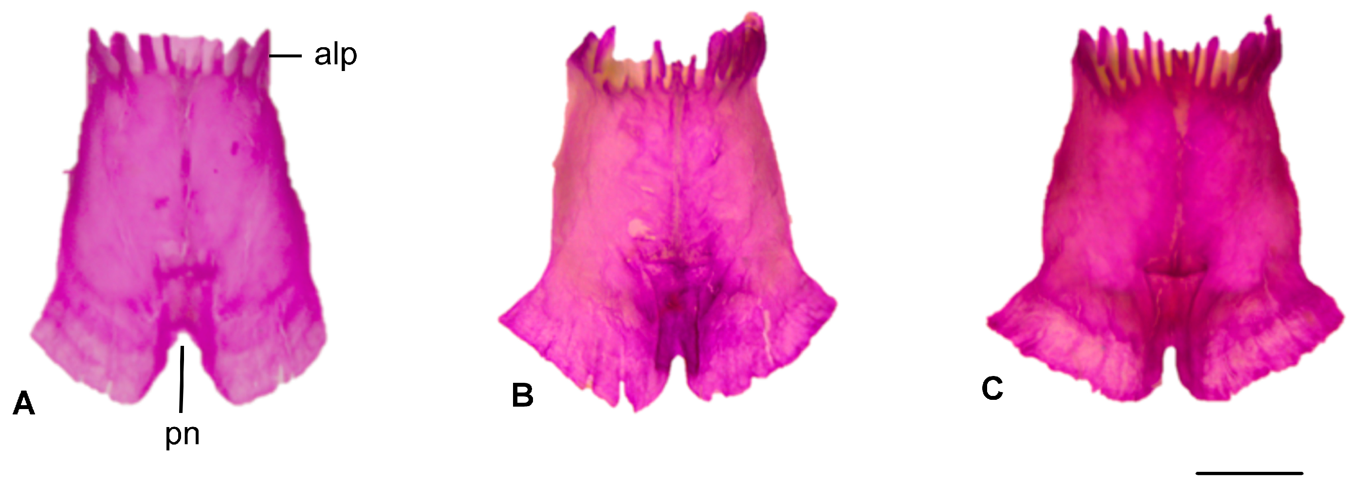
The facial process shows species-specific projections in the dorsal end of the process. B. alexandri has two projections, while B. aporus and B. strauchi exhibit three. All three species typically possess four maxillary teeth that gradually decrease in size posteriorly (Figure 3).

3.1.3. Parietal

A pair of parietal plates are detected, separated by a parietal notch at the posterior edge of the bone. This notch is wider and rounder in Blanus alexandri, whereas it is narrower and rounder in the other two blanid species (Figure 4).

3.1.4. Prefrontal

The prefrontal bone is one of the small, paired bones located anterolaterally in the skull. The anterodorsal process is situated at the dorsal boundary of the orbitonasal flange, while the posteroventral process is observed on the ventrolateral side. The dorsal process, which tapers to a point, is located on the posterodorsal side. In Blanus alexandri, the dorsal edge of the prefrontal bone is more concave, whereas in other species, it is flatter. In Blanus aporus, the posteroventral process is less developed (Figure 5).

3.1.5. Squamosal

The squamosal bone appears as a thin, short, and reduced structure. It is present in all three Blanus species. In B. strauchi and B. aporus, the squamosal bone is highly reduced (Figure 4). However, in B. alexandri, the bone is approximately twice as long as in the other two species (Figure 6).

3.1.6. Vomer

The posterior process of Blanus alexandri is nearly twice as long as those of B. aporus and B. strauchi. The lateral wing is well developed and oriented anteriorly in B. alexandri and B. aporus, whereas it is oriented posteriorly in B. strauchi (Figure 7).

3.1.7. Dentary

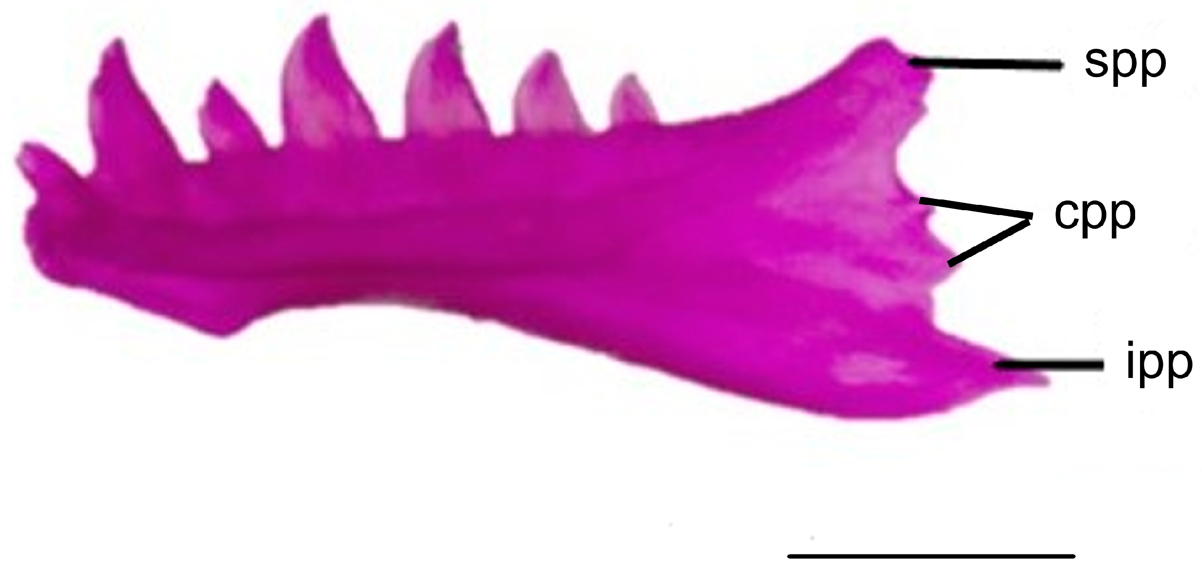
The posterior end of the dental bone contains three processes. The superior posterior process extends posterodorsally and wraps laterally around the coronoid process. This process exhibits a wide and flat termination in Blanus aporus and B. strauchi, while in B. alexandri, it is long and tapers to a point. The inferior posterior process protrudes posteriorly, covering the ventrolateral side of the compound bone. The inferior posterior process is long and tapers in B. alexandri, B. strauchi, and B. aporus. The central posterior process is located between the inferior and superior posterior processes. In B. alexandri, the central posterior process is short and rounded, while in B. strauchi, it is also short but tapers to a point. In B. aporus, unlike the other two blanid species, the central posterior process is present in two instances (Figure 8).

3.1.8. Coronoid

The coronoid bone features three prominences: the anteromedial, posteromedial, and coronoid processes. The anteromedial process is narrow and pointed, while the posteromedial process is broad, tapering to a point in Blanus strauchi and B. aporus and rounded in B. alexandri. The anteromedial and posteromedial processes are separated at a wider angle along the median line in B. strauchi (Figure 9).

3.2. Osteological Characters Exhibiting Variation Among Blanus Species and Related Taxa

Several osteological features display considerable variation among Blanus species and closely related amphisbaenian taxa. These characters provide critical insights into the morphological diversity and evolutionary adaptations of these reptiles (see Appendix B).

3.3. General Postcranial Shape of Blanus Species

3.3.1. The Vertebral Column

The vertebral column is composed of four distinct regions: cervical, thoracic, lumbar, and caudal. The number of vertebrae in each region is given in Table 2. The vertebral column is primarily distinguished by the presence or absence of ribs and the characteristics of the transverse processes. Cervical vertebrae are ribless, while thoracic vertebrae are rib-bearing. Although thoracic vertebrae are longer than those in the lumbar region, they are uniquely characterized by ribs that attach to each vertebra. In contrast, caudal vertebrae lack ribs but possess short transverse processes.

3.3.2. Pelvic and Pectoral Girdles

The pectoral girdle of Blanus species consists of a small clavicular element and a rod-like coracoid, scapula, and suprascapula. In these species, none of the forelimb elements are present, except for a reduced humerus. All components of the pelvic girdle are present in Blanus species, although the pubis and ischium are both reduced in size and less developed than ilium. In the hindlimbs, only the femur is present, though it is reduced and slender in form (Figure 10).

3.4. Geometric Morphometric Analyses

3.4.1. Dorsal Cranial Morphometrics

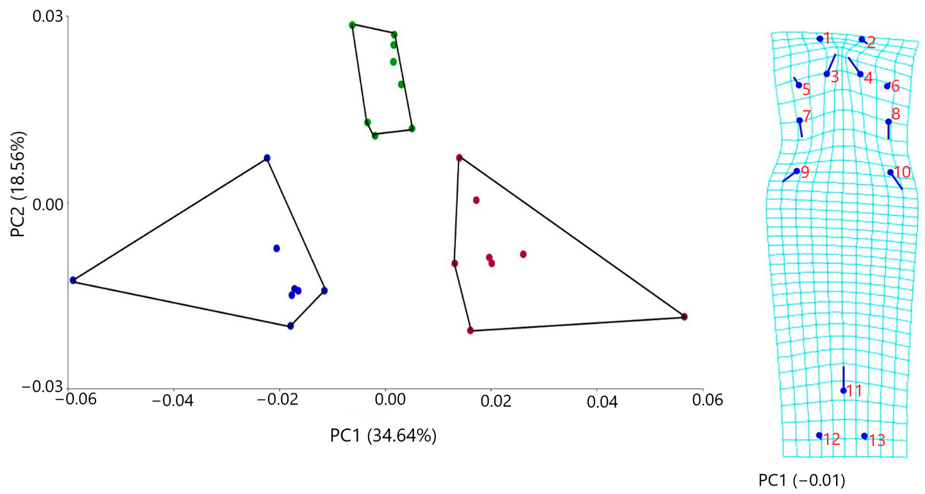
The PCA analysis indicated three main axes of group differentiation: PC1, PC2, and PC3 and explained, respectively, 34.64%, 18.56%, and 10.99% of the variation. The first three components account for 64.20% of the total variance. This indicates that a significant portion of the data can be effectively represented by these components, suggesting that a substantial amount of the underlying structure of the data is captured within them.
Geometric morphometric analyses of dorsal cranial osteological features demonstrated clear morphological differentiation among the three Blanus species. Principal component 1 (PC1) effectively separated Blanus alexandri from B. aporus and B. strauchi, with the former exhibiting distinctly negative scores along this axis. The majority of shape variation was localized in the anterior region of the dorsal skull (Figure 11). Specifically, B. alexandri was characterized by a comparatively elongated and more medially oriented anteromedial process of the nasal bone, a longer and posteriorly directed facial process of the maxilla, a more extended and posterolaterally oriented dorsal process of the prefrontal, and a markedly deeper parietal notch (Figure 11). These osteological distinctions support the morphological separation of B. alexandri from its congeners and provide additional evidence for its species-level differentiation.

3.4.2. Ventral Cranial Morphometrics

The PCA analysis revealed four principal axes of group differentiation, with the first four components explaining 70.14% of the total variance. Geometric morphometric analyses based on osteological features of the ventral skull demonstrated clear morphological differences among the three species. Along PC1, Blanus alexandri exhibited negative scores in comparison with the other two species (B. aporus and B. strauchi). Notably, the parasphenoid rostrum was shorter and more posteriorly oriented, while the posterior process of the vomer was shorter and more anteriorly directed (Figure 12). In addition, the posterior process of the maxilla was shorter and anteriorly oriented, and both the posterior process of the maxilla and the articulation between the ectopterygoid and maxilla were more medially directed (Figure 12).

3.5. Parsimony Reconstruction

In the initial analysis, the ‘New Technology’ search reconstructed 32 most parsimonious trees (MPTs) with a best score of 114. The ‘Traditional search’, using the trees from RAM, inferred 10,000 MPTs (maximum memory). The majority-rule consensus tree provided better-resolved taxonomic implications; however, relationships among all Blanus species, except for the Turkish clade, were ambiguously reconstructed, resulting in a polytomy. Within the monophyletic study group, B. strauchi and B. aporus formed a sister clade, whereas B. alexandri branched as a more distantly related lineage (Figure 13).

4. Discussion

This study presents a comprehensive comparative osteological analysis of three Blanus species—B. alexandri, B. aporus, and B. strauchi—highlighting key differences in both cranial and postcranial skeletal elements. Morphological variations, particularly in the nasal, maxilla, squamosal, vomer, parietal, prefrontal, dentary, and coronoid bones, offer valuable insights into the evolutionary history and constraints of the genus. These osteological distinctions help address taxonomic challenges by supplementing traditionally limited morphological data with more diagnostic skeletal characteristics, thereby bridging the gap between morphological and molecular differentiation.
Our cranial osteological findings differ from those reported by Villa et al. [28] in several important aspects. Notable differences in the nasal, maxilla, squamosal, and vomer bones among Blanus species emphasize the importance of osteological features in distinguishing closely related taxa. For instance, in B. aporus, the anteromedial process of the nasal bone is distinctly longer than the anterolateral process, unlike in B. alexandri and B. strauchi, where both processes are nearly equal in length. Similarly, the variation in the number of projections from the maxillary facial process further underscores the species-specific distinctions observed in the cranial structure. These differences suggest that despite the overall morphological similarity among Blanus species, subtle osteological variations play a significant role in differentiating these species. Further diagnostic differences observed include the number of projections from the dorsal end of the facial process of the maxilla, the shape of the ascending nasal process of the premaxilla, the number of labial foramina on the maxilla, the number of finger-like projections on the posterior margin of each frontal bone, the number of finger-like projections on the anterior margin of the parietal bone, and the sagittal crest of the parietal bone. These osteological characters, in combination with clear biogeographic distributions [4], reinforce the evolutionary distinctiveness of the studied species and support Anatolia’s role as a biodiversity hotspot.
Although approximately 184 species are currently recognized within the Amphisbaenidae family [40], studies focused on cranial osteology remain scarce (Table 1). This study provides comparative cranial data from three Blanus species and seven amphisbaenian species (Zygaspis quadrifrons [41]; Amphisbaena alba [42]; Diplometopon zarudnyi [43]; Rhineura hatcherii [44]; Amphisbaena arda and A. vermicularis [45]; and Spathorhychus fossorium [46]). Our findings reveal differences in the nasal, frontal, parietal, premaxillary, and maxillary bones across these taxa. Species with a posterior process of the nasal bone approximately equal in length to the distal end of the ascending nasal process include Z. quadrifrons [41], D. zarudnyi [43], R. hatcherii [44], and two amphisbaenian species [45]. In contrast, species with a longer posterior process of the nasal bone include A. alba [42] and S. fossorium [46]. The frontal bones interlock with each other in a tongue-and-groove articulation, which is particularly well developed towards the distal ends of the bones in three Blanus species. Three species of Amphisbaena [42,45], R. hatcherii [44], and S. fossorium [46] exhibit similarities to the three Blanus species examined, while in Z. quadrifrons [41] and D. zarudnyi [43], the sutures between the frontal bones are observed to be straight. A. alba [42], A. arda, and A. vermicularis [45] exhibit six finger-like projections on the parietal bones. The parietal notch is narrow and rounded in A. arda and A. vermicularis [45], while it is narrow and blunt in A. alba [42]. The premaxillary teeth consist of seven teeth in Z. quadrifrons [41], A. alba [42], species of Blanus [28], and A. arda and A. vermicularis [45], whereas the alveolar plates of D. zarudnyi [43], S. fossorium [46], and R. hatcherii [44] hold three teeth. The alveolar plate projects forward and develops into a rostral process. This condition is similar to that observed in D. zarudnyi [43], Z. quadrifrons [41], R. hatcherii [44], and species of Blanus [28]. However, this feature differs in other amphisbaenians, such as A. alba [42] and A. arda and A. vermicularis [45]. The facial process of the maxilla is wider than it is tall in Z. quadrifrons [41], R. hatcherii [44], species of Blanus [28], A. alba [42], S. fossorium [46], and A. arda and A. vermicularis [45]. However, D. zarudnyi [43] differs from other amphisbaenians in its longer body structure. On the dorsal part of the facial process of the maxilla, projections extend posterodorsally, with the first being the longest and pointed, while the others are shorter and rounder in the three Blanus species examined. The protrusions at the dorsal end of the facial process are single in A. alba [42], A. arda and A. vermicularis [45], Z. quadrifrons [41], R. hatcherii [44], and S. fossorium [46] and bifurcated in D. zarudnyi [43]. The base of the facial process of the maxilla is pierced in the anteroposterior direction by the superior alveolar canal located on the medial surface of the process. The number of labial foramina is two in D. zarudnyi [43] and S. fossorium [46], and the number is greater in A. alba [42], R. hatcherii [44], B. cinereus, and B. vandellii [28] and variable in A. arda and A. vermicularis [45]. The maxilla carries four teeth in Blanus species [28] and A. arda and A. vermicularis [45], three in D. zarudnyi [43] and Z. quadrifrons [41], six in R. hatcherii [44], and seven in S. fossorium [46]. The largest tooth in the maxillary bone varies among amphisbaenians. In three species of Amphisbaena [42,45], the first tooth is the largest, similar to species of Blanus [28]. In contrast, in R. hatcherii [44], S. fossorium [46], and Z. quadrifrons [41], the second tooth is the largest.
The fossil record of the genus Blanus has been the subject of several detailed morphological studies, particularly focusing on cranial and dental characteristics to clarify taxonomic relationships within the group. Delfino [30] reported the presence of eight teeth in the dentary, seven teeth in the premaxilla (with the middle tooth being the largest), and five teeth in the maxilla (where the first is very small and the second is the largest). In a later study, Čerňanskỳ and Venczel [31] could not provide detailed cranial and dentary character descriptions due to the limited and fragmentary nature of their specimen, which consisted of a small and insufficient sample. Bolet et al. [32] investigated the cranial and limited vertebral osteological characteristics of the fossil species Blanus mendezi sp. nov., focusing on the morphology of the premaxilla, quadrate, sutural structures, and dentary tooth arrangement, and revealed significant similarities with extant Blanus clades. Georgalis et al. [33] likewise noted a total of eight teeth on the dental bone. Most recently, Syromyatnikova et al. [34] provided a new Blanus fossil record, describing dental features—specifically, the presence of an enlarged second tooth and a small third dentary tooth—which support a close relationship with the Blanus strauchi complex (eastern clade).
The postcranial skeleton of Blanus also revealed interesting patterns, particularly in the vertebral column and pelvic structures. The number of presacral vertebrae in the three Blanus species examined showed notable differences, particularly in the thoracic and caudal vertebrae. The presacral vertebra counts are as follows: Blanus aporus < B. alexandri < B. strauchi. Variation in the number of presacral vertebrae among lizards reflects a complex interplay of phylogenetic history, ecological adaptation, and functional demands. Phylogenetic divergence contributes significantly to these differences, with specific vertebral structures serving as indicators of evolutionary relationships [47,48,49,50]. Functionally, the number of presacral vertebrae is closely linked to locomotor strategies: maneuverability typically requires a high degree of body flexibility, which is supported by a greater number of vertebrae relative to body length [51,52,53,54,55]. In contrast, a stiffer trunk, characterized by fewer vertebrae per unit body length, enhances maximum speed and acceleration capacity. Habitat type also plays a crucial role: lizards inhabiting open environments tend to have fewer presacral vertebrae, whereas those in structurally complex habitats possess more [55,56]. Overall, variation in presacral vertebrae provides valuable insights into the evolutionary trajectories and ecological specializations of lizards, highlighting the functional significance of axial morphology across diverse adaptive contexts.
Geometric morphometric analyses revealed distinct differences in dorsal and ventral cranial shapes among the three species. PCA effectively captured the variation in dorsal and ventral cranial morphology, with B. alexandri exhibiting more negative scores along PC1. Similarly, in the geometric morphometric analysis conducted by Sindoca et al. [2] based on the outer plate morphology of the dorsal skull, B. alexandri displayed a positive score on the PC2 axis, distinguishing it from B. strauchi and B. aporus. These shape differences provide additional evidence that the cranial morphology of Blanus species is subject to evolutionary pressures that influence their functional and ecological adaptations.
Phylogenetic analysis based on osteological data also yielded insights into the evolutionary relationships among Blanus species. Our results corroborate previous molecular findings, positioning B. alexandri as a distinct lineage from the closely related B. aporus and B. strauchi. While molecular studies [2] grouped B. alexandri and B. aporus as sister taxa, our osteological data suggest a more nuanced relationship, underscoring the importance of incorporating skeletal traits into phylogenetic frameworks, especially for taxa exhibiting subtle external morphological variation.
Ultimately, this study highlights the enduring relevance of osteological characters in taxonomy, particularly when integrated with molecular data. Furthermore, the observed variations in cranial and postcranial features could be indicative of ecological adaptations, with each species of Blanus evolving specific traits in response to their respective environments. In conclusion, this research emphasizes the importance of integrating osteological and geometric morphometric approaches to study the evolutionary dynamics of Blanus and related taxa. Future studies incorporating a broader sampling of amphisbaenian taxa and more detailed morphological data will further clarify evolutionary relationships and help elucidate the adaptive strategies underlying skeletal variation in these cryptic reptiles.

Author Contributions

Conceptualization, K.C., E.Y.C., Ç.I., and Y.K.; methodology, E.Y.C., E.B.Y., E.Ş., E.B.H., and E.A.Ç.; validation, E.Y.C. and K.C.; formal analysis, E.Y.C., E.B.Y., E.Ş., E.B.H., E.A.Ç. S.B., and A.G.K.; investigation, E.Y.C., E.B.Y., E.Ş., E.B.H., E.A.Ç., S.B., and A.G.K.; resources, K.C., A.G.K., Ç.I., and Y.K.; data curation, E.Y.C.; writing—original draft preparation, E.Y.C. and Ç.I.; writing—review and editing, K.C., Ç.I., and S.G.; visualization, E.Y.C.; project administration, Y.K.; funding acquisition, S.G. All authors have read and agreed to the published version of the manuscript.

Funding

This research was funded by Dokuz Eylül University Scientific Research Coordination Unit, grant number FBA-2022-2730, and the APC was funded by the Recep Tayyip Erdoğan University Development Foundation (Grant number: 02025007001578).

Institutional Review Board Statement

Ethical approval was not required for our study.

Informed Consent Statement

This study does not involve human participants.

Data Availability Statement

The original contributions presented in this study are included in the article.

Conflicts of Interest

The authors declare no conflicts of interest.

Appendix A

Table A1. Summary of the osteological data available for amphisbaenians.
SpeciesOsteological DataSource
Amphisbaenian sp.Postcranial osteology[26]
Amphisbaenian sp.Postcranial osteology[27]
Slavoia darevskii (extinct sp.)Postcranial osteology[29]
Zygaspis quadrifronsPostcranial osteology[57]
Blanus sp.Cranial osteology[28]
Blanus mendezi sp. Nov. (extict sp.)Cranial osteology[32]
Amphisbaenian sp.Cranial osteology[36]
Zygaspis quadrifronsCranial osteology[41]
Amphisbaenia albaCranial osteology[42]
Diplometopon zarudnyiCranial osteology[43]
Rhineura hatcherii (extinct sp.)Cranial osteology[44]
Spathorhynchus fossorium (extinct sp.)Cranial osteology[46]
Amphisbaenian sp.Cranial osteology[58]
Diplometalon zarudnyiCranial osteology[59]
Genus ZygaspisCranial osteology[60]
Diplometopon zarudnyiCranial osteology[61]
Amphisbaenian sp.Cranial osteology[62]

Appendix B

The list for observed variation in key osteological traits, offering a detailed comparison of their presence, form, and structure.
  • Premaxilla: the number of teeth: (0) 3, (1) 7.
  • Premaxilla: rostrol process: (0) absent, (1) present.
  • Premaxilla: ascending nasal process: (0) tapers posteriorly with a slight projection, (1) tapers posteriorly with a slight depression, (2) narrows towards the truncated or pointed dorsal end, (3) tapers with an arrow shaped*, (4) narrow and elongated end*, (5) appears to be short and peg-like, (6) tapers posteriorly with rounded end.
  • Premaxilla: ascending nasal process contacts to: (0) anteromedial process of the frontal, (1) nasal.
  • Maxilla: The number of teeth: (0) 3, (1) 4, (2) 5, (3) 6, (4) 7.
  • Maxilla: the larger teeth: (0) first teeth, (1) second teeth.
  • Maxilla: facial process: (0) wider, (1) taller.
  • Maxilla: the number of the projection from the dorsal end of the facial process: (0) 1, (1) 2, (2) 3.
  • Maxilla: the number of labial foramen: (0) 1, (1) 2, (2) more than two, (3) variable, (4) more than two and each side variable.
  • Nasal: the posterior process of the nasal: (0) the posterior process of the nasal bone is shorter than the length of the distal portion of the ascending nasal process of the premaxilla, (1) the posterior process of the nasal bone is approximately equal in length to the distal portion of the ascending nasal process of the premaxilla. (2) the posterior process of the nasal bone is longer than the length of the distal portion of the ascending nasal process of the premaxilla.
  • Frontal: suture between the frontals: (0) straight articulation, (1) tongue-and-groove articulation.
  • Frontal: number of maximum finger-like projections on the posterior margin of each frontal bone: (0) 3, (1) 4, (2) 5, (3) 6, (4) 7.
  • Parietal: number of maximum finger-like projections on the anterior margin of the parietal bone: (0) 5, (1) 6, (2) 7, (3) 8, (4) 9, (5) 10, (6) 11.
  • Parietal: parietal notch: (0) narrow, (1) wide.
  • Parietal: the lateralmost finger-like projection touches the prefrontal: (0) yes, (1) no, (2) both, (3) approach but not contact.
  • Parietal: parietal (pineal) foramen: (0) present, (1) absent.
  • Parietal: sagittal crest; (0) distinct, (1) indistinct, (3) absent.
  • Parietal: otic-occipital lappet; (0) wide, (1) narrow.
  • Parietal: the length of the finger-like projection; (0) relatively equal, (1) not equal.
  • Prefrontal: (0) present, (1) absent.
  • Squamosal: the length of the bone: (0) present, (1) absent, (2) fused with the otic-occipital complex.
  • Quadrate: the projection of the posteromedial part of the pillar: (0) distinct, (1) thin, (2) poor development.
  • Quadrate: quadrate foramen; (0) present and one, (1) absent, (2) present and more than one.
  • Epipterygoid: (0) present, (1) absent, (2) both.
  • Vomer: rostral process: (0) bifurcated, (1) long and slender, (2) wide and triangular-shaped posteriorly.
  • Vomer: posterior process: (0) long and narrow with an acute end, (1) long and slender.
  • Vomer: lateral wings: (0) slender and anteriorly directed, (1) posteriorly directed and then tapers, (2) posteriorly directed and widens, (3) widens laterally at approximately its midpoint, (4) widens and anteriorly directed, (5) slender and posteriorly directed.
  • Palatine: the laminar expansion of the lateral margin of the bone: (0) long, (1) short, (2) poor development, (3) absent.
  • Palatine: anterior processes: (0) the length of the vomerine process > the maxillar process, (1) the length of the vomerine process < the maxillar process, (2) the length of the vomerine process = the maxillar process, (3) absent.
  • Palatine: pterygoid process: (0) laterally oriented, long, and pointed, (1) flat and ends in a pointed tip, (2) projects posteriorly, (3) posteriorly directed and further tapers.
  • Palatine: palatine foramen: (0) present, (1) absent.
  • Pterygoid recess: (0) straight, (1) wide angle, (2) narrow angle, (3) absent.
  • Pterygoid: teeth: (1) present, (2) absent.
  • Dentary: the longest tooth of the dentary: (0) first, (1) second, (2) third, (3) first and forth.
  • Dentary: the number of ventral posterior process of the dentary: (0) 0, (1) 1, (2) 2, (3) 3.
  • Dentary: the first foramen of the dentary: (0) the level of the 2–3 teeth, (1) the level of the 1–2 teeth, (2) the level of the 1 teeth, (3) on the 2th teeth, (4) mental foramina are variable on the left and right sides.
  • Dentary: The second foramen of the dentary: (0) the level of the 3–4 teeth, (1) the level of the 2–3 teeth, (2) on the 2nd teeth, (3) on the 4th teeth, (4) mental foramina are variable on the left and right sides.
  • Dentary: The third foramen of the dentary: (0) the level of the 6–7 teeth, (1) the level of the 5–6 teeth, (2) the level of 3–4 teeth, (3) the level of 4–5 teeth, (4) on the 5th teeth, (5) mental foramina are variable on the left and right sides.
  • Dentary: the number of teeth: (0) 6, (1) 7, (2) 8, (3) 7 or 8.
  • Presacral vertebra: the average number of the presacral vertebrae: (0) 118, (1) 123, (2) 128.

Appendix C

Data matrix of Blanus group and outgroup taxa. Symbol “?”: not applicable. See Appendix B for descriptions of osteological characters.
SpeciesData MatrixReferenceCitations
Blanus alexandri1100100310135111100001020001001112330031Present Study
Blanus aporus1110100310125111100000020001001112315030Present Study
Blanus strauchi1110100240136131100010220001001112310032Present Study
Blanus cineraus112010012011301100000100000100111233003?Villa et al., 2019[28]
Blanus tingitanus112010012012311100001101000100111231002?Villa et al., 2019[28]
Blanus mettatali11201001301131?100000200000100111231003?Villa et al., 2019[28]
Blanus vandelli112010012011301100000200000100111231003?Villa et al., 2019[28]
Amphisbaena alba10402000221120210100?20?002???111230302?Montero & Gans, 1999[42]
Amphisbaena arda10401000311120210000??0?102?021?1224451?Paiva et al., 2024[45]
Amphisbaena vermicularis10401000311100210000??0?102?021?1224451?Paiva et al., 2024[45]
Zygaspis quadrifrons11160100210430111000?10?005300011222233?Bell et al., 2023[41]
Diplometopon zarudyni01300011210151110011001?004302031003110?Maisano et al., 2006[43]
Rhineura hatcherii01513100211??0010110221?003302121331141?Kearney et al., 2005[44]
Spathorhychus fossorium0?614100221???010??0211?201?230?1332222?Müller et al., 2016[46]

References

  1. Albert, E.M.; Fernández, A. Evidence of cryptic speciation in a fossorial reptile: Description of a new species of Blanus (Squamata: Amphisbaenia: Blanidae) from the Iberian Peninsula. Zootaxa 2009, 2234, 56–68. [Google Scholar] [CrossRef]
  2. Sindaco, R.; Kornilios, P.; Sacchi, R.; Lymberakis, P. Taxonomic reassessment of Blanus strauchi (Bedriaga, 1884) (Squamata: Amphisbaenia: Blanidae), with the description of a new species from southeast Anatolia (Turkey). Zootaxa 2014, 3795, 311–326. [Google Scholar] [CrossRef]
  3. Ceríaco, L.M.P.; Bauer, A.M. An integrative approach to the nomenclature and taxonomic status of the genus Blanus Wagler, 1830 (Squamata: Blanidae) from the Iberian Peninsula. J. Nat. Hist. 2018, 52, 849–880. [Google Scholar] [CrossRef]
  4. Baran, İ.; Avcı, A.; Kumlutaş, Y.; Olgun, K.; Ilgaz, Ç. Türkiye Amfibi ve Sürüngenleri; Palme: Ankara, Turkey, 2021; p. 223. [Google Scholar]
  5. von Bedriaga, J. Amphisbaena cinerea und A. strauchi v. Bedr. Erster Beitrag zur Kenntniss der Deppelscleichen. Arch. Für Naturgeschicte 1884, 50, 23–77. [Google Scholar]
  6. Werner, F. Über einige neue Reptilien und einen neuen Frosch aus dem cilicischen Taurus. Zool. Anz. 1898, 21, 217–223. [Google Scholar]
  7. Busack, S.D. Biochemical and morphological differentiation in Spanish and Moroccan populations of Blanus and the description of a new species from northern Morocco (Reptilia, Amphisbaenia, Amphisbaenidae). Copeia 1988, 1988, 101–109. [Google Scholar] [CrossRef]
  8. Sampaio, F.L.; Harris, D.J.; Perera, A.; Salvi, D. Phylogenetic and diversity patterns of Blanus worm lizards (Squamata: Amphisbaenia): Insights from mitochondrial and nuclear gene genealogies and species tree. J. Zool. Syst. Evol. Res. 2015, 53, 45–54. [Google Scholar] [CrossRef]
  9. Hipsley, C.A.; Rentinck, M.N.; Röder, M.O.; Müller, J. Ontogenetic allometry constrains cranial shape of the head-first burrowing worm lizard Cynisca leucura (Squamata: Amphisbaenidae). J. Morphol. 2016, 277, 1159–1167. [Google Scholar] [CrossRef]
  10. Hohl, L.S.L.; Barros-Filho, J.D.; Rocha-Barbosa, O. Skull variation in a shovel-headed amphisbaenian genus, inferred from the geometric morphometric analysis of five South American Leposternon species. J. Morphol. 2018, 279, 1665–1678. [Google Scholar] [CrossRef] [PubMed]
  11. Hohl, L.S.L.; Azorit, C.; Vassallo, A.I.; Casinos, A.; Machado, A.S.; Lopes, R.T.; Rocha-Barbosa, O. Ontogenetic skull variation in a shovel-headed amphisbaenian species. J. Morphol. 2023, 284, e21643. [Google Scholar] [CrossRef]
  12. Gans, C.; Wever, E.G. The Amphisbaenian Ear: Blanus cinereus and Diplometopon zarudnyi. Proc. Natl. Acad. Sci. USA 1975, 72, 1487–1490. [Google Scholar] [CrossRef] [PubMed]
  13. Martín, J.; López, P.; Salvador, A. Microhabitat selection of the Amphisbaenian Blanus cinereus. Copeia 1991, 1991, 1142–1146. [Google Scholar] [CrossRef]
  14. Díaz-Paniagua, C.; Blázoquez, M.C.; Keller, C.; Andreu, A.C.; Olmedo, G.; Mateo, J.A. Observations on seasonal and diel surface activity of the amphisbaenian Blanus cinereus in South-Western Spain. Herpetol. J. 1995, 5, 217–220. [Google Scholar]
  15. Vaconcelos, R.; Carretero, M.A.; Harris, D.J. Phylogeography of the genus Blanus (worm lizards) in Iberia and Morocco based on mitochondrial and nuclear markers—Preliminary analysis. Amphib.-Reptilia 2006, 27, 339–346. [Google Scholar] [CrossRef]
  16. Albert, E.M.; Zardoya, R.; García-París, M. Phylogeographical and speciation patterns in subterranean worm lizards of the genus Blanus (Amphisbaenia: Blanidae). Mol. Ecol. 2007, 16, 1519–1531. [Google Scholar] [CrossRef]
  17. Yıldırımhan, H.S.; Yılmaz, N.; İncedoğan, S. Helminth Fauna of the Anatolian Worm Lizard, Blanus strauchi (Bedriaga, 1884) From Hatay. Türkiye Parazitoloji Dergisi. 2009, 33, 327–329. [Google Scholar]
  18. Yıldız, M.Z.; Akman, B.; Göçmen, B.; Yalçınkaya, D. New locality records for the Turkish worm lizard, Blanus strauchi aporus (Werner, 1898) (Sauria: Amphisbaenidae) in Southeast Anatolia, Turkey. North-West. J. Zool. 2009, 5, 379–385. [Google Scholar]
  19. Düşen, S.; Uğurtaş, İ.H.; Aydoğdu, A. Nematode parasites of the two limbless lizards: Turkish worm lizard, Blanus strauchi (Bedriaga, 1884) (Squamata: Amphisbaenidae), and slow worm, Anguis fragilis Linnaeus 1758 (Squamata: Anguidae), from Turkey. Helminthologia 2010, 47, 158–163. [Google Scholar] [CrossRef]
  20. Tok, C.V.; Tosunoğlu, M.; Ayaz, D.; Çiçek, K.; Mutlu, H.S. New records of the Anatolian Worm Lizard, Blanus strauchi (Bedriaga, 1884), from Turkey. Herpetozoa 2012, 24, 107–111. [Google Scholar]
  21. Jablonski, D.; Sadek, R.A. The species identity and biogeography of Blanus (Amphisbaenia: Blanidae) in Lebanon. Zool. Middle East. 2019, 65, 208–214. [Google Scholar] [CrossRef]
  22. Gervais, P. Recherches sur l’osteologie de plusieurs especes d’Amphisbenes, et remarques sur la classification de ces reptiles. Ann. Sci. Naturelles 1853, 20, 293–312. [Google Scholar]
  23. Fürbringer, M. Die Knochen und Muskeln der Extremitaten bei den Schlangenahnlichen Sauriern: Vergleichend-Anatomische Abhandlung; W. Engelmann: Leipzig, Germany, 1869; p. 135. [Google Scholar]
  24. Fürbringer, M. Zur vergleichenden Anatomie des Brustschulterapparates und der Schultermuskeln. Jenaische Z. Für Naturwissenschaft 1900, 34, 215–718. [Google Scholar]
  25. Cope, E.D. The osteology of the Lacertilia. Proc. Am. Philos. Soc. 1892, 30, 185–221. [Google Scholar]
  26. Zangerl, R. Contributions to the osteology of the post-cranial skeleton of the Amphisbaenidae. Am. Midl. Nat. 1945, 33, 764–780. [Google Scholar] [CrossRef]
  27. Kearney, M. Appendicular skeleton in Amphisbaenians (Reptilia: Squamata). Copeia 2002, 2002, 719–738. [Google Scholar] [CrossRef]
  28. Villa, A.; Kirchner, M.; Alba, D.M.; Bernardini, F.; Bolet, A.; Lujan, A.H.; Fortuny, J.; Hipsley, A.C.; Müller, J.; Sindaco, R.; et al. Comparative cranial osteology of Blanus (Squamata: Amphisbaenia). Zool. J. Linn. Soc. 2019, 185, 693–716. [Google Scholar] [CrossRef]
  29. Talanta, M. Evolution of postcranial skeleton in worm lizards inferred from its status in the Cretaceous stem-amphisbaenian Slavoia darevskii. Acta Palaeontol. Pol. 2017, 62, 9–23. [Google Scholar] [CrossRef]
  30. Delfino, M. Blanus from the Early Pleistocene of Southern Italy: Another small tessera from a big mosaic. In Herpetologia Bonnensis; Böhme, W., Bischoff, W., Ziegler, T., Eds.; Societas Europae Herpetologica: Bonn, Germany, 1997; pp. 89–97. [Google Scholar]
  31. Čerňanskỳ, A.; Venczel, M. An amphisbaenid reptile (Squamata, Amphisbaenidae) from the Lower Miocene of Northwest Bohemia (MN 3, Czech Republic). Neues Jahrb. Fur Geol. Und Palaontol. Abhandlungen 2011, 260, 73–77. [Google Scholar] [CrossRef]
  32. Bolet, A.; Delfino, M.; Fortuny, J.; Almécija, S.; Robles, J.M.; Alba, D.M.; Rook, L. An amphisbaenian skull from the European Miocene and the evolution of Mediterranean worm lizards. PLoS ONE 2014, 9, e98082. [Google Scholar] [CrossRef]
  33. Georgalis, G.L.; Halaçlar, K.; Mayda, S.; Kaya, T.; Ayaz, D. First fossil find of the Blanus strauchi complex (Amphisbaenia, Blanidae) from the Miocene of Anatolia. J. Vertebr. Paleontol. 2018, 38, e1437044. [Google Scholar] [CrossRef]
  34. Syromyatnikova, E.V.; Kovalenko, E.S.; Kaloyan, A.A. A fossil record of the Eastern clade of Blanus (Amphisbaenia: Blanidae) from the late Miocene of Ukraine. Geobios 2021, 69, 69–75. [Google Scholar] [CrossRef]
  35. Wassersug, R. A procedure for differential staining of cartilage and bone in whole formalin fixed vertebrates. Stain Technol. 1976, 51, 131–134. [Google Scholar] [CrossRef]
  36. Gans, C.; Montero, R. An atlas of amphisbaenian skull anatomy. In Biology of the Reptilia: Morphology I. The skull and Appendicular Locomotor Apparatus of Lepidosauria; Gans, C., Gaunt, A., Adler, K., Eds.; Society for the Study of Amphibians and Reptiles: Ithaca, Greece, 2008; pp. 621–738. [Google Scholar]
  37. Rohlf, F.J. tpsDig; SUNY at Stony Brook Department of Ecology and Evolution, State University of New York, Stony Brook: New York, NY, USA, 2010; 11794–524. [Google Scholar]
  38. Goloboff, P.A.; Farris, J.S.; Nixon, K.C. TNT, a free program for phylogenetic analysis. Cladistics 2008, 24, 774–786. [Google Scholar] [CrossRef]
  39. Goloboff, P.A.; Catalano, S.A. TNT version 1.5, including a full implementation of phylogenetic morphometrics. Cladistics 2016, 32, 221–238. [Google Scholar] [CrossRef]
  40. Uetz, P.; Freed, P.; Aguliar, R.; Reyes, F.; Kudera, J.; Hošek, J. The Reptile Database. Available online: http://reptile-database.org (accessed on 11 June 2025).
  41. Bell, C.J.; Cadena, C.; Meza, A.; Rudie, L.; Lewis, P.J. Cranial anatomy of the “round-headed” Amphisbaenian Zygaspis quadrifrons (Squamata, Amphisbaenia) based on high-resolution x-ray computed tomography. Anat. Rec. 2023, 307, 495–532. [Google Scholar] [CrossRef]
  42. Montero, R.; Gans, C. The head skeleton of Amphisbaenia alba Linneaus. Ann. Carnegie Mus. 1999, 68, 15–80. [Google Scholar] [CrossRef]
  43. Maisano, J.A.; Kearney, M.; Rowe, T. Cranial anatomy of the spade-headed Amphisbaenian Diplometopon zarudnyi (Squamata, Amphisbaenia) based on high-resolution X-ray Ccmputed tomography. J. Morphol. 2006, 267, 70–102. [Google Scholar] [CrossRef]
  44. Kearney, M.; Maisano, J.A.; Rowe, T. Cranial anatomy of the extinct amphisbaenian Rhineura hatcherii (Squamata, Amphisbaenia) based on high-resolution X-ray computed tomography. J. Morphol. 2005, 264, 1–33. [Google Scholar] [CrossRef] [PubMed]
  45. Paiva, C.L.; Hipsley, C.A.; Müller, J.; Zaher, H.; Costa, H.C. Comparative skull osteology of Amphisbaena arda and Amphisbaena vermicularis (Squamata: Amphisbaenidae). J. Morphol. 2024, 285, e21702. [Google Scholar] [CrossRef] [PubMed]
  46. Müller, J.; Hipsley, C.A.; Maisano, J.A. Skull osteology of the Eocene amphisbaenian Spathorhynchus fossorium (Reptilia, Squamata) suggests convergent evolution and reversals of fossorial adaptations in worm lizards. J. Anat. 2016, 229, 615–630. [Google Scholar] [CrossRef]
  47. Gans, C. Tetrapod limblessness: Evolution and functional corollaries. Am. Zool. 1975, 15, 455–467. [Google Scholar] [CrossRef]
  48. Lee, M.S.Y. Convergent evolution and character correlation in burrowing reptiles: Towards a resolution of squamate relationships. Biol. J. Linn. Soc. 1998, 65, 369–453. [Google Scholar] [CrossRef]
  49. Wiens, J.J.; Brandley, M.C.; Reeder, T.W. Why does a trait evolve multiple times within a clade? Repeated evolution of snake-like body form in squamate reptiles. Evolution 2006, 60, 123–141. [Google Scholar] [CrossRef]
  50. Camaiti, M.; Evans, A.R.; Hipsley, C.A.; Chapple, D.G. A fare-well to arms and legs: A review of limb reduction in squamates. Biol. Rev. 2021, 96, 1035–1050. [Google Scholar] [CrossRef]
  51. Jayne, B.C. Comparative morphology of the semispinalisspinalis muscle of snakes and correlations with locomotion and constriction. J. Morphol. 1982, 172, 83–96. [Google Scholar] [CrossRef] [PubMed]
  52. Jayne, B.C. Swimming in constricting (Elaphe g. guttata) and nonconstricting (Nerodia fasciata pictiventris) colubroid snakes. Copeia 1985, 1985, 195–208. [Google Scholar] [CrossRef]
  53. Gasc, J.P.; Gans, C. Tests on locomotion of the elongate and limbless lizard Anguis fragilis (Squamata: Anguidae). Copeia 1990, 1990, 1055–1067. [Google Scholar] [CrossRef]
  54. Kelly, K.C.; Arnold, S.J.; Gladstone, J. The effect of substrate and vertebral number on locomotion in the garter snake Thamnophis elegans. Funct. Ecol. 1997, 11, 189–198. [Google Scholar] [CrossRef]
  55. Arnold, E.N. Structural niche, limb morphology and locomotion in lacertid lizards (Squamata Lacertidae); a preliminary survey. Bull. Br. Mus. (Nat. Hist.) Zool. 1998, 64, 63–89. [Google Scholar]
  56. Arnold, E.N. Osteology, genitalia and the relationships of Acanthodactylus (Reptilia: Lacertidae). Bull. Br. Mus. (Nat. Hist.) Zool. 1983, 44, 291–339. [Google Scholar]
  57. Urben, C.C.; Daza, J.D.; Cadena, C.; Lewis, P.J.; Thies, M.L. The Homology of the Pelvic Elements of Zygaspis quadrifrons (Squamata: Amphisbaenia). Anat. Rec. 2014, 297, 1407–1413. [Google Scholar] [CrossRef] [PubMed]
  58. Gans, C. The characteristics and affinities of the Amphisbaenia. Trans. Zool. Soc. Lond. 1978, 34, 347–416. [Google Scholar] [CrossRef]
  59. Hawkins, R.; Bell, C.; Olori, J.C.; Stocker, M.R. Intraspecific variation in the cranial osteology of Diplometopon zarudnyi (Squamata: Amphisbaenia: Trogonophidae). J. Morphol. 2022, 283, 1359–1375. [Google Scholar] [CrossRef]
  60. Meza, A.; Bell, C.J.; Daza, J.D.; Thies, M.L.; Lewis, P.J. Variation in the cranial osteology of the amphisbaenian genus Zygaspis based on high-resolution x-ray computed tomography. Anat. Rec. 2023, 307, 475–494. [Google Scholar] [CrossRef] [PubMed]
  61. Abo-Eleneen, R.E.; Othman, S.I.; Al-Harbi, H.M.; Abdeen, A.M.; Allam, A.A. Anatomical study of the skull of amphisbaenian Diplometopon zarudnyi (Squamata, Amphisbaenia). Saudi J. Biol. Sci. 2019, 26, 503–513. [Google Scholar] [CrossRef] [PubMed]
  62. Zangerl, R. Contributions to the osteology of the skull of the Amphisbaenidae. Am. Midl. Nat. 1944, 31, 417–454. [Google Scholar] [CrossRef]
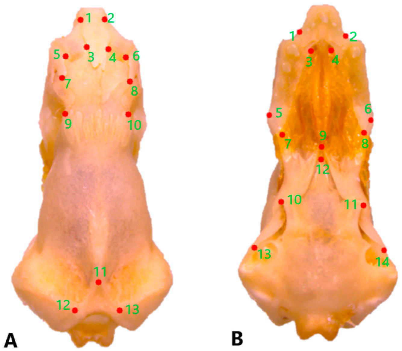
Figure 1. Position of the landmarks (A) on the dorsal and (B) on the ventral surfaces of a skull of Blanus alexandri.
Life 15 01263 g001
Figure 2. Dorsal view of the nasal bone. (A). Blanus alexandri, (B). Blanus aporus, and (C). Blanus strauchi. alp, anterolateral process; amp, anteromedial process. Scale bar = 2 mm.
Life 15 01263 g002
Figure 3. Dorsal view of the maxilla bone. (A). Blanus alexandri, (B). Blanus aporus, and (C). Blanus strauchi. dp, dorsal projections of the facial process. Scale bar = 2 mm.
Life 15 01263 g003
Figure 4. Parietal bone in dorsal view: (A). Blanus alexandri, (B). Blanus aporus, and (C). Blanus strauchi. alp, anterolateral process; pn, parietal notch. Scale bar = 2 mm.
Life 15 01263 g004
Figure 5. Prefrontal bone in dorsal view: (A). Blanus alexandri, (B). Blanus aporus, and (C). Blanus strauchi. adp, anterodorsal process; dp, dorsal process, pvp, posteroventral process. Scale bar = 2 mm.
Life 15 01263 g005
Figure 6. Squamosal bone in lateral view of the skull: (A). Blanus alexandri, (B). Blanus aporus, and (C). Blanus strauchi. sq, squamosal. Scale bar = 2 mm.
Life 15 01263 g006
Figure 7. Vomer bone in the ventral view. (A). Blanus alexandri, (B). Blanus aporus, and (C). Blanus strauchi. lw, lateral wing; pp, posterior process. Scale bar = 2 mm.
Life 15 01263 g007
Figure 8. Dentary bone in lingual view of Blanus aporus. cpp, central posterior process; ipp, inferior posterior process; spp, superior posterior process. Scale bar = 2 mm.
Life 15 01263 g008
Figure 9. Coronoid bone in the lingual view. (A). Blanus alexandri, (B). Blanus aporus, and (C). Blanus strauchi. amp, anteromedial process; cp, coronoid process; pmp, posteromedial process. Scale bar = 2 mm.
Life 15 01263 g009
Figure 10. (A). Pectoral girdle in Blanus alexandri and (B). Pelvic girdle in Blanus aporus. cl, clavicula; co, coracoid; f, femur; h, humerus; il, ilium; isc, ischium; pb, pubis; sc, scapula; ssc, suprascapula. Scale bar = 2 mm.
Life 15 01263 g010
Figure 11. Shape changes in dorsal skulls among three blanids using a principal component analysis (blue dots, B. alexandri; red dots, B. aporus; green dots, B. strauchi).
Life 15 01263 g011
Figure 12. Shape changes in ventral skulls among three blanids using a principal component analysis (blue dots, B. alexandri; red dots, B. aporus; green dots, B. strauchi).
Life 15 01263 g012
Figure 13. Majority-rule consensus tree from the parsimony analysis. Numbers above the nodes indicate the percentage of most parsimonious trees supporting each node. Numbers below the nodes represent Bremer support (decay index) values.
Life 15 01263 g013
Table 1. Description of specimens.
SpeciesRegion (City of Türkiye)Number of Sample
Blanus alexandriAdıyaman3
Mardin2
Urfa3
Blanus aporusAntalya2
Adana2
Mersin2
Hatay2
Blanus strauchiMarmaris (Muğla)3
Fethiye (Muğla)3
Muğla2
Table 2. The average number of vertebrae in three Blanus species.
SpeciesCervical
Vertebrae
Thoracic
Vertebra
Lumbar
Vertebrae
Caudal
Vertebrae
B. aporus2 (atlas and axis)98216
B. alexandri2 (atlas and axis)100219
B. strauchi2 (atlas and axis)105219
Disclaimer/Publisher’s Note: The statements, opinions and data contained in all publications are solely those of the individual author(s) and contributor(s) and not of MDPI and/or the editor(s). MDPI and/or the editor(s) disclaim responsibility for any injury to people or property resulting from any ideas, methods, instructions or products referred to in the content.