Article

Susceptibility of Lactobacillaceae Strains to Aminoglycoside Antibiotics in the Light of EFSA Guidelines

by
Marta Dec
1,*,
Klaudia Herman-Ostrzyżek
1,
Aldert Zomer
2,3 and
Renata Urban-Chmiel
1
1
Department of Veterinary Prevention and Avian Diseases, University of Life Sciences in Lublin, 20-612 Lublin, Poland
2
Division of Infectious Diseases and Immunology, Faculty of Veterinary Medicine, Utrecht University, 3584 CL Utrecht, The Netherlands
3
WHO Collaborating Centre for Reference and Research on Campylobacter and Antimicrobial Resistance from a One Health Perspective/WOAH Reference Laboratory for Campylobacteriosis, 3584 CL Utrecht, The Netherlands
*
Author to whom correspondence should be addressed.
Life 2025, 15(5), 732; https://doi.org/10.3390/life15050732
Submission received: 27 February 2025 / Revised: 25 April 2025 / Accepted: 29 April 2025 / Published: 30 April 2025
(This article belongs to the Special Issue Natural Substances in Nutrition and Health of Animals: 2nd Edition)

Abstract

:
Lactobacillaceae is a large family of bacteria from which probiotic strains often originate. Microorganisms used as feed additives in the EU must meet a number of formal criteria, some of which concern antimicrobial susceptibility. In this study, we determined the susceptibility of 19 reference strains and 121 wild-type strains of Lactobacillaceae to aminoglycoside antibiotics using the broth microdilution method based on the ISO 10932:2010/IDF 223:2010 standard. Strains were categorized as resistant or susceptible according to European Food Safety Authority (EFSA) guidelines. Resistance genes were detected by whole genome sequence (WGS) analysis or by PCR. The MICs read after 48 h of incubation showed that 36.8% of reference strains were resistant to kanamycin, 26.3% to streptomycin, and 5.3% to gentamicin, with no aminoglycoside resistance genes detected in any genome. As many as 93.2% of field isolates of Ligilactobacillus salivarius, 85% of Ligilactobacillus agilis, and 58.8% of Lactiplantibacillus plantarum were classified as resistant to kanamycin, with the aac(6)-Ie-aph(2)-Ia gene detected only in two isolates. In six of 12 streptomycin-resistant strains, the ant(6)-Ia gene was identified, which usually coexisted with the spw gene. Three isolates with high neomycin MICs harbored the ant(4′)-Ia gene. In Lactobacillus gallinarum strain LMG 9435, characterized by streptomycin MIC value > 1024 µg/mL, a potential resistance-causing mutation in the rpsL gene (Lys56 → Arg) was detected. The results of the study indicate that some genera of Lactobacillaceae, in particular L. salivarius and L. agilis, exhibit natural resistance to aminoglycoside antibiotics, mainly kanamycin. Therefore, there is a need to update the EFSA guidelines on antimicrobial susceptibility testing of Lactobacillaceae, so that strains lacking resistance genes and/or chromosomal mutations are not considered to be resistant.

1. Introduction

Lactobacillaceae are gram-positive, aerotolerant or anaerobic, non-spore-forming bacilli or cocci. They are the most numerous group of lactic acid bacteria (LAB), which produce lactic acid as the major metabolic end-product of carbohydrate fermentation [1]. The Lactobacillaceae family currently includes 35 genera with >460 species. Of these, 19 are referred to as ‘candidatus’, meaning that they have provisional taxonomic names because they were characterized based on metagenome sequencing without obtaining pure cultures [2,3,4]. Lactobacillaceae inhabit nutrient-rich environments, such as the mucous membranes of humans and animals, milk and dairy products, plants, and fermented foods, and most species of this family are considered non-pathogenic. This makes them ideal candidates for probiotics, which are widely used in humans, livestock, and companion animals [5].
Probiotic strains should meet a number of criteria, some of which concern antimicrobial susceptibility. They cannot contain acquired resistance genes, especially those located on mobile genome elements, due to the risk of transferring them to other bacteria (e.g., in the host’s intestine), including opportunistic and pathogenic bacteria [6].
According to the EFSA Panel on Additives and Products or Substances used in Animal Feed (FEEDAP) guidelines published in 2018 [6], microorganisms used as feed additives or as production organisms in the European Union (EU) must be assessed for antimicrobial susceptibility based on both a phenotypic test and a whole-genome sequence analysis for the detection of possible resistance genes. In the phenotypic test for Lactobacillaceae, the MIC values of nine antimicrobial substances should be determined, including three aminoglycosides, i.e., kanamycin, gentamicin, and streptomycin [6]. However, details of the antimicrobial susceptibility testing (AST) have not been specified. Both the agar dilution method and the broth dilution method are accepted, and the AST ‘should be performed in accordance with internationally recognized standards such as the European Committee on Antibacterial Susceptibility Testing (EUCAST), the Clinical and Laboratory Standards Institute (CLSI), the ISO standard or similar’. FEEDAP only stipulates that the culture medium should enable the growth of the strains tested, and in the case of some LAB, LAB susceptibility medium (LSM) [7] can be used.
EFSA guidelines for determining the antimicrobial susceptibility of Lactobacillaceae are therefore imprecise, and the breakpoints are the same for different laboratory protocols. This approach is flawed because the MIC value depends on many factors, e.g., the composition of the culture medium, inoculum density, incubation time, temperature, and atmosphere [7,8,9,10,11,12,13]. In addition, although a new taxonomic division of the former genus Lactobacillus was published in 2020 [4], the EFSA breakpoints still refer to the old nomenclature and metabolic groups, i.e., Lactobacillus obligate heterofermentative (OHE), Lactobacillus obligate homofermentative (OHO), and Lactobacillus facultative heterofermentative (FHE). Separate cut-off points were set for the Lactobacillus acidophilus group (although EFSA does not specify which species should be included in this group), the species Lactobacillus reuteri (currently Limosilactobacillus reuteri), Lactobacillus plantarum/pentosus (currently Lactiplantibacillus plantarum), Lactobacillus rhamnosus (currently Lacticaseibacillus rhamnosus), and L. casei/paracasei (currently Lacticaseibacillus casei/paracasei), and the genera Pediococcus and Leuconostoc. It is quite surprising that for the closely related species L. rhamnosus and L. casei/L. paracasei, which are FHE, the FEEDAP Panel established different cut-off points. This causes some confusion regarding the categorization of other species of the L. casei phylogenetic group, i.e., L. zeae. Literature data indicate that EFSA guidelines need to be improved because the level of antimicrobial susceptibility of individual taxa belonging to a given metabolic group may vary [14]. Significant differences have been observed even between species belonging to the same genus, e.g., L. delbrueckii and L. gasseri/L. paragasseri [15]. Moreover, in the case of kanamycin and streptomycin, the EFSA’s cut-off points are in contradiction with several reports [14,16,17,18], including the findings of Mayrhofer et al. in 2010 [17], which are referred to by the FEEDAP [6]. In that study, many Lactobacillaceae strains were classified as resistant to kanamycin or streptomycin (using EFSA cut-off points) while lacking resistance genes.

2. Materials and Methods

2.1. Purpose and Scope of the Research

The aim of the study was to determine the susceptibility of Lactobacillaceae strains (n = 140), both reference and wild-type, to aminoglycoside antibiotics (kanamycin, streptomycin, gentamicin, neomycin, spectinomycin) and to compare the results of the phenotypic test obtained based on EFSA breakpoints [6] with the results of genotypic analyses, i.e., detection of resistance genes and mutations in the rpsL gene. The study’s workflow is illustrated in Figure 1.

2.2. Strains and WGSs Used in the Study

The study used 19 reference strains and 121 wild-type strains of the Lactobacillaceae family (formerly the genus Lactobacillus) (Table 1). Wild-type strains were isolated from fecal samples from birds (chickens, turkeys, geese, pigeons) (n = 99), dogs (n = 12), and from commercial probiotic preparations (n = 6). Detailed information about the isolates and their sources of origin is provided in Table S1. The isolates were identified using MALDI-TOF mass spectrometry [18,19,20], and some were additionally identified based on the analysis of the 16S–23S rDNA regions [21] and the 16S rDNA gene [22].
The WGSs of the reference strains were downloaded from the GenBank database (Table 1). The WGSs of two randomly selected aminoglycoside-resistant wild-type isolates, i.e., Lactiplantibacillus plantarum G2Lp and Ligilactobacillus salivarius T25a, and one aminoglycoside-susceptible strain of Lactobacillus crispatus (T31e), were generated by nanopore sequencing according to protocol SQK-RBK114.96 with Flow Cell version R10.4 on a MinION device (FLO-MIN106D; Oxford Nanopore, Oxford, UK), using the super-accurate base-calling method in MinKNOW v22.12.7. Reads were trimmed and down-sampled to 200× coverage using Filtlong v0.20 and assembled into circular contigs using Flye v2.9.1 [23]. Genomes were polished using Medaka v1.11.0 and Homopolish v0.3.4 [24] and annotated using Prokka v1.14.5 [25].

2.3. Antimicrobial Susceptibility Testing

The antimicrobial susceptibility of the Lactobacillaceae strains was determined by the broth microdilution method using LSM containing 90% Iso-sensitest broth (Thermo Scientific, Basingstoke, UK) and 10% MRS (Biocorp, Warsaw, Poland) [7]. The antimicrobial agents included in the study were kanamycin (A&A Biotechnology, Gdynia, Poland), gentamicin, streptomycin, neomycin, and spectinomycin (Merck, Warsaw, Poland), although the latter was used only for reference strains. The analysis was based on the ISO 10932:2010/IDF 223:2010 standard [26]. Briefly, bacteria were suspended in 0.85% NaCl solution to obtain a turbidity of 1.0–1.1 according to the McFarland scale. The inocula were then diluted 1:500 in LSM broth. Stock solutions of antibiotics (20 mg/mL) were prepared in water (kanamycin and streptomycin) or in BRII buffer (16.73 g K2HPO4 and 0.523 g KH2PO4 dissolved in 1 L H2O, pH = 7.9) (gentamicin and neomycin), taking into account the product’s potency (given by the manufacturer and expressed in µg/1 mg of powder). Antibiotic dilutions were performed manually on a 96-well microplate in LSM. Finally, 50 µL of bacterial suspension was added to 50 µL of antibiotic solution [20]. For quality control, the Lacticaseibacillus paracasei ATCC 334 strain was used in each test repetition, in accordance with the ISO 10932:2010/IDF 223:2010 standard [26]. Plates were incubated at 37 °C in 5% CO2. MIC values (the lowest concentration of an antimicrobial agent at which no visible growth was observed) were read visually after 24 h [7] (two independent replicates for reference strains; no replicates for most field isolates) and after 48 h [26] (three independent replicates for reference strains; no replicates for most field isolates).
The categorization of strains into susceptible or resistant was based on the MIC cut-offs recommended by EFSA [6]. If different MIC values were obtained in subsequent test repetitions, the MIC with the highest value was finally taken into account. The categorization did not include neomycin and spectinomycin due to the lack of available guidelines. For strains of the species L. acidophilus, Lactobacillus johnsonii, Lactobacillus paragasseri, Lactobacillus gallinarum, Lactobacillus amylovorus, Lactobacillus kitasatonis, and Lactobacillus crispatus, EFSA breakpoints for the L. acidophilus group were used [17]. Strains of Limosilactobacillus oris and Limosilactobacillus ingluviei were assessed according to the criteria for OHE lactobacilli, and strains of Ligilactobacillus salivarius and Ligilactobacillus agilis according to the criteria for FHE lactobacilli [1,6].

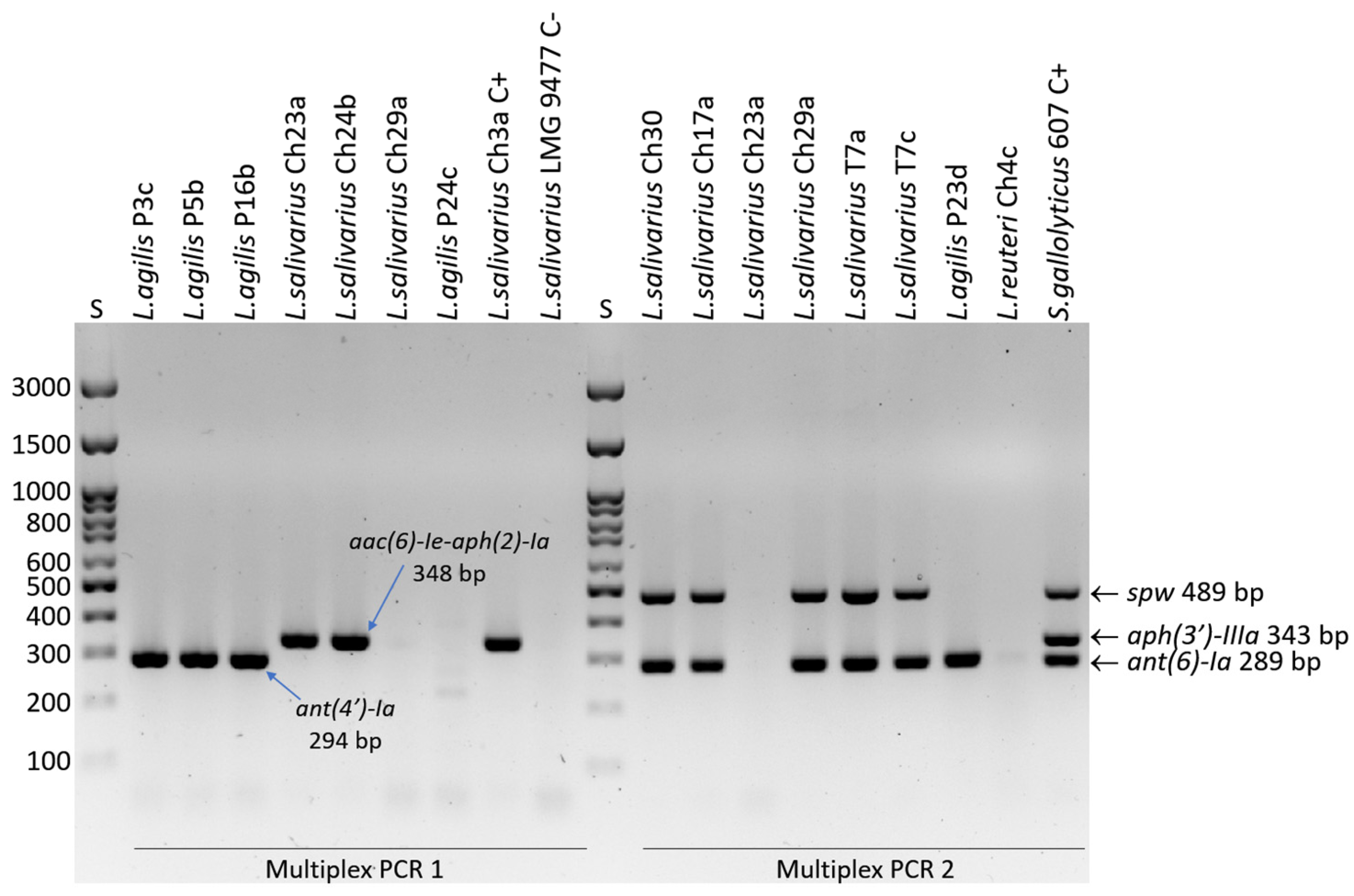
2.4. Detection of Resistance Genes

For the Lactobacillaceae reference strains and three wild-type strains for which WGSs were obtained (G2Lp, T25a, T31e), resistance genes were detected based on WGS analysis using Resfinder 4.1 [27], Resistance Gene Identifier (RGI) ver. 6.0.3 [28], and ABRicate ver. 0.8 [29]. In the remaining field isolates (n = 118), aminoglycoside resistance genes were detected using PCR. The aac(6)-Ie-aph(2)-Ia, ant(4′)-Ia, aph(2″)-Ib, aph(2″)-Ic and aph(2″)-Id genes were detected by multiplex PCR 1 [30], and the multiplex PCR 2 developed by us earlier was used to detect the ant(6)-Ia (aadE), spw, and aph(3′)-IIIa genes [31]. Primer sequences, amplicon sizes, and annealing temperature are given in Table S2. Wild-type LAB strains containing resistance genes were used as positive controls (Table S3).

2.5. Analysis of Mutations in the rpsL Gene

To explain the mechanism of resistance of the reference Lactobacillaceae strains to streptomycin, the sequence of the rpsL gene encoding the 30S subunit ribosomal protein S12 was analyzed. Due to the particularly high streptomycin MIC value (>1024 µg/mL) of L. gallinarum strain LMG 9435, the analysis also included other isolates of this species (with unknown susceptibility to streptomycin) (n = 4) and strains of other species belonging to the L. delbrueckii phylogenetic group with a previously determined degree of susceptibility to streptomycin (n = 8) [14] (Table A1). DNA sequences of the rpsL gene and putative protein sequences obtained by translation using ORF Finder (https://www.ncbi.nlm.nih.gov/orffinder/, accessed on 26 January 2025) were aligned in MEGA XI ver. 11.0.13.

3. Results

3.1. Results of AST and Resistance Gene Detection

According to EFSA criteria [6], after 48 h of incubation in LSM, 36.8% (7/19) of reference Lactobacillaceae strains showed resistance to kanamycin, 26.3% (5/19) were resistant to streptomycin, and 5.3% (1/19) showed resistance to gentamicin. The range of neomycin and spectinomycin MICs ranged from 1 to 64 µg/mL and from 4 to 256 µg/mL, respectively. Despite the significant percentage of resistant strains, no genes conferring resistance to aminoglycosides were detected in any genome (in several genomes, only genes associated with resistance to other antimicrobials were detected) (Table 2 and Table S4).
In wild-type isolates, resistance to kanamycin was most frequently recorded (78/121; 64.5%), and less frequently to streptomycin (12/121; 9.9%) and gentamicin (3/121, 2.5%). Genes determining aminoglycoside resistance were detected in only 11 of 78 (14.1%) phenotypically resistant isolates (Table S1). As many as 93% of L. salivarius isolates, 85% of L. agilis isolates, and 58.8% of L. plantarum isolates, as well as 26.4–33.3% of L. ingluviei and L. reuteri isolates, were classified as resistant to kanamycin. However, no strains were found to contain the aph(3′)-IIIa gene, which usually confers resistance to kanamycin in bacteria, but two L. salivarius isolates with very high MICs for kanamycin (≥1024 µg/mL) and gentamicin (≥128 µg/mL) revealed the aac(6)-Ie-aph(2)-Ia gene (coding for bifunctional aminoglycoside acetyltransferase). Interestingly, one of the aac(6)-Ie-aph(2)-Ia-positive strains also had a very high MIC of streptomycin (>1024 µg/mL) (Table S1). Streptomycin and gentamicin resistance were recorded only in isolates of L. salivarius and L. agilis. In six of 12 isolates resistant to streptomycin and characterized by high MICs (≥512 µg/mL), the ant(6)-Ia (aadE) gene (encoding aminoglycoside nucleotidyltransferase) was detected, and in L. salivarius isolates it coexisted with the spw gene (encoding aminoglycoside nucleotidyltransferase of the ANT(9) family). In 3 L. agilis strains with high neomycin MICs (128–256 µg/mL), the presence of the ant(4′)-Ia gene (encoding aminoglycoside O-nucleotidyltransferase) was confirmed (Table 3 and Table S1, Figure 2).
For all reference and wild-type strains, MIC values could only be read after 24 h of incubation, although L. acidophilus strain ATCC 4356 and some L. reuteri isolates displayed poor growth. The most intensive growth in LSM medium, assessed visually, was observed in isolates of L. salivarius and L. agilis. The MICs were usually one order of magnitude lower than the reading after 48 h, which resulted in a slightly lower percentage of resistant strains (Table 2, Tables S1 and S2).

3.2. Mutations in the rpsL Gene

Mutations in the rpsL gene encoding the S12 protein, which is part of the small ribosomal subunit, were detected in two of the 22 strains tested, i.e., L. gallinarum strain LMG 9435 and L. amylovorus LMG 9496 (Table 2). The mutation was recorded in L. gallinarum LMG 9435 at position 167 (AAG → AGG), translating into a Lys56 → Arg substitution, which most likely determined the high streptomycin MIC value of > 1024 µg/mL. At the same time, no cross-resistance to other aminoglycoside antibiotics has been reported. By analyzing the rpsL sequences of other L. gallinarum strains (n = 4) (of unknown susceptibility to streptomycin), we ruled out the possibility that the mutation at this position was a constant feature of strains of this species. Additionally, based on AST results published by Campedelli et al. [14], we showed that the mutation at position 167 does not occur in streptomycin-susceptible strains (of various species) of the L. delbrueckii phylogenetic group. However, the same aa substitution (Lys56 → Arg) was noted in the streptomycin-resistant L. gigeriorum strain DSM 23908 (Table A1). The mutation noted in L. amylovorus strain LMG 9496 at position 303 (AAG → ACG), which translates into a substitution (Lys101 → Thr), does not seem to affect the susceptibility of this strain to streptomycin (MIC 16–32 µg/mL) (Table A1).

4. Discussion

In this study, in an antimicrobial susceptibility test based on the ISO 10932:2010/IDF 223:2010 standard [23] and using the EFSA/FEEDAP Panel cut-off points [6], 52.6% (10/19) of reference Lactobacillaceae strains and 64.5% (78/121) of wild-type strains were classified as resistant to aminoglycosides, most of them lacking resistance genes. Resistance to kanamycin was most frequently noted, less frequently to streptomycin and gentamicin. Our observations are consistent with results reported by several other authors who assessed the antimicrobial susceptibility of Lactobacillaceae according to the same protocol.
The widespread occurrence of kanamycin resistance (61%) among reference Lactobacillaceae strains of various species (former genus Lactobacillus) has also been noted by Campedelli et al. [14], who detected (by whole-genome analysis) genes that could determine resistance to this antibiotic, i.e., aac(3) and aph(3), in only 11% of the phenotypically resistant strains. The same study also showed a significant percentage of strains to be resistant to streptomycin (27%) and gentamicin (14.2%), while the ant(6) and ant(9) resistance genes were identified in only in a few strains [14]. Mayrhofer et al. [17] reported resistance to kanamycin (MIC = 128 µg/mL) and streptomycin in 23.8% (24/101) and 8.9% (9/101) of strains of the L. acidophilus phylogenetic group, respectively, and did not detect aminoglycoside resistance genes in any strain. Results consistent with ours were also recently demonstrated by scientists from Denmark [15], who examined a total of 170 strains from the Lactobacillaceae family (from the Chr. Hansen’s Culture collection). According to the EFSA cut-off points, kanamycin resistance was demonstrated by 92% of L. salivarius strains, 11% of L. delbrueckii strains, and 13% of L. gasseri/L. paragasseri strains. Streptomycin resistance was recorded for 33% of L. salivarius strains, 60% of L. sakei strains, 4% of L. delbrueckii strains, and 6% of L. gasseri/L. paragasseri strains. No strain exceeded the FEEDAP Panel threshold values for gentamicin, and WGS analysis showed that none of the strains contained genes determining resistance to aminoglycosides [15]. In another study conducted on 65 LAB strains, including 57 from the former genus Lactobacillus, kanamycin resistance was recorded in 32.3% (21/65) of the strains, streptomycin-resistant strains accounted for 21.4% (12/56), and gentamicin-resistant for 3.1% (2/65). The aph(3)-IIIa gene was detected in only four of 21 (19%) kanamycin-resistant strains, and the str(A)/str(B) gene was detected in just one of 12 streptomycin-resistant strains (and in one phenotypically susceptible strain) [16]. In our previous paper, we showed widespread resistance of Lactobacillaceae isolates from pigeons to kanamycin (89%) and streptomycin (63%), and 14% of isolates were resistant to gentamicin [18]. However, it should be noted that the antimicrobial susceptibility test was performed in Iso-sensitest medium containing 25% MRS (not 10% as indicated in the ISO 10932:2010/IDF 223:2010 standard), which may have affected the MIC value. A high frequency of resistance to streptomycin has also been recorded in Lactobacillaceae strains (former genus Lactobacillus) from chickens (31%) and turkeys (31%), including L. agilis (33–100% of strains showed resistance to streptomycin), L. crispatus (17–23%), L. salivarius (50–52%), L. saerimneri (100%), and L. johnsonii (14%). However, the presence of the ant(6)-Ia gene was confirmed in only a few phenotypically resistant L. salivarius strains [19,20]. Danielsen and Wind [32], who used E-tests and MRS medium to assess the antimicrobial susceptibility of Lactobacillaceae (former genus Lactobacillus), reported high kanamycin and streptomycin MIC values (≥128 µg/mL) for 86% and 61% of isolates tested, respectively.
The results of this study, as well as the other studies cited above, suggest that the high frequency of kanamycin resistance in the examined species of the genus Ligilactobacillus and Limosilactobacillus is mainly due to intrinsic resistance, which is most likely linked to the lack of cytochrome-mediated drug transport [33]. The theory of intrinsic resistance is also supported by the unimodal distribution of kanamycin and streptomycin MIC values in Lactobacillaceae, including L. salivarius and L. agilis, also observed by other authors [15,18,32]. According to CLSI (document M100-Ed34) [34], intrinsic resistance is defined as ‘inherent or innate (not acquired) antimicrobial resistance, which is reflected in wild-type antimicrobial patterns of all or almost all representatives of a species’. Furthermore, the CLSI states that ‘intrinsic resistance is so common that susceptibility testing is unnecessary’. Therefore, it must be clearly determined which, if any, species of Lactobacillaceae are naturally resistant to aminoglycoside antibiotics, so that individual antimicrobial substances from this group can be excluded from laboratory tests. At the same time, it should be noted that EFSA does not rule out the use of naturally resistant strains as microbial feed additives. The guidelines state that ‘detection of the MIC above the cut-off values proposed by the FEEDAP Panel for one or more antimicrobials requires further investigation using genomic data to determine the nature of the resistance’, but ‘if no known AMR gene is identified that can be linked to the phenotype, no further studies are required’ [6].
Lactobacillaceae resistance to aminoglycosides may also be caused by mutations in chromosomal genes, e.g., those encoding ribosomal proteins, as we have shown in the case of streptomycin-resistant L. gallinarum strain LMG 9435. The mutation at position 167 (AAG → AGG) of the rpsL gene, translating into a Lys56 → Arg substitution, has previously been identified in streptomycin-resistant L. plantarum [35] and L. rhamnosus [36] strains. In the latter species, a mutation leading to a substitution for lysine was also recorded at position 303 [36]. In other taxonomic groups of bacteria, mutations determining resistance to streptomycin have been identified at other positions of the rpsL gene, i.e., at position 129 (Lys43 → Arg/Thr/Asn)) in Bifidobacterium bifidum [37], Yersinia pestis [38], and Pectobacterium carotovorum [39]; at position 128 (Lys42 → Thr) and 263 (Lys86 → Arg) in E. coli [40] and in Mycobacterium tuberculosis [41]. Interestingly, in almost all cases, streptomycin resistance was associated with a change from lysine to arginine or lysine to threonine. In agreement with our results are the findings of Bernard et al. [39] that streptomycin-resistant Pectobacterium carotovorum subsp. carotovorum (formerly Erwinia carotovora subsp. carotovora) strains with a mutation in the rpsL gene did not show cross-resistance to spectinomycin, kanamycin, or gentamicin.
We have demonstrated that the same cut-off points should not be used for different susceptibility testing protocols, as incubation time affects the MIC value, often leading to a change in the categorization of a strain from susceptible (reading after 24 h of incubation) to resistant (reading after 48 h). Similar conclusions have been reached by other authors analyzing the effect of incubation time and inoculum density on the antimicrobial susceptibility of LAB [8].
It should also be considered whether AST requires incubation of the microplate for as long as 48 h, given that many Lactobacillaceae species, especially L. salivarius (based on visual assessment), grow very well in LSM broth after 24 h. Longer incubation substantially decreases the pH of the medium due to lactic acid production, which may affect the activity of antimicrobial substances as well as the growth rate and viability of bacteria [42,43]. In addition, some antimicrobial substances, including neomycin, have limited stability in the culture medium [44]. Another possible undesirable effect of 48 h incubation may be partial evaporation of the medium from the microplate wells, which may also affect the MIC value.
Neomycin was included in this study because, unlike kanamycin, it is often used in poultry, pig, and cattle farming. Natural resistance to neomycin would certainly be a desirable feature to take into account when selecting probiotic strains for these groups of animals. We therefore believe that it would be worth extending the EFSA guidelines to include neomycin. Establishing cut-off points for this antibiotic, however, requires testing on a large number of Lactobacillaceae strains.

5. Conclusions

Our study shows that categorization of Lactobacillaceae strains (representing the former genus Lactobacillus) according to the cut-off points established by the EFSA FEEDAP Panel in 2018 [6] and the use of the antimicrobial susceptibility protocol according to ISO 10932:2010/IDF 223:2010 [26] can lead to the recognition of strains that do not contain resistance genes as resistant, and thus eliminate them from further studies on the selection of probiotic strains. It is true that in addition to the phenotypic test, EFSA also requires WGS analyses to detect resistance genes. However, in the standard laboratory procedure, the phenotypic test is performed first, and only those strains that do not exceed the cut-off points are referred for WGS (due to the high cost of genomic sequencing).
Our results, as well as other reports mentioned above, have shown that the cut-off points established by EFSA for aminoglycoside antibiotics, especially kanamycin, should be re-examined, defined at the genus and/or species level, and adapted to the given protocol of AST. It would also be appropriate to expand the guidelines to include neomycin, which is commonly used in animal treatment. The data presented in this paper may be useful for revising the current EFSA guidelines on the AST of Lactobacillaceae.

Supplementary Materials

The following supporting information can be downloaded at https://www.mdpi.com/article/10.3390/life15050732/s1: Table S1: Results of antimicrobial susceptibility testing (MIC values) and detection of resistance genes in Lactobacillaceae isolates; Table S2: Primers used for detection of aminoglycoside resistance genes; Table S3: Bacterial strains used as positive control during detection of resistance genes by PCR; Table S4: MIC values (µg/mL) obtained in the antimicrobial susceptibility test for reference Lactobacillaceae strains.

Author Contributions

Conceptualization, M.D.; methodology, M.D.; validation, M.D. and A.Z.; formal analysis, M.D. and A.Z.; investigation, M.D., K.H.-O. and A.Z.; resources, M.D., K.H.-O. and A.Z.; data curation, M.D. and A.Z.; writing—original draft preparation, M.D.; writing—review and editing, M.D., A.Z. and R.U.-C.; supervision, M.D. and R.U.-C. All authors have read and agreed to the published version of the manuscript.

Funding

This research received no external funding.

Institutional Review Board Statement

Not applicable.

Informed Consent Statement

Not applicable.

Data Availability Statement

The WGSs reported in this paper have been deposited in the NCBI GenBank database under the following accession numbers: CP168064-CP168068 (GCA_041531555.1)—strain G2Lp, CP180626-CP180627 (GCA_047782765.1)—strain T31e, CP183830-CP183833 (GCA_048572735.1)—strain T25a.

Acknowledgments

The authors would like to thank Marian Broekhuizen-Stins for her technical assistance with the genomic sequencing of Lactobacillaceae strains.

Conflicts of Interest

The authors declare no conflicts of interest.

Abbreviations

The following abbreviations are used in this manuscript:
ASTAntimicrobial susceptibility testing
CLSIClinical and Laboratory Standards Institute
EFSAEuropean Food Safety Authority
FEEDAPEFSA Panel on Additives and Products or Substances used in Animal Feed
IDFInternational Dairy Federation
ISOInternational Organization for Standardization
LABLactic acid bacteria
LSMLAB susceptibility test medium
MRSDe Man-Rogosa-Sharpe
WGSWhole genome sequence

Appendix A

Table A1. Results of mutation detection in the rpsL gene encoding ribosomal protein S12 in strains of the L. delbrueckii phylogenetic group.
StrainMetabolic GroupPhylogenetic GroupGenBank Acc. No.Susceptibility to STRMIC ValueMutations in rpsL GeneAminoglycoside RGs
L. gallinarum LMG 9435OHOL. delbrueckiiGCA_001434975.1R>1024Lys56 → ArgNo
L. gallinarum An153OHOL. delbrueckiiGCA_002160635.1NDND No
L. gallinarum An101OHOL. delbrueckiiGCA_002161165.1NDND No
L. gallinarum J07OHOL. delbrueckiiGCA_947381795.1NDND No
L. gallinarum Chicken_20_mag_156OHOL. delbrueckiiGCA_904419645.1NDND No
L. acidophilus ATCC 4356OHOL. delbrueckiiGCA_034298135.1S4–16 No
L. amylovorus LMG 9496OHOL. delbrueckiiGCA_002706375.1S/R8–32Lys101 → ThrNo
L. crispatus LMG 9479OHOL. delbrueckiiGCA_018987235.1S/R16–32 No
L. crisptus T31eOHOL. delbrueckiiGCA_047782765.1S8–16 No
L. amylolyticus DSM 11664OHOL. delbrueckiiGCA_004354545.1S [14]UN No
L. gigeriorum DSM 23908OHOL. delbrueckiiGCA_001436575.1R [14]UNLys56 → ArgNo
L. delbrueckii subsp. jakobsenii DSM 26046OHOL. delbrueckiiGCA_001888925.1R [14]UN No
L. delbruckii subsp. delbrueckii DSM 20074OHOL. delbrueckiiGCA_001908495.1S [14]UN No
L. helveticus LMG 22464OHOL. delbrueckiiGCA_001437535.1S [14]UN No
L. taiwanensis DSM 21401OHOL. delbrueckiiGCA_001436695.1S [14]UN No
L. pasteurii DSM 23907FHEL. delbrueckiiGCA_004354755.1S [14]UN No
L. apis LMG 26964FHEL. delbrueckiiGCA_002837055.1R [14]UN No
OHO—obligate homofermentative; FHE—facultative heterofermentative; R—resistant; S—susceptible; ND—not determined; UN—unknown; RGs—resistance genes.

References

  1. Sun, Z.; Yu, J.; Dan, T.; Zhang, W.; Zhang, H. Phylogenesis and evolution of lactic acid bacteria. In Lactic Acid Bacteria; Zhang, H., Cai, Y., Eds.; Springer: Dordrecht, The Netherlands, 2014; pp. 1–101. [Google Scholar] [CrossRef]
  2. Gilroy, R.; Ravi, A.; Getino, M.; Pursley, I.; Horton, D.L.; Alikhan, N.F.; Baker, D.; Gharbi, K.; Hall, N.; Watson, M.; et al. Extensive microbial diversity within the chicken gut microbiome revealed by metagenomics and culture. PeerJ 2021, 9, e10941. [Google Scholar] [CrossRef]
  3. NCBI Database. Available online: https://www.ncbi.nlm.nih.gov/taxonomy (accessed on 27 December 2024).
  4. Zheng, J.; Wittouck, S.; Salvetti, E.; Franz, C.M.A.P.; Harris, H.M.B.; Mattarelli, P.; O’Toole, P.W.; Pot, B.; Vandamme, P.; Walter, J.; et al. taxonomic note on the genus Lactobacillus: Description of 23 novel genera, emended description of the genus Lactobacillus Beijerinck 1901, and union of Lactobacillaceae and Leuconostocaceae. Int. J. Syst. Evol. Microbiol. 2020, 70, 2782–2858. [Google Scholar] [CrossRef] [PubMed]
  5. Bhogoju, S.; Nahashon, S. Recent Advances in Probiotic Application in Animal Health and Nutrition: A Review. Agriculture 2022, 12, 304. [Google Scholar] [CrossRef]
  6. EFSA. Guidance on the characterisation of microorganisms used as feed additives or as production organisms. EFSA J. 2018, 16, 5206. [Google Scholar]
  7. Klare, I.; Konstabel, C.; Müller-Bertling, S.; Reissbrodt, R.; Huys, G.; Vancanneyt, M.; Swings, J.; Goossens, H.; Witte, W. Evaluation of new broth media for microdilution antibiotic susceptibility testing of Lactobacilli, Pediococci, Lactococci, and Bifidobacteria. Appl. Environ. Microbiol. 2005, 71, 8982–8986. [Google Scholar] [CrossRef]
  8. Egervärn, M.; Lindmark, H.; Roos, S.; Huys, G.; Lindgren, S. Effects of inoculum size and incubation time on broth microdilution susceptibility testing of lactic acid bacteria. Antimicrob. Agents Chemother. 2007, 5, 394–396. [Google Scholar] [CrossRef] [PubMed]
  9. Mayrhofer, S.; Zitz, U.; Birru, F.H.; Gollan, D.; Gołoś, A.K.; Kneifel, W.; Domig, K.J. Comparison of the CLSI guideline and ISO/IDF standard for antimicrobial susceptibility testing of Lactobacilli. Microb. Drug Resist. 2014, 20, 591–603. [Google Scholar] [CrossRef]
  10. Łepecka, A.; Szymański, P.; Rutkowska, S.; Iwanowska, K.; Kołożyn-Krajewska, D. The Influence of Environmental Conditions on the Antagonistic Activity of Lactic Acid Bacteria Isolated from Fermented Meat Products. Foods 2021, 10, 2267. [Google Scholar] [CrossRef]
  11. Tscherne, A.; Mantel, E.; Boskani, T.; Budniak, S.; Elschner, M.; Fasanella, A.; Feruglio, S.L.; Galante, D.; Giske, C.G.; Grunow, R.; et al. Adaptation of Brucella melitensis Antimicrobial Susceptibility Testing to the ISO 20776 Standard and Validation of the Method. Microorganisms 2022, 10, 1470. [Google Scholar] [CrossRef]
  12. Sabath, L.D. Chemical and physical factors influencing methicillin resistance of Staphylococcus aureus and Staphylococcus epidermidis. J. Antimicrob. Chemother. 1977, 3 (Suppl. C), 47–51. [Google Scholar] [CrossRef]
  13. Mendonça, A.A.; de Morais, M.A.; Cabrera, M.Z., Jr. Cysteine induces resistance of lactobacilli to erythromycin and azithromycin. Int. J. Antimicrob. Agents. 2019, 53, 352–353. [Google Scholar] [CrossRef] [PubMed]
  14. Campedelli, I.; Mathur, H.; Salvetti, E.; Clarke, S.; Rea, M.C.; Torriani, S.; Ross, R.P.; Hill, C.; O’Toole, P.W. Genus-Wide Assessment of Antibiotic Resistance in Lactobacillus spp. Appl. Environ. Microbiol. 2018, 85, e01738-18. [Google Scholar] [CrossRef]
  15. Nøhr-Meldgaard, K.; Struve, C.; Ingmer, H.; Koza, A.; Al-Nakeeb, K.; Agersø, Y. Antimicrobial susceptibility testing and tentative epidemiological cut-off values for Lactobacillaceae family species intended for ingestion. Front. Antibiot. 2023, 2, 1162636. [Google Scholar] [CrossRef] [PubMed]
  16. Stefańska, I.; Kwiecień, E.; Jóźwiak-Piasecka, K.; Garbowska, M.; Binek, M.; Rzewuska, M. Antimicrobial Susceptibility of Lactic Acid Bacteria Strains of Potential Use as Feed Additives—The Basic Safety and Usefulness Criterion. Front. Vet. Sci. 2021, 8, 687071. [Google Scholar] [CrossRef]
  17. Mayrhofer, S.; van Hoek, A.H.; Mair, C.; Huys, G.; Aarts, H.J.; Kneifel, W.; Domig, K.J. Antibiotic susceptibility of members of the Lactobacillus acidophilus group using broth microdilution and molecular identification of their resistance determinants. Int. J. Food Microbiol. 2010, 144, 81–87. [Google Scholar] [CrossRef] [PubMed]
  18. Dec, M.; Stępień-Pyśniak, D.; Nowaczek, A.; Puchalski, A.; Urban-Chmiel, R. Phenotypic and genotypic antimicrobial resistance profiles of fecal lactobacilli from domesticated pigeons in Poland. Anaerobe. 2020, 65, 102251. [Google Scholar] [CrossRef]
  19. Dec, M.; Nowaczek, A.; Stępień-Pyśniak, D.; Wawrzykowski, J.; Urban-Chmiel, R. Identification and antibiotic susceptibility of lactobacilli isolated from turkeys. BMC Microbiol. 2018, 18, 168. [Google Scholar] [CrossRef]
  20. Dec, M.; Urban-Chmiel, R.; Stępień-Pyśniak, D.; Wernicki, A. Assessment of antibiotic susceptibility in Lactobacillus isolates from chickens. Gut Pathog. 2017, 9, 54. [Google Scholar] [CrossRef]
  21. Dec, M.; Urban-Chmiel, R.; Gnat, S.; Puchalski, A.; Wernicki, A. Identification of Lactobacillus strains of goose origin using MALDI-TOF mass spectrometry and 16S-23S rDNA intergenic spacer PCR analysis. Res. Microbiol. 2014, 165, 190–201. [Google Scholar] [CrossRef]
  22. Dec, M.; Puchalski, A.; Urban-Chmiel, R.; Wernicki, A. 16S-ARDRA and MALDI-TOF mass spectrometry as tools for identification of Lactobacillus bacteria isolated from poultry. BMC Microbiol. 2016, 16, 105. [Google Scholar] [CrossRef]
  23. Kolmogorov, M.; Yuan, J.; Lin, Y.; Pevzner, P.A. Assembly of long, error-prone reads using repeat graphs. Nat. Biotechnol. 2019, 37, 540–546. [Google Scholar] [CrossRef]
  24. Huang, Y.T.; Liu, P.Y.; Shih, P.W. Homopolish: A method for the removal of systematic errors in nanopore sequencing by homologous polishing. Genome Biol. 2021, 22, 95. [Google Scholar] [CrossRef]
  25. Seemann, T. Prokka: Rapid prokaryotic genome annotation. Bioinformatics 2014, 30, 2068–2069. [Google Scholar] [CrossRef]
  26. ISO 10932:2010 (IDF 223:2010); Milk and Milk Products—Determination of the Minimal Inhibitory Concentration (Mic) of Antibiotics Applicable to Bifidobacteria and Non-Enterococcal Lactic Acid Bacteria (LAB). International Organization for Standardization: Geneva, Switzerland, 2010. Available online: https://www.iso.org/standard/46434.html (accessed on 13 January 2025).
  27. Bortolaia, V.; Kaas, R.S.; Ruppe, E.; Roberts, M.C.; Schwarz, S.; Cattoir, V.; Philippon, A.; Allesoe, R.L.; Rebelo, A.R.; Florensa, A.F.; et al. ResFinder 4.0 for predictions of phenotypes from genotypes. J. Antimicrob. Chemother. 2020, 75, 3491–3500. [Google Scholar] [CrossRef] [PubMed]
  28. Alcock, B.P.; Raphenya, A.R.; Lau, T.T.Y.; Tsang, K.K.; Bouchard, M.; Edalatmand, A.; Huynh, W.; Nguyen, A.V.; Cheng, A.A.; Liu, S.; et al. CARD 2020: Antibiotic resistome surveillance with the comprehensive antibiotic resistance database. Nucleic Acids Res. 2020, 48, D517–D525. [Google Scholar] [CrossRef] [PubMed]
  29. Seemann, T. Abricate, Github. Available online: https://github.com/tseemann/abricate (accessed on 15 January 2025).
  30. Vakulenko, S.B.; Donabedian, S.M.; Voskresenskiy, A.M.; Zervos, M.J.; Lerner, S.A.; Chow, J.W. Multiplex PCR for detection of aminoglycoside resistance genes in enterococci. Antimicrob. Agents Chemother. 2003, 47, 1423–1426. [Google Scholar] [CrossRef] [PubMed]
  31. Dec, M.; Nowak, T.; Webster, J.; Wódz, K. Serotypes, Antimicrobial Susceptibility, and Potential Mechanisms of Resistance Gene Transfer in Erysipelothrix rhusiopathiae Strains from Waterfowl in Poland. Int. J. Mol. Sci. 2024, 25, 12192. [Google Scholar] [CrossRef]
  32. Danielsen, M.; Wind, A. Susceptibility of Lactobacillus spp. to antimicrobial agents. Int. J. Food Microbiol. 2003, 82, 1–11. [Google Scholar] [CrossRef]
  33. Hummel, A.S.; Hertel, C.; Holzapfel, W.H.; Franz, C.M. Antibiotic resistances of starter and probiotic strains of lactic acid bacteria. Appl. Environ. Microbiol. 2007, 73, 730–739. [Google Scholar] [CrossRef]
  34. CLSI Document M100-Ed34; Performance Standards for Antimicrobial Susceptibility Testing CLSI M100 Includes Updated Tables for the Clinical and Laboratory Standards Institute Antimicrobial Susceptibility Testing Standards CLSI M02, M07, and M11. A CLSI Supplement for Global Application. Clinical Lab Standards Institute: Malvern, PA, USA, 2024; p. 318.
  35. Zhang, F.; Gao, J.; Wang, B.; Huo, D.; Wang, Z.; Zhang, J.; Shao, Y. Whole-genome sequencing reveals the mechanisms for evolution of streptomycin resistance in Lactobacillus plantarum. J. Dairy Sci. 2018, 101, 2867–2874. [Google Scholar] [CrossRef]
  36. Namai, F.; Nishiyama, K.; Kitazawa, H.; Shimosato, T. Introduction of Spontaneous Mutations Using Streptomycin as a Method for Lactic Acid Bacteria Breeding. Methods Mol. Biol. 2024, 2851, 15–21. [Google Scholar] [CrossRef]
  37. Sirichoat, A.; Flórez, A.B.; Vázquez, L.; Buppasiri, P.; Panya, M.; Lulitanond, V.; Mayo, B. Antibiotic Susceptibility Profiles of Lactic Acid Bacteria from the Human Vagina and Genetic Basis of Acquired Resistances. Int. J. Mol. Sci. 2020, 21, 2594. [Google Scholar] [CrossRef] [PubMed]
  38. Dai, R.; He, J.; Zha, X.; Wang, Y.; Zhang, X.; Gao, H.; Yang, X.; Li, J.; Xin, Y.; Wang, Y.; et al. A novel mechanism of streptomycin resistance in Yersinia pestis: Mutation in the rpsL gene. PLoS Negl. Trop. Dis. 2021, 15, e0009324. [Google Scholar] [CrossRef]
  39. Barnard, A.M.L.; Simpson, N.J.L.; Lilley, K.S.; Salmond, G.P.C. Mutations in rpsL that confer streptomycin resistance show pleiotropic effects on virulence and the production of a carbapenem antibiotic in Erwinia carotovora. Microbiology 2010, 156, 1030–1039. [Google Scholar] [CrossRef]
  40. Pelchovich, G.; Schreiber, R.; Zhuravlev, A.; Gophna, U. The contribution of common rpsL mutations in Escherichia coli to sensitivity to ribosome targeting antibiotics. Int. J. Med. Microbiol. 2013, 303, 558–562. [Google Scholar] [CrossRef] [PubMed]
  41. Khosravi, A.D.; Etemad, N.; Hashemzadeh, M.; Khandan Dezfuli, S.; Goodarzi, H. Frequency of rrs and rpsL mutations in streptomycin-resistant Mycobacterium tuberculosis isolates from Iranian patients. J. Glob. Antimicrob. Resist. 2017, 9, 51–56. [Google Scholar] [CrossRef] [PubMed]
  42. Vera-Peña, M.Y.; Rodriguez Rodriguez, W.L. Effect of pH on the growth of three lactic acid bacteria strains isolated from sour cream. Univ. Sci. 2020, 25, 341–358. [Google Scholar] [CrossRef]
  43. Giraud, E.; Lelong, B.; Raimbault, M. Influence of pH and initial lactate concentration on the growth of Lactobacillus plantarum. Appl. Microbiol. Biotechnol. 1991, 36, 96–99. [Google Scholar] [CrossRef]
  44. Kerek, Á.; Ecsedi, B.G.; Szabó, Á.; Szimrók, Z.; Paliczné Kustán, B.; Jerzsele, Á.; Nagy, G. Stability Studies of the Dilution Series of Different Antibiotic Stock Solutions in Culture Medium Incubated at 37 °C. Antibiotics 2024, 13, 549. [Google Scholar] [CrossRef]
Figure 1. The study’s workflow.
Life 15 00732 g001
Figure 2. Electrophoretic separation of PCR products (amplicons of resistance genes) in a 2% agarose gel. S—DNA size standard; C+—positive control; C−—negative control.
Life 15 00732 g002
Table 1. Reference and wild-type Lactobacillaceae strains used in the study.
Reference StrainOther Culture Collection NumbersWGS
[GenBank Acc. No.]
Ligilactobacilluis salivarius LMG 9476DSM 20554; ATCC 11742GCA_002079585.1
Ligilactobacillus salivarius LMG 9477DSM 20555; ATCC 11741; JCM 1231GCA_001435955.1
Ligilactobacillus agilis LMG 9186DSM 20509; LMG 9186GCA_001436215.1
Lactiplantibacillus plantarum ATCC 8014DSM 20205; JCM 1057; LMG 1284GCA_002631775.1
Lacticaseibacillus rhamnosus ATCC 7469DSM 20021; JCM 1136GCA_001435405.1
Lacticaseibacillus casei ATCC 393DSM 20011; JCM 1134GCA_000829055.1
Lacticaseibacillus zeae LMG 17315DSM 20178; JCM 11302; ATCC 15820GCA_001433745.1
Limosilactobacillus reuteri subsp. reuteri LMG 9213ATCC 23272; JCM 1112; DSM 20016GCA_000010005.1
Limosilactobacillus reuteri LMG 18238ATCC 55148ERR3330657
Limosilactobacillus ingluviei LMG 20380JCM 12531; DSM 15946; CCUG 45722GCA_001435775.1
Limosilactibacillus ingluviei LMG 22056DSM 14792; JCM 11425GCA_001437235.1
Limosilactibacillus oris LMG 9848DSM 4864; ATCC 49062GCA_001434465.1
Lactobacillus paragasseri LMG 13134LMG 11444; JCM 5344; ATCC 9857GCA_003307295.1
Lactobacillus acidophilus ATCC 4356DSM 20079; JCM 1132; NCIB 8690GCA_034298135.1
Lactobacillus gallinarum LMG 9435JCM 2011; ATCC 33199; DSM 10532GCA_001434975.1
Lactobacillus kitasatonis LMG 23133LMG 22685; JCM 1039, DSM 16761GCA_000615285.1
Lactobacillus amylovorus LMG 9496JCM 1126; ATCC 33620; DSM 20531GCA_002706375.1
Lactobacillus johnsonii LMG 9436ATCC 33200; JCM 2012; DSM 10533GCA_001433975.1
Lactobacillus crispatus LMG 9479ATCC 33820; DSM 20584; JCM 1185GCA_018987235.1
Wild-type Strains
Ligilactobacillus salivarius (n = 44)
Ligilactobacillus agilis (n = 20)
Lactiplantibacillus plantarum (n = 17)
Limosilactobacillus ingluviei (n = 20)
Limosilactobacillus reuteri (n = 16)
Lactobacillus crispatus (n = 4)
Table 2. MIC values (µg/mL) obtained in the antimicrobial susceptibility test.
Antibiotic→KanamycinStreptomycinSpectinomycinGentamicinNeomycinResistance Genes Detected
EFSA Cut-Offs→R > 64 µg/mLR > 64 µg/mL or
R > 32 µg/mL A or
R > 16 µg/mL B
NAR > 16 µg/mL or
R > 8 µg/mL C or
R > 32 µg/mL D
NA
Strain24 h48 h24 h48 h48 h24 h48 h24 h48 h
L. salivarius LMG 947664–128128–5123232–64324–888–168–16
L. salivarius LMG 947764–128128–2563232–64644–84–88–168–16
L. agilis LMG 9186512512128128–25625616–3216–3216–3232–64
L. reuteri subsp. reuteri LMG 921316–3216–328–168–321280.5–10.5–21–42–4
L. reuteri LMG 1823864–256128–25616–6432–6412844–81616
L. oris LMG 984816–3232–648–1616–641280.5–112–42–4
L. ingluviei LMG 20380326464–12864–1282562–42–488tetW, tetM, tetL, ermB
L. ingluviei LMG 2205664–128128–2563232–6425624–81616tetW, lnuC
L. plantarum ATCC 80144–8164–84–161280.250.251–21–4
L. rhamnosus ATCC 74691632–6448–16641–22–42–84–16
L. casei ATCC 3931632–64816321–2248
L. zeae LMG 1731532–6464–12816–323264448–168–32
L. johnsonii LMG 943632–64648–88–163244–88–328–32
L. paragasseri LMG 1313464642–44–882–42–416–3232–64
L. acidophilus ATCC 43564484–16161–21–21–44
L. gallinarum LMG 94351616>1024>1024160.250.25–0.548tetW, rpsLArg56
L. kitasatonis LMG 231338–168–161–22–81622–43232–64
L. amylovorus LMG 94968–16168–168–32412816rpsLThr101
L. crispatus LMG 94791281281616–32324–8816–3232
Total resistant6
[31.6%]
7
[36.8%]
3
[15.8%]
5
[26.3%]
NA1
[5.3%]
1
[5.3%]
NANA
Gray-highlighted values indicate resistance; A—applies to L. rhamnosus; B—applies to species from the L. acidophilus group; C—applies to L. reuteri; D—applies to L. casei/paracasei and L. zeae; NA—not applicable.
Table 3. Distribution of MIC values of aminoglycoside antibiotics in wild-type Lactobacillaceae strains.
MIC Value→
[µg/mL]
≤0.250.51248163264128256512≥1024No. [%] of Resistant
Kanamycin
L. salivarius (n = 44) 32019 2 bif41 [93.2]
L. agilis (n = 20) 31042 ant(4)Ia117 [85.0]
L. plantarum (n = 17) 32282 10 [58.8]
L. ingluviei (n = 18) 12951 6 [33.3]
L. reuteri (n = 15) 1154 4 4 [26.7]
L. crispatus (n = 4) 13 0
Streptomycin
L. salivarius (n = 44) 313193 2 spw
aadE
4 spw(3)
aadE(3)
9 [20.4]
L. agilis (n = 20) 13582 1 aadE3 [15.0]
L. plantarum (n = 17) 12761 NA
L. ingluviei (n = 20) 2810 0
L. reuteri (n = 16) 36241 0
L. crispatus (n = 4) 21 1 0
Gentamicin
L. salivarius (n = 44) 1514175 1 bif 1 bif2 [4.5]
L. agilis (n = 20) 239411 1 [5.0]
L. plantarum (n = 17) 22616 0
L. ingluviei (n = 20) 1212311 0
L. reuteri (n = 16)35332 0
L. crispatus (n = 4) 22 0
Neomycin
L. salivarius (n = 41) 4112051 NA
L. agilis (n = 20) 15361 3 ant(4)Ia(2)1 ant(4)Ia NA
L. plantarum (n = 11) 131321 NA
L. ingluviei (n = 20) 17831 NA
L. reuteri (n = 13)2243 11 NA
L. crispatus (n = 4) 121 NA
Grey-highlighted values (µg/mL) indicate resistance; NA—not applicable; bif—aac(6)-Ie-aph(2)-Ia gene; the number of strains carrying the gene in question is given in brackets after the name of gene; the absence of any number following the name of gene means that all isolates contain the gene
Disclaimer/Publisher’s Note: The statements, opinions and data contained in all publications are solely those of the individual author(s) and contributor(s) and not of MDPI and/or the editor(s). MDPI and/or the editor(s) disclaim responsibility for any injury to people or property resulting from any ideas, methods, instructions or products referred to in the content.

