Article

Edge-Preferential Graphitization and Nanoscale Structural Heterogeneity in Coal-Derived Carbon Revealed by High-Resolution Transmission Electron Microscopy (HRTEM)


1
School of Geology and Mining Engineering, Xinjiang University, Ürümqi 830047, China
2
Xinjiang Key Laboratory for Geodynamic Processes and Metallogenic Prognosis of the Central Asian Orogenic Belt, Xinjiang University, Ürümqi 830047, China
3
Xinjiang Key Laboratory of Coalbed Methane Exploration and Development, Ürümqi 830063, China
4
Mineral Experimental Research Center, Xinjiang Bureau of Geology (Xinjiang Quality Testing Centre of Gemstones), Ürümqi 830099, China
5
School of Natural Resources Science and Technology, Xinjiang University of Technology, Hetian 848023, China
6
Xilingol Mengdong Mining Co., Ltd., Xilinhaote City 026000, China
7
Coal Chemistry Branch, China Coal Research Institute, Beijing 100013, China
8
School of Geoscience and Surveying Engineering, China University of Mining and Technology (Beijing), Beijing 100083, China
*
Authors to whom correspondence should be addressed.
Minerals 2026, 16(8), 780; https://doi.org/10.3390/min16080780
Submission received: 11 June 2026 / Revised: 24 July 2026 / Accepted: 25 July 2026 / Published: 27 July 2026

Abstract

The nanoscale structural heterogeneity of carbons plays a crucial role in regulating the surface activity, catalytic performance, and wettability of substances. Herein, we quantitatively investigate the nanostructural evolution of a coal-derived carbon during high-temperature treatment, using high-resolution transmission electron microscopy (HRTEM). To elucidate the intrinsic nanoscale structural heterogeneity of carbonaceous materials during thermal treatment, a particle-edge preferential graphitization is hypothesized in this study. Based on quantitative HRTEM analysis of coal-derived carbon heat-treated up to 3000 °C, this study demonstrates that particle boundaries act as intrinsic geometric templates and stress concentrators, governing the carbon nanostructural evolution. The length enhancement of the aromatic structural transformation occurs at 300 °C, 1000 °C, and 2550 °C, respectively, with substantially decreased small-size lattice fringes and substantially increased medium- and large-size lattice fringes. The average curvature displays a non-monotonic decrease and a transient increase during the second carbonization and pregraphitization processes, driven by the formation of concentric and twisted nanostructures. During graphitization, lattice fringe curvature becomes geometrically constrained by particle edges, resulting in an unexpected positive correlation with fringe length. Lattice fringes at particle edges develop highly ordered, boundary-parallel orientations, whereas interior regions experience delayed ordering with randomly oriented graphitic domains. Edge-preferential graphitization explains the non-uniform evolution of carbon structures, providing a crucial theoretical basis for the comprehensive utilization of complex amorphous carbon precursors, including coal and biomass-derived fuels.

1. Introduction

The thermal evolution of carbonaceous matter is a fundamental issue in energy conversion, the geological carbon cycle, and the manufacture of advanced carbon materials [1,2,3,4]. Natural thermal events, including magma- and tectonic-affected metamorphism [5,6], can expose organic matter to high temperatures [7,8]. The evolution of aromatic carbon, particularly its microcrystalline structure, provides a critical basis for reconstructing the temperature and origin of these geological events. In industrial applications, including pyrolysis [9,10], gasification [11,12,13], and the production of coal-derived graphite [14,15,16,17] and coal-based carbon electrode materials [18,19,20], and even in the field of aviation fuel [21], the degree of structure order, crystallite size, and structural orientation directly govern the performance and application potential of the products [22]. For example, the carbon deposits formed by diesel and biomass oil/diesel blends significantly affect the fuel injection characteristics and heat transfer performance [21]. Therefore, elucidating the evolution of the aromatic structure of carbonaceous matter from ambient to high temperature is of substantial theoretical significance and practical importance.
High-resolution transmission electron microscopy (HRTEM) provides a powerful approach for directly visualizing and quantitatively characterizing nanoscale structural evolutions of char, soot, coal, coke, and graphite-like carbon materials [23,24,25,26,27,28,29,30,31]. Tracking the coupled evolution of lattice fringe length, curvature, and orientation over a broad temperature range offers an opportunity to resolve the transition from disordered structures to graphite-like structures. In particular, during high-temperature graphitization, the lattice fringe growth, curvature confinement, and orientation reorganization may proceed in a coupled manner and collectively record the nucleation and growth of micro crystallite [32,33,34]. Therefore, distinguishing the structural responses of particle-edge and interior regions provides a critical perspective for understanding heterogeneous graphitization and the nanoscale-ordering pathways of carbonaceous matter under high-temperature conditions.
The graphitization and nanostructural evolution of carbonaceous materials are governed by multiple intrinsic and extrinsic factors. Numerous studies have explored the nanostructural evolution of coal-derived carbon during thermal treatment using HRTEM. Wang et al. (2026) constructed a curved graphite slit-pore model to study the adsorbed behavior of gases, underscoring the importance of curvature in the coal matrix [35]. Santos et al. (2024) studied the graphitization of coal char from aspects of microstructural and microtextural transformations, finding that hydrogen content, the spatial arrangement of aromatic structures, and coal macerals were closely related to graphitization, but how the initial structure influences graphitization remains to be studied [36]. Li et al. (2024) studied the homogeneous carbonization and heterogeneous graphitization of carbon structural evolution from coal to graphite: three stages, namely carbonized, initial graphitized, and late graphitized, were categorized [37]. Zhang et al. (2023) observed the evolution of the carbon nanostructure from coal to graphite, and identified non-graphitizable, successive graphitization, and abrupt graphitization [33]. Yang et al. (2022) studied aromatic fringe alterations of low-temperature (325 °C to 590 °C) pyrolyzed anthracite and found that the curvature tended to decrease significantly at high temperatures of >504 °C [26]. Li et al. (2022) found an increasing curvature with rising temperature, showing decreasing small cumulative angle and increasing medium and large cumulative angle with rising temperatures [38]. Li et al. (2022) systematically studied the structural evolution of aromatic fringes from coal to graphite, and proposed the making up mechanism of graphitization, and they found a negative relation between average curvature and Rmax [39]. Chen et al. (2021) distinguished aromatic clusters and graphite-like structures in thermally altered coal using HRTEM, indicating the heterogeneity of carbonaceous matter [40]. These advances have substantially improved our understanding of coal-to-graphite structural evolution. However, most existing studies emphasize average structural evolution during heat treatment, whereas the intrinsic heterogeneity of carbon structure remains insufficiently resolved. The coexistence of multiple structural evolution pathways indicates that graphitization is not a spatially uniform process, but rather a heterogeneous transformation controlled by local structural inheritance and reorganization. Therefore, systematically elucidating the nanoscale structural heterogeneity of high-temperature heat-treated coal is essential for revealing the mechanisms governing carbon structural evolution and graphitization.
In order to address the aforementioned challenges, we hypothesize that the graphitization and combustion-deposition processes of carbonaceous materials are spatially heterogeneous at the nanoscale. Specifically, the edges of particles or deposition layers, owing to greater thermal exposure, interfacial stress, and geometric constraints, undergo preferential structural organization, thereby promoting the growth, alignment, and stacking of aromatic layers. To test this hypothesis, coal samples were subjected to a series of high-temperature heat treatments, and the resulting nanostructures were quantitatively characterized by HRTEM image analysis. On this basis, this work aims to elucidate the coupled evolution of lattice fringe length, curvature, and orientation, identify critical temperature nodes associated with structural transitions, and establish edge-preferential crystallization. By providing direct nanoscale evidence for heterogeneous graphitization, this study advances the fundamental understanding of carbon structural evolution and also offers a theoretical basis for interpreting and controlling combustion-derived carbon deposition on key components such as diesel injector nozzles, while informing the optimized utilization of biomass-derived fuels.

2. Materials and Methods

2.1. Sample Properties and Pretreatment

A low-rank subbituminous coal sample was collected from a Jurassic coal-bearing formation in Shaanxi Province, China. The coal sample was crushed to 80-mesh size for proximate and ultimate analyses. Parameters of proximate analysis include moisture (M), ash (A), and volatile (V) were obtained. The ultimate analysis (including C, H, N, S) was obtained using a Germany Elementar-produced Vario EL Cube elemental analyzer on a dry-ash-free basis. The O content was obtained by difference. Analyses of a blank and a parallel sample were conducted simultaneously for accuracy. The accuracy of the ultimate analysis was below 0.1%. The macro-petrological characteristics were determined under the naked eye. The micro-petrological characteristics were determined on the 18-mesh coal sample under an optical microscope.
Table 1 summarizes the basic information of the coal sample. The sample has a maximum vitrinite reflectance (Rmax) of 0.49%, indicating a low coal rank. The contents of M, A and V are 2.03%, 5.55%, and 38.26%, respectively. The C, H, and O contents of the sample are 73.45%, 4,91%, and 19.90%, respectively, with around 0.90% content of N and S.
The petrological characteristics of the coal sample are summarized in Table 2. Macroscopically, the coal is classified as semi-bright coal. Inertinite is the dominant maceral group, accounting for 51.0%, followed by vitrinite at 43.8%, whereas liptinite is only present in a small amount, accounting for 0.2%. The mineral matter content is 4.8%.
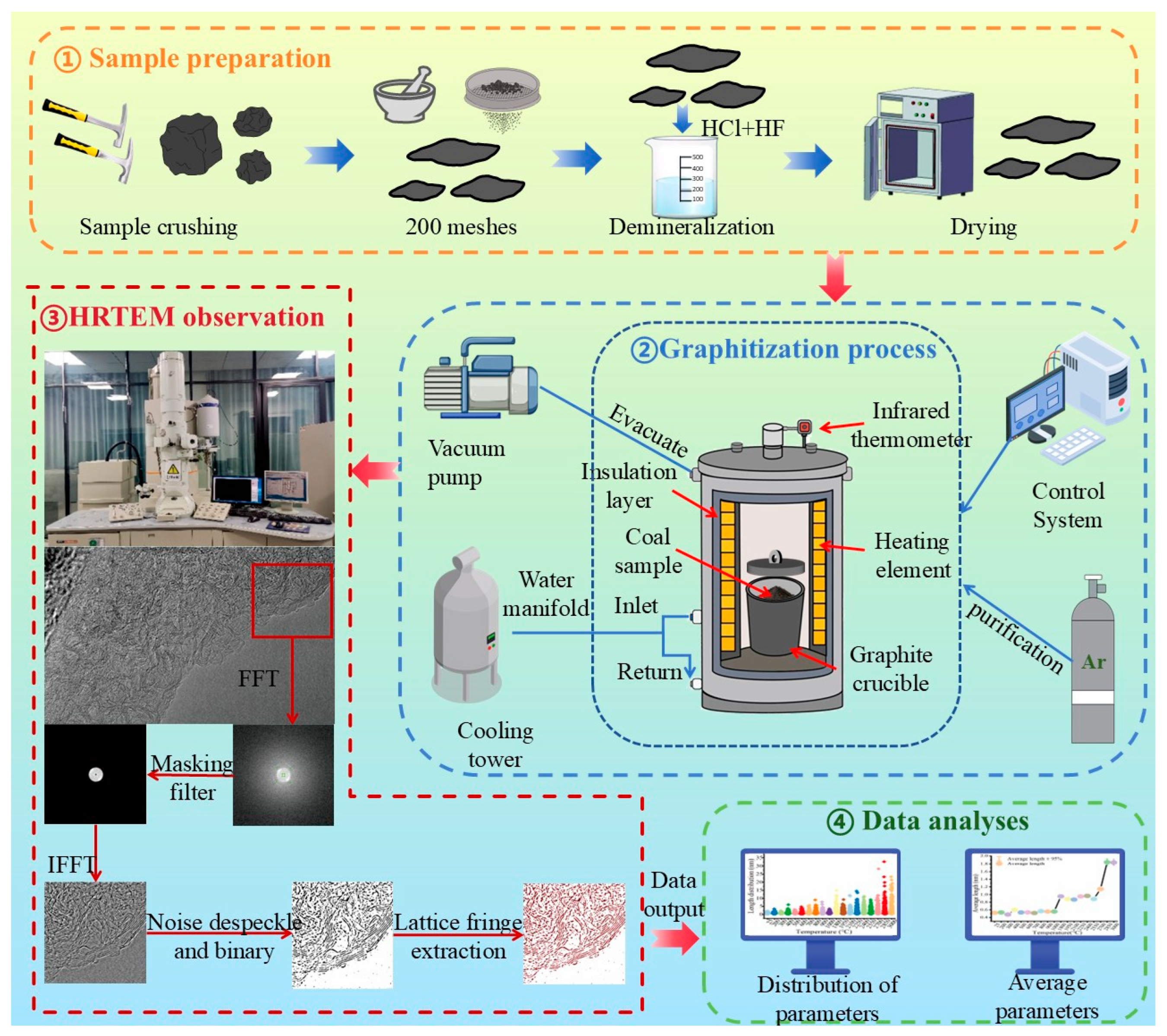
To eliminate the influence of mineral matter, demineralization with hydrochloric acid (HCl) and hydrofluoric acid (HF) was conducted on 200-mesh coal to remove carbonate and silicate minerals, respectively. The detailed demineralization procedure followed previously reported methods [31,33,41]. After acid treatment, the coal–acid mixture was repeatedly washed with deionized water until pH = 7. Then, the samples were dried for subsequent treatment, including graphitization and HRTEM observation. The detailed procedure is illustrated in Figure 1.

2.2. High-Temperature Treatment

The coal sample was randomly crushed and ground into 200-mesh size using an agate mortar. For each experiment, 5 g of the 200-mesh coal sample was placed into a graphite crucible for high-temperature treatment. Heat treatments were conducted from ambient temperature to 3000 °C in a high-temperature furnace, at a heating rate of 10 °C per minute and holding for 3 h at the target temperature. The furnace operated at a power of 60 kW, with a frequency of 2500 Hz and a voltage of 380 V. Two holes with a diameter of 2 mm were drilled in the upper wall of the graphite crucible. During the heat treatment, the coal sample was sealed in the graphite crucible with a closed lid under an inert argon atmosphere with a purity of 99.99%. After treatment, the samples were naturally cooled to room temperature and collected for subsequent HRTEM observation.

2.3. HRTEM Observation and Image Processing

HRTEM micrographs were acquired using a field-emission transmission electron microscope (JEM-2100) with a point resolution of 0.23 nm and a line resolution of 0.14 nm. Powdered samples below 200 meshes were ultrasonically dispersed in anhydrous ethanol for 15 min, after which a droplet of the suspension was deposited onto a copper microgrid for HRTEM observation. Low-magnification imaging was first performed to examine particle morphology, whereas high-magnification imaging was used for lattice fringes. The standard TEM operation process was conducted for choosing the regions of interest [33,42]. For each sample, 5–8 micrographs were collected to ensure representativeness. In each sample, more than 3000 lattice fringes were obtained for quantitative analysis.
Lattice fringes were extracted from HRTEM images through image-processing workflow based on previous publications [40,42,43,44,45,46,47], as illustrated in Figure 1. First, selected square regions from the raw HRTEM images were subjected to Fast Fourier Transform (FFT) and Inverse Fast Fourier Transform (IFFT) to enhance lattice fringe contrast; the filter options were default 5 and keeping the area masked. Second, image noise was reduced through smoothing (once) and filtering, followed by binarization to generate skeletonized images, and a despeckling process was applied. Finally, lattice fringes were extracted, and quantitative structural parameters were exported into Excel for further analysis. The detailed procedures of image processing can be referred to in the works of Mathews [46] and Sharma et al. [48]. For each HRTEM image, more than 3000 lattice fringes were extracted, and five images from each sample were analyzed to obtain representative nanostructural statistics.

3. Results and Discussion

High-resolution transmission electron microscopy (HRTEM) is a robust and widely recognized technique for the quantitative characterization of carbon nanostructures and the qualitative analysis of morphological features [23,24,26,28,39,45]. Nanostructural parameters, including lattice fringe length, curvature, and orientation, have been evaluated to investigate aromatic carbon structures [32,42,43,45,47]. In this study, HRTEM was applied to a low-rank Jurassic coal subjected to progressive heat treatment, covering carbonization (from ambient to 1000 °C), secondary and pre-graphitization (from 1000 to 2000 °C), and graphitization (from 2000 to 3000 °C), based on elemental analysis, XRD and HRTEM in our previous study [33]. For these heat-treated coal samples, lattice fringes in both edge and interior regions were observed qualitatively and analyzed quantitatively. The representative HRTEM images with corresponding structural characteristics were elaborated.
Generally, the HRTEM images reveal an increase in lattice fringe length, enhanced lattice fringe alignment, and improved orientation with increasing heating temperatures. Nevertheless, pronounced heterogeneity remains evident, including the coexistence of different types of nanostructured carbon and spatial variations between edge and interior regions. To systematically elucidate the structural evolution of high-temperature-treated coal, parameters including lattice fringe length, lattice fringe curvature, and lattice fringe orientation were quantitatively analyzed and discussed.

3.1. Evolution of Lattice Fringe Length with High-Temperature Treatment

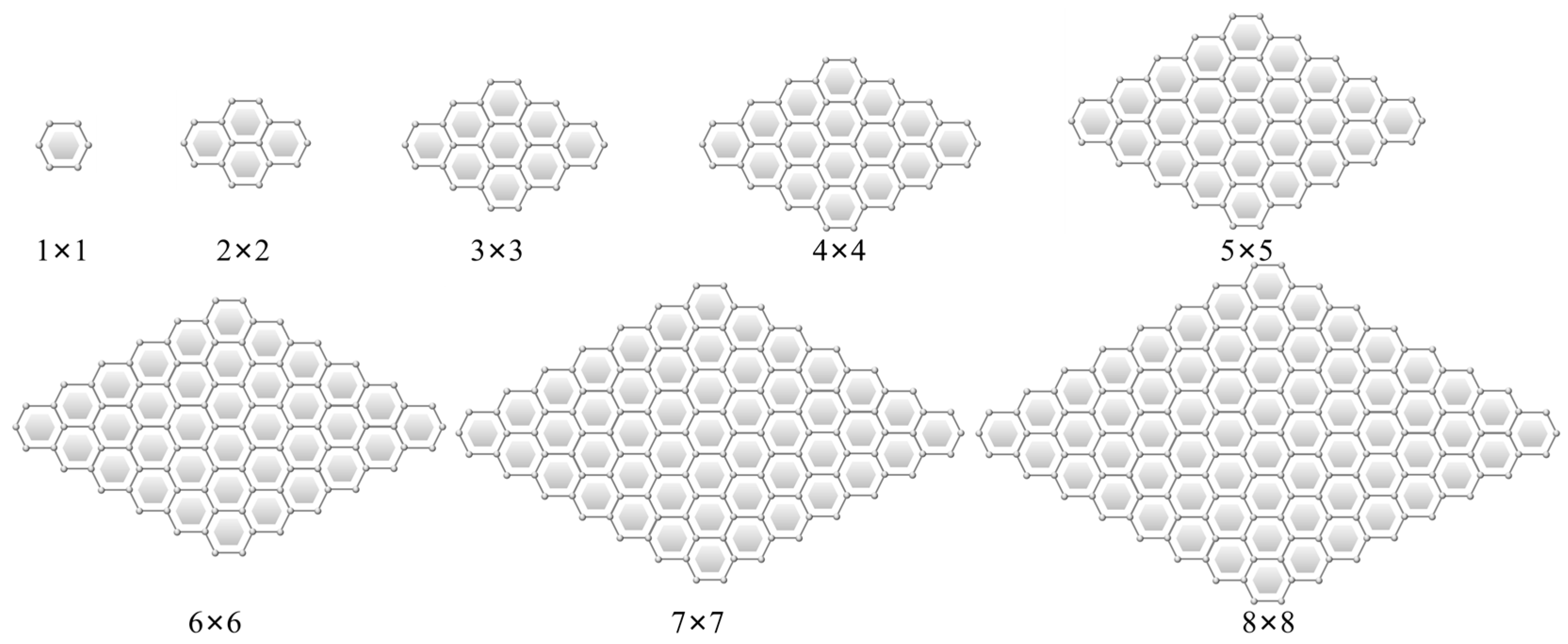
Lattice fringe length is a direct reflection of the lateral dimension of aromatic carbon structures [32]. Integrating findings from previous literature [27,31,45] and the present experimental results, the lattice fringes were categorized into three size regimes, including small-sized, medium-sized, and large-sized lattice fringes, as summarized in Table 3. Schematic representations of polycyclic aromatic carbons with different sizes are shown in Figure 2. Small-sized aromatic layers correspond to 1 × 1, with lattice fringe lengths ranging from 0.30 to 0.54 nm. Medium-sized aromatic layers include 2 × 2 and 3 × 3, with lattice fringe lengths ranging from 0.54 to 1.14 nm. Large-sized aromatic layers are defined as those with lattice fringe lengths exceeding 1.14 nm.
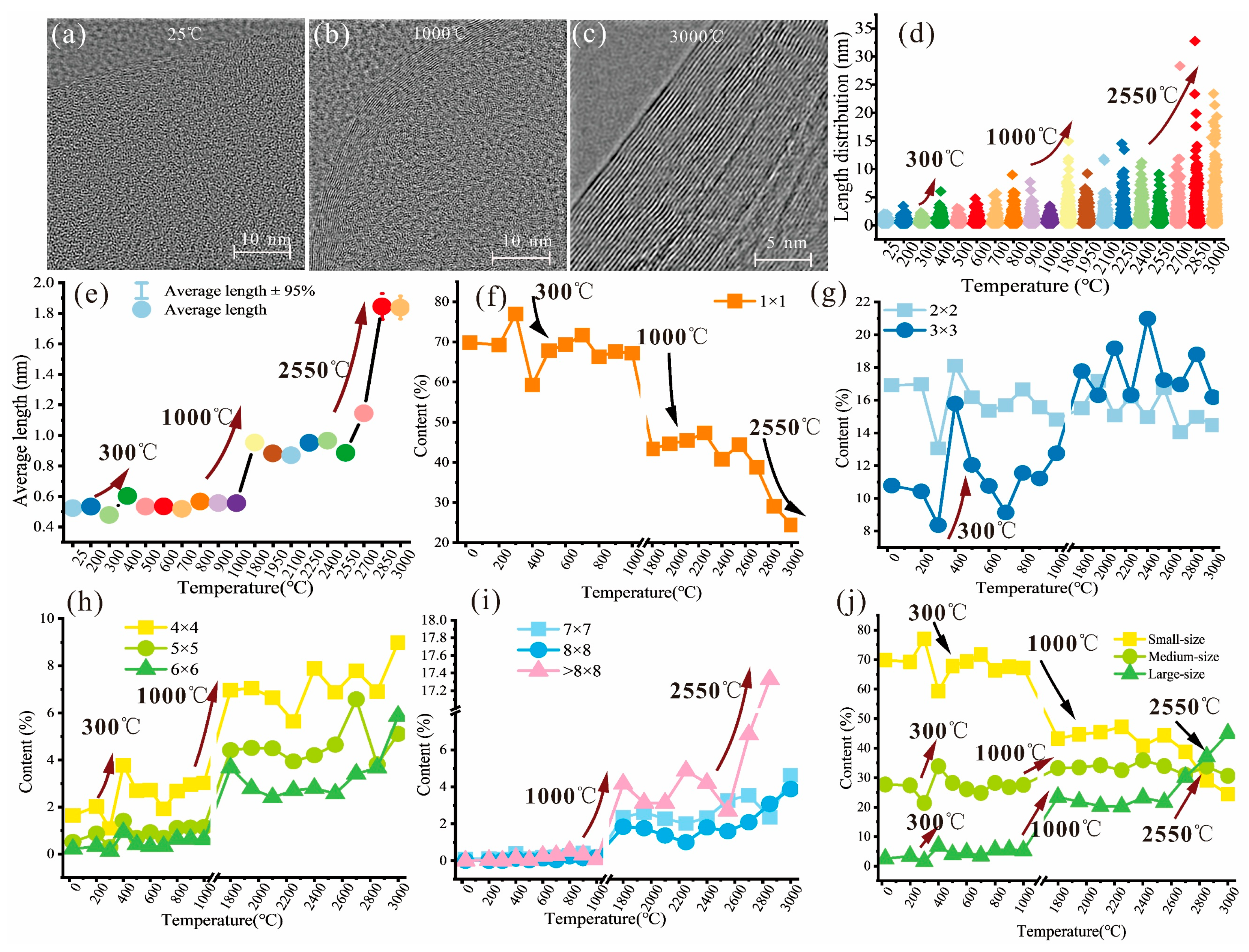
The lattice fringe length distributions and corresponding average lengths of heat-treated coal samples are shown in Figure 3 and Table 4. With increasing heat-treatment temperature, the lattice fringes show increased length and ordered alignment, as shown in Figure 3a–c. Correspondingly, both the lattice fringe length distributions (Figure 3d) and average lattice fringe length (Figure 3e) exhibit increasing trends. This trend is also evidenced by a decreasing proportion of small-sized aromatic layers and increasing proportions of medium- and large-sized aromatic layers, as illustrated in Figure 3f–j. These observations indicate the progressive growth and coalescence of aromatic carbon layers during thermal treatment, consistent with structural ordering associated with coalification maturation and thermal conversion [49].
The evolution data of lattice fringe length presents three obvious abrupt temperature points at 300 °C, 1000 °C, and 2550 °C. Here, 300 °C is the onset of chemical reactions, 1000 °C is the end of the carbonization process, and 2550 °C is within the graphitization stage. The characteristic temperatures are evident in the lattice fringe length distributions (Figure 3d). During carbonization, the chemical structure of coal undergoes decomposition and reorganization when temperatures are above 300 °C [50], followed by pronounced polymerization and condensation reactions at temperatures exceeding 700 °C [49,51,52], resulting in decreased small-sized lattice fringes (Figure 3f) and increased medium- and large-sized lattice fringes (Figure 3g–i).
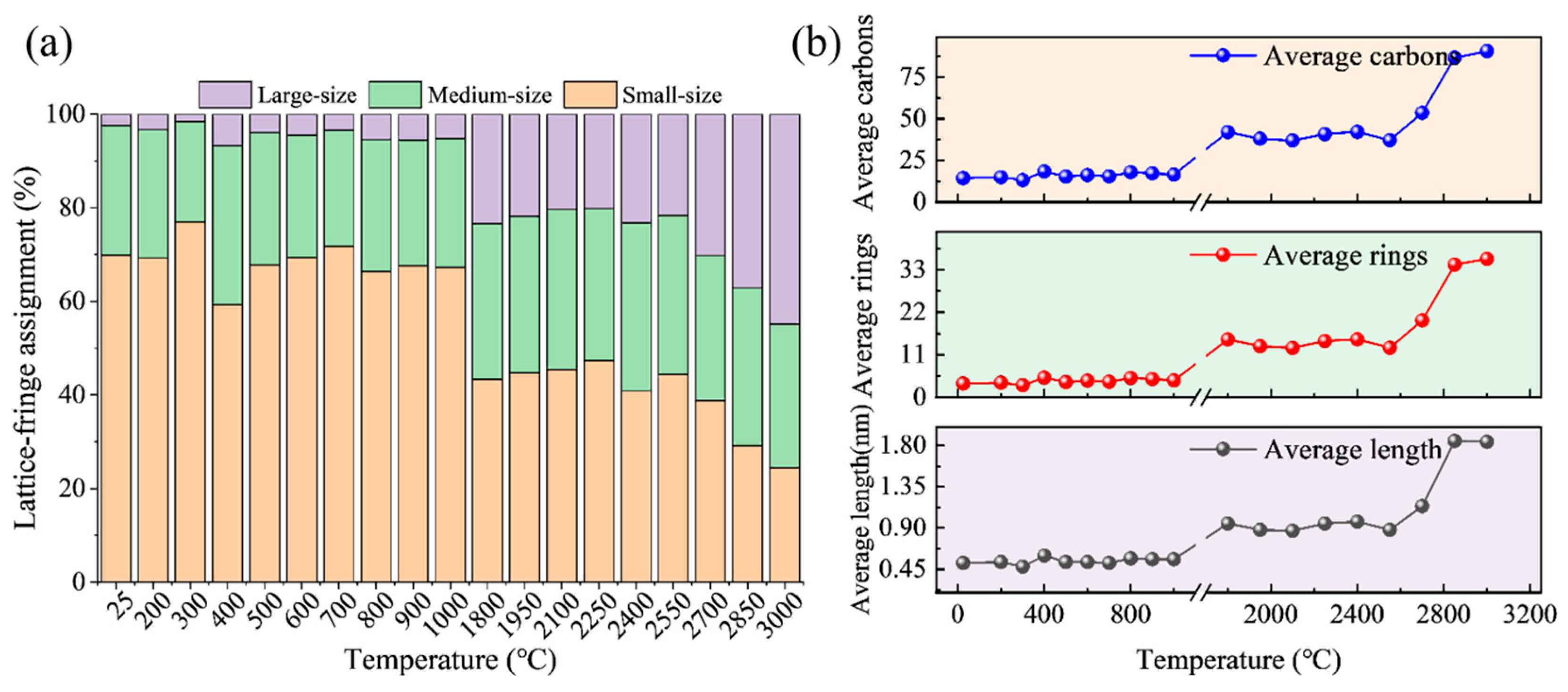
Figure 3j presents the compositional distribution of lattice fringe lengths in the raw coal and heat-treated samples. In both raw coal and carbonized coal, small-sized aromatic layers dominate the lattice fringes, accounting for more than half of the total lattice fringe. This observation is consistent with previous findings for other low-rank coals [38,53] and even anthracite [26]. With increasing heat-treatment temperature, the aromatic layers become progressively larger, as supported by the HRTEM images of the heat-treated coal samples (Figure 3a–c) and the lattice fringe length distributions (Figure 3d). The increase in average lattice fringe length is mainly attributed to the staged increase in medium-sized lattice fringes (Figure 3g) and the continuous growth of large-sized lattice fringes (Figure 3h,i). Specifically, aromatic layers corresponding to 4 × 4 to 6 × 6 increase preferentially at intermediate temperatures (1000–1800 °C), whereas layers larger than 6 × 6 increase markedly at higher temperatures (2550 °C). Large-sized lattice fringes only become dominant in graphitized samples, where their proportion exceeds that of both medium- and small-sized fringes (Figure 3j). The contribution of large-sized lattice fringes to the increase in average lattice fringe length is also directly reflected in Figure 4a. The average lattice fringe length is positively correlated with the average number of aromatic rings and aromatic carbon atoms (Figure 4b), indicating the progressive lateral growth of aromatic carbon domains during heat treatment.
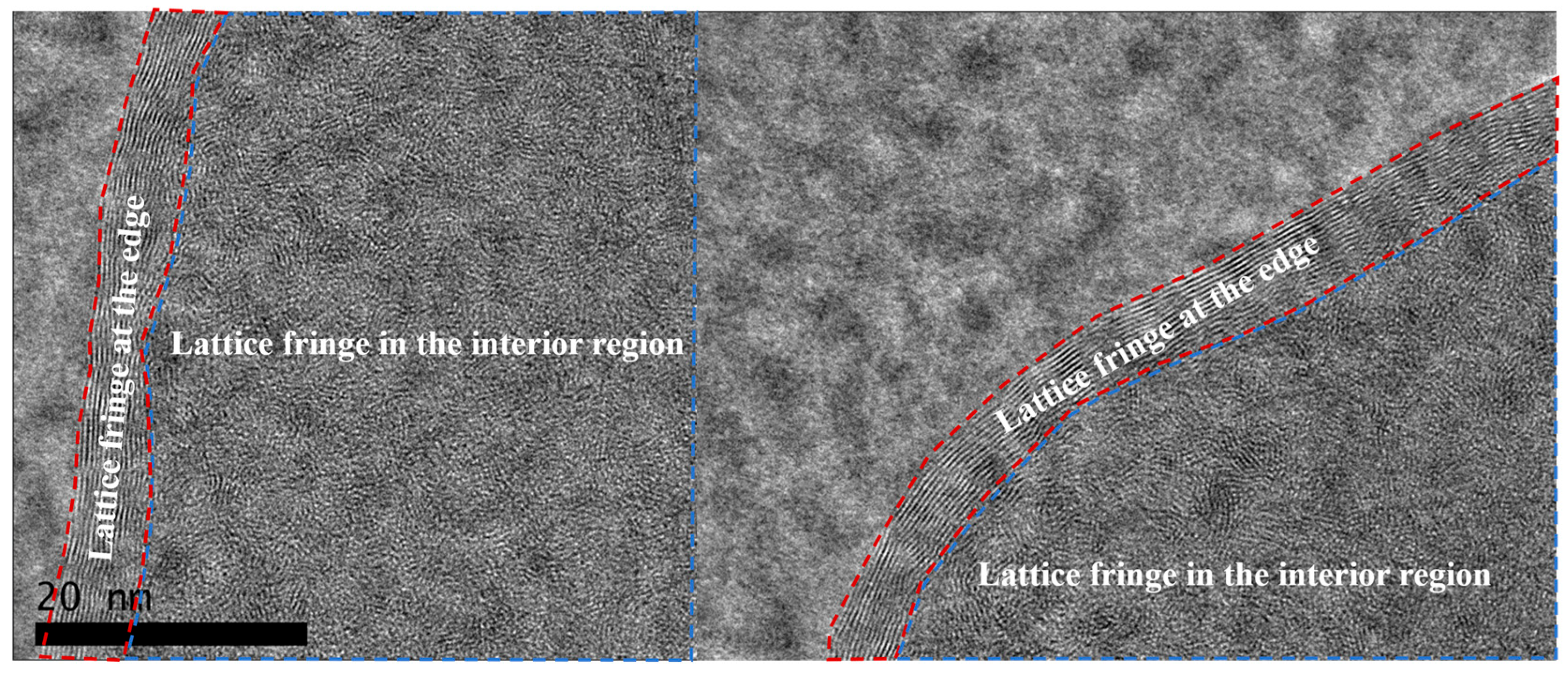
Moreover, lattice fringe growth is more pronounced at the edges of coal particles than in their interiors, indicating spatially heterogeneous graphitization. Similar edge-preferential structural ordering has also been observed in natural coaly graphite [34,39,54] and experimentally heat-treated coals [30,33,55,56]. Due to the uneven particle size of samples, the quantitative distance of particles varies, so only qualitative descriptions of lattice fringes at edges and in the interior are shown in Figure 5. The red-line circled regions are at edges, and the blue-line circled regions are in the interior of the particle.

3.2. Curvature-Limited Evolution of Lattice Fringes with High-Temperature Treatment

Lattice fringe curvature is a critical indicator of alignment and distortion of aromatic carbon layers [27,42,57]. Curvature is also reflected by segment angle, cumulative angle, and tortuosity [23,25,27,29]. Because these parameters (segment angle, cumulative angle, and tortuosity) generally show negative correlations with length and temperature, tortuosity was used in this study as a representative descriptor of lattice fringe curvature in high-temperature-treated samples.
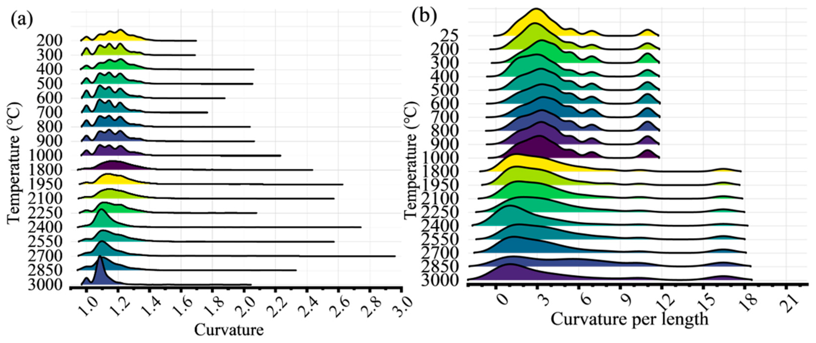
The distributions of lattice fringe curvature and curvature per length in the heat-treated samples are presented in Figure 6a and Figure 6b, respectively. With increasing heat-treatment temperature, the shape of the curvature distribution evolves from multiple peaks to a single peak, indicating a transition from fluctuating curvature to a relatively stable state. The curvature per length at different temperatures is relative stable, and decreases slightly with rising temperatures. However, the ranges of curvature per length in temperatures above 1000 °C are notably wide, suggesting persistent local structural variability despite overall thermal ordering.
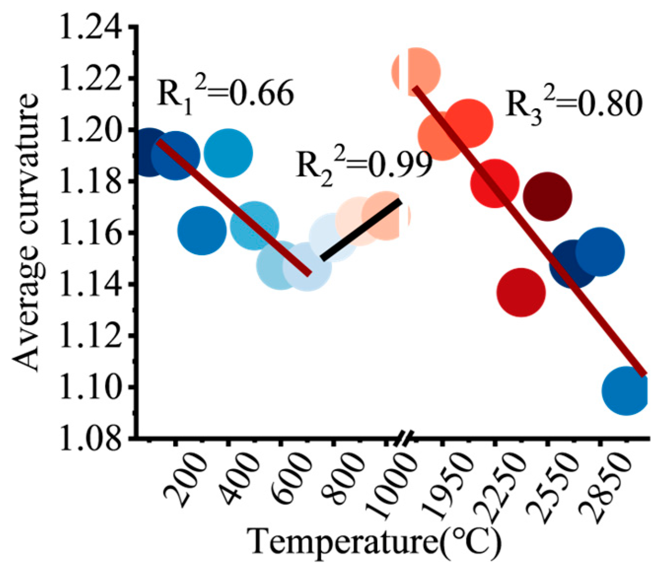
As shown in Figure 7, the average curvature exhibits two pronounced declines and one distinct increase during heat treatment. Specifically, from ambient temperature to 700 °C, the average curvature decreases from 1.19 to 1.14, with a coefficient of determination of R2 = 0.66. Between 1800 and 3000 °C, it further decreases from 1.22 to 1.09, with a stronger correlation of R2 = 0.80. These decreasing trends are consistent with the structural evolution previously reported for natural coaly graphite [39] and calcined petroleum coke [25], reflecting progressive flattening and alignment of aromatic carbon layers at elevated temperatures.
By contrast, the average curvature increases between 700 °C and 1800 °C rather than decreasing monotonically. This trend corresponds well to the enlarged diameter of concentric carbon nanostructures observed in heat-treated carbons in our previous work [33,58], which leads to an apparent increase in lattice fringe curvature in HRTEM images. The increase in curvature may also be associated with flexible out-of-plane pseudo-crystalline structures and twisted graphene layers [58,59]. Such twisted graphene tends to develop spontaneous two-dimensional curvature [57], thereby contributing to the transient curvature enhancement.
With increasing heat-treatment temperature, the average lattice fringe length increases, whereas the average curvature decreases, consistent with the structural evolution of the coalification process [32,60] and thermally treated carbonaceous materials [1,2,40,47,61]. Theoretically, lattice fringe curvature is expected to decrease as lattice fringe length increases, because longer aromatic layers generally undergo progressive flattening and alignment. This inverse relationship has also been supported by the correlation between average lattice fringe length and average curvature reported in previous studies by Li et al. [39].
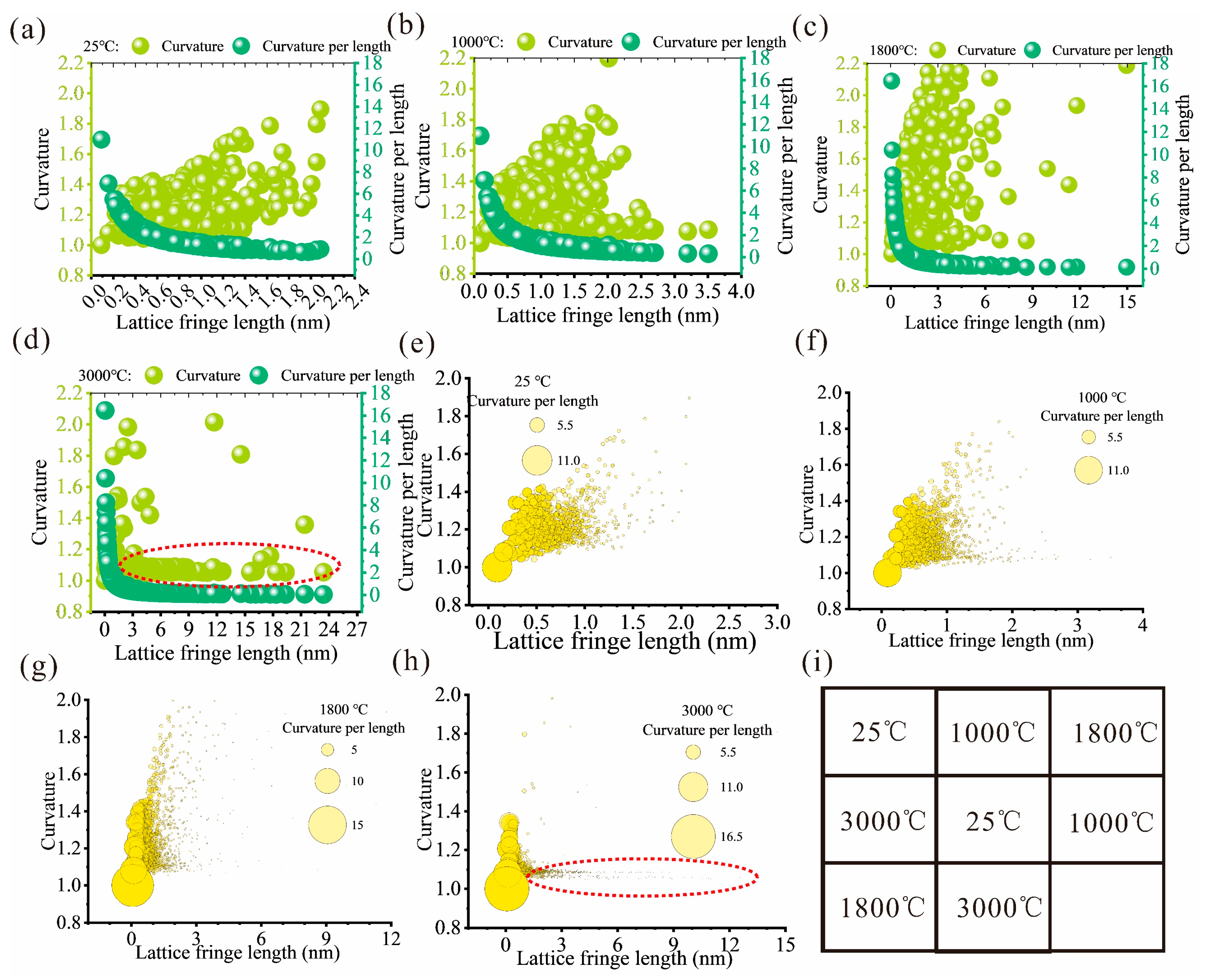
However, for samples heat-treated from ambient temperature to 3000 °C, the plot of lattice fringe curvature versus lattice fringe length exhibited a positive correlation (light green plots in Figure 8a–d), rather than the expected negative trend. This result suggests that longer lattice fringes may still retain, or even develop, higher curvature during structural evolution. This phenomenon is further corroborated by the bubble charts (Figure 8e–h). On the contrary, curvature per length (curvature/length) exhibits a clear negative correlation with lattice fringe length, as shown by the dark green plots in Figure 8a–d. This relationship agrees with the theoretical expectation that the normalized curvature decreases as aromatic layers grow laterally, indicating that curvature evolution is constrained by lattice fringe length and cannot be interpreted solely from absolute curvature values.
In graphitized coal samples treated above 2000 °C, lattice fringe curvature becomes constrained to a certain extent, as highlighted by the red circle in Figure 8d,h. Under these conditions, lattice fringe curvature remains relatively stable despite the continued increase in lattice fringe length. This behavior is attributed to the geometric confinement imposed by particle edges: constrained lattice fringes preferentially grow along particle margins and exhibit a maximum curvature that is comparable to that of the particle edge. We therefore define this phenomenon as edge-preferential crystallization.
Edge-preferential crystallization constrains lattice fringe curvature during graphitization, allowing aromatic layers to extend laterally while maintaining curvature compatible with the particle boundary. This behavior is analogous to the crystallographic-preferred orientation observed in mineral petrology [62,63], where crystal growth and alignment are governed by spatial constraints and boundary-controlled orientation. Thus, the curvature-limited evolution of lattice fringes reflects not only thermal ordering, but also the influence of particle-scale geometry on nanoscale graphitic structural development.

3.3. Edge-Interior Heterogeneity of Lattice Fringe Orientation Induced by Edge-Preferential Crystallization

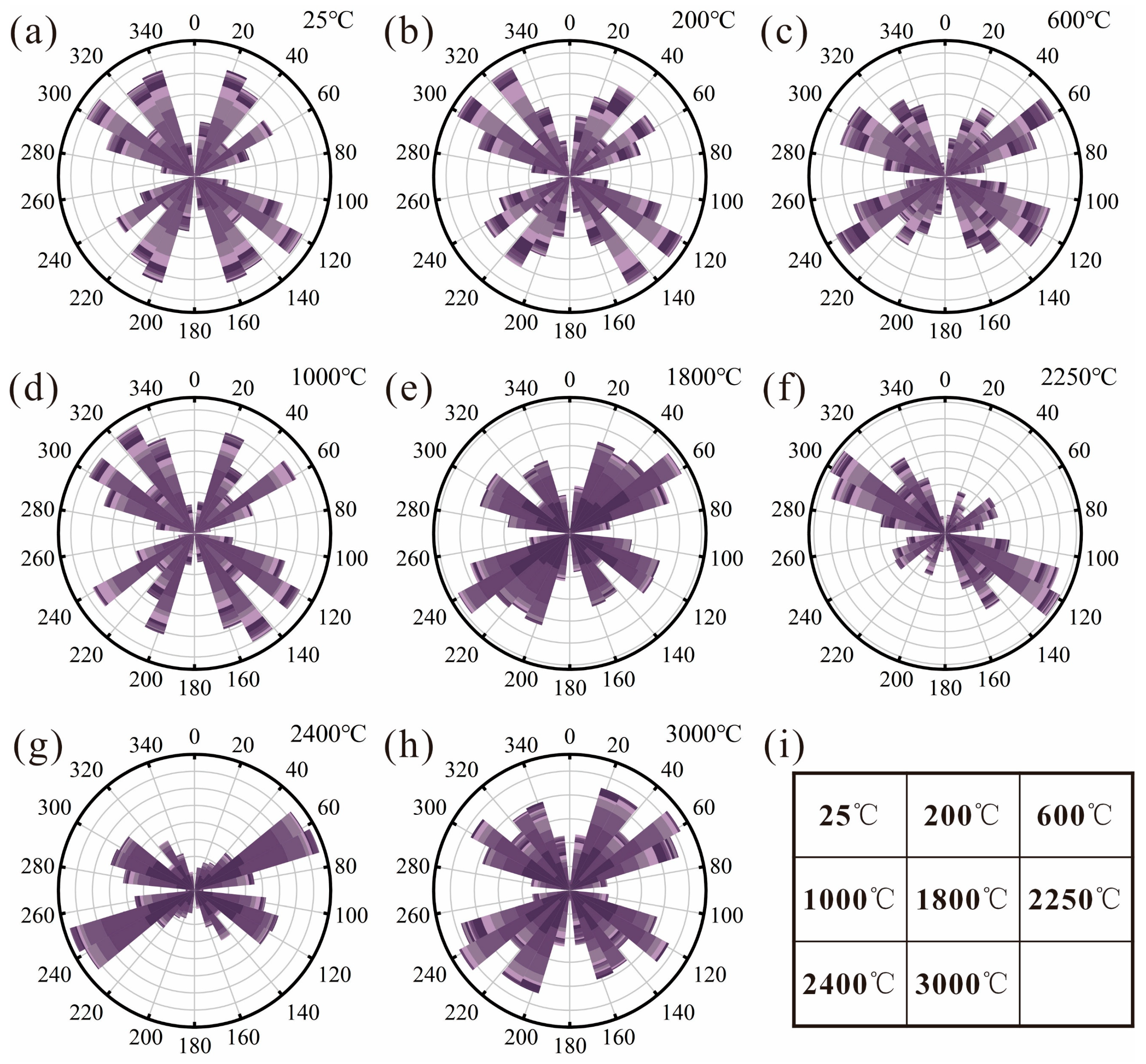
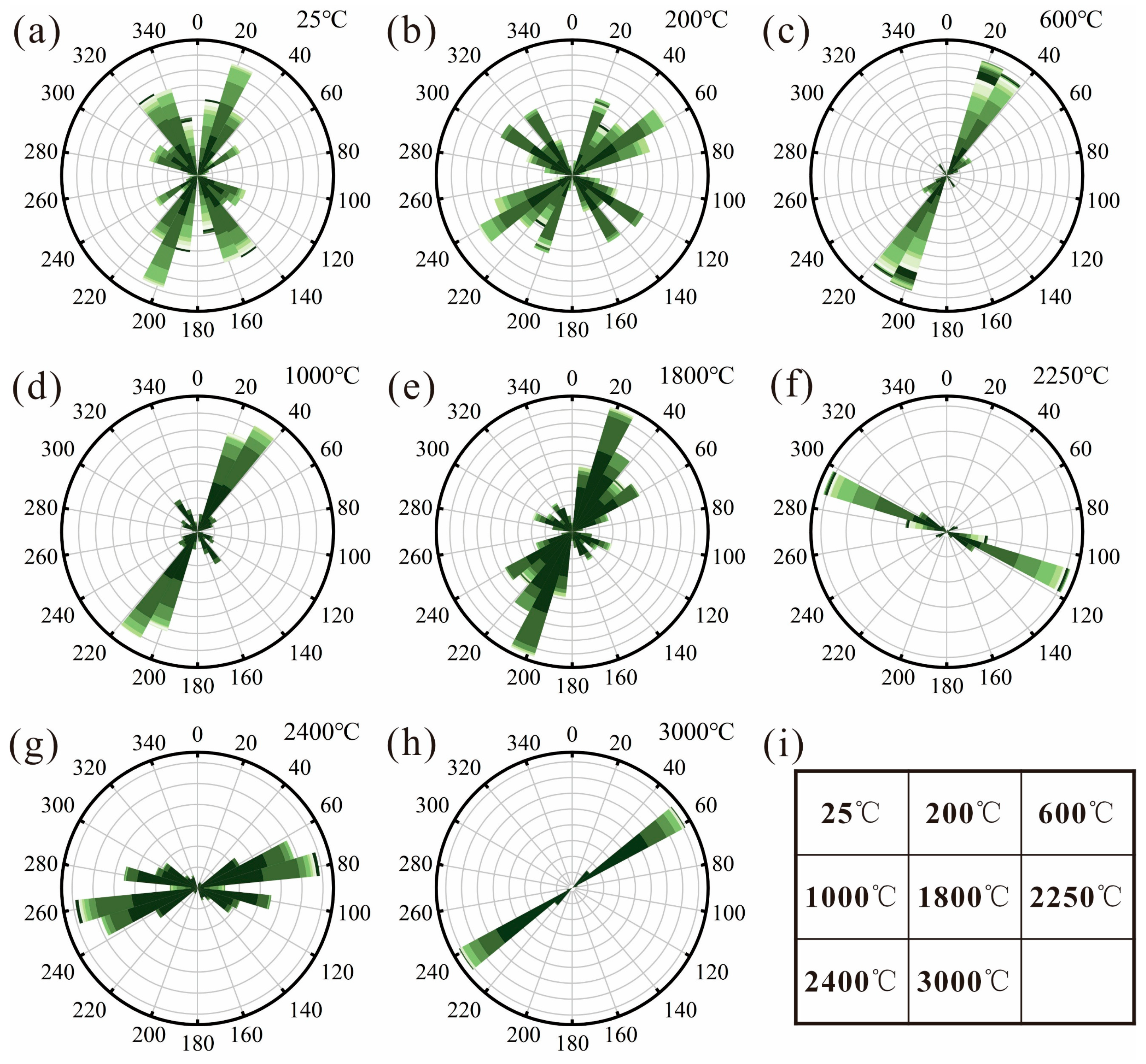
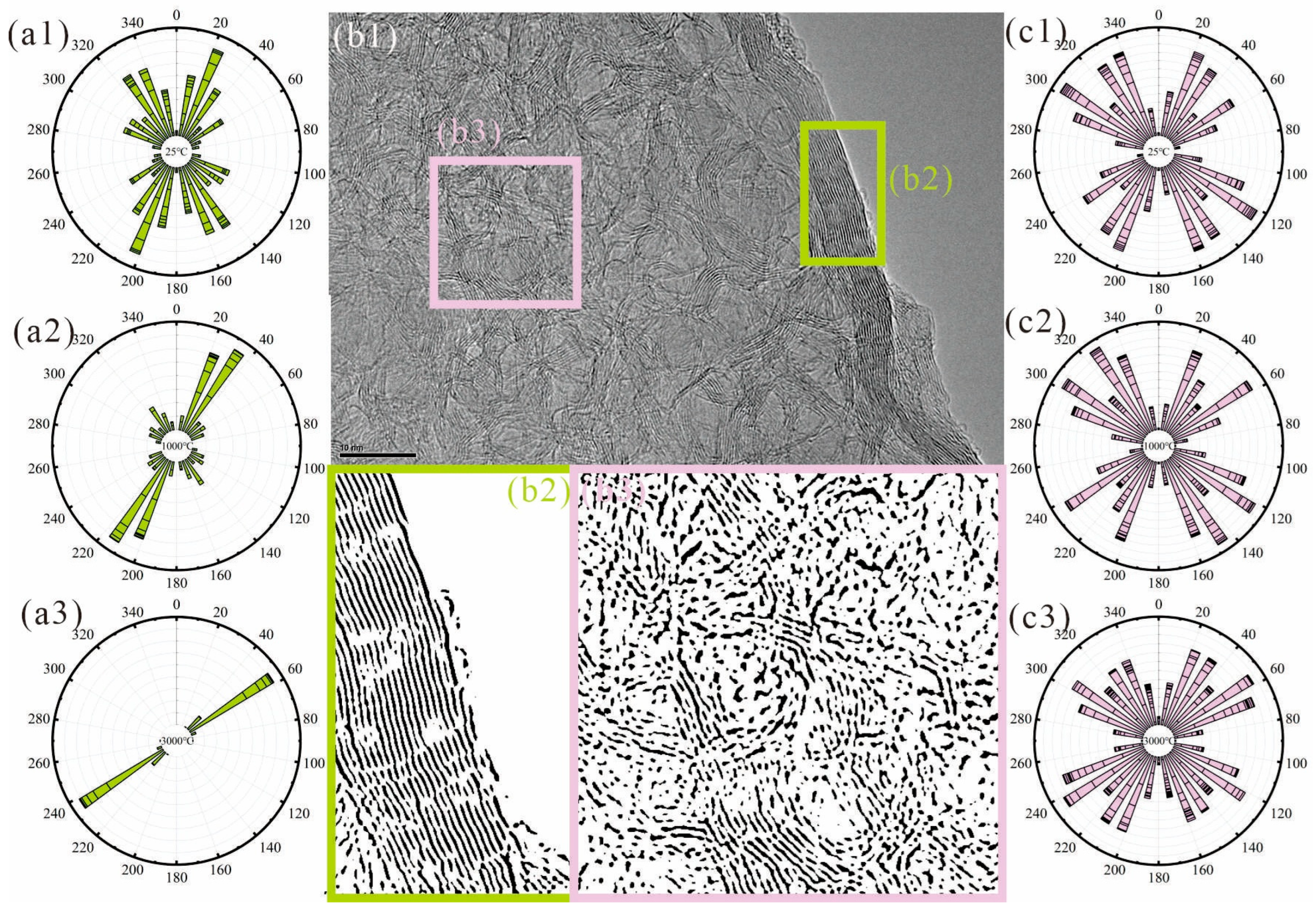
Lattice fringe orientation is a measure of the chemical structural order and thermal maturation of carbonaceous materials [26,55,64]. A higher degree of lattice fringe orientation indicates a more ordered arrangement of the carbon layers [44,55]. Therefore, a better lattice fringe orientation has been observed in HRTEM images with increasing coalification [29,58,65] and graphitization [33,66], and increasing heat-treated coal samples [26,38,55]. The total increasing lattice fringe orientation with increasing heat-treatment temperatures was observed in this work, and it was characterized by edge-preferential development. Figure 9 is a rose diagram of lattice fringes in interior regions at different temperatures. The narrower and longer the segment, the higher the frequency of lattice fringes appearing in the direction, and the better the general orientation of lattice fringes. With increasing temperature, the orientations showed slight improvement, whereas the rose diagram of lattice fringes at the edges (Figure 10) showed considerable enhancement, especially when temperatures were above 1000 °C. For both concentric carbon nanostructure and graphite-like carbon nanostructure, the lattice fringe orientation at the edges (Figure 11(b2)) exhibited a far more distinct pattern compared with that of the interior regions (Figure 11(b3)). Rose diagrams for the total lattice fringes in the edge and interior regions with increasing heat-treatment temperatures are also presented in Figure 11.
With increasing temperatures, the lattice fringe orientations in the edge (Figure 11(a1–a3)) exhibited more order, while the lattice fringe orientations of interior regions showed no regularity; this observation occurred in both natural coaly graphite [39,54] and heat-treated coal samples [30,33,56]. Furthermore, the orientation in the edges was consistently superior to that in the interior regions (Figure 11(c1–c3)), which is frequently considered to result from the more favorable and uniform heating conditions applied to the exterior [30,58]. In addition, this phenomenon also demonstrated that edge-preferential crystallization played a key role in promoting the ordered orientation of lattice fringes. Coal particle boundaries served as endogenous stressors to induce internal stress during the nucleation and growth of aromatic structures, thereby prompting the preferential orientation of lattice fringes.

3.4. Particle-Edge Preferential Graphitization of Coal-Derived Carbon

During the graphitization of high-temperature-treated coal-derived carbon, the three-stage lattice fringe growth, non-monotonic and curvature-limited evolution, and heterogeneous lattice fringe orientation were observed by HRTEM. Combined with previously reported findings [38,67], these phenomena demonstrate that the development of aromatic carbon layers is controlled not merely by thermally induced polymerization and condensation [40,67], but also by particle-edge preferential or confinement. Based on the above structural evolution characteristics, a particle-edge preferential graphitization is proposed to explain the nanoscale structural heterogeneity of coal-derived carbon materials.
In particle-edge preferential graphitization, aromatic layers grow progressively through polymerization, condensation, and vertical stacking during heat treatment, behaving as three stages at 300 °C, 1000 °C, and 2550 °C. However, their spatial evolution is strongly affected by particle boundaries. At particle edges, aromatic layers are more readily exposed to heat and mass transfer, and the boundary geometry provides a preferential template for fringe extension and alignment. As a result, lattice fringes at the edges grow more advantageously (Figure 10), exhibit constrained curvature compatible with the particle boundary (Figure 8), and develop stronger preferred orientation (Figure 11(a1–a3)). Particle boundaries provide advantages in lattice fringe length and orientation, and also limit the development of lattice fringe curvature. In contrast, the interior regions experience more complex structural constraints, leading to less ordered lattice fringe orientation and graphitic ordering.
Therefore, edge-preferential graphitization provides a theoretical explanation for the coexistence of easily graphitized carbon and difficultly graphitization, as observed by HRTEM at the nanoscale [33,58]. It also provides evidence for the limitation of the lattice fringe and the heterogeneity of edge-interior orientation.

4. Conclusions

The nanoscale structural heterogeneity and edge-preferential graphitization of coal-derived carbon in heat-treated conditions were investigated by HRTEM observation and analysis, revealing the intrinsic spatial heterogeneity of carbon structures. The edge-preferential graphitization phenomena provide a theoretical basis for the efficient and comprehensive utilization of coal.
The lattice fringe length of coal-derived carbon exhibits a staged evolution during high-temperature treatment. The lattice fringe length grows with increasing temperature and increases significantly with increasing thermal conversion temperature, accompanied by a transition from small-sized (0.30–0.54 nm, 1 × 1 rings) to medium-sized (0.54–1.14 nm, 2 × 2–3 × 3 rings) and large-sized (>1.14 nm, ≥4 × 4 rings) aromatic structures. Three abrupt temperature points (300 °C, 1000 °C, 2550 °C) are identified, corresponding to the onset of depolymerization, the end of carbonization, and intensive graphitization, respectively. In graphitized coal samples, large-sized lattice fringes dominate, accounting for the highest proportion.
Non-monotonic and curvature-limited structural evolution are affected by the particle edge. Contrary to the theoretical expectation of a continuous decrease, the average curvature of lattice fringes exhibits an increase between 700 °C and 1800 °C, primarily driven by the formation of concentric and twisted carbon nanostructures. Furthermore, during the high-temperature graphitization stage, the curvature of lattice fringes exhibits a positive correlation with length. This indicates that curvature evolution is geometrically constrained by the particles rather than solely governed by heat treatment.
Graphitization is a structurally heterogeneous process rather than a uniform evolution. Lattice fringes at particle edges demonstrate a highly ordered, boundary-parallel preferential orientation. In contrast, the interior regions experience delayed graphitic ordering, characterized by randomly oriented graphitic domains.
In the particle-edge preferential graphitization of coal-derived carbon, particle boundaries act as intrinsic geometric templates and stress concentrators, triggering edge-preferential crystallization. This boundary confinement not only accelerates the localized growth and alignment of aromatic layers at the edges, but also limits curvature, fundamentally explaining the nanoscale structural heterogeneity of coal-derived carbon materials.

Author Contributions

Conceptualization, X.Z. and H.C.; Methodology, H.C.; Software, X.C., Y.J., H.Z., M.S., and S.D.; Formal Analysis, X.C. and Y.J.; Investigation, X.Z.; Writing—Original Draft Preparation, X.Z.; Writing—Review and Editing, Y.J., J.S., L.M., Q.G., Y.Z., H.C., and S.W.; Funding Acquisition, X.Z. and H.C. All authors have read and agreed to the published version of the manuscript.

Funding

This research was funded by the “Tianchi Talent” program of the Xinjiang Uygur Autonomous Region Introduction Plan, grant number 5105260180j; the Science and Technology Program of Xinjiang Uyghur Autonomous Region, grant number 2025b01009-2; 2025b01009; and the Natural Science Foundation of Xinjiang Uygur Autonomous Region (2026).

Data Availability Statement

All data supporting the results are presented in the paper; the raw data are available on request from the corresponding author.

Acknowledgments

We greatly appreciate Zhuzhou Nuotian Electric Heating Technology Co., Ltd. for technical instructions. All authors also thank the editors and the anonymous reviewers for their careful review of the publication.

Conflicts of Interest

Qiang Guo is employee of Xilingol Mengdong Mining Co., Ltd. The paper reflects the views of the scientists and not the company. The authors declare no conflicts of interest.

References

  1. Zhao, F.; Yang, Z.; Zhang, L.; Zhang, C.; Wang, T.; Zhang, H. The effect of temperature on pyrolysis products during oil shale thermal decomposition. Sci. Rep. 2025, 15, 26135. [Google Scholar] [CrossRef] [Scilit] [PubMed]
  2. Xue, L.; Tang, Y.; Gao, S. Structural evolution characteristics of sulfur in coal during gold-tube thermal simulation. Fuel Process. Technol. 2025, 268, 108183. [Google Scholar] [CrossRef] [Scilit]
  3. Zhang, Z.; Ding, M.; Cheng, T.; Qiao, R.; Zhao, M.; Luo, M.; Wang, E.; Sun, Y.; Zhang, S.; Li, X.; et al. Continuous epitaxy of single-crystal graphite films by isothermal carbon diffusion through nickel. Nat. Nanotechnol. 2022, 17, 1258–1264. [Google Scholar] [CrossRef] [Scilit] [PubMed]
  4. Tang, Y.; Xiao, L.; Yang, F.; Xiaodan, W.U.; Zhan, S.U.; Zhang, Y.; Jiang, S.; Yang, P. Thermal transfer performance of downhole electric heaters for in-situ pyrolysis in tar-rich coal. Bull. Pol. Acad. Sci. Tech. Sci. 2025, 73, 153839. [Google Scholar] [CrossRef] [Scilit]
  5. Ding, D.; Liu, G.; Fu, B.; Yuan, Z.; Chen, B. Influence of magmatic intrusions on organic nitrogen in coal: A case study from the Zhuji mine, the Huainan coalfield, China. Fuel 2018, 219, 88–93. [Google Scholar] [CrossRef] [Scilit]
  6. Dun, W.; Guijian, L.; Ruoyu, S.; Shancheng, C. Influences of magmatic intrusion on the macromolecular and pore structures of coal: Evidences from Raman spectroscopy and atomic force microscopy. Fuel 2014, 119, 191–201. [Google Scholar] [CrossRef] [Scilit]
  7. Beyssac, O.; Rouzaud, J.-N.; Goffé, B.; Brunet, F.; Chopin, C. Graphitization in a high-pressure, low-temperature metamorphic gradient: A Raman microspectroscopy and HRTEM study. Contrib. Mineral. Petrol. 2002, 143, 19–31. [Google Scholar] [CrossRef] [Scilit]
  8. Beyssac, O.; Goffé, B.; Chopin, C.; Rouzaud, J.N. Raman spectra of carbonaceous material in metasediments: A new geothermometer. J. Metamorph. Geol. 2002, 20, 859–871. [Google Scholar] [CrossRef] [Scilit]
  9. Zhang, X.; Zhang, X.; Li, X.; Feng, S.; Han, C.; Gao, X.; Wang, X.; Dong, Z. Influence of Pressure and Coal-Forming Environment on the Pyrolysis of Tar-Rich Block Coal from the Santanghu Basin in Xinjiang. ACS Omega 2026, 11, 20559–20568. [Google Scholar] [CrossRef] [Scilit] [PubMed]
  10. Yu, J.; Qian, Z.; Zenglin, G.; Yongming, X.; Baolin, Z.; Qi, W.; Wei, H. Macro maceral separation of low-rank coal and the pyrolysis behavior of the maceral-rich fractions. Sep. Purif. Technol. 2025, 353, 128061. [Google Scholar] [CrossRef] [Scilit]
  11. Yang, H.; Pisupati, S.V.; Hu, H. Modeling char surface area evolution during coal pyrolysis: Effect of swelling and gasification at high pressures. Proc. Combust. Inst. 2021, 38, 4151–4159. [Google Scholar] [CrossRef] [Scilit]
  12. Yafei, S. Chars as carbonaceous adsorbents/catalysts for tar elimination during biomass pyrolysis or gasification. Renew. Sustain. Energy Rev. 2015, 43, 281–295. [Google Scholar] [CrossRef] [Scilit]
  13. Ahmed, I.I.; Gupta, A.K. Experiments and stochastic simulations of lignite coal during pyrolysis and gasification. Appl. Energy 2013, 102, 355–363. [Google Scholar] [CrossRef] [Scilit]
  14. Kim, K.-J.; Huynh, N.T.; Lee, Y.; Nguyen, T.; Pham, V.H.; Gao, Y.; Li, Y.; Bhandari, G.; Wang, C.; Matranga, C. Highly crystalline, low-ash, graphite from coal using an Fe2O3-based catalytic process with recovery and reuse of catalyst and process acid. Carbon 2026, 246, 120920. [Google Scholar] [CrossRef] [Scilit]
  15. Bhattacharyya, S.; Roy, S.; Lin, X.; Campagnol, N.; Vlad, A.; Ajayan, P.M. Graphite: The new critical mineral. Nat. Rev. Mater. 2026, 11, 65–78. [Google Scholar] [CrossRef] [Scilit]
  16. Wang, Y.; Yao, S.; Zheng, H.; Zuo, Z.; Liu, Y. Nanomechanical properties of anthracite and graphite: The role of heteroatom functional groups and structural evolution. Int. J. Coal Geol. 2025, 301, 104714. [Google Scholar] [CrossRef] [Scilit]
  17. Li, J.; Qin, Y.; Shen, J.; Chen, Y.; Li, G.; Zhu, P. Petrolographic characteristics of graphitic carbon in coal-based graphite: Implications for the applicability of reflectance. Int. J. Coal Geol. 2025, 31, 104884. [Google Scholar] [CrossRef] [Scilit]
  18. Li, G.; Li, Q.; Li, L.; Fan, J.; Ge, Q.; Xie, D.; Zheng, J.; Li, G. Surface element segregation and electrical conductivity of lithium layered transition-metal oxide cathode materials. Appl. Surf. Sci. 2018, 427, 226–232. [Google Scholar] [CrossRef] [Scilit]
  19. Mochidzuki, K.; Soutric, F.; Tadokoro, K.; Antal, M.J.; Tóth, M.; Zelei, B.; Várhegyi, G. Electrical and Physical Properties of Carbonized Charcoals. Ind. Eng. Chem. Res. 2003, 42, 5140–5151. [Google Scholar] [CrossRef] [Scilit]
  20. Seehra, M.S.; Pavlovic, A.S. X-Ray diffraction, thermal expansion, electrical conductivity, and optical microscopy studies of coal-based graphites. Carbon 1993, 31, 557–564. [Google Scholar] [CrossRef] [Scilit]
  21. Wo, H.; Dearn, K.D.; Song, R.; Hu, E.; Xu, Y.; Hu, X. Morphology, composition, and structure of carbon deposits from diesel and biomass oil/diesel blends on a pintle-type fuel injector nozzle. Tribol. Int. 2015, 91, 189–196. [Google Scholar] [CrossRef] [Scilit]
  22. Feng, B.; Bhatia, S.K.; Barryb, J.C. Structural ordering of coal char during heat treatment and its impact on reactivity. Carbon 2002, 40, 481–496. [Google Scholar] [CrossRef] [Scilit]
  23. Yang, W.; Song, Y.; Li, W. Nanostructural Characteristic Changes in High-Volatile Bituminous Coal in Response to Differential Tectonic Deformation: Insights Gained from Quantified HRTEM Aromatic Fringes. Energy Fuels 2023, 37, 10166–10176. [Google Scholar] [CrossRef] [Scilit]
  24. Chen, X.; Li, M.; Xiang, J.; Shao, Y. Micropores manipulation of medium-rank coal through aggregate structure evolution during coalification: A study based on HRTEM analysis. Fuel 2023, 346, 128380. [Google Scholar] [CrossRef] [Scilit]
  25. You, Z.; Xiao, J.; Mao, Q.; Zhang, X.; Zhong, Q. Microstructural features transformation at various temperature stages and multi-scale atomistic representations of calcined petroleum coke based on HRTEM. Fuel 2022, 330, 125521. [Google Scholar] [CrossRef] [Scilit]
  26. Yang, W.; Toth, P.; Song, Y.; Li, W. Structural alterations of aromatic fringes by HRTEM for Xinjing Vitrinite-rich anthracite: Impact of low-temperature pyrolysis. Thermochim. Acta 2022, 713, 179230. [Google Scholar] [CrossRef] [Scilit]
  27. Wang, X.; Wang, S.; Chen, H.; Zhao, Y.; Song, X. Quantifying orientation and curvature in HRTEM lattice fringe micrographs of naturally thermally altered coals: New insights from a structural evolution perspective. Fuel 2022, 309, 122180. [Google Scholar] [CrossRef] [Scilit]
  28. Qiao, P.; Ju, Y.; Yu, K.; Ju, L.; Xiao, L.; Feng, H.; Yao, Y.; Nie, B.; Li, X.; Tian, J.; et al. Nanoscale quantitative characterization of microstructure evolution of partly graphitized high rank coal: Evidence from AFM and HRTEM. Fuel 2022, 324, 124802. [Google Scholar] [CrossRef] [Scilit]
  29. Liu, L.; Du, M.; Li, G.; Schobert, H.H.; Fan, J.; Liu, J.; Wang, Q. Structure and evolution features of cutinite with different coal rank from stacking and arrangement of aromatic fringes in HRTEM. Fuel 2022, 326, 124998. [Google Scholar] [CrossRef] [Scilit]
  30. Wang, S.; Chen, H.; Zhang, X. Transformation of aromatic structure of vitrinite with different coal ranks by HRTEM in situ heating. Fuel 2020, 260, 116309. [Google Scholar] [CrossRef] [Scilit]
  31. Zhang, X.; Wang, S.; Chen, H.; Guo, Q.; Li, L.; Luo, G. Aromatic structural characterization of different-rank vitrinites: Using HRTEM, XRD and AFM. Polycycl. Aromat. Compd. 2019, 41, 1319–1330. [Google Scholar] [CrossRef] [Scilit]
  32. Qiao, P.; Xiao, L.; Tao, L.; Qian, J.; Yu, K.; Ge, M. Evolution of molecular structure during coalification and graphitization: Evidence from multiple characterization methods. Fuel 2026, 406, 136990. [Google Scholar] [CrossRef] [Scilit]
  33. Zhang, X.; Wang, S.; Chen, H.; Wang, X.; Deng, J.; Li, X.; Zhang, Y. Observation of carbon nanostructure and evolution of chemical structure from coal to graphite by high temperature treatment, using componential determination, X-ray diffraction and high-resolution transmission electron microscope. Fuel 2023, 332, 126145. [Google Scholar] [CrossRef] [Scilit]
  34. Zhang, S.; Liu, Q.; Zhang, H.; Ma, R.; Li, K.; Wu, Y.; Teppen, B.J. Structural order evaluation and structural evolution of coal derived natural graphite during graphitization. Carbon 2020, 157, 714–723. [Google Scholar] [CrossRef] [Scilit]
  35. Wang, B.W.; Wei, Z.; Fan, C.Y.; Liu, C.Y. Study on the adsorption of CH4, CO2 and N2 in curved graphite slit pores. Mol. Simul. 2026, 52, 272–282. [Google Scholar] [CrossRef] [Scilit]
  36. Santos, A.C.; Badenhorst, C.; Białecka, B.; Cameán, I.; Guedes, A.; Moreira, K.; Predeanu, G.; Suárez-Ruiz, I.; Wagner, N.; Valentim, B. Graphitization: Microstructural and microtextural transformations of residual char from international coal combustion ash. Int. J. Coal Geol. 2024, 285, 104470. [Google Scholar] [CrossRef] [Scilit]
  37. Li, K.; Zhu, Y.; Cao, H.; Zhang, H.; Wu, Y.; Li, X.; Xu, Z.; Liu, Q. Graphite Made from Coal by High-Temperature Treatment: An Insight into the Nanometric Carbon Structural Evolution. Minerals 2024, 14, 1092. [Google Scholar] [CrossRef] [Scilit]
  38. Li, X.; Zeng, Q. HRTEM analysis of the aggregate structure and ultrafine microporous characteristics of Xinjiang Zhundong coal under heat treatment. Sci. Rep. 2022, 12, 4994. [Google Scholar] [CrossRef] [Scilit] [PubMed]
  39. Li, J.; Qin, Y.; Chen, Y.; Song, Y.; Wang, Z. HRTEM observation of morphological and structural evolution of aromatic fringes during the transition from coal to graphite. Carbon 2022, 187, 133–144. [Google Scholar] [CrossRef] [Scilit]
  40. Chen, H.; Wang, S.; Tang, Y.; Zeng, F.; Schobert, H.H.; Zhang, X. Aromatic cluster and graphite-like structure distinguished by HRTEM in thermally altered coal and their genesis. Fuel 2021, 292, 120373. [Google Scholar] [CrossRef] [Scilit]
  41. Meshram, P.; Purohit, B.K.; Sinha, M.K.; Sahu, S.K.; Pandey, B.D. Demineralization of low grade coal–A review. Renew. Sustain. Energy Rev. 2015, 41, 745–761. [Google Scholar] [CrossRef] [Scilit]
  42. Wang, C.a.; Huddle, T.; Huang, C.-H.; Zhu, W.; Vander Wal, R.L.; Lester, E.H.; Mathews, J.P. Improved quantification of curvature in high-resolution transmission electron microscopy lattice fringe micrographs of soots. Carbon 2017, 117, 174–181. [Google Scholar] [CrossRef] [Scilit]
  43. Wang, C.a.; Huddle, T.; Lester, E.H.; Mathews, J.P. Quantifying curvature in high-resolution transmission electron microscopy lattice fringe micrographs of coals. Energy Fuels 2016, 30, 2694–2704. [Google Scholar] [CrossRef] [Scilit]
  44. Wang, C.a.; Watson, J.K.; Louw, E.; Mathews, J.P. Construction Strategy for Atomistic Models of Coal Chars Capturing Stacking Diversity and Pore Size Distribution. Energy Fuels 2015, 29, 4814–4826. [Google Scholar] [CrossRef] [Scilit]
  45. Mathews, J.P.; Fernandez-Also, V.; Daniel Jones, A.; Schobert, H.H. Determining the molecular weight distribution of Pocahontas No. 3 low-volatile bituminous coal utilizing HRTEM and laser desorption ionization mass spectra data. Fuel 2010, 89, 1461–1469. [Google Scholar] [CrossRef] [Scilit]
  46. Mathews, J.P.; Sharma, A. The structural alignment of coal and the analogous case of Argonne Upper Freeport coal. Fuel 2012, 95, 19–24. [Google Scholar] [CrossRef] [Scilit]
  47. Chen, H.; Wang, S.; Zhang, X.; Zhao, Y.; Zhang, H. A study of chemical structural evolution of thermally altered coal and its effect on graphitization. Fuel 2021, 283, 119295. [Google Scholar] [CrossRef] [Scilit]
  48. Sharma, A.; Kyotani, T.; Tomita, A. A new quantitative approach for microstructural analysis of coal char using HRTEM images. Fuel 1999, 78, 1203–1212. [Google Scholar] [CrossRef] [Scilit]
  49. Wang, Z.; Wang, Z.; Wang, T. Research progress on heat effects of coal pyrolysis process. J. Anal. Appl. Pyrolysis 2024, 183, 106827. [Google Scholar] [CrossRef] [Scilit]
  50. Li, X.; Tian, J.; Ju, Y.; Chen, Y. Permeability variations of lignite and bituminous coals under elevated pyrolysis temperatures (35–600 °C): An experimental study. Energy 2022, 254, 124187. [Google Scholar] [CrossRef] [Scilit]
  51. Wu, D.; Zhang, W.; Fu, B.; Hu, G. Chemical structure and gas products of different rank coals during pyrolysis. J. Therm. Anal. Calorim. 2018, 136, 2017–2031. [Google Scholar] [CrossRef] [Scilit]
  52. Chang, H.; Deng, H.; Yang, Q.; Du, S.; Hu, F.; Jia, C. Interaction of vitrinite and inertinite of Bulianta coal in pyrolysis. Fuel 2017, 207, 643–649. [Google Scholar] [CrossRef] [Scilit]
  53. Hanlin, L.; Caineng, Z.; Zhen, S.; Vandeginste, V.; Qun, Z.; Yanpeng, C.; Ze, D.; Guangjun, F.; Sijian, Z.; Deke, S.; et al. Structural alignment of PAH in vitrinite-rich anthracite and its impact on methane adsorption. Fuel 2026, 405, 136719. [Google Scholar] [CrossRef] [Scilit]
  54. Cao, D.; Zhang, H.; Dong, Y.; Yang, C. Nanoscale Microscopic Features and Evolution Sequence of Coal-Based Graphite. J. Nanosci. Nanotechnol. 2017, 17, 6276–6283. [Google Scholar] [CrossRef] [Scilit]
  55. Shao, Y.; Li, M.; Liu, P.; Cui, X.; Li, Y.; Meng, Y. The structural evolution characteristics for high volatile bituminous coal by in-situ heating in electronical microscope. J. Anal. Appl. Pyrolysis 2022, 168, 105751. [Google Scholar] [CrossRef] [Scilit]
  56. Wang, L.; Qin, R.; Li, Y.; Zhang, H. On the difference of graphitization behavior between vitrinite- and inertinite-rich anthracites during heat treatment. Energy Sources Part A Recovery Util. Environ. Eff. 2019, 44, 4991–5003. [Google Scholar] [CrossRef] [Scilit]
  57. Gao, Y.; Deng, F.; He, R.; Zhong, Z. Spontaneous curvature in two-dimensional van der Waals heterostructures. Nat. Commun. 2025, 16, 717. [Google Scholar] [CrossRef] [Scilit] [PubMed]
  58. Sha, J.; Wang, S.; Zhang, X.; Zhang, J. Evidence of a connection between turbostratic structure and twisted graphene. Cryst. Growth Des. 2025, 25, 7074–7080. [Google Scholar] [CrossRef] [Scilit]
  59. Wang, S.; Sha, J.; Lv, W.; Zhang, Y.; Li, X.; Tang, Y.; Hower, J.C.; French, D.; Schobert, H.H.; Guo, X.; et al. The stacking of graphene in a pseudocrystallite. Carbon 2025, 238, 120273. [Google Scholar] [CrossRef] [Scilit]
  60. Jingyu, J.; Weihua, Y.; Yuanping, C.; Ke, Z. Molecular structure characterization of middle-high rank coal via XRD, Raman and FTIR spectroscopy: Implications for coalification. Fuel 2019, 239, 559–572. [Google Scholar] [CrossRef] [Scilit]
  61. Hao, C.; Shaoqing, W.; Jinsong, D.; Xiaomei, Z.; Yu, L.; Xiaoguang, L. Petrologic characteristics and chemical structures of macerals in a suite of thermally altered coals by confocal raman. ACS Omega 2021, 6, 33409–33418. [Google Scholar] [CrossRef] [Scilit] [PubMed]
  62. Barrie, C.D.; Boyce, A.J.; Boyle, A.P.; Williams, P.J.; Blake, K.; Ogawara, T.; Akai, J.; Prior, D.J. Growth controls in colloform pyrite. Am. Mineral. 2009, 94, 415–429. [Google Scholar] [CrossRef] [Scilit]
  63. Ohfuji, H.; Akai, J. Icosahedral domain structure of framboidal pyrite. Am. Mineral. 2002, 87, 176–180. [Google Scholar] [CrossRef] [Scilit]
  64. Mathews, J.P.; Chaffee, A.L. The molecular representations of coal—A review. Fuel 2012, 96, 1–14. [Google Scholar] [CrossRef] [Scilit]
  65. Zhang, Y.; Hu, S.; Zhong, Q.; Zhuo, J.; Mathews, J.P. A large-scale molecular model of Fenghuangshan anthracite coal. Fuel 2021, 295, 120616. [Google Scholar] [CrossRef] [Scilit]
  66. Ju, Y.; Li, X. New research progress on the ultrastructure of tectonically deformed coals. Prog. Nat. Sci. 2009, 19, 1455–1466. [Google Scholar] [CrossRef] [Scilit]
  67. Zhu, H.; Yu, P.; Zhao, X.; Tian, L.; Li, H.; Chen, X.; Dou, J.; Yu, J. Study on the mechanism evolution of carbon structure during blending coal coking. Fuel 2026, 408, 137678. [Google Scholar] [CrossRef] [Scilit]
Figure 1. Schematic diagram of experimental procedures and the nanostructural characteristic analysis of high-temperature-treated samples.
Minerals 16 00780 g001
Figure 2. Schematic diagram of polycyclic aromatic hydrocarbons from 1 × 1 to 8 × 8.
Minerals 16 00780 g002
Figure 3. Lattice fringe length of heat-treated coal samples from ambient to 3000 °C. (ac) are HRTEM images of the sample at ambient, 1000 °C, and 3000 °C. (d) is the lattice fringe length distribution of the sample from ambient to 3000 °C; (e) is the average length of the lattice fringe from ambient to 3000 °C; (fi) are the average of different-sized lattice fringes from ambient to 3000 °C; and (j) is the lattice fringe length variations of small-sized, medium-sized, and large-sized lattice fringes from ambient to 3000 °C.
Minerals 16 00780 g003
Figure 4. Distributions of different-sized lattice fringes and related skeletal parameters. (a) is content of different-sized lattice fringes of heated samples; (b) is carbon structural parameters of heated samples.
Minerals 16 00780 g004
Figure 5. Different lattice fringes at the edge and interior of the particle.
Minerals 16 00780 g005
Figure 6. Curvature distribution of lattice fringes at different temperatures. (a) is curvature of lattice fringes at different temperatures; (b) is curvature per length of lattice fringes at different temperatures.
Minerals 16 00780 g006
Figure 7. Average lattice fringe curvature at different temperatures.
Minerals 16 00780 g007
Figure 8. Curvature characteristics of heat-treated coal samples. (ad) are plot charts between lattice fringe length and curvature (curvature per length) of samples at ambient temperature, 1000 °C, 1800 °C, and 3000 °C; (eh) are bubble charts between lattice fringe length and curvature (curvature per length) of samples at ambient temperature, 1000 °C, 1800 °C, and 3000 °C; (i) is the temperatures corresponding to (ah).
Minerals 16 00780 g008
Figure 9. Lattice fringe orientation in the interior region of heat-treated coal samples. (ah) are rose diagrams of lattice fringe orientation at different temperatures; (i) is the temperatures corresponding to (ah).
Minerals 16 00780 g009
Figure 10. Lattice fringe orientation on the edge region of heat-treated coal samples. (ah) are rose diagrams of lattice fringe orientation at different temperatures; (i) is the temperatures corresponding to (ah).
Minerals 16 00780 g010
Figure 11. Oriented characteristics of edge-preferential crystallization. (a1a3) are lattice fringe orientations in the interior region of samples at ambient temperature, 1000 °C, and 3000 °C; (b1) is a HRTEM image of heated sample; (b2) is skeletal HRTEM image at edge; (b3) is skeletal HRTEM image in interior; (c1c3) are lattice fringe orientations on the edge region of samples at ambient temperature, 1000 °C, and 3000 °C.
Minerals 16 00780 g011
Table 1. Basic properties of coal sample.
Rmax(%)Proximate Analysis (%)Ultimate Analysis (%)
MadAdVdafCdafHdafNdafSdafOdaf *
0.492.035.5538.2673.454.910.870.8819.90
Note: Rmax is maximum vitrinite reflectance, Mad is moisture content on air-dry basis, Vdaf is volatile content on dry-ash-free basis, C is carbon content, H is hydrogen content, N is nitrogen content, S is sulfur content, O is oxygen content, daf is dry-ash-free basis, * is obtained by difference.
Table 2. Petrological components of coal sample.
Rmax(%)Maceral Component (%)Mm/%Macroscopic Lithotype
VIL
0.4943.851.00.24.8Semi bright coal
Note: V is vitrinite, I is inertinite, L is liptinite, Mm is mineral matter.
Table 3. Length distribution and assignment of aromatic layers [30].
Aromatic RingsLattice Length (nm)Aromatic Size
<0.30Rejected
1 × 10.30–0.54Small
2 × 20.54–0.74Medium
3 × 30.74–1.14
4 × 41.14–1.44Large
5 × 51.44–1.74
6 × 61.74–2.04
7 × 72.04–2.44
8 × 82.44–2.84
>8 × 8>2.84
Table 4. Lattice fringe assignments and average parameters of heat-treated samples.
Temperature (°C)Lattice Assignment (%)Average Length (nm)Average RingsAverage Carbons
SmallMediumLarge
2569.84 27.69 2.47 0.52 3.56 14.40
20069.22 27.39 3.39 0.53 3.78 14.96
30077.00 21.41 1.59 0.48 3.15 13.25
40059.32 33.89 6.78 0.60 5.10 18.44
50067.82 28.22 3.96 0.53 3.95 15.43
60069.35 26.11 4.55 0.53 4.30 16.19
70071.68 24.83 3.49 0.52 4.03 15.46
80066.32 28.21 5.47 0.57 5.01 17.93
90067.63 26.79 5.59 0.56 4.72 17.25
100067.18 27.56 5.25 0.56 4.38 16.51
180043.30 33.27 23.42 0.95 14.95 42.01
195044.66 33.47 21.87 0.88 13.24 37.98
210045.44 34.22 20.34 0.87 12.79 36.94
225047.30 32.53 20.17 0.95 14.56 40.78
240040.81 35.94 23.25 0.97 14.99 42.16
255044.38 33.96 21.67 0.88 12.82 37.07
270038.77 30.99 30.23 1.14 19.89 53.58
285029.07 33.77 37.15 1.85 34.33 86.57
300024.41 30.65 44.94 1.84 35.81 90.49
Disclaimer/Publisher’s Note: The statements, opinions and data contained in all publications are solely those of the individual author(s) and contributor(s) and not of MDPI and/or the editor(s). MDPI and/or the editor(s) disclaim responsibility for any injury to people or property resulting from any ideas, methods, instructions or products referred to in the content.