Article

Rb2Ca3(SO4)4: Crystal Structure, Thermal Expansion, Phase Transformations and Comparison with Cs2Ca3(SO4)4 and Langbeinite Structure Type


by
Andrey P. Shablinskii
1,
Sofya V. Demina
1,
Margarita S. Avdontceva
2,*,
Alexey V. Povolotskiy
3,
Rimma S. Bubnova
1,
Maria G. Krzhizhanovskaya
2,
Svetlana Yu. Janson
4,
Valery L. Ugolkov
1 and
Stanislav K. Filatov
2
1
Grebenchikov Institute of Silicate Chemistry, Makarova Embankment 2, Saint Petersburg 199034, Russia
2
Department of Crystallography, Institute of Earth Sciences, St. Petersburg State University, Universitetskaya Embankment 7/9, Saint Petersburg 199034, Russia
3
Institute of Chemistry, St. Petersburg State University, Universitetskaya Embankment 7/9, Saint Petersburg 199034, Russia
4
The Center for Microscopy and Microanalysis, St. Petersburg State University, Universitetskaya Embankment 7/9, Saint Petersburg 199034, Russia
*
Author to whom correspondence should be addressed.
Minerals 2026, 16(5), 548; https://doi.org/10.3390/min16050548
Submission received: 20 April 2026 / Revised: 7 May 2026 / Accepted: 14 May 2026 / Published: 19 May 2026
(This article belongs to the Special Issue Crystal Chemistry of Sulfate Minerals and Synthetic Compounds)

Abstract

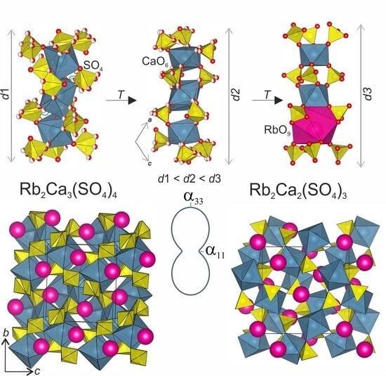
The Rb2Ca3(SO4)4 compound was obtained by rapid cooling of the stoichiometric melt. The crystal structure was solved and refined using single crystal X-ray diffraction analysis (P21/c, a = 9.2847(9), b = 9.4094(6), c = 9.2917(8) Å, β = 114.646(1)°, V = 737.80(12) Å3, R1 = 0.051). The thermal behavior of Rb2Ca3(SO4)4 was investigated by high-temperature powder X-ray diffraction in the range 25–1000 °C. Thermal decomposition of the Rb2Ca3(SO4)4 phase occurs at 300 °C, forming Rb2Ca2(SO4)3 and CaSO4. The decomposition is complete at 450 °C, and the mixture of Rb2Ca2(SO4)3 + CaSO4 persists up to 890 °C. Homogenization of the phases occurs at 900 °C, resulting in the formation of the Rb2Ca3(SO4)4 compound again at 970 °C. A structural interpretation of this thermal phase transformation is presented, and the relationship between the crystal structures of Rb2Ca3(SO4)4 and Rb2Ca2(SO4)3 of the langbeinite structure type is demonstrated. Thermal expansion of Rb2Ca3(SO4)4 is highly anisotropic: α11 = 23.9(4), αb = 19.2(3), α33 = 7.7(1), αβ = −1.9(7), αV = 50.8(9) × 10−6 °C−1 at 25 °C and α11 = −7(2), αb = 17(5), α33 = 25(7), αβ = −1.1(1), αV = 35(9) × 10−6 °C−1 at 1000 °C. The anisotropy of the thermal expansion is described in comparison with the Rb2Ca3(SO4)4 crystal structure. The optical band gap for the Rb2Ca3(SO4)4 compound was determined to be 3.7 eV from absorption spectroscopy data.

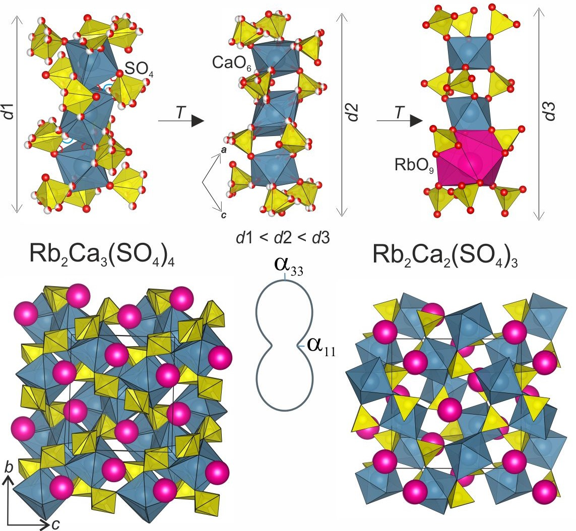
Graphical Abstract

1. Introduction

Complex alkali- and alkaline-earth-containing sulfates have been attracting significant attention recently as birefringent materials due to a wide transparency window, ease of crystal growth, environmental friendliness and thermal stability. These materials can be used for advanced light-matter coupling, polarizers and waveplates (e.g., [1]). However, many sulfates have relatively long UV absorption edges, which limit their use as birefringent materials in the ultraviolet region. Therefore, the search for new sulfates that are transparent in the ultraviolet region is highly relevant. Knowledge of thermal behavior and expansion of these compounds is quite important. For successful application in optical devices, their thermal behavior must be studied to prevent thermal stress and predict the shape change of the optical element with temperature.
Several structurally similar sulfates transparent in the ultraviolet region and exhibiting relatively high birefringence are known: Rb2Mg3(SO4)4, Cs2Mg3(SO4)4 [2], and Cs2Ca3(SO4)4 [3]. Cs2Mg3(SO4)4 crystallizes in the noncentrosymmetric space group P212121, whereas Rb2Mg3(SO4)4 and Cs2Ca3(SO4)4 crystallize in the centrosymmetric space group P21/c. The authors attributed the differences in symmetry and structure to the influence of cation size. The structures were described as heteropolyhedral frameworks composed of SO4 tetrahedra and MO6 (M = Mg, Ca) octahedra connected via shared vertices. Alkali metal cations (Rb+ and Cs+) occupy the cavities within the framework. As early as 1962 [4], it was hypothesized that the compounds Cs2Ca3(SO4)4 and Rb2Ca3(SO4)4 exist and undergo several polymorphic transformations upon heating. The compound Cs2Ca3(SO4)4 was successfully synthesized, and its structure was solved in [3]; however, no structural or physicochemical data have been reported for Rb2Ca3(SO4)4. High-temperature studies of Cs2Ca3(SO4)4 revealed changes in its diffraction pattern at 600 °C, though no definitive conclusions were drawn regarding the nature of these changes [5]. The present work is specifically aimed at addressing these gaps in knowledge.
Since we have established a structural relationship between the compound studied in this work and the structure type of langbeinite, it is pertinent to discuss the properties and existing research on this family. Named after the mineral, langbeinite-based materials offer distinct advantages for applications in functional materials due to their exceptional thermal stability and highly flexible crystal structures capable of accommodating a wide variety of cationic substitutions. In nature, there are five members of langbeinite group of minerals: langbeinite K2Mg2(SO4)3 (e.g., [6,7]), manganolangbeinite K2Mn2(SO4)3 [8], efremovite (NH4)2Mg2(SO4)3 [9], ferroefremovite (NH4)2Fe2+(SO4)3 [10], calciolangbeinite K2Ca2(SO4)3 [11]. Synthetic analog of K2Mg2(SO4)3 langbeinite is widely used as fertilizer for plants [12]. Members of the langbeinite crystal family exhibit a diverse range of functional properties, including ferroelectricity and ferroelasticity [13,14,15]. These attributes have positioned them as promising host matrices for the long-term immobilization of radioactive waste [16]. In optoelectronics, langbeinite-type rare-earth-doped zirconium phosphates are actively being investigated for use in white light-emitting diodes (LEDs) and plasma display panels [17]. Iron-substituted analogs, meanwhile, exhibit complex magnetic interactions that continue to attract fundamental research interest [18]. K2Mg2(SO4)3:Dy displays both thermoluminescent and mechanoluminescent responses following gamma irradiation [19]. Recent condensed matter studies have further identified both sulfate and phosphate langbeinite-type compounds as potential quantum spin liquid candidates, a frontier area in low-dimensional magnetism [20,21,22]. As characterization techniques and synthesis methods advance, research into the structural, photonic, and quantum properties of the langbeinite family is expected to accelerate, paving the way for novel functional materials and device architectures.
In this article, we report the synthesis, crystal structure, thermal behavior, Raman, IR and absorption spectroscopy of the Rb2Ca3(SO4)4 compound. A comparison of the Rb2Ca3(SO4)4 compound with the similar Cs2Ca3(SO4)4 and langbeinite-type compounds (e.g., Rb2Ca2(SO4)3, Cs2Ca2(SO4)3) was provided. The anisotropy of the Rb2Ca3(SO4)4 thermal expansion is described in comparison with the crystal structure.

2. Experimental

2.1. Raw Materials

The Rb2Ca3(SO4)4 compound was prepared by combining stoichiometric amounts of pre-calcined Rb2SO4 and CaSO4 (both Neva Reaktiv, (St. Petersburg, Russia) 99.93% purity). Then, the mixture was kept at 300 °C for 3 h in a LOIP LF 7/13-G1 muffle furnace.

2.2. Synthesis

The calcined mixture was ground for 1 h in an agate mortar and pestle; the powder was pressed into 1 mm pellets at 80 bar. The pellets were subsequently melted at 1000 °C for 30 min in a Nabertherm HTC furnace. The resulting melt was rapidly quenched on a steel plate to obtain the Rb2Ca3(SO4)4 compound. The Cs2Ca3(SO4)4 compound was previously synthesized by our group [5].

2.3. Methods

2.3.1. Powder X-Ray Diffraction

The powder X-ray diffraction data were collected using a Rigaku Miniflex II diffractometer (CuKα radiation, 30 kV and 10 mA, Bragg–Brentano geometry, 1D PSD D/tex Ultra, 2θ = 5–70°, step 0.02°) (Rigaku holding corporation, Tokyo, Japan). The phase composition was determined using PDXL integrated X-ray powder diffraction software [23] and PDF-2 database, ICDD, 2020. X-ray phase analysis revealed that the polycrystalline sample of Rb2Ca3(SO4)4 contains small impurity of Rb2Ca2(SO4)3 and CaSO4. The compound is monoclinic at ambient conditions (P21/c, a = 9.28446 (9), b = 9.40721 (9), c = 9.30017 (9) Å, β = 114.709 (3)°, V = 737.91 (4) Å3 and Z = 2).

2.3.2. Crystal Structure Determination

A single crystal of Rb2Ca3(SO4)4 was studied using a Rigaku XtaLAB Synergy-S diffractometer equipped with a high-stability microfocus X-ray source PhotonJet-S and a high-speed hybrid photon counting detector HyPix-6000HE (MoKα radiation, 50 kV and 1.0 mA) (Rigaku holding corporation, Tokyo, Japan). The crystal was kept at room temperature. Data collection parameters: frame width 1.0° and exposure time 220 s per frame. CrysAlisPro software (version 44.85) was used for further processing. An absorption correction was introduced using SCALE3 ABSPACK algorithm. The crystal structure was solved by direct methods and refined by least square techniques in the monoclinic space group P21/c to R1 = 0.051 (wR2 = 0.087) for 1786 unique observed reflections with I ≥ 2σ(I) using Shelx program package [24] within Olex2 shell [25]. Crystallographic data and refinement parameters, atomic coordinates and isotropic displacement parameters, atomic anisotropic displacement parameters and selected bond lengths are summarized in Table 1, Table 2 and Tables S1 and S2. The bond-valence calculations were performed using empirical parameters taken from [26]. The results are presented in Table S2. Some deviation of the bond-valence sums from the expected value may be explained by the positional disorder of the oxygen sites. Further details of the crystal structure investigations can be obtained from the Cambridge Structural Database by quoting the depository numbers CSD 2547613.

2.3.3. Thermal Analysis

Thermal analysis experiments (DSC + TG) were performed on a STA 449 C NETZSCH simultaneous thermal analysis instrument equipped with a platinum–rhodium sample holder (dynamic air atmosphere, air flow 50 cm3/min, temperature range 25–1100 °C, heating rate 20 °C/min). The pellets for the experiments were weighted with an accuracy of 0.01 mg (the mass was approximately 10 mg) and placed in an open platinum–rhodium crucible. The temperatures of thermal effects were determined using NETZSCH Proteus software, version 9.7 by the onset temperature on DSC curves. According to the TG data, no mass losses occurred.

2.3.4. High-Temperature Powder X-Ray Diffraction

The thermal behavior of Rb2Ca3(SO4)4 was studied using in situ high-temperature powder X-ray diffraction in the range 25–1000 °C on a Rigaku Ultima IV diffractometer (CuKα) (Rigaku holding corporation, Tokyo, Japan) with the SHT–1500 high-temperature camera. Unit cell parameters, coefficients of approximation of its temperature dependencies, eigenvalues and figures of the thermal expansion tensor were calculated using RTT software, version 5.3 [28].

2.3.5. Absorption Spectroscopy

The optical bandgap of polycrystalline powders was determined using the Tauc method based on absorption spectra. Absorption spectra were recorded using a research-grade PerkinElmer Lambda 1050 spectrophotometer. To account for scattering from polycrystalline samples, measurements were performed in an integrating sphere, with a polycrystalline BaSO4 sample used as a reference. Absorption spectra were measured with a step size of 1 nm and an integration time of 1 s per point.

2.3.6. Raman Spectroscopy

Raman spectra were measured using a Bruker Senterra spectrometer with laser excitation at a wavelength of 785 nm and a power of 100 mW. The laser beam was focused onto the surface of the polycrystalline sample using a 20× objective lens (numerical aperture 0.40); the Raman signal was collected in backscattering geometry and recorded for 30 s with averaging over two acquisitions. The spectral resolution was 3 cm−1.

2.3.7. IR Spectroscopy

Vibrational spectra were complemented by Fourier-transform infrared (FTIR) spectroscopy. IR spectra were measured using a Thermo Scientific Nicolet 8700 FTIR spectrometer (Thermo Fisher Scientific, Waltham, MA, USA) equipped with an attenuated total reflection (ATR) accessory and an MCT-A detector. IR absorption spectra were recorded in the spectral range of 4000–650 cm−1 at a resolution of 2 cm−1. To minimize the contribution of water vapor and CO2, the spectrometer was purged with nitrogen.

2.3.8. Energy-Dispersive X-Ray Spectroscopy

The chemical composition was studied using a system with focused electron and ion probes, QUANTA 200 3D (FIA, Paris, France), with an energy-dispersive spectroscopy analytical complex, Pegasus 4000 (EDAX, Pleasanton, CA, USA). The analytical spectra were obtained from a smooth crystal surface at an operating voltage of 20 kV, with a beam size of 1 μm. The samples were coated with carbon. Analytical results are given in Table S3. The empirical formula calculated for 16 O is Rb1.98Ca3.03S3.97O16.

3. Results and Discussion

3.1. Thermal Behavior

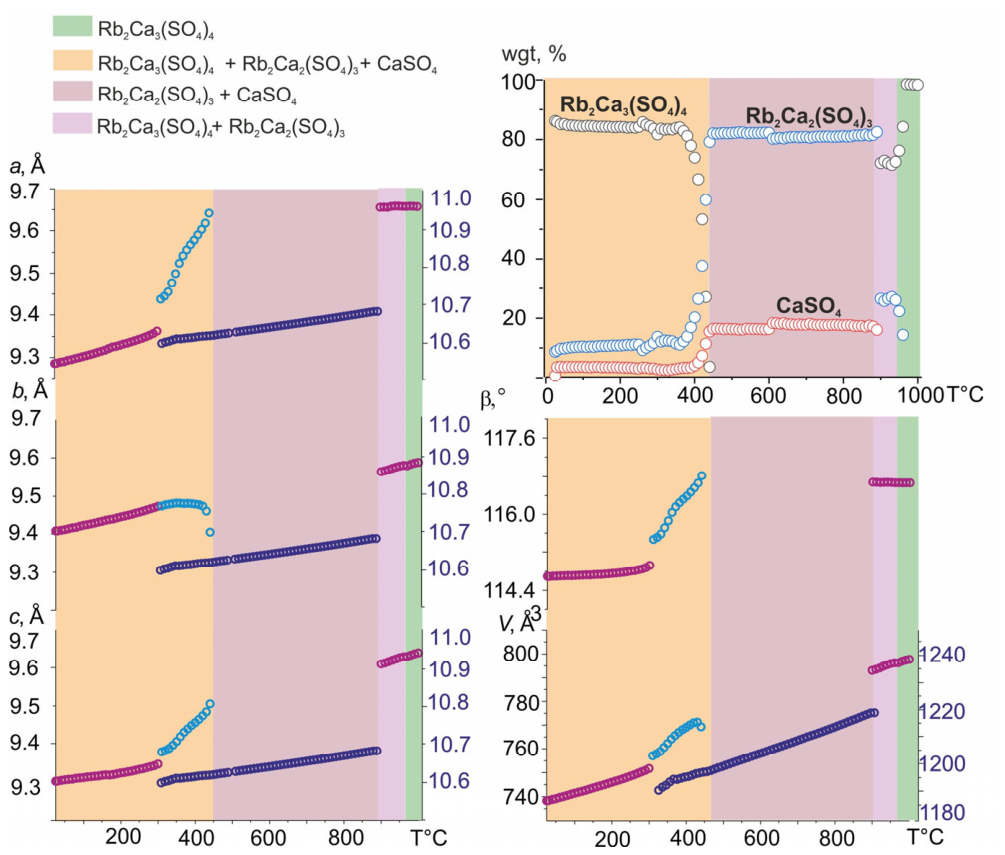
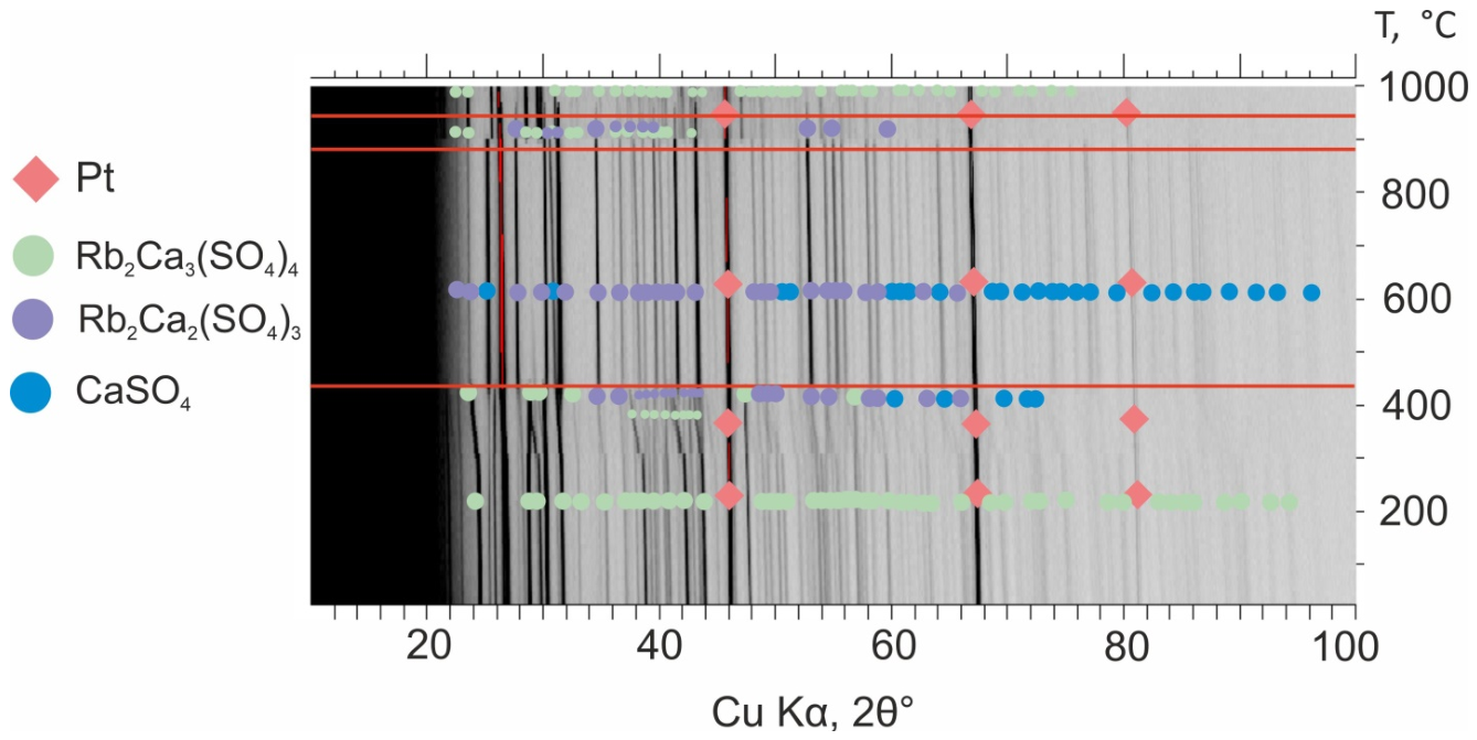
Figure 1 presents the diffraction patterns obtained from the high-temperature X-ray diffraction experiment for Rb2Ca3(SO4)4, while Figure S1 shows the corresponding data for Cs2Ca3(SO4)4. According to the present study, both phases exhibit two distinct temperature ranges of stability: 25–300 and 900–1000 °C for Rb2Ca3(SO4)4, 25–540 and 840–900 °C for Cs2Ca3(SO4)4.
The Rb2Ca3(SO4)4 compound is stable up to 300 °C. Upon heating above this temperature, it begins to decompose into Rb2Ca2(SO4)3 and CaSO4. The decomposition is fully completed at 450 °C, and the resulting two-phase mixture (Rb2Ca2(SO4)3 + CaSO4) persists up to 890 °C. At 900 °C, homogenization occurs, leading to the reformation of the Rb2Ca3(SO4)4 phase at 970 °C, which remains stable up to 1000 °C before congruent melting.
A similar reversible decomposition pathway is observed for the cesium analog, Cs2Ca3(SO4)4: the phase is stable up to 540 °C, decomposes into Cs2Ca2(SO4)3 + CaSO4 in the intermediate range, and reforms at 840 °C, remaining stable up to 900 °C.
Further temperature increase leads to congruent melting of both compounds.

3.2. Crystal Structure of Rb2Ca3(SO4)4 Compound

The crystal structure of Rb2Ca3(SO4)4 is closely related to that of Cs2Ca3(SO4)4, differing primarily in the positional disorder of the SO4 tetrahedra (i.e., splitting of the oxygen atom sites). It is worth noting that Rb2Ca3(SO4)4 compound crystallizes in a new structure type because atomic sites are not the same as in the Cs2Ca3(SO4)4 due to positional disorder of the oxygen sites. The compound crystallizes in the monoclinic system, space group P21/c (a = 9.2847 (9), b = 9.4093 (6), c = 9.2917 (8) Å, β = 114.646 (10)°, V = 737.80 (12) Å3). The asymmetric unit contains one Rb site, two Ca sites, two S sites, and fourteen O sites. Only three oxygen sites (O8, O12 and O13) are fully occupied, whereas the remaining eleven are split.
The S1 site is coordinated by seven oxygen atoms with bond lengths ranging from 1.37 to 1.67 Å, while the S2 site is surrounded by seven oxygen atoms with bond lengths of 1.36–1.56 Å. The mean bond distances <S1–O>7 and <S2–O>7 are consistent with the S–O bond lengths reported in the comprehensive review [29]. The Ca1 and Ca2 sites are each coordinated by twelve oxygen atoms, with bond lengths of 2.19–2.71 Å and 2.09–3.05 Å, respectively. The Rb1 site also exhibits a coordination number of twelve, with Rb1–O bond distances varying in the range 2.89–3.28 Å.
If one considers a hypothetical ordered structure of Rb2Ca3(SO4)4 isotypical with Cs2Ca3(SO4)4, the coordination environment would be as follows: the S sites would display tetrahedral coordination, the Ca sites octahedral coordination, and the coordination number of the Rb1 site is eight.
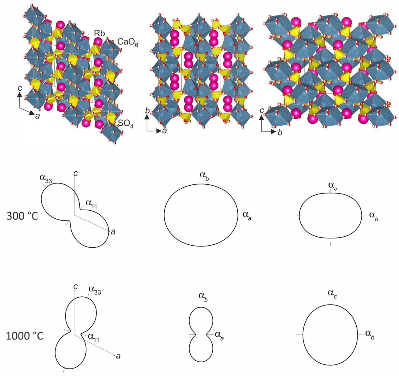
The crystal structure can be described as a mixed framework composed of SO4 tetrahedra and CaO6 octahedra. The CaO6 octahedra share edges with one another and are interconnected via three SO4 tetrahedra. Layers parallel to the (111) plane can be identified within the structure; their stacking generates a framework featuring tunnels running along the a-axis, which accommodate the Rb+ cations (Figure 2).
Furthermore, if we viewed the crystal structure as ordered, the structure can be considered in terms of rods built from Ca(SO4)6 structural units. Each unit consists of a central CaO6 octahedron connected via vertices to six SO4 tetrahedra (Figure 3, Figure 4 and Figure 5). These motifs closely resemble the M(TO4)6 microblocks introduced in [30] and discussed in detail in [31].
Comparison with the langbeinite structure type. A distinct structural relationship exists between the crystal structures of Rb2Ca3(SO4)4 and Rb2Ca2(SO4)3, as well as between Cs2Ca3(SO4)4 and Cs2Ca2(SO4)3. Since the latter pair consists of fully ordered phases, their comparison is more straightforward.
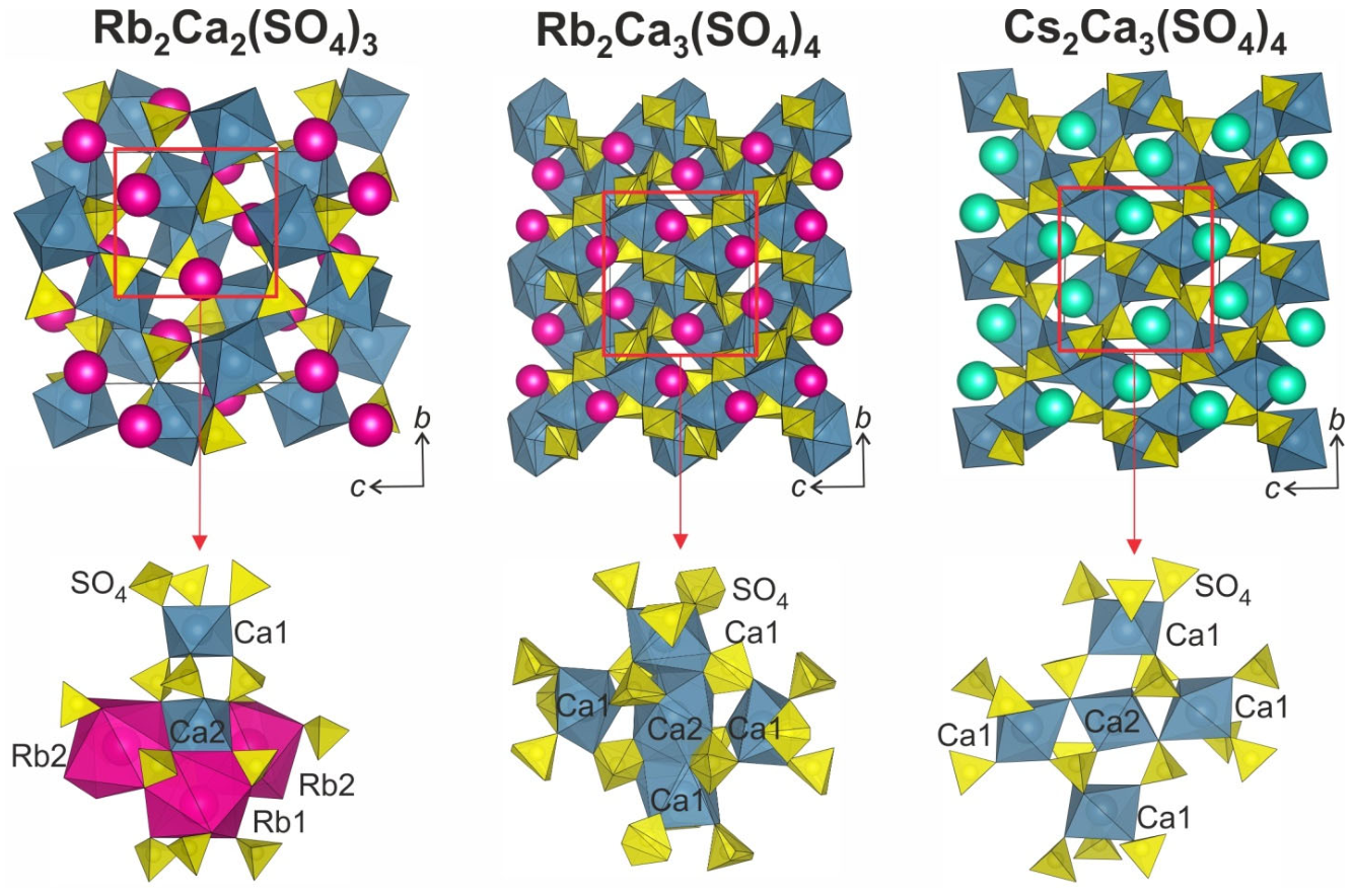
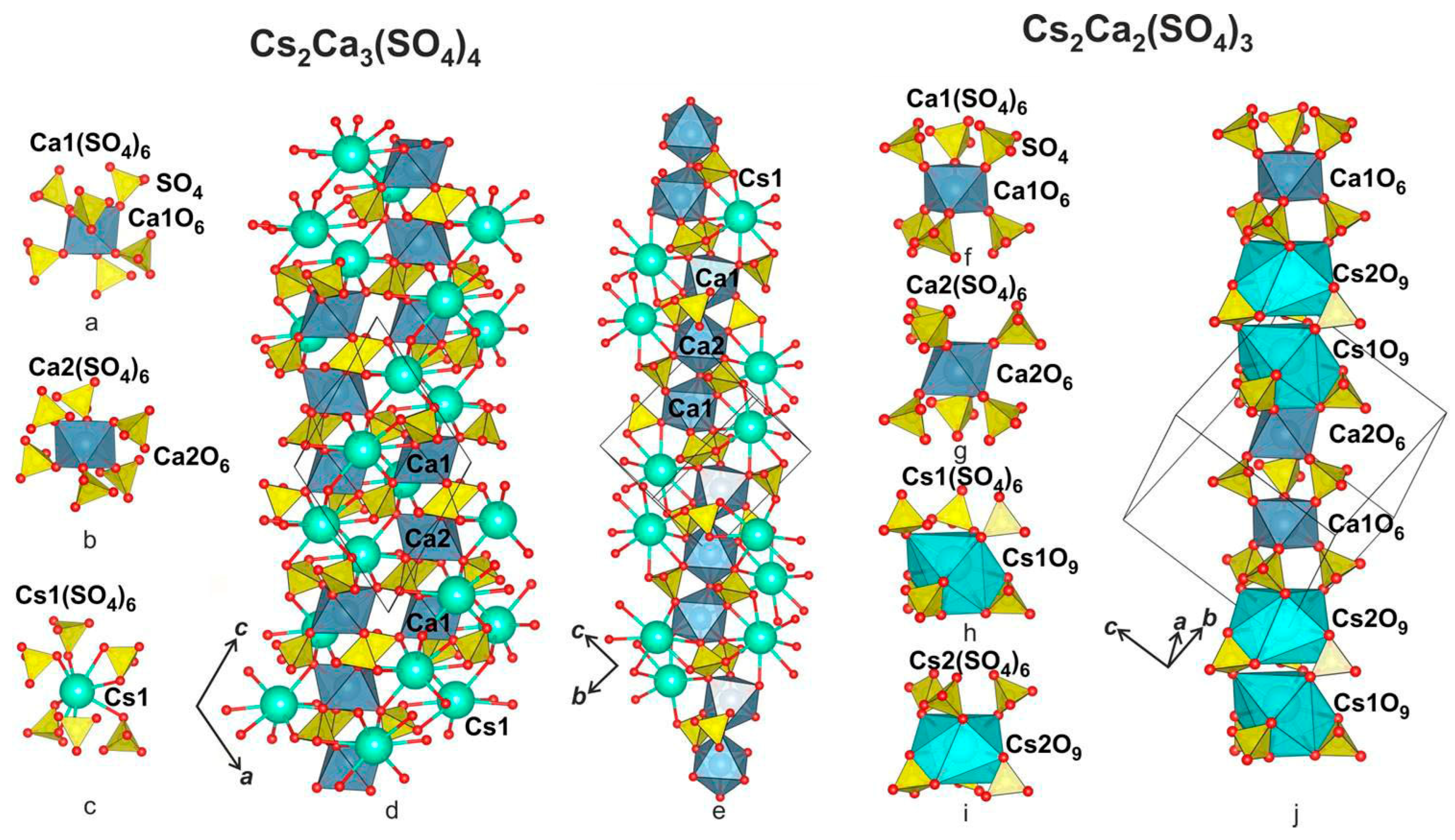
The cb-projection of the Cs2Ca3(SO4)4 crystal structure exhibits a pseudo-cubic appearance along the apparent fourfold axis (L4) and resembles the orthogonal projections characteristic of the cubic langbeinite structure type (e.g., Rb2Ca2(SO4)3) (Figure 3). Within the framework, columns composed of three linked Ca(SO4)6 microblocks extend along the directions corresponding to the apparent threefold axes. A direct structural comparison of these crystal structures and columns is presented in Figure 3.
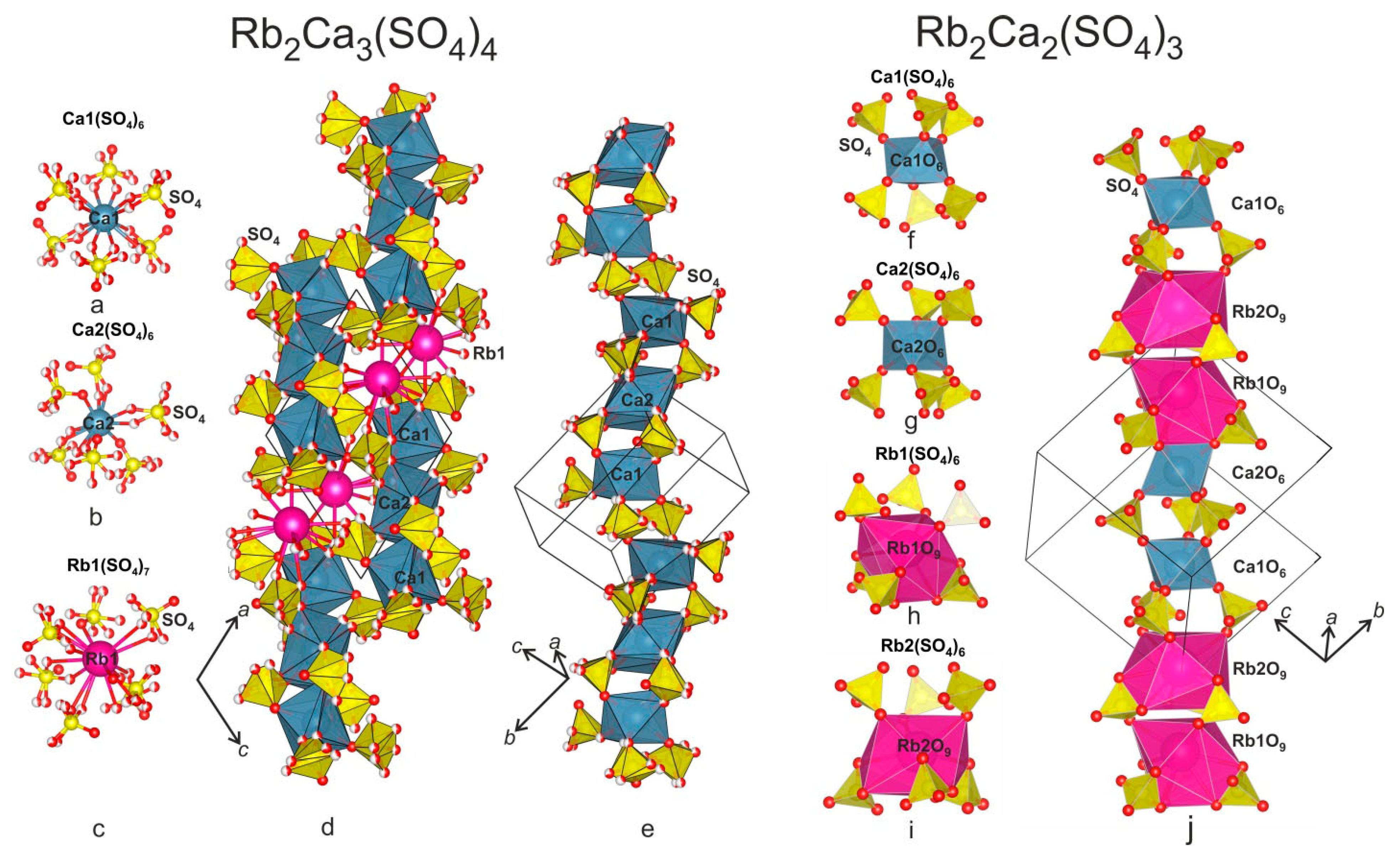
The crystal structure of cubic langbeinite structure type can be described as an M(TO4)6 framework, with alkali metal cations occupying its cavities [30,32,33]. This framework is built from M(TO4)6 microblocks, first introduced in [30], which connect through three TO4 tetrahedra to form M2(TO4)9 dimers elongated along the threefold axes. In the present case, for Rb2Ca2(SO4)3 and Cs2Ca2(SO4)3, these correspond to Ca(SO4)6 microblocks and Ca2(SO4)9 dimers. Interestingly, if the truncated coordination environment of the Cs+ cations is considered, Cs(SO4)6 microblocks can also be identified, with the lower part of the microblock rotated by 60° relative to the upper part. Similar microblocks can be identified in the structures of Rb2Ca3(SO4)4 and Cs2Ca3(SO4)4. The Ca(SO4)6 and Cs(SO4)6 microblocks connect via three SO4 tetrahedra, or via three SO4 tetrahedra and vertex of octahedra, to form rods along the threefold axes (Figure 4 and Figure 5). Viewed this way, the langbeinite structure can be described as a framework assembled from such columns. For Rb2Ca2(SO4)3, the microblock sequence along a given column is Ca2(SO4)6, Ca1(SO4)6, Rb2(SO4)6 and Rb1(SO4)6.
The crystal structure of Cs2Ca3(SO4)4 can similarly be described as consisting of slightly curved columns elongated along the unit cell diagonals. These directions correspond to the threefold axes of the cubic langbeinite structure type but are distorted due to the structural transformation. The columns are constructed from Ca(SO4)6 microblocks linked through SO4 tetrahedra and vertices of CaO6 octahedra (Figure 4d,e and Figure 5d,e). Both column systems can be described as exclusively comprising Ca(SO4)6 microblocks, while the cesium polyhedra do not integrate into the columns but instead frame them laterally. In columns of the first type, three Ca(SO4)6 microblocks are sequentially linked via one vertex of CaO6 octahedra and three SO4 tetrahedra. This is followed by a slight shift of the rod, after which the subsequent triplet of Ca(SO4)6 microblocks connects through two SO4 tetrahedra and two CsO8 polyhedra. In rods of the second type, triplets of Ca(SO4)6 microblocks are linked through three SO4 tetrahedra. These triplets connect to one another along the column direction via two CsO8 polyhedra. Rods of the second type are interconnected with each other through shared SO4 tetrahedra at the terminal microblocks of each triplet.
Comparing the structures of Rb2Ca3(SO4)4 and Cs2Ca3(SO4)4, the former can be regarded as a disordered analog of the latter. However, a key distinction is that rods connected via three tetrahedra and those connected via three tetrahedra and vertex of CaO6 octahedra elongate in opposite directions in the two structures. In the Cs2Ca3(SO4)4 crystal structure, rods of Ca(SO4)6 microblocks linked through three SO4 tetrahedra extend along the long diagonal of the ac parallelogram (Figure 4d), whereas rods connected via three SO4 tetrahedra and vertex of CaO6 octahedra extend along the short diagonal of the ac parallelogram (Figure 4e). In Rb2Ca3(SO4)4, the situation is reversed, despite the positional disorder of the oxygen sites (Figure 5d,e).
Upon heating, the Rb2Ca3(SO4)4 and Cs2Ca3(SO4)4 phases obtained by crystallization from the melt decompose to form Rb2Ca2(SO4)3 + CaSO4 and Cs2Ca2(SO4)3 + CaSO4, respectively. Initially, before a detailed analysis of high-temperature phases was conducted, it was believed that polymorphic transformations occur with increasing temperature [4,5].
The structural transformations Rb2Ca2(SO4)3 + CaSO4 → Rb2Ca3(SO4)4 and Cs2Ca2(SO4)3 + CaSO4 → Cs2Ca3(SO4)4 can be caused by the metastability of the Rb2Ca3(SO4)4 and Cs2Ca3(SO4)4 phases, coupled with a structural tendency to increase symmetry through the incorporation of Rb+ and Cs+ cations into the rods. The driving force for this transformation is thermal atomic motion, which induces cation migration and order–disorder processes for Rb, Cs, and Ca, as well as potential disordering of the SO4 tetrahedra at elevated temperatures. This interpretation is indirectly supported by the observed structural disorder in Rb2Ca3(SO4)4.
As the temperature increases, the difference between Rb and Ca atoms decreases, and the size of the polyhedra in the rod also decreases due to disordering and rotation of the tetrahedra. Therefore, Rb atoms are displaced from the rods into the framework cavities, and their place is taken by Ca cations. Since the rod length decreases in this case, a shift or displacement of the rods occurs, resulting in fragments of three linked Ca(SO4)6 microblocks.
Due to the order–disorder processes of Ca–Rb and Ca–Cs and the displacement of alkali framework cations, a distortion of the crystal structure occurs, which leads to the formation of layers in the (111) plane and tunnels for alkali cations elongated along the a axis (Figure 1).
Another point that needs to be considered is why the crystal structure of Rb2Ca3(SO4)4 is disordered, while Cs2Ca3(SO4)4 is ordered. Presumably, this is due to the sizes of the ionic radii. The ionic radius of [8]Rb is 1.75 Å, while [8]Cs is 1.88 Å [34], apparently due to the longer average bond length due to the ionic radius; Cs stabilizes the crystal structure by forming bonds with SO4 tetrahedra, and the length of these bonds is sufficient for the SO4 tetrahedra to be ordered. In the structure of Rb2Ca3(SO4)4, the length of the Rb–O bonds is only sufficient to form bonds with disordered SO4 tetrahedra.

3.3. Thermal Expansion

Temperature dependencies of the Rb2Ca3(SO4)4 and Cs2Ca3(SO4)4 unit cell parameters and volume are shown in Figure 6 and Figure S2. These dependencies were approximated by second-order polynomial functions (Table 3) in 25–300 and 900–1000 °C temperature ranges. Table 4 and Table S4 present the main coefficients of the thermal expansion tensor at selected temperatures.
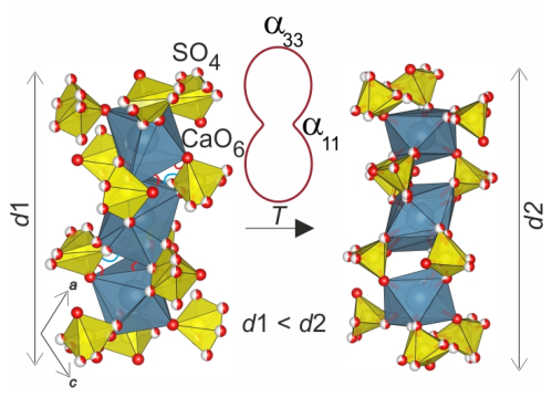
The maximum and minimum thermal expansion of Rb2Ca3(SO4)4 occurs in the monoclinic ac plane. The maximum thermal expansion is close to the long diagonal of the ac parallelogram, while the minimum is close to the short diagonal of the ac parallelogram. Thermal expansion can also be described as a classical expansion of a layered structure, since layers can be distinguished in the structure in the (111) plane (see Section 3.1), and the expansion between the layers will be maximum. Also, this nature of thermal expansion indicates shear deformations (e.g., [35,36]), consistent with observations for Cs2Ca3(SO4)4 [5]. These shear deformations are associated with the straightening of the rods (Figure 5d) along the long diagonal of the ac parallelogram, driven by the rocking (initiation of rotation) of the SO4 tetrahedra. Perpendicularly, thermal expansion is minimal. Rotations of the tetrahedra can lead to the disordered CaO6 octahedra in such a column being linked through three SO4 tetrahedra (Figure 7). Thermal deformations of similar structural groups have been previously considered by us [5,37,38]. The formation of a connection of octahedra through three tetrahedra partially reduces the corrugation of the rods, which leads to positive thermal expansion along the z-axis of the rod and restrains thermal expansion along the x- and y-axes (Figure 7). In rods of the second type (Figure 5e), the disordered CaO6 octahedra are already linked through three SO4 tetrahedra, and apparently, the thermal deformations of this rod are not so anisotropic. In the Rb2Ca2(SO4)3 compound, into which Rb2Ca3(SO4)4 transforms upon heating, the octahedra in the rod are also connected to each other via three SO4 tetrahedra (Figure 5). Due to the structural similarity of Rb2Ca3(SO4)4 and Rb2Ca2(SO4)3, the principle of continuity of structural deformations of crystals during thermal expansion is partially fulfilled in this case. This principle states that “the characteristic features of structural rearrangement during thermal polymorphic transformation are likely to tend to manifest themselves in thermal deformations of a less symmetric modification” [39].
The thermal expansion of Rb2Ca3(SO4)4 and Cs2Ca3(SO4)4 differs significantly in the low-temperature range, and is virtually identical in the high-temperature range. The thermal expansion of Cs2Ca3(SO4)4 in the temperature range of 25–540 °C was studied previously, and an interpretation of the thermal expansion anisotropy was provided in [5]. The maximum positive thermal expansion occurs along the b-axis, and a strong negative one occurs along the long diagonal of the ac parallelogram. Along the short diagonal of the ac parallelogram, the thermal expansion is almost the same as along αb (Figure 2). It should be noted once again that the crystal structure of Cs2Ca3(SO4)4 is completely ordered relative to the crystal structure of Rb2Ca3(SO4)4, which affects the difference in the thermal expansion of these two compounds. The difference in thermal expansion can be explained by the fact that in the rods extended along the long diagonal of the ac parallelogram in the crystal structure of Cs2Ca3(SO4)4, the ordered octahedra are already connected through three SO4 tetrahedra (Figure 3), and due to the vibrations of oxygen atoms perpendicular to the S–O and Ca–O bonds in the Ca(SO4)6 microblocks that make up this column, the polyhedra rock relative to each other, which leads to a strong negative thermal expansion along the direction of the rods’ extension along the long diagonal of the ac parallelogram and to a positive expansion in perpendicular directions.
In the temperature range of 900–1000 °C, the thermal expansion of Rb2Ca3(SO4)4 is similar to that of Cs2Ca3(SO4)4. It is slightly negative in the direction close to the long diagonal of the ac parallelogram and is maximum perpendicular to this direction in the monoclinic plane. Apparently, this is due to the same crystallochemical factors as in Cs2Ca3(SO4)4. And for the compound Cs2Ca3(SO4)4 in the temperature range of 840–900 °C, the thermal expansion has a similar character: maximum expansion along the short diagonal of the ac parallelogram, and minimal, but not negative, along the long diagonal. The fact that large negative thermal expansion did not occur in these temperature ranges is most likely explained by the maximum stress of the structure before subsequent melting.

3.4. Thermal Analysis

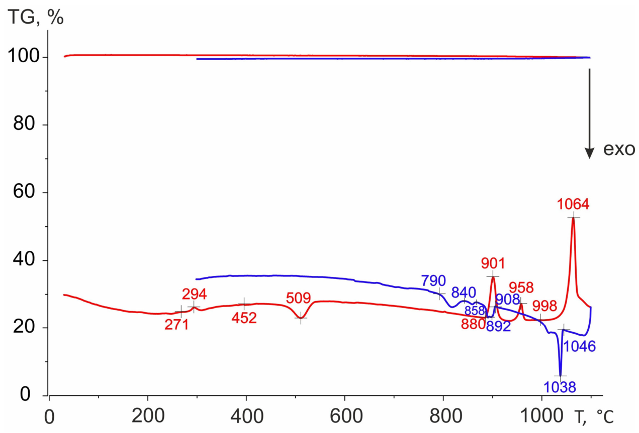
Figure 8 shows the DSC curve of the Rb2Ca3(SO4)4 compound during heating and cooling. Earlier, the thermal analysis of Rb2Ca3(SO4)4 compound was conducted in [4], and our data generally confirm the previously obtained results, except for their interpretation. Our interpretation is further supported by the results of high-temperature powder X-ray diffraction. An endothermic peak at 271 °C with a maximum at 294 °C can be attributed as thermal decomposition of the Rb2Ca3(SO4)4 phase with formation of Rb2Ca2(SO4)3 and CaSO4. A peak at 452 °C with a maximum at 509 °C can be described as the completion of thermal decomposition. The Rb2Ca3(SO4)4 phase completely disappears at this temperature. The next peak at 880 °C with a maximum at 901 °C corresponds to the reappearance of the Rb2Ca3(SO4)4 phase, and peak at 932 °C with a maximum at 958 °C corresponds to the homogenization of Rb2Ca3(SO4)4 phase. Finally, the Rb2Ca3(SO4)4 compound melts at 998 °C, with a maximum at 1064 °C. Thermogravimetric analysis revealed no mass change in the sample when heated to 1100 °C.
During cooling, several thermal effects were observed. The crystallization temperature of the Rb2Ca3(SO4)4 compound was 1046 °C. At 908 °C, the decomposition of the Rb2Ca3(SO4)4 phase and the formation of the Rb2Ca2(SO4)3 and CaSO4 occurred. Finally, the process of thermal decomposition was completed at 840 °C.

3.5. Absorption Spectroscopy

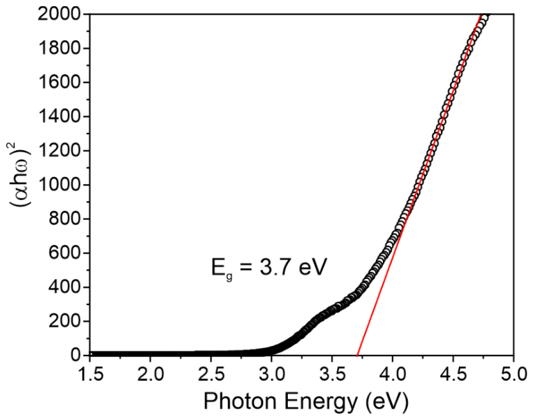
Absorption spectroscopy was used to determine the optical bandgap energy of polycrystalline Rb2Ca3(SO4)4 samples. To this end, the absorption spectra measured in an integrating sphere to account for scattered light were plotted in Tauc coordinates (Figure 9). Based on data from the related but ordered structure of the Cs2Ca3(SO4)4 phase, this material exhibits direct bandgap transitions; therefore, when calculating the optical bandgap of the Rb2Ca3(SO4)4 phase, we consider it to be a direct bandgap material. Approximation of the linear region of the fundamental absorption edge in Tauc coordinates yields an optical bandgap energy of 3.70 ± 0.02 eV for Rb2Ca3(SO4)4. It should be noted that this value is lower than the band gap of Cs2Ca3(SO4)4 [3], which may be attributed to positional disorder of the oxygen sites and the SO4 tetrahedra resulting from the substitution of Rb ions for Cs.

3.6. Vibrational Spectroscopy

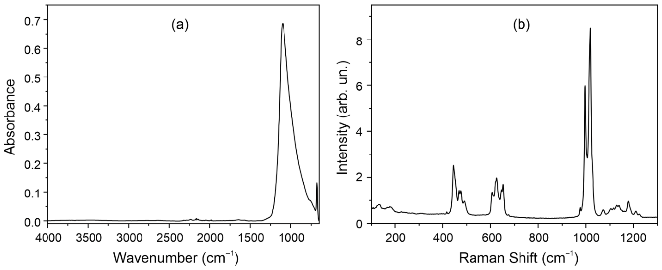
The structural features of the Rb2Ca3(SO4)4 compound were investigated using vibrational spectroscopy, including Fourier-transform infrared (FTIR) spectroscopy and Raman scattering spectroscopy. Typical vibrational spectra of Rb2Ca3(SO4)4 are shown in Figure 10. It should be noted that, according to the IR absorption data, -OH groups are absent, which is fully consistent with X-ray diffraction analysis and also indicates that the samples do not absorb water from the air. The maximum phonon energy, as derived from both IR absorption and Raman spectroscopy, does not exceed 1250 cm–1, and all bands in the vibrational spectra correspond to the vibrational modes of the [SO4]2– structural units. The absorption bands in the IR spectra are considerably overlapped, whereas the Raman bands are well resolved. Therefore, the Raman data confirmed the presence of two nonequivalent SO4 tetrahedra in the crystal structure, because all vibrational bands are double and well resolved. This is particularly evident for the symmetric stretching vibrations ν1(SO42−) near 1000 cm−1, which appear as two resolved bands with maxima at 997 cm−1 and 1019 cm−1. The position of the maximum of this band is determined by the environment of the tetrahedron. For example, for the orthorhombic phase of CaSO4 (Amma space group), the maximum of this band is at 1016 cm−1 [40], while for the orthorhombic phase of Rb2SO4 (Pmcn space group), the maximum lies at 976 cm−1 [41]. In the Rb2SO4–CaSO4–H2O system, the maximum of the vibrational band is located at 1027 cm−1 [42]. Thus, for the studied Rb2Ca3(SO4)4 compound, Raman spectroscopy confirms the presence of two nonequivalent SO4 tetrahedra, which manifests as the doubling of vibrational bands. Apart from the symmetric stretching vibrations, the Raman spectra exhibit two sets of bands for the ν2(SO42−) vibrations at frequencies 445, 468, 476, and 490 cm−1, for the ν3(SO42−) vibrations at 1073, 1104, 1116, 1130, 1140, 1179, 1210, and 1225 cm−1, and for the ν4(SO42−) vibrations at 608, 625, 645, and 653 cm−1 [43,44]. In the region of 135 and 180 cm−1, bands are observed that correspond to vibrational modes such as rotational and translational external modes [45].

4. Conclusions

The Rb2Ca3(SO4)4 compound was successfully synthesized via rapid cooling of the melt, and its crystal structure was fully solved and refined using single-crystal X-ray diffraction. The thermal behavior of Rb2Ca3(SO4)4 was studied over a wide temperature range. The thermal decomposition of the Rb2Ca3(SO4)4 phase begins at 300 °C, forming Rb2Ca2(SO4)3 and CaSO4, and it is fully completed at 450 °C. Homogenization of the phases occurs at 900 °C, resulting in the formation of the Rb2Ca3(SO4)4 compound again. A structural interpretation of this thermal phase transformation is presented. The anisotropy of the thermal expansion is described in comparison with the Rb2Ca3(SO4)4 crystal structure. The maximum and minimum thermal expansion of Rb2Ca3(SO4)4 occurs in the monoclinic ac plane; the maximum is close to the long diagonal of the ac parallelogram, and the minimum is close to the short diagonal of the ac parallelogram. These thermal deformations are associated with the straightening of the rods along the long diagonal of the ac parallelogram due to the rocking (initiation of rotation) of the SO4 tetrahedra. The structural relation between the Rb2Ca3(SO4)4, Cs2Ca3(SO4)4 and the langbeinite structure type (e.g., Rb2Ca2(SO4)3 and Cs2Ca2(SO4)3) was established. These relations partially explain the nature of the anisotropy of thermal expansion. Optical absorption spectroscopy determined a band gap of 3.7 eV. These results establish a comprehensive structure–property relationship for Rb2Ca3(SO4)4, highlight its highly anisotropic expansion behavior, and suggest its potential utility in optoelectronic applications.

Supplementary Materials

The following supporting information can be downloaded at: https://www.mdpi.com/article/10.3390/min16050548/s1.

Author Contributions

Conceptualization, A.P.S. and S.K.F.; methodology, M.S.A.; formal analysis, S.V.D., M.S.A., A.V.P., R.S.B. and M.G.K.; investigation, A.P.S., S.V.D., M.S.A., A.V.P., R.S.B., M.G.K., S.Y.J. and V.L.U.; writing—original draft, A.P.S.; writing—review & editing, A.P.S., S.V.D., M.S.A., A.V.P., R.S.B. and S.K.F.; visualization, A.P.S., S.V.D., M.S.A. and A.V.P.; supervision, A.P.S. and S.K.F.; project administration, A.P.S.; funding acquisition, A.P.S. All authors have read and agreed to the published version of the manuscript.

Funding

The synthesis was performed within a task of the Ministry of Science and Higher Education of the Russian Federation within the scientific tasks of the Grebenshchikov Institute of Silicate Chemistry [project number 1023033000085-7-1.4.3]. Experiments, data evaluation and interpretation were supported by the Russian Science Foundation (grant No. 23-77-10066).

Data Availability Statement

The original contributions presented in this study are included in the article/Supplementary Materials. Further inquiries can be directed to the corresponding author.

Acknowledgments

The authors acknowledge Saint-Petersburg State University for a research project: 125021702335-5. The Raman, IR and absorption spectra were collected at Center for Optical and Laser Materials Research, energy-dispersive X-ray spectroscopy was conducted at Center for Microscopy and Microanalysis, and the structure and powder X-ray diffraction data were carried out at the Center for X-Ray Diffraction Research of the Research Park of Saint-Petersburg State University.

Conflicts of Interest

The authors declare that they have no known competing financial interests or personal relationships that could have appeared to influence the work reported in this paper.

References

  1. Jung, G.Y.; Pravan, G.R.; Jayakanth, O.; Mishra, R.R. Design principles and identification of birefringent materials. Chem. Mater. 2025, 37, 5796–5804. [Google Scholar] [CrossRef] [Scilit]
  2. Wang, M.; Wei, D.; Liang, L.; Yan, X.; Lv, K. Centrosymmetric Rb2Mg3(SO4)4 and non-centrosymmetric Cs2Mg3(SO4)4 with a phase-matching nonlinear optical response. Inorg. Chem. Commun. 2019, 107, 107486. [Google Scholar] [CrossRef] [Scilit]
  3. Fang, P.; Tang, W.; Shen, Y.; Hong, J.; Li, Y.; Jia, J. Crystal structure and theoretical analysis of Cs2Ca3(SO4)4. Crystals 2022, 12, 126. [Google Scholar] [CrossRef] [Scilit]
  4. Plyushchev, V.E. System Rb2SO4–CaSO4. Russ. J. Inorg. Chem. 1962, 7, 709–712. [Google Scholar]
  5. Shablinskii, A.P.; Demina, S.V.; Biryukov, Y.P.; Bubnova, R.S.; Krzhizhanovskata, M.G.; Filatov, S.K. Thermal evolution and crystal structure features of Cs2SO4 and Cs2Ca3(SO4)4 sulfates. Crystallogr. Rep. 2025, 70, 358–368. [Google Scholar] [CrossRef] [Scilit]
  6. Zemann, A.; Zemann, J. Die kristallstruktur von langbeinit, K2Mg2(SO4)3. Acta Crystallogr. 1957, 10, 409–413. [Google Scholar] [CrossRef] [Scilit]
  7. Mereiter, K. Refinement of the crystal structure of langbeinite, K2Mg2(SO4)3. Neues Jahrb. Fur Mineral. Monatshefte 1979, 182–188. [Google Scholar]
  8. Zambonini, F.; Carobbi, G. Reale accademia delle scienze fisische e matematiche. Naples Rendus 1924, 30, 123. [Google Scholar]
  9. Shcherbakova, Y.P.; Bazhenova, L.F. Efremovite (NH4)2Mg2(SO4)3—Ammonium analogue of langbeinite—A new mineral. Zap. VMO 1989, 118, 84–87. [Google Scholar]
  10. Kasatkin, A.V.; Plášil, J.; Škoda, R.; Campostrini, I.; Chukanov, N.V.; Agakhanov, A.A.; Karpenko, V.Y.; Belakovskiy, D.I. Ferroefremovite, (NH4)2Fe2+2(SO4)3, a new mineral from Solfatara di Pozzuoli, Campania, Italy. Can. Miner. 2021, 59, 59–68. [Google Scholar] [CrossRef] [Scilit]
  11. Pekov, I.V.; Zelenski, M.E.; Zubkova, N.V.; Yapaskurt, V.O.; Chukanov, N.V.; Belakovskiy, D.I.; Pushcharovsky, D.Y. Calciolangbeinite, K2Ca2(SO4)3, a new mineral from the Tolbachik volcano, Kamchatka, Russia. Mineral. Mag. 2012, 76, 673–682. [Google Scholar] [CrossRef] [Scilit]
  12. Ulrich, E.G.; Neitzel, O.; Frederick, J. Production of Langbeinite from a Potassium Magnesium Sal. Patent US3814595A, 1 March 1973. [Google Scholar]
  13. Jona, F.; Pepinsky, R. Ferroelectricity in the Langbeinite System. Phys. Rev. 1956, 103, 1126. [Google Scholar] [CrossRef] [Scilit]
  14. Brzina, B.; Glogarová, M. New ferroelectric langbeinite Tl2Cd2(SO4)3. Phys. Status Solidi A 1972, 11, K39–K42. [Google Scholar]
  15. Norberg, S.T. New phosphate langbeinites, K2MTi(PO4)3 (M = Er, Yb or Y), and an alternative description of the langbeinite framework. Acta Crystallogr. 2002, B58, 743–749. [Google Scholar] [CrossRef] [Scilit]
  16. Orlova, A.I.; Orlova, V.A.; Orlova, M.P.; Bykov, D.M.; Stefanovskii, S.V.; Stefanovskaya, O.I.; Nikonov, B.S. The crystal-chemical principle in designing mineral-like phosphate ceramics for immobilization of radioactive waste. Radiochemistry 2006, 48, 330–339. [Google Scholar] [CrossRef] [Scilit]
  17. Chornii, V.; Hizhnyi, Y.; Nedilko, S.G.; Terebilenko, K.; Zatovsky, I.; Ogorodnyk, I.; Boyko, V. Synthesis, Crystal Structure, Luminescence and Electronic Band Structure of K2BiZr(PO4)3 Phosphate Compound. Solid State Phenom. 2015, 230, 55–61. [Google Scholar] [CrossRef] [Scilit]
  18. Slobodyanik, M.S.; Slobodyanik, N.S.; Terebilenko, K.V.; Ogorodnyk, I.V.; Zatovsky, I.V.; Seredyuk, M.; Baumer, V.N.; Gütlich, P. K2MIII2(MVIO4)(PO4)2 (MIII = Fe, Sc; MVI = Mo, W), Novel members of the lagbeinite-related family: Synthesis, structure, and magnetic properties. Inorg. Chem. 2012, 51, 1380–1385. [Google Scholar] [CrossRef] [Scilit]
  19. Panigrahi, A.K.; Dhoble, S.J.; Kher, R.S.; Moharil, S.V. Thermo and mechanoluminescence of Dy3+-activated K2Mg2(SO4)3 phosphor. Phys. Status Solidi A 2003, 198, 322–328. [Google Scholar] [CrossRef] [Scilit]
  20. Gonzalez, M.G.; Noculak, V.; Sharma, A.; Favre, V.; Soh, J.R.; Magrez, A.; Bewley, R.; Jeschke, H.O.; Reuther, J.; Rønnow, H.M.; et al. Dynamics of K2Ni2(SO4)3 governed by proximity to a 3D spin liquid model. Nat. Commun. 2024, 15, 7191. [Google Scholar] [CrossRef] [Scilit] [PubMed]
  21. Yao, W.; Huang, Q.; Xie, T.; Podlesnyak, A.; Brassington, A.; Xing, C.; Mudiyanselage, R.S.D.; Wang, H.; Xie, W.; Zhang, S.; et al. Continuous Spin Excitations in the Three-Dimensional Frustrated Magnet K2Ni2(SO4)3. Phys. Rev. Lett. 2023, 131, 146701. [Google Scholar] [CrossRef] [Scilit]
  22. Živković, I.; Favre, V.; Salazar Mejia, C.; Jeschke, H.O.; Magrez, A.; Dabholkar, B.; Noculak, V.; Freitas, R.S.; Jeong, M.; Hegde, N.G.; et al. Magnetic field induced quantum spin liquid in the two coupled trillium lattices of K2Ni2(SO4)3. Phys. Rev. Lett. 2021, 127, 157204. [Google Scholar] [CrossRef] [Scilit]
  23. Sasaki, A.; Himeda, A.; Konaka, H.; Muroyama, N. Ab initio crystal structure analysis based on powder diffraction data used PDXL. Rigaku J. 2010, 26, 10–14. [Google Scholar]
  24. Sheldrick, G.M. Crystal structure refinement with SHELXL. Acta Crystallogr. 2015, C71, 3–8. [Google Scholar]
  25. Dolomanov, O.V.; Bourhis, L.; Gildea, R.J.; Howard, J.A.K.; Puschmann, H. OLEX2: A complete structure solution, refinement and analysis program. J. Appl. Crystallogr. 2009, 42, 339–341. [Google Scholar] [CrossRef] [Scilit]
  26. Gagné, O.C.; Hawthorne, F.C. Comprehensive derivation of bond-valence parameters for ion pairs involving oxygen. Acta Crystallogr. 2015, B71, 562–578. [Google Scholar] [CrossRef] [Scilit]
  27. CRYSALISPRO Software System, version 1.171.39.44; Rigaku Oxford Diffraction: Oxford, UK, 2015.
  28. Bubnova, R.S.; Firsova, V.A.; Volkov, S.N.; Filatov, S.K. RietveldToTensor: Program for processing powder X-ray diffraction data under variable conditions. Glass Phys. Chem. 2018, 44, 33–40. [Google Scholar] [CrossRef] [Scilit]
  29. Hawthorne, F.C.; Krivovichev, S.V.; Burns, P.C. The crystal chemistry of sulfate minerals. Rev. Mineral. Geochem. 2000, 40, 1–112. [Google Scholar] [CrossRef] [Scilit]
  30. Voronkov, A.A.; Ilyukhin, V.V.; Belov, N.V. Crystal chemistry of mixed frameworks. Principles of their formation. Kristallografiya 1975, 20, 556–566. (In Russian) [Google Scholar]
  31. Shablinskii, A.P.; Filatov, S.K.; Biryukov, Y.P. Crystal structures inherited from parent high-temperature disordered microblocks: Ca2SiO4, Na2SO4–K2SO4 sulfates, and related minerals (bubnovaite and dobrovolskyite). Phys. Chem. Miner. 2023, 50, 30. [Google Scholar] [CrossRef] [Scilit]
  32. Sizova, R.G.; Blinov, V.A.; Voronkov, A.A.; Ilyukhin, V.V.; Belov, N.V. Refined structure of Na4Zr2(SiO4)3 and its place in the series of displaced frameworks with the general formula M2(TO4)3. Kristallografiya 1981, 26, 293–300. (In Russian) [Google Scholar]
  33. Dros, T.; Glaum, R. The langbeinite-type barium vanadium (III) orthophosphate, Ba3V4(PO4)6. Acta Crystallogr. 2004, E60, 58–60. [Google Scholar] [CrossRef] [Scilit]
  34. Shannon, R.D. Revised rffective ionic radii and systematic studies of interatomic distances in halides and chalcogenides. Acta Crystallogr. 1976, A32, 751–767. [Google Scholar] [CrossRef] [Scilit]
  35. Filatov, S.K.; Andrianova, L.V.; Bubnova, R.S. Regularities of thermal deformations in monoclinic crystals. Cryst. Res. Technol. 1984, 19, 563–569. [Google Scholar] [CrossRef] [Scilit]
  36. Evans, J.S.O.; Mary, T.A.; Sleight, A.W. Negative thermal expansion in Sc2(WO4)3. J. Solid State Chem. 1998, 137, 148–160. [Google Scholar] [CrossRef] [Scilit]
  37. Shablinskii, A.P.; Shorets, O.Y.; Povotskiy, A.V.; Bubnova, R.S.; Krzhizhanovskaya, M.G.; Janson, S.Y.; Ugolkov, V.L.; Filatov, S.K. Novel Y2(SO4)3:Eu3+ phosphors with anti-thermal quenching of luminescence due to giant negative thermal expansion. Crystals 2024, 14, 1074. [Google Scholar] [CrossRef] [Scilit]
  38. Shablinskii, A.P.; Demina, S.V.; Biryukov, Y.P.; Bubnova, R.S.; Krzhizhanovskaya, M.G.; Filatov, S.K. A structural origin of both positive and negative thermal expansion in langbeinite-, arcanite- and metathenardite-type and related Rb2SO4 and Rb2Ca2(SO4)3 compounds. Ceram. Int. 2025, 51, 51342–51350. [Google Scholar] [CrossRef] [Scilit]
  39. Filatov, S.K. General concept of increasing crystal symmetry with an increase in temperature. Crystallogr. Rep. 2011, 56, 953–961. [Google Scholar] [CrossRef] [Scilit]
  40. Ekdal, E.; Garcia Guinea, J.; Kelemen, A.; Ayvacikli, M.; Canimoglu, A.; Jorge, A.; Karali, T.; Can, N. Cathodoluminescence and Raman characteristics of CaSO4:Tm3+, Cu phosphor. J. Lumin. 2015, 161, 358–362. [Google Scholar] [CrossRef] [Scilit]
  41. Montero, S.; Schmölz, R.; Haussühl, S. Raman spectra of orthorhombic sulfate single crystals I: K2SO4, Rb2SO4, Cs2SO4 and Tl2SO4. J. Raman Spectrosc. 1974, 2, 101–113. [Google Scholar] [CrossRef] [Scilit]
  42. Freyer, D.; Losch, G.; Pannach, M.; Sohr, J. Solubility equilibria in the ternary systems Rb2SO4–CaSO4–H2O and Cs2SO4–CaSO4–H2O at ambient temperature, and the crystal structure of rubidium syngenite. Monatshefte Für Chem.-Chem. Mon. 2023, 154, 755–764. [Google Scholar] [CrossRef] [Scilit]
  43. Prieto-Taboada, N.; Fdez-Ortiz de Vallejuelo, S.; Veneranda, M.; Lama, E.; Castro, K.; Arana, G.; Larrañaga, A.; Madariaga, J.M. The Raman spectra of the Na2SO4-K2SO4 system: Applicability to soluble salts studies in built heritage. J. Raman Spectrosc. 2019, 50, 175–183. [Google Scholar] [CrossRef] [Scilit]
  44. Shi, E.; Wang, A.; Li, H.; Ogliore, R.; Ling, Z. Gamma-CaSO4 with abnormally high stability from a hyperarid region on Earth and from Mars. J. Geophys. Res. Planets 2022, 127, e2021JE007108. [Google Scholar] [CrossRef] [Scilit]
  45. Tsunawaki, Y.; Iwamoto, N.; Hattori, T.; Mitsuishi, A. Analysis of CaO-SiO2 and CaO-SiO2-CaF2 glasses by Raman spectroscopy. J. Non-Cryst. Solids 1981, 44, 369–378. [Google Scholar] [CrossRef] [Scilit]
Figure 1. Thermal phase transformations of Rb2Ca3(SO4)4. The red lines indicate the appearance or disappearance of the Rb2Ca3(SO4)4, Rb2Ca2(SO4)3 and CaSO4 phases.
Minerals 16 00548 g001
Figure 2. Comparison of the Rb2Ca3(SO4)4 crystal structure in the ac, ab and bc projections with sections of characteristic surface of tensor of the thermal expansion coefficients.
Minerals 16 00548 g002
Figure 3. The comparison of the langbeinite-type Rb2Ca2(SO4)3 crystal structure and its structural fragments with crystal structures and structural fragments of Rb2Ca3(SO4)4 and Cs2Ca3(SO4)4 compounds.
Minerals 16 00548 g003
Figure 4. Microblocks Ca1(SO4)6 (a), Ca2(SO4)6 (b), Cs1(SO4)6 (c) in the Cs2Ca3(SO4)4 crystal structure and rods (d,e) elongated along the unit cell diagonals. Microblocks Ca1(SO4)6 (f), Ca2(SO4)6 (g), Cs1(SO4)6 (h) and Cs2(SO4)6 (i) in the Cs2Ca2(SO4)3 crystal structure and a rod (j) elongated along L3 axis.
Minerals 16 00548 g004
Figure 5. Microblocks Ca1(SO4)6 (a), Ca2(SO4)6 (b), Rb1(SO4)7 (c) in the Rb2Ca3(SO4)4 crystal structure and rods (d,e) elongated along the unit cell diagonals. Microblocks Ca1(SO4)6 (f), Ca2(SO4)6 (g), Rb1(SO4)6 (h) and Rb2(SO4)6 (i) in the Rb2Ca2(SO4)3 crystal structure and a rod (j) elongated along L3 axis.
Minerals 16 00548 g005
Figure 6. Temperature dependencies of unit cell parameters and volume for Rb2Ca3(SO4)4 and phase composition vs. temperature. Different colors of balls indicate different temperature ranges. Violet balls are unit cell parameters and volume for Rb2Ca2(SO4)3 phase.
Minerals 16 00548 g006
Figure 7. Schematic representation of thermal deformations of the Rb2Ca3(SO4)4 structural fragment. d1, d2, d3, and d4 represent the dimensions of these structural fragments in various directions. Increasing angles are shown in red, while decreasing angles are shown in blue.
Minerals 16 00548 g007
Figure 8. DSC curves of the Rb2Ca3(SO4)4 compound: red line—heating, blue line—cooling.
Minerals 16 00548 g008
Figure 9. Tauc plot for Rb2Ca3(SO4)4 compound.
Minerals 16 00548 g009
Figure 10. (a) ATR absorption spectra; (b) Raman spectra of Rb2Ca3(SO4)4 compound.
Minerals 16 00548 g010
Table 1. Crystal data, data collection and refinement for Rb2Ca3(SO4)4.
Chemical formulaRb2Ca3(SO4)4
Mr675.42
Crystal system, space groupMonoclinic, P21/c
Temperature (K)300
CCDC №2547613
a, b, c (Å)9.2847 (9), 9.4093 (6), 9.2917 (8)
β (°)114.646 (10)
V3)737.80 (12)
Z2
Radiation typeMo Kα
µ (mm−1)8.33
Crystal size (mm)0.3 × 0.2 × 0.05
DiffractometerXtaLAB Synergy, Single source at home/near, HyPix
Absorption correctionMulti-scan CrysAlis PRO 1.171.42.102a [27] Empirical absorption correction using spherical harmonics, implemented in SCALE3 ABSPACK scaling algorithm.
Tmin, Tmax0.670, 1.000
No. of measured, independent and
observed [I> 2σ(I)] reflections
9844, 1786, 1576
Rint0.041
(sin θ/λ)max−1)0.661
R[F2> 2σ(F2)], wR(F2), S0.051, 0.087, 1.09
No. of reflections1786
No. of parameters175
Δρmax, Δρmin (e Å−3)1.85, −2.08
Table 2. Fractional atomic coordinates and equivalent isotropic displacement parameters (Å2) for Rb2Ca3(SO4)4.
AtomxyzUeqOcc. (<1)
Rb10.82198 (6)0.16646 (6)0.44323 (6)0.03061 (16)
Ca10.5000000.0000001.0000000.0565 (7)
Ca20.25231 (14)0.11824 (13)0.24321 (13)0.0306 (3)
S10.10372 (19)0.43304 (15)0.34326 (17)0.0292 (3)
S20.48400 (14)0.17194 (13)0.64087 (15)0.0191 (3)
O10.6561 (8)0.1263 (7)0.6988 (9)0.020 (2)0.493 (11)
O20.5788 (9)0.0899 (7)0.7646 (8)0.021 (2)0.507 (11)
O30.5609 (10)0.2143 (8)0.5300 (10)0.032 (2)0.501 (10)
O40.4413 (9)0.1299 (8)0.7810 (8)0.024 (2)0.499 (10)
O50.218 (3)0.5371 (13)0.386 (5)0.042 (4)0.67 (9)
O60.1168 (16)0.3255 (8)0.2306 (12)0.033 (3)0.67 (3)
O70.242 (3)0.548 (2)0.458 (8)0.034 (8)0.33 (9)
O80.0852 (5)0.3610 (5)0.4714 (5)0.0373 (11)
O9−0.075 (5)0.488 (3)0.243 (2)0.028 (6)0.45 (10)
O100.210 (3)0.3491 (16)0.294 (2)0.029 (6)0.33 (3)
O11−0.015 (5)0.515 (2)0.2383 (12)0.029 (6)0.55 (10)
O120.3589 (7)0.0995 (5)0.5193 (6)0.071 (2)
O130.439 (3)0.312 (2)0.685 (3)0.017 (5)
O140.473 (3)0.3253 (13)0.643 (4)0.026 (5)0.56 (9)
Table 3. Temperature dependencies of the unit cell parameters for Rb2Ca3(SO4)4 and Cs2Ca3(SO4)4 approximated as quadratic polynomials a0 + a1 × 10–3t + a2 × 10–6t2.
Parametera0a1a2
Rb2Ca3(SO4)4 (25–300 °C)
a(t) (Å)9.2828 (4)0.150 (6)0.32 (2)
b(t) (Å)9.4036 (3)0.167 (3)0.21 (1)
c(t) (Å)9.2975 (7)0.12 (1)0.15 (3)
β(t) (Å)114.707 (8)0.2 (1)2.7 (3)
V(t) (Å3)737.29 (4)35.5 (6)3.9 (2)
Rb2Ca3(SO4)4 (900–1000 °C)
a(t) (Å)9.611 (7)0.050(8)
b(t) (Å)9.35 (1)0.24 (1)
c(t) (Å)9.37 (1)0.27 (1)
β(t) (Å)116.78 (7)0.11 (8)
V(t) (Å3)751 (2)47 (1)
Cs2Ca3(SO4)4 (24–300 °C) [5]
a(t) (Å)9.891 (1)0.025 (9)−0.33 (1)
b(t) (Å)9.351 (2)0.14 (1)−0.18 (2)
c(t) (Å)9.7728 (9)−0.021 (8)−0.06 (1)
β(t) (Å)118.385 (9)−1.21 (7)2.2 (1)
V(t) (Å3)795.3 (2)21.2 (1)−1.5 (2)
Cs2Ca3(SO4)4 (740–900 °C)
a(t) (Å)9.88 (4)−0.17 (5)
b(t) (Å)9.24 (2)0.37 (3)
c(t) (Å)9.58 (2)0.21 (2)
β(t) (Å)117.5 (1)−1.1 (2)
V(t) (Å3)775 (2)−44 (3)
Table 4. Thermal expansion coefficients α (×106 °C–1) for Rb2Ca3(SO4)4.
T °Cα11αbα33α11^aα33^cαaαcαβ αV
2518.5 (3)18.8 (3)13.3 (1)20.14.617.9 (6)13.4 (9)−0.6 (8)50.7 (7)
10023.2 (1)22.1 (1)12.92 (8)7.432.123.0 (3)15.8 (5)3.0 (4)58.2 (4)
20031.2 (1)26.5 (1)10.50 (4)15.340.029.8 (2)19.1 (3)7.7 (3)68.2 (3)
30039.9 (4)31.1 (3)7.61 (8)17.942.836.8 (6)22.5 (9)12.7 (9)78.6 (8)
10004.2 (2)25 (1)30 (2)11.015.65.1 (8)25 (1)28 (1)60 (3)
Disclaimer/Publisher’s Note: The statements, opinions and data contained in all publications are solely those of the individual author(s) and contributor(s) and not of MDPI and/or the editor(s). MDPI and/or the editor(s) disclaim responsibility for any injury to people or property resulting from any ideas, methods, instructions or products referred to in the content.