Article

Dissolution of Diamond in Water–Chloride Fluids at Mantle P-T Conditions

V.S. Sobolev Institute of Geology and Mineralogy, SB RAS, Koptyug Ave. 3, Novosibirsk 630090, Russia
*
Author to whom correspondence should be addressed.
Minerals 2025, 15(9), 897; https://doi.org/10.3390/min15090897
Submission received: 27 June 2025 / Revised: 4 August 2025 / Accepted: 20 August 2025 / Published: 24 August 2025

Abstract

Syngenetic fluid inclusions in natural diamonds are indicators of the composition of fluids responsible for growth and crystallization conditions. The chloride concentration in saline fluid inclusions of natural diamonds reaches 50 wt%. We study the dissolution of diamonds in the H2O-KCl-NaCl system at temperatures of 1200 °C and 1400 °C and a pressure of 5.5 GPa using a BARS high-pressure multi-anvil apparatus. Two scenarios of diamond dissolution were experimentally investigated: (i) metasomatism by saline brines at high oxygen fugacity of the magnetite–hematite buffer; (ii) interaction with reduced carbon-unsaturated water–chloride fluid at low fO2 imposed by the iron–wüstite buffer. It is found that the presence of alkaline chlorides in the aqueous fluid significantly accelerates diamond dissolution at high oxygen fugacity but inhibits the process under reduced conditions. The morphology of diamond dissolution features is controlled by the presence of water in the fluid over the entire range of the studied P-T-fO2 conditions. Experimental results indicate that the interaction with oxidizing highly saline fluids during metasomatic events could negatively affect diamond preservation in mantle rocks and eventually lead to the formation of uneconomic kimberlites. Under reducing conditions, water–chloride fluids favor diamond preservation.

1. Introduction

Syngenetic inclusions provide reliable evidence of the growth medium and crystallization conditions of natural diamonds. Of special interest in this respect are microinclusions of high-density fluid (HDF), which faithfully record the composition of the diamond growth medium. Such inclusions have mostly been reported from fibrous and coated diamonds but have also been reported in monocrystalline diamonds [1]. Microinclusions in cubic/fibrous diamonds worldwide vary in composition between three endmember compositions: brine, carbonate-rich, or silicate-rich compositions [2,3,4,5,6]. For example, diamond cuboids from the De Beers kimberlite, South Africa, contain microinclusions with up to 39 wt% chlorine [7]. Microinclusions in fibrous diamonds from the Ekati and Diavik kimberlites, Canada, contain up to 50 mol% chlorine [4,6,8]. High chlorine contents (19–22 wt%) have also been found in microinclusions of diamonds from Koffiefontein, South Africa [5]. Such diamonds were presumably formed during a metasomatic event by saline fluids at lower temperatures and slightly higher oxygen fugacity than the host peridotite [8,9]. Meanwhile, the effect of saline fluids on the diamond formation and post-crystallization processes remains poorly constrained, both theoretically and experimentally.
Studies of possible diamond crystallization in KCl and NaCl melts [10,11] showed the latter to be a weakly active growth medium for diamonds. Diamonds grew at slow rates of <1 µm/h on seeds in KCl and NaCl melts in experiments at 6 GPa and 1620 °C [10] and crystallized spontaneously at higher pressures and temperatures (7–8 GPa and 1500–1700 °C) in a KCl-C system [11]. Experiments in a K2CO3-KCl-C system, which simulated high-density mantle fluids [12], revealed up to 10 µm growth layers on seed diamond crystals at 7.0–7.7 GPa and 1260 to 1420 °C. Experiments at 7.5 GPa and 1500–1600 °C with a KCl-K2CO3-H2O-C system [13] showed that the intensity of diamond formation decreased with increasing chloride contents in the system. However, limited data on the synthesis of diamonds that would enclose fluid microinclusions of such compositions are available [11,12].
As far as we know, the effect of saline fluid on the intensity and patterns of diamond dissolution has never been a focus of experimental studies. To investigate the interaction of diamonds with saline fluids, we performed experiments in the H2O-KCl-NaCl system at a pressure of 5.5 GPa and temperatures of 1200 °C and 1400 °C. The experiments simulated two different scenarios of diamond dissolution. Possible resorption of natural diamond by oxidizing water–chloride fluid was modeled at the oxygen fugacity of the magnetite–hematite (MH) buffer. Another set of experiments reproduced diamond resorption by a carbon-unsaturated water–chloride fluid at fO2 imposed by the iron–wüstite (IW) buffer.

2. Materials and Methods

2.1. Starting Compositions

The dissolution of diamond was studied experimentally using distilled water and the reagents NaCl and KCl with a purity of 99.99%. The experiments were applied to 0.5–0.7 mm colorless crystals of natural diamond from kimberlites sampled in Yakutia (Russia). The surface textures of diamond grains were examined before and after runs on a Carl Zeiss Axio Imager Z2m microscope (Carl Zeiss Microscopy, Jena, Germany). Low-relief features on crystal faces were visualized by differential-interference contrast (DIC) microscopy. The diamonds were natural octahedral crystals with smooth faces and sharp corners or octahedrons truncated with trigonal plates and stepped edges. Signatures of natural dissolution observed on some crystal faces appeared as negative trigons, ditrigonal or shield-shaped plates, or rounded edges. The surface textures produced by laboratory dissolution were identified in optical microscope images taken before and after each run. The diamonds were weighed on an electronic analytical scale to a precision of ±0.01 mg. The dissolution rate was estimated as (∆mg/mm2·min−1)·103, where ∆mg is the change in crystal weight, mm2 is the crystal surface area calculated from the crystal weight as the area of an ideal octahedron, and min is the run duration in minutes. The surface area of the crystals was determined from their mass, diamond density (3.52 mg/mm3), and their octahedral shape. The accuracy of rate estimates is thought to be ±0.025 mg/mm2·min−1, including analytical error and uncertainty in crystal shapes.
Water, alkali chlorides, and one diamond crystal were loaded into Pt capsules with an external diameter of 2 mm, a wall thickness of 0.2 mm, and a height of 5 mm. In each experiment, there were five capsules with different reagent contents: one capsule contained only water, two capsules contained 75 wt% H2O and 25 wt% of alkali chloride (NaCl or KCl), and two capsules contained equal amounts of water and chloride (Table 1). The capsules were welded using a Lampert PUK 4U (LampertWerktechnikGmbH, Werneck, Germany) high-frequency arc welder. The capsules were checked for failure and loss of volatiles by weight verification before and after the experiments.
In the first set of experiments, a two-capsule assembly was used to control fO2 at the Fe3O4-Fe2O3 (MH) buffer (Figure 1b). Five capsules containing diamonds and the initial reagents were pressed into hematite powder to which 10 wt% H2O was added [14,15]. The resulting Fe2O3 disk with capsules was placed in an outer platinum capsule (outer diameter 10 mm; wall thickness 3 mm; height 6.3 mm). Equilibrium hydrogen fugacity with the MH buffer was ensured by water dissociation in the outer capsule under the experimental P-T conditions. The redox equilibrium between the MH buffer and the samples from the inner capsules was ensured by hydrogen leakage through permeable Pt walls. After the experiments, the buffer always contained magnetite and hematite.
In the second set of experiments, another variant of the two-capsule assembly [14,15] was used to control fO2 at the Fe-FeO equilibrium (IW buffer). The outer capsule (10.0 mm outer diameter, 1.0 mm wall thickness, and 9.0 mm height) made of metallic iron served as both container and buffer (Figure 1a). It contained a cylindrical talc liner (~390 mg), which served as the water source under the experimental conditions. The capsules containing diamond crystals were pressed into CsCl powder, and the resulting CsCl cylinder with inner capsules was placed in the outer capsule. Dehydration of the talc resulted in the formation of aqueous liquid (~18 mg) and wustite on the inner surface of the Fe capsule, making it an IW buffer. The redox equilibrium between the IW buffer and the samples from the inner capsules was ensured in the same way by hydrogen leakage through permeable Pt walls.

2.2. High-Pressure Apparatus and Analytical Techniques

The experiments were performed at a pressure of 5.5 GPa using a split-sphere multi-anvil high-pressure apparatus. The prismatic high-pressure cell had dimensions of 21.1 × 21.1 × 25.4 mm with a graphite heater with a diameter of 12.2 mm and a height of 18.8 mm. The assembled capsules were loaded into the central low-gradient zone of a high-pressure cell. The capsule was isolated from the graphite heater by a MgO sleeve. The temperature was controlled in each experiment using a PtRh6/PtRh30 thermocouple with its junction next to the working capsule. Existing pressure and temperature calibrations for the high-pressure cell were used and were measured to an accuracy of ±0.1 GPa and ±20 °C, respectively. The experiments were terminated by switching off the power supply. The quenching rate was approximately 150 °C/s. After the experiment, the capsule containing the sample was cleaned of CsCl and iron oxides, dried, and weighed to check for leaks.

3. Results

The duration of the first 1200 °C run at fO2 of the MH buffer was chosen to be 5 h, proceeding from our previous experience with diamond dissolution [16,17,18], as well as from published data on diamond dissolution in pure H2O fluids [19]. However, this duration turned out to be excessive because the water–chloride mixtures were more aggressive to diamond than expected and completely dissolved all the diamond crystals. Therefore, the duration of the following runs was reduced to 1 h, which was enough for a 62% to 100% decrease in the initial diamond weight, depending on the composition of the fluid mixture (Table 1). The lowest diamond mass loss (62%) was in the presence of pure H2O fluid, and the dissolution rate was 2.90 (∆mg/mm2·min1)·103. In the capsule containing 50 wt% NaCl, the loss of initial diamond mass was 95%, and the dissolution rate increased accordingly to 4.7 (∆mg/mm2·min1)·103. In the capsule containing 50 wt% KCl, the diamond was completely dissolved.
Dissolution accelerated notably when the temperature rose to 1400 °C: the diamond crystals lost 45% to 89% of their initial weight in only 10 min (Table 1). In the same way as in the 1200 °C runs, the pure H2O fluid was the least active solvent, while the chloride-bearing mixtures were more effective. Dissolution in H2O at 1400 °C occurred at a rate of 12.8 (∆mg/mm2·min1)·103, or 4.4 times faster than at 1200 °C, and the rate increased proportionally with the chloride content. It was 23.4 (∆mg/mm2·min1)·103 at a NaCl content of 50 wt.% and reached 25.7 (∆mg/mm2·min1)·103 at a KCl content of 50 wt.% in the fluid.
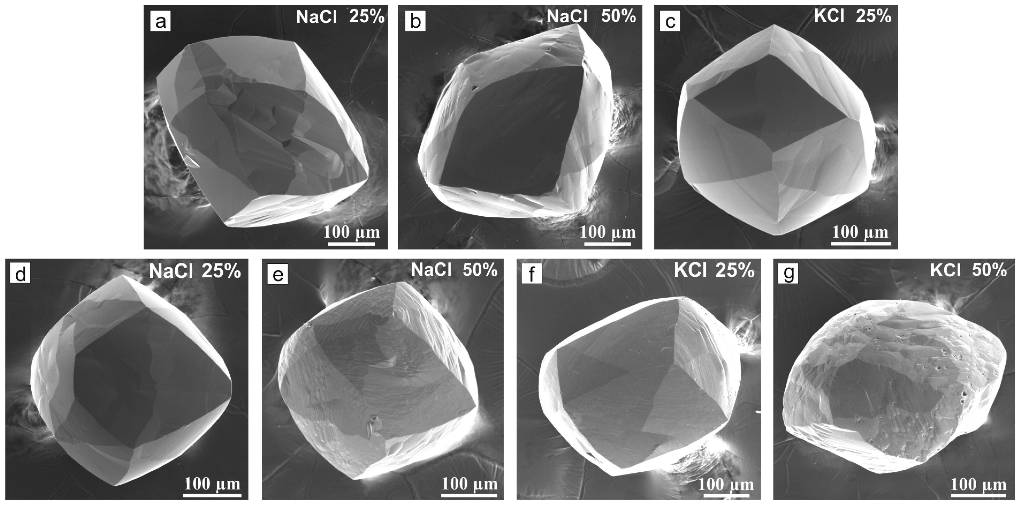
Dissolution of diamond in water and water–chloride systems at fO2 of the MH buffer produced a tetrahexahedroid morphology (Figure 2). The tetrahexahedroids were isometric or more or less elongate. All the original diamonds were isometric. Unfortunately, it was not possible to establish the position of the diamond crystals relative to the walls of the Pt capsules during the experiments.
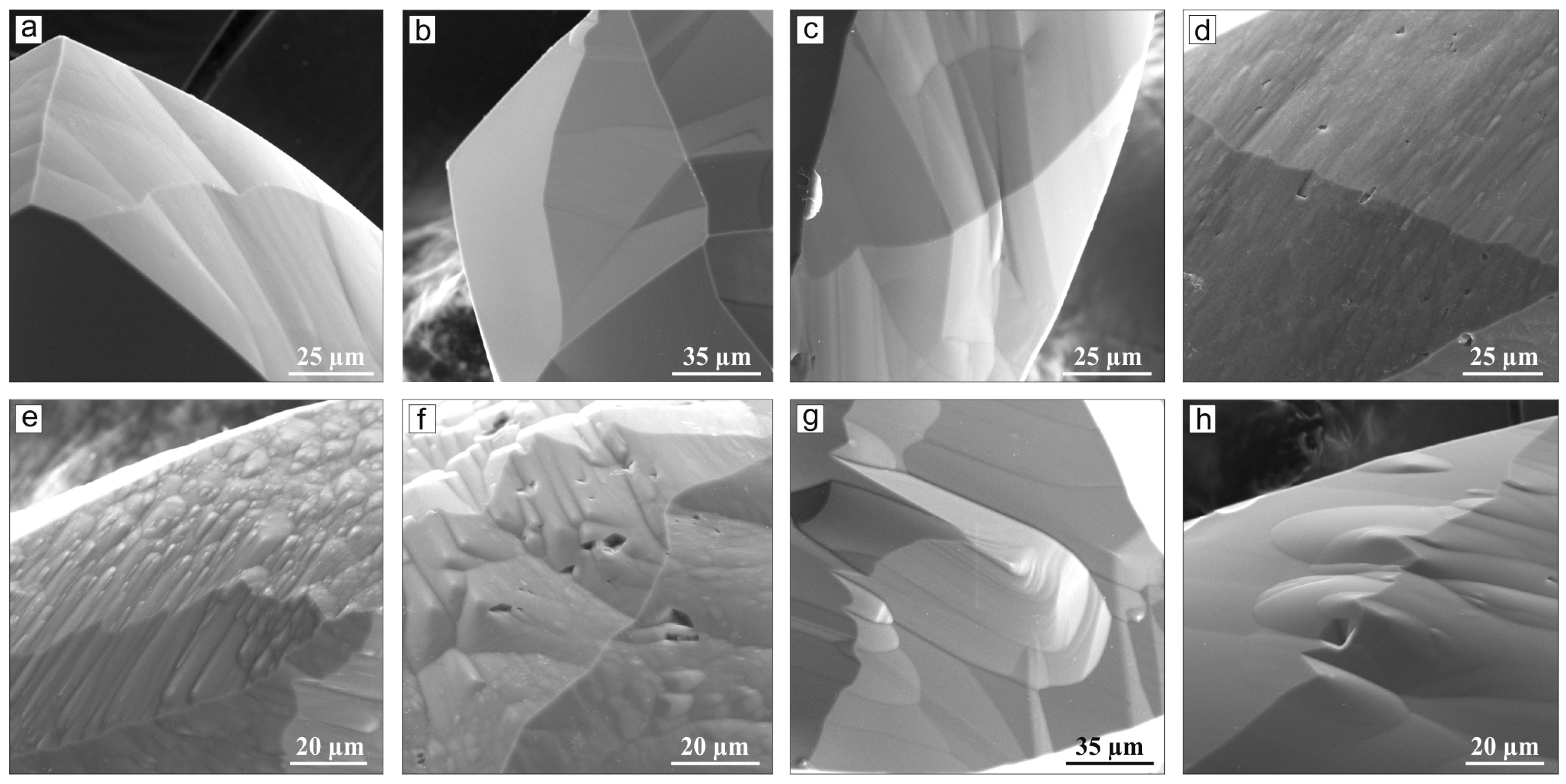
The surface microtopography differed in crystals from different capsules and often even within one crystal. Some faces were perfectly smooth while others were undulated or often covered with rounded drop-shaped or polyhedral hillocks (Figure 3). In the latter case, abundant small hillocks produced shagreen-like textures while larger hillocks formed mosaic patterns (Figure 3e,f). In some crystals, isolated large hillocks appeared along face sutures in the center (Figure 3h). Furthermore, some rounded tetrahexahedroid faces had elongate depressions in the center connecting the fourth-order corners; such depressions were especially prominent in the runs with the H2O solvent (Figure 3c).
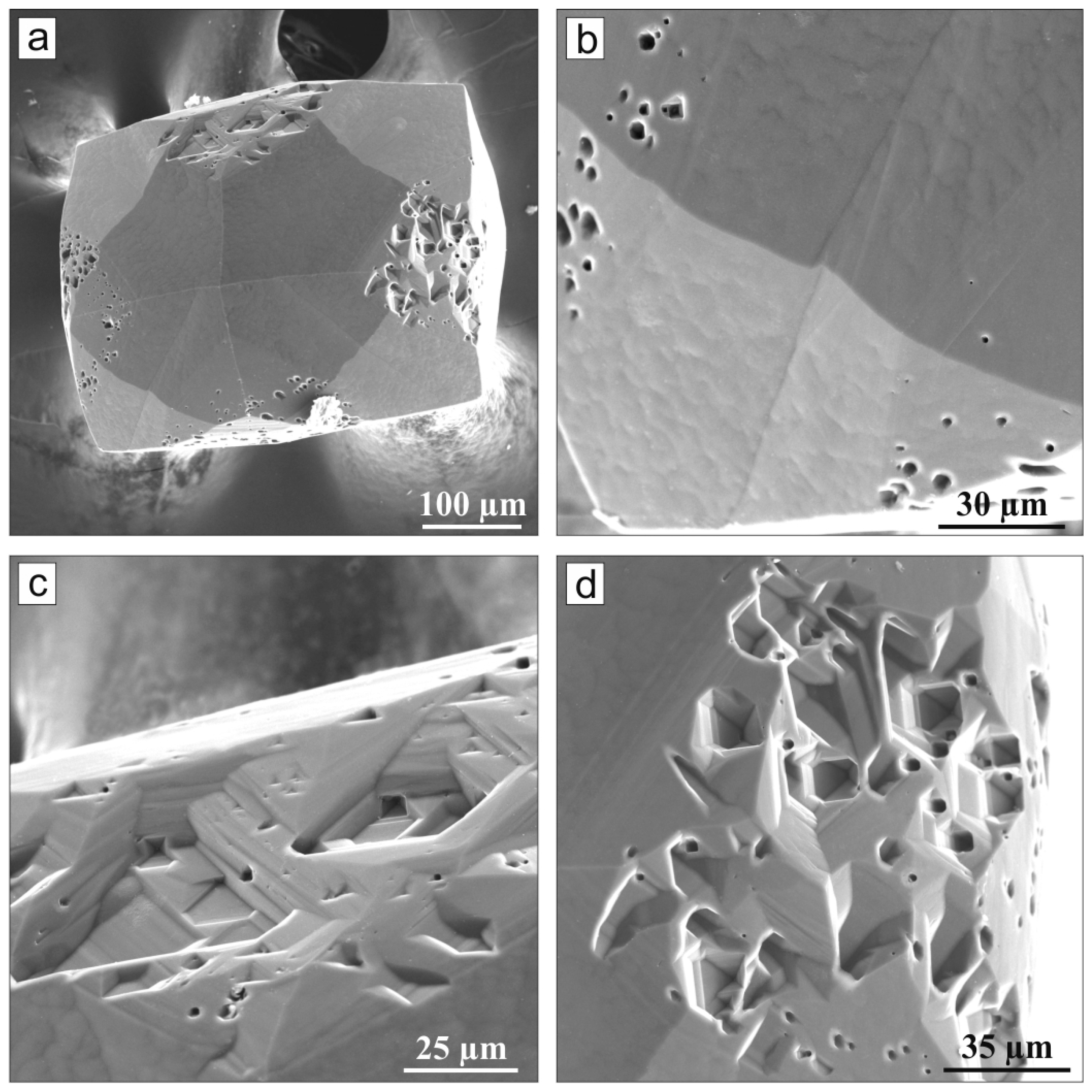
The tetrahexahedroids resulting from resorption of octahedral diamonds in the H2O fluid differed in the presence of numerous hexagonal or triangular pits on octahedral faces (Figure 4), with etch channels stretching towards the center of the crystal along the <100> direction from the pit base or lateral surfaces (Figure 4c,d). Similar etch channels, though smaller and less numerous, were observed in the runs with an H2O/KCl ratio of unity (by mass).
The dissolution rate at redox conditions of the IW buffer was unexpectedly low: the diamond crystals affected by the pure H2O fluid lost only 9% of their weight in a 40 h run at 1200 °C, with the respective dissolution rate being only 0.01 (∆mg/mm2·min1)·103, but dissolution in water–chloride systems was even slower. The weight of the diamonds after the experiments did not change with compositions containing 50% sodium and potassium chlorides (Table 1). Minor dissolution of the diamonds was recorded by changes in the microrelief of the faces. Pyramidal and flat-bottomed triangular etch pits were formed on all faces. (Figure 5). The maximum size of the pits (50 μm) was established in the aqueous fluid. Their size did not exceed 20 μm with a NaCl content of 50 wt% in the fluid, and with a KCl content of 50 wt%, the size of the pits did not exceed 7 μm. Dissolution by the pure H2O fluid led to rounding of the edges and shield-like terracing of the faces of diamond crystals. The initially existing shield-like plates became smaller and acquired better pronounced ditrigonal shapes in the H2O-NaCl system with the content of the component ratio of unity (by mass). Thus, the activity of solvents with respect to diamond decreased in the order of H2O > H2O + NaCl > H2O + KCl in the 1200 °C runs at fO2 of the IW buffer.
The rates of diamond dissolution in H2O became 12 times higher in the 1400 °C runs and reached 0.12 (∆mg/mm2·min1)·103: the crystal lost 28% of its weight in 10 h. Dissolution in chloride-containing aqueous fluids, whether NaCl (50 wt%) or KCl (50 wt%), occurred at a rate of 0.07 (∆mg/mm2 min−1) 103 (Table 1), and the weight loss was 16%–17%.
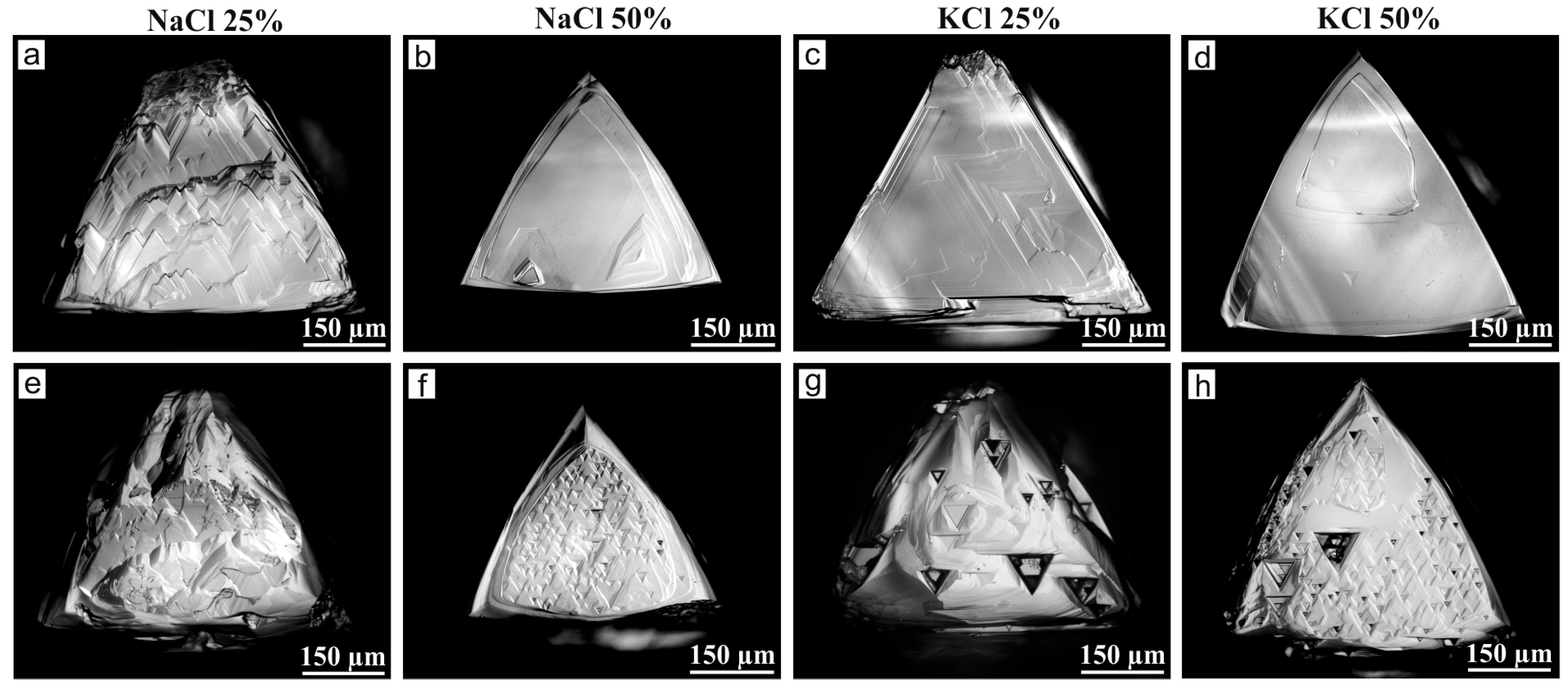
The crystals preserved their octahedral morphology but developed rounded tetrahexahedroid surfaces on the edges. The crystals dissolved in predominantly aqueous systems containing 100 wt.% or 75 wt.% H2O acquired a vicinal structure of relic {111} faces, with vicinal surfaces composed of ditrigonal layers. However, the vicinal features are only detectable by DIC microscopy (Figure 6 and Figure 7) because of the very low angles. The effect of the H2O fluid was quite strong and produced trigonal pits and related deep etch channels cutting the crystal faces into a group of vicinal pyramids (Figure 6). Resorption with chloride fluids was limited to poorly pronounced etching of {111} faces in the case of the H2O-KCl fluid, but no visible etch features were observed in the runs with the H2O-NaCl system (Figure 7).

4. Discussion

It is reasonable to begin the discussion with the experimental results obtained with the pure H2O solvent at different oxygen fugacities. At high fO2 of the MH buffer, the predominance of water in the system provided oxidation of diamond carbon with formation of CO2/CO and hydrogen; hydrogen diffused through the Pt capsule and reacted with the buffer material. The large amount of water and efficient buffering maintained high dissolution rates, up to 12.5 (∆mg/mm2·min−1)·103 at 1400 °C, with a weight loss of 45% from a 0.4 mg crystal for 10 min.
In contrast, dissolution in the pure H2O at reducing conditions of the IW buffer was very slow: a 0.29 mg crystal lost only 28% of its initial weight in 10 h, at a dissolution rate of 0.12 (∆mg/mm2·min1)·103. The low rates can be explained by different dissolution mechanisms. These redox conditions correspond to the stability field of elemental carbon (graphite or diamond). The preservation of diamond crystals in this case is controlled by the shift in the composition fluid from pure H2O and by the possibility of C0 dissolution in the fluid. At IW-buffered fO2, 5.5 GPa, and 1200–1400 °C (logfO2 = −7.8 to −9.8), the fluid equilibrated with diamond has an H2O-CH4 composition [20]. The formation of methane was provided by the reaction of diamond with hydrogen supplied from the external IW buffer. However, the previously observed textures of dissolution in anhydrous systems were mostly of trigonal–trioctahedral morphology [21]. The presence of ditrigonal or shield-like sculptures, and, finally, rounded tetrahexahedroids, typical in diamonds affected by aqueous fluids, indicates the participation of water in the dissolution process.
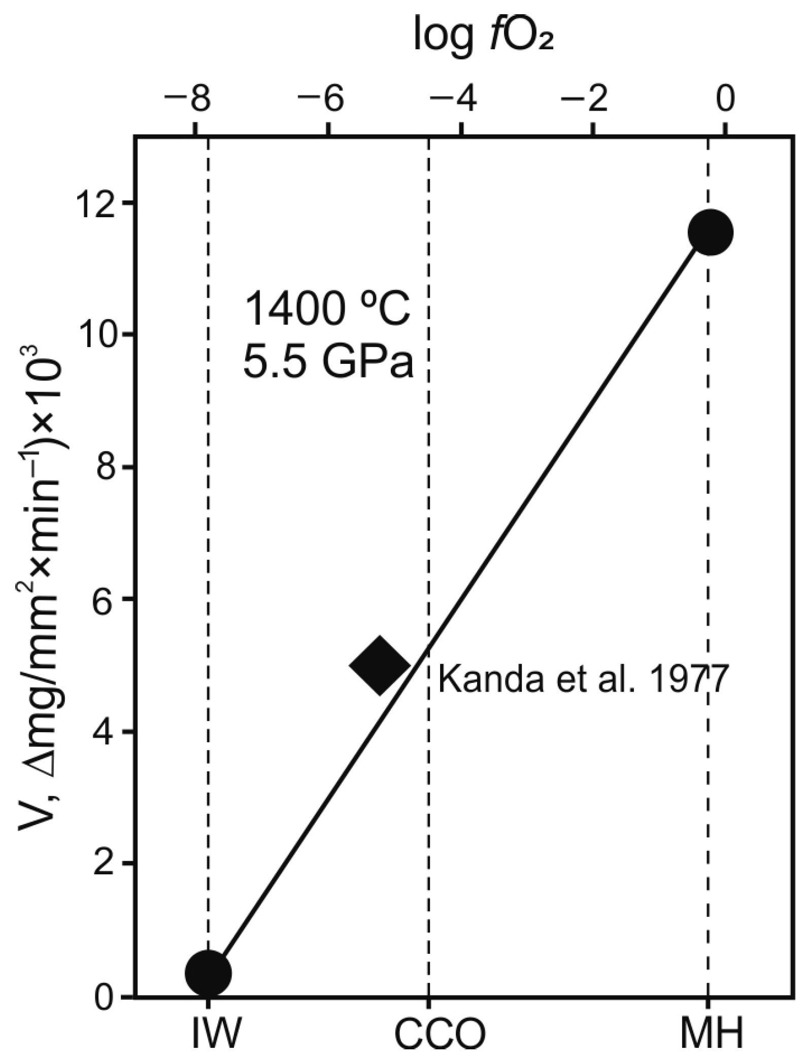
The correlation between dissolution rate and oxygen fugacity obtained by us (Figure 8) is consistent with earlier experimental results using water as an etchant of diamond at temperatures of 1100, 1300, and 1500 °C, as well as at a pressure of 5.0 GPa, without external buffering of fO2 [19]. Oxygen fugacity was apparently between the graphite-CO2-CO (CCO) equilibrium and the water–carbon (WC) maximum, judging by the material of the high-pressure cell and the graphite heater used in those experiments. In such conditions [19], water can dissolve diamond at a rate of ~5.0 (∆mg/mm2·min1)·103.
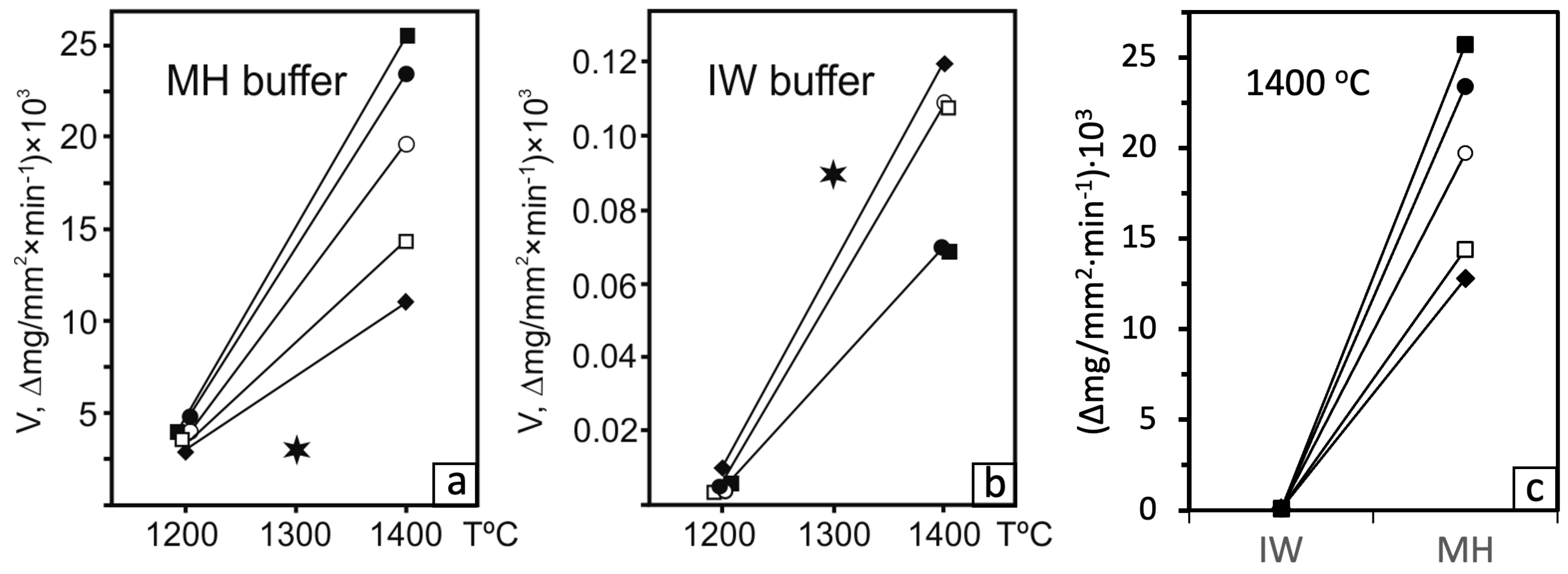
Good agreement was also obtained with the results of Kozai and Arima [22], who studied dissolution of diamond in kimberlite and lamproite melts at 1300 to 1420 °C and 1 GPa under the IW, WM, and MH buffers. Both of our estimates of dissolution rates at 1200 and 1400 °C and fO2 under the MH and IW buffers and those for kimberlite melt solvent containing 6.2 wt% H2O [22] showed slower dissolution at lower oxygen fugacity (Figure 9). However, the dissolution rates in the case of the MH buffer were lower and those at IW buffering were higher [22] than in our experiments with the water–chloride system, possibly due to differences in the composition of solving agents or pressure [18].
Figure 8. The rate of diamond dissolution in H2O fluid as a function of fO2. Solid rhombus [19] and solid circles—our data. The positions of the buffers are given according to the data: IW buffer according to Ballhaus et al. [23], MH buffer according to Kadik and Lukanin [24], and CCO equilibrium according to Frost and Wood [25].
Minerals 15 00897 g008
Figure 9. Temperature dependence of the diamond dissolution rate in the H2O-KCl-NaCl system at fO2 at the level of MH (a), IW (b), and buffers depending on redox conditions at 1400 °C (c). Solid rhombus—pure H2O fluid; solid squares—KCl = 50 wt%; open squares—KCl = 25 wt%; solid circles—NaCl = 50 wt%; open circles—NaCl = 25 wt%. The stars indicate experimental data from [21].
Minerals 15 00897 g009
The presence of chlorides in the system changes the rates of diamond dissolution significantly. The effect of chlorides on the dissolution of diamond can be compared with experimental data of Palyanov et al. [13] on diamond growth in the KCl-H2O-C system. A diamond formation study conducted at higher P-T conditions than our experiments (7.5 GPa and 1500–1600 °C versus 5.5 GPa and 1200–1400 °C) showed the same tendency: changing the fluid composition from pure H2O to a fluid with an H2O/KCl ratio of unity resulted in a 15-fold decrease in the degree of graphite conversion to diamond, from 60% to 4% [9]. In the case of diamond dissolution at fO2 of the IW buffer, the dissolution rate became 1.7 times lower at higher KCl content in the fluid (0.07 against 0.12 (∆mg/mm2·min−1)·103). Thus, the presence of alkali chlorides in the system inhibited both the diamond formation and dissolution processes at reduced conditions.
Conversely, the effect was opposite at oxygen fugacity as high as the MH buffer. Namely, the dissolution rates accelerated considerably at higher contents of chlorides: 1.8 times for KCl and 1.6 times for NaCl. This trend is consistent with the results of experiments on the catalytic activity of alkali metals in diamond oxidation [26,27]. These authors have demonstrated the highest resorption efficiency for transitional catalytic complexes with K ions. The rate of diamond dissolution in water vapor in K-bearing melts was the highest among other alkali metals and was almost twice as high as that for systems with NaCl. The difference was attributed to the proximity of the K ion radius to the length of the diamond C-C bond [26,27].
Dissolution of diamond in all water-bearing systems (aqueous fluids and water-bearing carbonate or silicate melts) is known to produce tetrahexahedroids [16,17,28,29,30,31,32]. Tetrahexahedroids also form in water–chloride systems within a large range of oxygen fugacities, from oxidizing to reducing conditions (MH to IW buffers, respectively). Tetrahexahedroids formed by dissolving diamond in water–chloride systems, in most cases, have smooth surfaces with large single hillocks, or block-type surfaces from a cluster of small hillocks. The surface microtopography of tetrahexahedroids is controlled by the interior structure of diamond crystals and their dissolution conditions. In a review of experimental work on diamond resorption [30], it was concluded that low-relief surfaces of tetrahexahedroids are formed only in an H2O-oversaturated melt at pressures of 1–3 GPa, as shown, for example, in [29]. The results presented in this paper and previously obtained in water-bearing carbonated melts at high oxygen fugacity [16,17] indicate that such surfaces can also form during diamond resorption under mantle conditions. Dissolution of diamond in water-bearing silicate melts usually leads to the formation of a rounded stepped relief on the surfaces—from fine striation to stepped sculptures. The hillocks on rounded surfaces apparently reflect the internal deformation structure of diamond crystals, as shown earlier [33].

5. Conclusions

The obtained experimental results on diamond dissolution in aqueous chloride fluid are of interest to elucidate the role of saline fluids in diamond resorption during mantle metasomatic events, especially shortly before the formation of kimberlite magma. There is increasing evidence that the fluids/melts that provided the metasomatic alteration of the peridotite source of the kimberlite melt contained a significant amount of chlorine. Moreover, they could be a medium for diamond crystallization. This is indicated by diamond fluid inclusions with a high chlorine content [1,2,3,4,5,6], as well as melt inclusions enclosed in kimberlite minerals (they contain 2 to 55 vol.% halite and sylvite) [34]. This may also be supported by high chlorine concentrations (up to several wt.%) in kimberlite from the Udachnaya–Vostochnaya Devonian tube in Siberia, which was not affected by postmagmatic changes [35]. The data obtained allow us to conclude that the low oxygen fluidity near the IW buffer should ensure the survival of diamonds in the mantle even in the presence of highly saline fluids. Intensive dissolution of diamond by H2O-NaCl-KCl fluids under oxidizing conditions may be the cause of uneconomic kimberlites. On the other hand, saline fluids can maintain the integrity of diamonds that have undergone mantle metasomatism under reducing conditions. At the present stage of the study, it is still difficult to draw final conclusions about the effect of chlorine complexes on the rate of diamond resorption. To obtain reliable information on this issue, further studies are needed on diamond dissolution in aqueous fluid containing other anionic complexes, such as [CO]−2, [SiO4]−4, etc.

Author Contributions

Conceptualization, A.K. (Alexander Khokhryakov) and A.S.; methodology, A.S.; validation, A.K. (Alexander Khokhryakov) and A.K. (Alexey Kruk); formal analysis, A.K. (Alexander Khokhryakov) and D.N.; investigation, A.K. (Alexey Kruk) and D.N.; writing—original draft preparation, A.S. and A.K. (Alexander Khokhryakov); writing—review and editing, A.S. and A.K. (Alexander Khokhryakov); visualization, A.K. (Alexander Khokhryakov) and D.N.; project administration, A.K. (Alexey Kruk); funding acquisition, A.K. (Alexey Kruk). All authors have read and agreed to the published version of the manuscript.

Funding

This research was funded by the Russian Science Foundation, grant number 24-77-10006.

Data Availability Statement

The original contributions presented in this study are included in the article. Further inquiries can be directed to the corresponding author.

Acknowledgments

The SEM studies of experimental samples were performed at the Analytical Center for Multi-elemental and Isotope Research (IGM SB RAS, Novosibirsk). The authors did not use GenAI during the preparation of this manuscript/study.

Conflicts of Interest

All authors declare that the research was conducted in the absence of any commercial or financial relationships that could be construed as a potential conflict of interest.

Abbreviations

The following abbreviations are used in this manuscript:
HDFHigh-density fluid
DICDifferential-interference contrast
SEMScanning electron microscope

References

  1. Jablon, B.M.; Navon, O. Most diamonds were created equal. Earth Planet. Sci. Let. 2016, 443, 41–47. [Google Scholar] [CrossRef]
  2. Navon, O. Diamond formation in the Earth’s mantle. Int. Kimberl. Conf. Ext. Abstr. 1998, 7, 618–621. [Google Scholar]
  3. Schrauder, M.; Navon, O.; Szafranek, D.; Kaminsky, F.; Galimov, E. Fluids in Yakutian and Indian diamonds. Mineral. Mag. 1994, 58, 813–814. [Google Scholar] [CrossRef]
  4. Izraeli, E.S.; Harris, J.W.; Navon, O. Brine inclusions in diamonds: A new upper mantle fluid. Earth Planet. Sci. Lett. 2001, 187, 323–332. [Google Scholar] [CrossRef]
  5. Izraeli, E.S.; Harris, J.W.; Navon, O. Fluid and mineral inclusions in cloudy diamonds from Koffiefontein, South Africa. Geochim. Cosmochim. Acta 2004, 68, 2561–2575. [Google Scholar] [CrossRef]
  6. Tomlinson, E.L.; Jones, A.P.; Harris, J.W. Co-existing fluid and silicate inclusions in mantle diamond. Earth Plan. Sci. Lett. 2006, 250, 581–595. [Google Scholar] [CrossRef]
  7. Weiss, Y.; Navon, O.; Goldstein, S.L.; Harris, J.W. Inclusions in diamonds constrain thermo-chemical conditions during Mesozoic metasomatism of the Kaapvaal cratonic mantle. Earth Planet. Sci. Let. 2018, 491, 134–147. [Google Scholar] [CrossRef]
  8. Klein-BenDavid, O.; Izraeli, E.S.; Hauri, E.; Navon, O. Fluid inclusions in diamonds from the Diavik mine, Canada and the evolution of diamond-forming fluids. Geochim. Cosmochim. Acta 2007, 71, 723–744. [Google Scholar] [CrossRef]
  9. Weiss, Y.; McNeill, J.; Pearson, D.G.; Nowell, G.M.; Ottley, C.J. Highly saline fluids from a subducting slab as the source for fluid-rich diamonds. Nature 2015, 524, 339–342. [Google Scholar] [CrossRef]
  10. Wang, Y.; Kanda, H. Growth of HPHT diamonds in alkali halides: Possible effects of oxygen contamination. Diam. Relat. Mater. 1998, 7, 57–63. [Google Scholar] [CrossRef]
  11. Litvin, Y.A. Alkaline-chloride components in processes of diamond growth in the mantle and high-pressure experimental conditions. Dokl. Earth Sci. 2003, 389, 388–391. [Google Scholar]
  12. Tomlinson, E.; Jones, A.; Milledge, J. High-pressure experimental growth of diamond using C–K2CO3–KCl as an analogue for Cl-bearing carbonate fluid. Lithos 2004, 77, 287–294. [Google Scholar] [CrossRef]
  13. Palyanov, Y.N.; Shatsky, V.S.; Sobolev, N.V.; Sokol, A.G. The role of mantle ultrapotassic fluids in diamond formation. Proc. Natl. Acad. Sci. USA 2007, 104, 9122–9127. [Google Scholar] [CrossRef]
  14. Luth, R.W. Natural versus experimental control of oxidation state; effects on the composition and speciation of COH fluids. Am. Min. 1989, 74, 50–57. [Google Scholar]
  15. Boettcher, A.L.; Mysen, B.O.; Allen, J.C. Techniques for the control of water fugacity and oxygen fugacity for experimentation in solid-media high-pressure apparatus. J. Geophys. Res. 1973, 78, 5898–5901. [Google Scholar] [CrossRef]
  16. Khokhryakov, A.F.; Palyanov, Y.N. Influence of the fluid composition on diamond dissolution forms in carbonate melts. Am. Mineral. 2010, 95, 1508–1514. [Google Scholar] [CrossRef]
  17. Khokhryakov, A.F.; Kruk, A.N.; Sokol, A.G. The effect of oxygen fugacity on diamond resorption in ascending kimberlite melt. Lithos 2021, 394–395, 106166. [Google Scholar] [CrossRef]
  18. Khokhryakov, A.F.; Kruk, A.N.; Sokol, A.G.; Nechaev, D.V. Experimental modeling of diamond resorption during mantle metasomatism. Minerals 2022, 12, 414. [Google Scholar] [CrossRef]
  19. Kanda, H.; Yamaoka, S.; Setaka, N.; Komatsu, H. Etching of diamond octahedrons by pressure water. J. Cryst. Growth 1977, 38, 1–7. [Google Scholar] [CrossRef]
  20. Stachel, N.; Luth, R.W. Diamond—Where, when and how? Lithos 2015, 200–223, 200–220. [Google Scholar] [CrossRef]
  21. Khokhryakov, A.F.; Pal’yanov, Y.N.; Sobolev, N.V. Crystal morphology as an indicator of redox conditions of natural diamond dissolution at the mantle PT parameters. In Doklady Earth Sciences; Pleiades Publishing, Ltd.: Moscow, Russia, 2002; Volume 385, pp. 534–537, (Translated from Doklady Akademii Nauk 2002, 384, 670–673). [Google Scholar]
  22. Kozai, Y.; Arima, M. Experimental study on diamond dissolution in kimberlitic and lamproitic melts at 1300–1420 °C and 1 GPa with controlled oxygen partial pressure. Am. Mineral. 2005, 90, 1759–1766. [Google Scholar] [CrossRef]
  23. Ballhaus, C.; Berry, R.F.; Green, D.H. High pressure experimental calibration of the olivine-orthopyroxene-spinel oxygen geobarometer: Implications for the oxidation state of the upper mantle. Contrib. Mineral. Petrol. 1991, 107, 27–40. [Google Scholar] [CrossRef]
  24. Kadik, A.A.; Lukanin, O.A. Degassing of the Upper Mantle During Melting; Nauka: Moscow, Russia, 1986; 120p. (In Russian) [Google Scholar]
  25. Frost, D.J.; Wood, B.J. Experimental measurements of the graphite C−O equilibrium and CO2 fugacities at high temperature and pressure. Contrib. Mineral. Petrol. 1995, 121, 303–308. [Google Scholar] [CrossRef]
  26. Rudenko, A.P.; Kulakova, I.I.; Shturman, V.L. Oxidation of natural diamond. In Novye Dannye o Mineralogii SSSR; Nauka: Moscow, Russia, 1979; Volume 28, pp. 105–125. (In Russian) [Google Scholar]
  27. Skvortsova, V.L.; Shiryaev, A.A.; Fedortchouk, Y. Influence of ions on diamond resorption. Diam. Relat. Mater. 2020, 104, 107764. [Google Scholar] [CrossRef]
  28. Khokhryakov, A.F.; Palyanov, Y.N. The evolution of diamond morphology in the process of dissolution: Experimental data. Am. Mineral. 2007, 92, 909–917. [Google Scholar] [CrossRef]
  29. Fedortchouk, Y.; Canil, D.; Semenets, E. Mechanisms of diamond oxidation and their bearing on the fluid composition in kimberlite magmas. Am. Mineral. 2007, 92, 1200–1212. [Google Scholar] [CrossRef]
  30. Fedortchouk, Y. A new approach to understanding diamond surface features based on a review of experimental and natural diamond studies. Earth-Sci. Rev. 2019, 193, 45–65. [Google Scholar] [CrossRef]
  31. Palyanov, Y.N.; Khokhryakov, A.F.; Kupriyanov, I.N. Crystallomorphological and crystallochemical indicators of diamond formation conditions. Crystallogr. Rep. 2021, 66, 142–155. [Google Scholar] [CrossRef]
  32. Luth, R.W.; Palyanov, Y.N.; Bureau, H. Experimental petrology applied to natural diamond growth. Rev. Mineral. Geochem. 2021, 88, 755–808. [Google Scholar] [CrossRef]
  33. Khokhryakov, A.F.; Palyanov, Y.N. Effect of crystal defects on diamond morphology during dissolution in the mantle. Am. Mineral. 2015, 100, 1528–1532. [Google Scholar] [CrossRef]
  34. Golovin, A.V.; Kamenetsky, V.S. Compositions of kimberlite melts: A review of melt inclusions in kimberlite minerals. Petrology 2023, 31, 143–178. [Google Scholar] [CrossRef]
  35. Kamenetsky, V.S.; Kamenetsky, M.B.; Golovin, A.V.; Sharygin, V.V.; Maas, R. Ultrafresh salty kimberlite of the Udachnaya–East pipe (Yakutia, Russia): A petrological oddity or fortuitous discovery? Lithos 2012, 152, 173–186. [Google Scholar] [CrossRef]
Figure 1. Cell assembly of high-pressure experiments, with oxygen fugacity buffering at the IW (a) and MH (b) equilibria: 1—outer Fe0 capsule; 2—talc; 3—CsCl; 4—inner Pt capsule with diamond and H2O ± NaCl ± KCl mixture; 5—outer Pt capsule; 6—Fe2O3 + H2O (10 wt%).
Minerals 15 00897 g001
Figure 2. SEM images of diamonds resorbed in H2O-NaCl-KCl systems at the MH oxygen buffer: (ac) run 95/4; (dg) run 37/8.
Minerals 15 00897 g002
Figure 3. SEM images of tetrahexahedroid textures produced by dissolution in H2O-NaCl-KCl systems at the MH oxygen buffer: (a) undulated face, run 95/4—KCl 25 wt%; (b) smooth face, run 95/4—NaCl 25 wt%; (c) face with elongate depression, run 95/4—NaCl 50 wt%; (d) fine shagreen texture, run 37/8—KCl 25 wt%; (e) shagreen-like texture, run 37/8—NaCl 50 wt%; (f) mosaic pattern, run 37/8—KCl 50 wt%; (g) single large hillock, run 95/4—NaCl 25 wt%; (h) isolated large hillocks along face suture, run 95/4—NaCl 50 wt%.
Minerals 15 00897 g003
Figure 4. Morphology of etch features on diamond crystals affected by an H2O solvent at the MH oxygen buffer and 1200 °C (run 95/4): (a) general view of the crystal; (b) surfaces with elongated depressions; (c,d) hexagonal pits and etching channels on relict octahedral faces.
Minerals 15 00897 g004
Figure 5. DIM images of {111} faces in a diamond: (ac) initial surfaces; (df) same faces after 1200 °C run at the IW oxygen buffer (run 67/3).
Minerals 15 00897 g005
Figure 6. Morphology of diamond crystals affected by H2O solvent at the IW oxygen buffer and 1400 °C (run 40/8): (a) SEM image; (b,c) DIM images of face {111} before and after run, respectively.
Minerals 15 00897 g006
Figure 7. DIM images of {111} faces before and after the run: (ad) initial surfaces; (eh) same faces after 1400 °C run at the IW oxygen buffer (run 40/8).
Minerals 15 00897 g007
Table 1. Experimental results on diamond dissolution (P = 5.5 GPa).
Run No.BufferT (°C)Time
(h)
Composition (mg)Diamond
H2OKClNaClInitial Weight (mg)Final Weight mg)Weight Loss (mg)Weight Loss (%)V 1
95/4MH120015.0--0.340.130.21622.9
3.7-1.30.320.050.27843.9
2.5-2.50.410.020.39954.7
3.71.3-0.380.110.27713.5
2.52.5-0.3500.35100≥5.0
37/8MH14000.175.0--0.400.220.184512.8
3.7-1.30.430.150.286519.7
2.5-2.50.380.080.307823.4
3.71.3-0.450.240.215314.4
2.52.5-0.360.040.328925.7
39/8IW1200105.0--0.400.40≤0.40≥0≥0
2.5-2.50.380.38000
3.7-1.30.260.26000
3.71.3-0.320.32000
2.52.5-0.350.35000
67/3IW1200405.0--0.350.320.0390.01
3.7-1.30.320.310.013≤0.01
3.7-2.50.26≤0.26≥0≥0≤0.01
2.51.3-0.35≤0.35≥0≥0≤0.01
2.52.5-0.30≤0.30≥0≥0≤0.01
40/8IW1400105.0--0.290.210.08280.12
3.7-1.30.350.270.08230.11
2.5-2.50.300.250.05170.07
3.71.3-0.290.220.07240.11
2.52.5-0.320.270.05160.07
1 Specific dissolution rate (∆mg/mm2·min−1) × 103.
Disclaimer/Publisher’s Note: The statements, opinions and data contained in all publications are solely those of the individual author(s) and contributor(s) and not of MDPI and/or the editor(s). MDPI and/or the editor(s) disclaim responsibility for any injury to people or property resulting from any ideas, methods, instructions or products referred to in the content.