Article

Kinetic Aspects of Chrysotile Asbestos Thermal Decomposition Process

1
Łukasiewicz Research Network—Institute of Ceramics and Building Materials, Cementowa 8, 31-983 Cracow, Poland
2
Department of Biology, Ecology and Earth Sciences, University of Calabria, Via P. Bucci, 15B, 87036 Arcavacata di Rende, Italy
*
Author to whom correspondence should be addressed.
Minerals 2025, 15(6), 609; https://doi.org/10.3390/min15060609
Submission received: 22 April 2025 / Revised: 2 June 2025 / Accepted: 3 June 2025 / Published: 5 June 2025

Abstract

:
Growing requirements in the field of environmental protection and waste management result in the need to search for new and effective methods of recycling various types of waste. From the perspective of technical and natural sciences, the disposal of hazardous waste, which can lead to environmental degradation, is of utmost importance. A particularly hazardous waste is asbestos, used until recently in many branches of the economy and industry. Despite the ban on the production and use of asbestos introduced in many countries, products containing it are still present in the environment and pose a real threat. This paper presents the results of research related to the process of asbestos neutralization, especially the chrysotile variety, by the thermal decomposition method. Changes in the mineralogical characteristics of asbestos waste were studied using the following methods: TG-DTA-EGA, XRD, SEM-EDS and XRF. The characteristics of the chrysotile asbestos sample were determined before and after thermal treatment at selected temperatures. The second part of the study focuses on the kinetic aspect of this process, where the chrysotile thermal decomposition process was measured by two techniques: ex situ and in situ. This study showed that the chrysotile structure collapsed at approximately 600–800 °C through dehydroxylation, and then the fibrous chrysotile asbestos was transformed into new mineral phases, such as forsterite and enstatite. The formation of forsterite was observed at temperatures below 1000 °C, while enstatite was created above this temperature. From the kinetic point of view, the chrysotile thermal decomposition process could be described by the Avrami–Erofeev model, and the calculated activation energy values were ~180 kJ mol−1 and ~220 kJ mol−1 for ex situ and in situ processes, respectively. The obtained results indicate that the thermal method can be successfully used to detoxify hazardous chrysotile asbestos fibers.

Graphical Abstract

1. Introduction

Asbestos is an industry term for a group of natural fibrous minerals that were used in industrial products. We distinguish here six types of asbestos minerals: chrysotile, actinolite, amosite, anthophyllite, crocidolite and tremolite. From the chemical point of view, all asbestos minerals are hydrated silicates of different metals, mainly magnesium, calcium, iron and sodium [1,2]. Although asbestos was a popular material in the past and was often used in the broader construction industry, it is now known that asbestos fibers have harmful effects on human health.
The first article on asbestos-related disease was published as early as 1924, and in the following years, research presented asbestos as a material whose properties can lead to pulmonary asbestosis and other deadly diseases, including cancer. Significant scientific evidence linking asbestos to lung cancer and mesothelioma was not obtained until the 1970s [3]. As a result, asbestos was designated as a Group 1 carcinogen by the International Agency for Research on Cancer (IARC) in 1987 [4,5,6].
When it was proven that asbestos is the cause of different diseases, the manufacturing and application of asbestos were outlawed [7,8]. Since then, directives have been successively introduced, the purpose of which was first to end the extraction of asbestos minerals, then the fabrication of asbestos materials, and finally, the complete removal of asbestos materials from the environment. Today, in more than 70 countries worldwide [7], the first two stages have been completed, while the third stage is still ongoing. Due to the inefficiency of current asbestos removal methods—primarily relying on controlled landfilling [9,10,11], which, while common and cost-effective, merely postpones rather than eliminates the risks associated with asbestos fiber release—this disposal strategy increasingly conflicts with sustainability goals and with the growing awareness of raw material depletion in the construction industry. As a result, efforts have recently intensified on two fronts. On one hand, there is a push toward more effective recycling methods; on the other, there is an increasing attempt to implement strategies aligned with the principles of the circular economy. Work on neutralizing asbestos fibers has been underway since the 1980s of the twentieth century, but widespread use for commercial purposes has not occurred due to regulations that have become increasingly stringent. Nevertheless, work on the development of a technology for the full-scale, commercial processing of asbestos materials is still ongoing, using various technologies. Among the proposed laboratory-scale methods are biological treatment, acid dissolution, and mechanical grinding [12,13]. In particular, physical treatments such as milling, while potentially effective, are associated with high energy demands, which significantly increase both operational costs and environmental impact [13]. Moreover, chemical processes demand large quantities of reagents, which can pose challenges from both an economic and ecological perspective. Bioremediation of asbestos-containing waste (ACW) also shows limited efficiency, making it difficult to achieve rapid and sustainable results. Finally, the management of end-products resulting from chemical and biological treatments remains a critical issue, as these materials must be handled with care to avoid potential risks to human health and the environment [13]. Among the available technologies, particular attention is paid to thermal treatment technologies, where, as it has been shown, it leads to the neutralization of carcinogenic properties of asbestos materials and the production of minerals that can successfully constitute a building raw material, building material or mineral binders [14,15,16,17,18,19,20,21,22,23]. Asbestos minerals are typically relatively stable under standard conditions; however, they can undergo structural or compositional changes when subjected to elevated temperatures or other aggressive conditions [24]. High temperatures favor the release of water molecules from the crystal structure, leading to its transformation into a more stable form under extreme conditions. Importantly, these changes are not identical for all asbestos varieties [25,26,27,28].
In this article, we focused on the most popular mineral among asbestos materials, which is chrysotile (with ideal chemical formula Mg3(OH)4Si2O5), which belongs to the group of serpentine asbestos, and its share in the industry compared to other asbestos was as much as 96% [1,29]. It is worth emphasizing that chrysotile asbestos (commonly known as white asbestos) is the only asbestos that is still mined. Worldwide consumption of asbestos fiber in 2015 was about 1.1 million tons per year, reaching a value of around 1.4 million tons per year in 2024 [30]. It is mined mainly by Russia, Kazakhstan, China and Brazil.
Since chrysotile asbestos belongs to the group of hydrated silicates, its thermal decomposition occurs in three main stages upon heating to a sufficiently high temperature [24]. First, at temperatures ranging from approximately 50 °C to even 600 °C, they lose water bound to the surface of the fibers through dehydration, but most of the water loss occurs at 200–300 °C [1,15,24,25,26,31,32,33]. Secondly, at a temperature of approximately 400–700 °C, chemically bound water (resulting from the dehydroxylation process) is released from the mineral structure. This stage is considered the most critical in the context of chrysotile asbestos thermal degradation. This process contributes to the complete breakdown of the mineral structure and the formation of an anhydrous phase [24,34,35,36]. The release of structured water leads to an irreversible deterioration of the strength properties of the fibers, which become brittle and easily break apart. The final step is the transformation of the material into new mineral phases [24,31,34,37] when the amorphous structure crystallizes. The first major product of this transformation is forsterite (Mg2SiO4, created from Mg-rich regions) and amorphous silica. The second is enstatite (MgSiO3), formed at a higher temperature when the temperature reaches 1000 °C or more [38]. Neither minerals exhibit carcinogenic properties [39,40]. Sometimes, distributions of impurities present in the chrysotile sample are also observed. They can be derived from brucite, organic impurities, and other compounds present in the sample [26,41].
As already mentioned, the main reaction of chrysotile under high temperatures is dehydroxylation, and this process can be studied from a kinetic perspective to understand how factors like time, temperature and others influence its rate. The rate of this process is influenced by several factors, and understanding this is crucial in terms of both industrial processes and safety regarding asbestos exposure. This study presents the first systematic investigation of the thermal decomposition kinetics of P-3-50-grade chrysotile-asbestos historically sourced from Kazakhstan and employed in electrolytic diaphragm production at the “Zachem” Chemical Plant (Bydgoszcz, Poland). The comprehensive kinetic analysis offers novel insights into a previously uncharacterized industrial material. Despite its industrial and historical significance, this specific material had never been subjected to such a detailed and systematic thermal analysis.
The novelty of the research lies not only in the unique origin and application of the sample but also in the dual-method approach adopted for its analysis. The isothermal decomposition process was investigated using two complementary techniques: ex situ thermal annealing in a laboratory furnace and in situ measurements carried out with a high-temperature X-ray diffraction (XRD) chamber. This integrated methodological framework has enabled a comprehensive understanding of the kinetic behavior of chrysotile asbestos during thermal decomposition, providing valuable new insights into a material of significant environmental and technological importance.

2. Materials and Methods

The material used in this study was a commercial chrysotile asbestos sample. The asbestos used was of P-3-50 grade (semi-rigid, long-fiber), supplied from Kazakhstan (previous USSR) to the “Zachem” Chemical Plant in Bydgoszcz (Poland) in the past, at the turn of the 20th and 21st centuries. It was used to produce electrolytic diaphragms in chlor-alkali technologies.
The characteristic of the asbestos specimen was first examined, and then the kinetic investigation was conducted. Various analytical methods were used to investigate properties of material before and after heat treatment. To this aim, the raw chrysotile asbestos sample was studied by thermal analysis with evolved gas analysis (DTA/TG/DTG-EGA), X-ray powder diffraction (XRD) as well as scanning electron microscopy with energy-dispersive X-ray spectroscopy (SEM-EDS). Moreover, chemical analysis was also performed by X-ray fluorescence spectroscopy (XRF). In addition, isothermal calcinations for 2 h of asbestos sample at different temperatures ranging from 500 to 1200 °C with step 50 °C were also performed.
The temperature-induced kinetic reaction of the chrysotile asbestos was studied by two methods. In the first, the ex situ experiment was conducted, where asbestos (initial mass of material was 5 g) was isothermally calcined in a ceramic crucible at selected temperatures—550, 575, 600, 625, 650, 675 and 700 °C—recording changes of mass at time intervals until a constant mass had been achieved. This isothermal treatment was performed in a laboratory electric furnace LT9/14 with B510 heating curve controller (Nabertherm, Lilienthal, Germany). The measurement of mass change was performed on a laboratory assay balance ALN220G (Axis, Gdańsk, Poland).
The second scheduled method was the in situ experiment by a conventional X-ray measurement in a heating chamber. In this case, isothermal runs for kinetic analysis in the angular ranges 10–14 and 22–27° 2Theta, in order to follow the decrease in the intensities of the main chrysotile reflections, were measured. We assumed that the progress of the reaction would be monitored based on the loss of the sum of the surface areas of both reflections in relation to area of raw chrysotile sample. The changes in these reflections were followed in isothermal conditions at 650, 700, 725, 750, 775, 800 and 825 °C. A chrysotile asbestos sample (starting amount of material was 0.2 g) was placed in an alumina pad heated by a platinum plate as a heating element of the heating chamber.
The chemical analysis supported by the L.O.I. (loss on ignition; determined by isothermal treatment at 1025 °C to constant mass) of the samples was performed using a Panalytical Magix PW-2424 spectrometer (Malvern PANalytical, Almelo, The Netherlands) following the fused cast-bead method described in PN-EN ISO 12677:2011 standard [42]. Simultaneous differential thermal analysis (DTA), thermogravimetric analysis (TG), differential thermogravimetric analysis (DTG), and measurement of gases evolved from the samples (EGA) were performed using a STA 409 PC Luxx thermal analyzer coupled with QMS 403 C Aeolos quadrupole mass spectrometer (Netzsch, Selb, Germany). All experiments were performed up to 1000 °C in the airflow of 30 mL min−1 at a heating rate of 20 °C min−1 using an open alumina crucible with 100 mg of sample. Microstructure by SEM analysis was made using a Mira 3 electron microscope (Tescan, Brno, Czech Republic) equipped with an AZtec Automated 3.1 EDS system (Oxford Instruments, Abingdon, UK). The analysis was performed at an accelerating voltage of 15 kV in the secondary electron (SE) mode. The asbestos sample was coated with a chromium layer by the Quorum Q150R ES sputter coaters (Quorum Technologies, Laughton, UK). The mineralogical phase study was obtained by X-ray powder diffraction using PANalytical X’pert Pro diffractometer, with copper Kα radiation, nickel filter, 40 kV, 30 mA, X’Celerator detector (Malvern PANalytical, Almelo, The Netherlands). The in situ high-temperature diffraction experiments were conducted using the same diffractometer equipped with an Anton Paar HTK2000 chamber (Anton Paar, Ashland, VA, USA). The diffraction patterns were analyzed using HighScore Plus software (version 5.2; Malvern PANalytical, Almelo, The Netherlands) and the ICDD PDF-5+ database.
All sample handling and preparation procedures were conducted in accordance with established safety protocols for hazardous airborne particulates. Specifically, operations involving raw or milled asbestos materials were carried out within a certified glove box equipped with HEPA filtration to prevent fiber release into the laboratory environment [43]. Personnel wore appropriate personal protective equipment (PPE), including disposable lab coats, nitrile gloves, and P3-rated respirators. Waste materials were collected in sealed, labeled containers and disposed of according to local regulations for hazardous waste. Work surfaces were regularly cleaned using wet-wipe methods to minimize dust dispersion, and air monitoring was performed periodically to ensure compliance with occupational exposure limits.

3. Results and Discussion

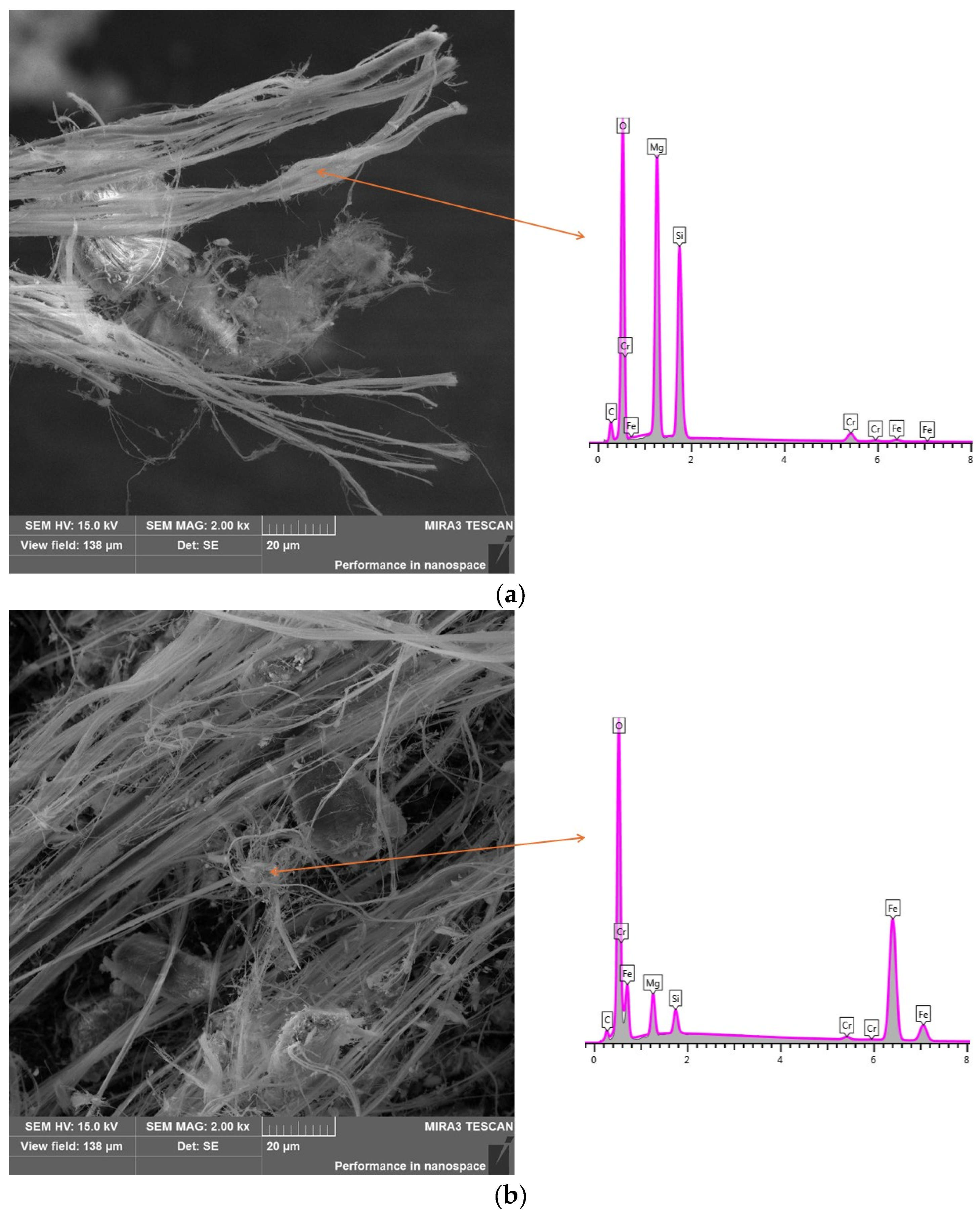
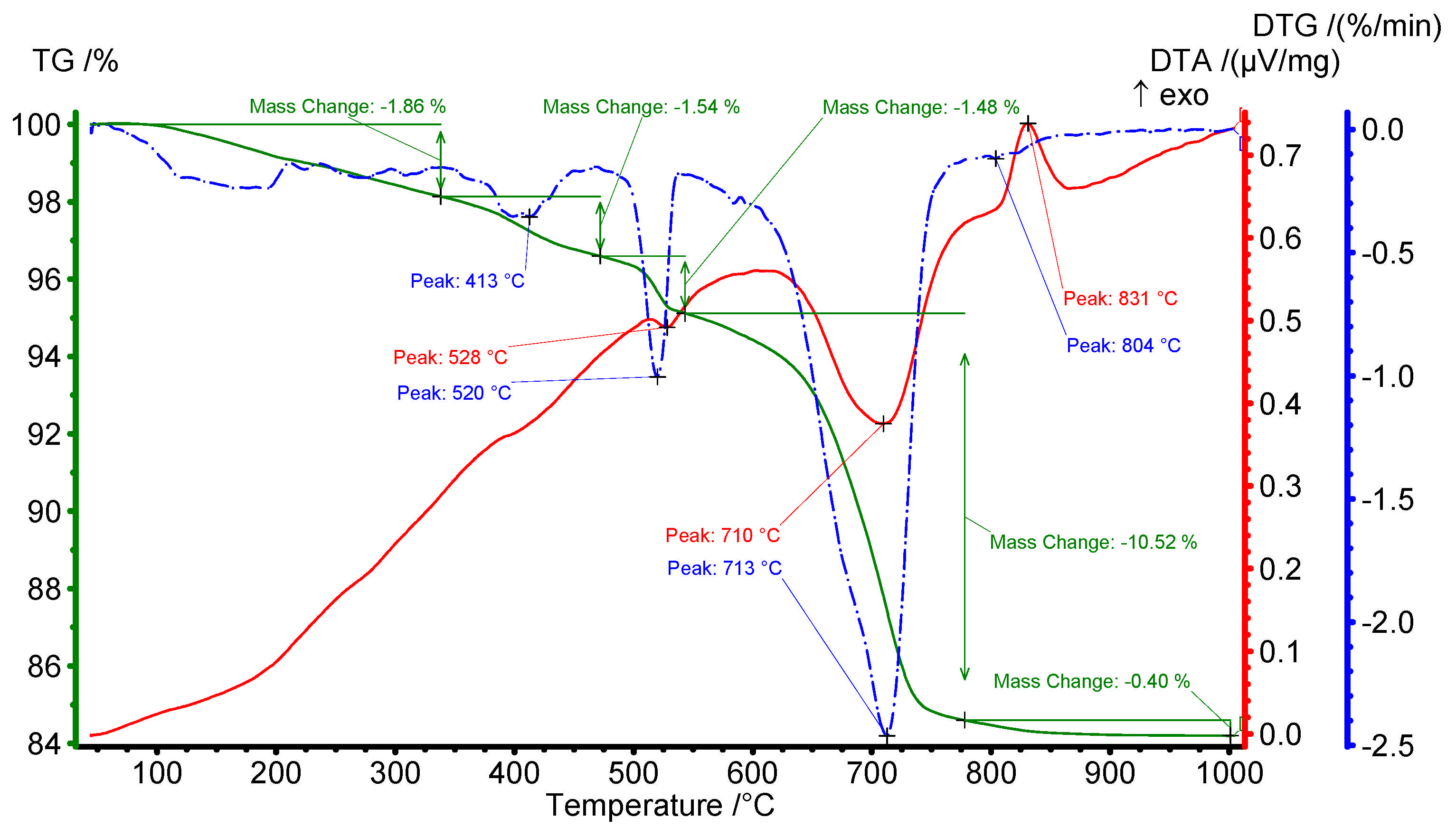
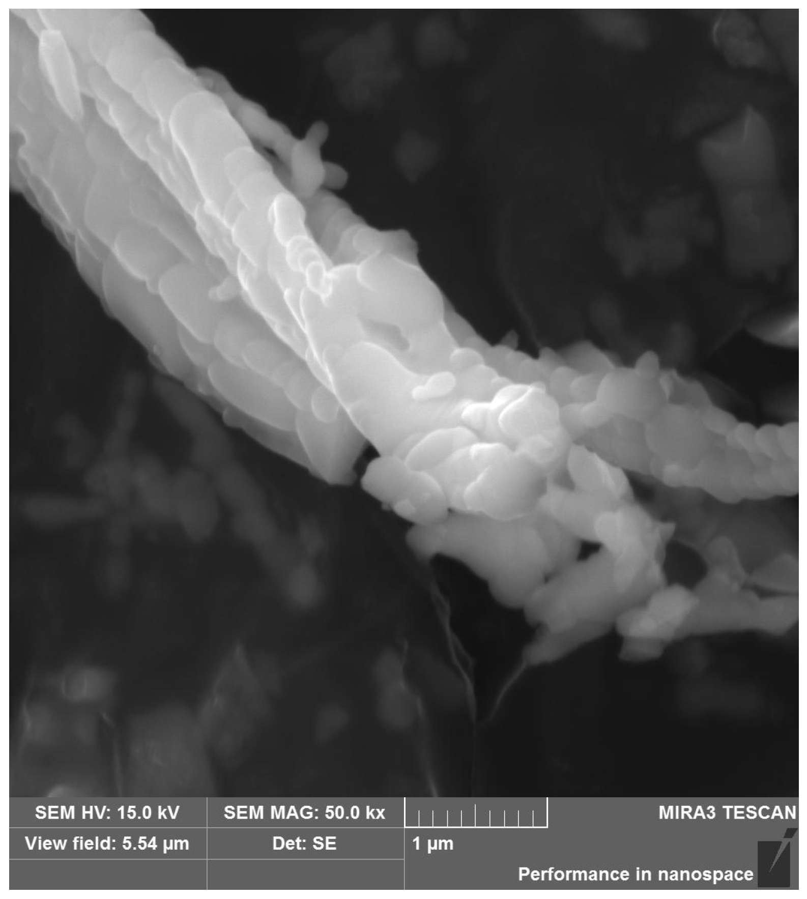
3.1. Characteristics of Chrysotile Asbestos
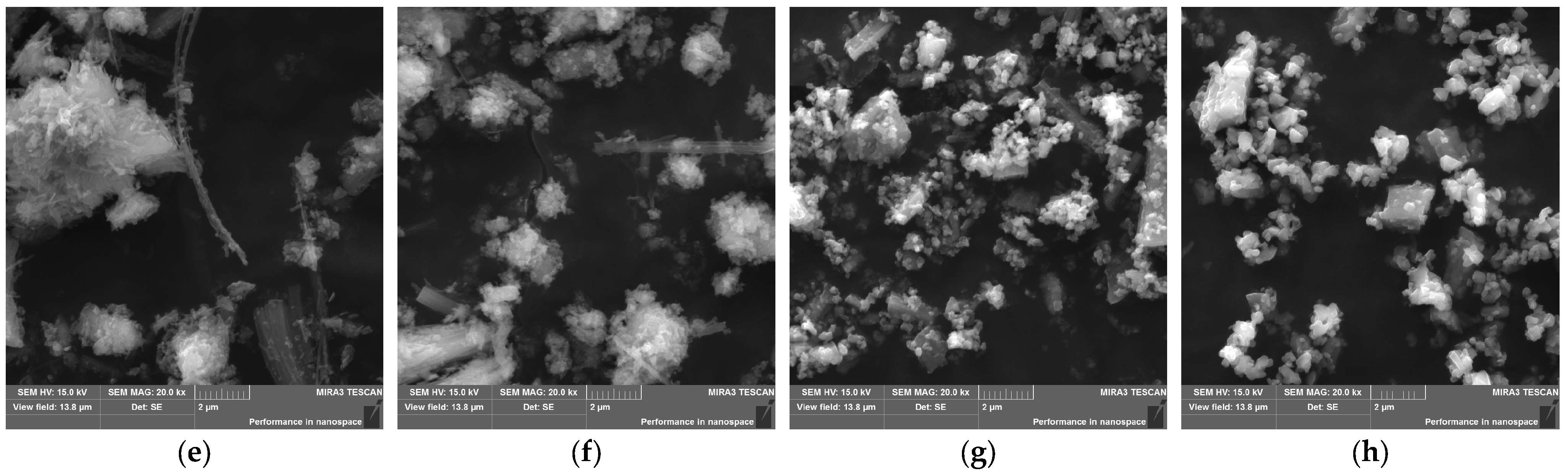
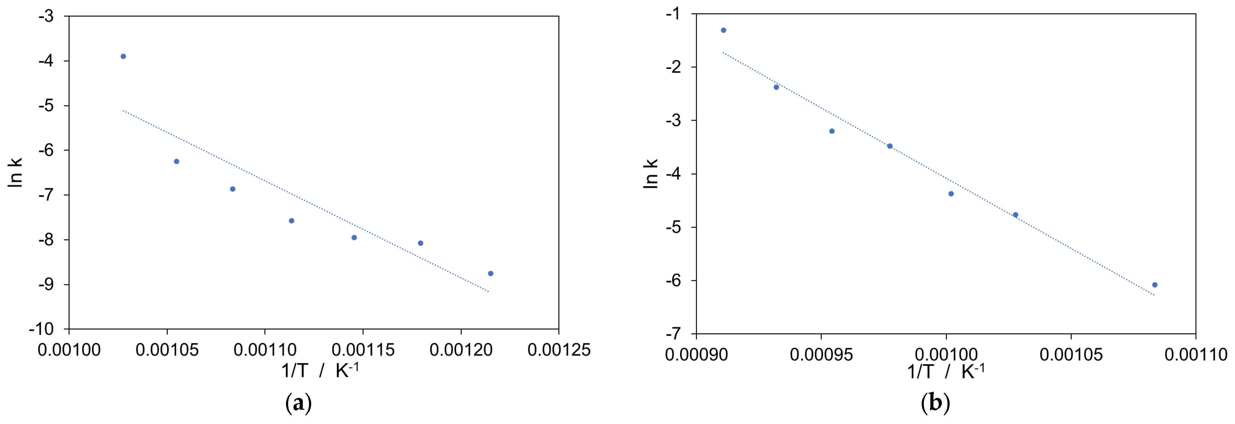
From the chemical point of view, the chemical composition related to oxides is typical for chrysotile asbestos (Table 1). The asbestos mainly consisted of MgO (~42 wt%) and SiO2 (38 wt%). Due to the presence of crystal water in the structure, this material has a high value of loss on ignition (~16 wt%). The theoretical weight loss of pure chrysotile (with ideal formula Mg3(OH)4Si2O5) is 13 wt%. In this study, the mass change determined by L.O.I. value was higher and achieved ~16 wt%. This indicates a lower level of purity and the presence of other mineral impurities, which could also undergo thermal decomposition. The analyzed sample of chrysotile asbestos is also characterized by a relatively high content of iron compounds. This may suggest that the material comes from the Zhitikara deposit in the southern Urals in Kazakhstan [44,45]. Compared to the chemical compositions of chrysotile asbestos samples from other deposits, the determined composition is closest to this one. According to the literature [44], the Zhitikara deposit was the second largest asbestos deposit in the USSR.
X-ray diffraction test clearly showed (Figure 1) that the main mineral component of the tested asbestos material was serpentinite minerals like chrysotile (Mg3(OH)4Si2O5, ICDD-PDF-00-027-1276) and lizardite (Mg3(OH)4Si2O5, ICDD-PDF 04-012-6172). Both minerals possess the same chemical formula and they are hydrated magnesium silicate but differ in the habit of the crystals. Due to common rock-forming processes, these minerals often co-occur together [45]. Beside these two hydrated magnesium silicates others minerals in accompanying and trace form from parent rock were also identified. In this chrysotile asbestos sample, the presence of another mineral phases like brucite (Mg(OH)2, ICDD-PDF 04-011-5938) as well as magnetite (Fe3O4, ICD-PDF 04-021-5000) were confirmed.
The characteristic fibrous form of asbestos was stated during SEM observations (Figure 2a). The SEM imaging process of the raw natural chrysotile asbestos showed the characteristic and typical bundles of asbestos fibers. Moreover, they tended defibration into smaller elementary fibers. Small, irregularly shaped grains were also found among the long, wavy asbestos fibers. This confirmed the presence of other mineral impurities from the parent rock. EDS analysis showed a higher amount of iron in these microregions (Figure 2b).
In Figure 3, the thermal behavior of the considered chrysotile sample was presented. There was an initial small mass loss due to adsorbed water around 100 °C, which was followed by two additional mass losses connected with endothermic effects recorded at ~410 °C and ~520 °C, respectively. Until about 530 °C, the sample loses ~5% of its starting mass. These effects could be connected with the presence of impurities in the sample and were related to the water and carbon dioxide release. The first effect in the temperature range 50–200 °C comes from the dehydratation of hygroscopic water and the release of water molecules closed between the fibrils of chrysotile. The second effect, visible on the DTG curve at 413 °C, could be connected with the brucite (Mg(OH)2) dehydroxylation. As was shown from X-ray powder diffraction, brucite was presented in minor amounts in the sample. The recorded mass change for this effect was 1.54 wt%, which can be recalculated on the ~5 wt% concentration of this mineral in raw chrysotile sample. The next minor recorded effect was a sharp mass loss of ~1.5% with an endothermic peak at 528 °C. Evolved gas analysis showed the visible effect of CO2 release. This effect is problematic and difficult to interpret. Instinctively, this effect could be linked to the thermal decomposition of carbonate minerals, especially magnesite (MgCO3) or siderite (FeCO3). The first mineral was often identified as accompanying minerals in chrysotile asbestos deposits [45]. The first thermally decomposed at ~600 °C, while the second underwent thermal dissociation at ~550 °C [46,47]. However, phase analysis did not reveal the presence of any minerals from the group of carbonates.
The temperature of thermal decomposition of these minerals strongly depends on the degree of sample crystallinity [47], which can also affect phase analysis by X-ray measurement. Due to the low crystallinity, these minerals may not be detected and may be present in so-called X-ray amorphous phase. On the other hand, the evolving of carbon dioxide for asbestos sample between 200 and 600 °C was also observed by other authors, and has been attributed to the evolution of CO2 adsorbed on the fiber surface [48] or attributed to the degradation of trace of intermediate carbonate minerals [49]. By assuming the presence of magnesite in the sample, the registered mass loss can be converted to 2.8 wt% of this carbonate in the chrysotile asbestos sample.
At 600–850 °C, a strong endothermic effect on DTA curve connected with the high mass change was observed. This effect was responsible for the dehydroxylation reaction of chrysotile asbestos [25,35,50,51,52,53,54,55] and the loss of the chemical-bonded water from its structure. This means the total breakdown of the mineral structure and forming of an amorphous mixture of silica and magnesia [39], which is also called serpentine anhydrite or metaserpentine phase [56]. A registered mass loss of 10.52 wt% shows that the tested sample contains c.a. 81 wt% of chrysotile asbestos. Because in the sample, two hydrated silicate minerals were identified, i.e., chrysotile and lizardite, there were also visible small effects that could be connected with lizardite thermal decomposition and breakdown. This may be indicated by a weak mass loss (~0.39 wt%) on the TG curve at a temperature of about 800 °C, or a more visible peak on the DTG curve with a maximum at 804 °C. Viti [57] shows that serpentine dehydroxylation takes place between 550 and 800 °C, with thermal analysis peak temperatures progressively decreasing in order: antigorite, lizardite, polygonal serpentine, and chrysotile. Similar to chrysotile, the recorded mass change for lizardite dehydroxylation allowed the calculation of the content of this mineral. Its content was estimated at 3 wt%.
The last thermal phenomenon visible on the DTA curve was recorded at 831 °C. The occurrence of relatively strong, sharp exothermic peaks in DTA curve without mass change indicates that serpentine asbestos sample undergoes recrystallization processes. The nature of this exotherm is attributed in literature to forsterite formation [58,59], or forsterite recrystallisation [60,61], or enstatite formation [57,62]. Forsterite (Mg2SiO4) and enstatite (MgSiO3) do not form directly from serpentine, but rather through a topotactic transition involving intermediate phases [56]. During serpentinite dehydroxylation, Mg-rich regions favor the formation of forsterite, while Si-rich regions favor the formation of enstatite [56]. The detailed description of the mechanism can be found in the literature on the subject [56,57,58,59,60,61,62]. The above changes can be summarized by the following chemical reactions:
Mg3(OH)4Si2O5 (chrysotile or lizardite) → Mg3Si2O7 (serpentine anhydrite) + 2H2O
And in the next step:
Mg3Si2O7 → Mg2SiO4 + MgSiO3
After thermal treatment, especially after the isothermal calcination process at different temperatures, the change in material color was observed (Figure 4). With the increase in isothermal treatment temperature, the obtained heated powders became pale orange-red and finally rusty-colored after calcination at the highest assumed temperature. This indicates the possible oxidation of ferrous iron, especially coming from the magnetite impurities in the sample. Moreover, as the calcination temperature increased, the resulting material became progressively more brittle and susceptible to crushing.
Figure 5 shows the XRD patterns of tested chrysotile asbestos samples after different calcination temperatures. The characteristic reflexes coming from raw natural chrysotile were observed for unheated sample and after 2 h of isothermal treatment at 450, 500 and 550 °C. After calcination at 600 °C, the diffraction reflexes of chrysotile showed a downward trend. At the same time, new X-ray reflections appeared. They corresponded with forsterite (Mg2SiO4, ICDD-PDF 04-007-2768) as well as hematite (Fe2O3, ICDD-PDF 04-005-4630). This phenomenon has often been observed by other researchers [41,63,64,65]. After calcination at higher temperatures (from 650 to 1000 °C), only forsterite was identified in the sample. Only calcination at temperatures of 1050 °C and above contributed to the appearance of new diffraction reflections related to enstatite (MgSiO3, ICDD-PDF 01-076-2427). The presence of forsterite in the first stage and later also enstatite indicates the total destruction of chrysotile asbestos. This is a key issue in the context of thermal disposal methods for waste containing chrysotile asbestos. This breakdown of the mineral structure and creation of intermediate and finally new mineral phases opens up new possibilities for the economic use of thermally processed asbestos wastes. The last study showed that forsterite and enstatite minerals transformed from chrysotile by heating could be considered non-carcinogens [39,40].
During isothermal heating, the fiber microstructure of chrysotile was changed despite maintaining a fibrous shape. High temperature causes the creation of new phases, which changes the structure completely at the molecular scale and decreases strength properties (Figure 6). This occurrence is called pseudomorphosis [66]. As can be seen, the fiber is not divided into long micrometric fibers, but into tiny grains; therefore, during mechanical processing, it is not divided along the fiber axis but along the boundaries of the formed grains. This enables the efficient grinding of the fiber prepared in this way [67,68]. Kim et al. [65] showed by TEM analysis that chrysotile asbestos after heat treatment at 800 and 900 °C did not show the hollow, tubular structures typical of chrysotile asbestos, but minerals in the form of lumps and columns, indicating the destruction of the hollow tubular structure of the chrysotile asbestos. This material is easily broken into round grains of new silicate minerals, which are apparent on the primary bundle of asbestos.
Comparison of grinded material of chrysotile asbestos samples after isothermal calcination at selected temperatures is presented in Figure 7. Calcination caused a change in the properties of asbestos fibers, which became brittle and susceptible to grinding. The higher the temperature of thermal treatment, the easier the obtained material was to grind. In the case of material obtained after calcination at 650 °C (Figure 7a,e), a clear presence of fibrous forms was still observed in the material. Visible asbestos bundles tend to divide into elementary fibers along the length of the original bundle. The material obtained after isothermal treatment at 700 °C (Figure 7b,f) contained significantly fewer fibrous forms, and the fibrous character of the original asbestos changed into beams or slats of a sharp-edged nature. In turn, for the ground materials after the calcination of asbestos at the highest temperatures (Figure 7c,d,g,h), only the presence of irregular or spherical grains was found in the material. Taking the above into account, methods based on the calcination of asbestos at high temperatures can be considered an effective and easy-to-implement method in the context of getting rid of the problem of accumulated asbestos waste.

3.2. Kinetic Study

Understanding kinetic factors, such as reaction order, activation energy, and rate constant, is important in indicating reaction mechanisms in solid phases. Therefore, a kinetic study was carried out on a chrysotile asbestos sample by two different techniques. The first (so-called ex situ) was connected with the isothermal calcination of material in a laboratory furnace where change of total mass during heat-treatment was measured. Because the studied asbestos material is not pure and contains other mineral impurities (please see Section 3.1), in this way, we can calculate the kinetic parameters for the whole thermal decomposition process. In contrary, the second way (so-called in situ) was connected with the measurement of main chrysotile X-ray diffraction reflex disappearance, so in this way, we could calculate the kinetic parameters of the dehydroxylation process of chrysotile asbestos.
The kinetics of a chemical process or reaction can be extended by the following equation:
dα/dt = k·f(α),
where α is the degree of conversion of the reaction/process, k is the rate constant at a given temperature and f(α) is the kinetic model function, usually an empirical function dependent on the mechanism of the reaction. The Avrami–Erofeev (A–E) equation has been extensively used to present the isothermal behavior of different objects and processes [69,70,71,72,73,74], so we decided to use it in this study. In this case, the degree of conversion α could be expressed as a function of time according to the A–E transformation equation:
α ( t ) = 1 e x p ( k t n ) ,
where k is the defined as a kinetic constant, t is time and n is the reaction order. Using the Arrhenius temperature dependency of k, i.e.,:
k = A   exp ( E a R T ) ,
where A is the pre-exponential factor, Ea is the apparent activation energy (in kJ mol−1), R is the universal gas constant (8.314 J K−1 mol−1) and T is the thermal process absolute temperature (in K), Equation (1) could be written as:
d α / d t = A   exp ( E a R T ) · f ( α )
Taking the logarithm of both sides of Equation (2) twice, the linearized form of the A–E could be obtained. It is expressed as:
ln   [ ln 1 α ] = ln k + n ln t
Thus, when the double logarithm side is taken as the vertical coordinate and ln t is taken as the horizontal coordinate, we can plot the so-called ln-ln graph from which the value of k (rate coefficient) and n (reaction order) can be easily calculated. In turn, the plot of ln(k) vs. 1/T from Equation (3) yields the apparent activation energy (Ea) from the slope of the linear curve and the preexponential factor (A) from the line intercept.
In Figure 8, the variations of degree of conversion α versus time t for both variants of kinetic tests were reported. Obviously, with the increase in the isothermal treatment temperature, the maximum degree of conversion was achieved faster. In the ex situ experiment (Figure 8a), the calcination time to obtain constant mass change varied over a wide range. For the highest temperature, the constant mass of sample was achieved after 30 min of isothermal calcination at 700 °C. When the calcination temperature decreased, the time required to reach constant mass was noticeably longer. Firing at 675, 650, 625 and 600 °C required a time of ~1.0, 1.5, 3.0 and 10 h, respectively. For the lowest calcination temperatures, the required times were even longer and are counted in days. The change in mass is connected mainly with the dehydroxylation of serpentine minerals; however, in the tested sample, other accompanying minerals contribute to thermal decomposition process and recorded mass change.
In relation to the results of the in situ experiment (Figure 8b), the degree of conversion was associated directly with the disappearance of the main asbestos diffraction reflections. Therefore, direct comparison of the results of both methods is not advisable. In this case, total conversion (α = 1) was achieved after ~20 min of isothermal treatment at 825 °C. With the decrease in temperature, the time to achieve full conversion was longer and changed from 4 h to 12 h for 800 and 700 °C, respectively. In the case of the lowest temperature of the in situ experiment, i.e., 650 °C, the test was stopped after one week of measurement. In this case, the final degree of conversion achieved a value close to 0.8. The different behavior of the asbestos sample during the in situ experiment can also be connected with different heating conditions, where the sample was placed on a corundum pad and heated on one side in a X-ray diffraction temperature chamber. The fact of the formation of the so-called serpentine anhydride [56,63] may also be significant, as indicated by the ex situ results at a relatively lower temperature. The results obtained may also indicate that the product being formed has a crystal structure similar to the original material, and only a higher temperature can generate crystallochemical transitions and the disappearance of reflections from asbestos.
The so-called ln-ln plots (the linearized form of the Avrami–Erofeev formula) for both experiments are presented in Figure 9, while plots of rate coefficient k against temperature are shown in Figure 10. For both cases, the obtained curves for different annealing temperatures show nearly the same slope and parallelism. This may indicate the same mechanism of thermal decomposition process of asbestos sample in the considered temperature range. The determined values of reaction order n for ex situ measurements ranged from ~0.7 to ~0.9, with an average value of 0.80. In turn, the calculated reaction order for in situ technique changed from 0.41 to 0.57, with an average value of 0.51 (Table 2). The obtained values of reaction order determined by the A–E method allow for the postulation that the rate-limiting step of chrysotile dehydroxylation process could be one-dimensional diffusion [35,75].
The obtained results suggest a complex mechanism of the chrysotile asbestos thermal decomposition process. Due to the presence of other mineral phases in the tested material, the overall reaction order connected with thermal decomposition by ex situ technique was higher than expected. Obviously, with the increase in the heat treatment temperature in both considered cases, the value of the rate coefficient k increased significantly (Table 2). Activation energy connected with the thermal decomposition of tested chrysotile asbestos sample changed from ~180 kJ mol−1 to ~220 kJ mol−1 (Table 3) for ex situ and in situ measurement, respectively. The activation energy represents the energy barrier that must be overcome to transform the material into another phase. In our case, this is related to the initiation of the asbestos dehydroxylation process and its changes at the crystalline level.
The values of activation energy for thermal decomposition of chrysotile were at a similar level to those found in the literature. Long et al. [64] show that the calculated activation energy for dehydroxylation of chrysotile fiber membrane or raw chrysotile fiber measured by thermal analysis method was ~243 or ~223 kJ mol−1, respectively. In turn, the calculated activation energy of the dehydroxylation reaction in the temperature range 620–750 °C was 184 kJ mol−1, and the rate-limiting step was one-dimensional diffusion with an instantaneous nucleation or a deceleratory rate of nucleation of the reaction product [35,51]. This value was lower than for others serpentine minerals like lizardite and antigorite, for which the determined activation energy by Avrami model in the temperature range 612–708 °C was 221 and 255 kJ mol−1, respectively [51]. In turn, data presented by [76], shows that chrysotile dehydroxylation experiments gave changeable activation energy values in the range of around 250–300 kJ mol−1 for thermal analysis test or 250–380 kJ mol−1 for the high-temperature X-ray powder diffraction measurement.

4. Conclusions

The thermal decomposition process of chrysotile asbestos from the mineralogical as well as kinetic point of view was described. This study determined the change in chrysotile asbestos material following thermal treatment at various temperatures. The presented study demonstrated that chrysotile asbestos can be converted into new mineral phases at sufficient temperatures. The minimum temperature of 600 °C is required for the thermal decomposition of chrysotile to initiate the process of dehydroxylation (dehydration) of the chrysotile mineral at a satisfactory rate. Moreover, a higher temperature is required to cause a clear disappearance of chrysotile diffraction reflections. As a result of thermal treatment in the first stage of chrysotile decomposition, forsterite is formed as a product, and at higher temperatures, above 1050 °C, enstatite also appears. The kinetics of chrysotile thermal decomposition was investigated under isothermal conditions by two methods: by ex situ with isothermal annealing in furnace or by in situ with the use of a high-temperature chamber of X-ray measurement. This heterogeneous solid-state reaction was analyzed by the Avrami–Erofeev kinetic model. Two different values of the activation energy, ~180 kJ mol−1 and ~220 kJ mol−1, for the above methods were obtained, respectively. These findings could be useful for the safe disposal and recycling of chrysotile asbestos-containing materials by thermal methods in the future. Within this framework, some researchers [14,15] have proposed the recycling of asbestos-containing materials (ACMs) as a raw material in the production of high-end traditional ceramics, such as porcelain stoneware (porcellanato grés) or fired-clay brick. In this application, asbestos can be completely converted into a mixture of non-hazardous silicate phases through thermal treatment at temperatures up to 1250 °C. This method appears promising, as the estimated recycling costs are up to ten times lower than those associated with the disposal of ACMs as hazardous waste. Additionally, the process generates no hazardous residues, thereby eliminating the risk of environmental contamination.

Author Contributions

Conceptualization, R.K. and A.G.; methodology, R.K. and A.G.; software, A.G.; validation, R.K., M.K. and A.G.; formal analysis, R.K., A.B. and A.G.; investigation, R.K., M.K. and A.G.; resources, R.K.; data curation, R.K. and A.B.; writing—original draft preparation, R.K. and M.K.; writing—review and editing, R.K., M.K., A.B. and A.G.; visualization, R.K., M.K. and A.G.; supervision, R.K.; project administration, R.K.; funding acquisition, R.K. All authors have read and agreed to the published version of the manuscript.

Funding

This research was funded in whole by the National Science Centre, Poland under “Sonata 17” grant number UMO-2021/43/D/ST5/00356.

Data Availability Statement

The data presented in the study are open. The experimental data that support the findings of this study are available in the RepOD repository with the identifier https://doi.org/10.18150/XCDVVX as well as https://doi.org/10.18150/B55A4G.

Acknowledgments

The authors would like to thank Elwira Cieślińska and Maria Pyka for their help in preparing samples for testing.

Conflicts of Interest

The authors declare no conflicts of interest.

References

  1. Virta, R.L. Mineral Commodity Profiles—Asbestos; Circular 1255-KK; U.S. Geological Survey: Reston, VA, USA, 2005.
  2. Sporn, T.A. Mineralogy of asbestos. In Malignant Mesothelioma; Tannapfel, A., Ed.; Springer: Berlin/Heidelberg, Germany, 2011; pp. 1–11. [Google Scholar]
  3. Bartrip, P.W. History of asbestos related disease. Postgrad. Med. J. 2004, 80, 72–76. [Google Scholar] [CrossRef]
  4. Musk, A.W.; de Klerk, N.; Reid, A.; Hui, J.; Franklin, P.; Brims, F. Asbestos-related diseases. Tuber. Lung Dis. 2020, 24, 562–567. [Google Scholar] [CrossRef] [PubMed]
  5. Stayner, L.; Welch, L.S.; Lemen, R. The worldwide pandemic asbestos-related diseases. Annu. Rev. Public Health 2013, 34, 205–216. [Google Scholar] [CrossRef] [PubMed]
  6. Kwak, K.; Kang, D.; Paek, D. Environmental exposure to asbestos and the risk of lung cancer: A systematic review and meta-analysis. J. Occup. Environ. Med. 2022, 79, 207–214. [Google Scholar] [CrossRef] [PubMed]
  7. The International Ban Asbestos Secretariat IBAS. Available online: www.ibasecretariat.org/index.htm (accessed on 10 January 2025).
  8. Lin, R.T.; Chien, L.C.; Jimba, M.; Furuya, S.; Takahashi, K. Implementation of national policies for a total asbestos ban: A global comparison. Lancet Planet. Health 2019, 3, 341–348. [Google Scholar] [CrossRef]
  9. Paglietti, F.; Malinconico, S.; Conestabile della Staffa, B.; Bellagamba, S.; De Simone, P. Classification and management of asbestos-containing waste: European legislation and the Italian experience. Waste Manag. 2016, 50, 130–150. [Google Scholar] [CrossRef]
  10. Promentilla, M.A.B.; Peralta, G.L. An Evaluation of landfill disposal of asbestos-containing waste and geothermal residues within a risk-assessment framework. J. Mater. Cycles Waste Manag. 2003, 5, 13–21. [Google Scholar] [CrossRef]
  11. Macher, G.Z.; Torma, A.; Beke, D. Examining the environmental ramifications of asbestos fiber movement through the water–soil continuum: A review. Int. J. Environ. Res. Public Health 2025, 22, 505. [Google Scholar] [CrossRef]
  12. Paolini, V.; Tomassetti, L.; Segreto, M.; Borin, D.; Liotta, F.; Torre, M.; Petracchini, F. Asbestos treatment technologies. J. Mater. Cycles Waste Manag. 2019, 21, 205–226. [Google Scholar] [CrossRef]
  13. Spasiano, D.; Pirozzi, F. Treatments of asbestos containing wastes. J. Environ. Manag. 2017, 204, 82–91. [Google Scholar] [CrossRef]
  14. Zaremba, T.; Krząkała, A.; Piotrowski, J.; Garczorz, D. Utilization of chrysotile asbestos for sintering ceramics production. Ceram. Mater. 2010, 62, 149–155. (In Polish) [Google Scholar]
  15. Gualtieri, A.F.; Tartaglia, A. Thermal decomposition of asbestos and recycling in traditional ceramics. J. Eur. Ceram. Soc. 2000, 20, 1409–1418. [Google Scholar] [CrossRef]
  16. Gualtieri, A.F.; Cavenati, C.; Zanatto, I.; Meloni, M.; Elmi, G.; Gualtieri, M.L. The transformation sequence of cement–asbestos slates up to 1200 °C and safe recycling of the reaction product in stoneware tile mixtures. J. Hazard. Mater. 2008, 152, 563–570. [Google Scholar] [CrossRef] [PubMed]
  17. Kusiorowski, R.; Zaremba, T.; Piotrowski, J.; Podwórny, J. Utilisation of cement-asbestos wastes by thermal treatment and the potential possibility use of obtained product for the clinker bricks manufacture. J. Mater. Sci. 2015, 50, 6757–6767. [Google Scholar] [CrossRef]
  18. Gualtieri, A.F.; Boccaletti, M. Recycling of the product of thermal inertization of cement–asbestos for the production of concrete. Constr. Build. Mater. 2011, 25, 3561–3569. [Google Scholar] [CrossRef]
  19. Kusiorowski, R.; Zaremba, T.; Piotrowski, J. The potential use of cement-asbestos waste in the ceramic masses destined for sintered wall clay brick manufacture. Ceram. Int. 2014, 40, 11995–12002. [Google Scholar] [CrossRef]
  20. Obmiński, A. Asbestos waste recycling using the microwave technique—Benefits and risks. Environ. Nanotechnol. Monit. Manag. 2021, 16, 100577. [Google Scholar] [CrossRef]
  21. Carneiro, G.O.; Santos, T.A.; Simonelli, G.; Ribeiro, D.V.; Cilla, M.S.; Dias, C.M.R. Thermal treatment optimization of asbestos cement waste (ACW) potentializing its use as alternative binder. J. Clean. Prod. 2021, 320, 128801. [Google Scholar] [CrossRef]
  22. Capitani, G.; Dalpiaz, M.; Vergani, F.; Campanale, F.; Conconi, R.; Odorizzi, S. Recycling thermally deactivated asbestos cement in mortar: A possible route towards a rapid conclusion of the “asbestos problem”. J. Environ. Manag. 2024, 355, 120507. [Google Scholar] [CrossRef]
  23. Singh, R.; Rao, B.; Asolekar, S.R. Geopolymer-based solidification and stabilization for environmentally sound disposal of asbestos-containing waste. J. Mater. Cycles Waste Manag. 2025, 27, 75–90. [Google Scholar] [CrossRef]
  24. Gualtieri, A.F. Mineral Fibers: Crystal Chemistry, Chemical-Physical Properties, Biological Interaction, and Toxicity; European Mineralogical Union: London, UK, 2017. [Google Scholar]
  25. Bloise, A.; Catalano, M.; Barrese, E.; Gualtieri, A.F.; Gandolfi, N.B.; Capella, S.; Belluso, E. TG/DSC study of the thermal behavior of hazardous mineral fibres. J. Therm. Anal. Calorim. 2016, 123, 2225–2239. [Google Scholar] [CrossRef]
  26. Kusiorowski, R.; Zaremba, T.; Piotrowski, J.; Adamek, J. Thermal decomposition of different types of asbestos. J. Therm. Anal. Calorim. 2012, 109, 693–704. [Google Scholar] [CrossRef]
  27. Bloise, A. Thermal behaviour of actinolite asbestos. J. Mater. Sci. 2019, 54, 11784–11795. [Google Scholar] [CrossRef]
  28. Bloise, A. On the thermal breakdown of tremolite: A new method for distinguishing between asbestos and non-asbestos tremolite samples. J. Mater. Sci. 2023, 58, 8779–8795. [Google Scholar] [CrossRef]
  29. Carmignano, O.R.; Vieira, S.S.; Brandão, P.R.; Bertoli, A.C.; Lago, R.M. Serpentinites: Mineral structure, properties and technological applications. J. Braz. Chem. Soc. 2020, 31, 2–14. [Google Scholar] [CrossRef]
  30. Flanagan, D.M. Mineral Commodity Summaries 2025, Ver. 1.2; U.S. Geological Survey: Reston, VA, USA, 2025; p. 38.
  31. Bloise, A.; Critelli, T.; Catalano, M.; Apollaro, C.; Miriello, D.; Croce, A.; Barrese, E.; Liberi, F.; Piluso, E.; Rinaudo, C.; et al. Asbestos and other fibrous minerals contained in the serpentinites of the Gimigliano-Mount Reventio Unit (Calabria, Italy). Environ. Earth Sci. 2014, 71, 3773–3786. [Google Scholar] [CrossRef]
  32. Teixeira, A.P.C.; Santos, E.M.; Vieira, A.F.P.; Lago, R.M. Use of chrysotile to produce highly dispersed K-doped MgO catalyst for biodiesel synthesis. Chem. Eng. J. 2013, 232, 104–110. [Google Scholar] [CrossRef]
  33. Sprynskyy, M.; Niedojadło, J.; Buszewski, B. Structural features of natural and acids modified chrysotile nanotubes. J. Phys. Chem. Solids 2011, 72, 1015–1026. [Google Scholar] [CrossRef]
  34. Jeyaratnam, M.; West, N.G. A study of heat-degraded chrysotile, amosite and crocidolite by X-ray diffraction. Ann. Occup. Hyg. 1994, 38, 137–148. [Google Scholar] [CrossRef]
  35. Cattaneo, A.; Gualtieri, A.F.; Artioli, G. Kinetic study of the dehydroxylation of chrysotile asbestos with temperature by in situ XRPD. Phys. Chem. Miner. 2003, 30, 177–183. [Google Scholar] [CrossRef]
  36. Hashimoto, S.; Yamaguchi, A. Detoxification technique of asbestos using low-temperature heating and grinding. J. Ceram. Soc. Jpn. 2006, 41, 856–858. [Google Scholar]
  37. Dellisanti, F.; Minguzzi, V.; Morandi, N. Experimental results from thermal treatment of asbestos containing materials. GeoActa 2001–2002, 1, 61–70. [Google Scholar]
  38. Brindley, G.W.; Hayami, R. Mechanism of formation of forsterite and enstatite from serpentine. Mineral. Mag. 1965, 35, 189–195. [Google Scholar] [CrossRef]
  39. Langer, A.M. Reduction of the biological potential of chrysotile asbestos arising from conditions of service on brake pads. Regul. Toxicol. Pharmacol. 2003, 38, 71–77. [Google Scholar] [CrossRef] [PubMed]
  40. Takata, A.; Yamauchi, H.; Yamashita, K.; Aminaka, M.; Hitomi, T.; Toya, T.; Kohyama, N. Mesothelioma carcinogenesis of chrysotile and forsterite compared and validated by intraperitoneal injection in rat. Ind. Health 2025, 63, 14–28. [Google Scholar] [CrossRef]
  41. Zaremba, T.; Krząkała, A.; Piotrowski, J.; Garczorz, D. Study on the thermal decomposition of chrysotile asbestos. J. Therm. Anal. Calorim. 2010, 101, 479–485. [Google Scholar] [CrossRef]
  42. PN-EN ISO 12677:2011; Chemical Analysis of Refractory Products by X-Ray Fluorescence (XRF)—Fused Cast-Bead Method. ISO: Geneva, Switzerland, 2011.
  43. Bloise, A.; Parisi, F.; La Russa, M.F.; Apollaro, C.; Godbert, N.; Aiello, I.; Giorno, E.; Croce, A.; Cagna, L.; López, A.J.; et al. Evaluation of asbestos dispersion during laser ablation of rocks containing Naturally Occurring Asbestos (NOA). Heliyon 2024, 10, e39624. [Google Scholar] [CrossRef]
  44. Łącki, J.W. Wydobycie i Zastosowanie Azbestu (Mining and Use of Asbestos), 1st ed.; Wydawnictwo Śląsk: Katowice, Poland, 1974. (In Polish) [Google Scholar]
  45. Shayakhetova, R.A.; Mukhametzhanova, A.A.; Akbayeva, D.N.; Terlikbaeva, A.Z.; Osipov, P.A.; Alimzhanova, A.M.; Zharmenov, A.A. Magnesium and silicon recovery from chrysotile asbestos waste of the deposit Zhitikara, Kazakhstan. Sci. Rep. 2024, 14, 31866. [Google Scholar]
  46. Földvári, M. Handbook of Thermogravimetric System of Minerals and Its Use in Geological Practice; Geological Institute of Hungary: Budapest, Hungary, 2011. [Google Scholar]
  47. Strzałkowska, E. Thermal Analysis of Minerals, Rocks and Mineral Waste Materials; Silesian University of Technology: Gliwice, Poland, 2019. (In Polish) [Google Scholar]
  48. Sanders, J.P.; Brosnan, D. Modelling the Thermal Decomposition of Chrysotile. 2005. Available online: https://www.jurispro.com/files/documents/doc-1066205184-article-1610.pdf (accessed on 14 April 2025).
  49. Khorami, J.; Choquette, D.; Kimmerle, F.M.; Gallagher, P.K. Interpretation of EGA and DTG analyses of chrysotile asbestos. Thermochim. Acta 1984, 76, 87–96. [Google Scholar] [CrossRef]
  50. Bloise, A.; Kusiorowski, R.; Lassinantti Gualtieri, M.; Gualtieri, A.F. Thermal behaviour of mineral fibres. In Mineral Fibers: Crystal Chemistry, Chemical-Physical Properties, Biological Interaction and Toxicity; Gualtieri, A.F., Ed.; European Mineralogical Union: London, UK; Mineralogical Society of Great Britain & Ireland Minerals: Twickenham, UK, 2017. [Google Scholar]
  51. Gualtieri, A.F.; Giacobbe, C.; Viti, C. The dehydroxylation of serpentine group minerals. Am. Mineral. 2012, 97, 666–680. [Google Scholar] [CrossRef]
  52. Belardi, G.; Piga, L. Influence of calcium carbonate on the decomposition of asbestos contained in end-of-life products. Thermochim. Acta 2013, 573, 220–228. [Google Scholar] [CrossRef]
  53. Malkov, A.A.; Korytkova, E.N.; Maslennikova, T.P.; Shtykhova, A.M.; Gusarov, V.V. Effect of heat treatment on structural-chemical transformations in magnesium hydrosilicate Mg3Si2O5(OH)4 nanotubes. Russ. J. Appl. Chem. 2009, 82, 2079–2086. [Google Scholar] [CrossRef]
  54. Leonelli, C.; Veronesi, P.; Boccaccini, D.N.; Rivasi, M.R.; Barbieri, L.; Andreola, F.; Lancellotti, I.; Rabitti, D.; Pellacani, G.C. Microwave thermal inertisation of asbestos containing waste and its recycling in traditional ceramics. J. Hazard. Mater. 2006, 135, 149–155. [Google Scholar] [CrossRef] [PubMed]
  55. Vast, P.; Andries, V.; Martines, M.A.U.; Auffredic, J.P.; Poulain, M.; Messaddeq, Y. Treatment and destruction of inorganic fibers wastes like asbestos by sodium polyphosphate. Phosphorus Res. Bull. 2004, 15, 68–82. [Google Scholar] [CrossRef] [PubMed]
  56. Dlugogorski, B.Z.; Balucan, R.D. Dehydroxylation of serpentine minerals: Implications for mineral carbonation. Renew. Sustain. Energy Rev. 2014, 31, 353–367. [Google Scholar] [CrossRef]
  57. Viti, C. Serpentine minerals discrimination by thermal analysis. Am. Mineral. 2010, 95, 631–638. [Google Scholar] [CrossRef]
  58. Martinez, E. The effect of particle size on the thermal properties of serpentine minerals. Am. Mineral. 1961, 46, 901–912. [Google Scholar]
  59. Coats, C.J.A. Serpentine minerals from Manitoba. Can. Mineral. 1968, 9, 322–347. [Google Scholar]
  60. Rao, V.A.N.; Murty, M.S. Thermal studies on soft and brittle asbestos. Curr. Sci. 1979, 48, 760–761. [Google Scholar]
  61. Perez-Rodriguez, J.L.; Franco, F.; Ramirez-Valle, V.; Perez-Maqueda, L.A. Modification of the thermal dehydroxylation of antigorite by ultrasound treatment. J. Therm. Anal. Calorim. 2005, 82, 769–774. [Google Scholar] [CrossRef]
  62. Viti, C.; Giacobbe, C.; Gualtieri, A.F. Quantitative determination of chrysotile in massive serpentinites using DTA: Implications for asbestos determinations. Am. Mineral. 2011, 96, 1003–1011. [Google Scholar] [CrossRef]
  63. Jeong, H.; Moon, W.; Roh, Y. Characterization of mineralogical changes of chrysotile and its thermal decomposition by heat treatment. Econ. Environ. Geol. 2016, 49, 77–88. [Google Scholar] [CrossRef]
  64. Long, J.; Liu, W.; Zhang, N.; Zhang, H.; Xiao, Q.; Huang, S. New insight into the phase transition and kinetics of the dehydroxylation of bulk-to-nano chrysotile. Phys. Chem. Miner. 2024, 51, 28. [Google Scholar] [CrossRef]
  65. Kim, C.; Kim, Y.; Roh, Y. Thermal decomposition and phase transformation of chrysotile in asbestos-containing waste. Minerals 2025, 15, 344. [Google Scholar] [CrossRef]
  66. Viani, A.; Gualtieri, A.F.; Mácová, P.; Pollastri, S. Transformations through pseudomorphosis of asbestos minerals in thermally processed asbestos-containing materials investigated through SEM/EDS and micro-Raman spectroscopy: Implications for recycling of hazardous wastes. In Proceedings of the 18th International Microscopy Congress, Prague, Czech Republic, 7–12 September 2014. [Google Scholar]
  67. Gualtieri, A.F. Recycling asbestos-containing material (ACM) from construction and demolition waste (CDW). In Handbook of Recycled Concrete and Demolition Waste; Woodhead Publishing: Sawston, UK, 2013; pp. 500–525. [Google Scholar]
  68. Giacobbe, C.; Gualtieri, A.F.; Quartieri, S.; Rinaudo, C.; Allegrina, M.; Andreozzi, G.B. Spectroscopic study of the product of thermal transformation of chrysotile-asbestos containing materials (ACM). Eur. J. Mineral. 2010, 22, 535–546. [Google Scholar] [CrossRef]
  69. Joraid, A.A.; Alamri, S.N.; Abu-Sehly, A.A.; Al-Raqa, S.Y.; Shipman, P.O.; Shipley, P.R.; Abd-El-Aziz, A.S. Isothermal kinetics and thermal degradation of an aryl azo dye-containing polynorbornene. Thermochim. Acta 2011, 515, 38–42. [Google Scholar] [CrossRef]
  70. Chen, G.; Shi, Z.; Yu, J.; Wang, Z.; Xu, J.; Gao, B.; Hu, X. Kinetic analysis of the non-isothermal decomposition of carbon monofluoride. Thermochim. Acta 2014, 589, 63–69. [Google Scholar] [CrossRef]
  71. Wang, S.; Shen, F.; Zheng, H.; Nie, X.; Jiang, X.; Gao, Q. Isothermal reduction and Avrami-Erofeev kinetic model for reducing iron ore pellets in a 70% N2-30%H2/CO atmosphere. J. Sustain. Metall. 2024, 10, 2337–2351. [Google Scholar] [CrossRef]
  72. Kusiorowski, R.; Gerle, A.; Kujawa, M. Kinetic analysis of cement-asbestos materials’ thermal decomposition process by an ex situ technique. Fibers 2025, 13, 43. [Google Scholar] [CrossRef]
  73. Gasparini, E.; Tarantino, S.C.; Ghigna, P.; Pia Riccardi, M.; Cedillo-González, E.I.; Siligardi, C.; Zema, M. Thermal dehydroxylation of kaolinite under isothermal conditions. Appl. Clay Sci. 2013, 80–81, 417–425. [Google Scholar] [CrossRef]
  74. Gurgul, S.J.; Seng, G.; Williams, G.R. A kinetic and mechanistic study into the transformation of calcium sulfate hemihydrate to dihydrate. J. Synchrotron Radiat. 2019, 26, 774–784. [Google Scholar] [CrossRef]
  75. Giacobbe, C. High temperature reactions of serpentine group minerals. Plinius 2012, 38, 93. [Google Scholar]
  76. Trittschack, R.; Grobéty, B.; Brodard, P. Kinetics of the chrysotile and brucite dehydroxylation reaction: A combined non-isothermal/isothermal thermogravimetric analysis and high-temperature X-ray powder diffraction study. Phys. Chem. Miner. 2014, 41, 197–214. [Google Scholar] [CrossRef]
Figure 1. XRD pattern of tested chrysotile sample.
Minerals 15 00609 g001
Figure 2. SEM microphotographs of chrysotile asbestos and corresponding EDS analysis for (a) fiber, and (b) grainy impurities; magnification 2000×.
Minerals 15 00609 g002
Figure 3. DTA-TG-DTG curves of chrysotile sample.
Minerals 15 00609 g003
Figure 4. Changes in asbestos color after isothermal calcination at selected temperatures.
Minerals 15 00609 g004
Figure 5. XRD patterns of chrysotile asbestos sample after calcination at selected temperatures.
Minerals 15 00609 g005
Figure 6. SEM microphotograph of pseudomorphosis obtained after isothermal calcination of chrysotile asbestos at 1100 °C where visible grains of new mineral phases were observed; magnification 50,000×.
Minerals 15 00609 g006
Figure 7. SEM microphotographs of chrysotile asbestos after isothermal calcination at 650 °C (a,e), 700 °C (b,f), 1100 °C (c,g), 1200 °C (d,h), where materials were additionally grinded in laboratory mill; magnification 2000× (ad) or 20,000× (eh).
Minerals 15 00609 g007aMinerals 15 00609 g007b
Figure 8. The degree of conversion, α, as a function of time at different isothermal calcination temperatures during (a) ex situ or (b) in situ experiments.
Minerals 15 00609 g008
Figure 9. Plots of ln[−ln(1 − α)] versus ln t at different temperatures for (a) ex situ or (b) in situ experiments.
Minerals 15 00609 g009
Figure 10. Plots of ln K versus 1/T for the isothermal degradation of chrysotile asbestos by (a) ex situ or (b) in situ experiments.
Minerals 15 00609 g010
Table 1. Chemical composition of chrysotile asbestos sample, wt%.
SiO2TiO2Al2O3Fe2O3MnOMgOCaONa2OK2OP2O5Cr2O3L.O.I.
38.080.010.214.210.1141.940.120.04bdlbdl0.0616.14
bdl—below detection limit.
Table 2. Calculated kinetic factors for the decomposition process of chrysotile asbestos sample performed by ex situ or in situ method.
Ex Situ Method
Temperature, °C550575600625650675700
Value of reaction order, n0.690.700.820.800.850.890.87
Rate coefficient 10−4, k1.573.123.535.1510.419.320.3
Correlation coefficient0.9910.9950.9940.9880.9880.9870.977
In Situ Method
Temperature, °C650700725750775800825
Value of reaction order, n0.480.570.550.550.450.550.41
Rate coefficient 10−3, k2.28.512.630.840.992.6271.1
Correlation coefficient0.9880.9880.9820.9910.9770.9660.970
Table 3. The apparent activation energy results received from the Arrhenius plots.
MethodEx SituIn Situ
Activation energy, Ea, kJ mol−1180.6 ± 14.5218.8 ± 15.3
Correlation coefficient0.8210.974
Disclaimer/Publisher’s Note: The statements, opinions and data contained in all publications are solely those of the individual author(s) and contributor(s) and not of MDPI and/or the editor(s). MDPI and/or the editor(s) disclaim responsibility for any injury to people or property resulting from any ideas, methods, instructions or products referred to in the content.

