
Review

Advancing Brain–Computer Interface Systems for Stroke Motor Recovery: An Umbrella Review of Meta-Analyses

1
Department of Human Movement Science, Incheon National University, Incheon 22012, Republic of Korea
2
Neuromechanical Rehabilitation Research Laboratory, Incheon National University, Incheon 22012, Republic of Korea
3
Division of Sport Science, Sport Science Institute & Health Promotion Center, Incheon National University, Incheon 22012, Republic of Korea
*
Author to whom correspondence should be addressed.
These authors contributed equally to this work.
Symmetry 2026, 18(9), 1484; https://doi.org/10.3390/sym18091484
Submission received: 21 July 2026 / Revised: 24 August 2026 / Accepted: 2 September 2026 / Published: 4 September 2026

Abstract

This review examined cumulative findings from recent meta-analyses to identify current challenges and possible suggestions for improving the effects of BCI systems on stroke motor recovery. Consistent with the Preferred Reporting Items for Systematic Reviews and Meta-Analyses guidelines, a systematic literature search was conducted using PubMed, Web of Science, and the Cochrane Library on 30 June 2026. A total of 17 systematic reviews and meta-analyses were included. Among three motor intent-induced (i.e., motor attempt, motor observation, and motor imagery) modalities, motor attempt was the modality most consistently associated with significant therapeutic effects across meta-analyses. Electrical stimulation was a consistently effective feedback modality, whereas robot-assisted and visual feedback showed heterogeneous effects. Higher weekly session frequencies and moderate session durations (approximately 20–60 min) showed consistent motor recovery. Stroke type, age, intervention period, total sessions, total training time, and long-term effect durability were inconsistent across the included evidence. These findings suggest that applying BCI-based training is an effective rehabilitation program for the functional recovery of upper extremities in patients with stroke who have moderate to severe motor impairments, potentially achieving greater therapeutic efficacy when combining motor attempts with electrical stimulation.

1. Introduction

Stroke remains the second leading cause of death worldwide [1]. In particular, patients with stroke have various motor impairments that significantly interfere with their quality of life [2,3]. After stroke, the recovery progress consists of three phases. The acute phase (i.e., a few days to one week after stroke) focuses on medical stabilization and preventing complications, while gentle mobilization and rehabilitation protocols during the subacute phase (i.e., 1 week–6 months after stroke) focus on intensive, multidisciplinary rehabilitation to restore motor functions. Furthermore, training programs for chronic stroke patients (i.e., after 6 months) mainly emphasize long-term rehabilitation goals to maintain function and enhance quality of life [4,5]. Despite the development of stroke recovery protocols [5], the degree of motor improvements is often limited because progress of neuroplasticity in the brain tends to decrease over time and musculoskeletal adaptations become fixed, facilitating muscle atrophy, spasticity, and joint stiffness [6,7]. Thus, identifying optimal rehabilitation strategies for stroke patients is a critical goal for stroke researchers and therapists.
The brain–computer interface (BCI), a novel neurological rehabilitation technique facilitating bidirectional communication between the brain and the environment, has been used for stroke motor rehabilitation protocols over the past two decades [8,9,10]. Typically, BCI-based training is based on a closed-loop approach including central–peripheral–central process [11,12]. Given that effectively detecting brain activity of stroke patients during a certain task is essential, non-invasive approaches for recording brain activation patterns such as electroencephalogram (EEG), magnetoencephalogram, and functional near-infrared spectroscopy are widely investigated because of their safety and portability [13]. At this stage, a task to prompt brain activations normally includes motor attempt, motor observation, or motor imagery. Then, a computer system interprets these brain signals and converts them into commands to generate feedback, such as through functional electrical stimulation (FES), robot assistance, or visual feedback supporting movement executions. This closed-loop process may improve the self-regulation of neurophysiological activities, presumably resulting in brain plasticity in stroke patients [14,15]. Given that neuroplasticity is highly facilitated by external drive rather than being spontaneous [16], administering a reliable task can selectively engage motor-related cortical regions, and providing effective sensory feedback can improve the perception–action coupling contributing to relearning motor control [17,18].
Several systematic reviews and meta-analyses have evaluated the effects of BCI-based training on stroke motor recovery [13,14,15]. However, these individual reviews indicated inconsistent conclusions regarding which task modality (motor attempt, observation, or imagery) and feedback modality (electrical stimulation, robot, or visual feedback) most reliably improves motor outcomes, as well as conflicting findings on optimal training volume parameters. Moreover, evidence across these meta-analyses has yet to be systematically synthesized, and their collective methodological quality and reliance on overlapping primary trials remain largely unexamined, information essential for gauging how much confidence the cumulative evidence warrants. Addressing this gap requires an umbrella review that evaluates these dimensions to clarify which reported effects are well supported and which remain uncertain. In this umbrella review study, we examined specific protocols of BCI-based training for effectively facilitating progress toward stroke motor recovery by focusing on cumulative findings from meta-analysis studies. In particular, we focused on current challenges in BCI-based stroke motor training that may influence its treatment effects: (a) reliable detection of motor intent, (b) effective neurofeedback for plasticity induction, and (c) potential heterogeneity of clinical responses. Exploring clinically relevant parameters can enhance therapeutic effect and help the application of BCI-based interventions to routine neurorehabilitation practice. We also provide future suggestions to overcome methodological limitations and optimize rehabilitative effects of BCI protocols for stroke patients.

2. Methods

2.1. Search Strategy

We conducted this umbrella review in accordance with the Preferred Reporting Items for Systematic Reviews and Meta-Analyses (PRISMA) guidelines [19], and registered the study protocol at an international systematic review registry, with the International Prospective Register of Systematic Reviews (PROSPERO; CRD420261380397). Two reviewers (R.K.K. and H.L.) independently performed a systematic literature search using PubMed, Web of Science, and the Cochrane Library by 30 June 2026. We used the following keywords to identify systematic reviews and meta-analyses that evaluated the effects of BCI-based training on motor function in patients with stroke: (stroke OR post-stroke OR cerebral infarction OR cerebral hemorrhage OR cerebrovascular accident OR cerebrovascular disease OR brain infarction) AND (brain-computer interface OR brain-machine interface OR BCI OR BMI) AND (systematic review OR meta-analysis).
Based on the Population, Intervention, Comparison, Outcome, and Study design (PICOS) framework, the qualified studies met the following criteria: (a) Population: patients with stroke; (b) Intervention: BCI-based training for motor rehabilitation; (c) Comparison: conventional therapy or sham BCI; (d) Outcome: quantitative motor function outcomes (e.g.; the Fugl–Meyer Assessment); and (e) Study design: systematic reviews with meta-analyses that conducted moderator analyses. Studies were excluded if they were (a) protocols, (b) without a meta-analysis, (c) not focused on patients with stroke, (d) not using BCI techniques, (e) not reporting motor function outcomes, or (f) not conducting moderator analyses.
For clarity, we define motor intent, motor attempt, and motor execution. Motor intent refers to a patient’s neurologically encoded intention to move, which BCI systems are designed to detect and translate into feedback. Motor attempt refers to a task in which patients with paresis try to perform a movement, generating detectable neural and residual motor signals despite incomplete or absent overt movement. Motor execution, defined as successful voluntary movement, is often difficult to achieve in stroke patients with moderate to severe paresis. In our umbrella review, we use the terms “motor attempt” instead of “motor execution”.

2.2. Studies Selection

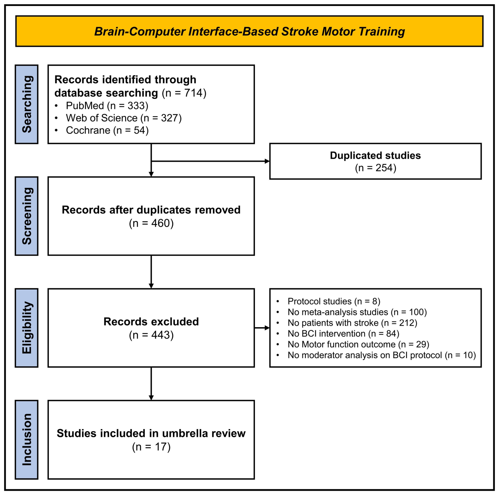
We used three databases: PubMed, Web of Science, and Cochrane. Additionally, duplicate studies were removed prior to screening. Two reviewers (R.K.K. and H.L.) independently screened titles and abstracts against the PICOS eligibility criteria described above, followed by independent manuscript screening. Finally, an initial systematic literature search identified 714 studies, including 333 from PubMed, 327 from Web of Science, and 54 from Cochrane. After excluding 254 duplicate studies, 443 studies were removed based on eligibility criteria: (a) 8 protocol studies, (b) 100 studies without meta-analyses, (c) 212 studies that did not focus on patients with stroke, (d) 84 studies that did not use BCI techniques, (e) 29 studies that did not report motor function outcomes, and (f) 10 studies that did not conduct moderator analyses to identify the effects of BCI protocols. Moderator variable analyses on task and feedback modality, participant characteristics, and training volume were essential for identifying potential heterogeneity of clinical responses. Thus, we included only meta-analyses that performed the moderator variable analyses. Finally, 17 meta-analysis studies were included in this umbrella review. Figure 1 shows the study identification procedures.

2.3. Characteristics of Included Studies

The included 17 meta-analysis studies were published between 2016 and 2026. Thirteen studies focused on a randomized controlled trial (RCT) design. Three studies used combination RCTs with non-RCT or single-group design, and one study only used single-group design. Regarding the stroke phase, the included meta-analyses predominantly focused on individuals in the subacute and chronic phase, with two studies including acute phase stroke patients. Typically, BCI intervention protocols consisted of tasks for the reliable detection of motor intent (motor attempt, observation, and imagery) and feedbacks for inducing neuroplasticity (electrical stimulation, robot, and visual feedback). Methodological quality and certainty of evidence were assessed via the Cochrane RoB, PEDro, and GRADE approaches. The specific characteristics of included studies are detailed in Table 1.
To evaluate upper extremity motor function in patients with stroke, clinical Fugl–Meyer Assessment (FMA) was determined as the primary outcome. In terms of statistical modeling, 10 meta-analyses applied a random-effects model, whereas seven meta-analyses used a fixed-effects model when the I2 value was below 50%. The effects of BCI-based training were evaluated based on the task, feedback, and moderator variables. The specific characteristics of included studies are shown in Supplementary Table S1.

2.4. Assessment of Methodological Quality

The methodological quality of each included meta-analysis was independently assessed by two reviewers (R.K.K. and H.L.) using the Assessing the Methodological Quality of Systematic Reviews 2 (AMSTAR-2) [36]. The AMSTAR-2 consists of 16 items, including seven critical domains, and the overall confidence for each domain was evaluated as high, moderate, low, or critically low based on the presence of critical and non-critical weaknesses. Disagreements between the two reviewers were resolved by a third reviewer (N.K.). The 17 included studies, according to a methodological quality assessment using the AMSTAR 2 tool revealed significant limitations among the 17 included studies. Specifically, eight studies (47.1%) were rated as critically low quality, eight studies (47.1%) as low quality, and only one (5.9%) as high quality. There was no moderate-quality study. Detailed evaluation outcomes were presented in Table 2.

2.5. Overlapping of Primary Studies

The CCA method was employed to evaluate the degree of overlap in primary studies among the 17 included in umbrella review. The results indicated a high overall level of overlap (CCA = 12.0%). For the CCA, one abstract and seven case reports were excluded. This pattern suggests that while most evidence syntheses were based on distinct sets of studies, several key clinical trials were repeatedly included across multiple meta-analyses. Specifically, four studies were included in 15 of 17 systematic reviews and meta-analyses [37,38,39,40]. A complete list of original studies included more than 11 times is provided in Supplementary Table S2.

2.6. Data Extraction and Synthesis

Two reviewers (R.K.K. and H.L.) independently extracted data into a standardized form. For each included meta-analysis, we extracted the first author, publication year, number and design of primary studies, total sample size, stroke phase, outcome measures, BCI task paradigm, feedback modality, pooled effect sizes with 95% confidence intervals, heterogeneity (I2), and the results of subgroup and meta-regression analyses. We qualitatively synthesized the results of each included meta-analysis rather than re-pooling primary data [41,42]. Effect sizes were interpreted as small at 0.2, medium at 0.5, and large at 0.8. A statistically significant p-value indicated that the result could be interpreted according to the magnitude of the effect size [43]. Effect sizes were presented as reported by each meta-analysis (i.e., standardized mean difference, Hedges’ g, or mean difference) with I2. Findings were organized into three domains: (a) task for detecting motor intent, (b) feedback for inducing neuroplasticity, and (c) moderator variables potentially influencing heterogeneity in treatment effects.
We quantified the degree of overlap using the corrected covered area (CCA) because primary studies may be included in more than one meta-analysis. This overlap may lead to double counting of evidence and reduced independence among the included meta-analyses [44]. We calculated the CCA using Pieper’s formula: CCA = (Nr)/(r × cr), where N is the total number of included primary studies counted across all reviews including double counting, r is the number of distinct primary studies, and c is the number of reviews [45]. The CCA was interpreted as slight (0–5%), moderate (6–10%), high (11–15%), or very high (>15%) overlap [46].

3. Comparative Study

3.1. Reliable Detection of Motor Intent

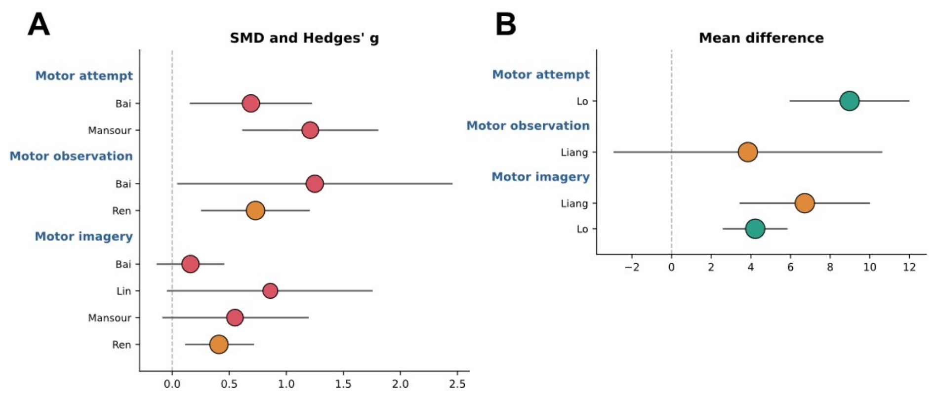
Six meta-analyses estimated the effects of motor intent-induced tasks (i.e., motor attempt, motor observation, and motor imagery) used to trigger and detect cortical activity during BCI-based training. Bai et al. (2020) found that motor attempt (SMD = 0.69, p = 0.010, I2 = 0%) and motor observation (SMD = 1.25, p = 0.040, I2 = 72%) significantly improved upper limb motor function, whereas motor imagery showed no significant improvements (SMD = 0.16, p = 0.290, I2 = 0%) [20]. Mansour et al. (2022) revealed that motor attempt significantly improved upper limb motor function (Hedges’ g = 1.21, p < 0.001, I2 = 42%), although motor imagery failed to show significant improvements (Hedges’ g = 0.55, p = 0.089, I2 = 78%) [27]. Lo et al. (2024), pooling uncontrolled pre-post changes in the FMA-UE, reported significant improvements for both motor attempt (8.98 points; 95% CI = 6.00–11.96; I2 = 0%) and motor imagery (4.22 points; 95% CI = 2.62–5.81; I2 = 0%), and only the motor attempt surpassed the minimal clinically important difference (MCID) of 5.25 [26].
Ren et al. (2024) found significant effects for both motor observation (SMD = 0.73; p = 0.002; I2 = 0%) and motor imagery (SMD = 0.41; p = 0.006; I2 = 13%) [31], whereas Liang et al. (2026) revealed a significant effect for motor imagery (MD = 6.72; p < 0.001; I2 = 30%) but not for motor observation training (MD = 3.85; p = 0.260; I2 = 66%) [24]. In contrast, Lin et al. (2026), a recent meta-analysis focused on motor imagery-based BCI training, found that motor imagery failed to significantly improve upper limb motor function (SMD = 0.86; p = 0.060; I2 = 91%) [25]. Meta-analytic findings indicated that motor attempt during BCI-based training may be a reliable way to trigger and detect motor intent in stroke patients, presumably contributing to motor improvements, whereas administering motor observation and motor imagery remain inconclusive in their treatment effects. However, these findings reflect an indirect, qualitative comparison across meta-analyses that differed in comparator groups and pooled uncontrolled pre-post changes rather than controlled contrasts in Lo et al. (2024) [26]. The forest plots illustrating the effects of different motor intent-induced tasks are shown in Figure 2.

3.2. Effective Neurofeedback for Plasticity Induction

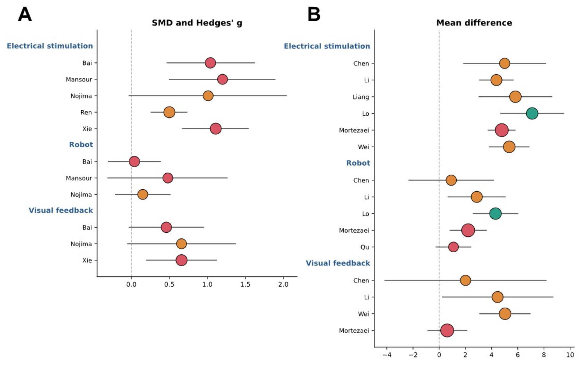
Twelve meta-analyses assessed the effects of BCI-based training combined with neurofeedback (i.e., electrical stimulation, robot, and visual feedback). Bai et al. (2020), Chen & Yun (2026), and Mansour et al. (2022) found that electrical stimulation (i.e., FES and neuromuscular electrical stimulation; NMES) was the only neurofeedback that significantly improved upper limb motor function (SMD = 1.04, p < 0.001, I2 = 37%; MD = 5.00, p = 0.010, I2 = 18%; Hedges’ g = 1.20, p = 0.001, I2 = 47%), and the between-subgroup difference was significant in Bai et al. (2020) (p = 0.010) [20,22,27]. Ren et al. (2024) and Liang et al. (2026), including only BCI-FES studies, confirmed that electrical stimulation improved upper limb motor function (SMD = 0.50, p < 0.001, I2 = 0%; MD = 5.82, p < 0.001, I2 = 39%) [24,31].
Xie et al. (2022) and Wei et al. (2026) found significant effects for both electrical stimulation (SMD = 1.11, p < 0.001, I2 = 11%; MD = 5.35, p < 0.001, I2 = 27%) and visual feedback (SMD = 0.66, p = 0.005, I2 = 4%; MD = 5.03, p < 0.001, I2 = 70%) [32,33]. Further, Lo et al. (2024), restricted to FES and powered exoskeleton and pooling uncontrolled pre-post changes, and Mortezaei et al. (2026) reported significant effects for both electrical stimulation (7.10 points, 95% CI = 4.70–9.50, I2 = 0%; MD = 4.78, 95% CI = 3.75–5.80) and robot (4.30 points, 95% CI = 2.60–6.00, I2 = 0%; MD = 2.21, 95% CI = 0.83–3.60) [26,28]. Li et al. (2025) showed that all three modalities significantly improved upper limb motor function: (a) electrical stimulation (MD = 4.37, p < 0.001, I2 = 36%), (b) robot (MD = 2.87, p = 0.010, I2 = 81%), and (c) visual feedback (MD = 4.46, p = 0.040, I2 = 27%) [14].
In contrast, Qu et al. (2024), restricted to BCI-robot research, found no significant benefit compared with robot therapy alone (MD = 1.09, p = 0.110, I2 = 0%) [30], and Nojima et al. (2022) reported no significant effects for any modality, including electrical stimulation (SMD = 1.01, 95% CI = −0.03–2.04, I2 = 49%), robot (SMD = 0.15, 95% CI = −0.21–0.51, I2 = 7%), and perceptual feedback (SMD = 0.66, 95% CI = −0.05–1.37, I2 = 0%) [29]. Overall, BCI-based training combined with electrical stimulation revealed the most consistent improvements for stroke motor recovery across the meta-analyses. However, several pooled mean differences failed to exceed the reported MCID of 5.25 points, and thus these improvements may not consistently reach a clinically meaningful level. For applying robot and visual feedback, further studies should be necessary for determining its optimal treatment effects. These findings were based on an indirect comparison across meta-analyses with differing comparators and uncontrolled pre-post designs in Lo et al. (2024) [26]. The forest plots illustrating the effects of different BCI-based training combined with neurofeedback are shown in Figure 3.

3.3. Potential Heterogeneity of Clinical Responses

3.3.1. Stroke Phase

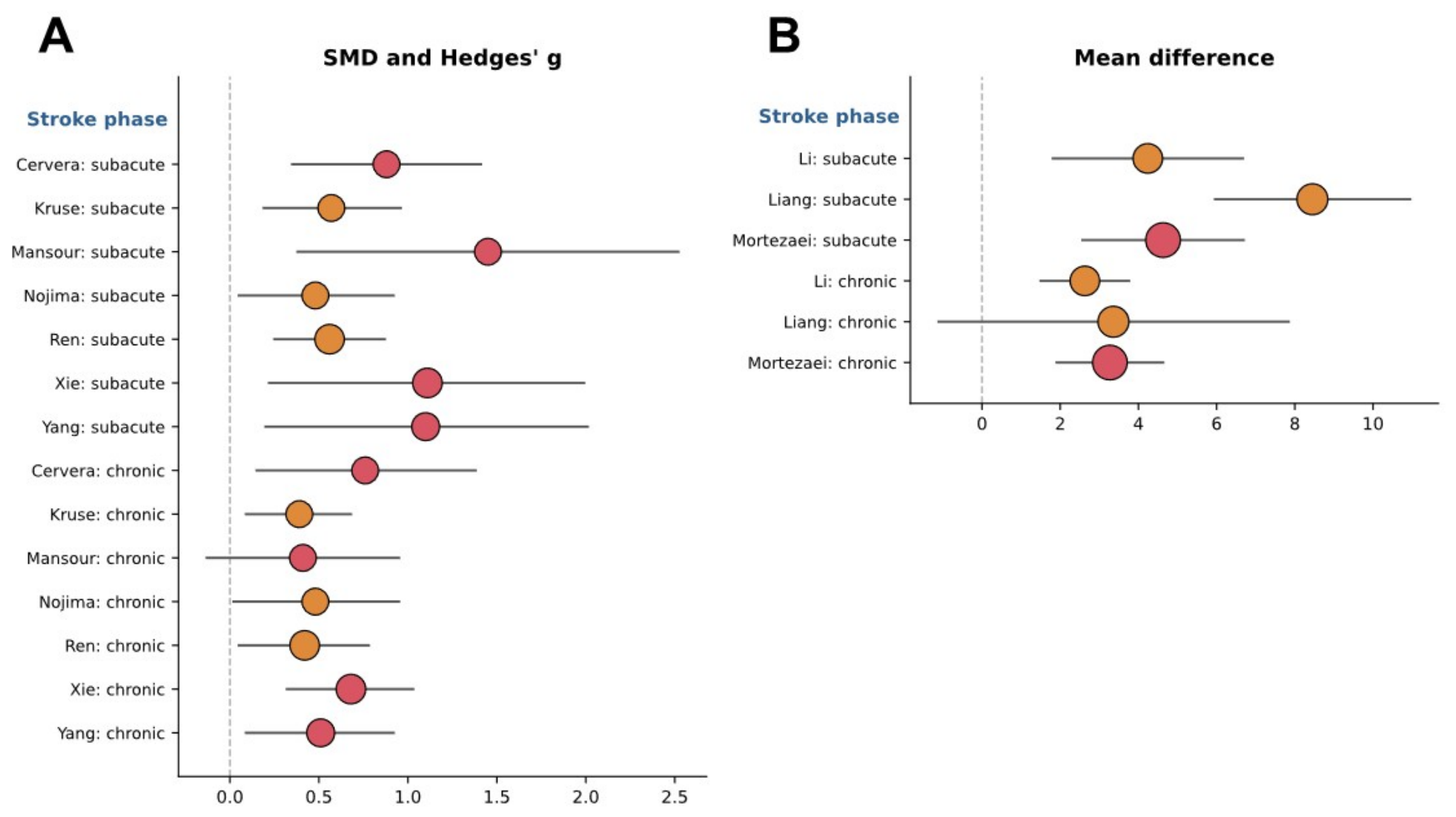
Twelve meta-analyses examined the effects of BCI-based training on upper limb motor function according to stroke phase. Mansour et al. (2022) and Liang et al. (2026) found significant improvements in upper limb motor function only in the subacute phase (Hedges’ g = 1.45; MD = 8.45) with a borderline difference in Liang (2026) (p = 0.050) [24,27]. Eight meta-analyses reported positive effects in both subacute and chronic phases [14,21,23,28,29,31,33,34]: (a) Cervera et al. (2018) (SMD = 0.88 vs. 0.76), (b) Kruse et al. (2020) (SMD = 0.57 vs. 0.39), (c) Nojima et al. (2022) (SMD = 0.48 vs. 0.48), (d) Xie et al. (2022) (SMD = 1.11 vs. 0.68), (e) Yang et al. (2022) (SMD = 1.10 vs. 0.51), (f) Ren et al. (2024) (SMD = 0.56 vs. 0.42), (g) Li et al. (2025) (MD = 4.24 vs. 2.63), and (h) Mortezaei et al. (2026) (MD = 4.63 vs. 3.27). Among these eight meta-analyses, the four meta-analyses that compared the between-phase difference failed to show a significant difference in effects between stroke phases [28,31,33,34]. Further, Wei et al. (2026), who compared time from onset (≤30 vs. >30 days), reported that significant improvements in FMA-UE were observed in both the ≤30 days (MD = 5.02) and >30 days (MD = 4.03), although the between-subgroup difference was not significant (p = 0.240) [32]. Lo et al. (2024) found no relationship between time since stroke and treatment effects (p = 0.790) [26]. These findings suggest that BCI-based training may improve upper limb motor function regardless of stroke phase, although the magnitude of these improvements remained below the MCID in several meta-analyses. The forest plots illustrating the effects of different stroke phases are shown in Figure 4.

3.3.2. Stroke Type

Two meta-analyses investigated the effects of BCI-based training on upper limb motor function according to stroke type. Mortezaei et al. (2026) found that a greater proportion of hemorrhagic stroke was significantly associated with motor function improvements (r = 0.28, p = 0.008) by meta-regression [28], whereas Lo et al. (2024) found no significant relationship between stroke type and treatment effects (p = 0.180) [26]. These findings suggest that the influence of stroke type on BCI-based training remains inconclusive and requires further investigation.

3.3.3. Age

Four meta-analyses evaluated the effects of BCI-based training on upper limb motor function according to patient age. Lo et al. (2024) found that patients ≤ 50 years old reached the minimal clinically important difference more often than those >50 years old (p = 0.002) [26]. Wei et al. (2026) reported that significant improvements in FMA-UE were observed in both patients ≤ 60 years (MD = 4.41) and >60 years (MD = 5.66), although the between-subgroup difference was not significant (p = 0.460) [32]. In contrast, Ren et al. (2024) and Mortezaei et al. (2026) found no association between age and effect size by meta-regression [28,31]. Overall, the evidence on patient age remains inconsistent.

3.3.4. Baseline Severity

Three meta-analyses examined the effects of BCI-based training on upper limb motor function according to baseline severity. Lo et al. (2024) found that patients with mild or moderate impairment reached the minimal clinically important difference more often than those with severe impairment (p = 0.042) [26]. Wei et al. (2026) found significant improvements at an initial FMA-UL ≤ 23 (MD = 6.34) [32]. Kruse et al. (2020) found no association between baseline impairment and effect size by meta-regression [23]. These findings suggest that the influence of baseline severity on BCI-based training remains limited.

3.3.5. Session Duration

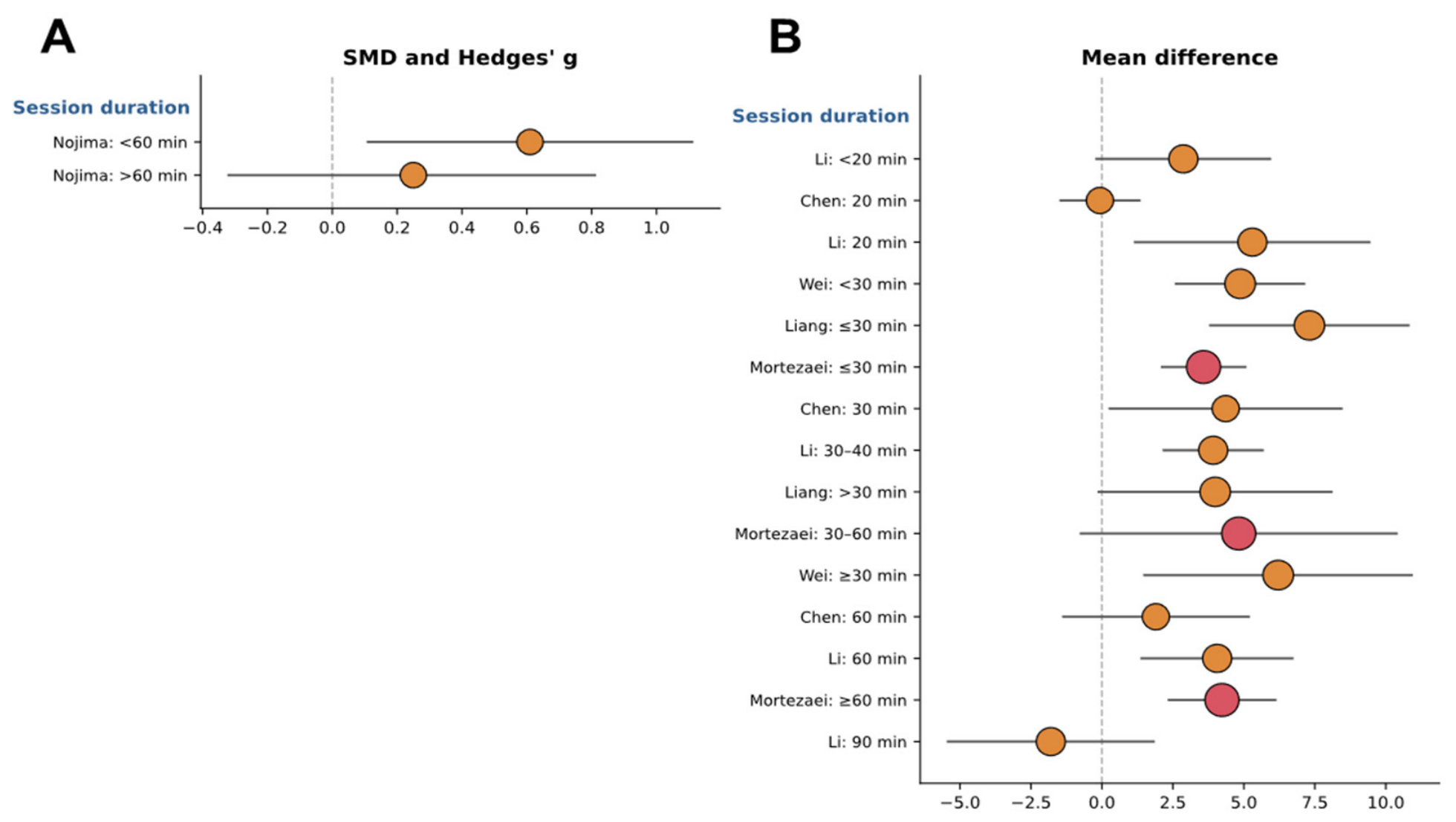
Seven meta-analyses assessed the effects of BCI-based training on upper limb motor function according to session duration. A significant effect appeared at <60 min (SMD = 0.61) in Nojima et al. (2022), at ≤30 min (MD = 7.31) in Liang et al. (2026), and only at 30 min (MD = 4.36) in Chen & Yun (2026) [22,24,29]. Mortezaei et al. (2026) reported significant effects at both ≤30 min (MD = 3.58) and ≥60 min (MD = 4.23) [28]. Further, Wei et al. (2026) showed significant effects at both <30 min (MD = 4.87) and ≥30 min (MD = 6.21) [32]. Li et al. (2025) found significant improvements at 20 min (MD = 5.30), 30–40 min (MD = 3.92), and 60 min (MD = 4.06), with no significant effects at <20 min or 90 min [14]. Finally, Ren et al. (2024) found no association between session duration and effect size by meta-regression [31]. Overall, significant effects appeared mainly at moderate durations (approximately 20–60 min). The forest plots illustrating the effects of different session duration are shown in Figure 5.

3.3.6. Session Frequency

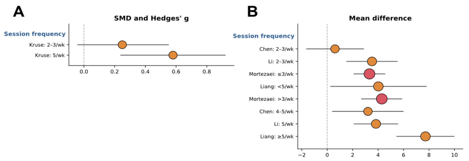
Five meta-analyses estimated the effects of BCI-based training on upper limb motor function according to session frequency. A significant effect appeared only at 4–5 sessions/week (MD = 3.20) in Chen & Yun (2026) and at 5 sessions/week (SMD = 0.58) in Kruse et al. (2020) [22,23]. Further, greater effects were observed at a higher frequency in Li et al. (2025) (5 sessions/week, MD = 3.83; 2–3 sessions/week, MD = 3.52), Liang et al. (2026) (≥5 sessions/week, MD = 7.72; <5 sessions/week, MD = 4.02), and Mortezaei et al. (2026) (>3 sessions/week, MD = 4.29; ≤3 times/week, MD = 3.32) [14,24,28]. Overall, significant effects consistently occurred at higher session frequency, although no between-subgroup difference reached significance. The forest plots illustrating the effects of different session frequency are shown in Figure 6.

3.3.7. Intervention Period

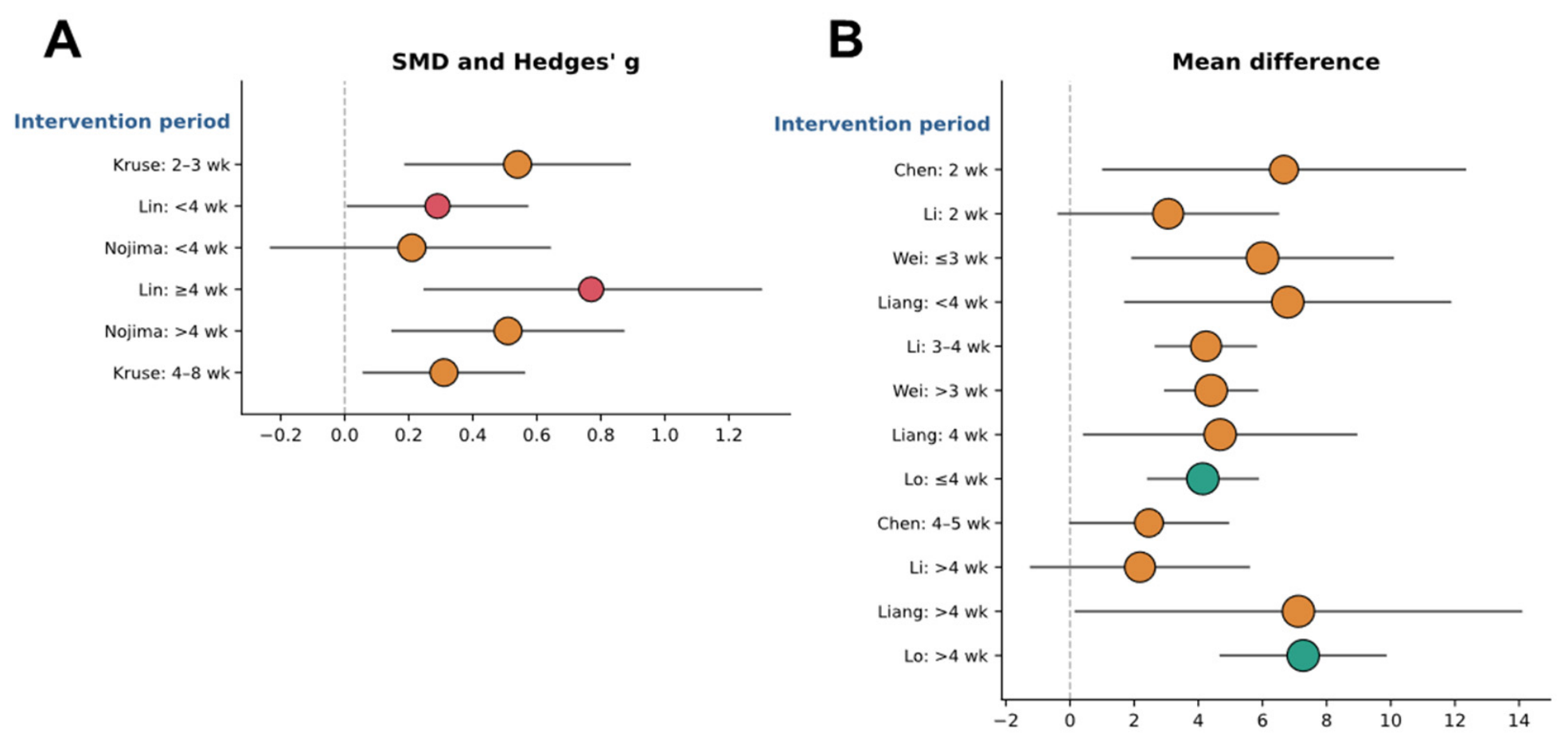
Nine meta-analyses examined the effects of BCI-based training on upper limb motor function according to the intervention period. Shorter periods (≤4 weeks) were favored in Li et al. (2025) (3–4 weeks, MD = 4.24) [14]. Longer periods (>4 weeks) were favored in Nojima et al. (2022) (>4 weeks, SMD = 0.51) [29]. Both shorter and longer periods were significant in Kruse et al. (2020) (2–3 weeks, SMD = 0.54; 4–8 weeks, SMD = 0.31), Lo et al. (2024) (≤4 weeks, MD = 4.14; >4 weeks, MD = 7.27), Chen & Yun (2026) (2 weeks, MD = 6.67; 4–5 weeks, MD = 2.46), Lin et al. (2026) (<4 weeks, SMD = 0.29; ≥4 weeks, SMD = 0.77), Liang et al. (2026) (<4 weeks, MD = 6.79; 4 weeks, MD = 4.68; >4 weeks, MD = 7.12), Mortezaei et al. (2026) (<1 month, MD = 5.74; 1 month, MD = 3.06; >1 month, MD = 3.70), and Wei et al. (2026) (≤3 weeks, MD = 6.00; >3 weeks, MD = 4.40) [22,23,24,25,26,28,32]. Among these meta-analyses, a significant between-subgroup difference was found only in Lo et al. (2024) [33], favoring >4 weeks (p = 0.047). Overall, the optimal intervention period remained inconsistent across meta-analyses. The forest plots illustrating the effects of different intervention periods are shown in Figure 7.

3.3.8. Total Sessions

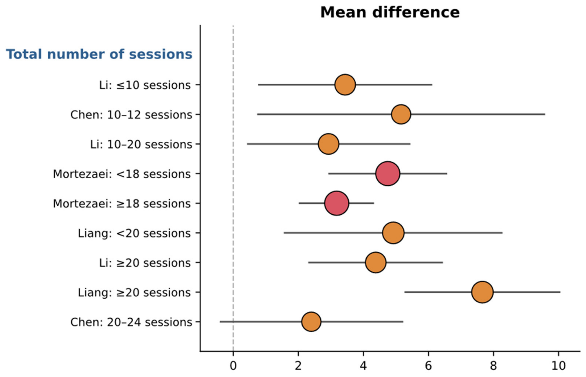
Six meta-analyses evaluated the effects of BCI-based training on upper limb motor function according to the total number of sessions. Chen & Yun (2026) found a significant effect only at 10–12 sessions (MD = 5.16) [22]. Greater effects with fewer sessions were reported in Mortezaei et al. (2026) (<18 sessions, MD = 4.75; ≥18 sessions, MD = 3.18) [28], whereas greater effects with more sessions were reported in Li et al. (2025) (≤10 sessions, MD = 3.44; 10–20 sessions, MD = 2.93; ≥20 sessions, MD = 4.38) and Liang et al. (2026) (<20 sessions, MD = 4.92; ≥20 sessions, MD = 7.66) [14,24]. In contrast, meta-regression in Bai et al. (2020) and Nojima et al. (2022) found no association between the number of sessions and effect size [20,29]. Overall, the optimal number of sessions remained inconsistent across meta-analyses, with no between-subgroup difference reaching significance. The forest plot illustrating the effects of different total sessions is shown in Figure 8.

3.3.9. Total Training Time

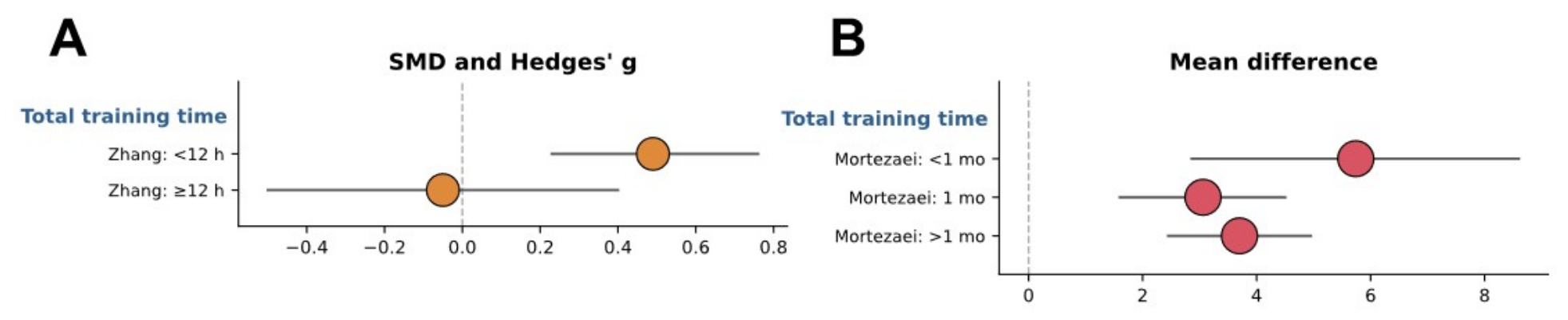
Three meta-analyses investigated the effects of BCI-based training on upper limb motor function according to the total training time. A significant effect appeared only at <12 h of cumulative training in Zhang et al. (2024) (SMD = 0.49), with no significant effect at ≥12 h [35]. In contrast, meta-regression in Bai et al. (2020) and Ren et al. (2024) found no association between cumulative training time and effect size [20,31]. Overall, current evidence is insufficient to determine the optimal total training time. The forest plots illustrating the effects of different total training time are shown in Figure 9.

3.3.10. Long-Term Effects

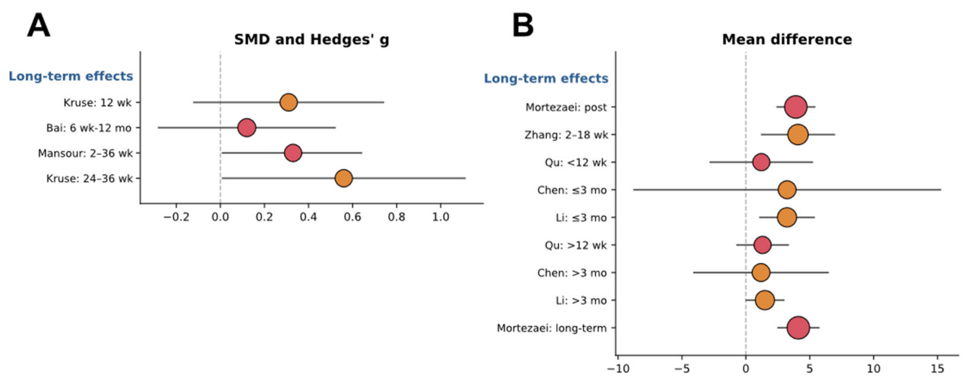
Eight meta-analyses assessed the long-term effects of BCI-based training on upper limb motor function. The improvements were sustained at follow-up in Kruse et al. (2020) (24–36 weeks, SMD = 0.56), Mansour et al. (2022) (4–36 weeks, Hedges’ g = 0.33), Zhang et al. (2024) (2–18 weeks, MD = 4.08), Li et al. (2025) (≤3 months, MD = 3.22; >3 months, MD = 1.49), and Mortezaei et al. (2026) (2–12 months, MD = 4.11) [14,23,27,28,35], whereas the benefits were not maintained in Bai et al. (2020) (6 weeks and 12 months), Qu et al. (2024) (<12 weeks and >12 weeks), and Chen & Yun (2026) (≤3 months and >3 months) [20,22,30]. Overall, the durability of BCI-based training effects remained inconsistent across meta-analyses. The forest plots illustrating the effects of different long-term effects are shown in Figure 10.

4. Discussion

This umbrella review provides integrated perspectives from the meta-analytic evidence investigating effects of BCI-based training on upper limb recovery after stroke. Among the three modalities of motor intent-induced tasks (i.e., motor attempt, motor observation, and motor imagery), motor attempt was the modality most consistently associated with significant therapeutic effects across meta-analyses, although this comparison is indirect and potentially confounded by differences in comparators and study designs across reviews. In contrast, the clinical utility of motor imagery and motor observation remained inconclusive due to inconsistent findings across meta-analyses. Electrical stimulation (i.e., FES and NMES) was the feedback modality most consistently associated with significant effects across meta-analyses, whereas robot-assisted and visual feedback showed heterogeneous effects. As with the task comparison above, this pattern reflects an indirect comparison across reviews with differing comparators and study designs. Regarding characteristics of stroke patients, BCI-based training was effective regardless of time since stroke onset, improving upper limb motor function in both subacute and chronic stroke patients. For training volume, higher weekly session frequencies (≥3–5 sessions/week) combined with moderate session durations (approximately 20–60 min) showed a consistent direction toward motor recovery. However, several pooled mean differences failed to exceed the reported MCID of 5.25 points. Thus, the statistical significance of these improvements may not consistently correspond to a clinically meaningful change. Finally, the long-term effects of BCI-based training on motor recovery were inconclusive across the included meta-analyses.

4.1. Detecting Motor Intent-Induced Brain Signals

Given that BCI-based training is mostly based on a closed-loop system, sensitively detecting brain activity from patients and appropriately triggering effectors may be critical for optimizing treatment effects. We found that motor attempt tasks during BCI-based training improved motor functions in stroke patients. In addition to motor dysfunctions, cognitive deficits such as impaired attention and executive functions normally appear in 30–70% of stroke survivors [47,48,49]. Then, top-down approaches such as motor imagery tasks requiring internally driven brain signals without external stimuli may be difficult for stroke patients because these processes induce more reliance on higher levels of focus, memory, and executive function potentially leading to mental fatigue [50,51]. Prior findings indicated that one critical challenge of motor imagery tasks is BCI illiteracy, where users fail to generate the necessary brain signals because of individual differences (e.g., different cognitive function and motivation levels) interfering with the effective use of BCI systems [52,53]. On the other hand, several studies argued that motor attempt tasks with paretic arms may naturally induce brain activity that can be easily monitored by EEG techniques, as compared with motor imagery tasks even for stroke patients who have additional cognitive impairments [54,55]. Moreover, motor observation tasks combined with BCI training may easily facilitate brain activation across motor simulation-related areas according to the mirror neuron theory assuming that motor cortical regions trigger both during the execution of a movement and the observation of comparable motor actions [56]. These findings suggest that the bottom-up approaches including motor attempt and motor observation presumably reduce cognitive loads in stroke patients via supports from external sensory inputs [57,58,59].
Another option to improve BCI performance is to apply the performance accommodation mechanism (PAM) that encourages users to actively participate in BCI training programs by adjusting the challenge level of the BCI system [60]. In fact, 10–30% of users experienced BCI illiteracy (i.e., inability to control the motor imagery–BCI system) potentially reducing motivation. Therefore, an alternative to deal with this limitation is necessary to improve BCI performances leading to brain plasticity in stroke patients. Interestingly, a recent study by Jochumsen and colleagues investigated effects of PAMs, including augmented success, mitigated failure, and input override perceived control and frustration [60]. The findings indicated that input override PAM (i.e., turning an unsuccessful BCI attempt into a successful output) significantly increased frustration because patients might want to execute the task by themselves, whereas augmented success did not reduce perceived control and increased frustration. Importantly, applying PAM advanced perceived control in patients with poor BCI performance. Presumably, developing individualized BCI protocols using various combinations of PAMs may optimize motor improvements in patients who have different levels of motor and cognitive functions. Finally, synchronizing motor imagery cues with respiration may improve motor performances in stroke patients [61]. Specifically, applying a fNIRS-based BCI and robot-assisted system with synchronization between motor imagery cues and the inhalation phase reduced the variability of oxyhemoglobin levels associated with better BCI decoding accuracy. Consequently, this enhanced decoding accuracy based on successful motor intent may promote precise neurofeedback improving motor functions in stroke patients.
Although previous findings reported relatively lower impacts of motor imagery tasks on motor improvements, BCI protocols using motor imagery tasks may be still useful for patients with more severe motor deficits. Recent studies suggest the possibility of combining tasks such as motor attempt with motor imagery or action observation with motor imagery, which may induce stronger activation and connectivity across sensorimotor areas than those for sole motor imagery tasks during BCI training [62,63]. For example, a BCI-based training requiring movement attempts of the paretic upper limb following motor imagery improved motor functions (i.e., FMA-UE and ARAT), and the improvements in ARAT scores were significantly correlated with increased movement-related cortical activity in the lesional hemisphere [64]. Further, performing motor attempt with motor imagery tasks enhanced motor-related brain activations, including supplementary motor area and primary motor cortex [62]. Executing motor observation with motor imagery tasks increased the brain network, including sensory and visual-related regions, due to the critical role of action observation in improving attention [63]. Taken together, motor attempt may provide a more reliable method for detecting motor intent during BCI-based training; further combined-task approaches may enhance sensorimotor engagement and neuroplastic adaptations, improving the effectiveness of neurofeedback-driven motor recovery in stroke patients.

4.2. Effective BCI Neurofeedback for Inducing Optimal Plasticity

After detecting brain activation patterns, a closed-loop BCI system normally controls and provides electrical, robotic, or visual feedback to the patient presumably contributing to motor learning and rewiring neural pathways. Applying electrical stimulation may successfully translate motor intentions into actual muscle contractions via a closed-loop feedback mechanism, potentially augmenting neuroplasticity and sensorimotor coupling so that both motor function and the patient’s motivation could be improved [31]. Further, previous studies suggested that BCI-FES directly stimulates muscle contraction for functional movement execution with simultaneous proprioceptive feedback as compared with other BCI combined protocols [65,66]. Theoretically, BCI-based rehabilitation programs are based on the Hebbian principles of associativity assuming that stronger correlation between brain activity and feedback strengthen synaptic connections [67,68]. Thus, administering the precise coupling between the motor intention and the afferent signal (i.e., electrical stimulation) during BCI system driving the Hebbian plasticity is important for optimizing functional improvements [8].
Although most BCI-based training studies have focused on functional recovery of upper extremity for stroke patients, BCI-based training would be a viable option for lower limb rehabilitation. In 2020, first meta-analytic findings reported that BCI-FES approaches failed to show significant improvements in lower limb function [23]. However, these findings from two studies could be affected by short period of intervention (e.g., three to five days) [8,69]. Recent two meta-analyses focused on lower limb found that BCI-based training significantly improved lower limb motor function and activities of daily living [70,71]. Contrary to conventional gait training, BCI-FES system requires no bodyweight support devices so that patients can receive the training while seated with minimal fall risk [72]. Potentially, the BCI-based training highlights the crucial role of neurofeedback in promoting functional lower limb recovery post stroke.

4.3. Potential Heterogeneity of Clinical Responses in Current BCI System

Although many meta-analysis studies reported the evidence of BCI-based training protocols for stroke motor recovery, investigating methodological issues that may influence treatment effects would be necessary. Firstly, treatment effects of BCI-based training may be affected by stroke phases because of progressively altered brain plasticity in stroke patients [73,74]. Previous studies mainly focused on motor recovery of subacute (i.e., shorter than six months post stroke) and chronic (i.e., longer than six months post stroke) patients. The cumulative findings from multiple meta-analyses indicated that BCI-based training protocols seem to be favorable methods for upper limb motor improvements across subacute and chronic stroke patients. Considering the traditional hypothesis that a critical window (i.e., first six months post stroke) for motor recovery may exist [74], significant motor improvements observed for both subacute and chronic stroke patients indicate that BCI-based training system is an attractive rehabilitation option regardless of time since stroke. Beyond stroke phase, other patient characteristics including stroke type, age, and baseline severity showed inconsistent effects. However, clinical implementation of BCI-based training is still limited because current systems normally require complicated setup procedures influencing patient comfort, greater calibration time (e.g., unstable EEG signal quality), and higher equipment cost. These practical barriers of the BCI system may hinder integration into standard clinical practice for subacute and chronic stroke patients. To enable intensive and frequent training beyond laboratory setting, recent studies suggest portable and cost-effective BCI platforms (e.g., dry EEG electrode or two-channel EEG recording and a head-mounted display for visual feedback) for improving usability [75,76]. Presumably, these innovative systems can strengthen the feasibility of implementing BCI-based training into routine clinical environments, further contributing to long-term motor improvements.
Next, levels of training volume that can be modulated by session duration, session frequency, intervention period, and the number of sessions may influence treatment effects of BCI-based rehabilitation. Across meta-analyses, higher frequency and moderate session duration (i.e., approximately 20–60 min) tended to show greater gains in upper limb motor function, whereas findings regarding the intervention period, the number of sessions, and the total training time were less consistent across meta-analyses. Meta-regression analyses found that none of these parameters of training volume including session duration, session frequency, the intervention period, the number of sessions, and the total training time were significantly related to BCI-based training effects [20,23,28,29,31]. Several subgroup analyses within the included meta-analyses, however, reported significantly greater effects at longer session durations (e.g., ≥30–60 min) [28,32], a greater number of sessions (e.g., ≥20 sessions) [14,24], or a longer intervention period (e.g., >4 weeks) [26]. Taken together, these findings suggest that the absence of a significant moderator effect may reflect insufficient current evidence to determine an optimal training volume for BCI-based rehabilitation. A BCI system normally requires more concentration from participants on instructions and feedback, leading to greater cognitive fatigue [29]. In fact, a prior study reported that most patients felt subjective fatigue after 20–30 min of BCI training [77]. Future studies may therefore systematically investigate training volume parameters such as session duration, frequency, and intervention period, while incorporating strategies to reduce task-related cognitive fatigue during implementation, including simplifying task demands, providing more intuitive feedback, and incorporating short rest periods. Further, individualizing training intensity based on patient’s motor and cognitive impairments could increase the sustainability of treatment effects with less heterogeneity.
Finally, the sustainability of motor improvements after BCI-based training are still controversial. Five out of eight studies reported that upper limb motor improvements may appear in both immediate posttest and follow-up test although long-term effects are carefully interpreted because of insufficient sample size (two to seven included studies) and various follow-up periods (two weeks to 12 months) [14,23,27,28,35]. As Li and colleagues suggested [14], incorporating long-term follow-up tests into a study would be further required to build more effective treatment regimens advancing the sustainability of treatment effects.

4.4. Limitations and Future Research Directions

This umbrella review has several limitations. Methodologically, 16 of 17 included meta-analyses were rated low or critically low on AMSTAR-2. These lower ratings appear to have been mainly driven by reporting deficiencies. First, most studies did not report lists of excluded studies. Second, funding sources were frequently unreported. Third, protocol registration was only partially complied with. Given that previous umbrella reviews in stroke rehabilitation have similarly reported a majority of low or critically low AMSTAR-2 ratings [78,79], these methodological limitations may reflect a structural characteristic of the field rather than a deficiency specific to the present review.
Further, the CCA of 12.0% indicated a high degree of overlap among the primary studies analyzed across the included meta-analyses. Thus, the current findings may not represent fully independent replications of evidence. Given that outcome measurements differed across the meta-analyses and this review primarily aimed to summarize outcome variables rather than to perform a quantitative synthesis, we did not apply statistical corrections for the overlapping studies [78,80]. Future studies may focus on large-scale clinical trials for strengthening the current evidence on BCI-based motor rehabilitation after stroke.
Additionally, potential heterogeneity exceeded 50% in seven meta-analyses that applied a fixed-effects model, as the evidence base for BCI-based stroke rehabilitation was limited by data availability. This may have overstated the precision of the affected estimates and should be interpreted cautiously. Standardized large-scale neural data from stroke patients remain relatively scarce. Widely used benchmark datasets were mostly collected from healthy individuals. These datasets presumably do not fully reflect the altered neural signal patterns characteristic of stroke. This limitation appears consistent with the structural constraint identified in the present review. A small number of primary trials were repeatedly reused across the included meta-analyses without independent verification. Nonetheless, several stroke-specific datasets are increasingly available. These include an EEG motor imagery dataset in acute stroke patients, a lower-limb motor imagery dataset with longitudinal follow-up, and a multi-session NIRS dataset. These datasets were summarized in Supplementary Table S3 [81,82,83,84,85].
Although this umbrella review focused on motor function of stroke patients, the included meta-analyses mainly reported an FMA of unilateral motor impairment rather than bilateral motor function requiring interhemispheric balance. Stroke disrupts the normal interhemispheric inhibition, leading to greater interhemispheric asymmetry [86]. The observed motor improvement following BCI-based training could potentially be related to increased interhemispheric balance or enhanced interhemispheric brain function [87]. BCI-based rehabilitation interventions promote favorable interhemispheric neuroplastic changes after stroke, including reduced abnormal interhemispheric inhibition, and strengthened interhemispheric functional connectivity [88,89,90]. For example, BCI-based robot training reduced interhemispheric asymmetry and improved lower limb motor function, suggesting BCI approach may enhance motor function by balancing theses neurophysiological changes [88]. Considering that BCI-based training operates through a central–peripheral–central loop that directly links cerebral cortical activity with peripheral feedback, this closed-loop mechanism may be particularly well suited for strengthening interhemispheric balance. Therefore, future research should evaluate interhemispheric symmetry to clarify the effects of BCI-based rehabilitation.

5. Conclusions

BCI-based training may improve upper extremity motor function in patients with stroke who have moderate to severe motor impairments. These improvements were most consistent when motor attempt was combined with electrical stimulation. To overcome the inconclusive long-term effects and high heterogeneity of alternative modalities (such as motor imagery, motor observation, robotic, or visual feedback), future studies should conduct well-designed, rigorously controlled clinical trials to clarify whether BCI–NIBS integration systems can reveal reliable and clinically meaningful benefits on stroke motor rehabilitation.

Supplementary Materials

The following supporting information can be downloaded at https://www.mdpi.com/article/10.3390/sym18091484/s1, Table S1: Results of the included meta-analyses; Table S2: Overlapping of primary studies; Table S3: Summary of publicly available, stroke-specific open-source datasets relevant to BCI-based motor rehabilitation research.

Author Contributions

Conceptualization, N.K.; methodology, N.K.; software, R.K.K. and H.L.; validation, N.K.; formal analysis, N.K.; investigation, R.K.K. and H.L.; resources, N.K.; data curation, R.K.K. and H.L.; writing—original draft preparation, R.K.K. and H.L.; writing—review and editing, N.K.; visualization, N.K.; supervision, N.K.; project administration, N.K.; funding acquisition, N.K. All authors have read and agreed to the published version of the manuscript.

Funding

This work was supported by Incheon National University Research Grant in 2025 (2025-0016) to NK.

Data Availability Statement

No new data were created or analyzed in this study. Data sharing is not applicable to this article.

Acknowledgments

During the preparation of this manuscript, the author(s) used Claude (3.5 Sonnet version) and Gemini (2.0 Flash version) for the purposes of figures, language editing, grammar checking, and improving text readability during manuscript preparation. The authors have reviewed and edited the output and take full responsibility for the content of this publication.

Conflicts of Interest

The authors declare no conflicts of interest.

Abbreviations

The following abbreviations are used in this manuscript:
ARATAction Research Arm Test
BCIBrain–Computer Interface
CCACorrected Covered Area
EEGElectroencephalogram
FESFunctional Electrical Stimulation
FMAFugl–Meyer Assessment
FMA-UEFugl–Meyer Assessment of the Upper Extremity
MBIModified Barthel Index
NIBSNon-invasive Brain Stimulation
NMESNeuromuscular Electrical Stimulation
PRISMAPreferred Reporting Items for Systematic Reviews and Meta-Analyses
RCTRandomized Controlled Trial
VRVirtual reality
WMFTWolf Motor Function Test

References

  1. Feigin, V.L.; Brainin, M.; Norrving, B.; Martins, S.O.; Pandian, J.; Lindsay, P.; Grupper, M.F.; Rautalin, I. World Stroke Organization: Global Stroke Fact Sheet 2025. Int. J. Stroke 2025, 20, 132–144. [Google Scholar] [CrossRef] [Scilit] [PubMed]
  2. Kim, Y.W. Update on Stroke Rehabilitation in Motor Impairment. Brain Neurorehabil. 2022, 15, e12. [Google Scholar] [CrossRef] [Scilit] [PubMed]
  3. Cauraugh, J.H.; Kang, N. Bimanual Movements and Chronic Stroke Rehabilitation: Looking Back and Looking Forward. Appl. Sci. 2021, 11, 10858. [Google Scholar] [CrossRef] [Scilit]
  4. Mazzoleni, S.; Puzzolante, L.; Zollo, L.; Dario, P.; Posteraro, F. Mechanisms of motor recovery in chronic and subacute stroke patients following a robot-aided training. IEEE Trans. Haptics 2014, 7, 175–180. [Google Scholar] [CrossRef] [Scilit] [PubMed]
  5. Li, X.; He, Y.; Wang, D.; Rezaei, M.J. Stroke rehabilitation: From diagnosis to therapy. Front. Neurol. 2024, 15, 1402729. [Google Scholar] [CrossRef] [Scilit] [PubMed]
  6. Cabral, D.F.; Fried, P.; Koch, S.; Rice, J.; Rundek, T.; Pascual-Leone, A.; Sacco, R.; Wright, C.B.; Gomes-Osman, J. Efficacy of mechanisms of neuroplasticity after a stroke. Restor. Neurol. Neurosci. 2022, 40, 73–84. [Google Scholar] [CrossRef] [Scilit] [PubMed]
  7. Li, S. Spasticity, Motor Recovery, and Neural Plasticity after Stroke. Front. Neurol. 2017, 8, 120. [Google Scholar] [CrossRef] [Scilit] [PubMed]
  8. Mrachacz-Kersting, N.; Jiang, N.; Stevenson, A.J.; Niazi, I.K.; Kostic, V.; Pavlovic, A.; Radovanovic, S.; Djuric-Jovicic, M.; Agosta, F.; Dremstrup, K.; et al. Efficient neuroplasticity induction in chronic stroke patients by an associative brain-computer interface. J. Neurophysiol. 2016, 115, 1410–1421. [Google Scholar] [CrossRef] [Scilit] [PubMed]
  9. Silvoni, S.; Ramos-Murguialday, A.; Cavinato, M.; Volpato, C.; Cisotto, G.; Turolla, A.; Piccione, F.; Birbaumer, N. Brain-computer interface in stroke: A review of progress. Clin. EEG Neurosci. 2011, 42, 245–252. [Google Scholar] [CrossRef] [Scilit] [PubMed]
  10. Cantillo-Negrete, J.; Carino-Escobar, R.I.; Ortega-Robles, E.; Arias-Carrion, O. A comprehensive guide to BCI-based stroke neurorehabilitation interventions. MethodsX 2023, 11, 102452. [Google Scholar] [CrossRef] [Scilit] [PubMed]
  11. Mang, J.; Xu, Z.; Qi, Y.; Zhang, T. Favoring the cognitive-motor process in the closed-loop of BCI mediated post stroke motor function recovery: Challenges and approaches. Front. Neurorobot. 2023, 17, 1271967. [Google Scholar] [CrossRef] [Scilit] [PubMed]
  12. Wang, W.; Collinger, J.L.; Perez, M.A.; Tyler-Kabara, E.C.; Cohen, L.G.; Birbaumer, N.; Brose, S.W.; Schwartz, A.B.; Boninger, M.L.; Weber, D.J. Neural interface technology for rehabilitation: Exploiting and promoting neuroplasticity. Phys. Med. Rehabil. Clin. N. Am. 2010, 21, 157–178. [Google Scholar] [CrossRef] [Scilit] [PubMed]
  13. Shou, Y.Z.; Wang, X.H.; Yang, G.F. Verum versus Sham brain-computer interface on upper limb function recovery after stroke: A systematic review and meta-analysis of randomized controlled trials. Medicine 2023, 102, e34148. [Google Scholar] [CrossRef] [Scilit] [PubMed]
  14. Li, D.; Li, R.; Song, Y.; Qin, W.; Sun, G.; Liu, Y.; Bao, Y.; Liu, L.; Jin, L. Effects of brain-computer interface based training on post-stroke upper-limb rehabilitation: A meta-analysis. J. Neuroeng. Rehabil. 2025, 22, 44. [Google Scholar] [CrossRef] [Scilit] [PubMed]
  15. Peng, Y.; Wang, J.; Liu, Z.; Zhong, L.; Wen, X.; Wang, P.; Gong, X.; Liu, H. The Application of Brain-Computer Interface in Upper Limb Dysfunction After Stroke: A Systematic Review and Meta-Analysis of Randomized Controlled Trials. Front. Hum. Neurosci. 2022, 16, 798883. [Google Scholar] [CrossRef] [Scilit] [PubMed]
  16. Di Pino, G.; Pellegrino, G.; Assenza, G.; Capone, F.; Ferreri, F.; Formica, D.; Ranieri, F.; Tombini, M.; Ziemann, U.; Rothwell, J.C.; et al. Modulation of brain plasticity in stroke: A novel model for neurorehabilitation. Nat. Rev. Neurol. 2014, 10, 597–608. [Google Scholar] [CrossRef] [Scilit] [PubMed]
  17. Irimia, D.C.; Cho, W.; Ortner, R.; Allison, B.Z.; Ignat, B.E.; Edlinger, G.; Guger, C. Brain-Computer Interfaces With Multi-Sensory Feedback for Stroke Rehabilitation: A Case Study. Artif. Organs 2017, 41, E178–E184. [Google Scholar] [CrossRef] [Scilit] [PubMed]
  18. Ono, T.; Shindo, K.; Kawashima, K.; Ota, N.; Ito, M.; Ota, T.; Mukaino, M.; Fujiwara, T.; Kimura, A.; Liu, M.; et al. Brain-computer interface with somatosensory feedback improves functional recovery from severe hemiplegia due to chronic stroke. Front. Neuroeng. 2014, 7, 19. [Google Scholar] [CrossRef] [Scilit] [PubMed]
  19. Page, M.J.; McKenzie, J.E.; Bossuyt, P.M.; Boutron, I.; Hoffmann, T.C.; Mulrow, C.D.; Shamseer, L.; Tetzlaff, J.M.; Akl, E.A.; Brennan, S.E. The PRISMA 2020 statement: An updated guideline for reporting systematic reviews. BMJ 2021, 372, n71. [Google Scholar] [CrossRef] [Scilit] [PubMed]
  20. Bai, Z.; Fong, K.N.K.; Zhang, J.J.; Chan, J.; Ting, K.H. Immediate and long-term effects of BCI-based rehabilitation of the upper extremity after stroke: A systematic review and meta-analysis. J. Neuroeng. Rehabil. 2020, 17, 57. [Google Scholar] [CrossRef] [Scilit] [PubMed]
  21. Cervera, M.A.; Soekadar, S.R.; Ushiba, J.; Millan, J.D.R.; Liu, M.; Birbaumer, N.; Garipelli, G. Brain-computer interfaces for post-stroke motor rehabilitation: A meta-analysis. Ann. Clin. Transl. Neurol. 2018, 5, 651–663. [Google Scholar] [CrossRef] [Scilit] [PubMed]
  22. Chen, H.; Yun, G. Efficacy of Brain-Computer Interface Therapy for Upper Limb Rehabilitation in Chronic Stroke: Systematic Review and Meta-Analysis of Randomized Controlled Trials. J. Med. Internet Res. 2026, 28, e79132. [Google Scholar] [CrossRef] [Scilit] [PubMed]
  23. Kruse, A.; Suica, Z.; Taeymans, J.; Schuster-Amft, C. Effect of brain-computer interface training based on non-invasive electroencephalography using motor imagery on functional recovery after stroke—A systematic review and meta-analysis. BMC Neurol. 2020, 20, 385. [Google Scholar] [CrossRef] [Scilit] [PubMed]
  24. Liang, F.; Chen, X.; Li, B.; Yang, B. Brain-Computer Interface Combined with Functional Electrical Stimulation for Post-Stroke Upper Limb Motor Recovery: A Systematic Review and Meta-Analysis. Clin. EEG Neurosci. 2026, 15500594261441055. [Google Scholar] [CrossRef] [Scilit] [PubMed]
  25. Lin, Y.; Yuan, Y.; Chen, J.; Lin, X. Motor imagery combined with brain-computer interface for stroke patients: A meta-analysis. Front. Neurol. 2026, 17, 1672882. [Google Scholar] [CrossRef] [Scilit] [PubMed]
  26. Lo, Y.T.; Lim, M.J.R.; Kok, C.Y.; Wang, S.; Blok, S.Z.; Ang, T.Y.; Ng, V.Y.P.; Rao, J.P.; Chua, K.S.G. Neural interface-based motor neuroprosthesis in poststroke upper limb neurorehabilitation: An individual patient data meta-analysis. Arch. Phys. Med. Rehabil. 2024, 105, 2336–2349. [Google Scholar] [CrossRef] [Scilit] [PubMed]
  27. Mansour, S.; Ang, K.K.; Nair, K.P.S.; Phua, K.S.; Arvaneh, M. Efficacy of Brain-Computer Interface and the Impact of Its Design Characteristics on Poststroke Upper-limb Rehabilitation: A Systematic Review and Meta-analysis of Randomized Controlled Trials. Clin. EEG Neurosci. 2022, 53, 79–90. [Google Scholar] [CrossRef] [Scilit] [PubMed]
  28. Mortezaei, A.; Al-Saidi, N.; Taghlabi, K.M.; Hussein, A.; Hallak, H.; Pouratian, N.; Faraji, A.H. Brain-computer interfaces in poststroke rehabilitation: A meta-analysis of randomized clinical trials. Neurosurg. Focus 2026, 60, E7. [Google Scholar] [CrossRef] [Scilit] [PubMed]
  29. Nojima, I.; Sugata, H.; Takeuchi, H.; Mima, T. Brain–computer interface training based on brain activity can induce motor recovery in patients with stroke: A meta-analysis. Neurorehabil. Neural Repair 2022, 36, 83–96. [Google Scholar] [CrossRef] [Scilit] [PubMed]
  30. Qu, H.; Zeng, F.; Tang, Y.; Shi, B.; Wang, Z.; Chen, X.; Wang, J. The clinical effects of brain–computer interface with robot on upper-limb function for post-stroke rehabilitation: A meta-analysis and systematic review. Disabil. Rehabil. Assist. Technol. 2024, 19, 30–41. [Google Scholar] [CrossRef] [Scilit] [PubMed]
  31. Ren, C.; Li, X.; Gao, Q.; Pan, M.; Wang, J.; Yang, F.; Duan, Z.; Guo, P.; Zhang, Y. The effect of brain-computer interface controlled functional electrical stimulation training on rehabilitation of upper limb after stroke: A systematic review and meta-analysis. Front. Hum. Neurosci. 2024, 18, 1438095. [Google Scholar] [CrossRef] [Scilit] [PubMed]
  32. Wei, J.-X.; Zhang, Y.-M.; Liang, S.-S.; Li, Q.-F.; Huang, L.; Dai, Y.-H.; Bi, Z.-T.; Xiao, J.-H.; Xu, J.-W.; Zhang, Y.-S. Effects of brain-computer interface-based rehabilitation on upper limb function, activities of daily living, and adverse events in patients with early stroke: A systematic review and meta-analysis. Front. Aging Neurosci. 2026, 18, 1737740. [Google Scholar] [CrossRef] [Scilit] [PubMed]
  33. Xie, Y.-l.; Yang, Y.-x.; Jiang, H.; Duan, X.-Y.; Gu, L.-j.; Qing, W.; Zhang, B.; Wang, Y.-x. Brain-machine interface-based training for improving upper extremity function after stroke: A meta-analysis of randomized controlled trials. Front. Neurosci. 2022, 16, 949575. [Google Scholar] [CrossRef] [Scilit] [PubMed]
  34. Yang, W.; Zhang, X.; Li, Z.; Zhang, Q.; Xue, C.; Huai, Y. The effect of brain–computer interface training on rehabilitation of upper limb dysfunction after stroke: A meta-analysis of randomized controlled trials. Front. Neurosci. 2022, 15, 766879. [Google Scholar] [CrossRef] [Scilit] [PubMed]
  35. Zhang, M.; Zhu, F.; Jia, F.; Wu, Y.; Wang, B.; Gao, L.; Chu, F.; Tang, W. Efficacy of brain-computer interfaces on upper extremity motor function rehabilitation after stroke: A systematic review and meta-analysis. NeuroRehabilitation 2024, 54, 199–212. [Google Scholar] [CrossRef] [Scilit] [PubMed]
  36. Shea, B.J.; Reeves, B.C.; Wells, G.; Thuku, M.; Hamel, C.; Moran, J.; Moher, D.; Tugwell, P.; Welch, V.; Kristjansson, E. AMSTAR 2: A critical appraisal tool for systematic reviews that include randomised or non-randomised studies of healthcare interventions, or both. BMJ 2017, 358, j4008. [Google Scholar] [CrossRef] [Scilit] [PubMed]
  37. Ang, K.K.; Chua, K.S.; Phua, K.S.; Wang, C.; Chin, Z.Y.; Kuah, C.W.; Low, W.; Guan, C. A Randomized Controlled Trial of EEG-Based Motor Imagery Brain-Computer Interface Robotic Rehabilitation for Stroke. Clin. EEG Neurosci. 2015, 46, 310–320. [Google Scholar] [CrossRef] [Scilit] [PubMed]
  38. Biasiucci, A.; Leeb, R.; Iturrate, I.; Perdikis, S.; Al-Khodairy, A.; Corbet, T.; Schnider, A.; Schmidlin, T.; Zhang, H.; Bassolino, M.; et al. Brain-actuated functional electrical stimulation elicits lasting arm motor recovery after stroke. Nat. Commun. 2018, 9, 2421. [Google Scholar] [CrossRef] [Scilit] [PubMed]
  39. Kim, T.; Kim, S.; Lee, B. Effects of Action Observational Training Plus Brain-Computer Interface-Based Functional Electrical Stimulation on Paretic Arm Motor Recovery in Patient with Stroke: A Randomized Controlled Trial. Occup. Ther. Int. 2016, 23, 39–47. [Google Scholar] [CrossRef] [Scilit] [PubMed]
  40. Ramos-Murguialday, A.; Broetz, D.; Rea, M.; Laer, L.; Yilmaz, O.; Brasil, F.L.; Liberati, G.; Curado, M.R.; Garcia-Cossio, E.; Vyziotis, A.; et al. Brain-machine interface in chronic stroke rehabilitation: A controlled study. Ann. Neurol. 2013, 74, 100–108. [Google Scholar] [CrossRef] [Scilit] [PubMed]
  41. Borenstein, M.; Hedges, L.V.; Higgins, J.P.; Rothstein, H.R. Introduction to Meta-Analysis; John Wiley & Sons: Hoboken, NJ, USA, 2021. [Google Scholar]
  42. Chandler, J.; Cumpston, M.; Li, T.; Page, M.J.; Welch, V. Cochrane Handbook for Systematic Reviews of Interventions; Wiley: Hoboken, NJ, USA, 2019. [Google Scholar]
  43. Cohen, J. Statistical Power Analysis for the Behavioral Sciences; Routledge: New York, NY, USA, 2013. [Google Scholar]
  44. Hennessy, E.A.; Johnson, B.T. Examining overlap of included studies in meta-reviews: Guidance for using the corrected covered area index. Res. Synth. Methods 2020, 11, 134–145. [Google Scholar] [CrossRef] [Scilit] [PubMed]
  45. Pieper, D.; Antoine, S.-L.; Mathes, T.; Neugebauer, E.A.; Eikermann, M. Systematic review finds overlapping reviews were not mentioned in every other overview. J. Clin. Epidemiol. 2014, 67, 368–375. [Google Scholar] [CrossRef] [Scilit] [PubMed]
  46. Kirvalidze, M.; Abbadi, A.; Dahlberg, L.; Sacco, L.B.; Calderón-Larrañaga, A.; Morin, L. Estimating pairwise overlap in umbrella reviews: Considerations for using the corrected covered area (CCA) index methodology. Res. Synth. Methods 2023, 14, 764–767. [Google Scholar] [CrossRef] [Scilit] [PubMed]
  47. Sun, J.H.; Tan, L.; Yu, J.T. Post-stroke cognitive impairment: Epidemiology, mechanisms and management. Ann. Transl. Med. 2014, 2, 80. [Google Scholar] [PubMed]
  48. Al-Qazzaz, N.K.; Ali, S.H.; Ahmad, S.A.; Islam, S.; Mohamad, K. Cognitive impairment and memory dysfunction after a stroke diagnosis: A post-stroke memory assessment. Neuropsychiatr. Dis. Treat. 2014, 10, 1677–1691. [Google Scholar] [CrossRef] [Scilit] [PubMed]
  49. El Husseini, N.; Katzan, I.L.; Rost, N.S.; Blake, M.L.; Byun, E.; Pendlebury, S.T.; Aparicio, H.J.; Marquine, M.J.; Gottesman, R.F.; Smith, E.E.; et al. Cognitive Impairment After Ischemic and Hemorrhagic Stroke: A Scientific Statement From the American Heart Association/American Stroke Association. Stroke 2023, 54, e272–e291. [Google Scholar] [CrossRef] [Scilit] [PubMed]
  50. Jacquet, T.; Lepers, R.; Poulin-Charronnat, B.; Bard, P.; Pfister, P.; Pageaux, B. Mental fatigue induced by prolonged motor imagery increases perception of effort and the activity of motor areas. Neuropsychologia 2021, 150, 107701. [Google Scholar] [CrossRef] [Scilit] [PubMed]
  51. Talukdar, U.; Hazarika, S.M.; Gan, J.Q. Motor imagery and mental fatigue: Inter-relationship and EEG based estimation. J. Comput. Neurosci. 2019, 46, 55–76. [Google Scholar] [CrossRef] [Scilit] [PubMed]
  52. Kim, D.H.; Shin, D.H.; Kam, T.E. Bridging the BCI illiteracy gap: A subject-to-subject semantic style transfer for EEG-based motor imagery classification. Front. Hum. Neurosci. 2023, 17, 1194751. [Google Scholar] [CrossRef] [Scilit] [PubMed]
  53. Vidaurre, C.; Blankertz, B. Towards a cure for BCI illiteracy. Brain Topogr. 2010, 23, 194–198. [Google Scholar] [CrossRef] [Scilit] [PubMed]
  54. Chen, S.; Shu, X.; Wang, H.; Ding, L.; Fu, J.; Jia, J. The Differences Between Motor Attempt and Motor Imagery in Brain-Computer Interface Accuracy and Event-Related Desynchronization of Patients With Hemiplegia. Front. Neurorobot. 2021, 15, 706630. [Google Scholar] [CrossRef] [Scilit] [PubMed]
  55. Behboodi, A.; Kline, J.; Gravunder, A.; Phillips, C.; Parker, S.M.; Damiano, D.L. Development and evaluation of a BCI-neurofeedback system with real-time EEG detection and electrical stimulation assistance during motor attempt for neurorehabilitation of children with cerebral palsy. Front. Hum. Neurosci. 2024, 18, 1346050. [Google Scholar] [CrossRef] [Scilit] [PubMed]
  56. Garrison, K.A.; Aziz-Zadeh, L.; Wong, S.W.; Liew, S.L.; Winstein, C.J. Modulating the motor system by action observation after stroke. Stroke 2013, 44, 2247–2253. [Google Scholar] [CrossRef] [Scilit] [PubMed]
  57. O’Shea, H.; Moran, A. Does Motor Simulation Theory Explain the Cognitive Mechanisms Underlying Motor Imagery? A Critical Review. Front. Hum. Neurosci. 2017, 11, 72. [Google Scholar] [CrossRef] [Scilit] [PubMed]
  58. Gatti, R.; Tettamanti, A.; Gough, P.M.; Riboldi, E.; Marinoni, L.; Buccino, G. Action observation versus motor imagery in learning a complex motor task: A short review of literature and a kinematics study. Neurosci. Lett. 2013, 540, 37–42. [Google Scholar] [CrossRef] [Scilit] [PubMed]
  59. Rungsirisilp, N.; Chaiyawat, P.; Techataweesub, S.; Meesrisuk, A.; Wongsawat, Y. Applying Action Observation During a Brain-Computer Interface on Upper Limb Recovery in Chronic Stroke Patients. IEEE Access 2023, 11, 4931–4943. [Google Scholar] [CrossRef] [Scilit]
  60. Jochumsen, M.; Hougaard, B.I.; Kristensen, M.S.; Knoche, H. Implementing Performance Accommodation Mechanisms in Online BCI for Stroke Rehabilitation: A Study on Perceived Control and Frustration. Sensors 2022, 22, 9051. [Google Scholar] [CrossRef] [Scilit] [PubMed]
  61. Premchand, B.; Zhang, Z.; Ang, K.K.; Yu, J.; Tan, I.O.; Lam, J.P.W.; Choo, A.X.Y.; Sidarta, A.; Kwong, P.W.H.; Chung, L.H.C. A Personalized Multimodal BCI-Soft Robotics System for Rehabilitating Upper Limb Function in Chronic Stroke Patients. Biomimetics 2025, 10, 94. [Google Scholar] [CrossRef] [Scilit] [PubMed]
  62. He, J.; Yuan, Z.; Quan, L.; Xi, H.; Guo, J.; Zhu, D.; Chen, M.; Yang, B.; Cui, Z.; Zhu, S.; et al. Multimodal assessment of a BCI system for stroke rehabilitation integrating motor imagery and motor attempts: A randomized controlled trial. J. Neuroeng. Rehabil. 2025, 22, 185. [Google Scholar] [CrossRef] [Scilit] [PubMed]
  63. Wang, Z.; Yang, L.; Wang, M.; Zhou, Y.; Chen, L.; Gu, B.; Liu, S.; Xu, M.; He, F.; Ming, D. Motor Imagery and Action Observation Induced Electroencephalographic Activations to Guide Subject-Specific Training Paradigm: A Pilot Study. IEEE Trans. Neural Syst. Rehabil. Eng. 2023, 31, 2457–2467. [Google Scholar] [CrossRef] [Scilit] [PubMed]
  64. Bhagat, N.A.; Yozbatiran, N.; Sullivan, J.L.; Paranjape, R.; Losey, C.; Hernandez, Z.; Keser, Z.; Grossman, R.; Francisco, G.E.; O’Malley, M.K.; et al. Neural activity modulations and motor recovery following brain-exoskeleton interface mediated stroke rehabilitation. Neuroimage Clin. 2020, 28, 102502. [Google Scholar] [CrossRef] [Scilit] [PubMed]
  65. Remsik, A.B.; van Kan, P.L.E.; Gloe, S.; Gjini, K.; Williams, L., Jr.; Nair, V.; Caldera, K.; Williams, J.C.; Prabhakaran, V. BCI-FES With Multimodal Feedback for Motor Recovery Poststroke. Front. Hum. Neurosci. 2022, 16, 725715. [Google Scholar] [CrossRef] [Scilit] [PubMed]
  66. Zhang, Y.; Gao, Y.; Zhou, J.; Zhang, Z.; Feng, M.; Liu, Y. Advances in brain-computer interface controlled functional electrical stimulation for upper limb recovery after stroke. Brain Res. Bull. 2025, 226, 111354. [Google Scholar] [CrossRef] [Scilit] [PubMed]
  67. Mrachacz-Kersting, N.; Kristensen, S.R.; Niazi, I.K.; Farina, D. Precise temporal association between cortical potentials evoked by motor imagination and afference induces cortical plasticity. J. Physiol. 2012, 590, 1669–1682. [Google Scholar] [CrossRef] [Scilit] [PubMed]
  68. Niazi, I.K.; Mrachacz-Kersting, N.; Jiang, N.; Dremstrup, K.; Farina, D. Peripheral electrical stimulation triggered by self-paced detection of motor intention enhances motor evoked potentials. IEEE Trans. Neural Syst. Rehabil. Eng. 2012, 20, 595–604. [Google Scholar] [CrossRef] [Scilit] [PubMed]
  69. Chung, E.; Park, S.I.; Jang, Y.Y.; Lee, B.H. Effects of brain-computer interface-based functional electrical stimulation on balance and gait function in patients with stroke: Preliminary results. J. Phys. Ther. Sci. 2015, 27, 513–516. [Google Scholar] [CrossRef] [Scilit] [PubMed]
  70. Guo, X.; Li, P.; Liu, H.; Ding, S. A systematic review of the effects of brain-computer interface on lower limb motor function, balance function, and activities of daily living in stroke patients. Front. Neurosci. 2026, 19, 1641843. [Google Scholar] [CrossRef] [Scilit] [PubMed]
  71. Liu, C.; Han, J.; Wang, Y.; Liang, X.; Meng, X. Effects of brain-computer interface-based rehabilitation on lower limb function and activities of daily living after stroke: A systematic review and meta-analysis. Front. Neurol. 2026, 17, 1746958. [Google Scholar] [CrossRef] [Scilit] [PubMed]
  72. Sebastian-Romagosa, M.; Cho, W.; Ortner, R.; Sieghartsleitner, S.; Von Oertzen, T.J.; Kamada, K.; Laureys, S.; Allison, B.Z.; Guger, C. Brain-computer interface treatment for gait rehabilitation in stroke patients. Front. Neurosci. 2023, 17, 1256077. [Google Scholar] [CrossRef] [Scilit] [PubMed]
  73. Alia, C.; Spalletti, C.; Lai, S.; Panarese, A.; Lamola, G.; Bertolucci, F.; Vallone, F.; Di Garbo, A.; Chisari, C.; Micera, S.; et al. Neuroplastic Changes Following Brain Ischemia and their Contribution to Stroke Recovery: Novel Approaches in Neurorehabilitation. Front. Cell. Neurosci. 2017, 11, 76. [Google Scholar] [CrossRef] [Scilit] [PubMed]
  74. Ballester, B.R.; Maier, M.; Duff, A.; Cameirao, M.; Bermudez, S.; Duarte, E.; Cuxart, A.; Rodriguez, S.; San Segundo Mozo, R.M.; Verschure, P. A critical time window for recovery extends beyond one-year post-stroke. J. Neurophysiol. 2019, 122, 350–357. [Google Scholar] [CrossRef] [Scilit] [PubMed]
  75. Fu, J.; Chen, S.; Shu, X.; Lin, Y.; Jiang, Z.; Wei, D.; Gao, J.; Jia, J. Functional-oriented, portable brain-computer interface training for hand motor recovery after stroke: A randomized controlled study. Front. Neurosci. 2023, 17, 1146146. [Google Scholar] [CrossRef] [Scilit] [PubMed]
  76. Hashimoto, Y.; Kakui, T.; Ushiba, J.; Liu, M.; Kamada, K.; Ota, T. Portable rehabilitation system with brain-computer interface for inpatients with acute and subacute stroke: A feasibility study. Assist. Technol. 2022, 34, 402–410. [Google Scholar] [CrossRef] [Scilit] [PubMed]
  77. Frolov, A.A.; Mokienko, O.; Lyukmanov, R.; Biryukova, E.; Kotov, S.; Turbina, L.; Nadareyshvily, G.; Bushkova, Y. Post-stroke Rehabilitation Training with a Motor-Imagery-Based Brain-Computer Interface (BCI)-Controlled Hand Exoskeleton: A Randomized Controlled Multicenter Trial. Front. Neurosci. 2017, 11, 400. [Google Scholar] [CrossRef] [Scilit] [PubMed]
  78. Bargeri, S.; Scalea, S.; Agosta, F.; Banfi, G.; Corbetta, D.; Filippi, M.; Sarasso, E.; Turolla, A.; Castellini, G.; Gianola, S. Effectiveness and safety of virtual reality rehabilitation after stroke: An overview of systematic reviews. eClinicalMedicine 2023, 64, 102220. [Google Scholar] [CrossRef] [Scilit] [PubMed]
  79. Hao, J.; Crum, G.; Siu, K.C. Effects of virtual reality on stroke rehabilitation: An umbrella review of systematic reviews. Health Sci. Rep. 2024, 7, e70082. [Google Scholar] [CrossRef] [Scilit] [PubMed]
  80. Fang, E.; Guan, H.; Du, B.; Ma, X.; Ma, L. Effectiveness of virtual reality for functional disorders in cerebral palsy: An overview of systematic reviews and meta-analyses. Front. Neurol. 2025, 16, 1582110. [Google Scholar] [CrossRef] [Scilit] [PubMed]
  81. Isaev, M.R.; Mokienko, O.A.; Lyukmanov, R.K.; Ikonnikova, E.S.; Cherkasova, A.N.; Suponeva, N.A.; Piradov, M.A.; Bobrov, P.D. A multiple session dataset of NIRS recordings from stroke patients controlling brain-computer interface. Sci. Data 2024, 11, 1168. [Google Scholar] [CrossRef] [Scilit] [PubMed]
  82. Liu, H.; Wei, P.; Wang, H.; Lv, X.; Duan, W.; Li, M.; Zhao, Y.; Wang, Q.; Chen, X.; Shi, G.; et al. An EEG motor imagery dataset for brain computer interface in acute stroke patients. Sci. Data 2024, 11, 131. [Google Scholar] [CrossRef] [Scilit] [PubMed]
  83. Liu, Y.; Gui, Z.; Yan, D.; Wang, Z.; Gao, R.; Han, N.; Chen, J.; Wu, J.; Ming, D. Lower limb motor imagery EEG dataset based on the multi-paradigm and longitudinal-training of stroke patients. Sci. Data 2025, 12, 314. [Google Scholar] [CrossRef] [Scilit] [PubMed]
  84. Lu, R.; Luo, J.; Zhong, S.H.; Gao, T. An Upper-Limb Motor Imagery EEG Dataset of Chronic Stroke Patients. Sci. Data 2026. [Google Scholar] [CrossRef] [Scilit] [PubMed]
  85. Ma Thi, C.; Nguyen The, H.-A.; Nguyen Minh, K.; Vu Thanh, L.; Nguyen Dinh, H.; Huynh Thi, N.-Y.; Ha Thi, T.-H.; Hoang Tien, T.-N.; Au Dao, D.-T.; Nguyen Hoang, K.-L. UET175: EEG dataset of motor imagery tasks in Vietnamese stroke patients. Front. Neurosci. 2025, 19, 1580931. [Google Scholar] [CrossRef] [Scilit] [PubMed]
  86. Mane, R.; Chew, E.; Phua, K.S.; Ang, K.K.; Vinod, A.P.; Guan, C. Quantitative EEG as biomarkers for the monitoring of post-stroke motor recovery in BCI and tDCS rehabilitation. In Proceedings of the 2018 40th Annual International Conference of the IEEE Engineering in Medicine and Biology Society (EMBC), Honolulu, HI, USA, 18–21 July 2018; pp. 3610–3613. [Google Scholar]
  87. van Putten, M.J. The revised brain symmetry index. Clin. Neurophysiol. 2007, 118, 2362–2367. [Google Scholar] [CrossRef] [Scilit] [PubMed]
  88. Zhai, X.; Hao, Z.; Wang, X.; Li, C.; Cao, Y.; Peng, B.; Qi, X.; Ni, X.; Xie, R.; Dou, W.; et al. Effect of Brain-Computer Interface-Controlled Ankle Robot Training on Post-Stroke Motor Rehabilitation and Resting QEEG Neuroplasticity: A Randomized Controlled Trial. Neurorehabil. Neural Repair 2026, 40, 278–290. [Google Scholar] [CrossRef] [Scilit] [PubMed]
  89. Johnson, N.N.; Carey, J.; Edelman, B.J.; Doud, A.; Grande, A.; Lakshminarayan, K.; He, B. Combined rTMS and virtual reality brain-computer interface training for motor recovery after stroke. J. Neural Eng. 2018, 15, 016009. [Google Scholar] [CrossRef] [Scilit] [PubMed]
  90. Sinha, A.M.; Nair, V.A.; Prabhakaran, V. Brain-Computer Interface Training With Functional Electrical Stimulation: Facilitating Changes in Interhemispheric Functional Connectivity and Motor Outcomes Post-stroke. Front. Neurosci. 2021, 15, 670953. [Google Scholar] [CrossRef] [Scilit] [PubMed]
Figure 1. Preferred Reporting Items for Systematic Reviews and Meta-Analyses (PRISMA) flowchart.
Symmetry 18 01484 g001
Figure 2. Comparison of effect size across different motor intent-induced tasks. (A) Standardized mean difference (SMD) and Hedges’ g. (B) Mean difference (MD). The x-axis displays the effect size, while the y-axis displays individual studies. The circle sizes represent the sample size included in each analysis, and the level of evidence quality is designated by circle colors: (a) green for high quality, (b) orange for low quality, and (c) red for critically low quality.
Symmetry 18 01484 g002
Figure 3. Comparison of effect size across different BCI-based training combined with neurofeedback. (A) Standardized mean difference (SMD) and Hedges’ g. (B) Mean difference (MD). The x-axis displays the effect size, while the y-axis displays individual studies. The circle sizes represent the sample size included in each analysis, and the level of evidence quality is designated by circle colors: (a) green for high quality, (b) orange for low quality, and (c) red for critically low quality.
Symmetry 18 01484 g003
Figure 4. Comparison of effect size across different stroke phases. The x-axis displays the effect size, while the y-axis displays individual studies. (A) Standardized mean difference (SMD) and Hedges’ g. (B) Mean difference (MD). The x-axis displays the effect size, while the y-axis displays individual studies. The circle sizes represent the sample size included in each analysis, and the level of evidence quality is designated by circle colors: (a) orange for low quality and (b) red for critically low quality.
Symmetry 18 01484 g004
Figure 5. Comparison of effect size across different session duration. (A) Standardized mean difference (SMD) and Hedges’ g. (B) Mean difference (MD). The x-axis displays the effect size, while the y-axis displays individual studies. The circle sizes represent the sample size included in each analysis, and the level of evidence quality is designated by circle colors: (a) orange for low quality and (b) red for critically low quality.
Symmetry 18 01484 g005
Figure 6. Comparison of effect size across different session frequency. (A) Standardized mean difference (SMD) and Hedges’ g. (B) Mean difference (MD). The x-axis displays the effect size, while the y-axis displays individual studies. The circle sizes represent the sample size included in each analysis, and the level of evidence quality is designated by circle colors: (a) orange for low quality and (b) red for critically low quality.
Symmetry 18 01484 g006
Figure 7. Comparison of effect size across different intervention period. (A) Standardized mean difference (SMD) and Hedges’ g. (B) Mean difference (MD). The x-axis displays the effect size, while the y-axis displays individual studies. The circle sizes represent the sample size included in each analysis, and the level of evidence quality is designated by circle colors: (a) green for high quality, (b) orange for low quality, and (c) red for critically low quality.
Symmetry 18 01484 g007
Figure 8. Comparison of effect size across different total sessions, presented as mean difference (MD). The x-axis displays the effect size, while the y-axis displays individual studies. The circle sizes represent the sample size included in each analysis, and the level of evidence quality is designated by circle colors: (a) orange for low quality and (b) red for critically low quality.
Symmetry 18 01484 g008
Figure 9. Comparison of effect size across different total training time. (A) Standardized mean difference (SMD) and Hedges’ g. (B) Mean difference (MD). The x-axis displays the effect size, while the y-axis displays individual studies. The circle sizes represent the sample size included in each analysis, and the level of evidence quality is designated by circle colors: (a) orange for low quality and (b) red for critically low quality.
Symmetry 18 01484 g009
Figure 10. Comparison of effect size across different long-term effects. (A) Standardized mean difference (SMD) and Hedges’ g. (B) Mean difference (MD). The x-axis displays the effect size, while the y-axis displays individual studies. The circle sizes represent the sample size included in each analysis, and the level of evidence quality is designated by circle colors: (a) orange for low quality and (b) red for critically low quality.
Symmetry 18 01484 g010
Table 1. Characteristics of the included meta-analyses.
StudySearch RangeData BaseIncluded Study
(Design)
NStroke PhaseIntervention
(Task/Feedback)
Quality
Assessment Tool
Bai 2020 [20]to August 2019CINAHL, Cochrane, Embase, MEDLINE, PEDro, PsycINFO, PubMed33 (13 RCT, 2 nRCT, 18 single-group)562Subacute and chronicMA, MO, MI/FES, robot, VFPEDro
Cervera 2018 [21]to December 2016Cochrane, MEDLINE, PEDro9 (RCT)235Subacute and chronicMI/FES, robot, VFCochrane RoB
Chen & Yun 2026 [22]to October 2025Cochrane, Embase, PubMed, Scopus, Wanfang, Web of Science21 (RCT)650ChronicMI/FES, robot, VFCochrane RoB 2, GRADE
Kruse 2020 [23]to March 2019Cochrane, Embase, IEEE Xplore, MEDLINE14 (RCT)362Subacute and chronicMI/FES, robotCochrane RoB 2, GRADE
Li 2025 [14]to September 2024Cochrane, PubMed, Web of Science21 (RCT)886Subacute and chronicMI/FES, robot, VFPEDro, Cochrane RoB, GRADE
Liang 2026 [24]to September 2025Cochrane, Embase, PubMed, Web of Science12 (RCT)619Subacute and chronicMA, MO, MI/FESPEDro, GRADE
Lin 2026 [25]to June 2025CBM, CNKI, Cochrane, Embase, PubMed, VIP, Wanfang, Web of Science8 (RCT)357Acute, subacute, and chronicMI/FES, robot, VFCochrane RoB
Lo 2024 [26]to February 2022Cochrane, Embase, PubMed46 (single-group)617Subacute and chronicMA, MI/FES, robotNIH quality assessment tool
Mansour 2022 [27]to April 2020Cochrane, PEDro, PubMed12 (RCT)298Subacute and chronicMA, MI/FES, robot, VFPEDro
Mortezaei 2026 [28]to August 2025PubMed, Scopus, Web of Science32 (RCT)1187Subacute and chronicMI/NMES, robot, VFCochrane RoB 2
Nojima 2022 [29]to April 2021Cochrane, MEDLINE, PEDro, Web of Science16 (12 RCT, 4 nRCT)382Subacute and chronicMI/NMES, robot, VFCochrane RoB
Qu 2024 [30]2010–2020CINAHL, EBSCO, Embase, PubMed, Web of Science19 (11 RCT, 8 single-group)413Subacute and chronicMI/robotCochrane RoB
Ren 2024 [31]to October 2023Cochrane, Embase, PubMed, ScienceDirect, Web of Science10 (RCT)290Subacute and chronicMO, MI/FESPEDro
Wei 2026 [32]to August 2025CINAHL, CNKI, Cochrane, Embase, PubMed9 (RCT)642Acute and subacuteMI/FES, robot, VFCochrane RoB, GRADE
Xie 2022 [33]to April 2022Cochrane, Embase, PubMed, Scopus, Web of Science17 (RCT)410Subacute and chronicMI/FES, robot, VFCochrane RoB, GRADE
Yang 2022 [34]to 2021Cochrane, MEDLINE, PubMed, ScienceDirect, Web of Science13 (RCT)258Subacute and chronicMI/FES, robot, VFCochrane RoB, GRADE
Zhang 2024 [35]to July 2023Cochrane, Embase, PEDro, PubMed, ScienceDirect, Web of Science25 (RCT)730Subacute and chronicMI/FES, robotCochrane RoB
Abbreviations: CBM, Chinese biomedical literature database; CINAHL, cumulative index to nursing and allied health literature; CNKI, China national knowledge infrastructure; EBSCO, Elton B. Stephens Company; Embase, excerpta medica database; FES, functional electrical stimulation; MA, motor attempt; MEDLINE, medical literature analysis and retrieval system online; MI, motor imagery; MO, motor observation; NMES, neuromuscular electrical stimulation; nRCT, non-randomized controlled trial; PEDro, physiotherapy evidence database; PsycINFO, American psychological association psychology database; RCT, randomized controlled trial; VF, visual feedback; VIP, Chinese Science and technology periodical database.
Table 2. Quality assessment of included meta-analyses using AMSTAR 2.
Study12345678910111213141516Final
Rating
Bai 2020 [20]YNNP.YYYNYYNYYYYYYC.L
Cervera 2018 [21]YNYP.YYYNYYNYNNYYYC.L
Chen & Yun 2026 [22]YYYP.YYYNYYNYYYYYYLow
Kruse 2020 [23]YYYYYNNYYNYYYYYYLow
Li 2025 [14]YYYYYYNYYNYYYYYYLow
Liang 2026 [24]YYYP.YYYNYP.YNYYYYYYLow
Lin 2026 [25]YNYP.YYYNYYNYNYYNYC.L
Lo 2024 [26]YYYYYYP.YYYNYYYYYYHigh
Mansour 2022 [27]YNYP.YYYNYYNYNYYYYC.L
Mortezaei 2026 [28]YNYP.YYYNYYNYNYYYYC.L
Nojima 2022 [29]YYYYYYNYYNYYYYYYLow
Qu 2024 [30]YNYYYYNYYNYNYNYYC.L
Ren 2024 [31]YYYP.YYYNYYNYYYYYYLow
Wei 2026 [32]YYYYYYNYYNYYYYYYLow
Xie 2022 [33]YNYP.YYYNYYNYYYYYYC.L
Yang 2022 [34]YNYP.YYYNYYNYNYYYYC.L
Zhang 2024 [35]YYYYYYNYYNYYYYYYLow
C.L: critically low; Y: yes; N: no; P.Y: partial yes. AMSTAR 2: a Measurement Tool to Assess the Methodological Quality of Systematic Reviews. AMSTAR 2 evaluation items (the items in bold are considered critical): 1, PICO description; 2, a priori protocol registered; 3, research design; 4, literature search strategy; 5, study selection in duplicate; 6, data extraction in duplicate; 7, a list of excluded individual studies; 8, a detailed description of the included studies; 9, a satisfactory technique to assess risk of bias for the original studies; 10, source of funding for individual studies; 11, an appropriate statistical method; 12, effect of risk of bias on the results of single studies; 13, account for the risk of bias when interpreting the results; 14, consideration the observed heterogeneity; 15, status of publication; 16, conflict of interest declared. AMSTAR 2 rating: High: no or one 1 non-critical weakness. Moderate: more than 1 non-critical weakness. Low: 1 critical flaw, with or without non-critical weaknesses. Critically low: more than 1 critical flaw, with or without non-critical weaknesses.
Disclaimer/Publisher’s Note: The statements, opinions and data contained in all publications are solely those of the individual author(s) and contributor(s) and not of MDPI and/or the editor(s). MDPI and/or the editor(s) disclaim responsibility for any injury to people or property resulting from any ideas, methods, instructions or products referred to in the content.