Article

Favorable Symmetric Structures of Radiopharmaceutically Important Anionic (2-) Cyclen-Based Ligands

European Commission, Joint Research Centre (JRC), Karlsruhe, Germany
Symmetry 2025, 17(9), 1466; https://doi.org/10.3390/sym17091466
Submission received: 20 May 2025 / Revised: 23 July 2025 / Accepted: 4 August 2025 / Published: 5 September 2025
(This article belongs to the Special Issue Symmetry/Asymmetry in Computational and Theoretical Chemistry)

Abstract

Cyclen-based ligands are among the most preferred ones in radiopharmacy, where they are mainly applied for transferring radioisotopes through the human body. A crucial criterion is the stability of their metal–ligand complexes, which depends on the stabilization of the free ligand in solution. However, these flexible ligands can have numerous conformations, and for a reliable evaluation of the dissociation energy, the most stable one(s) in solution must be known. In the present study, the low-energy conformational space of four anionic (2-) cyclen-based ligands has been elucidated in aqueous solution by a joint molecular mechanics (MM)/Density Functional Theory (DFT) procedure. The results revealed a significant preference for C2 symmetric structures, more or less resembling the arrangements in their metal complexes. The computed dissociation energies agree with the experimentally found stability trend for the Pb2+ complexes with ligands containing picolinate pendant arms. For complexes with mixed donor groups (carboxyl, amide, pyridine), significant thermodynamic stabilities were predicted.

1. Introduction

Macrocycles appear in numerous drugs for medical applications [1,2]. One important field is radiopharmacy, where such chelating ligands are responsible for a stable transfer of the metal radioisotope through the human body in both diagnostic [3,4,5,6] and therapeutic applications [7,8,9,10]. The latter application is a sophisticated procedure, with the drug binding temporarily to tumor cells and destroying them with high-energy radiation. These drugs contain two main parts with important functions: a large biomolecule (e.g., antibody, called targeting vector) that recognizes the tumor cells and binds to them [11,12,13,14], and the conjugated small chelating ligand that keeps the radioisotope captured during the whole process. Otherwise, free radioisotopes would be assembled in human organs (mostly in the liver) and damage them.
Macrocycles with multidentate coordination possibilities are advantageous chelating ligands as their complexes can be highly stable. A popular group of such ligands is based on the tetradentate 1,4,7,10-tetraazacyclododecane (cyclen) macrocycle. The cyclen backbone can be extended with various pendant functional groups, resulting in thermodynamically more stable complexes, particularly of large metal ions. The best-known cyclen-based ligand is 1,4,7,10-tetraazacyclododecane-1,4,7,10-tetraacetate (DOTA), forming highly stable eight-coordinate complexes with prominent radiopharmaceutical metals, like Sc3+, Ga3+, In3+, Y3+, Lu3+ [15], Bi3+ [16], Pb2+ [17], Ac3+ [18].
The development of radiopharmaceuticals is a long and expensive experimental procedure due to the challenges and high costs associated with radioisotopes. The development costs can be reduced by computer-assisted drug design, as computational chemistry can efficiently be applied to probe possible ligand molecules by modeling the thermodynamic stability of the metal complexes in solution. The property in focus here is the dissociation energy, evaluated from the electronic energies of the solvated complex vs. those of the solvated dissociation products (metal ion and free ligand). From these species, the generally rigid complexes and the solvated metal ions can be easily computed. In contrast, identification of the most stable solvated ligand conformers can require extensive computational work.
The present paper is a follow-up to a conformational analysis of neutral cyclen-based ligands [19]. Here, anionic pendant arms on the cyclen backbone are probed, elucidating the most stable conformers in solution and their characteristic structural properties. The selected four ligands are presented in Figure 1.
The most known of the four ligands is MeDO2PA. It achieves octacoordination with two pendant arms utilizing both the pyridine N and carboxylate O donors of the picolinate arm in addition to the cyclen N-s. It was recently investigated for Bi3+ [20,21], and greater complex stability compared to the DOTA ligand was found [20,21]. Complexation with Pb2+ ions resulted also in a highly stable complex, though somewhat weaker than with the DOTA ligand [21]. The crystal structures of Bi(MeDO2PA)(NO3) [20] and Pb(MeDO2PA)·6H2O [21] revealed octacoordination. In addition, both the Bi3+ and Pb2+ complexes presented fast formation and very good kinetic inertness [21]. The latter study also included H2DO2PA [21]; its complexes with Bi3+ and Pb2+ proved to be somewhat less stable than those of MeDO2PA. In the study, DFT calculations were also performed on the structural properties of the isolated complex molecules.
The ligands with mixed pendant arms (DO2A2AM and DO2A2Py) are less well-known. They appeared in a recent comparative theoretical study on Pb2+ complexes [22]. The bonding energies in terms of natural energy decomposition analysis [23,24] were found to be comparable to that of MeDO2PA while somewhat greater than that of H2DO2PA. For estimation of the in vivo stabilities, however, the dissociation energies in solution (requiring the most stable solvated ligand conformer) are necessary.

2. Materials and Methods

The most stable conformers of the four ligands in aqueous solution were determined using a stepwise molecular mechanics (MM)/Density Functional Theory (DFT) procedure [25]: (1) A conformational search at the cost-effective MM level provided an initial set of low-energy conformers. (2) For more reliable energetics, conformers falling into an appropriate low-energy window were pre-filtered with simple DFT calculations. (3) The final selection and energies were evaluated at a sophisticated DFT level applied to a small set of the lowest-energy structures from step (2).
The MM conformational search was carried out with the MacroModel code [26,27] using the OPLS2005 force field [28] in conjunction with the GB/SA continuum solvation model for aqueous solution [29], as implemented in the Schrödinger suite [30]. The initial ligand structures were taken from their recently reported Pb complexes [22]. The generation of conformers was performed using the mixed torsional/low-mode algorithm [31] running up to 5000 steps for each compound. Default values were used for the probability of a torsion rotation/molecule translation (0.5), as well as for the minimum and maximum distance for low-mode moves (3.0 and 6.0 Å, respectively). For energy minimizations, the Polak–Ribiere conjugate gradient (PRCG) algorithm [32], with iteration steps up to 10,000 and a gradient convergence criterion of 0.01 kJ/(Å∙mol), was used. Redundant conformers were eliminated on the basis of a distance threshold of 0.5 Å between any pair of heavy atoms. Visual analysis of the obtained conformers was performed with the Maestro 14.2 module of the Schrödinger suite [30].
Pre-filtering of the MM conformers (a few hundred ones within 35 kJ/mol) was carried out with the low-cost B97-3c composite DFT method in aqueous solution using the conductor-like polarizable continuum model (CPCM) [33]. This composite method is based on the B97 exchange–correlation functional, including D3 dispersion correction [34] with three-body contribution, a short-range bond length correction, and a stripped-down triple-ζ basis [35].
The low-energy B97-3c structures within the energy window of ca. 10 kJ/mol were treated at a more reliable PBE0 [36,37] level, applying a high integration grid (Orca keywords: IntAccX 5,5,5; GridX 3,3,4 [38]), VeryTight optimization convergence criteria, the D4 dispersion correction [39,40], the CPCM solvation model [33], and the resolution-of-the-identity (RI) approximation [41] for the four-index electron repulsion integrals. Because of the planned evaluation of stabilities of the Pb2+ complexes, a relativistically recontracted ZORA-def2-TZVPP basis set [42] was utilized, where the Zero-Order Regular Approximation [43] accounts for scalar relativistic effects being important for Pb. These energies are given as ∆ECPCM in the tables. The numbers of treated structures in the three steps are provided in Table S1 of the Supplementary Material.
The optimized structures were minima on the respective potential energy surfaces as confirmed by analytical frequency calculations. The thermochemical data were evaluated for 298 K and 1354 atm (mimicking the condensed phase, as derived from p = ρwRT, where ρw = 997.02 kg/m3 is the experimental density of liquid water at 298 K [44]) using the rigid rotor approximation and neglecting electronic contributions. The obtained data are given as ∆GCPCM in the tables.
A more sophisticated continuum solvation model based on quantum mechanical charge density of the solute molecule interacting with a continuum description of the solvent (SMD) by Truhlar and co-workers [45] was also considered, taking over those radii and non-electrostatic terms for the CPCM calculation. Based on the reported [19] and here also found failing geometry optimizations at the latter level, only single-point SMD calculations were performed on the CPCM optimized geometries. They were extended with the thermal contributions from the above-mentioned (CPCM) frequency analyses, resulting in the tabulated ∆GSMD data. The DFT computations were performed with the Orca 6.0 version [46,47,48].
The relative energies of selected low-energy structures obtained from the different computational levels are compared in Tables S2–S5.

3. Results and Discussion

3.1. Characteristic Conformational Properties of Cyclen-Based Ligands

The following nomenclature will be used in the discussion of the conformational properties:
  • The conformation of the cyclen ring is determined by the four-ring N–C–C–N torsional angles. They can occur as δ- or λ-gauche arrangements (positive or negative sign, respectively, according to Corey and Bailar [49]). In the metal complexes of cyclen-based ligands, generally uniform cyclen conformations—clockwise (δδδδ) or equivalent enantiomeric anticlockwise (λλλλ)—have been found [15,22,50].
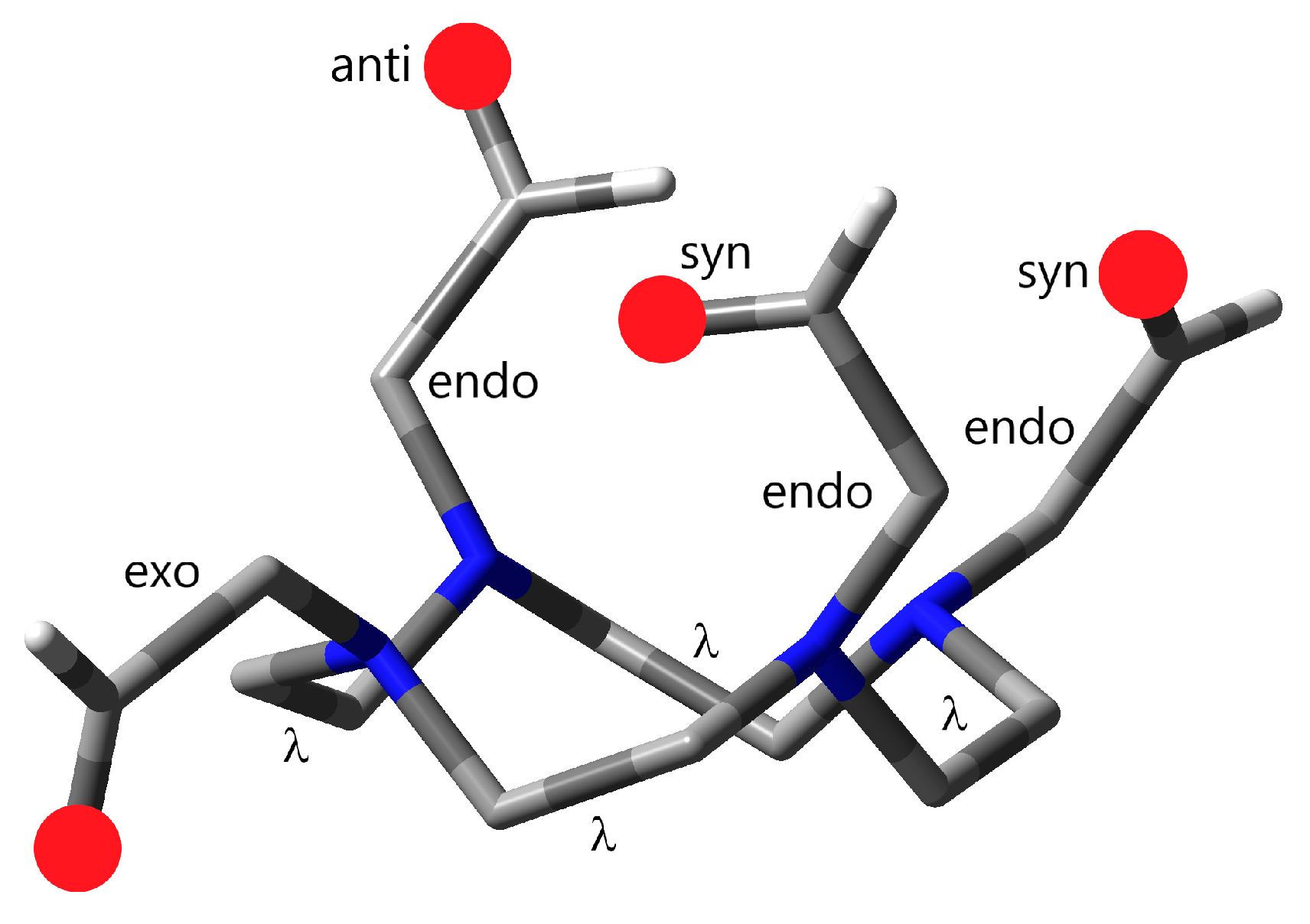
  • The pendant arms at the cyclen N-s can turn towards the cavity of the cyclen ring or away from the cavity (endo or exo orientation, respectively; Figure 2).
  • Similarly, the donor groups of the pendant arms can point towards the cavity (in the complexes coordinating to the encapsulated metal ions in this way, syn orientation) or can turn outside for interactions with solvent molecules (anti orientation; Figure 2). The syn/anti orientations are relevant only for endo pendant arms.

3.2. MeDO2PA

This ligand has two pendant arms in which the picolinate (PA) groups contain two donors: carboxylate O and aromatic N. Generally, both donors form strong bonds with encapsulated metal ions, and the resulting complexes have C2 symmetry [19,21,22,51].
The five lowest-energy solvated MeDO2PA conformers found in the present study are shown in Figure 3, and their relevant energy data are given in Table 1. Though the energy ordering is not consistent for all three energy types, they agree in the preference of conformer MeDO2PA-1.
Among the lowest-energy conformers, MeDO2PA-1 and MeDO2PA-3 are quite similar. In fact, MeDO2PA-3 corresponds to the ligand structure in the metal complexes, just relaxed in the absence of metal. Its picolinate O and N donors are more or less oriented towards the cavity of the cyclen ring, whereas those of MeDO2PA-1 are turned by ca. 60 degrees. In terms of the properties defined in Figure 2, the PA groups have 2× endo arrangements in both conformers. Both conformers have C2 symmetry in solution, similarly to the reported computed structures of the metal complexes of MeDO2PA [19,21,22,51].
In the metal complexes, the two CO bond distances differ significantly: the syn CO bond is longer by ca. 0.02 Å due to the coordination to the metal [22]. In contrast, in the free ligand, the delocalized carboxylate group is nearly symmetric with a marginal difference of ca. 0.003 Å in the CO bond distances. In both conformers, the slightly longer CO bonds point towards the solvent shell.
An interesting structural feature of MeDO2PA-1 is the facing position of the two picolinate groups in the manner of staggered π-stacking [52,53]. In MeDO2PA-3, weak HMe∙∙∙π contacts could be recognized with a distance of 3.12 Å to the center of the rings. Occasional very weak contacts of the methyl hydrogens with the negatively charged picolinate O and N donors are also over 3 Å.
The other three presented low-energy conformers differ by rotations around the NcycCH2 and/or CH2-CPA bonds and are asymmetric. The conformation of the cyclen ring (δδδδ) is preserved in these conformers too. Occasionally, a few weak C-H···O or C-H···N contacts could be observed in the MeDO2PA-2, -4, and -5 structures with distances ≥ 2.7 Å. They do not seem to bear much significance in the stability of these conformers.
Rotation of a pendant arm into the exo orientation (MeDO2PA-4) results in a small loss (ca. 10 kJ/mol) of stability. The energy change upon this endo-to-exo rotation is additive: the conformer with two exo-oriented PA rings (having C2 symmetry) is by ca. 19 kJ/mol higher in energy than MeDO2PA-1.

3.3. H2DO2PA

Though the MeDO2PA and H2DO2PA ligands differ only by the substitution on two opposite cyclen N-s (methyl versus H), they show significantly different conformational properties. The lowest-energy conformers of H2DO2PA are presented in Figure 4, while their relevant energy data are given in Table 2. The five structures are within 4.9 kJ/mol, this energy range being significantly smaller than the one for MeDO2PA (11 kJ/mol, Table 1). The energy ordering is nearly consistent for the three energy types, the only exception being H2DO2PA-1 and -2 in terms of ∆ECPCM. The more sophisticated ∆G data agree in the preference of conformer H2DO2PA-1, though with a rather small energetic advantage only. This structure differs significantly from the global minimum MeDO2PA-1 structure, particularly by the rotated PA rings (Figure 3 and Figure 4).
The H2DO2PA analogue of the MeDO2PA-1 structure was proven to be higher than H2DO2PA-1 by 24.0 kJ/mol (∆GSMD). Meanwhile, the MeDO2PA analogue of H2DO2PA-1 went through a significant change: the NH hydrogens in H2DO2PA oriented inside towards the picolinate arms, which is not feasible for the larger methyl substituents from steric interactions. The related MeDO2PA structure (of C2 symmetry) with outward-oriented methyl groups is higher than MeDO2PA-1 by 11.6 kJ/mol.
The other four lowest-energy conformers of H2DO2PA agree with H2DO2PA-1 in the δδδδ conformation of the cyclen rings (similarly to the known MeDO2PA complexes [20,21,22]) and in the endo orientation of the pendant arms. The differences are manifested in the Ccyc-Ncyc-C-CPA and Ncyc-C-CPA-NPA torsion angles. The PA rings point away from each other in all five lowest-energy conformers; there is no interaction between them (in contrast to MeDO2PA-1, vide supra). Accordingly, the picolinate N and O donors prefer an anti orientation, facilitating advanced interactions with the solvent. These similar orientations of the characteristic groups in the five lowest-energy conformers may explain the slight stability differences between them.
The property highlighting the most stable H2DO2PA-1 structure among the five lowest-energy conformers is its C2 symmetry.
The H2DO2PA conformers presented above are significantly more stable than the one derived from the metal complex [22], which, after relaxation in the absence of metal, proved to be higher than H2DO2PA-1 by 38 kJ/mol in terms of ∆GSMD.

3.4. DO2A2AM

The two types of pendant arms (CH2COO¯ and CH2CONH2) of this ligand have similar steric properties. Due to their strongly polar character, both groups participate in advanced interactions with polar solvent molecules. The solute–solvent interactions compete with the intramolecular hydrogen bonds in the ligand. The five lowest-energy conformers are presented in Figure 5, and the related energy data are compiled in Table 3. The presented DO2A2AM conformers have some significant structural differences; these are also included in Table 3.
The DO2A2AM structures show several interesting structural features. In contrast to the above-discussed MeDO2PA and H2DO2PA cases, the lowest-energy DO2A2AM conformers show changes in the cyclen ring conformation: beyond the usual δδδδ structure in three conformers, the δδδλ and δδλλ structures appear in two of the lowest-energy ones (DO2A2AM-3 and -5, respectively). The main point is, however, that the most stable DO2A2AM-1 conformer has a δδδδ cyclen, as found generally for cyclen-based ligands and their metal complexes [13,15,19,50].
DO2A2AM-1 has a compact structure, resembling that of the metal complexes of the ligand [22]. The main difference is the orientation of the amide groups: while in the metal complexes the carbonyl groups are turned inside towards the metal and form donor–acceptor interactions, in the free solvated ligand, the NH2 groups are turned inside and establish intramolecular hydrogen bonding (vide infra). The anti orientations of the COO oxygens are characterized by slightly (0.003 Å) longer C=O distances of the outward-oriented oxygens.
An additional noteworthy result is the significant gap between DO2A2AM-1 and the next conformers (ca. 10 kJ/mol; Table 3), whereas the free ligand structure relaxed from that in the metal complex is higher by 40 kJ/mol.
Two of the five lowest-energy conformers are symmetric: DO2A2AM-1 has C2 symmetry, similarly to the Pb(DO2A2AM) complex [22], whereas DO2A2AM-5 has Ci symmetry. The prerequisite of the latter symmetry was the twist of the cyclen ring to the δδλλ conformation, resulting in pendant positions at both sides of the cyclen plane.
In the above-discussed cases of MeDO2PA and H2DO2PA, no significant intramolecular hydrogen bonding was found. In contrast, the lowest-energy DO2A2AM conformers are stabilized by several hydrogen bonding interactions in five- and six-membered rings. They include NH and CH hydrogen donor groups with O and N acceptors (Table 3). The formed hydrogen bonds and distances are depicted in Figure S1 in the Supporting Information. Most of the interactions are weak with distances well above 2 Å.
The strongest hydrogen bonding interactions are likely the N-H···O and N-H···N ones. In most conformers, the shortest ones are two N-H···N interactions with distances between 1.96 and 2.10 Å. The exception is DO2A2AM-3 with only one such short N-H···N interaction (2.04 Å) and with an extremely short N-H···O one (1.83 Å). On the other hand, this structure has the fewest hydrogen bonding contacts (Table 3).
Another interesting case is that DO2A2AM-2 and DO2A2AM-4 differ only in the torsion of the exo COO group, yet their energy difference amounts to a few kJ/mol. The rotation of the exo COO groups results in slightly longer C-H···O hydrogen bonding distances in the latter conformer, which may contribute to its somewhat higher energy. In addition, this single torsion may change the solvation effects too, as seen in the variation in energies in Table 3.
Altogether, while the above-shown intramolecular hydrogen bonds surely play some role in the stability of the individual conformers, they do not solely determine it. Further significant contributions are the steric interactions and the solute–solvent interactions, which vary according to the chemical groups on the periphery.

3.5. DO2A2Py

The two types of pendant arms (CH2COO¯ and CH2Py) of this ligand have different chemical characteristics. The small carboxylate groups are very polar with negatively charged O donors, while the larger Py groups contain polarized N donors and the aromatic rings may exert π interactions. By rotations of the pendant arms and donor groups, several low-energy conformers can be formed. From them, five characteristic conformers are presented in Figure 6, and the related energy data are compiled in Table 4.
The most stable solvated conformer (DO2A2Py-1) resembles the one in the metal complexes of the ligand [22], with the pendants keeping their endo orientations. The difference is that the pyridine N donors—in the complex turned towards the metal for the donor–acceptor interactions—are here oriented outside towards the solvent. On the other hand, the anti orientations of the COO oxygens are characterized by slightly (0.006 Å) longer C=O distances of the outward-oriented oxygens. This structure has C2 symmetry.
As a unique case among the four ligands studied here, the conformer corresponding to the ligand structure in the metal complexes (endo orientations of the pendant arms and syn positions of the N donor atoms, DO2A2Py-2) has quite low energy: it is higher than DO2A2Py-1 only by 3.1 kJ/mol in terms of ∆GSMD. However, lacking the shielding and bonding effects of the metal ion, the relative positions of the acetylate pendants are changed significantly with respect to their positions in the metal complex: they moved away from each other to an O···O distance of 6.57 Å compared with 4.93 Å in the Pb(DO2A2Py) complex. Parallel, with the steric hindrance diminished, the Py groups moved towards each other, reducing the NPy···NPy distance of 5.24 Å in the complex to 3.73 Å in DO2A2Py-2. The relative positions of the N atoms in the cyclen ring changed in the same manner, though only slightly.
The exo orientation of an acetylate pendant arm is also less favored (DO2A2Py-3: 4.9 kJ/mol in terms of ∆GSMD), while that of one Py pendant requires significantly more energy (DO2A2Py-4: 11.4 kJ/mol; Table 4).
The DO2A2Py-5 conformer is characterized by two exo acetylate pendants and two endo Py ones (C2 symmetry). Its peculiar feature is the favored syn orientation of both pyridine N-s, because their anti orientation (interacting with solvent) was predicted to be less stable by 2 kJ/mol.
In spite of several strong proton acceptor heteroatoms (COO, aromatic N) in DO2A2Py, intramolecular hydrogen bonding interactions seem to be less significant than in the case of the DO2A2AM ligand. There are only a few weak intramolecular C-H···O hydrogen bonds in the lowest-energy DO2A2Py conformers, as compiled in Figure S2. Most C-H···π contacts are above 3 Å; only in conformers DO2A2Py-3 and -5 appear contacts with distances around 2.7 Å. The strengths of these interactions may be comparable to those of the steric ones and solvation differences.

3.6. Stabilities of Complexes with Pb2+

Experimental stability constants are available for the Pb2+ and Bi3+ complexes with MeDO2PA and H2DO2PA [21]. With both metals, the MeDO2PA complex was found to be somewhat more stable than the H2DO2PA one. In addition, theoretical Pb2+–ligand interaction energies in isolated complex structures of all four ligands have been reported [22]. As for the Pb2+ complexes, more literature data are available; they were selected in the present study for comparison with computations. The stability of the complexes is estimated by the computed dissociation energies using the here-determined most stable solvated free ligand conformers as the ligand dissociation product.
In order to obtain the ∆GSMD data of the Pb2+–complexes, their molecular geometries were optimized at the present computational level. In agreement with the literature information [22], the optimizations converged to structures with C2 symmetry (Figure 7) with slight differences in the geometrical parameters from those in Ref. [22], as usual in the case of competent DFT levels (see Table S6 in the Supplementary Materials).
The computed dissociation energies in aqueous solution and the available experimental stability constants of the Pb2+ complexes are compared in Table 5.
The present computed dissociation energies reflect the experimentally found [21] greater stability of the Pb(MeDO2PA) complex with respect to Pb(H2DO2PA). The present results imply even greater stability for the Pb(DO2A2AM) and Pb(DO2A2Py) complexes.
It is interesting to see the relation of the theoretical Pb2+–ligand interaction energy data (∆Eint) to the practically more relevant dissociation energies and experiment in Table 5. The interaction energy model lacks solvent effects and the optimum ligand conformations, as it operates with fragments fixed in the geometry of the complexes. Yet, the trend agrees with ∆Gdiss and experiment for the two DO2PA-derivative complexes. On the other hand, the relation is worse for Pb(DO2A2AM) and Pb(DO2A2Py): while their ∆Eint values are still greater than those of Pb(H2DO2PA), there is less agreement with their high ∆Gdiss data.

4. Conclusions

The four ligands covered in the present study are known to form symmetric (C2) complexes with metal ions. Less obvious would be the symmetry in aqueous solution due to the extensive solvation interactions of water with these polar ligand molecules. The present study also confirmed the preference for the C2 symmetry of the most stable free ligand conformers in the solution. A significant difference from the complexes is, however, that the CO and pyridine N donors are generally turned outside for advanced interactions with the water solvent. Some additional low-energy symmetric (C2, Ci) structures appear for the ligands with mixed pendant arms, varying their anti and syn orientations.
A remarkable structural feature is the preference of the δδδδ cyclen ring conformation in the most stable ligand structures. Torsion of cyclen in the lowest-energy conformers (to δδδλ and δδλλ) was observed only in the case of DO2A2AM.
The stability of the conformers seems to be maintained by solvation and steric effects. Substantial intramolecular hydrogen bonding was found only in DO2A2AM, where it may play a comparable role beside the above two factors.
The adequate performance of the applied composite MM/DFT computational procedure is reflected by the agreement of computed dissociation energies with the experimentally found stability trend for Pb(MeDO2PA) and Pb(H2DO2PA) [21]. The computed thermodynamic stability for the Pb2+ complexes corresponds to DO2A2Py > DO2A2AM > MeDO2PA > H2DO2PA.

Supplementary Materials

The following supporting information can be downloaded at: https://www.mdpi.com/article/10.3390/sym17091466/s1, Table S1: Number of structures treated at the various computational levels; Tables S2–S5: Comparison of relative energies of selected low-energy ligand conformers in aqueous solution. Table S6: Compilation of Pb–ligand bond distances. Figures S1–S2: Short contacts in characteristic DO2A2AM and DO2A2Py conformers; Cartesian coordinates of the most significant optimized structures.

Funding

This research received no external funding.

Data Availability Statement

All data obtained in the study are included in this article. If more information is needed, it can be available on request from the corresponding author.

Acknowledgments

Zoltán Varga is acknowledged for providing scripts used in the analysis of conformational search results.

Conflicts of Interest

The author declares no conflicts of interest.

References

  1. Driggers, E.M.; Hale, S.P.; Lee, J.; Terrett, N.K. The exploration of macrocycles for drug discovery—An underexploited structural class. Nat. Rev. Drug Discov. 2008, 7, 608–624. [Google Scholar] [CrossRef] [PubMed]
  2. Giordanetto, F.; Kihlberg, J. Macrocyclic Drugs and Clinical Candidates: What Can Medicinal Chemists Learn from Their Properties? J. Med. Chem. 2014, 57, 278–295. [Google Scholar] [CrossRef]
  3. Wei, W.; Rosenkrans, Z.T.; Liu, J.; Huang, G.; Luo, Q.-Y.; Cai, W. ImmunoPET: Concept, Design, and Applications. Chem. Rev. 2020, 120, 3787–3851. [Google Scholar] [CrossRef]
  4. Mota, F.; Ordonez, A.A.; Firth, G.; Ruiz-Bedoya, C.A.; Ma, M.T.; Jain, S.K. Radiotracer Development for Bacterial Imaging. J. Med. Chem. 2020, 63, 1964–1977. [Google Scholar] [CrossRef]
  5. Kręcisz, P.; Czarnecka, K.; Królicki, L.; Mikiciuk-Olasik, E.; Szymański, P. Radiolabeled Peptides and Antibodies in Medicine. Bioconjug. Chem. 2021, 32, 25–42. [Google Scholar] [CrossRef]
  6. Llop, J.; Lammers, T. Nanoparticles for Cancer Diagnosis, Radionuclide Therapy and Theranostics. ACS Nano 2021, 15, 16974–16981. [Google Scholar] [CrossRef]
  7. Sgouros, G.; Bodei, L.; McDevitt, M.R.; Nedrow, J.R. Radiopharmaceutical therapy in cancer: Clinical advances and challenges. Nat. Rev. Drug Discov. 2020, 19, 589–608. [Google Scholar] [CrossRef]
  8. St. James, S.; Bednarz, B.; Benedict, S.; Buchsbaum, J.C.; Dewaraja, Y.; Frey, E.; Hobbs, R.; Grudzinski, J.; Roncali, E.; Sgouros, G.; et al. Current Status of Radiopharmaceutical Therapy. Int. J. Radiat. Oncol. Biol. Phys. 2021, 109, 891–901. [Google Scholar] [CrossRef]
  9. Rondon, A.; Rouanet, J.; Degoul, F. Radioimmunotherapy in Oncology: Overview of the Last Decade Clinical Trials. Cancers 2021, 13, 5570. [Google Scholar] [CrossRef]
  10. Zhang, T.; Lei, H.; Chen, X.; Dou, Z.; Yu, B.; Su, W.; Wang, W.; Jin, X.; Katsube, T.; Wang, B.; et al. Carrier systems of radiopharmaceuticals and the application in cancer therapy. Cell Death Discov. 2024, 10, 16. [Google Scholar] [CrossRef]
  11. Bazak, R.; Houri, M.; El Achy, S.; Hussein, W.; Refaat, T. Passive targeting of nanoparticles to cancer: A comprehensive review of the literature. Mol. Clin. Oncol. 2014, 2, 904–908. [Google Scholar] [CrossRef]
  12. Kleynhans, J.; Grobler, A.F.; Ebenhan, T.; Sathekge, M.M.; Zeevaart, J.-R. Radiopharmaceutical enhancement by drug delivery systems: A review. J. Control. Release 2018, 287, 177–193. [Google Scholar] [CrossRef]
  13. Kostelnik, T.I.; Orvig, C. Radioactive Main Group and Rare Earth Metals for Imaging and Therapy. Chem. Rev. 2019, 119, 902–956. [Google Scholar] [CrossRef]
  14. Peltek, O.O.; Muslimov, A.R.; Zyuzin, M.V.; Timin, A.S. Current outlook on radionuclide delivery systems: From design consideration to translation into clinics. J. Nanobiotechnol. 2019, 17, 90. [Google Scholar] [CrossRef]
  15. Price, E.W.; Orvig, C. Matching chelators to radiometals for radiopharmaceuticals. Chem. Soc. Rev. 2014, 43, 260–290. [Google Scholar] [CrossRef]
  16. Csajbók, É.; Baranyai, Z.; Bányai, I.; Brucher, E.; Király, R.; Muller-Fahrnow, A.; Platzek, J.; Raduchel, B.; Schäfer, M. Equilibrium, 1H and 13C NMR spectroscopy, and X-ray diffraction studies on the complexes Bi(DOTA) and Bi(DO3A-Bu). Inorg. Chem. 2003, 42, 2342–2349. [Google Scholar] [CrossRef]
  17. Tosato, M.; Lazzari, L.; Marco, V.D. Revisiting Lead(II)-1,4,7,10-tetraazacyclododecane-1,4,7,10-tetraacetic Acid Coordination Chemistry in Aqueous Solutions: Evidence of an Underestimated Thermodynamic Stability. ACS Omega 2022, 7, 15596–15602. [Google Scholar] [CrossRef]
  18. Deal, K.A.; Davis, I.A.; Mirzadeh, S.; Kennel, S.J.; Brechbiel, M.W. Improved in vivo stability of actinium-225 macrocyclic complexes. J. Med. Chem. 1999, 42, 2988–2992. [Google Scholar] [CrossRef]
  19. Kovács, A. Favorable symmetric structures of radiopharmaceutically important neutral cyclen-based ligands. Symmetry 2024, 16, 1668. [Google Scholar] [CrossRef]
  20. Lima, L.M.P.; Beyler, M.; Oukhatar, F.; Le Saec, P.; Faivre-Chauvet, A.; Platas-Iglesias, C.; Delgado, R.; Tripier, R. H2Me-do2pa: An attractive chelator with fast, stable and inert natBi3+ and 213Bi3+ complexation for potential a-radioimmunotherapy applications. Chem. Commun. 2014, 50, 12371–12374. [Google Scholar] [CrossRef]
  21. Lima, L.M.P.; Beyler, M.; Delgado, R.; Platas-Iglesias, C.; Tripier, R. Investigating the Complexation of the Pb2+/Bi3+ Pair with Dipicolinate Cyclen Ligands. Inorg. Chem. 2015, 54, 7045–7057. [Google Scholar] [CrossRef]
  22. Kovács, A.; Varga, Z. Theoretical Study of Metal–Ligand Interactions in Lead Complexes with Radiopharmaceutical Interest. Molecules 2024, 29, 4198. [Google Scholar] [CrossRef]
  23. Glendening, E.D.; Streitwieser, A. Natural energy decomposition analysis: An energy partitioning procedure for molecular interactions with application to weak hydrogen bonding, strong ionic, and moderate donor–acceptor interactions. J. Chem. Phys. 1994, 100, 2900–2909. [Google Scholar] [CrossRef]
  24. Glendening, E.D. Natural Energy Decomposition Analysis:  Extension to Density Functional Methods and Analysis of Cooperative Effects in Water Clusters. J. Phys. Chem. A 2005, 109, 11936–11940. [Google Scholar] [CrossRef]
  25. Gutten, O.; Bím, D.; Řezáč, J.; Rulíšek, L. Macrocycle Conformational Sampling by DFT-D3/COSMO-RS Methodology. J. Chem. Inf. Model. 2018, 58, 48–60. [Google Scholar] [CrossRef]
  26. Mohamadi, F.; Richards, N.G.J.; Guida, W.C.; Liskamp, R.; Lipton, M.; Caufield, C.; Chang, G.; Hendrickson, T.; Still, W.C. Macromodel—An integrated software system for modeling organic and bioorganic molecules using molecular mechanics. J. Comput. Chem. 1990, 11, 440–467. [Google Scholar] [CrossRef]
  27. Watts, K.S.; Dalal, P.; Tebben, A.J.; Cheney, D.L.; Shelley, J.C. Macrocycle Conformational Sampling with MacroModel. J. Chem. Inf. Model. 2014, 54, 2680–2696. [Google Scholar] [CrossRef]
  28. Shivakumar, D.; Harder, E.; Damm, W.; Friesner, R.A.; Sherman, W. Improving the Prediction of Absolute Solvation Free Energies Using the Next Generation OPLS Force Field. J. Chem. Theory Comput. 2012, 8, 2553–2558. [Google Scholar] [CrossRef]
  29. Still, W.C.; Tempczyk, A.; Hawley, R.C.; Hendrickson, T. Semianalytical treatment of solvation for molecular mechanics and dynamics. J. Am. Chem. Soc. 1990, 112, 6127–6129. [Google Scholar] [CrossRef]
  30. Schrödinger Release 2024-1; Schrödinger, LLC.: New York, NY, USA, 2024.
  31. Olanders, G.; Alogheli, H.; Brandt, P.; Karlén, A. Conformational analysis of macrocycles: Comparing general and specialized methods. J. Comput. Aided Mol. Des. 2020, 34, 231–252. [Google Scholar] [CrossRef]
  32. Polak, E.; Ribiere, G. Note sur la convergence de méthodes de directions conjuguées. ESAIM 1969, 3, 35–43. [Google Scholar] [CrossRef]
  33. Barone, V.; Cossi, M. Quantum calculation of molecular energies and energy gradients in solution by a conductor solvent model. J. Phys. Chem. A 1998, 102, 1995–2001. [Google Scholar] [CrossRef]
  34. Grimme, S.; Antony, J.; Ehrlich, S.; Krieg, H. A consistent and accurate ab initio parametrization of density functional dispersion correction (DFT-D) for the 94 elements H-Pu. J. Chem. Phys. 2010, 132, 154104. [Google Scholar] [CrossRef]
  35. Brandenburg, J.G.; Bannwarth, C.; Hansen, A.; Grimme, S. B97-3c: A revised low-cost variant of the B97-D density functional method. J. Chem. Phys. 2018, 148, 064104. [Google Scholar] [CrossRef]
  36. Perdew, J.P.; Burke, K.; Ernzerhof, M. Generalized gradient approximation made simple. Phys. Rev. Lett. 1996, 77, 3865–3868, Errata in Phys. Rev. Lett. 1997, 78, 1396. [Google Scholar] [CrossRef]
  37. Adamo, C.; Barone, V. Toward reliable density functional methods without adjustable parameters: The PBE0 model. J. Chem. Phys. 1999, 110, 6158–6170. [Google Scholar] [CrossRef]
  38. Orca, Input Library Webpage. Available online: https://sites.google.com/site/orcainputlibrary/numerical-precision (accessed on 20 June 2025).
  39. Caldeweyher, E.; Bannwarth, C.; Grimme, S. Extension of the D3 dispersion coefficient model. J. Chem. Phys. 2017, 147, 034112. [Google Scholar] [CrossRef]
  40. Caldeweyher, E.; Ehlert, S.; Hansen, A.; Neugebauer, H.; Spicher, S.; Bannwarth, C.; Grimme, S. A generally applicable atomic-charge dependent London dispersion correction. J. Chem. Phys. 2019, 150, 154122. [Google Scholar] [CrossRef]
  41. Dunlap, B.I. Robust and variational fitting. Phys. Chem. Chem. Phys. 2000, 2, 2113–2116. [Google Scholar] [CrossRef]
  42. Pantazis, D.A.; Chen, X.-Y.; Landis, C.R.; Neese, F. All-Electron Scalar Relativistic Basis Sets for Third-Row Transition Metal Atoms. J. Chem. Theory Comput. 2008, 4, 908–919. [Google Scholar] [CrossRef]
  43. van Lenthe, E.; Baerends, E.J.; Snijders, J.G. Relativistic total energy using regular approximations. J. Chem. Phys. 1994, 101, 9783–9792. [Google Scholar] [CrossRef]
  44. Martin, R.L.; Hay, P.J.; Pratt, L.R. Hydrolysis of Ferric Ion in Water and Conformational Equilibrium. J. Phys. Chem. A 1998, 102, 3565–3573. [Google Scholar] [CrossRef]
  45. Marenich, A.V.; Cramer, C.J.; Truhlar, D.G. Universal Solvation Model Based on Solute Electron Density and on a Continuum Model of the Solvent Defined by the Bulk Dielectric Constant and Atomic Surface Tensions. J. Phys. Chem. B 2009, 113, 6378–6396. [Google Scholar] [CrossRef]
  46. Neese, F. The ORCA program system. WIREs Comput. Mol. Sci. 2012, 2, 73–78. [Google Scholar] [CrossRef]
  47. Neese, F.; Wennmohs, F.; Becker, U.; Riplinger, C. The ORCA quantum chemistry program package. J. Chem. Phys. 2020, 152, 224108. [Google Scholar] [CrossRef]
  48. Neese, F. Software update: The ORCA program system—Version 5.0. WIREs Comput. Mol. Sci. 2022, 12, e1606. [Google Scholar] [CrossRef]
  49. Corey, E.J.; Bailar, J.C. The Stereochemistry of Complex Inorganic Compounds. XXII. Stereospecific Effects in Complex Ions. J. Am. Chem. Soc. 1959, 81, 2620–2629. [Google Scholar] [CrossRef]
  50. Viola-Villegas, N.; Doyle, R.P. The coordination chemistry of 1,4,7,10-tetraazacyclododecane-N,N′,N″,N‴-tetraacetic acid (H4DOTA): Structural overview and analyses on structure-stability relationships. Coord. Chem. Rev. 2009, 253, 1906–1925. [Google Scholar] [CrossRef]
  51. Kovács, A.; Varga, Z. Metal–ligand interactions in complexes of cyclen-based ligands with Bi and Ac. Struct. Chem. 2021, 32, 1719–1731. [Google Scholar] [CrossRef]
  52. Martinez, C.R.; Iverson, B.L. Rethinking the term “pi-stacking”. Chem. Sci. 2012, 3, 2191–2201. [Google Scholar] [CrossRef]
  53. Lewis, M.; Bagwill, C.; Hardebeck, L.; Wireduaah, S. Modern Computational Approaches to Understanding Interactions of Aromatics. In Aromatic Interactions: Frontiers in Knowledge and Application; Johnson, D.W., Hof, F., Eds.; The Royal Society of Chemistry: Cambridge, UK, 2016; pp. 1–17. [Google Scholar] [CrossRef]
Figure 1. The 2D structures of the four cyclen-based ligands: 6,6′-((4,10-dimethyl-1,4,7,10-tetraazacyclododecane-1,7-diyl)bis(methylene)) dipicolinate (MeDO2PA), 6,6′-((1,4,7,10-tetraazacyclododecane-1,7-diyl)bis(methylene)) dipicolinate (H2DO2PA), 1,7-bis(2-carbamoyl-methyl)-4,10-diacetyl-1,4,7,10-tetraazacyclododecane (DO2A2AM), and 1,7-bis(pyridin-2-ylmethyl)-4,10-diacetyl-1,4,7,10-tetraazacyclododecane (DO2A2Py).
Symmetry 17 01466 g001
Figure 2. Demonstration of the specific conformational properties of a hypothetical cyclen-based ligand. The donor groups are represented by red balls; cyclen hydrogens are omitted for clarity. Atoms are represented by the following colors: C, gray; N, blue; H, white.
Symmetry 17 01466 g002
Figure 3. Lowest-energy MeDO2PA conformers. Atoms are represented by the following colors: C, gray; N, blue; O, red. Hydrogens are omitted for clarity. The C2 axis in conformers 1 and 3 is running vertically through the center of the structures.
Symmetry 17 01466 g003
Figure 4. Lowest-energy H2DO2PA conformers. Atoms are represented by the following colors: C, gray; N, blue; O, red. Hydrogens are omitted for clarity. The C2 axis in conformer 1 is running vertically through the center of the structure.
Symmetry 17 01466 g004
Figure 5. Lowest-energy DO2A2AM conformers. Atoms are represented by the following colors: C, gray; N, blue; O, red. Hydrogens are omitted for clarity. The C2 axis in conformer 1 is running vertically through the center of the structure.
Symmetry 17 01466 g005
Figure 6. Selected characteristic low-energy DO2A2Py conformers. Atoms are represented by the following colors: C, gray; N, blue; O, red. Hydrogens are omitted for clarity. The C2 axis in conformers 1, 2, and 5 is running vertically through the center of the structures.
Symmetry 17 01466 g006
Figure 7. Optimized structures of the Pb complexes with ligands MeDO2PA, H2DO2PA, DO2A2AM, DO2A2Py. Atoms are represented by the following colors: Pb, green; C, gray; N, blue; O, red. Hydrogens are omitted for clarity. The C2 axis is running vertically through the center of the structures.
Symmetry 17 01466 g007
Table 1. Lowest-energy conformers of MeDO2PA in aqueous solution 1.
1 (C2)23 (C2)45
Pendants 2Arm2 × endo2× endo2 × endo1 × endo
1 × exo
2 × endo
PA2 × syn1 × anti
1 × syn
2 × syn1 × syn2 × anti
∆ECPCM 0.02.54.410.46.2
∆GCPCM 0.00.44.94.21.7
∆GSMD 0.04.09.910.911.0
1 The geometries were optimized and thermal contributions were calculated with the CPCM solution model. The SMD solution model was applied in single-point energy calculations on the latter geometries. Electronic energies (∆E) at 0 K and Gibbs free energies (∆G) at 298 K and 1354 atm are given in kJ/mol. 2 Orientations of the pendant arms are characterized with respect to the cyclen ring (see text and Figure 2). The anti/syn orientations of the picolinate (PA) groups are meaningful only in the cases of endo conformations.
Table 2. Lowest-energy conformers of H2DO2PA in aqueous solution 1.
1 (C2)2345
Pendants 2Arm2 × endo2 × endo2 × endo2 × endo2 × endo
PA2 × anti1 × anti
1 × syn
1 × anti
1 × syn
2 × anti2 × anti
∆ECPCM 1.00.02.16.06.5
∆GCPCM 0.00.32.34.34.7
∆GSMD 0.01.82.64.64.9
1 The geometries were optimized and thermal contributions were calculated with the CPCM solution model. The SMD solution model was applied in single-point energy calculations on the latter geometries. Electronic energies (∆E) at 0 K and Gibbs free energies (∆G) at 298 K and 1354 atm are given in kJ/mol. 2 Orientations of the pendant arms are characterized with respect to the cyclen ring (see text and Figure 2). The anti/syn orientations of the picolinate (PA) groups are meaningful only in cases of endo conformations.
Table 3. Lowest-energy conformers of DO2A2AM in aqueous solution 1.
1 (C2)2345 (Ci)
Pendants 2COO-Arm2 × endo1 × endo
1 × exo
1 × endo
1 × exo
1 × endo
1 × exo
2 × endo
CONH2-Arm2 × endo2 × endo2 × endo2 × endo2 × endo
C=OCOO2 × anti1 × anti1 × syn1 × anti2 × anti
C=OCONH22 × anti2 × anti2 × anti2 × anti2 × anti
∆ECPCM 0.010.417.116.716.1
∆GCPCM 0.08.511.515.412.7
∆GSMD 0.09.011.811.912.0
Cyclen conformation 3 δδδδδδδδδδδλδδδδδδλλ
Hydrogen bondingN-H···O
N-H···N
C-H···O
C-H···N
Σ1413101414
1 The geometries were optimized, and thermal contributions were calculated with the CPCM solution model. The SMD solution model was applied in single-point energy calculations on the latter geometries. Electronic energies (∆E) at 0 K and Gibbs free energies (∆G) at 298 K and 1354 atm are given in kJ/mol. 2 Orientations of the pendant arms are characterized with respect to the cyclen ring (see text and Figure 2). The anti/syn orientations of the CO donors are meaningful only in the cases of endo conformations. 3 Nomenclature according to Corey and Bailar [49].
Table 4. Selected characteristic low-energy conformers of DO2A2Py in aqueous solution 1.
1 (C2)2 (C2)345 (C2)
Pendants 2COO-Arm2 × endo2 × endo1 × endo
1 × exo
2 × endo2 × exo
Py-Arm2 × endo2 × endo2 × endo1 × endo
1 × exo
2 × endo
C=O2 × anti2 × anti1 × anti2 × anti-
NPy2 × anti2 × syn2 × anti1 × anti2 × syn
∆ECPCM 0.06.66.113.614.0
∆GCPCM 0.06.12.09.710.9
∆GSMD 0.03.14.911.412.7
1 The geometries were optimized and thermal contributions were calculated with the CPCM solution model. The SMD solution model was applied in single-point energy calculations on the latter geometries. Electronic energies (∆E) at 0 K and Gibbs free energies (∆G) at 298 K and 1354 atm are given in kJ/mol. 2 Orientations of the pendant arms are characterized with respect to the cyclen ring (see text and Figure 2). The anti/syn orientations of the C=O/NPy donors are meaningful only in the cases of endo conformations.
Table 5. Computed stability data and available experimental stability constants of Pb2+ complexes.
Complex (PbL)∆Eint 1∆Gdiss 2log KPbL 3
Pb(MeDO2PA)−2523326.118.44(2)
Pb(H2DO2PA)−2471313.116.44(2)
Pb(DO2A2AM)−2520335.0-
Pb(DO2A2Py)−2513342.0-
1 Pb2+–ligand interaction energies (kJ/mol) in the isolated complexes from Ref. [22]. 2 The Gibbs free energies of dissociation (kJ/mol) to Pb2+ + L were evaluated from ∆GSMD data at 298 K and 1354 atm. 3 Experimental stability constants from Ref. [21].
Disclaimer/Publisher’s Note: The statements, opinions and data contained in all publications are solely those of the individual author(s) and contributor(s) and not of MDPI and/or the editor(s). MDPI and/or the editor(s) disclaim responsibility for any injury to people or property resulting from any ideas, methods, instructions or products referred to in the content.