Article

Automatic Brain Tumor Detection and Volume Estimation in Multimodal MRI Scans via a Symmetry Analysis


1
Department of Electrical and Electronics Engineering, Ankara University, 06830 Ankara, Turkey
2
Department of Biomedical Engineering, TOBB University of Economics and Technology, 06560 Ankara, Turkey
3
Department of Biomedical Engineering, Başkent University, 06790 Ankara, Turkey
*
Author to whom correspondence should be addressed.
Symmetry 2023, 15(8), 1586; https://doi.org/10.3390/sym15081586
Submission received: 30 April 2023 / Revised: 16 June 2023 / Accepted: 31 July 2023 / Published: 14 August 2023
(This article belongs to the Section E: Life Sciences)

Abstract

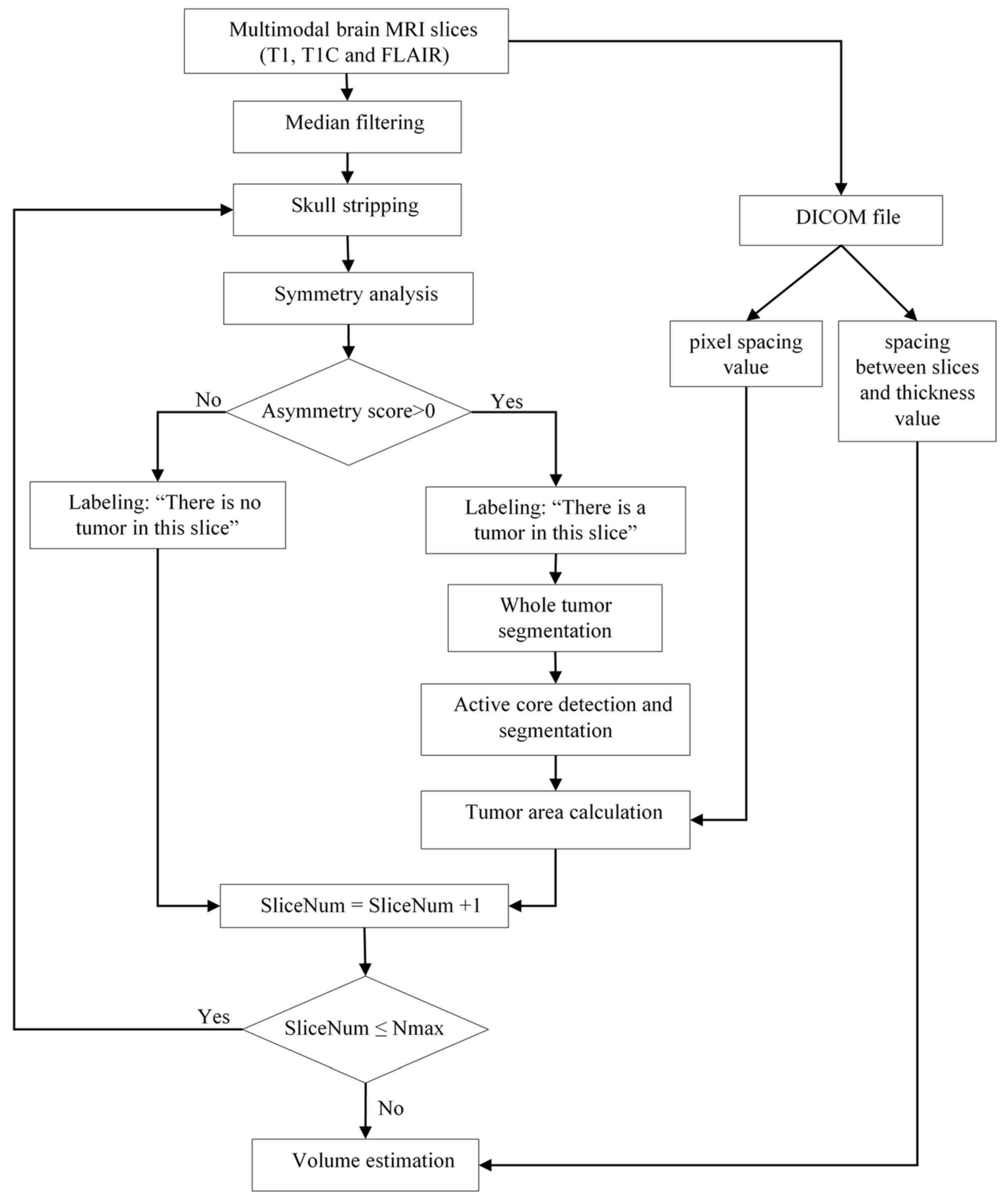
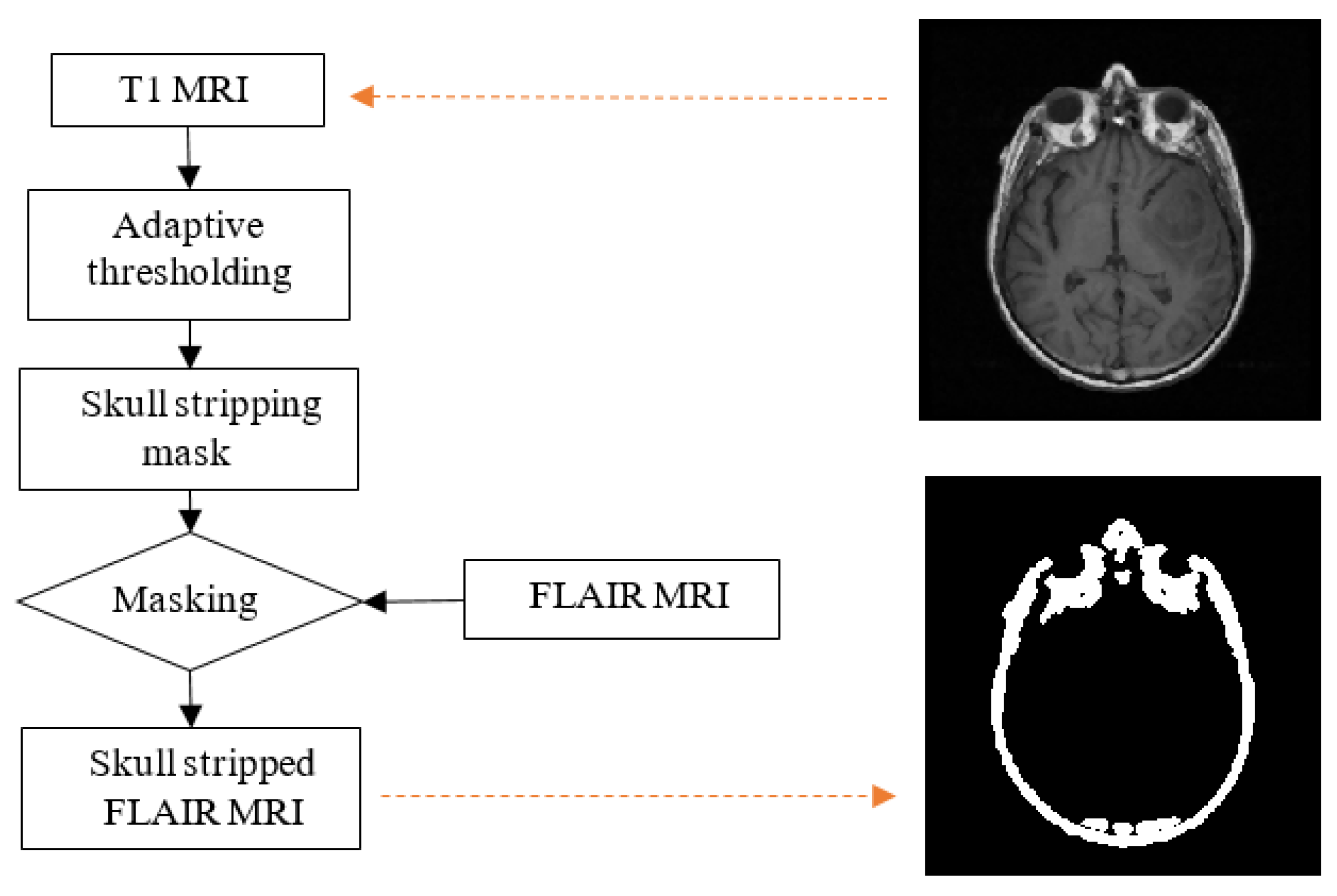
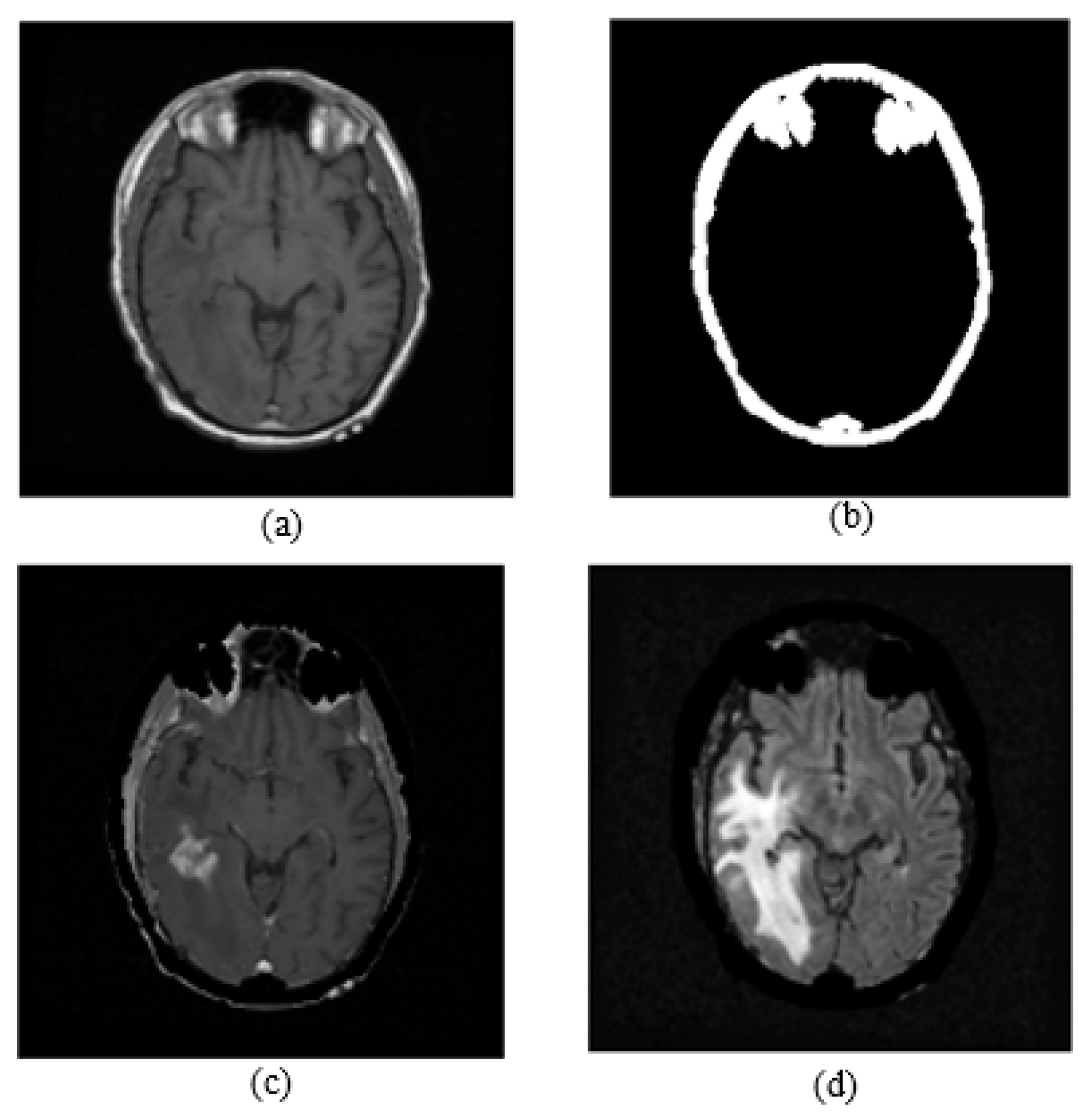
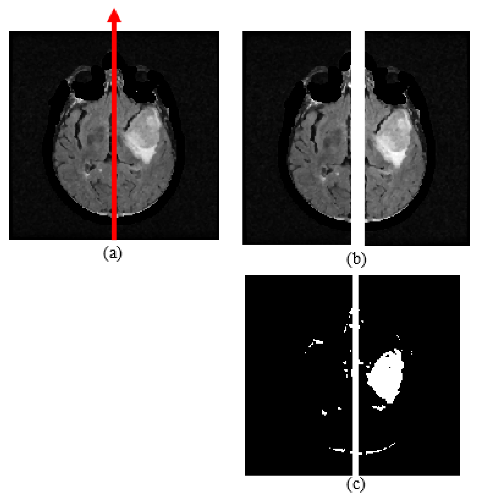
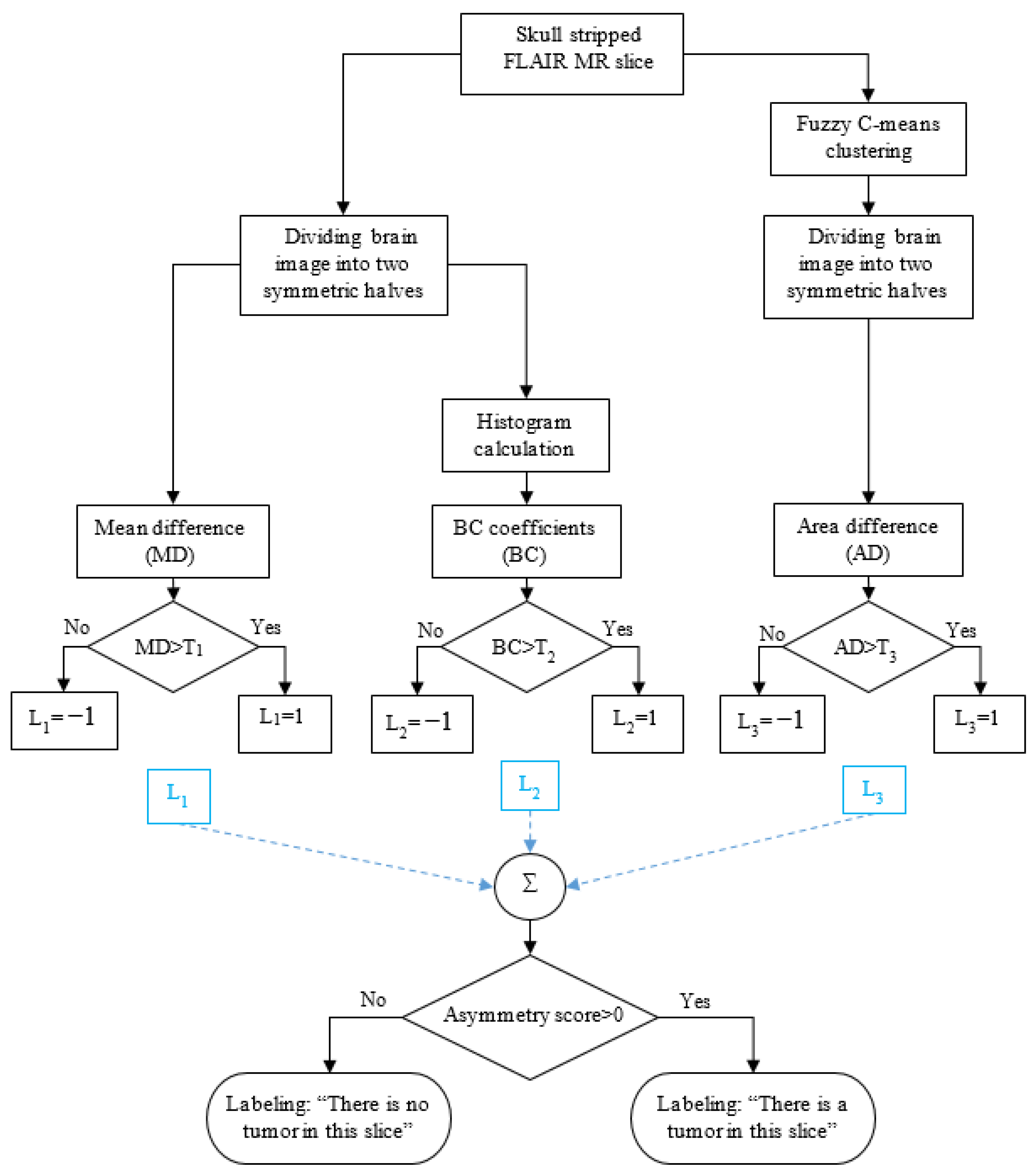
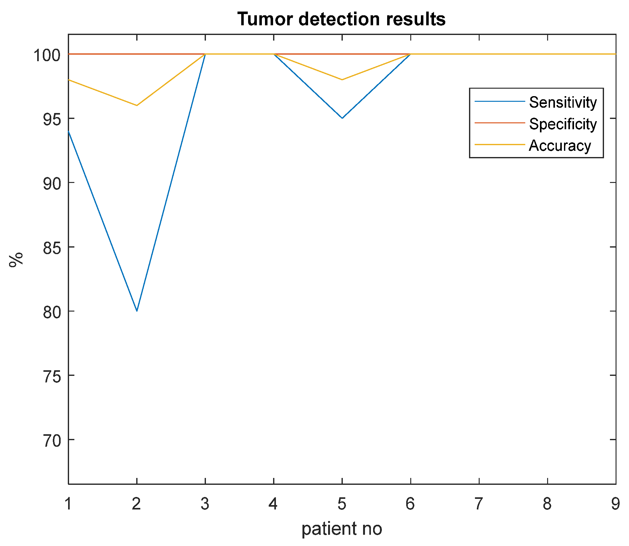
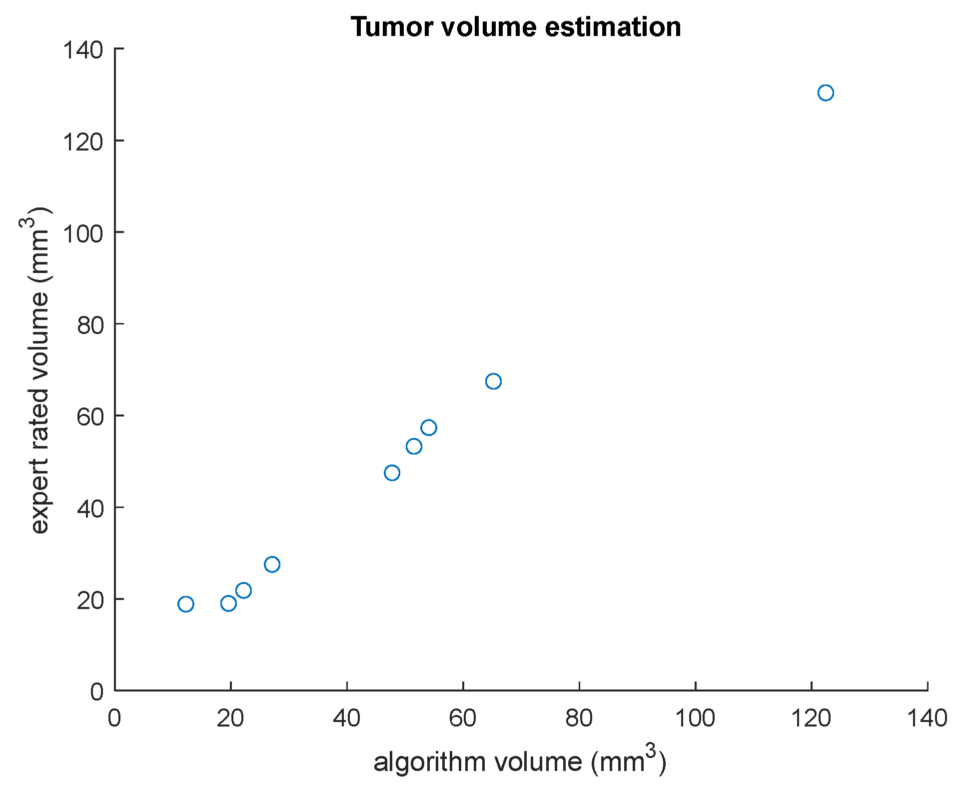
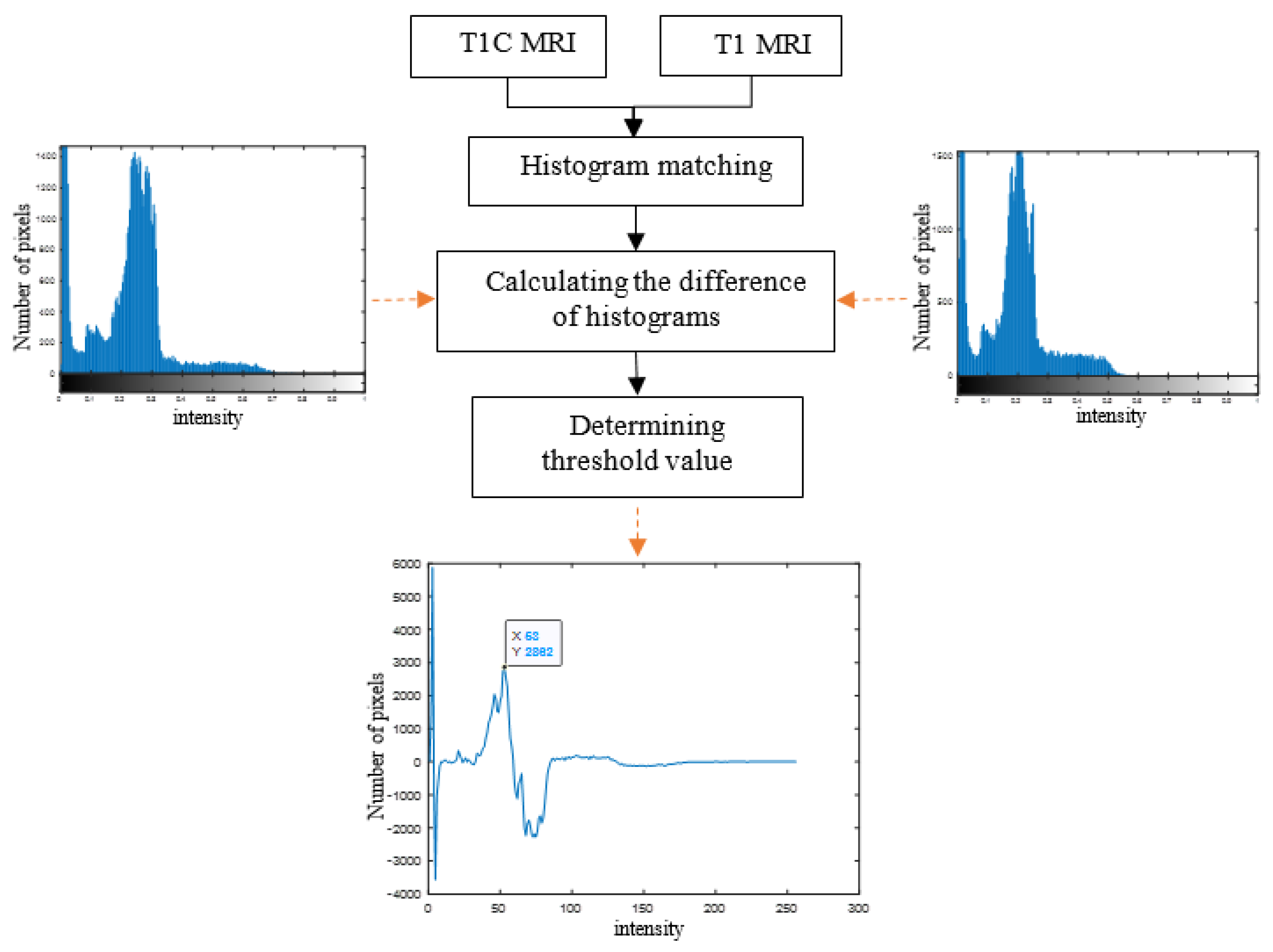
In this study, an automated medical decision support system is presented to assist physicians with accurate and immediate brain tumor detection, segmentation, and volume estimation from MRI which is very important in the success of surgical operations and treatment of brain tumor patients. In the proposed approach, first, tumor regions on MR images are labeled by an expert radiologist. Then, an automated medical decision support system is developed to extract brain tumor boundaries and to calculate their volumes by using multimodal MR images. One advantage of this study is that it provides an automated brain tumor detection and volume estimation algorithm that does not require user interactions by determining threshold values adaptively. Another advantage is that, because of the unsupervised approach, the proposed study realized tumor detection, segmentation, and volume estimation without using very large labeled training data. A brain tumor detection and segmentation algorithm is introduced that is based on the fact that the brain consists of two symmetrical hemispheres. Two main analyses, i.e., histogram and symmetry, were performed to automatically estimate tumor volume. The threshold values used for skull stripping were computed adaptively by examining the histogram distances between T1- and T1C-weighted brain MR images. Then, a symmetry analysis between the left and right brain lobes on FLAIR images was performed for whole tumor detection. The experiments were conducted on two brain MRI datasets, i.e., TCIA and BRATS. The experimental results were compared with the labeled expert results, which is known as the gold standard, to demonstrate the efficacy of the presented method. The performance evaluation results achieved accuracy values of 89.7% and 99.0%, and a Dice similarity coefficient value of 93.0% for whole tumor detection, active core detection, and volume estimation, respectively.
Keywords: histogram analysis; adaptive thresholding; skull stripping; symmetry analysis; fuzzy c-means clustering histogram analysis; adaptive thresholding; skull stripping; symmetry analysis; fuzzy c-means clustering