Abstract
Currently, inertial sensors are often used to study balance in an upright stance. There are various options for recording balance data with different locations and numbers of sensors used. Methods of data processing and presentation also differ significantly in published studies. We propose a certain technical implementation of the method and a previously tested method for processing primary data. In addition, the data were processed along three mutually perpendicular planes. The study was conducted on 109 healthy adults. A specially developed inertial sensor, commercially available for medical purposes, was used. Thus, this work can outline the limits of normative values for the calculated stabilometric measures. Normative data were obtained for three oscillation planes with the sensor located on the sacrum. The obtained parameters for the vertical component of the oscillations are of the same order as for the frontal and sagittal components. Normative parameters are required in any clinical study, as the basis from which we start in the evaluation of clinical data. In this study, such normative parameters are given for one of the most commonly used Romberg’s tests. The obtained normative data can be used for scientific and clinical research.
1. Introduction
An instrumental examination of balance function upon quiet standing was introduced in clinical practice in the second half of the 20th century. Various methods were tried out in the process of creating this examination technique. Since the end of the last century, the method using a special platform with force sensors has been most commonly used. This method has several names: stabilometry, posturography, stabilography, and some others.
The first fundamental work devoted to the physical foundations of the method was published in 1973 [1]. The later works focused on the main approaches to the registration technique and the parameters used for the analysis. Technical and methodological standards for the recording devices have been developed and proposed [2].
Traditional stabilometry, as it is, also has certain limitations which are associated with the registration method. To obtain data in the patient’s coordinate system (i.e., taking into account the position of his/her feet), it is required to position the patient’s feet on the platform in a certain way. There are even corresponding standards of feet positioning [3,4]. In some cases, the size of the feet is used to determine the coordinate system in which the results of the examination will be shown [3]. In rare cases, stabilometric platforms have fairly large overall dimensions and weight. This is caused by the technical basis of registration, which requires a high rigidity of the top plate of the platform [2].
At the end of the 20th century and the beginning of the 21st century, inertial sensors also began to be used for balance studies. These are three-component accelerometers or more complex devices based on them, e.g., an inertial measurement unit (IMU). IMU systems are based on a special chip, in which sensors are located in three mutually perpendicular planes: accelerometric, gyroscopic, magnetometric. They have been used for the assessment of balance function in neurological practice for over 15 years [5].
IMU sensors are usually positioned on the patient’s sacrum, and sometimes on other body parts [6]. The sensors register oscillations in all three planes at once. Besides, their frequency range is much higher than in the old stabilometric platforms or in platforms based on pressure sensors. Depending on the aims of the study, either individual sensors are used, or sensors in combination with mathematical processing of information using rather complex algorithms. The existing approaches can be divided into several methods. These are registration and analysis of only accelerometric data, data of sensor inclination, and movement in the frontal and sagittal planes. Each method has its own advantages and limitations. Accelerometry is a method of directly registering of oscillations which is technically convenient; however, as a source of the final information, it is quite difficult for the user to understand. The sensor inclination data and stabilometric platform give quite similar information [7]. However, physically, these are completely different values, which can be confusing. In the study of Lesinski M, et al. [8], the authors concluded that such devices cannot be used interchangeably with the force plate. The sensor movement data are physically rather close to what can be obtained with a stabilometric platform. However, they are values with a significant error, since the process of obtaining them is associated with double integration of the initial accelerometric data.
The IMU method has another significant difference from stabilometry using platforms; it cannot correlate the position of the centre of gravity in the patient’s (platform) coordinates. It should be mentioned that some traditional stabilometric platforms also do not provide this information, since the platform is reset after the patient has stepped on it. Thus, the centre of pressure (CoP) is always in the centre of the platform. Inertial systems, due to their physical characteristics, do not work in the external coordinate system. However, they are able to assess body oscillations in the process of balance upon standing still.
The advantages of this method are also directly related to the technical characteristics of registration. For example, it has a higher range of recorded oscillation frequencies.
Another benefit of these sensors is that they can be worn all the time. Thus, they record not only balance, but also any movements in general. This gave a boost to the development of a new method of movement studies, which is called actimetry. Since at least a two-component accelerometer is built into modern smartphones, scientists have tried consistently to use this device to assess balance function [9].
There is another circumstance that allows or does not allow us to obtain the required information. Regardless of the registration frequency, the studied oscillations are in a relatively low frequency range. This is relevant for the old types of platforms based on force sensors and pressure sensors. The problem is that the platform loaded with the weight of the body has a certain natural resonant frequency of oscillations, which is higher, the smaller the mass of the entire system. However, taking into account the average weight of a healthy adult or even a teenager, it still will be low. The same is for true of devices with pressure sensors, and even to a greater extent than for platforms. The hysteresis properties of most sensors do not allow them to have a high natural frequency of data registration. This means that the first and second types of devices can only correctly show the relatively low frequency range of the oscillations: on average, up to 10 Hz or even lower. In comparison, the inertial sensors can record oscillations in a wider range of 50 Hz and above. These processes require further studies.
The question of how comparable the data obtained by IMU sensors and old type stabilometric platforms are was raised right away. Over the past few years, a number of experiments have been conducted that allow us to draw certain conclusions. Although the recorded information from both devices is different, these data are comparable and reliable [10]. Studies of balance upon quiet standing using video analysis also proved that registration of oscillation using IMU can be successfully applied in practice [11,12]. In the study of the comparability of data from the force plate and the IMU sensor [13] the authors came to the conclusion that the issues related to the visual system were better for examining using a force plate, and the pathology of the low limb was better studied study using the IMU sensor. In some cases, in addition to the force plate, video analysis is also used to record the oscillations of the human body. This method in its modern iteration has reached the required accuracy and resolution. This study [12] showed good comparability between video analysis and IMU technologies.
The data of the balance in norm are given in numerous studies, as a control group or a special study [13,14]. However, the study methods, the position of the sensor, and the calculation of the final measures differ significantly. According to the review [6], one of the positions frequently used by researchers in the area of the sacrum is not strictly fixed. The position of the sensor “fluctuates” from study to study, from L5 to S2. At the same time, L5 is the most common place for sensor fixation, and it was found in 42.6% of all studies. According to other researchers [13] this location is used in 65% of the published studies. It should be mentioned that this is also the most convenient place for sensor fixation. The registration time is rarely less than 20 s and does not exceed 60 s, with the most common time being 30 s [6].
Most often, the study is carried out using Romberg’s test, i.e., the same amount of time with open and closed eyes [6]. Such a study allows us to assess the functioning of the visual and proprioceptive analyzers, which are most involved in the regulation of balance upon quiet standing.
The balance data on quiet standing, as it follows from the vast majority of literature, including those cited in this work, are obtained in the anterior–posterior and lateral planes. At the same time, such a standard device for this study, a traditional stabilometric platform based on force sensors, allows us, by its physical nature, to perform a study in three mutually perpendicular planes. The same applies to the force platform used for this purpose. In the vertical direction, a number of oscillation processes are also produced, which are potentially valuable for diagnosis [15,16]. However, in the literature, there are only a few works devoted to the study of oscillation processes upon quiet standing in a three-dimensional plane [15,17,18,19]. The intentional use of three-dimensional registration systems, both traditional platforms and magnetic and inertial sensors [6,20], does not affect the process in any way, and the registration, just as it is traditionally, can be performed in only two planes [21]. One of the probable reasons for this is a tradition of sway path registration that dates back to the beginning of the last century, when the registration of human body balance oscillations was carried out by simpler devices on a horizontal plane.
Recent works related to this problem presented studies that were carried out both on the traditional type of platform [22], and using IMU sensors technologies [19].
This method has several names. These are spatial or 3D stabilometry [19] and three-dimensional statokinesigrams [22].
The aim of this study was to determine the limits of the nominative values for 3D stabilometry and the Romberg’s test using, in particular, an IMU sensor.
2. Materials and Methods
2.1. Participants
Some 109 healthy adults participated in the stabilometric study. There were 77 males and 32 females, with a mean age of 22 years (range 16–38, median—21). All examined subjects were healthy at the beginning of the study, with no orthopaedic or neurological diseases that could affect the balance state upon quiet standing. The condition of participants’ eyesight function was generally normal; some participants were constantly using contact lenses for vision correction, but they could see without them.
The study was conducted according to the principles of the Declaration of Helsinki and was approved by the local ethics committee. All patients gave written informed consent before the tests.
2.2. Test Procedures
The study was conducted using Neurosens IMU sensor (Neurosoft company, Ivanovo, Russia).
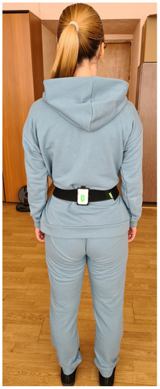
The study method met the recommendations [23,24]. The part of the study specific to IMU sensors was carried out in accordance with the results of the aforementioned [6] research. The sensor was attached to the patient’s sacrum area using a special sensor holder with an elastic band (Figure 1).

Figure 1.
Examination with Neurosens IMU sensor during the study.
The sensor is most commonly placed on the sacrum area [6] from L2 to S2. Such positioning was found in 68.1% of studies. This tendency has a physical explanation; it is in front of the promontorium of the pelvis, where the centre of mass of the human body is located. Thus, the sensor location in this area is closest to the centre of mass of the body. The placement of the sensor above this level is quite rare. In addition, in this case, oscillations of the body relative to the pelvis are also recorded.
Both a special sensor holder and an elastic band were originally designed to comfortably and tightly hold the sensor.
The subject was standing in a free symmetrical pose with arms beside the body. After 2–3 min of quiet standing, the registration began. Until that moment, the patient had been instructed on how to stand (quiet, straight, silently, without voluntary movements, etc.). The first registration was performed with open eyes for 30 s. This was followed by a command to close the eyes (there were no other movements). We waited for about 30 s to eliminate the influence of transient processes and started the second recording session for another 30 s.
The data from the three-component accelerometer integrated into the sensor were recorded at a frequency of 200 Hz and transmitted via Wi-Fi directly to the special software on the PC.
The used measures were calculated according to the method described in the following work [25], which is also used within the Steadys-Balance software for the Neurosens IMU sensor. One measure, namely the projection area of the 95% confidence ellipsoid of acceleration (S m2/s4), is missing in the cited work, but we considered it to be quite interesting and calculated it according to the method described in the work of Hanakova L. et al. [26]. The abbreviations of the measures and their descriptions are specified in Table 1.

Table 1.
The abbreviation and description of calculated measures.
2.3. Statistical Analysis
Statistical analysis was performed using Excel software. A total of 109 people participated in the study and, accordingly, there were 109 results for each measure. For each measure, using the standard method of interval estimation with an unknown distribution of a random variable, 95% of the values were selected with the exception of 5% of the sample values (data for 104 subjects remained). The limitations of values in the 95% were the minimal (Min) and maximal (Max) values for each measure with eyes open (EO) and with eyes closed (EC).
3. Results
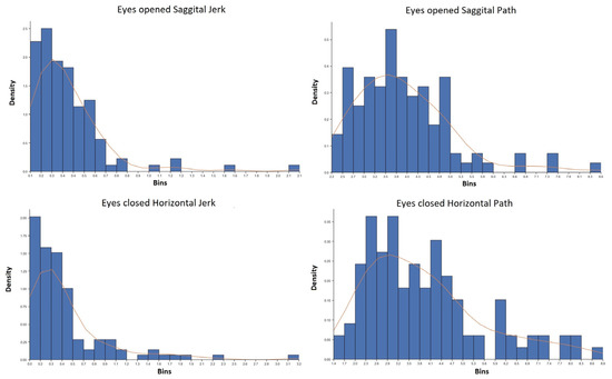
Checking for each parameter of the characteristics of the data distribution showed that for all parameters, the distributions are not normal. As an example, we show graphs for two parameters (Jerk and Path) in the EO and EC positions in Figure 2.

Figure 2.
Distribution plots for Jerk and Path parameters in the EO and EC position, where the vertical axis is Density, and the horizontal axis is Bins.
The results of the study parameters are given in Table 2, Table 3 and Table 4, namely the minimal and maximal values for each measure.

Table 2.
Measures for both positions in frontal plane.

Table 3.
Measures for both positions in horizontal plane.

Table 4.
Measures for both positions in sagittal plane.
Specialists working with traditional stabilometric platforms know that usually a healthy person (with normal eyesight) shows an increased value of stabilometric parameters in the position with EC [3,16]. The measures obtained during inertial 3D stabilometry are quite different due to its physical nature. A significant part of these measures (presented in this paper) also increases. However, there are some differences. Thus, the same measure can change depending on the registration plane. The response may differ not only due to the registration plane, but also depending on what value we are analyzing: minimum or maximum.
Of particular interest in this study are the data on the horizontal plane, since these data are usually ignored in most studies. In this regard, we should say that the measures in this plane and the characteristics of their change practically do not differ from the sagittal and frontal planes. This proves the assumption that oscillation processes, due to various physiological reasons, have common patterns for all directions. Oscillation processes are exactly the same both in the vertical direction and in the lateral and anterior–posterior direction.
4. Discussion
The obtained data were collected from a significant number of young healthy people of both sexes. The participants were in good physical shape at the moment of the study. Some of them had minor refractive errors such as myopia up to 1 diopter, and did not use constant vision correction. Thus, this work can outline the limits of normative values for the calculated stabilometric measures.
The attempt to analyze the obtained values with similar ones reported in the literature showed that there are serious discrepancies in the study design, sensor positioning, examination timing, examination conditions, data registration requirements, the method of measures calculation, etc. This does not allow direct comparison of data collected from the different studies. In addition, there are still not many potential studies for comparison.
However, for healthy people, according to the inverted pendulum model, the amplitude of oscillation increases with increasing the length of the pendulum arm, i.e., the sensor height. The oscillation frequency changes in the same way, but in the opposite pattern. With an increase in the length of the pendulum arm, it decreases. The listed regularities refer not only to the fundamental features of pendulum oscillations, but also to the 1/f noise law or flicker noise. The action of this law is noted not only for various complex systems, but also for the functioning of the systems of the human body, and the balance control system, in particular [27,28]. Among other technical factors, the resolution and registration limits of accelerometers and gyroscopes, the frequency at which the resulting data is received, the methods of sensor calibration, and a number of other technical parameters matter.
The control group of the [29] study consists of healthy subjects, and the algorithm of data processing is equivalent to one used in our work [25], which, in theory, lets us conduct the direct comparison. However, despite the fact that the authors used an IMU chip with similar technical characteristics, the study itself and the frequency of data processing were significantly different. The sensor was strapped at the lumbar area but not at the sacrum which probably could not affect the compared values. The main difference is that the study was carried out on the foam mat (a foamy polymer of medium density) which very significantly increased the amplitude of oscillations. The position of arms also differed. In our study, the arms were placed at the sides of the body, but in one cited work, the arms were crossed, with the palms on the shoulders. The frequency of data registration also has a significant impact on quantitative values. In our study, the frequency was 200 Hz, and in the analyzed work, it was 120 Hz. There were some other differences as well. For the reasons listed above, it was not possible to conduct a direct comparison of obtained data with already reported ones. In general, we should say that the standards for such studies are rather variable [6]. In an analytical review [30] based on an analysis of data from 19 selected sources, where a similar method of studying balance upon quiet standing was used, and information on the registration frequency from the IMU sensor was available, options from 20 to 400 Hz, are given with most values in the 100 Hz range. There is still no consensus on frequency range in this part of the methodology. The same can be noted for a much more mature technique that already has certain standardization, namely, studies using a force platform. At the same time, the standards proposed for the force platform [2] cannot be correctly applied due to the different physical nature of the method.
The analytical review [30] analyses another significant factor that affects the possibility of comparing balance studies performed even on the same equipment, but with different durations. In modern studies, the duration of registration varies from 10 s to 3 min [30]. In each case, the duration of the study is determined by its design and purpose. So, if any agreement can be reached, it is only for special cases.
However, the obvious advantages of the method and its availability make this type of stabilometry more and more common. Based on the results of the study of patients with neurological pathology, the authors [31] concluded that statistical stabilometry using IMU technology can be used for the assessment of the treatment dynamics. The most noticeable changes can be seen in the oscillation parameter in the frontal plane, and the oscillations frequency. In this work, the authors used the same measures as in our work, which were suggested by Mancini M, et al. [25].
In another study of patients with vestibular impairments [10] the same parameters recommended by [25] were used. The authors assess this method as highly reliable and comparable, under control conditions, to the gold standard stabilometric platform measures.
There is another approach to the stabilometric data analysis, unlike the traditional linear method; nonlinear methods of data analysis can be more sensitive [32,33]. Nonlinear measures quantify the regularity, stability, adaptability to the environment, dimensionality, and complexity of the human postural system. The evaluation of such information is of considerable practical interest. Detrended fluctuation analysis using the scaling exponent allows ut to assess the presence or absence of correlations in a time series using the scaling exponent. This method may be of undoubted interest for the analysis of data obtained from IMU sensors. The implementation of this method is technically possible using native data received from the sensor. At the same time, at the final stage, as for other data processing methods, it is required to connect the patterns obtained in healthy people with data that will be obtained for various pathologies. This topic is interesting and can be developed in future studies.
The ratio of the balance parameters obtained by us, as well as by other researchers, with those obtained with the force platform is as follows. In this case, we can operate only with the balance parameters themselves, i.e., parameters similar to the amplitudes of oscillations in a particular plane, the frequency of oscillations, the range of oscillations, etc. Some of the force platforms also only give these values, but the majority also allow you to evaluate the position of the projection of the centre of gravity of the body on the support plane, taking into account the position of the feet. This gives two main parameters: the average position of the projection of the centre of gravity of the body in the sagittal and frontal planes when using the appropriate coordinate system. Any existing inertial systems do not operate with these parameters due to technical features. However, in accordance with the research [11,13,34] the information obtained with the help of inertial sensors and the force platform is quite comparable. However, it should be noted that due to their low weight, inertial sensors allow recording higher frequency oscillations than the force platform, which can be considered as a definite advantage of IMU technology. At the same time, there will be no data comparability with respect to this part of the spectrum of oscillations, due to the lower natural resonant frequency of the oscillations of the examined platform loaded with body weight. Thus, we can outline the comparability of the data of two different systems in the region of low-frequency oscillations.
5. Conclusions
Normative parameters are required in any clinical study, as the basis from which we start in the evaluation of clinical data. In this study, such normative parameters are given for one of the most commonly used Romberg tests. The obtained data were processed for three mutually perpendicular planes. This made it possible to estimate that the oscillations of the body in the vertical plane, which are usually ignored, are of the same order as for the sagittal or frontal. The vertical plane is no less informative, but so far, insufficient attention has been paid to it. The data obtained, like any other parameters, significantly depend on the technical basis of the method and the specific settings for obtaining primary data. The extent to which the balance data in the vertical rack depend on the technical parameters of the device used when they change (for example, the frequency of registration of parameters, or the sensitivity of the sensor elements of the IMU chip) will be the subject of further research.
The obtained normative data are intended to assess the balance control system in various pathological conditions. The developed software allows you to obtain results immediately after the test. On the basis of the known patterns of changes in the nature of the balance, the amplitude of oscillations and their frequency, this allows us to assess the dynamics of the balance function, and the success of its training and rehabilitation measures. However, the values obtained can only be considered a certain guideline for subsequent clinical evaluation.
Author Contributions
D.S.—Conceptualization, methodology, project administration, investigation, writing—original draft preparation; N.P.—formal analysis, data curation, validation. All authors have read and agreed to the published version of the manuscript.
Funding
This research received no external funding.
Data Availability Statement
Any additional information or data can be requested from the authors.
Acknowledgments
The authors wish to express their gratitude to Koroleva S.V. for participation in the registration of data and Daria Korytova for translating the article.
Conflicts of Interest
The authors declare no conflict of interest.
References
- Gurfinkel, E.V. Physical foundations of stabilography. Agressologie 1973, 14, (Spec No C). 9–13. [Google Scholar] [PubMed]
- Bizzo, G.; Guillet, M.; Patat AGagey, P.M. Specifications for building a vertical force platform designed for clinical stabilometry. Med. Biol. Eng. Comput. 1985, N23, 474–476. [Google Scholar] [CrossRef]
- Gagey, P.M.; Weber, B. Posturologie. In Regulation et Dereglements de la Station Debout; Masson: Paris, France, 1995; p. 145. [Google Scholar]
- Winter, D.A. A.B.C. of Balance during Standing and Walking; University of Waterloo Press: Waterloo, ON, Canada, 1995; p. 56. [Google Scholar]
- Zampogna, A.; Mileti, I.; Palermo, E.; Celletti, C.; Paoloni, M.; Manoni, A.; Mazzetta, I.; Costa, G.D.; Pérez-López, C.; Camerota, F.; et al. Fifteen Years of Wireless Sensors for Balance Assessment in Neurological Disorders. Sensors 2020, 20, 3247. [Google Scholar] [CrossRef] [PubMed]
- Ghislieri, M.; Gastaldi, L.; Pastorelli, S.; Tadano, S.; Agostini, V. Wearable Inertial Sensors to Assess Standing Balance: A Systematic Review. Sensors 2019, 19, 4075. [Google Scholar] [CrossRef] [PubMed]
- Zemková, E.; Ďurinová, E.; Džubera, A.; Chochol, J.; Koišová, J.; Šimonová, M.; Zapletalová, L. Simultaneous measurement of centre of pressure and centre of mass in assessing postural sway in healthcare workers with non-specific back pain: Protocol for a cross-sectional study. BMJ Open 2021, 11, e050014. [Google Scholar] [CrossRef]
- Lesinski, M.; Muehlbauer, T.; Granacher, U. Concurrent validity of the Gyko inertial sensor system for the assessment of vertical jump height in female sub-elite youth soccer players. BMC Sports Sci. Med. Rehabil. 2016, 8, 35. [Google Scholar] [CrossRef]
- Pinho, A.S.; Salazar, A.P.; Hennig, E.M.; Spessato, B.C.; Domingo, A.; Pagnussat, A.S. Can We Rely on Mobile Devices and Other Gadgets to Assess the Postural Balance of Healthy Individuals? A Systematic Review. Sensors 2019, 19, 2972. [Google Scholar] [CrossRef]
- Alessandrini, M.; Micarelli, A.; Viziano, A.; Pavone, I.; Costantini, G.; Casali, D.; Paolizzo, F.; Saggio, G. Body-worn triaxial accelerometer coherence and reliability related to static posturography in unilateral vestibular failure. Acta Otorhinolaryngol. Ital. 2017, 37, 231–236. [Google Scholar] [CrossRef]
- Ekvall Hansson, E.; Tornberg, Å. Coherence and reliability of a wearable inertial measurement unit for measuring postural sway. BMC Res. Notes 2019, 12, 201. [Google Scholar] [CrossRef]
- Noamani, A.; Nazarahari, M.; Lewicke, J.; Vette, A.H.; Rouhani, H. Validity of using wearable inertial sensors for assessing the dynamics of standing balance. Med. Eng. Phys. 2020, 77, 53–59. [Google Scholar] [CrossRef]
- Lee, C.H.; Sun, T.L. Evaluation of postural stability based on a force plate and inertial sensor during static balance measurements. J. Physiol. Anthr. 2018, 37, 27. [Google Scholar] [CrossRef] [PubMed]
- Reynard, F.; Christe, D.; Terrier, P. Postural control in healthy adults: Determinants of trunk sway assessed with a chest-worn accelerometer in 12 quiet standing tasks. PLoS ONE 2019, 14, e0211051. [Google Scholar] [CrossRef] [PubMed]
- Onell, A. The vertical ground reaction force for analysis of balance? Gait Posture 2000, 12, 7–13. [Google Scholar] [CrossRef]
- Skvortsov, D.V. Diagnosis of Motor Pathology with Instrumental Methods: Gait Analysis, Stabilometry. Nauch.-Med. Firma MBN. 2007; p. 640. Available online: https://rehabrus.ru/Docs/Diagn_dvig_patalogii_2007.pdf (accessed on 20 April 2018). (In Russian)
- Pagnacco, G.; Heiss, D.G.; Oggero, E. Muscular contractions and their effect on the vertical ground reaction force during quiet stance. Part I: Hypothesis and experimental investigation. Biomed. Sci. Instrum. 2001, 37, 227–232. [Google Scholar] [PubMed]
- Conforto, S.; Schmid, M.; Camomilla, V.; D’Alessio, T.; Cappozzo, A. Hemodynamics as a possible internal mechanical disturbance to balance. Gait Posture 2001, 14, 28–35. [Google Scholar] [CrossRef]
- Zagorodniy, N.V.; Polyaev, B.A.; Skvortsov, D.V.; Karpovich, N.I.; Damazh, A.V. Spatial stabilometry with the use of three-component telemetric accelerometers (piloting study). Lech. Fiscultura I Sport. Med. 2013, 3, 4–10. [Google Scholar]
- Borges, A.P.; Carneiro, J.A.; Zaia, J.E.; Carneiro, A.A.; Takayanagui, O.M. Evaluation of postural balance in mild cognitive impairment through a three-dimensional electromagnetic system. Braz. J. Otorhinolaryngol. 2016, 82, 433–441. [Google Scholar] [CrossRef]
- Conceição, L.B.; Baggio, J.A.O.; Mazin, S.C.; Edwards, D.J.; Santos, T.E.G. Normative data for human postural vertical: A systematic review and meta-analysis. PLoS ONE 2018, 13, e0204122. [Google Scholar] [CrossRef]
- Ferreira Ade, S.; Baracat, P.J. Test-retest reliability for assessment of postural stability using center of pressure spatial patterns of three-dimensional statokinesigrams in young health participants. J. Biomech. 2014, 47, 2919–2924. [Google Scholar] [CrossRef]
- Ruhe, A.; Fejer, R.; Walker, B. The test-retest reliability of centre of pressure measures in bipedal static task conditions—A systematic review of the literature. Gait Posture 2010, 32, 436–445. [Google Scholar] [CrossRef] [PubMed]
- Scoppa, F.; Capra, R.; Gallamini, M.; Shiffer, R. Clinical stabilometry standardization. Basic definitions – acquisition interval – sampling frequency. Gait Posture 2013, 37, 290–292. [Google Scholar] [CrossRef] [PubMed]
- Mancini, M.; Salarian, A.; Carlson-Kuhta, P.; Zampieri, C.; King, L.; Chiari, L.; Horak, F.B. ISway: A sensitive, valid and reliable measure of postural control. J. Neuroeng. Rehabil. 2012, 9, 59. [Google Scholar] [CrossRef] [PubMed]
- Hanakova, L.; Socha, V.; Kutílek, P. Assessmetn of postural instability in patients with a neurological disorder using a tri-axial accelerotmeter. Acta Polytech. 2015, 55, 229. [Google Scholar] [CrossRef]
- Lauk, M.; Chow, C.C.; Pavlik, A.E.; Collins, J.J. Human balance out of equilibrium: Nonequilibrium statistical mechanics in posture control. Phys. Rev. Lett. 1998, 80, 413–416. [Google Scholar] [CrossRef]
- Musha, T. 1/f fluctuations in biological systems. In Sixth International Conference on Noise in Physical Systems; Meijer, P.H.E., Mountain, R.D., Soulen, R.J., Jr., Eds.; Department of Commerce and National Bureau of Standards: Washington, DC, USA, 1981; pp. 143–146. [Google Scholar]
- Solomon, A.J.; Jacobs, J.V.; Lomond, K.V.; Henry, S.M. Detection of postural sway abnormalities by wireless inertial sensors in minimally disabled patients with multiple sclerosis: A case-control study. J. Neuroeng. Rehabil. 2015, 12, 74. [Google Scholar] [CrossRef]
- Baker, N.; Gough, C.; Gordon, S.J. Inertial Sensor Reliability and Validity for Static and Dynamic Balance in Healthy Adults: A Systematic Review. Sensors 2021, 21, 5167. [Google Scholar] [CrossRef]
- Hansen, C.; Beckbauer, M.; Romijnders, R.; Warmerdam, E.; Welzel, J.; Geritz, J.; Emmert, K.; Maetzler, W. Reliability of IMU-Derived Static Balance Parameters in Neurological Diseases. Int. J. Environ. Res. Public Health. 2021, 18, 3644. [Google Scholar] [CrossRef]
- Tigrini, A.; Verdini, F.; Fioretti, S.; Mengarelli, A. Long term correlation and inhomogeneity of the inverted pendulum sway time-series under the intermittent control paradigm. Commun. Nonlinear Sci. Numer. Simul. 2022, 108, 106198. [Google Scholar] [CrossRef]
- Saraiva, M.; Vilas-Boas, J.P.; Fernandes, O.J.; Castro, M.A. Effects of Motor Task Difficulty on Postural Control Complexity during Dual Tasks in Young Adults: A Nonlinear Approach. Sensors 2023, 23, 628. [Google Scholar] [CrossRef]
- Felius, R.A.W.; Geerars, M.; Bruijn, S.M.; Wouda, N.C.; Van Dieën, J.H.; Punt, M. Reliability of IMU-based balance assessment in clinical stroke rehabilitation. Gait Posture 2022, 98, 62–68. [Google Scholar] [CrossRef] [PubMed]
Disclaimer/Publisher’s Note: The statements, opinions and data contained in all publications are solely those of the individual author(s) and contributor(s) and not of MDPI and/or the editor(s). MDPI and/or the editor(s) disclaim responsibility for any injury to people or property resulting from any ideas, methods, instructions or products referred to in the content. |
© 2023 by the authors. Licensee MDPI, Basel, Switzerland. This article is an open access article distributed under the terms and conditions of the Creative Commons Attribution (CC BY) license (https://creativecommons.org/licenses/by/4.0/).