Article

Perfusion, Stance and Plantar Pressure Asymmetries on the Human Foot in the Absence of Disease—A Pilot Study

by
Luis Monteiro Rodrigues
1,*,
Sérgio Loureiro Nuno
1,2,3,4,
Tiago Granja
1,
Margarida Esteves Florindo
1,2,5,
João Gregório
1 and
Tiago Atalaia
5,6
1
CBIOS—Research Center for Biosciences & Health Technologies, Universidade Lusófona, Campo Grande 376, 1749-024 Lisbon, Portugal
2
PhD Program Health Sciences, Universitad de Alcala, 28871 Alcalá de Henares, Spain
3
Clínica São João de Deus—CTD, 1749-024 Lisbon, Portugal
4
ESTeSL Lisboa, Polytechnical Institute, 1749-024 Lisbon, Portugal
5
ESSCVP—Department of Physiotherapy, The Portuguese Red Cross Health School, 1749-024 Lisbon, Portugal
6
MovLab, CICANT-Universidade Lusófona, 1749-024 Lisbon, Portugal
*
Author to whom correspondence should be addressed.
Symmetry 2022, 14(3), 441; https://doi.org/10.3390/sym14030441
Submission received: 21 December 2021 / Revised: 9 February 2022 / Accepted: 21 February 2022 / Published: 23 February 2022

Abstract

:
Physiological perfusion asymmetries in the lower limb are known, although poorly understood, as are asymmetries reported in plantar pressure and stance. This preliminary study aims to explore potential relationships between perfusion and pressure variables in humans. A convenience sample of eight healthy individuals (25.25 ± 5.37 years old) of both sexes, was selected. Chosen variables were perfusion, plantar pressure, and stance. Perfusion was measured in both feet by laser Doppler flowmetry (LDF) and polarized light spectroscopy (PSp), and plantar pressure and stance obtained by a pressure plate. These were measured in baseline (Phase I) in a repeated squatting (Phase II), and in recovery (Phase III). A 95% confidence interval was adopted. Intraindividual significant perfusion asymmetries between both feet were detected by LDF in Phase I. These disappeared in Phase II and returned in Phase III. PSp did not detect any asymmetries. Plantar pressure was also asymmetric and differently distributed along both feet with no statistical significance except in the hindfoot. Significant correlations were found between BMI and mean Plantar Pressure in Phase I and Phase III, and an inverse correlation between LDF perfusion and Plantar Pressure in Phase I. These results seem to suggest an interesting direction for exploration and study of these asymmetries in the absence of disease.

1. Introduction

Peripheral arterial disease and arterial blood pressure differences in the arm and leg were identified and described in the mid-nineteenth century [1,2] but only with modern imaging technology has our attention been drawn to lower limb circulatory asymmetries in the absence of disease [3,4,5]. Physiological perfusion asymmetries may be defined as differences in baseline perfusion between paired limbs. The significance of these asymmetries remains unknown. Sex-related interindividual baseline differences have been reported [6,7,8,9,10,11] while age and BMI seem the be critical determinants, as recently published [11,12].
Muscular asymmetries, that is, “the inability to produce a force of contraction that is equal in both lower extremities” [13], gained particular relevance in sports physiology related to strength and training conditioning [14,15]. Lower limb blood flow seems to be directly related to muscle mass [2], which means that perfusion stress might favour vascular and muscle-perfusion impairment [1,2,3,16]. A significant inverse relationship between force asymmetry and muscular performance was reported [17,18], and interlimb asymmetries have been suggested to involve higher non-contact injury risk likely accentuated by the sporting activity [19,20]. Nevertheless, the distal activation of both limbs, no matter the asymmetries, seems to demand equivalent perfusion levels even for common activities such as bipedal walking [21,22].
Plantar pressure of the foot is regarded as an important determinant of gait, and although every individual presents a normal range of plantar pressure, the pressure is asymmetric between paired feet [23,24]. Foot pathologies are known as major causes of plantar pressure modifications that accentuate those asymmetries. The upright stance relates to plantar pressure, and plantar sensory inputs influence control of stance, gait, and foot perfusion [23,24,25]. Plantar pressure is primarily related to the structure of the foot, meaning that its centre might be used as a reference for transversal (medial-lateral) and longitudinal (posterior-anterior) displacements [24,25,26], and through these to access the course of biomechanical variables with hemodynamics during movement.
Our group’s research has been focused in understanding these physiological perfusion asymmetries in the lower limb, including distal microcirculatory adaptive mechanisms prior and after movement [8,12,27,28,29]. In the present paper, we explore these themes further by studying potential relationships among perfusion and plantar pressure variables in the feet of a healthy group of young participants.

2. Materials and Methods

2.1. Participants

This exploratory study involved a convenience sample of eight young, healthy participants (25.2 ± 5.4 years old) of both sexes recruited from the university’s student body. Selection took place after informed consent and involved specific predefined inclusion/non-inclusion criteria used for this type of study [26,27,28]. Participants were normotensive, with normal body mass index (BMI) reporting a normal vascular condition as confirmed by the ankle-brachial index (ABI), a good clinical indicator of vascular health [30]. Furthermore, all participants were non-smokers, self-referring regular physical activity, and free of any regular consumption of dietary supplements or medications. Energy drinks (including coffee) and alcoholic beverages were not allowed in the 24 h preceding the experiments. The general characteristics of the participants panel are summarised in Table 1.

2.2. Experimental

All procedures complied with the principles of good clinical practice adopted for human research in accordance with the Declaration of Helsinki and subsequent amendments [31]. The study was previously approved by the institutional ethics committee (EC.ECTS/P03.20).
Participants were allowed to adapt for 15–20 min to the laboratory conditions (temperature of 21 ± 1 °C, relative humidity of 40 to 60%) before experiments. The applied protocol involved a sequence of three phases with the uninterrupted measurement of perfusion and plantar pressure variables in both feet as follows:
-
Phase I, baseline register for 5 min in the orthostatic position;
-
Phase II, register during continuous bipodal squatting for 2 min (25 to 30 complete movements per minute);
-
Phase III, recovery register for 5 min in the standing position.
The continuous assessment of blood perfusion on both feet is a procedure that we demonstrated to reduce variability [10,27,32]. Laser Doppler flowmetry (LDF, Perimed PF5010, Stockholm, Sweden) sensors were applied to the dorsum of the foot between the 3rd and 4th toes. The LDF signal was recorded at a frequency of 32 Hz and data quantified in terms of blood perfusion (BP) expressed in arbitrary units (BPUs). We also assessed the perfusion of the dorsal region of each foot by a non-contact polarized light spectroscopy (PSp) system, the Tissue Viability Imager TiVi 700 (WheelsBridge AB, Stockholm, Sweden) registering means from each period. Here, blood perfusion (the TiVi index) corresponds to the concentration of red blood cells (CRBC, expressed in arbitrary units) in a selected region of interest (ROI) in all images from each phase.
Systolic (sAP) and diastolic (dAP) arterial pressures were recorded in the arm using a portable digital device (Tensoval Comfort, Hartman, Germany) 2 min before the protocol, at minute 3 of Phases I and III, and 2 min after the experimental protocol was completed. Peripheral oxygen saturation (SpO2) and heart rate (bpm) were assessed by a pulse oximeter (NoninOnyx® model 9500, Plymouth, MA, USA).
The plantar pressure data were obtained at 100 Hz with a FootScan® RsScan International® Balance pressure plate (Olen, Belgium). For image analysis and to obtain the maximum plantar pressure values (in N), we divided the foot into three regions—hindfoot, midfoot, and forefoot (Figure 1)—according to previously established functional criteria and according to the length and width of the plantar surface of each individual [23].
In addition to plantar pressure distribution, we also analysed:
-
the displacement oscillation of the centre of pressure (CoP) defining the total length of the path marked by the CoP, expressed in mm;
-
the average velocity of the CoP, referring to the average speed at which the CoP moves. This parameter indicates the speed of changes in the CoP location, which reflects the speed of postural reactions on standing, expressed in mm/s.
-
the area of the ellipse (AoE) representing the size of the area marked by the CoP. The area of the ellipse includes 95% of the CoP measurement points, and this parameter allows us to evaluate the size of the area of CoP movement (bipodal) on the support surface expressed in mm2.
For all of these variables, higher scores indicate greater sway and stance instability.

2.3. Statistical Analysis

Statistical analysis was performed with Prism (GraphPad Software Inc., Version 9.2.0, San Diego, CA, USA) and jamovi softwares (Version 2.2, Sydney, Australia).
After verifying the normal distribution of the sample data with the Shapiro-Wilk test, parametric (repeated measures ANOVA, with the Post hoc Tukey test for pairwise comparisons) or non-parametric (Wilcoxon signed rank and Friedman test with Paired comparisons correction) tests were used for comparative analysis. Since data was continuous, the analysis of correlation between variables was performed using the Pearson’s test. A 95% confidence interval was adopted

3. Results

Perfusion and hemodynamic changes registered in both feet during the experimental protocol are summarized in Table 2.
LDF detected significant differences between the right and left foot in all participants in Phase I, showing significantly higher values in the left foot (p = 0.007). PSp could not detect any statistically significant differences between feet, although consistently showing higher values for the right foot than the left (Table 2). Bipodal squatting, the challenge movement in Phase II, increased perfusion in both feet, particularly apparent with LDF compared with PSp records (Table 2 and Table 3). Both technologies suggested that (baseline) perfusion asymmetries disappear in Phase II, and LDF detected their reappearance in Phase III (p = 0.015) (Table 2). Perfusion comparisons with PSp in Phase III have shown that perfusion was closer to the baseline values of Phase I during the measured time. The same was not observed with LDF, as only the left foot returned to baseline values while the right foot maintained higher perfusion values (Table 3).
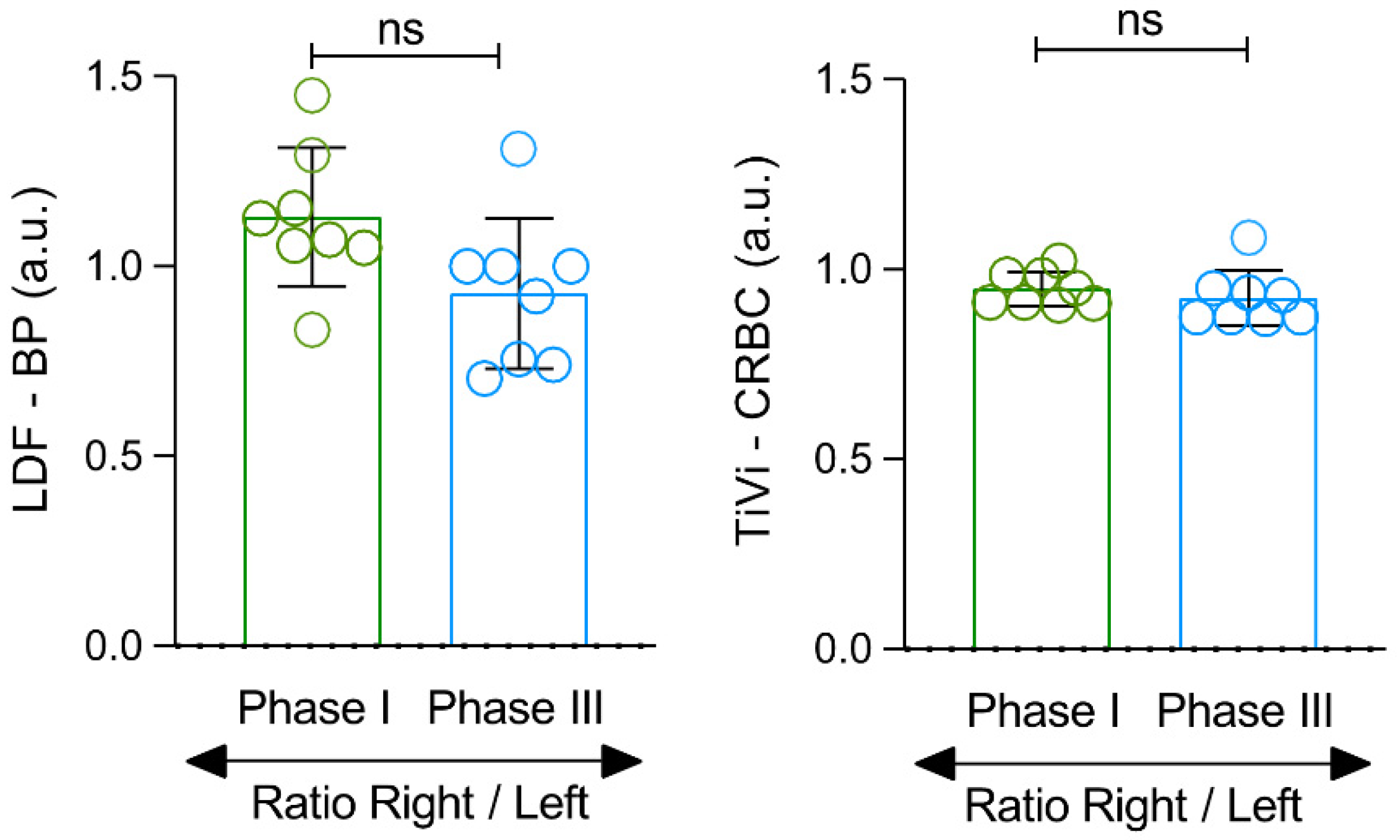
Calculating the right-left perfusion ratio between both feet, a common indicator of the lower limb perfusion asymmetry [6,7,8,9,10], we see no statistically significant differences in Phase I or Phase III perfusions as detected by LDF or PSp in the studied conditions (Figure 2).
Plantar pressure in the standing position was asymmetric (Figure 3). Plantar pressures differed between feet and were not equally distributed in the foot (considering the different areas of pressure as illustrated in Figure 1). Our results indicated that these differences were not statistically significant in the forefoot and in the midfoot, and that the squat increased plantar pressure in both these two regions (Figure 3). In the hindfoot, differences between left and right feet were always present and statistically significant in each phase of the experimental protocol. Not surprisingly, squat reduced the registered pressure in Phase 2 (Figure 3). Concerning the pressure related variables of CoP velocity and displacement and AoE, we noted that squat evoked a significant change (p = 0.0021) in all variables (Figure 4).
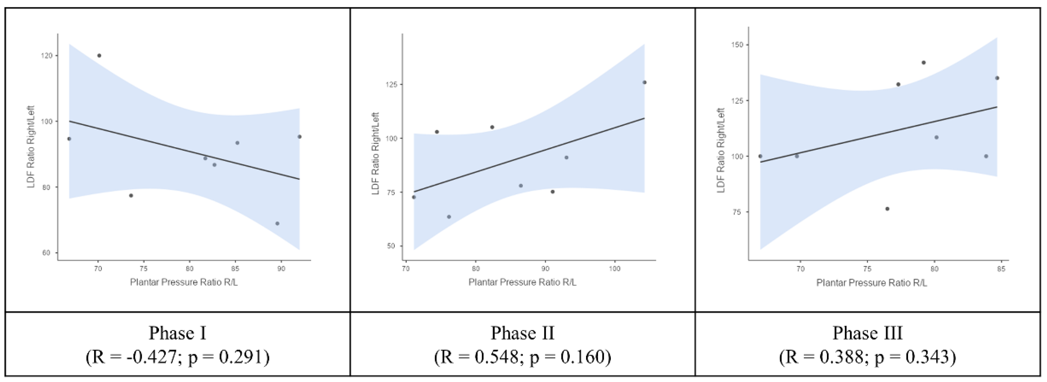
The Pearson’s correlation analysis of these asymmetries detected between both feet for LDF perfusion and plantar pressure (Figure 5) could not detect any significant differences in any phase except a tendency that is evoked by the squat (discussed ahead).
Further analysis of correlations among variables suggests important relationships in Phases I and III, while it is not possible to identify statistically significant correlations in Phase II. As seen in Figure 6 and Figure 7, mean perfusion measured by LDF in Phase I is inversely correlated with the centre of pressure (CoP) velocity (p = 0.025) and CoP displacement (p = 0.024), while the body mass index (BMI) and mean plantar pressure are positively correlated (p = 0.022). In Phase III, no significant correlations are observed between plantar pressure variables and stance and perfusion. However, the correlation of BMI with mean pressure remains significant (p = 0.014).

4. Discussion

Pathological blood perfusion differences in lower limbs are known [33,34,35]. This unevenness has also been described in the absence of disease and has been related to age, sex and BMI [6,11,12,29]. Thus, the interlimb asymmetries here detected were expected. The apparently contradictory results of a left foot perfusion “dominance” as measured by LDF and right foot perfusion “dominance” as measured by PSp (Table 2) are, in fact, complementary. The two technologies used in our study share a common optical basis, but the interaction of the respective laser lights with the skin employs very specific and differing mechanisms. LDF uses a red light with a 780 nm wavelength, providing a signal assumed to be linearly related to the velocity and concentration of moving erythrocytes [32,36,37,38]. The perfusion estimations provided by LDF are based in a small vascular volume, likely at a depth of around 1 mm, since contact with the skin allows the light to penetrate deeper and to access larger vessels and higher volumes of blood [38,39]. In turn, the PSp is a non-contact system using a white light with a wavelength of 633 nm and measures in a sub-epidermal area at an estimated depth of 0.5 mm, where light is scattered and absorbed primarily by the haemoglobin molecule in the red blood cells [37,40]. It is clear that PSp reads more superficial areas with smaller vessels and blood volumes. Therefore, considering the peculiar organisation of skin circulation involving a superficial plexus at the dermis and a deeper structure with larger vessels crossing the adipose tissue and beyond, these measurements are in agreement. Higher blood volumes are present in deeper, larger vessels, while the most superficial vessels are smaller, containing necessarily lower blood volume. Both systems detected perfusion increases in both feet in Phase II (Table 3), along with the disappearance of the Phase I asymmetries. These asymmetries reappear in Phase III as perfusion decreaases. This last finding indicates a rapid recovery capacity, keeping in mind that all participants are healthy and active (Figure 1). The significant increase in blood pressure and heart rate in Phase II are clearly associated with the squatting activity (Table 2).
Movement and exercise are known to influence lower limb vascular perfusion and pooling, and muscle recruitment [41]. Stance modifies heart rate, mean arterial pressure, and blood accumulation in the foot, and stance alterations were recently associated with lower limb discomfort [42]. Vascular diseases such as peripheral vascular disease (PVD) and type 2 diabetes mellitus (T2D) are known to determine perfusion asymmetries in the lower limb and modify muscular biomechanics and movement (gait) [43]. This might be accentuated in older adults in the presence of common comorbidities since ageing per se seems not to significantly modify gait function [44]. Nevertheless, PVD and T2D patients are known to be prone to unfavorable ankle and knee joint modifications, likely due to compensatory changes in gait [43,44]. Gait adaptation is also a common consequence of an increase of plantar pressure asymmetries—where wide asymmetries reflect an unequal loading and mechanics of the paired feet—especially in the presence of vascular impairment [23,25,26,43,45].
Under this view, we decided to explore potential relations between stance, blood perfusion, and plantar pressure to better understand these lower limb asymmetries and their implications. Although not equally distributed along the foot, plantar pressure differences were present in all individuals at Phase I (Figure 3). Higher plantar pressure was registered in the hindfoot and here the left foot showed statistically significant higher values when compared with the right foot, such as perfusion measured by LDF. The squat in Phase II reduced plantar pressure displaced to the other regions but accentuated these statistically significant differences in Phases II and III (Figure 3). Forefoot and midfoot pressures were lower, and their differences were not statistically significant in any of the phases, but the squat of Phase II increased the plantar pressure in these regions (Figure 3).
Regarding the stance variables related to plantar pressure, we notice that the Phase 2 squat significantly increased all variables (Figure 4). The Pearson’s correlation analysis did not show any relevant correlation between plantar pressure and LDF perfusion (data not shown) in both feet. We repeated this correlation analysis with right/left foot ratios as a practical method to assess bi-lateral asymmetries (6–10) for both variables (Figure 5). Here we found an interesting tendency—in Phase I, an inverse relationship between perfusion and plantar pressure asymmetries was present, suggesting that higher pressure in one foot favours perfusion in the opposite foot. However, in Phases II and III, the correlation was reversed, more evident in Phase II (R = 0.55), as in Phase III the tendency seemed to recover the Phase I relationship (Figure 5).
We further analysed potential correlations within these variables (Pearson’s correlation test). As shown in Figure 6 and Figure 7, a significant directly proportional correlation between BMI and mean Plantar Pressure scores was detected in Phase I and in Phase III, and a significant inverse correlation between LDF blood perfusion and CoP velocity and displacement found in Phase I.
The exploratory nature of these results should draw our attention to some obvious limitations, including (i) the reduced number of participants restraining any extrapolations and the identification of other potential determinants; (ii) the exclusive use of healthy participants, excluding specific groups of typical patients (e.g., those with vascular, muscular, osteoarticular impairments) and (iii) the lack of movement kinetics and muscular strength variables necessary to better understand other asymmetry-related relationships. We will address these limitations in future studies, including the evaluation of “non-healthy” individuals, to better recognise its potential clinical utility. Nevertheless, this exploratory approach seems to justify our view on the interest of studying potential relationships among blood perfusion, biomechanics, and postural indicators as plantar pressure variables, to better understand the significance of intraindividual functional asymmetries between the lower limbs in the absence of disease.

Author Contributions

L.M.R. and M.E.F. conceptual design; S.L.N., T.A. and M.E.F. experimental; T.G., J.G. and S.L.N. data curation and analysis; L.M.R. and T.A. reviewed and L.M.R. wrote the manuscript in its final version. All authors have read and agreed to the published version of the manuscript.

Funding

This research is funded by FCT—Foundation for Science and Technology I.P., through grant UIDB/04567/2020 to CBIOS. J.G. is funded by FCT—Foundation for Science and Technology I.P., with the grant CEEC/CBIOS/EPH/2018 for Scientific Employment Stimulus.

Informed Consent Statement

Informed consent was obtained from all subjects involved in the study.

Acknowledgments

The authors wish to thank João Abrantes from MovLab—Iteraction and Interface Technologies Laboratory.

Conflicts of Interest

The authors declare no conflict of interest.

References

  1. Bouley, J. Claudication Intermittent des Membres Posterieurs, determinee par L’obliteration des Arteres Femorales. Rec. Med. Vet. Ec. Alfort. 1831, 8, 517–527. [Google Scholar]
  2. Zusmanovich, F.; Elizarova, S. Perfusion pressure dynamics in lower extremities at rest and after exercise. Fiziol. Cheloveka 2002, 28, 133. (In Russian) [Google Scholar] [PubMed]
  3. Siegel, M.E.; Siemsen, J.K. A new noninvasive approach to peripheral vascular disease: Thallium-201 leg scans. AJR 1978, 131, 827–830. [Google Scholar] [CrossRef] [Green Version]
  4. Seder, J.S.; Botvinick, E.H.; Rahimtoola, S.H.; Goldstone, J.; Price, D.C. Detecting and localizing peripheral arterial disease: Assessment of 201Tl scintigraphy. AJR 1981, 137, 373–380. [Google Scholar] [CrossRef] [PubMed] [Green Version]
  5. Collins, R.; Burch, J.; Cranny, G.; Aguiar-Ibáñez, R.; Craig, D.; Wright, K.; Berry, E.; Gough, M.; Kleijnen, J.; Westwood, M. Duplex ultrasonography, magnetic resonance angiography, and computed tomography angiography for diagnosis and assessment of symptomatic, lower limb peripheral arterial disease: Systematic review. BMJ Clin. Res. 2007, 334, 1257. [Google Scholar] [CrossRef] [Green Version]
  6. Mayrovitz, H.N.; Larsen, P.B. Pulsatile blood flow asymmetry in paired human legs. Clin. Physiol. 1996, 16, 495–505. [Google Scholar] [CrossRef]
  7. Nuno, S.; Atalaia, T.; Gregório, J.; Florindo, M.; Granja, T.; Abrantes, J.; Rodrigues, L.M. Influence of posture on the foot perfusion in the upright position. Physiol. 2021 Proc. Physiol. Soc. 2021, 48, PC013. Available online: https://www.physoc.org/abstracts/influence-of-posture-on-the-foot-perfusion-in-the-upright-position/ (accessed on 21 December 2021).
  8. Gregório, J.; Florindo, M.; Nuno, S.; Rodrigues, L.M. Insights into human healthy. Physiol. 2021 Proc. Physiol. Soc. 2021, 48, OC04. Available online: https://www.physoc.org/abstracts/insights-into-human-healthy-lower-limb-perfusion-asymmetry-during-rest/ (accessed on 21 December 2021).
  9. Rocha, C.; Silva, H.; Ferreira, H.; Rodrigues, L.M. Evidence of a Physiological Perfusion Balance Between Human Limb Pairs. Europhysiology 2018. Abstract Book 2018. Available online: https://www.europhysiology2018.org/sites/default/files/files/Europhysiology%202018_ABSTRACTS_ONLINE.pdf (accessed on 21 December 2021).
  10. Rodrigues, L.M.; Rocha, C.; Ferreira, H.T.; Silva, H.M. Lower limb massage in humans increases local perfusion and impacts systemic hemodynamics. J. Appl. Physiol. 2020, 128, 1217–1226. [Google Scholar] [CrossRef]
  11. Gregório, J.; Silva, H.; Rocha, C.; Rodrigues, L.M. Perfusion is sex related but response to massage evokes the same hemodynamic adaptation in both sexes—Results from an exploratory factor analysis. Proceed Physioma 2019—1st Int Meeting Portuguese Physiological Society. Biomed. Biopharm. Res. 2019, 16, 31. [Google Scholar] [CrossRef]
  12. Rodrigues, L.M.; Rocha, C.G.; Florindo, M.E.; Gregório, J. Lower Limb Perfusion Asymmetries in Humans at Rest and Following Activity—A Collective View. Symmetry 2021, 13, 2348. [Google Scholar] [CrossRef]
  13. Keeley, D.W.; Plummer, H.A.; Oliver, G.D. Predicting Asymmetrical Lower Extremity Strength Deficits in College-Aged Men and Women Using Common Horizontal and Vertical Power Field Tests: A Possible Screening Mechanism. J. Strength Cond. Res. 2011, 25, 1632–1637. [Google Scholar] [CrossRef]
  14. Bishop, C.; Read, P.; Chavda, S.; Turner, A. Asymmetries of the Lower Limb: The Calculation Conundrum in Strength Training and Conditioning. Strength Cond. J. 2016, 38, 27–32. [Google Scholar] [CrossRef] [Green Version]
  15. Lanshammar, K.; Ribom, E.L. Differences in muscle strength in dominant and non-dominant leg in females aged 20–39 years—A population-based study. Phys. Ther. Sport 2011, 12, 76–79. [Google Scholar] [CrossRef]
  16. Manevska, N.; Gjorceva, D.P.; Ahmeti, I.; Todorovska, L.; Stojanoski, S.; Kocovska, M.Z. Tissue-Muscle Perfusion Scintigraphy of the Lower Limbs in a Patient with Type 2 Diabetes Mellitus and Peripheral Arterial Disease. Mol. Imaging Radionucl. Ther. 2016, 25, 42–46. [Google Scholar] [CrossRef] [PubMed]
  17. Bell, D.R.; Sanfilippo, J.L.; Binkley, N.; Heiderscheit, B.C. Lean mass asymmetry influences force and power asymmetry during jumping in collegiate athletes. J. Strength Cond. 2014, 28, 884–891. [Google Scholar] [CrossRef] [PubMed] [Green Version]
  18. Hoffman, J.R.; Ratamess, N.A.; Klatt, M.; Faigenbaum, A.D.; Kang, J. Do bilateral power deficits influence direction-specific movement patterns? Res. Sports Med. 2007, 15, 125–132. [Google Scholar] [CrossRef]
  19. Maloney, S.J. The Relationship Between Asymmetry and Athletic Performance: A Critical Review. J. Strength Cond. 2019, 33, 2579–2593. [Google Scholar] [CrossRef]
  20. Heil, J.; Loffing, F.; Büsch, D. The Influence of Exercise-Induced Fatigue on Inter-Limb Asymmetries: A Systematic Review. Sports Med. 2020, 6, 39. [Google Scholar] [CrossRef]
  21. Kadoguchi, T.; Horiuchi, M.; Kinugawa, S.; Okita, K. Heterogeneity in the vasodilatory function of individual extremities. Vascular 2020, 28, 87–95. [Google Scholar] [CrossRef]
  22. Jungmann, P.M.; Pfirrmann, C.; Federau, C. Characterization of lower limb muscle activation patterns during walking and running with Intravoxel Incoherent Motion (IVIM) MR perfusion imaging. Magn. Reson. Imaging 2019, 63, 12–20. [Google Scholar] [CrossRef] [PubMed]
  23. Wafai, L.; Zayegh, A.; Woulfe, J.; Aziz, S.M.; Begg, R. Identification of Foot Pathologies Based on Plantar Pressure Asymmetry. Sensors 2015, 15, 20392–20408. [Google Scholar] [CrossRef] [PubMed] [Green Version]
  24. Menz, H.B.; Morris, M.E. Clinical determinants of plantar forces and pressures during walking in older people. Gait Posture 2006, 24, 229–236. [Google Scholar] [CrossRef]
  25. Erdoğanoğlu, Y.; Sayaca, Ç.; Çalık, M.; Noyan, C.O.; Çetin, A.; Yertutanol, D.K.; Taşcılar, L.N.; Kaya, D. Evaluation of Plantar Foot Sensation, Balance, Physical Performance, and Fear of Movement in Substance Use Disorders. J. Am. Podiat. Med. Assoc. 2020, 110, 5. [Google Scholar] [CrossRef] [PubMed]
  26. de Cock, A.; Vanrenterghem, J.; Willems, T.; Witvrouw, E.; de Clercq, D. The trajectory of the centre of pressure during barefoot running as a potential measure for foot function. Gait Posture 2008, 27, 669–675. [Google Scholar] [CrossRef] [PubMed]
  27. Florindo, M.; Silva, H.; Rodrigues, L.M. Impact of the isometric contraction of the calf on the local microcirculation. Biomed. Biopharm. Res. 2017, 14, 179–186. [Google Scholar] [CrossRef]
  28. Nuno, S.; Florindo, M.; Silva, H.; Rodrigues, L.M. Studying the impact of different body positioning, squatting, and unipodal flexion on perfusion in the lower limb—An exploratory approach complemented with optical spectroscopy (TiVi). Biomed. Biopharm. Res. 2020, 17, 1–10. [Google Scholar] [CrossRef]
  29. Florindo, M.; Nuno, S.L.; Rodrigues, L.M. Lower limb dynamic activity significantly reduces foot skin perfusion—Exploring data with different optical sensors in age-grouped healthy adults. Skin Pharmacol. Physiol. 2022, 35, 13–22. [Google Scholar] [CrossRef]
  30. Aboyans, V.; Criqui, M.H.; Abraham, P.; Allison, M.A.; Creager, M.A.; Diehm, C.; Fowkes, F.G.R.; Hiatt, W.R.; Jönsson, B.; Lacroix, P.; et al. Measurement and interpretation of the ankle-brachial index: A scientific statement from the American Heart Association. Circulation 2012, 126, 2890–2909. [Google Scholar] [CrossRef] [Green Version]
  31. World Medical Association. World Medical Association Declaration of Helsinki: Ethical principles for medical research involving human subjects. JAMA 2013, 310, 2191–2194. [Google Scholar] [CrossRef] [Green Version]
  32. Rocha, C.; Silva, H.; Ferreira, H.; Rodrigues, L.M. About the in vivo discriminatory capacity of photoplethysmography versus laser Doppler flowmetry. Biomed. Biopharm. Res. 2017, 14, 37–44. [Google Scholar] [CrossRef]
  33. Srivaratharajah, K.; Abramson, B.L. Women and Peripheral Arterial Disease: A Review of Sex Differences in Epidemiology, Clinical Manifestations, and Outcomes. Can. J. Cardiol. 2018, 34, 356–361. [Google Scholar] [CrossRef]
  34. Xu, X.; Wang, B.; Ren, C.; Hu, J.; Greenberg, D.A.; Chen, T.; Xie, L.; Jin, K. Age-related Impairment of Vascular Structure and Functions. Aging Dis. 2017, 8, 590–610. [Google Scholar] [CrossRef] [PubMed] [Green Version]
  35. Abiri, B.; Vafa, M. Dietary Restriction, Cardiovascular Aging and Age-Related Cardiovascular Diseases: A Review of the Evidence. Adv. Exp. Med. Biol. 2019, 1178, 113–127. [Google Scholar] [CrossRef]
  36. Bergstrand, S.; Lindberg, L.G.; Ek, A.C.; Lindén, M.; Lindgren, M. Blood flow measurements at different depths using photoplethysmography and laser Doppler techniques. Ski. Res. Technol. ISBS ISDIS ISSI 2009, 15, 139–147. [Google Scholar] [CrossRef] [PubMed]
  37. Nilsson, G.E.; Tenland, T.; Oberg, P.A. Evaluation of a laser Doppler flowmeter for measurement of tissue blood flow. IEEE Trans. Bio-Med. Eng. 1980, 27, 597–604. [Google Scholar] [CrossRef]
  38. Nilsson, G.E.; Salerud, E.G.; Stromberg, N.O.T.; Wardell, K. Laser Doppler perfusion monitoring and imaging. In Biomedical Photonics Handbook; Vo-Dinh, I.T., Ed.; CRC Press: Boca Raton, FL, USA, 2003; pp. 1–24. [Google Scholar]
  39. Rodrigues, L.M.; Rocha, C.; Ferreira, H.; Silva, H. Different lasers reveal different skin microcirculatory flowmotion—Data from the wavelet transform analysis of human hindlimb perfusion. Sci. Rep. 2019, 9, 16951. [Google Scholar] [CrossRef]
  40. O’Doherty, J.; Henricson, J.; Anderson, C.; Leahy, M.J.; Nilsson, G.E.; Sjöberg, F. Sub-epidermal imaging using polarized light spectroscopy for assessment of skin microcirculation. Skin Res. Technol. 2007, 13, 472–484. [Google Scholar] [CrossRef]
  41. Raffetto, J.D.; Khalil, R.A. Mechanisms of varicose vein formation: Valve dysfunction and wall dilation. Phlebology 2008, 23, 85–98. [Google Scholar] [CrossRef]
  42. Antle, D.M.; Cormier, L.; Findlay, M.; Miller, L.L.; Côté, J.N. Lower limb blood flow and mean arterial pressure during standing and seated work: Implications for workplace posture recommendations. Prev. Med. Rep. 2018, 10, 117–122. [Google Scholar] [CrossRef]
  43. Thorne, C.S.; Bartolo, E.; Gatt, A.; Formosa, C. The Impact of Peripheral Artery Disease (PAD) on Lower Limb Kinematics in Type 2 Diabetes Mellitus. Rev. Diabet. Stud. RDS 2021, 17, 11–16. [Google Scholar] [CrossRef] [PubMed]
  44. Myers, S.A.; Applequist, B.C.; Huisinga, J.M.; Pipinos, I.I.; Johanning, J.M. Gait kinematics and kinetics are affected more by peripheral arterial disease than by age. JRRD J. Rehabil. Res. Dev. 2016, 53, 229–238. [Google Scholar] [CrossRef] [PubMed]
  45. Boulton, A.J.; Hardisty, C.A.; Betts, R.P.; Franks, C.I.; Worth, R.C.; Ward, J.D.; Duckworth, T. Dynamic foot pressure and other studies as diagnostic and management aids in diabetic neuropathy. Diabetes Care 1983, 6, 26–33. [Google Scholar] [CrossRef] [PubMed]
Figure 1. Illustrative pedobarographic image of one participant depicting distribution areas of plantar pressure while standing in the upright position (see text).
Symmetry 14 00441 g001
Figure 2. Perfusion ratios (right/left feet) as an indicator of the individual asymmetry measured with LDF and PSp (TiVi) instruments at Phase I and Phase III (see text). Comparisons between phases are also shown (ns—non-significant).
Symmetry 14 00441 g002
Figure 3. Plantar pressure (PP) asymmetries measured in three areas of both feet during the different phases of the experimental protocol. Comparison between feet in each foot area depicts a significance difference in plantar pressure asymmetry at the hindfoot in all the phases of the protocol (* p < 0.05; ns—non-significant). Note the differing Y-axis values in the far right panel (hindfoot).
Symmetry 14 00441 g003
Figure 4. Posture related changes expressed as Velocity of the Centre of Pressure (Veloc. CoP) Maximum Area of the Ellipse (AoE) and CoP Displacement registered during the experimental protocol (* p < 0.05; ns—non-significant).
Symmetry 14 00441 g004
Figure 5. Pearson’s correlations between LDF perfusion (right foot/left foot) ratio and mean Plantar Pressure (right/left feet) ratio (see text).
Symmetry 14 00441 g005
Figure 6. Variable’s correlation matrix (Pearson) found in Phase I (left) and Phase III (right). No correlations could be found in Phase II (see text).
Symmetry 14 00441 g006
Figure 7. Graphical evolution of variables’ correlation (Pearson’s) during the experimental protocol. Statistically significant correlations are marked in red (see text).
Symmetry 14 00441 g007
Table 1. Participants’ characterisation (baseline). When applicable, results are presented as medians and Q1–Q3 (25th empirical quartile–75th empirical quartile) (* p < 0.05).
MENWOMENp-Value
N (%) 4 (50)4 (50)_
Smokers (%) 0 (100)0 (100)_
Age, years (Q1–Q3) 28.8(20–32)21.8(21–22)0.098
Body mass, kg (Q1–Q3) 74.5 (68.0–85.0)61.5 (58.0–68.0)0.201
Height, m (Q1–Q3) 1.8 (1.7–1.8)1.6 (1.6–1.7)<0.001 *
BMI, kg/m2 (Q1–Q3) 23.9 (22.9–24.9)22.8 (22.1–24.9)0.546
SYSTP, mmHg (Q1–Q3) 122.0 (113.7–129.0)120.9 (111.7–135.3)0.670
DIASP, mmHg (Q1–Q3) 82.4 (74.7–88.0)78.1 (75.0–78.7)0.424
ABI (Q1–Q3) 1.0 (1.0–1.1)1.0 (1.0–1.1)0.062
PR, bpm (Q1–Q3) 68.8 (61.5–77.3)65.5 (59.0–69.5)0.088
SpO2 (%), bpm (Q1–Q3) 98.5 (98–99)98.3 (98–99)0.951
BMI, Body Mass Index; SYSTP, Systolic pressure; DIASP, Diastolic Pressure; ABI, ankle-brachial index; PR, Pulse Rate; bpm, beats per minute; SpO2 oxygen saturation.
Table 2. Cardiovascular dynamics changes registered in the studied experimental conditions. Perfusion (mean + sd) obtained by LDF and PSp instruments was measured and compared in both feet at baseline (Phase I), challenge (Phase II) and recovery (Phase III) (p). Other hemodynamic variables (cardiac frequency and blood arterial pressure) are compared with baseline (p 🢗) (* p/p 🢗 < 0.05). ⸸—Statistical comparison between lower limbs with the Friedman test with Paired Comparisons correction (Durbin-Conover); ¥—Repeated measures ANOVA, with the Post hoc Tukey test for pairwise comparisons.
Phase 1Phase 2Phase 3
Right FootLeft FootRight FootLeft FootRight FootLeft Foot
LDF_BPU (AU) ⸸6.0 ± 1.36.7 ± 1.412.1 ± 4.313.9 ± 4.87.7 ± 2.16.9 ± 1.3
p-value0.007 *0.5710.015 *
PSp_CRBC (AU) ¥217.2 ± 14.8206.0 ± 18.0227.2 ± 12.0222.9 ± 14.8217.1 ± 13.7200.0 ± 11.0
p-value0.0940.6910.125
PR (p-value 🢗) ¥63.1 ± 9.873.6 ± 8.9 (0.002) *65.5 ± 9.0 (0.020) *
sAP (p-value 🢗) ¥123.0 ± 7.0130.5 ± 5.8 (0.004) *124.9 ± 7.8 (0.495)
dAP (p-value 🢗) ¥65.0 ± 7.170.3 ± 4.8 (0.049) *67.3 ± 7.0 (0.079)
AU: arbitrary units; PR: pulse rate; bpm: beats per minute; sAP: systolic arterial pressure; dAP: diastolic arterial pressure; * p < 0.05.
Table 3. Statistical differences between experimental phases in each limb as measured with LDF and PSp systems. ⸸—Statistical comparison between lower limbs with the Friedman test with Paired Comparisons correction (Durbin-Conover). ¥—Repeated measures ANOVA, with the Post hoc Tukey test for pairwise comparisons (* p < 0.05).
LDF ⸸
right foot Phase I
6.0 ± 1.3
right foot Phase II
12.1 ± 4.3
<0.001 *
right foot Phase I
6.0 ± 1.3
right foot Phase III
7.7 ± 2.1
<0.001 *
left foot Phase I
6.7 ± 1.4
left foot Phase II
13.9 ± 4.8
<0.001 *
left foot Phase I
6.7 ± 1.4
left foot Phase III
6.9 ± 1.3
0.207
PSp ¥
right foot Phase I
217.2 ± 14.8
right foot Phase II
227.2 ± 12.0
0.080
right foot Phase I
217.2 ± 14.8
right foot Phase III
217.1 ± 13.7
1.000
left foot Phase I
206.0 ± 18.0
left foot Phase II
222.9 ± 14.8
0.016 *
left foot Phase I
206.0 ± 18.0
left foot Phase III
200.0 ± 11.0
0.613
Publisher’s Note: MDPI stays neutral with regard to jurisdictional claims in published maps and institutional affiliations.