Article

Movement Coordination during Functional Single-Leg Squat Tests in Healthy, Recreational Athletes

by
Korbinian Sebastian Hermann Ksoll
1,*,†,
Matthias Cotic
2,†,
Kathrin Schmalzl
3,
Knut Beitzel
4,
Andrea Achtnich
2,
Andreas Imhoff
2,
Ansgar Schwirtz
5,
Florian Kreuzpointner
3 and
Wolfgang Seiberl
1
1
Department of Human Sciences, Institute of Sport Sciences, University of the Bundeswehr Munich, 85577 Neubiberg, Germany
2
Department of Orthopaedic Sports Medicine, Klinikum Rechts der Isar, Technical University of Munich, 81675 Munich, Germany
3
Prevention Center, Department of Sport and Health Sciences, Technical University of Munich, 80809 Munich, Germany
4
ATOS Orthoparc Clinic GmbH, 50858 Cologne, Germany
5
Biomechanics in Sport, Department of Sport and Health Sciences, Technical University Munich, 80992 Munich, Germany
*
Author to whom correspondence should be addressed.
These authors contributed equally to this project and share first authorship.
Symmetry 2022, 14(2), 388; https://doi.org/10.3390/sym14020388
Submission received: 14 December 2021 / Revised: 1 February 2022 / Accepted: 10 February 2022 / Published: 15 February 2022

Abstract

:
The single-leg squat (SLS) represents a functional movement task for determining leg function. Objective movement analysis is required to evaluate inter-limb symmetry and movement coordination. Therefore, this study aimed to investigate inter-limb symmetry of SLS kinematics and movement coordination using the modified vector coding technique. A 3D motion capture system and electromyography were used to assess SLS execution and muscle activation of hip ab- and adductors of 17 healthy, recreational athletes. Coordination patterns of hip, knee, and ankle joint movement were assessed by the modified vector coding technique. Statistical parametric mapping revealed no significant differences between both legs (p > 0.05). Inter-limb symmetry also appeared in movement coordination (p > 0.05). Additionally, the analysis of movement coordination indicates knee-dominant, in-phase coordination. However, coordination patterns were different between downward movement, change of direction, and upward movement (p < 0.001). Since perturbations during SLS execution, such as moments of imbalance, occur as anti-phase coordination patterns, the analysis of coordination patterns can be used as a new evaluation method for SLS performance. Furthermore, the modified vector coding technique might be helpful to analyze different compensation strategies during the SLS in symptomatic individuals.

1. Introduction

Functional movement tests are commonly used in sports clubs, rehabilitation centers, and clinics to determine limb and muscle function. These tests are typically easy to assess and are known to be an important criterion in injury prevention and return-to-play decisions [1,2,3]. Furthermore, clinical decision making, as well as reports on the rehabilitation process, are based in part on the rating of these functional movement tasks [1,4,5]. One common functional movement test is the single-leg squat (SLS) [6]. The SLS simulates both different activities of daily living, such as ascending and descending stairs, as well as sporting activities [7,8,9]. The SLS task increases pressure in the patellofemoral joint and is known to provoke and thus identify symptoms of anterior knee pain, also known as patellofemoral pain syndrome [10,11,12]. One reason for the increased patellofemoral joint pressure is the dynamic knee valgus, a multiplanar movement in the hip (adduction and internal rotation), knee (abduction and external rotation), and ankle joint (foot pronation). The dynamic knee valgus leads to patella maltracking, inducing lateral shear stress on the patellar and femoral articular surface [10,12,13]. Furthermore, the dynamic knee valgus is known as a risk factor for lower limb injuries, most dominant the injury of the anterior cruciate ligament [11,14,15]. Thus, the SLS test is an important test in the clinical setting [5,9,16].
In clinical use, the assessment of SLS performance is often subjective, based on a predefined rating catalog. For example, the qualitative rating of SLS performance according to the method proposed by Crossley et al. [5] focuses on the following five criteria: (1) overall impression, (2) trunk posture, (3) movement of the pelvis, (4) hip joint movement, and (5) knee joint movement. Although the SLS performance rating is valid and reliable [5,17], objective and quantitative movement analysis, by 3D motion capture or inertial measurement units [9,17,18], allows deeper insights into the biomechanics of the SLS movement. Hence, 3D motion analysis is able to quantify the dynamic knee valgus. This multiplanar movement in the hip, knee, and ankle joint is reported to provoke excessive medial knee displacement [11,15,19].
An important aspect in the interpretation of objective motion analysis during functional movement tests is the inter-limb symmetry [20,21,22]. Inter-limb symmetry refers to the equality of movement in both legs [21,23]. When assessing inter-limb symmetry and inter-limb asymmetry, respectively, intra-limb variation must be considered [21]. In terms of the functional SLS test, intra-limb variation refers to the variation of movement kinematics within different SLS trials of one leg and is also known as coordinative variability [21,24]. It is assumed that coordinative variability differs between novice and experienced test persons, as well as between symptomatic and asymptomatic individuals [24]. In order to evaluate the complex interaction between two segments or joints, the modified vector coding technique is able to distinguish human movements in different coordination patterns [24,25]. These coordination patterns and the coordinative variability are assumed to be different according to different compensation strategies in asymptomatic individuals. To our knowledge, less is known about the movement coordination of SLS execution in healthy individuals using the modified vector coding technique. The determination of SLS movement coordination might serve as a new, additional measure of lower leg function, thus expanding the clinical value of the functional SLS test. Therefore, the aim of the present study was to objectively investigate inter-limb symmetry and movement coordination in SLS execution of healthy, recreational athletes using 3D motion capture, electromyography, and the modified vector coding technique. We hypothesized that, in a healthy population, the kinematic movement patterns of the SLS occur symmetrically in both limbs. With respect to intra-limb coordination, we additionally hypothesized that adjacent joints show in-phase coordination patterns during SLS execution. In this context, we analyzed the movement coordination in terms of differences between the downward movement, upward movement, and the change of direction phase of the SLS task.

2. Materials and Methods

2.1. Subject Information and Ethics

In this study, only healthy men and women between 18 and 35 years were included. Subjects were excluded if they met one or more of the following exclusion criteria, which were defined according to Weeks et al. [26] and Crossley et al. [5]: (1) multi-ligamentous instabilities; (2) markedly decreased range of motion (arc of motion < 140°, flexion contracture > 5°); (3) previous musculoskeletal surgery in the last 10 years of the spine, hip, knee and ankle, which might affect coordination abilities of the lower extremities; (4) femorotibial rest pain; (5) neurologic disorders; (6) psychological disorders; (7) pregnancy; (8) inflammation of the musculoskeletal system; (9) genu varum or genu valgum; (10) subjective perception of knee pain. Thus, 17 male (n = 8) and female (n = 9) participants voluntarily took part in this study. All participants were physically active multiple times per week, healthy, and had no knee pathologies (Table 1). The Tegner Activity Scale [27] was used to quantify the sporting activity of the participants. For each participant, a subjective knee evaluation was assessed based on the knee injury and Osteoarthritis Outcome Score (KOOS) questionnaire according to the KOOS User’s Guide. Knee pain was recorded according to the visual analog scale for pain (VAS), ranging from no pain (0) to worst pain possible (10), before, during, and after physical activity [28]. One female participant reported minor pain during (VAS = 2) and after activity (VAS = 1). As the clinical examination of the SLS execution according to Crossley et al. [5] did not reveal any restrictions or abnormalities, this participant was not excluded from further analysis. The study was carried out in accordance with the declaration of Helsinki [29] and was approved by the university’s local ethics committee (Project No.: 333/17S). Informed consent was obtained from all participants.

2.2. Data Collection

A 3D motion capture system (11 cameras, Vicon Motion Systems Ltd., Oxford, UK) recorded the SLSs with a sampling rate of 200 fps for kinematic data and a sampling rate of 1000 Hz for analog data. The sampling frequency was determined in relation to SLS movement velocity and is in accordance with comparable research designs [18,30]. In total, 16 retroreflective markers were placed at anatomical landmarks of the lower body and on the lateral side of the thigh and shank according to the Vicon Plug-In Gait lower body model [31,32]. Additional EMG sensors (Myon AG, Schwarzenberg, Switzerland) were used to receive information about muscle activity of hip abductors and adductors. To this end, one pair of EMG electrodes (Kendall H124SG, Covidien Deutschland GmbH, Neustadt/Donau, GER, inter-electrode distance 20 mm) was placed on the left and right m. gluteus medius (GM) halfway between crista iliaca and trochanter major according to the SENIAM recommendations for sensor location representing the hip abductors [33]. Representing the muscle activity of hip adductor muscles (ADD), two additional EMG sensors measured the superposed EMG signal of m. adductor magnus, m. adductor brevis and m. adductor longus of both legs. Electrode pairs were placed at the palpated m. adductor magnus about half-length of the line between Spina iliaca anterior superior and patella, on the medial side of the thigh. Prior to the SLS execution, isometric strength of hip ad- and abductors was measured during a maximum voluntary contraction (MVC) for EMG normalization using a hand-held dynamometer (microFET®2, Hoggan Scientific LLC, Salt Lake City, UT, USA). For the MVCs of hip ab- and adduction muscles, the participants lay on their side with 0° hip and knee flexion. Measuring maximum isometric hip abduction force, the participants had to push against the dynamometer, placed proximal of the lateral femur condyle. In contrast, the dynamometer was placed proximal to the medial femur condyle to measure the maximum isometric force of hip adduction muscles.
Each participant performed four sets of three consecutive SLSs on each side. To simulate a clinical application of the SLS execution the subjects were instructed as follows: “Stand on one leg, raise the free leg to the front, squat as deep as possible while maintaining balance and being able to come up again. Perform three subsequent repetitions.” The order of sides alternated with each set, starting on the left side. The leg at which participants individually reached higher knee flexion in terms of the mean of the 12 SLSs was defined as the better-performing leg (BPL). The other one was defined as the worse-performing leg (WPL).

2.3. Data Processing

Kinematic data were captured and processed with Vicon Nexus 2.7 (Vicon Motion Systems Ltd., Oxford, UK). Kinematic data were smoothed by a 15 Hz Woltring filter. Pelvis, hip, knee, and ankle motion were derived from the Plug-In Gait lower body model [31,32].
As the medial knee displacement (MKD) was not provided by the Plug-In Gait lower body model, we calculated the frontal plane knee motion based on marker trajectories. The MKD was defined by the perpendicular distance between the knee joint center and a reference plane. According to Krosshaug et al. [34] and Ellenberger et al. [18], the reference plane was first defined by the ankle, knee, and hip joint center at a reference frame and afterward, during the movement, linked to the hip joint center. For the upcoming three SLSs, the reference plane was set one frame before exceeding 35° knee flexion (0° referring to the fully extended leg). For further calculations, the data were interpolated and time normalized from 0% to 100% movement cycle, where 0% marked the beginning (exceeding 35° knee flexion in downward movement), and 100%, the end of the SLS (falling below 35° knee flexion in upward movement).
To evaluate lower limb movement coordination, the modified vector coding technique was applied following the methodology of recent studies [24,25,35,36]. Accordingly, vector coding describes the relation of two joint movements as an angle between the vector of two consecutive timepoints in an angle–angle plot and the horizontal. The so-called coupling angle describes the movement of one joint in relation to the other (e.g., hip vs. knee flexion). If only one joint is moving and the other one keeps its joint angle constant, the resulting vector is in parallel to the x- or y-axis of the angle–angle plot and the respective coupling angle, therefore, would be either 360°/0°, 90°, 180°, or 270°, depending on the joint moving and related axis. If both joints have identical flexion or extension patterns the coupling angle describes an “in-phase” (45° or 225°) coordination. Thus, “anti-phase” coordination (coupling angle of 135° or 315°) represents the situation in which one joint angle increases while the other one decreases (Figure 1, [24,25,35,36]). The variability in movement coordination was calculated as the standard deviation from the previously calculated coupling angle [25]. The coupling angle was derived by the following angle–angle plots: hip flexion–knee flexion, and knee flexion–ankle flexion. Additionally, the coupling angle of knee flexion–medial knee displacement was assessed. To reduce the effect of different movement velocities, each SLS was divided into three parts—namely, the downward movement phase (DOWN, 35° knee flexion to 5° below maximum knee flexion), the upward movement phase (UP, 5° below maximum knee flexion to 35° knee flexion), and the change of direction phase (CHANGE, phase between DOWN and UP). Furthermore, we calculated to what percentage a certain coordination pattern occurred during the downward, change of direction, and upward phases of the SLS movement. This should help to interpret the coupling angles during each SLS phase and identify possible pattern dominances. We defined eight coordination patterns to sort the coupling angles. These bins for coordination pattern classification are 360°/0–44°, 45–89°, 90–134°, 135–179°, 180–224°, 225–269°, 270–314°, and 315–359° (compare Figure 1). Accordingly, a 70° coupling angle between hip and knee motion in the sagittal plane indicates an “in-phase, knee-dominant” flexion coordination pattern.
The EMG signal was centered on zero and filtered by a Butterworth bandpass filter (10–500 Hz, fourth order, zero phase filtering). Since the frequency range of surface EMG signals is between 10 Hz and 500 Hz, the bandpass filter removes undesired low- and high-frequency components [37,38]. Further, the filtered signal was rectified and smoothed by a 250 ms moving average. The highest EMG value of the strongest MVC trial was used to normalize the processed amplitude of the EMG signal. Finally, an EMG ratio between GM and ADD was calculated to receive information about activation patterns of hip ab- and adductors.

2.4. Statistical Analysis

The data were extracted, processed, and analyzed with MATLAB (R2020b, The MathWorks, Inc., Natick, MA, USA) using custom-written codes. We used paired t-tests to analyze leg-specific differences in maximum isometric muscle strength between the BPL and WPL. Temporal differences in joint kinematics and muscle activation were analyzed by statistical parametric mapping (SPM). Thus, time normalized SLS kinematics of pelvis, hip, knee, and ankle motion, as well as GM:ADD ratio, were tested for significant differences between BPL and WPL using separate SPM paired t-tests. All SPM analyses were based on the MATLAB functions developed by Todd Pataky (www.spm1d.org, last access on 13 December 2021, [39]). To investigate differences in coordination patterns, 8 × 3 × 2 multifactorial within-subjects ANOVA was used with respect to the dominance of the coupling angle in eight coordination patterns, the three movement phases, and both legs. Statistical significance was set at α = 0.05. Violations of the sphericity assumption were corrected by the Greenhouse–Geisser method. Bonferroni correction was used in post hoc tests to avoid α-error cumulation by multiple testing.

3. Results

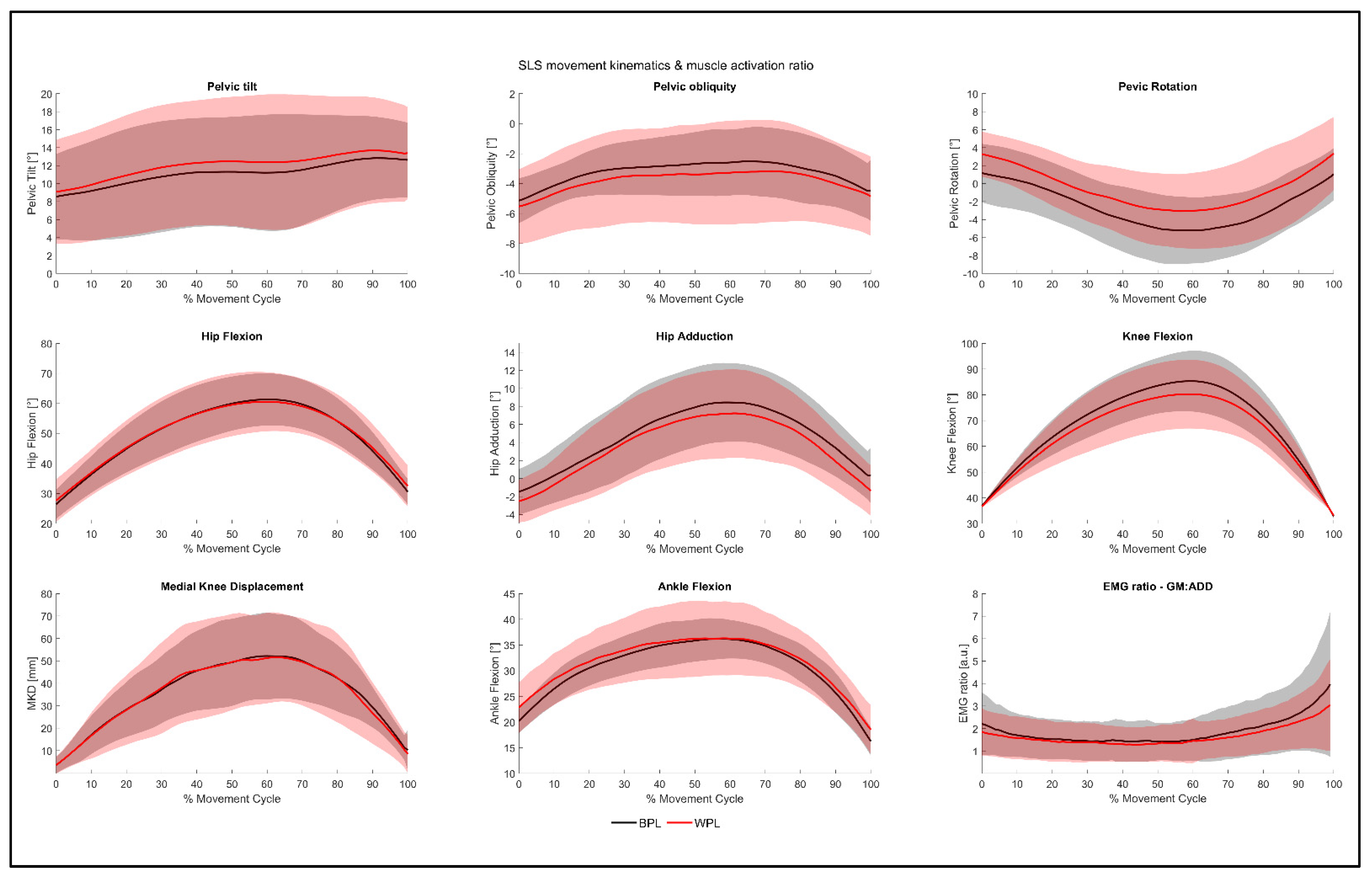
3.1. Inter-Limb Symmetry

Isometric strength tests of hip abductor and adductor muscles revealed no significant differences between both legs (hip abductors: p = 0.344, hip adductors: p = 0.263, Table 1). The mean difference in hip abductors between BPL and WPL was −23 N (95% CI (−72 N, 27 N)), while the mean difference in hip adductors was −29 N (95% CI (−84 N, 24 N)).
The kinematics of the pelvis, hip, knee, and ankle joint, as well as the EMG ratio between hip abductors and adductors during SLS, are presented in Figure 2. To compare joint kinematics of BPL and WPL, an SPM paired t-test analysis was conducted for each joint movement. The SPM test statistics did not exceed the critical thresholds in any parameter of the SLS kinematics. Thus, according to the SPM analysis, there was no significant difference in joint kinematics between both legs.

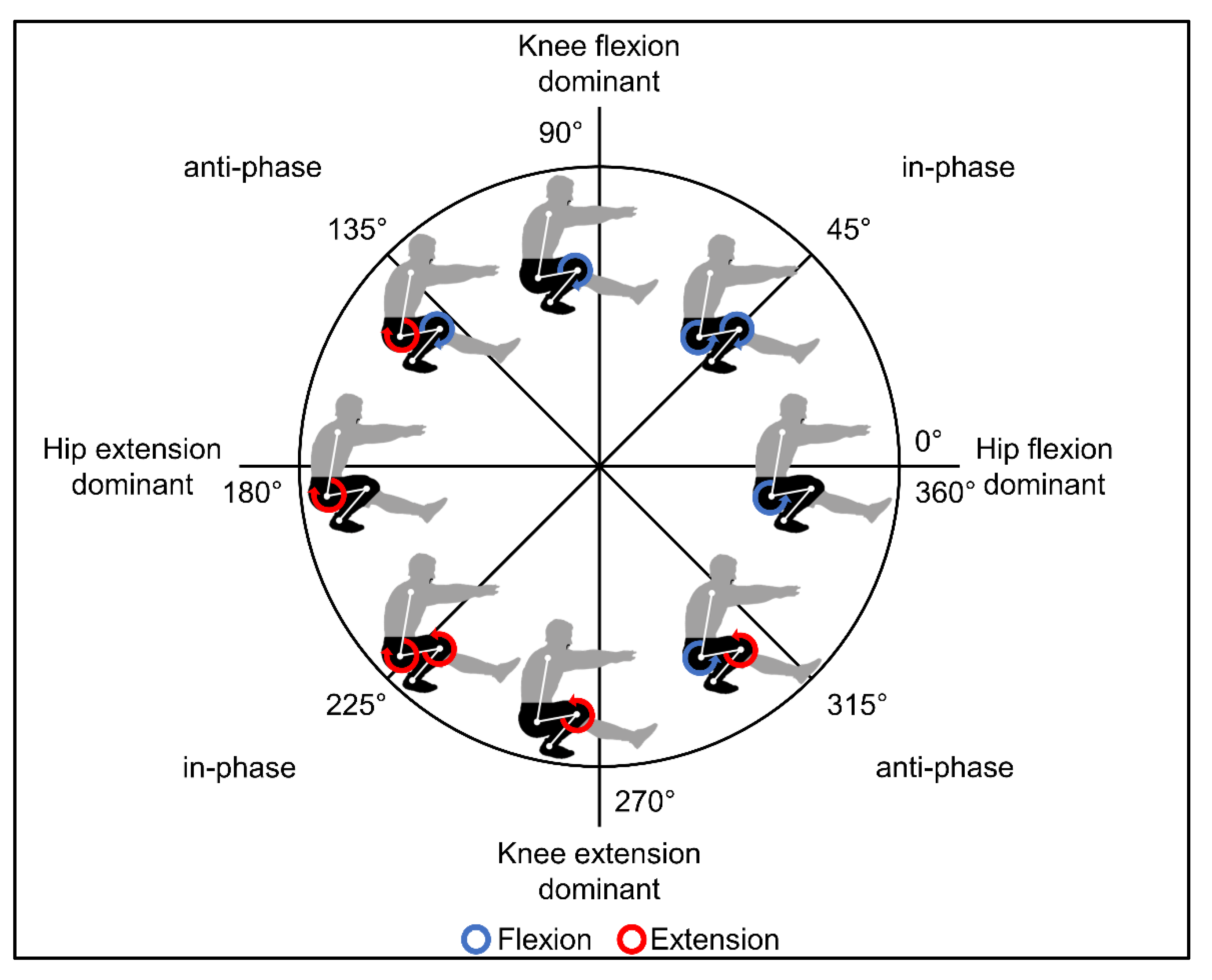
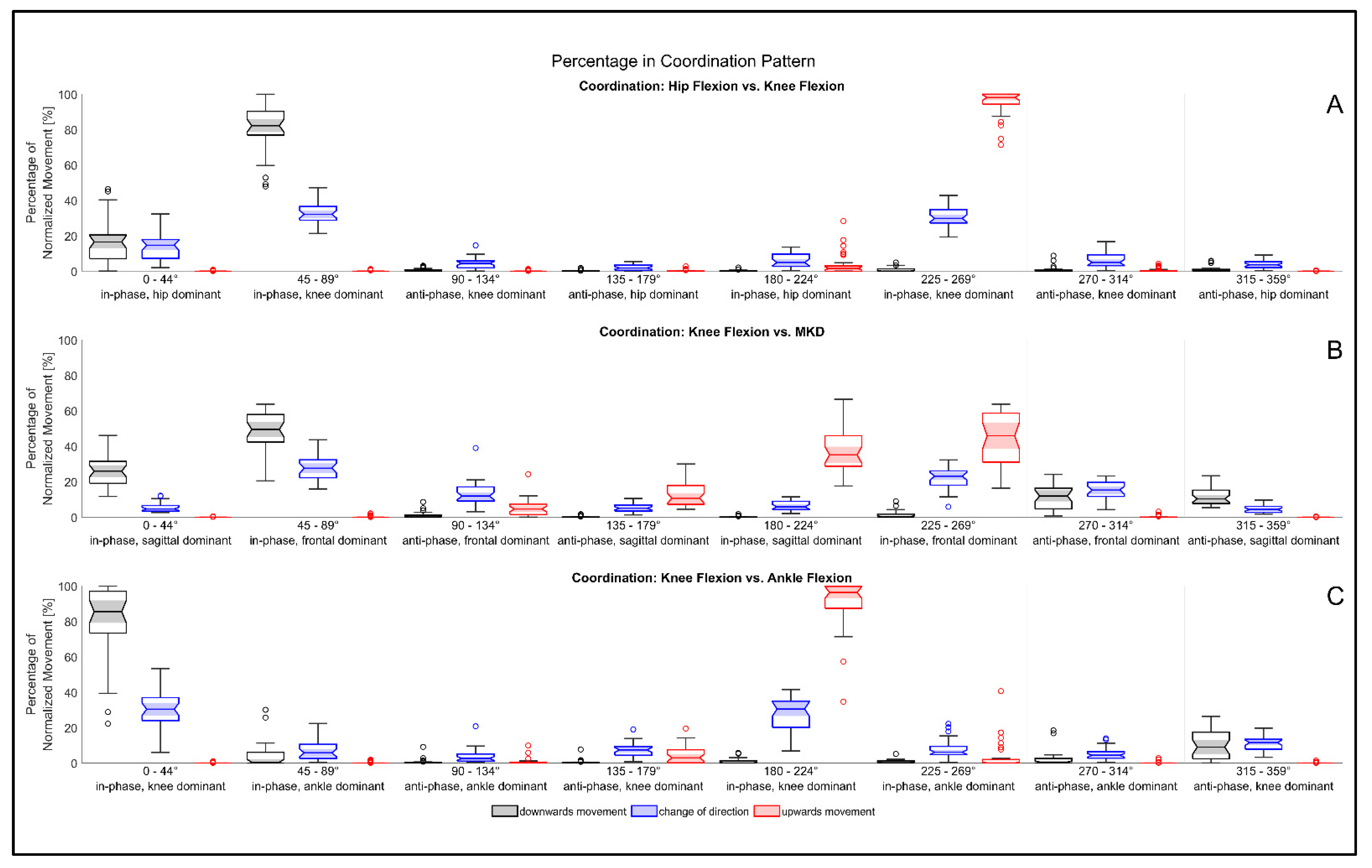
3.2. Movement Coordination

The coordination pattern during downward movement, change of direction, and upward movement with respect to both legs was statistically analyzed by 8 × 3 × 2 multifactorial, within-design ANOVA. All coordination patterns were not significantly different between both legs (p > 0.05). The coordination of sagittal hip and knee movement had significant differences between the eight coordination patterns of the coupling angle (F (1.548, 24.768) = 509.507, p < 0.001, η2 = 0.970). In addition, the coordination patterns were different depending on the SLS movement phase (F (2.097, 33.552) = 482.743, p < 0.001, η2 = 0.968). This significant interaction is visualized in Figure 3A, in which the dominance of the hip flexion vs. knee flexion coordination patterns are differently distributed between the SLS phases.
The coordination of sagittal and frontal knee motion was significantly different in the percentages of normalized time of each coordination pattern (F (2.177, 34.828) = 70.694, p < 0.001, η2 = 0.815). Similar to the coordination of hip and knee flexion, a significant interaction effect was shown between movement phase and the dominance of coordination pattern (F (3.13, 50.092) = 161.197, p < 0.001, η2 = 0.910, Figure 3B).
Furthermore, the coupling angle, investigating the coordination of sagittal knee and ankle motion, revealed significant differences in the distribution of the coordination pattern (F (1.364, 21.822) = 270.412, p < 0.001, η2 = 0.944). The significant interaction effect between coordination pattern and SLS phase was observed in the coordination of knee flexion vs. ankle flexion, too (F (1.905, 30.488) = 424.630, p < 0.001, η2 = 0.964, Figure 3C).

4. Discussion

The purpose of the present study was to measure SLS kinematics and SLS movement coordination in healthy, recreational athletes as a measure of lower leg function. Additionally, we used the modified vector coding technique to gain a better understanding of movement coordination and coordinative variability during the SLS as indicators of leg function and performance.

4.1. Inter-Limb Symmetry, Movement Kinematics, and Hip Muscle Activity

Maximum isometric hip muscle strength was assessed during MVC using a hand-held dynamometer. The measurement with hand-held dynamometers is reported to be a valid and reliable measurement tool in clinical settings [40,41]. The present results confirm the assumption of symmetry in a healthy population [42] and indicate no significant differences in the maximum isometric force of hip adductor and abductor muscles between BPL and WPL. Since the SLS test is known as a predictor of hip muscle function [5,43,44], the symmetry of isometric hip adductor and abductor forces might represent the symmetry of frontal plane hip and knee kinematics. Further, the movement kinematics of the pelvis, hip, knee, and ankle do not differ between both legs. Thus, we can assume movement symmetry in healthy, recreational athletes. The SLS kinematics are comparable to recent studies [30,45]. However, it must be mentioned, that different SLS techniques (i.e., different position of the free leg) affects the lower limb kinematics [45,46]. Aside from the squat technique, the calculation of joint motion, especially the calculation of MKD, also the direct comparison between different studies needs to be done with caution. As we calculated the MKD in the same manner as Ellenberger et al. [18], it is possible to compare the MKD between both studies. Although the same calculation technique was used for MKD, we used a higher knee flexion angle for the reference frame, resulting in lower MKD values and, thus, an underestimation of MKD, compared with reference frames with lower knee flexion as used by Ellenberger et al. [18]. However, the mean MKD of 55 ± 18 mm is higher than the MKD of male (about 48 mm) and female elite skiers (about 26 mm) [18]. This might be due to the different training levels and types of sports of both cohorts. Thus, the difference between recreational athletes and elite skiers might be larger than proposed by the present results if 100% identical MKD calculation techniques were used.
The hip abductor muscles and hip external rotator muscles are considered to be important protective factors against possible dangerous MKD [5,47,48]. It was shown that the hip abduction torque [5] and the activation of hip abductors [48] are different in subjects with good and poor knee control. In a healthy control, Mauntel et al. presented an activation ratio between the GM muscle and the ADD muscles, which demonstrates four times higher activation of the GM and hip abductor muscles, respectively [48]. Our results also indicate an activation ratio in favor of hip abduction muscle activation during the whole SLS movement, which is likely due to different functions of m. gluteus medius. As hip abductor and external rotator, the GM muscle is responsible for a neutral pelvic obliquity and controls the knee position in the medial plane [5,47,48]. Furthermore, the increase in activation ratio in the second half of SLS execution can be assigned to the leg extension. The concentric muscle activation of leg extensor muscles, including m. gluteus medius, requires increased activation to overcome gravity, compared with the downward movement phase [49].

4.2. Movement Coordination

Although the SLS is a commonly used functional test [26,43,50], less is known about the coordination of SLS execution, compared with the kinematic analysis. To our knowledge, we were the first to use the modified vector coding technique to investigate the SLS movement coordination. This technique represents the coordination of two segments or joint motions as one coupling angle [25,35,36]. The statistical analysis proposed a symmetric coordination pattern between both limbs, a significant difference in the percentages of coordination patterns, as well as a significant interaction between coordination patterns and the SLS phase. The first effect represents the differences in the percentages of coordination patterns independent of the movement phase. Hence, different in-phase and anti-phase coordination patterns are present in the SLS. In-phase coordination is predominant in hip, knee, and ankle sagittal movement. Furthermore, the knee flexion is dominant, compared with hip and ankle flexion. The second, significant effect represents the interaction between coordination pattern and SLS movement phase. In each of the movement phases, i.e., downward movement, upward movement, and the change of direction phase, a knee-dominant coordination pattern can be observed in sagittal motion. The results reveal that 80 ± 16% of sagittal hip and knee motion are defined by an in-phase, knee-dominant (coupling angle between 45–89°) coordination pattern, and 81 ± 19% of the coordination pattern of knee and ankle flexion are defined by in-phase, knee-dominant coupling angle (0–44°). During the upward movement, a phase shift of 180° results in hip and knee extension (95 ± 7%, in-phase, knee-dominant angle 225–269°), as well as knee and ankle extension (91 ± 14%, in-phase, knee-dominant angle 180–224°). During the change of direction phase, the coordination pattern shifts from in-phase leg flexion to in-phase leg extension via different anti-phase coordination patterns (Figure 3A,C). The interpretation of sagittal and frontal knee coordination must be taken with caution, due to the different units (knee flexion [°]; MKD [mm]) in the angle-distance plot, which was used for the calculation of the coupling angle. That means that the in-phase, mediolateral coordination patterns are predominant in our results (Figure 3B), because of an increase of more than 1 mm MKD per degree of knee flexion during downward movement. Accordingly, concerning the upwards movement, MKD decreases more than 1 mm per degree of knee extension. Nevertheless, the results demonstrate the in-phase coupling of knee flexion and mediolateral knee motion and therefore a positive correlation between sagittal and frontal knee motion.
Furthermore, the variability in coordination patterns differs between movement phases. The different coordinative variability is characterized by the different proportion of coordination patterns during each movement phase (Figure 3). Due to the anti-phase change of the movement direction, the coordinative variability is highest in the change of direction phase. Although downward and upward movements have predominantly in-phase and knee-dominant patterns, the downward movement is more often interrupted by other coordination patterns. These interruptions mainly consist of anti-phase coordination patterns that arise during moments of imbalance when the movement of one joint stops, or even changes direction, while the other joint moves further in the same direction. For example, the knee moves in the frontal plane while knee flexion stops, to maintain balance during knee stabilization. It can be assumed that the descending movement has a higher demand for neuromuscular control, compared with the ascending movement. The high demand for neuromuscular control might be justified by the closed-loop, feedback-driven lowering of the body, combined with many degrees of freedom [51,52]. During the controlled lowering, the participants permanently have to regulate the movement velocity, the position of the trunk, hip, knee, and ankle while maintaining balance. In contrast, the upward movement represents a movement to a known end position, i.e., a single-leg standing position with a fully extended leg, typically executed with a faster movement velocity. The reduced variability during the upward movement phase was also found in bodyweight squats analyzed by King and Hannan [53]. They proposed that coordination patterns during the upward movement are characterized by a higher redundancy reducing the coordinative variability [53]. Both the low redundancy in coordinative patterns and higher neuromuscular demands during the downward movement explain the importance of the downward movement phase in the assessment of SLS performance. Thus, our findings suggest the analysis of coordination patterns as a new performance criterion, i.e., the amount of anti-phase, as well as hip-dominant or ankle-dominant movement patterns during SLS, might be an indicator of different compensation strategies in symptomatic individuals.

4.3. Clinical Relevance

Our findings produce clinically relevant and new insights for clinicians, physiotherapists, and trainers in SLS evaluation as follows: Graduation of the SLS, e.g., according to the method proposed by Crossley et al. [5], does not differentiate between movement phases. Due to the significant differences in variability in coordination patterns between movement phases, we recommend additional differentiation between downward movement, change of direction phase, and upward movement in the subjective evaluation. Due to the higher neuromuscular demand resulting in higher coordinative variability, we recommend the subjective rating to focus on the downward movement and change of direction phase of SLS tests. During these phases, we expect different compensation strategies dependent on clinical findings. In patients with hip abductor deficits, we expect a high increase in MKD during SLS tests. Hence, the coordination pattern would be more dominant in favor of the frontal plane motion, whereas patellofemoral cartilage damages might result in more perturbations and, therefore, a high percentage of anti-phase coordination patterns. These anti-phase coordination patterns might arise from the high patellofemoral stress and the compensation by the shift to less painful position of the patella with respect to the patellofemoral joint.

4.4. Strengths and Limitations

The SLS test is a common functional test in sports clubs, clinics, and rehabilitation centers, in which SLS execution is often unrestricted in terms of squat depth and movement velocity. To simulate the assessment of SLS performance in those settings, the SLS execution was also unrestricted in our study, to the detriment of movement standardization. To account for individual differences in movement execution and to enable between-subject analysis, we used time normalization and divided the SLS movement into upward, downward, and change of direction phases.
From a technical point of view, the methods used in the present study has also strengths and limitations. Marker-based 3D motion capture is considered a gold standard that is commonly used in clinics, rehabilitation centers, and biomechanical laboratories. Nevertheless, this method has also limitations. Although the accuracy and precision of optical marker tracking systems are high, the calculated output of the kinematic model can be affected by poor marker placement, soft tissue artifacts, and kinematic crosstalk [54,55,56]. These factors influence the joint coordinate system and the definition of the axis of rotation and are, therefore, the main causes for errors in the output parameters of the kinematic model. With a limited range of motion, errors are expected to be higher in frontal and transversal planes [57]. Using joint center trajectories for calculation, the calculation of MKD is independent of the joint rotation axis and, therefore, robust against errors of the kinematic model [34]. Furthermore, the calculation of MKD is easy to use and also transferable to 2D video analysis [58].
As a first study investigating coordination patterns of SLS tests using the modified vector coding technique, our research provides a dataset of healthy, recreational athletes, which can serve as a baseline for further research. The modified vector coding technique allows new insights in continuously measured inter-segmental coordination but is limited to two segments at the same time, typically arranged in angle–angle plots. Thus, only considering spatial (angle or distance) information, the modified vector coding technique loses temporal information in coordination patterns [25,59]. Therefore, for example, potentially interesting and meaningful information on movement velocity is lost. To compensate for this issue, we divided the SLS movement into upward movement, downward movement, and change of direction phase with respect to the knee flexion angle.
Since this study aimed to analyze general coordination patterns, sex-specific differences were not considered. Further, the present study included only healthy individuals. In the next step, the investigation of symptomatic individuals and their compensating coordination patterns, compared with those of healthy individuals, might be relevant in rehabilitation centers for back-to-sport decisions, as well as in injury risk screening at sports clubs.

5. Conclusions

The aim of the present study was to investigate inter-limb symmetry and movement coordination in SLS execution of healthy, recreational athletes. Symmetric kinematics between both legs are predominant in healthy individuals. The modified vector coding technique analyzed the coordination patterns in different movement phases during SLS execution. The SLS movement consists of in-phase, knee-dominant coordination patterns with respect to sagittal hip and ankle joint movement during leg flexion and leg extension. Moments of imbalance and other movement perturbations are mostly characterized by anti-phase coordination patterns. Thus, the analysis of coordination patterns based on modified vector coding revealed new insights in the performance assessment of SLS. Further, the knowledge of coordination patterns can be used to investigate different compensation strategies in patients.

Author Contributions

Conceptualization, M.C., K.B., A.A., A.I., A.S., F.K. and W.S.; methodology, K.S.H.K., M.C., A.A., F.K. and W.S.; software, validation, investigation, data curation, K.S.H.K., M.C., K.S., F.K. and W.S.; resources, project administration, funding acquisition, M.C., A.A. and F.K.; writing—original draft preparation, visualization, K.S.H.K.; writing—review and editing, all authors; supervision, M.C., F.K. and W.S. All authors have read and agreed to the published version of the manuscript.

Funding

This research was funded by a German Knee Society Grant (Deutsche Kniegesellschaft e.V., #8810003018).

Institutional Review Board Statement

The study was conducted in accordance with the Declaration of Helsinki, and approved by the Ethics Committee of TUM Faculty of Medicine (protocol code 333/17S, 3 August 2017).

Informed Consent Statement

Informed consent was obtained from all subjects involved in the study.

Data Availability Statement

All data, tables, and figures presented in this manuscript are original. Further inquiries can be directed to the corresponding author.

Acknowledgments

We acknowledge financial support by FORscience Research Fund of the University of the Bundeswehr Munich.

Conflicts of Interest

The authors declare no conflict of interest.

References

  1. Davies, G.J.; McCarty, E.; Provencher, M.; Manske, R.C. ACL Return to Sport Guidelines and Criteria. Curr. Rev. Musculoskelet. Med. 2017, 10, 307–314. [Google Scholar] [CrossRef] [PubMed] [Green Version]
  2. Minthorn, L.M.; Fayson, S.D.; Stobierski, L.M.; Welch, C.E.; Anderson, B.E. The Functional Movement Screen’s Ability to Detect Changes in Movement Patterns After a Training Intervention. J. Sport Rehabil. 2015, 24, 322–326. [Google Scholar] [CrossRef] [PubMed]
  3. Fox, A.S.; Bonacci, J.; McLean, S.G.; Spittle, M.; Saunders, N. A Systematic Evaluation of Field-Based Screening Methods for the Assessment of Anterior Cruciate Ligament (ACL) Injury Risk. Sports Med. Auckl. N. Z. 2016, 46, 715–735. [Google Scholar] [CrossRef] [PubMed]
  4. Batty, L.M.; Feller, J.A.; Hartwig, T.; Devitt, B.M.; Webster, K.E. Single-Leg Squat Performance and Its Relationship to Extensor Mechanism Strength After Anterior Cruciate Ligament Reconstruction. Am. J. Sports Med. 2019, 47, 3423–3428. [Google Scholar] [CrossRef]
  5. Crossley, K.M.; Zhang, W.-J.; Schache, A.G.; Bryant, A.; Cowan, S.M. Performance on the Single-Leg Squat Task Indicates Hip Abductor Muscle Function. Am. J. Sports Med. 2011, 39, 866–873. [Google Scholar] [CrossRef]
  6. Begalle, R.L.; Distefano, L.J.; Blackburn, T.; Padua, D.A. Quadriceps and Hamstrings Coactivation during Common Therapeutic Exercises. J. Athl. Train. 2012, 47, 396–405. [Google Scholar] [CrossRef] [Green Version]
  7. Warner, M.B.; Wilson, D.A.; Herrington, L.; Dixon, S.; Power, C.; Jones, R.; Heller, M.O.; Carden, P.; Lewis, C.L. A Systematic Review of the Discriminating Biomechanical Parameters during the Single Leg Squat. Phys. Sport 2019, 36, 78–91. [Google Scholar] [CrossRef]
  8. Zeller, B.L.; McCrory, J.L.; Kibler, W.B.; Uhl, T.L. Differences in Kinematics and Electromyographic Activity between Men and Women during the Single-Legged Squat. Am. J. Sports Med. 2003, 31, 449–456. [Google Scholar] [CrossRef]
  9. Gianola, S.; Castellini, G.; Stucovitz, E.; Nardo, A.; Banfi, G. Single Leg Squat Performance in Physically and Non-Physically Active Individuals: A Cross-Sectional Study. BMC 2017, 18, 299. [Google Scholar] [CrossRef]
  10. Petersen, W.; Rembitzki, I.; Liebau, C. Patellofemoral Pain in Athletes. Open Access J. Sports Med. 2017, 8, 143–154. [Google Scholar] [CrossRef] [Green Version]
  11. Wilczyński, B.; Zorena, K.; Ślęzak, D. Dynamic Knee Valgus in Single-Leg Movement Tasks. Potentially Modifiable Factors and Exercise Training Options. A Literature Review. Int. J. Environ. Res. Public Health 2020, 17, 8208. [Google Scholar] [CrossRef] [PubMed]
  12. Petersen, W.; Ellermann, A.; Gösele-Koppenburg, A.; Best, R.; Rembitzki, I.V.; Brüggemann, G.-P.; Liebau, C. Patellofemoral Pain Syndrome. Knee Surg. Sports Traumatol. Arthrosc. 2014, 22, 2264–2274. [Google Scholar] [CrossRef] [PubMed] [Green Version]
  13. Souza, R.B.; Draper, C.E.; Fredericson, M.; Powers, C.M. Femur Rotation and Patellofemoral Joint Kinematics: A Weight-Bearing Magnetic Resonance Imaging Analysis. J. Orthop. Sport Phys. 2010, 40, 277–285. [Google Scholar] [CrossRef] [PubMed]
  14. Hewett, T.E.; Myer, G.D.; Ford, K.R.; Heidt, R.S., Jr.; Colosimo, A.J.; McLean, S.G.; Van Den Bogert, A.J.; Paterno, M.V.; Succop, P. Biomechanical Measures of Neuromuscular Control and Valgus Loading of the Knee Predict Anterior Cruciate Ligament Injury Risk in Female Athletes: A Prospective Study. Am. J. Sports Med. 2005, 33, 492–501. [Google Scholar] [CrossRef] [PubMed] [Green Version]
  15. Koga, H.; Nakamae, A.; Shima, Y.; Iwasa, J.; Myklebust, G.; Engebretsen, L.; Bahr, R.; Krosshaug, T. Mechanisms for Noncontact Anterior Cruciate Ligament Injuries. Am. J. Sports Med. 2010, 38, 2218–2225. [Google Scholar] [CrossRef] [PubMed]
  16. Carroll, L.A.; Kivlan, B.R.; Martin, R.L.; Phelps, A.L.; Carcia, C.R. The Single Leg Squat Test: A “Top-Down” or “Bottom-Up” Functional Performance Test? Int. J. Sports Phys. 2021, 16, 360–370. [Google Scholar] [CrossRef]
  17. Ageberg, E.; Bennell, K.L.; Hunt, M.A.; Simic, M.; Roos, E.M.; Creaby, M.W. Validity and Inter-Rater Reliability of Medio-Lateral Knee Motion Observed during a Single-Limb Mini Squat. BMC Musculoskelet. 2010, 11, 265. [Google Scholar] [CrossRef] [Green Version]
  18. Ellenberger, L.; Oberle, F.; Lorenzetti, S.; Frey, W.O.; Snedeker, J.G.; Spörri, J. Dynamic Knee Valgus in Competitive Alpine Skiers: Observation from Youth to Elite and Influence of Biological Maturation. Scand. J. Med. Sci. Sports 2020, 30, 1212–1220. [Google Scholar] [CrossRef]
  19. Hewett, T.E.; Ford, K.R.; Myer, G.D. Anterior Cruciate Ligament Injuries in Female Athletes. Am. J. Sports Med. 2006, 34, 490–498. [Google Scholar] [CrossRef]
  20. Steidl-Müller, L.; Hildebrandt, C.; Müller, E.; Fink, C.; Raschner, C. Limb Symmetry Index in Competitive Alpine Ski Racers: Reference Values and Injury Risk Identification According to Age-Related Performance Levels. J. Sport Health Sci. 2018, 7, 405–415. [Google Scholar] [CrossRef]
  21. Exell, T.A.; Irwin, G.; Gittoes, M.J.R.; Kerwin, D.G. Implications of Intra-Limb Variability on Asymmetry Analyses. J. Sport Sci. 2012, 30, 403–409. [Google Scholar] [CrossRef] [PubMed]
  22. Bishop, C. Interlimb Asymmetries: Are Thresholds a Usable Concept? Strength Cond. J. 2020, 43, 32–36. [Google Scholar] [CrossRef]
  23. Bishop, C.; Turner, A.; Read, P. Effects of Inter-Limb Asymmetries on Physical and Sports Performance: A Systematic Review. J. Sport Sci. 2018, 36, 1135–1144. [Google Scholar] [CrossRef] [PubMed]
  24. Hamill, J.; Palmer, C.; Emmerik, R.E.A.V. Coordinative Variability and Overuse Injury. Sports Med. Arthrosc. Rehabil. Ther. Technol. 2012, 4, 45. [Google Scholar] [CrossRef] [PubMed] [Green Version]
  25. Weir, G.; van Emmerik, R.; Jewell, C.; Hamill, J. Coordination and Variability during Anticipated and Unanticipated Sidestepping. Gait Posture 2019, 67, 1–8. [Google Scholar] [CrossRef]
  26. Weeks, B.K.; Carty, C.P.; Horan, S.A. Kinematic Predictors of Single-Leg Squat Performance: A Comparison of Experienced Physiotherapists and Student Physiotherapists. BMC Musculoskelet. 2012, 13, 207. [Google Scholar] [CrossRef] [Green Version]
  27. Tegner, Y.; Lysholm, J. Rating Systems in the Evaluation of Knee Ligament Injuries. Clin. Orthop. Relat. Res. 1985, 198, 42–49. [Google Scholar] [CrossRef]
  28. Gould, D.; Kelly, D.; Goldstone, L.; Gammon, J. Examining the Validity of Pressure Ulcer Risk Assessment Scales: Developing and Using Illustrated Patient Simulations to Collect the Data POINT: Visual Analogue Scale. J. Clin. Nurs. 2001, 10, 697–706. [Google Scholar] [CrossRef]
  29. Association, W.M. World Medical Association Declaration of Helsinki: Ethical Principles for Medical Research Involving Human Subjects. JAMA 2013, 310, 2191–2194. [Google Scholar] [CrossRef] [Green Version]
  30. Horan, S.A.; Watson, S.L.; Carty, C.P.; Sartori, M.; Weeks, B.K. Lower-Limb Kinematics of Single-Leg Squat Performance in Young Adults. Physiother. Can. 2014, 66, 228–233. [Google Scholar] [CrossRef] [Green Version]
  31. Davis, R.B.; Õunpuu, S.; Tyburski, D.; Gage, J.R. A Gait Analysis Data Collection and Reduction Technique. Hum. Mov. Sci. 1991, 10, 575–587. [Google Scholar] [CrossRef]
  32. Kadaba, M.; Ramakrishnan, H.; Wootten, M. Measurement of Lower Extremity Kinematics during Level Walking. J. Orthop. Res. Off. Publ. Orthop. Res. Soc. 1990, 8, 383–392. [Google Scholar] [CrossRef] [PubMed]
  33. Hermens, H.J.; Freriks, B.; Disselhorst-Klug, C.; Rau, G. Development of Recommendations for SEMG Sensors and Sensor Placement Procedures. J. Electromyogr. Kines 2000, 10, 361–374. [Google Scholar] [CrossRef]
  34. Krosshaug, T.; Steffen, K.; Kristianslund, E.; Nilstad, A.; Mok, K.-M.; Myklebust, G.; Andersen, T.E.; Holme, I.; Engebretsen, L.; Bahr, R. The Vertical Drop Jump Is a Poor Screening Test for ACL Injuries in Female Elite Soccer and Handball Players. Am. J. Sports Med. 2016, 44, 874–883. [Google Scholar] [CrossRef] [PubMed] [Green Version]
  35. Chang, R.; Emmerik, R.V.; Hamill, J. Quantifying Rearfoot–Forefoot Coordination in Human Walking. J. Biomech. 2008, 41, 3101–3105. [Google Scholar] [CrossRef]
  36. Needham, R.A.; Naemi, R.; Chockalingam, N. A New Coordination Pattern Classification to Assess Gait Kinematics When Utilising a Modified Vector Coding Technique. J. Biomech. 2015, 48, 3506–3511. [Google Scholar] [CrossRef] [PubMed]
  37. Luca, C.J.D. The Use of Surface Electromyography in Biomechanics. J. Appl. Biomech. 1997, 13, 135–163. [Google Scholar] [CrossRef] [Green Version]
  38. Merletti, R.; Cerone, G.L. Tutorial. Surface EMG Detection, Conditioning and Pre-Processing: Best Practices. J. Electromyogr. Kines 2020, 54, 102440. [Google Scholar] [CrossRef]
  39. Pataky, T.C.; Vanrenterghem, J.; Robinson, M.A. Zero- vs. One-Dimensional, Parametric vs. Non-Parametric, and Confidence Interval vs. Hypothesis Testing Procedures in One-Dimensional Biomechanical Trajectory Analysis. J. Biomech. 2015, 48, 1277–1285. [Google Scholar] [CrossRef] [Green Version]
  40. Bohannon, R.W. Reference Values for Extremity Muscle Strength Obtained by Hand-Held Dynamometry from Adults Aged 20 to 79 Years. Arch. Phys. Med. Rehabil. 1997, 78, 26–32. [Google Scholar] [CrossRef]
  41. Douma, R.K.; Soer, R.; Krijnen, W.P.; Reneman, M.; Schans, C.P. van der Reference Values for Isometric Muscle Force among Workers for the Netherlands: A Comparison of Reference Values. BMC Sports Sci. Med. Rehabil. 2014, 6, 10. [Google Scholar] [CrossRef] [PubMed] [Green Version]
  42. Sadeghi, H.; Allard, P.; Prince, F.; Labelle, H. Symmetry and Limb Dominance in Able-Bodied Gait: A Review. Gait Posture 2000, 12, 34–45. [Google Scholar] [CrossRef]
  43. Hall, M.P.; Paik, R.S.; Ware, A.J.; Mohr, K.J.; Limpisvasti, O. Neuromuscular Evaluation with Single-Leg Squat Test at 6 Months After Anterior Cruciate Ligament Reconstruction. Orthop. J. Sports Med. 2015, 3, 2325967115575900. [Google Scholar] [CrossRef] [Green Version]
  44. Lubahn, A.J.; Kernozek, T.W.; Tyson, T.L.; Merkitch, K.W.; Reutemann, P.; Chestnut, J.M. Hip Muscle Activation and Knee Frontal Plane Motion during Weight Bearing Therapeutic Exercises. Int. J. Sports Phys. 2011, 6, 92–103. [Google Scholar]
  45. Khuu, A.; Foch, E.; Lewis, C.L. Not All Single Leg Squats Are Equal: A Biomechanical Comparison of Three Variations. Int. J. Sports Phys. 2016, 11, 201–211. [Google Scholar]
  46. Khuu, A.; Lewis, C.L. Position of the Non-Stance Leg during the Single Leg Squat Affects Females and Males Differently. Hum. Mov. Sci. 2019, 67, 102506. [Google Scholar] [CrossRef] [PubMed]
  47. Willson, J.D.; Ireland, M.L.; Davis, I. Core Strength and Lower Extremity Alignment during Single Leg Squats. Med. Sci. Sports Exerc. 2006, 38, 945–952. [Google Scholar] [CrossRef] [PubMed] [Green Version]
  48. Mauntel, T.C.; Begalle, R.L.; Cram, T.R.; Frank, B.S.; Hirth, C.J.; Blackburn, T.; Padua, D.A. The Effects of Lower Extremity Muscle Activation and Passive Range of Motion on Single Leg Squat Performance. J. Strength Cond. Res. 2013, 27, 1813–1823. [Google Scholar] [CrossRef] [Green Version]
  49. Duchateau, J.; Enoka, R.M. Neural Control of Lengthening Contractions. J. Exp. Biol. 2016, 219, 197–204. [Google Scholar] [CrossRef] [Green Version]
  50. Mehl, J.; Diermeier, T.; Herbst, E.; Imhoff, A.B.; Stoffels, T.; Zantop, T.; Petersen, W.; Achtnich, A. Evidence-Based Concepts for Prevention of Knee and ACL Injuries. 2017 Guidelines of the Ligament Committee of the German Knee Society (DKG). Arch. Orthop. Trauma Surg. 2017, 138, 51–61. [Google Scholar] [CrossRef]
  51. Scott, S.H. Inconvenient Truths about Neural Processing in Primary Motor Cortex. J. Physiol. 2008, 586, 1217–1224. [Google Scholar] [CrossRef] [PubMed]
  52. Bernstein, N. The Co-Ordination and Regulation of Movements; Pergamon Press Ltd.: London, UK, 1967. [Google Scholar]
  53. King, A.; Hannan, K. Segment Coordination Variability During Double Leg Bodyweight Squats at Different Tempos. Int. J. Sports Med. 2019, 40, 725–731. [Google Scholar] [CrossRef] [PubMed]
  54. Mok, K.-M.; Kristianslund, E.K.; Krosshaug, T. The Effect of Thigh Marker Placement on Knee Valgus Angles in Vertical Drop Jumps and Sidestep Cutting. J. Appl. Biomech. 2015, 31, 269–274. [Google Scholar] [CrossRef] [PubMed]
  55. Piazza, S.J.; Cavanagh, P.R. Measurement of the Screw-Home Motion of the Knee Is Sensitive to Errors in Axis Alignment. J. Biomech. 2000, 33, 1029–1034. [Google Scholar] [CrossRef]
  56. Cappello, A.; Cappozzo, A.; Palombara, P.F.L.; Lucchetti, L.; Leardini, A. Multiple Anatomical Landmark Calibration for Optimal Bone Pose Estimation. Hum. Mov. Sci. 1997, 16, 259–274. [Google Scholar] [CrossRef]
  57. Ehrig, R.M.; Taylor, W.R.; Duda, G.N.; Heller, M.O. A Survey of Formal Methods for Determining Functional Joint Axes. J. Biomech. 2007, 40, 2150–2157. [Google Scholar] [CrossRef]
  58. Nilstad, A.; Petushek, E.; Mok, K.-M.; Bahr, R.; Krosshaug, T. Kiss Goodbye to the ‘Kissing Knees’: No Association between Frontal Plane Inward Knee Motion and Risk of Future Non-Contact ACL Injury in Elite Female Athletes. Sport Biomech. 2021, 1–15. [Google Scholar] [CrossRef]
  59. Hamill, J.; Haddad, J.M.; McDermott, W.J. Issues in Quantifying Variability from a Dynamical Systems Perspective. J. Appl. Biomech. 2000, 16, 407–418. [Google Scholar] [CrossRef] [Green Version]
Figure 1. Interpretation of the vector modified coding technique using a polar plot previously described by Needham et al. [36] and Weir et al. [25]. The blue arrows represent hip and knee flexion movements. Hip and knee extension movements are marked by blue arrows.
Symmetry 14 00388 g001
Figure 2. Kinematic analysis of pelvis, knee, and ankle joint movement, as well as EMG ratio of hip ab- and adductors during SLS, presented as mean ± SD of the better-performing leg (BPL; black) and the worse-performing leg (WPL; red). The movement cycle is defined as the time between exceeding 35° knee flexion (=0%) to 35° knee extension (=100%).
Symmetry 14 00388 g002
Figure 3. Coupling angle bins for coordination pattern classification of the single-leg squat: Coordination of sagittal hip and knee movement (A), sagittal and frontal knee movement (B), and sagittal knee and ankle movement (C) during downward movement (black), change of direction (blue), and upward movement (red) represented by the percentages in eight different coupling angle coordination pattern: 360°/0–44°, 45–89°, 90–134°, 135–179°, 180–224°, 225–269°, 270–314°, and 315–359°. For instance, for the coordination of sagittal hip and knee movement (A), it can be noted that about 90–100% of the time during downward and upward movements is spent in an in-phase, knee-dominant flexion and extension, respectively.
Symmetry 14 00388 g003
Table 1. Subject characteristics, maximum isometric strength during maximum voluntary contraction (MVC) of the better-performing leg (BPL) and worse-performing leg (WPL), and clinical scores (VAS, KOOS, TEGNER).
Subject CharacteristicsMale (n = 8)Female (n = 9)
Age [years]23 ± 226 ± 4
Mass [kg]79 ± 568 ± 7
Height [cm]181 ± 11170 ± 6
BMI [kg/cm2]24.0 ± 2.223.4 ± 2.5
MVC [N]Hip abductors (BPL)474 ± 93369 ± 32
Hip abductors (WPL)473 ± 88373 ± 40
Hip adductors (BPL)385 ± 56320 ± 66
Hip adductors (WPL)404 ± 43309 ± 52
VASBefore activity0 ± 00 ± 0
During activity0 ± 00 ± 1
After activity0 ± 00 ± 0
KOOSOverall100 ± 098 ± 3
Pain100 ± 099 ± 2
Symptoms99 ± 197 ± 6
Activities of daily living100 ± 0100 ± 0
Sporting activities100 ± 0100 ± 0
Quality of daily living100 ± 097 ± 8
Tegner Activity Scale6 ± 17 ± 2
Publisher’s Note: MDPI stays neutral with regard to jurisdictional claims in published maps and institutional affiliations.

