
Article

Mechanism of Competitive Reduction of Fe(III) and As(V) Mediated by Electron Shuttles and Bacteria

1
Hubei Key Laboratory of Yangtze Catchment Environmental Aquatic Science, School of Environmental Studies, China University of Geosciences, Wuhan 430074, China
2
Changjiang River Scientific Research Institute, Changjiang Water Resources Commission, Wuhan 430010, China
3
College of Food Science and Technology, Wuhan Business University, Wuhan 430056, China
4
Yangtze Ecological Environmental Protection Industrial Technology Research Institute, Wuhan 430200, China
*
Authors to whom correspondence should be addressed.
Water 2026, 18(8), 956; https://doi.org/10.3390/w18080956
Submission received: 7 March 2026 / Revised: 13 April 2026 / Accepted: 15 April 2026 / Published: 17 April 2026
(This article belongs to the Section Water Quality and Contamination)

Abstract

Arsenic (As) contamination in groundwater represents a critical global environmental health issue. The reductive dissolution of arsenic-bearing iron oxides by dissimilatory metal-reducing bacteria (DMRB) is a key biogeochemical process driving arsenic mobilization and release in groundwater. However, the mechanism of exogenous electron shuttles in this process remains poorly understood. This study investigated the impact of the quinone-based electron shuttle anthraquinone-2,6-disulfonate (AQDS) on the reductive dissolution of arsenic-loaded goethite by the model DMRB Shewanella putrefaciens CN32 (S.P CN32). The mobilization and transformation behaviors of arsenic and iron were compared under different pH conditions and using different arsenic-loading methods (coprecipitation vs. adsorption). Results demonstrated that AQDS acted as an electron transfer mediator. It significantly enhanced the reductive dissolution of Fe(III). It also significantly enhanced the reduction of As(V). These actions collectively accelerated arsenic release and mobilization. The study also revealed a competitive preferential order in microbial reduction, where the thermodynamically more favorable Fe(III) reduction preceded As(V) reduction. Environmental pH co-regulated this process. Its influence worked through microbial activity and mineral surface properties. A neutral pH was most conducive to the AQDS-mediated bioreduction of arsenic and iron. This study elucidates the critical role of electron shuttles in the biogeochemical cycling of arsenic in contaminated sites, providing a scientific basis for a deeper understanding of the formation mechanisms and risk assessment of high-arsenic groundwater.

1. Introduction

Arsenic (As), a naturally occurring metalloid, ranks as the 20th most abundant element in the Earth’s crust [1]. The contamination of groundwater by arsenic represents one of the most severe and widespread threats to environmental and human health, with approximately 150 million people worldwide currently exposed to high-arsenic groundwater [2,3].
Metal-reducing bacteria play a pivotal role in the mobilization and transformation of arsenic in groundwater systems, potentially accelerating the migration of arsenic and iron and posing uncertain environmental health risks [4,5]. These bacteria can utilize various forms of Fe(III) as terminal electron acceptors, including soluble Fe(III) and Fe(III) (hydrogen) oxides [6]. Dissimilatory iron reduction is a crucial component of the iron cycle, a process occurring in both bacteria and archaea that relies on dissimilatory iron reductases [7]. Inorganic Fe(III) precipitates and various complexed Fe(III) species can serve as terminal electron acceptors for dissimilatory iron reduction [8]. Labile dissolved organic matter promotes the microbial reduction of Fe(III) (hydr) oxides and arsenate by acting as an essential electron donor. This process facilitates electron transfer to the iron mineral surfaces, which ultimately drives the mobilization and enrichment of arsenic in groundwater [9]. The introduction of exogenous substances into the microinterfaces of biological reactions in groundwater environments can alter reaction pathways. Among them, exogenous electron shuttles can accelerate electron transfer from microorganisms to the surfaces of insoluble extracellular electron acceptors, facilitating electron exchange between reactants in biological or chemical processes [10,11]. Notably, such electron-shuttle-mediated electron transfer is predominantly confined to the microbe–mineral interface, where electron shuttles adsorb onto iron oxide surfaces, reduce interfacial electron transfer resistance, and bridge the electron flow between cellular outer membrane proteins and solid mineral phases [12]. Quinone-based electron shuttles have been verified to mediate extracellular electron transfer at mineral interfaces and significantly enhance interfacial electron transfer efficiency [4]. The reductive dissolution of arsenic-bearing iron (hydrogen) oxides, coupled with the reduction of As(V) to the more mobile As(III), is a critical process controlling arsenic enrichment in groundwater [7]. Studies by Aromokeye et al. [8] have elucidated the reduction and dissolution of iron hydroxides, demonstrating that both endogenous and exogenous electron-transfer mechanisms can effectively promote this transformation. Quinone groups are recognized as the primary electron-accepting and donating moieties in reactive organic matter [13]. Consequently, the key role of quinone-based electron shuttles in promoting electron transfer from metal-reducing bacteria to arsenic-bearing Fe(III) (hydrogen) oxide minerals cannot be overlooked [7]. Furthermore, AQDS offers the advantage of good biocompatibility; its addition to cultures of Shewanella oneidensis MR-1 has been shown to enhance Fe(III) reduction significantly [14,15,16].
In arsenic-contaminated groundwater systems, reduction induced by dissimilatory metal-reducing bacteria (DMRB) plays a dominant role in controlling arsenic mobilization and enrichment, making DMRB crucial for predicting the environmental fate of arsenic [17]. In arsenic-contaminated groundwater systems, reduction induced by dissimilatory metal-reducing bacteria (DMRB) plays a dominant role in controlling arsenic mobilization and enrichment, making DMRB crucial for predicting the environmental fate of arsenic [18,19]. The competitive reduction between Fe(III) and As(V) driven by DMRB represents a core biogeochemical process governing arsenic mobilization and transformation in groundwater systems, which directly determines the geochemical speciation and ecological risks of arsenic. A strict electron acceptor preference mechanism is ubiquitous in natural environments [20]. Previous studies have demonstrated that the metal-reducing bacterium Geobacter spp. preferentially reduces Fe(III) over U(VI) when both electron acceptors are available, clearly illustrating the competitive selection of different electron acceptors during microbial respiration [21]. The extracellular electron transfer (EET) pathway mediated by DMRB and electron shuttles such as AQDS has been fully validated in previous studies. The reducing power generated by intracellular bacterial metabolism is transferred to the extracellular space via the EET chain (e.g., cytochrome Cyt series). AQDS then acts as an electron shuttle to efficiently deliver electrons to Fe(III) and As(V) on the goethite surface, driving the reductive dissolution of iron minerals and the release of arsenic [22].
Although such extracellular electron transfer processes have been widely recognized, most existing studies have focused on mechanistic validation in simplified single systems. A systematic comparative investigation is still lacking regarding how AQDS regulates the competitive reduction, kinetic differences, and interfacial processes between Fe(III) and As(V) under realistic groundwater scenarios, including systems with coexisting adsorbed/coprecipitated arsenic and varying pH conditions. Accordingly, within the well-established framework of extracellular electron transfer, this study focused on the following objectives: (1) to explore the priority sequence, kinetic patterns underlying AQDS-mediated Fe(III)–As(V) reduction; (2) to reveal the mobilization and transformation of iron and arsenic co-mediated by strain CN32 and AQDS. These findings provide novel insights into the electron shuttle-mediated biogeochemical cycling of arsenic and iron in high-arsenic sediments. The results also establish a vital scientific basis for future efforts to clarify extracellular electron transfer pathways in DMRB and to unravel the molecular mechanisms by which AQDS mediates microbial mobilization and transformation of arsenic and iron in high-arsenic groundwater environments.

2. Materials and Methods

2.1. Biological Material

S.P CN32 was obtained from the China Center for Type Culture Collection (CCTCC, accession number AB 2013239). The bacterium was cultured in a minimal salts medium (MSM). The MSM contained 1 mM KH2PO4, 17 mM NaCl, 6.7 mM KCl, 3 mM MgCl2·6H2O, 5 mM NH4Cl, 1 mM CaCl2, 20 mM pyruvate, and 0.05% yeast extract. The medium was supplemented with 40 mM Bis-Tris as a buffer.

2.2. Effect of AQDS on the Reduction of As(V)/Fe(III) by Strain CN32

To investigate the effect of AQDS to mediate the reduction of As(V) and Fe(III) by S.P CN32, batch incubation assays were set up in triplicate. The experiments used a modified MSM. This medium was supplemented with: (1) 10 mM ferric citrate and 0, 0.1, 0.5, or 1 mM AQDS; (2) 1.0 mM sodium arsenate and 0, 0.1, 0.5, or 1 mM AQDS; or (3) 10 mM ferric citrate, 1.0 mM sodium arsenate, and 0, 0.1, 0.5, or 1 mM AQDS. All treatments were buffered with 40.0 mM Bis-Tris to keep pH near 7.0. Experiments were conducted in 100 mL anaerobic bottles. Each treatment was performed in triplicate. The initial OD600 was set to 0.02 for all cultures. Regular sampling was performed in an anaerobic glove box incubator. This allowed monitoring of arsenic and iron species and concentrations in the medium.

2.3. AQDS-Mediated Mobilization and Transformation of As/Fe in Goethite by S. putrefaciens CN32

Pure goethite (Gt) was synthesized according to the method published by Schwertmann and Cornell (2003) [23]. The effects of AQDS and pH on the mobilization of As/Fe from arsenic-loaded goethite by S.P CN32 were investigated. Experiments were conducted in modified MSM with pH values ranging from 6 to 8, supplemented with the following treatments: 10 mM pure goethite (Gt), arsenic-coprecipitated goethite (Gt-As), and arsenic-adsorbed goethite (Gt∗As) (Table 1). To evaluate the impact of electron shuttling, 0.1 mM AQDS was added to the nine treatments described above. All experimental groups were set up in triplicate.

2.4. Analytical Methods for Aqueous Samples and Solid-Phase Characterization

Bacterial growth was monitored by measuring the optical density at 600 nm (OD600) using a UV/Vis spectrophotometer. Fe(II)dis (dissolved Fe(II)) refers to Fe(II) in the filtrate obtained by filtering samples through a 0.22 μm cellulose acetate membrane, while Fe(II)HCl refers to HCl-extractable Fe(II). Both fractions were determined using a modified ferrozine assay at 562 nm. Samples were collected in an anaerobic glove box and filtered through 0.22 μm polycarbonate membranes. Arsenic species were separated using a strong anion-exchange LC-SAX solid-phase extraction cartridge: 2 mL of diluted filtrate was passed through the column at 1 mL/min to collect As(III), and the retained As(V) was eluted with 2 mL of 1 M HCl solution. Total arsenic and arsenic species concentrations were quantified by atomic fluorescence spectrometry (AFS-9600, Beijing Haiguang Instrument Co., Ltd., Beijing, China) with a pre-reduction step using 2.5% thiourea, 2.5% ascorbic acid, and 5% HCl. Arsine was generated using 2% KBH4–0.5% KOH as the reducing agent in 5% HCl medium, with a detection limit of 2 μg/L, R2 > 0.999, and RSD < 2%. To prevent oxidation of As(III), filtered samples were acidified to pH < 2 with ultrapure nitric acid, stored at 4 °C in the dark, and analyzed within 24 h. Surface morphology was observed using a scanning electron microscope (SU8010, Hitachi High-Tech Corporation, Tokyo, Japan) at an accelerating voltage of 10–15 kV and an electron beam current of 30–40 μA. X-ray diffraction patterns were recorded from 10° to 90° 2θ at a scanning speed of 2.5° 2θ min−1 using a Bruker D8 Advance (Bruker AXS GmbH, Karlsruhe, Germany) diffractometer.

2.5. Data Analysis

Data processing was performed using Origin and Microsoft Excel software. All experiments were conducted in triplicate. Results are expressed as mean values ± standard error (SE). Graphs were generated using Origin 8 (OriginLab, Northampton, MA, USA).

3. Results and Analysis

3.1. Effects of AQDS and As(V) on the Reduction of Fe(III) by Strain CN32

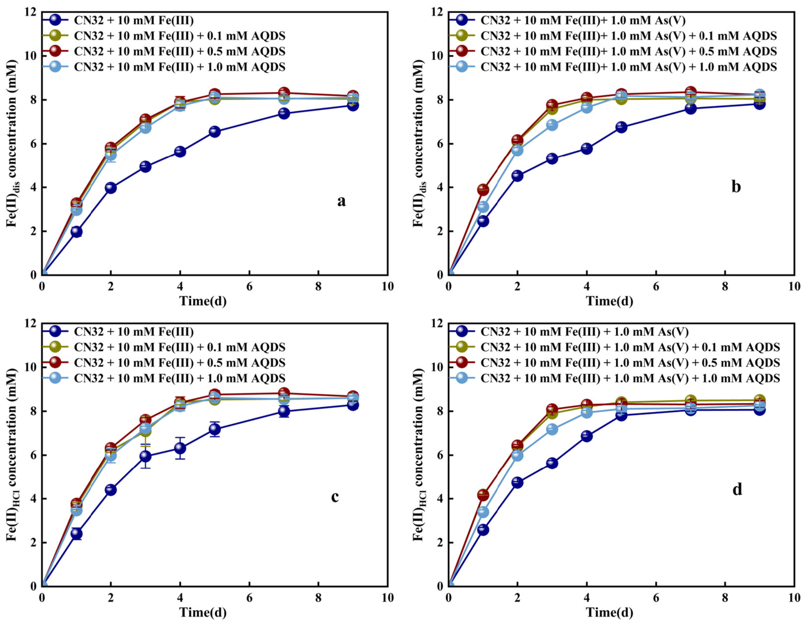
To investigate the combined effects of the electron shuttle AQDS and the presence of As(V) on the bioreduction of Fe(III), four concentrations of AQDS (0.0, 0.1, 0.5, and 1.0 mM) and/or As(V) were introduced during Fe(III) metabolism by S.P CN32. The experimental results showed that strain CN32 reduced Fe(III) via extracellular electron transfer (Figure 1). Figure S1 shows the content of remaining As(T) in the medium when As(V) is reduced alone or in the presence of 10 mM Fe(III), as well as OD600 nm. The addition of AQDS significantly enhanced this reductive process (Figure 1). In the absence of AQDS, strain CN32 continuously reduced Fe(III), generating 7.8 mM Fe(II) by day 9. In contrast, both the 0.1 and 0.5 mM AQDS groups achieved rapid Fe(III) reduction within 4 days. Although the 1.0 mM AQDS group also reduced Fe(III) rapidly, it exhibited a slight inhibitory effect. This was evidenced by a reduction that was slower or less complete than that in the lower-concentration groups. These data indicate that AQDS, as an electron shuttle, enhances electron transfer from strain CN32 to Fe(III). Notably, a high concentration of 1.0 mM AQDS did not further enhance the reduction efficiency, indicating that the electron-shuttling effect of AQDS has an optimal concentration range.
The reduction of Fe(III) by strain CN32 was also affected by the presence of As(V). After adding As(V), the Fe(II) production rate increased in all four treatment groups (Figure 1). With 0.1, 0.5, and 1 mM AQDS, strain CN32 reacted quickly in the first three days, nearly completing Fe(III) bioreduction by day 3. These groups had much higher reaction rates than the group without AQDS. This suggests that As(V) did not inhibit Fe(III) bioreduction but instead promoted it. Through simulation calculations, when the concentration of AQDS in the culture medium was 0.0, 0.1, 0.5, and 1.0 mM, the first-order kinetic constants for the generation of Fe(II) during the cultivation period were 0.179, 0.2701, 0.325, and 0.238 d−1, respectively (Table 2). The addition of As(V) did not inhibit the biological reduction of Fe(III). On the contrary, As(V) induced strain CN32 to promote the extracellular electron transfer efficiency through a unique pathway, thereby accelerating the generation and release of Fe(II) in the liquid phase.

3.2. Effects of AQDS and Fe(III) on the Reduction of As(V) by Strain CN32

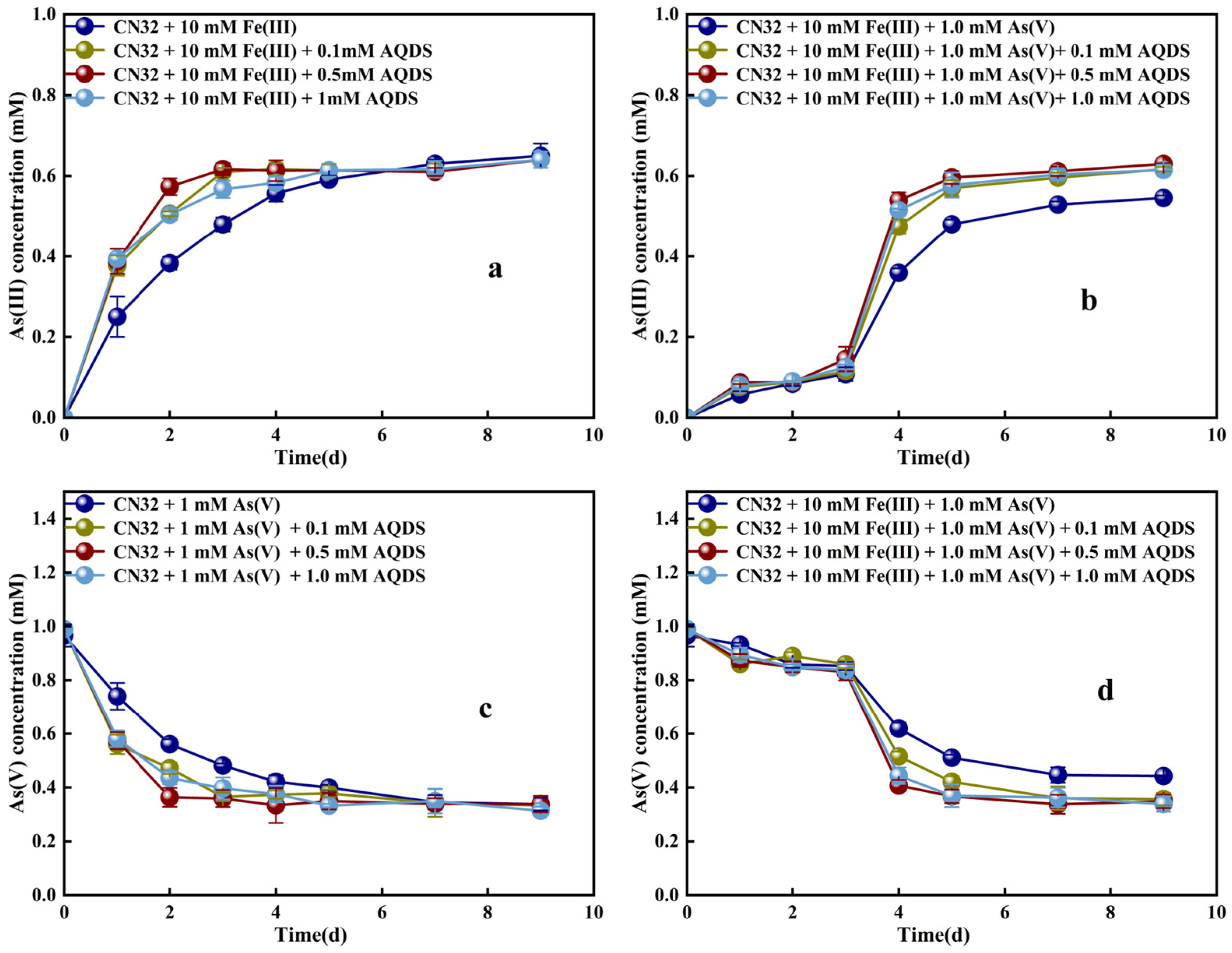
In addition to the influence of the electron shuttle AQDS, the reduction of As(V) by strain CN32 was also affected by the presence of Fe(III). To determine the effects of AQDS and Fe(III) on the reduction of As(V) by strain CN32, four different concentrations of AQDS (0.0, 0.1, 0.5, and 1.0 mM) were added to the medium containing 1 mM As(V), and the residual concentrations of arsenate and arsenite in the medium were measured. In the absence of AQDS, strain CN32 continuously reduced As(V) throughout the reaction process, reaching 0.65 mM by day 9. In contrast, treatment groups supplemented with 0.1, 0.5, and 1.0 mM AQDS all achieved rapid reduction of As(V) within 4 days, indicating that AQDS effectively enhanced the efficiency of electron transfer between the bacteria and arsenate by serving as an electron shuttle. Similarly, the addition of 1.0 mM AQDS exhibited a slight inhibitory effect on As(V) reduction compared to the additions of 0.1 and 0.5 mM.
Following the addition of Fe(III), a significant inhibitory effect on the As(V) reduction process was observed. As shown in Figure 2, the reduction rate of As(V) was very slow in all treatment groups during the first three days of the reaction. By day 3, the extent of reduction in each group did not exceed 20%. However, starting from day 3, the reductive activity in the groups amended with 0.1, 0.5, and 1.0 mM AQDS was markedly activated. The extent began to increase rapidly and eventually stabilized by day 7, with an overall reduction efficiency of approximately 60%. This result clearly demonstrates that the exogenous Fe(III) exerted a strong competitive inhibition, particularly in the early stage of the reaction, significantly delaying the reduction process.

3.3. AQDS-Mediated Iron Mobilization and Transformation During the Bioreduction of Three Types of Goethite

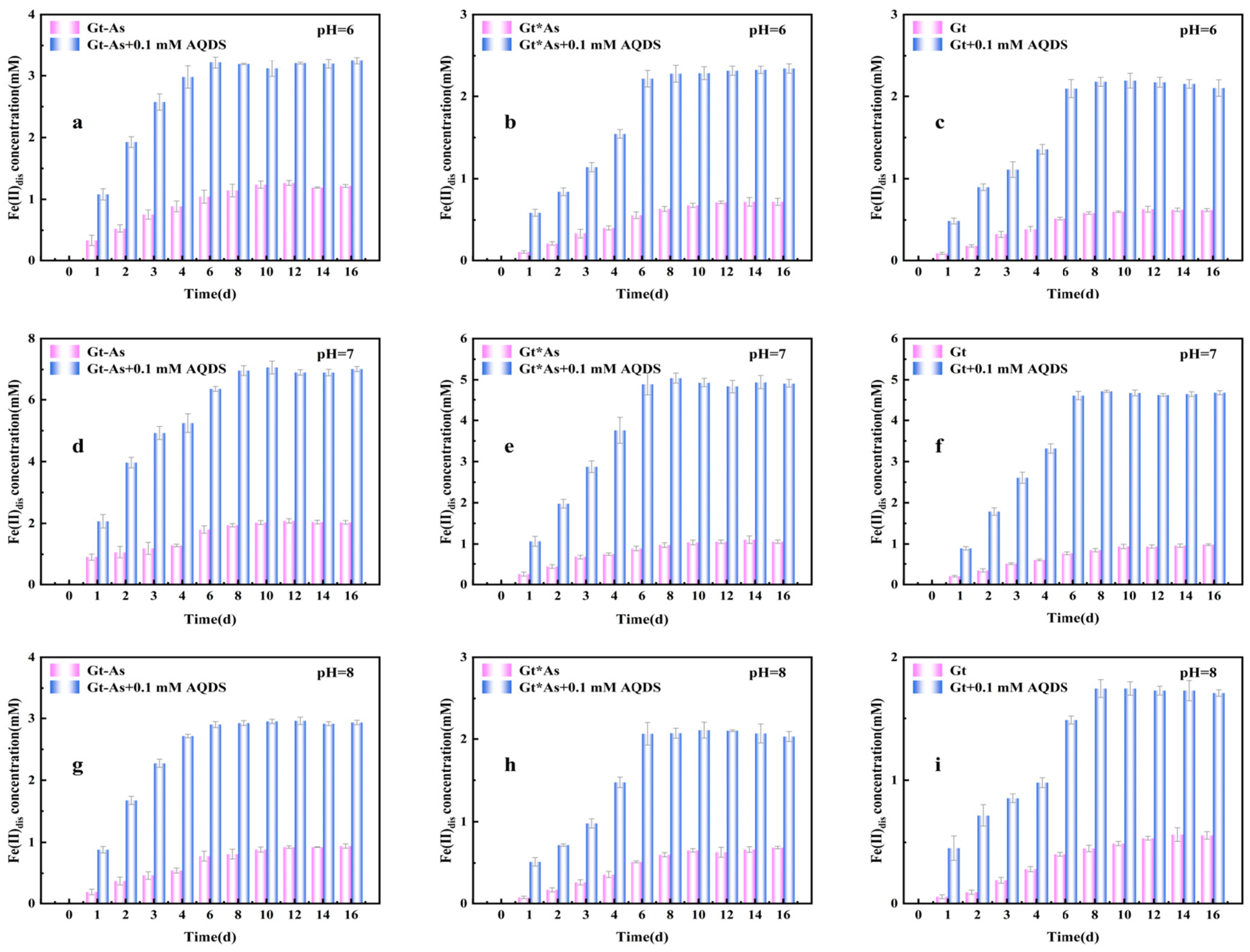
This study investigated the bioreduction of three different forms of goethite. The forms tested were Gt, Gt-As, or Gt∗As (Figure 3). The process was mediated by S.P CN32 and AQDS, with effects on iron release examined. The results are shown in Figure 3. The Gt-As had the strongest reducibility. Without AQDS, the Gt-As group released Fe(II)dis at 1.26 mM, 2.03 mM, and 0.92 mM at pH 6.0, 7.0, and 8.0 over 16 days. With AQDS, Fe(II)dis release increased to 3.24 mM (pH 6.0), 5.86 mM (pH 7.0), and 2.93 mM (pH 8.0). These are 2.57, 2.88, and 3.18 times higher compared to the respective treatments without AQDS.
Furthermore, as shown in Figure S4, the addition of AQDS also significantly enhanced the release of Fe(II)dis in both the Gt and Gt-As groups. The Fe(II)dis concentrations in the Gt group reached 2.19 mM, 4.08 mM, and 1.74 mM under the three pH conditions, which were 3.47 times, 4.16 times, and 3.10 times higher, respectively, than those without AQDS. Similarly, the Gt-As group showed increases of 3.23 times, 4.66 times, and 2.98 times, respectively, compared to their counterparts without AQDS. At pH 6, 7, and 8, the Fe(II)HCl reduction extent in the Gt group rose by 3.00-fold, 2.73-fold, and 2.00-fold, respectively. The Gt-As group showed enhancements of 2.28-fold, 2.83-fold, and 2.25-fold. The Gt∗As group showed increases of 2.95-fold, 1.77-fold, and 2.03-fold. In summary, the introduction of AQDS significantly promoted the release of both dissolved Fe(II)dis and Fe(II)HCl from all three goethite types. This demonstrates a substantial promoting effect of AQDS on microbial goethite reduction.
Figure S3 shows the growth amount of bacterial CN32 under different pH conditions. At pH 7, all three goethite systems showed higher reducibility than at pH 6 or pH 8. After AQDS addition, concentrations of released dissolved Fe(II)dis were 4.67 mM, 7.01 mM, and 4.90 mM for Gt, Gt-As, and Gt∗As, respectively. Compared to pH 6 and pH 8, iron reduction was enhanced by 2.20-fold and 2.73-fold for Gt. For Gt-As, enhancements were 2.16-fold and 2.41-fold, and for Gt∗As, they were 2.10-fold and 2.41-fold, respectively. These results clearly indicate all three goethites were more reducible at a neutral pH.

3.4. AQDS-Mediated Arsenic Mobilization and Transformation During the Bioreduction of Arsenic-Loaded Goethite

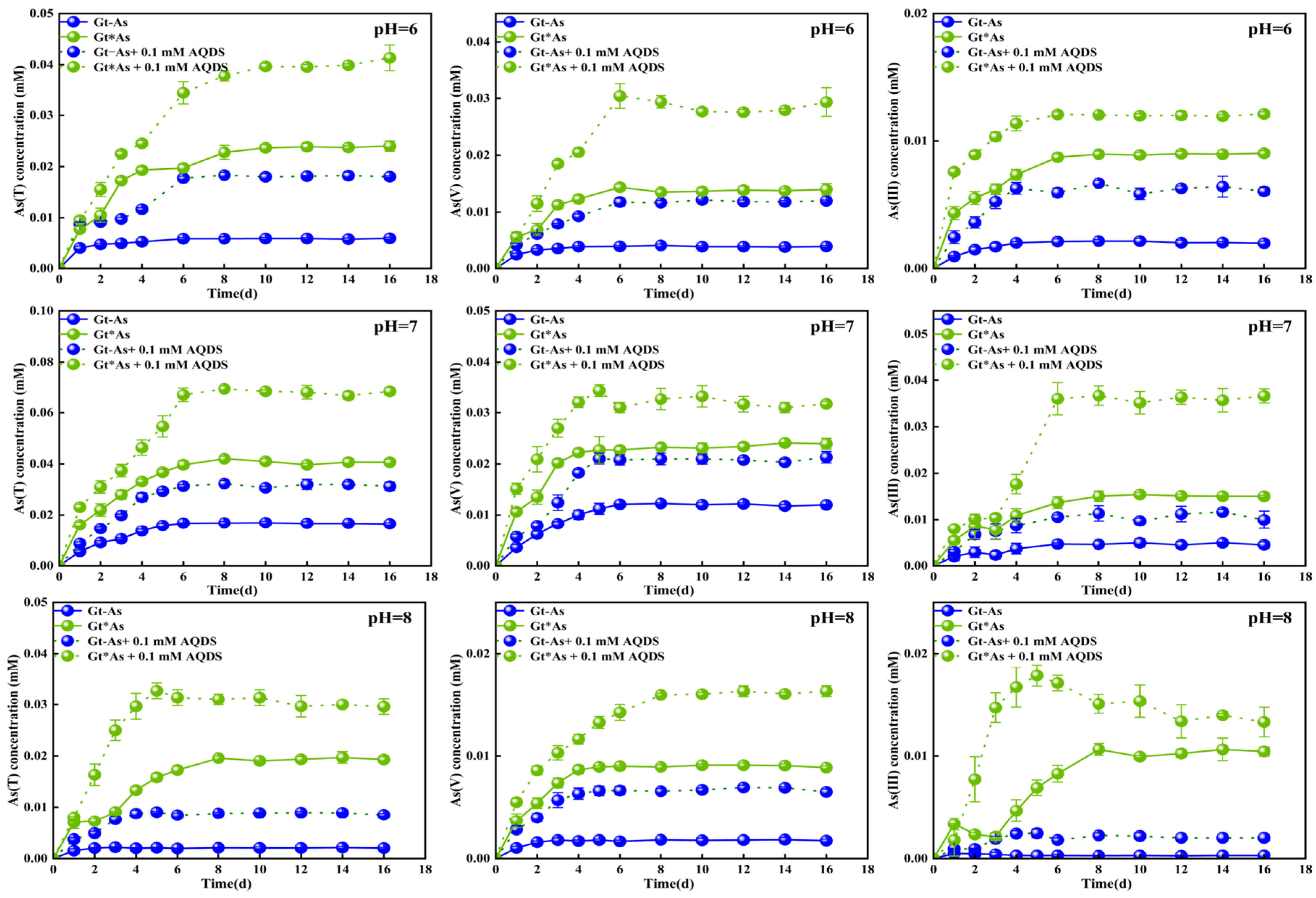
This study further analyzed the effect of AQDS on arsenic release from goethite under different pH conditions (Figure 4). In the Gt-As samples without AQDS addition, the release of As(T) over the 16-day incubation period was 0.0058 mM at pH 6.0, 0.0168 mM at pH 7.0, and 0.0029 mM at pH 8.0. Following the addition of AQDS, the As(T) release increased to 3.10-fold, 1.84-fold, and 3.00-fold of the values without AQDS, respectively. Concurrently, the concentration of dissolved As(III) at the end of the incubation also increased to 3.15-fold, 2.22-fold, and 1.92-fold of the corresponding treatments without AQDS. In the Gt∗As samples without AQDS, the As(T) release was 0.024 mM at pH 6.0, 0.042 mM at pH 7.0, and 0.019 mM at pH 8.0. With the addition of AQDS, the As(T) release increased to 1.70-fold, 1.61-fold, and 1.57-fold of the values without AQDS, respectively, while the dissolved As(III) concentration increased to 1.33-fold, 1.66-fold, and 1.40-fold, respectively. Together, these results indicate that AQDS, serving as an electron shuttle, significantly promotes arsenic release by enhancing the microbial reduction of goethite.
Furthermore, environmental pH is a critical factor influencing arsenic release and speciation transformation. Building on earlier findings on AQDS, the experimental results demonstrate that the concentrations of As(T), As(V), and As(III) were significantly lower under acidic or alkaline conditions than under neutral conditions. Specifically, in the Gt-As samples at pH = 7, the As(T) release was 2.75-fold and 8.25-fold higher than that at pH = 6 and pH = 8, respectively. In the Gt∗As samples at pH = 7, the As(T) release was 1.66-fold and 2.10-fold higher than that at pH = 6 and pH = 8, respectively. Overall, these data indicate that under neutral pH conditions, the release of total arsenic from the goethite systems was greater, and the bioreduction of As(V) to As(III) was also more pronounced.

3.5. Solid-Phase Characterization of Three Goethite Types After AQDS-Mediated Bioreduction

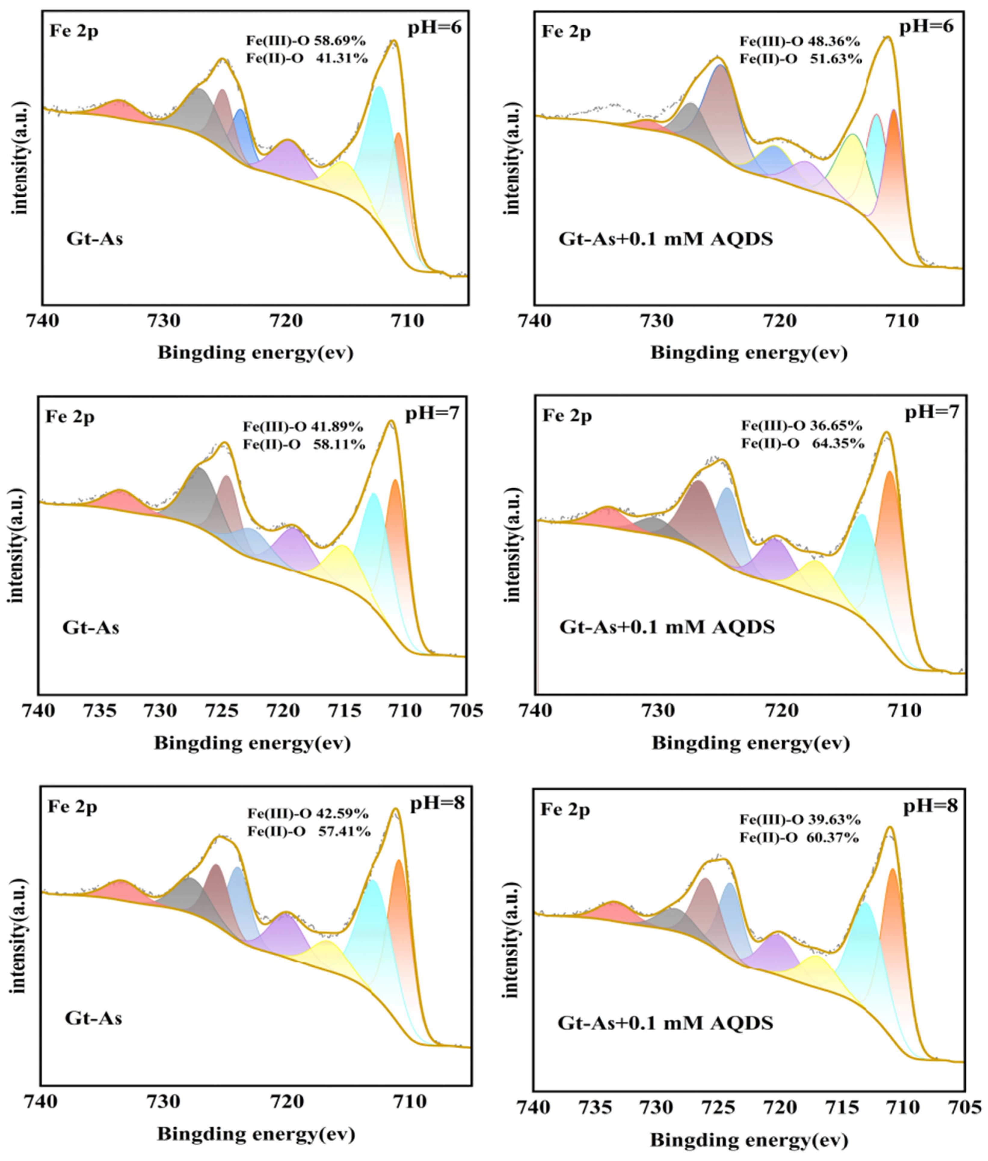
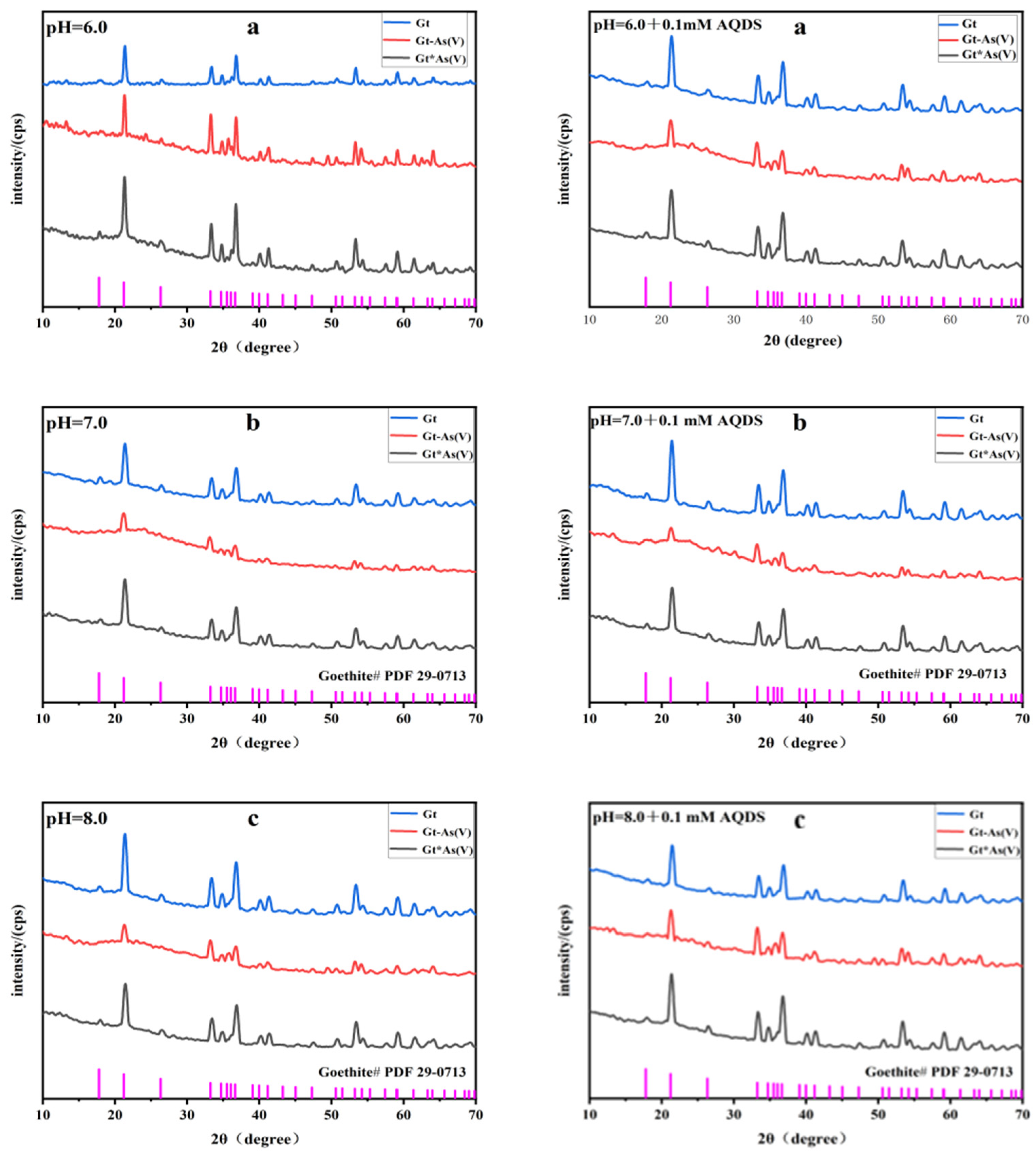
To examine goethite mineral phases after bioreduction by S.P CN32 with AQDS, XRD, XPS and SEM analyses were used. As shown in Figure 5, after bioreduction of the three goethite types at pH 6.0, 7.0, and 8.0, the mineral composition showed no significant change. Figure 6 shows the Fe2p orbital fitting analysis of the mineral XPS after Gt-As biological reduction under different pH conditions. The peak areas of the integrated spectra after peak fitting were used to calculate the percentage of Fe(III) and Fe(II) on the mineral surface. After adding AQDS, the percentage of Fe(II) on the mineral surface increased from 41.31%, 58.11%, and 57.41% at pH = 6, 7, and 8 to 51.63%, 64.35%, and 57.41%, further indicating the promoting effect of AQDS input on the biological reduction of arsenic-bearing goethite. At neutral pH, the reduction effect of arsenic-bearing goethite was the most significant. Goethite remained the dominant phase. SEM images of goethite are shown in Figure S2. No characteristic peaks of other minerals appeared. Formation of Fe(II) secondary minerals depends on multiple factors, such as the initial iron reduction rate, Fe(II) concentration, and pH. Although adding AQDS promoted iron reduction, no new minerals were detected. This result suggests that reduced Fe(II) was not concentrated enough to cause mineral transformation.
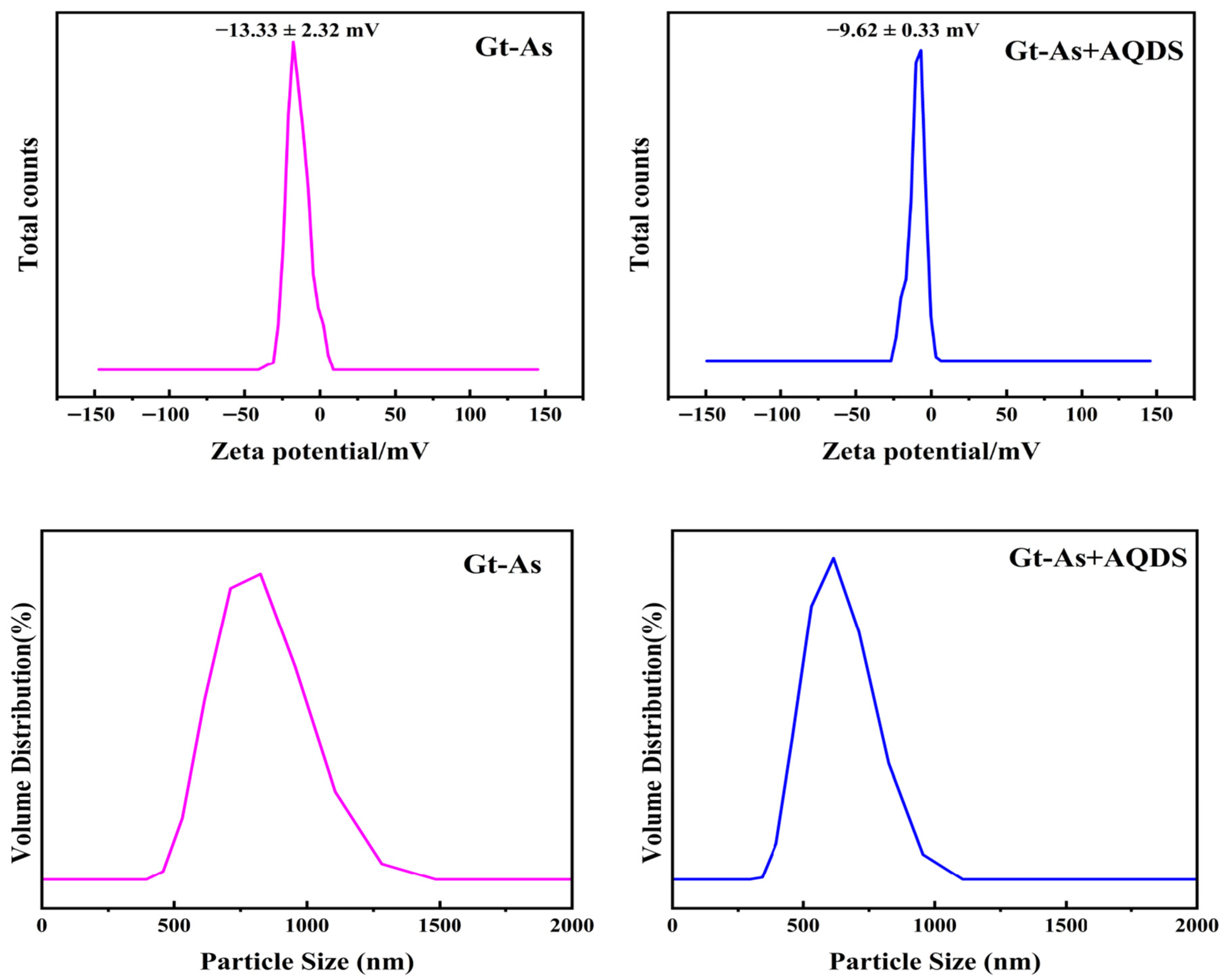
The interfacial properties of arsenic-bearing goethite before and after AQDS-mediated bioreduction at pH 7 were analyzed by measuring zeta potential and particle size (Figure 7). Both Gt-As and Gt-As + AQDS samples exhibited negative zeta potentials (−13.33 ± 2.32 mV and −9.62 ± 0.33 mV, respectively), confirming the negatively charged surface of goethite under neutral conditions. The significant increase in zeta potential following AQDS addition verified that negatively charged AQDS effectively adsorbed onto the mineral surface, neutralized partial surface charge, and facilitated interfacial electron transfer between bacterial cells and goethite. Regarding particle size distribution, the Gt-As sample showed a broad distribution with a peak at 700–800 nm, whereas the AQDS-amended sample exhibited a narrower distribution centered at 500–600 nm. This indicates that AQDS adsorption improved the dispersion of goethite particles, inhibited aggregation, and increased the effective reactive surface area. Collectively, these interfacial changes promoted the microbial reduction of Fe(III) and As(V) in the AQDS-supplemented system.

4. Discussion

4.1. AQDS Enhances the Reduction of Fe(III) and As(V) by Strain CN32

This study demonstrates that AQDS acts as an effective electron transfer mediator between S.P CN32 and As(V)/Fe(III), significantly accelerating the production of As(III)/Fe(II). After the addition of AQDS, the first-order rate constant of As(III)/Fe(II) generation is notably improved. The sharp increase in the Fe(II) generation rate constant caused by 0.1 mM AQDS directly confirms that AQDS can effectively mediate electron transfer and accelerate redox reactions in the aqueous phase. Solid-phase experiments and XPS results show that the Fe(II) content on the goethite surface increases significantly after adding AQDS. The elevated Fe(II) content on the mineral surface verifies the role of AQDS as an exogenous electron shuttle: strain CN32 transfers metabolic electrons to AQDS via outer membrane cytochromes, reducing AQDS from its oxidized state to the reduced state.
As a dissimilatory metal-reducing bacterium, the extracellular Fe(III) reduction capability of strain CN32 mainly relies on the Mtr pathway. Shewanella species exhibit a strong capacity for exogenous electron transfer. For instance, S. oneidensis MR-1 can reduce hexavalent chromium via the outer membrane cytochromes MtrC and OmcA [24]. And the expression of MtrC and UndA in CN32 confirms its extracellular electron transfer mechanism [25]. In the CN32 system, AQDS accelerates electron transfer in both the liquid and solid phases, facilitating transfer from the outer membrane to insoluble iron minerals such as goethite. Bacteria can enhance the efficiency of electron transfer from the cytoplasm to the extracellular environment by regulating their cell membrane permeability or surface protein distribution. Additionally, they can improve Fe(III) reduction kinetics by optimizing their contact with Fe(III) minerals [26]. The zeta potential of goethite confirmed the adsorption of AQDS onto the mineral surface. Meanwhile, AQDS alters the surface properties of goethite, improves the dispersion of goethite particles and increases the effective reactive surface area, thereby further facilitating interfacial electron transfer, consequently promoting the reduction of As(V)/Fe(III) [4].

4.2. Competitive Reduction Process Between Fe(III) and As(V)

The results demonstrated that Fe(III) was more readily reduced than As(V) in the coexisting system, and the microbial reduction process exhibited significant competitive selectivity, where Fe(III) was reduced preferentially over As(V). Thermodynamically, the redox potential of electron acceptors directly determines the free energy released during microbial reduction; a higher potential provides more energy for microbial metabolism and stronger electron affinity. The free energy released during electron transfer can be more efficiently captured by the electron transport chain and converted into ATP [27,28]. The kinetic data further support this prediction: Fe(III) was rapidly reduced to Fe(II) in the early stage, whereas As(V) reduction was significantly inhibited. The first-order rate constant of Fe(II) production was significantly higher than that of As(V) reduction, confirming faster and preferential reduction of Fe(III).
Similar selective reduction has been reported in Geobacter spp., which preferentially reduce Fe(III) over U(VI) when encountering both electron acceptors, indicating clear electron competition among metals [21]. Likewise, competitive adsorption of Cr3+, Co2+, and Ni2+ on bacterial surfaces follows a clear priority sequence (Cr3+ > Co2+ ≈ Ni2+) [29]. At the electron transfer level, electroactive proteins such as c-type cytochromes on the outer membrane of Shewanella and Geobacter directly interact with mineral surfaces to form a “bio-mineral interface” that facilitates transmembrane electron transfer [30]. Methanogenic archaea employ the transmembrane c-type cytochrome MmcA to transfer electrons extracellularly and preferentially reduce Fe(III) oxides [30,31] Accordingly, the electron transport chain of S.P CN32 tends to direct electrons toward thermodynamically more favorable Fe(III), leading to inhibited As(V) reduction in the early reaction stage. Collectively, the preferential reduction of Fe(III) over As(V) is jointly governed by thermodynamic potential differences, kinetic rates, and molecular electron transfer pathways. This competitive mechanism provides a theoretical basis for understanding microbially mediated arsenic mobilization in multi-electron-acceptor environments.

4.3. Influence of pH on AQDS-Mediated Biotic Mobilization and Transformation of Fe/As

Environmental pH plays a crucial role in bacterial growth and reproduction. It not only regulates the surface charge and permeability of the cell membrane to directly affect nutrient uptake and energy metabolism, but also alters the surface chemical properties of minerals, thereby jointly affecting bio-mineral interactions [32,33]. The influence of pH on the system is reflected in many aspects, including arsenic speciation distribution, redox behavior of AQDS, and microbial metabolic activity.
Experimental data under different pH conditions show that the Fe(II) production rates of Gt∗As and Gt-As at neutral pH (7.0) are significantly higher than those in acidic and alkaline environments. From the perspective of mineral stability, pH significantly affects the solubility, structural stability, and reactivity of goethite [34]. Under acidic pH, the mineral surface is highly protonated, and H+ tends to compete with AQDS for Fe(III) surface active sites, thereby reducing the binding efficiency and electron transfer ability of AQDS to minerals [35]. Under alkaline conditions, a high pH environment may alter the surface chemical properties of goethite or induce surface structural rearrangement, further affecting the effective contact between AQDS and the mineral interface [36]. From the microbial perspective, the OD600 nm value of strain CN32 in the blank group at pH 7.0 was significantly higher than at pH 6.0 and pH 8.0 (Figure S3). The higher biomass at neutral pH indicates more vigorous metabolic activity and stronger Fe(III) and As(V) reduction ability [37,38]. Meanwhile, pH directly determines the speciation of arsenic in the system: it is dominated by H2AsO4 in the range of pH 6.0–7.0 and shifts to HAsO42− at pH 8.0. Different arsenic species differ significantly in mineral binding capacity and bioavailability, which further affects the reduction and release rates.
Overall, the influence of pH on electron transfer is not simply a change in thermodynamic reaction tendency, but a combined effect through multiple key pathways: first, regulating the redox activity of AQDS and affecting its electron acceptance and transfer ability; second, affecting the metabolic and growth status of the strain and determining the efficiency of electron supply; third, changing the speciation of arsenic and affecting its biotransformation pathway.

5. Conclusions

This study demonstrates that the electron shuttle AQDS significantly promotes the reductive dissolution of arsenic-bearing goethite by S.P CN32, thereby enhancing the mobilization and transformation of As(V)/Fe(III) reduction. Fe(III) reduction is thermodynamically and kinetically preferred over As(V) reduction in the coexisting system, which is governed by the difference in redox potentials and further modulated by microbial extracellular electron transfer and interfacial processes. Neutral pH conditions are most favorable for microbial activity, interfacial electron transfer, and the electron-shuttling role of AQDS, leading to the highest reduction efficiency. Excessively high AQDS concentrations may exert mild inhibitory effects on bacterial activity, such that the promoting effect does not increase continuously with concentration.
By integrating kinetic analyses, interfacial characterization, and arsenic speciation determination, this study systematically reveals the key characteristics of AQDS-mediated microbe–mineral interfacial electron transfer and the regulatory effects of pH, AQDS concentration, and arsenic occurrence form on As/Fe transformation. These findings improve our understanding of the biogeochemical cycling of iron and arsenic in shallow high-arsenic groundwater systems and provide a scientific basis for further investigations on microbially driven arsenic mobilization in complex groundwater environments.

Supplementary Materials

The following supporting information can be downloaded at https://www.mdpi.com/article/10.3390/w18080956/s1: Figure S1: The effects of AQDS and Fe(III) on the reduction of As(V) by CN32. The figure shows the content of remaining As(T) in the medium when As(V) is reduced alone or in the presence of 10 mM Fe(III), as well as OD600 nm; Figure S2: The initial SEM images of three types of goethite; Figure S3: The growth amount of bacterial CN32 under different pH conditions; Figure S4: The effect of CN32 on the reduction of Fe(III) in three types of arsenic-loaded goethites. The figure includes the extracted state of hydrochloric acid Fe(II)HCl.

Author Contributions

Conceptualization: W.L. and J.W.; Methodology: J.W. and W.L.; Software: W.L.; Validation: Y.Y., M.C., C.Z. and Y.L.; Formal analysis: Y.L., Y.Y. and M.C.; Resources: J.W.; Data curation: W.L.; Writing—original draft: W.L.; Writing—review & editing: Z.X.; Supervision: Z.X.; Funding acquisition: J.W. and Z.X. All authors have read and agreed to the published version of the manuscript.

Funding

This study was jointly supported by The National Natural Science Foundation of China (Grant No. 42307115), Hubei Provincial Natural Science Foundation (Gran No. JCZRYB202501363), the Special Fund of Chinese Central Government for Basic Scientific Research Operations in Commonweal Research Institutes (Grant Nos. CKSF2025533+NY and CKSF2024338/NY), and the National Natural Science Foundation of China (Grant No. 42577070).

Data Availability Statement

The data presented in this study are available on request from the corresponding author due to participant privacy, and the raw data cannot be deposited in a public repository.

Conflicts of Interest

The authors declare no conflicts of interest.

References

  1. Mandal, B.K.; Suzuki, K.T. Arsenic round the world: A review. Talanta 2002, 58, 201–235. [Google Scholar] [CrossRef] [PubMed]
  2. Ahmad, S.A.; Khan, M.H.; Haque, M. Arsenic contamination in groundwater in Bangladesh: Implications and challenges for healthcare policy. Risk Manag. Healthc. Policy 2018, 11, 251–261. [Google Scholar] [CrossRef] [PubMed]
  3. Palma-Lara, I.; Martínez-Castillo, M.; Quintana-Pérez, J.C.; Arellano-Mendoza, M.G.; Tamay-Cach, F.; Valenzuela-Limón, O.L.; García-Montalvo, E.A.; Hernández-Zavala, A. Arsenic exposure: A public health problem leading to several cancers. Regul. Toxicol. Pharmacol. 2020, 110, 104539. [Google Scholar] [CrossRef]
  4. Wang, J.; Xie, Z.M.; Wang, Y.X.; Yang, Y.; Chen, M.N. Microbial reduction and migration of As/Fe mediated by electron shuttle: Differences between incorporated and adsorbed As(V). Water Biol. Secur. 2022, 1, 100035. [Google Scholar] [CrossRef]
  5. Xie, Z.M.; Wang, J.; Wei, X.F.; Li, F.; Chen, M.N.; Wang, J.; Gao, B. Interactions between arsenic adsorption/desorption and indigenous bacterial activity in shallow high arsenic aquifer sediments from the Jianghan Plain, Central China. Sci. Total Environ. 2018, 644, 382–388. [Google Scholar] [CrossRef]
  6. Luu, Y.S.; Ramsay, J.A. Review: Microbial mechanisms of accessing insoluble Fe(III) as an energy source. World J. Microbiol. Biotechnol. 2003, 19, 215–225. [Google Scholar] [CrossRef]
  7. Wang, J.; Xie, Z.M.; Wei, X.F.; Chen, M.N.; Luo, Y.; Wang, Y.X. An indigenous bacterium Bacillus XZM for phosphate enhanced transformation and migration of arsenate. Sci. Total Environ. 2020, 719, 137183. [Google Scholar] [CrossRef]
  8. Aromokeye, D.A.; Oni, O.E.; Tebben, J.; Yin, X.R.; Richter-Heitmann, T.; Wendt, J.; Nimzyk, R.; Littmann, S.; Tienken, D.; Kulkarni, A.C.; et al. Crystalline iron oxides stimulate methanogenic benzoate degradation in marine sediment-derived enrichment cultures. Isme J. 2021, 15, 965–980. [Google Scholar] [CrossRef]
  9. Pi, K.F.; Wang, Y.X.; Xie, X.J.; Huang, S.B.; Yu, Q.; Yu, M. Geochemical effects of dissolved organic matter biodegradation on arsenic transport in groundwater systems. J. Geochem. Explor. 2015, 149, 8–21. [Google Scholar] [CrossRef]
  10. Bai, Y.; Mellage, A.; Cirpka, O.A.; Sun, T.R.; Angenent, L.T.; Haderlein, S.B.; Kappler, A. AQDS and Redox-Active NOM Enables Microbial Fe(III)-Mineral Reduction at cm-Scales. Environ. Sci. Technol. 2020, 54, 4131–4139. [Google Scholar] [CrossRef] [PubMed]
  11. Lovley, D.R.; Malvankar, N.S. Seeing is believing: Novel imaging techniques help clarify microbial nanowire structure and function. Environ. Microbiol. 2015, 17, 2209–2215. [Google Scholar] [CrossRef] [PubMed]
  12. Lovley, D.R.; Coates, J.D.; BluntHarris, E.L.; Phillips, E.J.P.; Woodward, J.C. Humic substances as electron acceptors for microbial respiration. Nature 1996, 382, 445–448. [Google Scholar] [CrossRef]
  13. de Melo, B.A.G.; Motta, F.L.; Santana, M.H.A. Humic acids: Structural properties and multiple functionalities for novel technological developments. Mater. Sci. Eng. C-Mater. Biol. Appl. 2016, 62, 967–974. [Google Scholar] [CrossRef] [PubMed]
  14. Chen, Z.; Wang, Y.P.; Jiang, X.L.; Fu, D.; Xia, D.; Wang, H.T.; Dong, G.W.; Li, Q.B.A. Dual roles of AQDS as electron shuttles for microbes and dissolved organic matter involved in arsenic and iron mobilization in the arsenic-rich sediment. Sci. Total Environ. 2017, 574, 1684–1694. [Google Scholar] [CrossRef]
  15. Menezes, O.; Kocaman, K.; Wong, S.; Rios-Valenciana, E.E.; Baker, E.J.; Hatt, J.K.; Zhao, J.S.; Madeira, C.L.; Krzmarzick, M.J.; Spain, J.C.; et al. Quinone Moieties Link the Microbial Respiration of Natural Organic Matter to the Chemical Reduction of Diverse Nitroaromatic Compounds. Environ. Sci. Technol. 2022, 56, 9387–9397. [Google Scholar] [CrossRef]
  16. Peng, H.; Pearce, C.I.; N’Diaye, A.T.; Zhu, Z.L.; Ni, J.R.; Rosso, K.M.; Liu, J. Redistribution of Electron Equivalents between Magnetite and Aqueous Fe2+ Induced by a Model Quinone Compound AQDS. Environ. Sci. Technol. 2019, 53, 1863–1873. [Google Scholar] [CrossRef]
  17. Das, S.; Liu, C.C.; Jean, J.S.; Lee, C.C.; Yang, H.J. Effects of microbially induced transformations and shift in bacterial community on arsenic mobility in arsenic-rich deep aquifer sediments. J. Hazard. Mater. 2016, 310, 11–19. [Google Scholar] [CrossRef]
  18. Brooks, S.C.; Fredrickson, J.K.; Carroll, S.L.; Kennedy, D.W.; Zachara, J.M.; Plymale, A.E.; Kelly, S.D.; Kemner, K.M.; Fendorf, S. Inhihition of bacterial U(VI) reduction by calcium. Environ. Sci. Technol. 2003, 37, 1850–1858. [Google Scholar] [CrossRef]
  19. Lovley, D.R. Dissimilatory Metal Reduction. Annu. Rev. Microbiol. 1993, 47, 263–290. [Google Scholar] [CrossRef]
  20. Lopez-Adams, R.; Newsome, L.; Moore, K.L.; Lyon, I.C.; Lloyd, J.R. Dissimilatory Fe(III) Reduction Controls on Arsenic Mobilization: A Combined Biogeochemical and NanoSIMS Imaging Approach. Front. Microbiol. 2021, 12, 640734. [Google Scholar] [CrossRef]
  21. Lovley, D.R.; Phillips, E.J.P. Competitive Mechanisms for Inhibition of Sulfate Reduction and Methane Production in The Zone of Ferric Iron Reduction in Sediments. Appl. Environ. Microbiol. 1987, 53, 2636–2641. [Google Scholar] [CrossRef]
  22. Wang, J.; Xie, Z.M.; Wang, Y.X.; Yang, Y.; Chen, M.N. Synergy between indigenous bacteria and extracellular electron shuttles enhances transformation and mobilization of Fe(III)/As(V). Sci. Total Environ. 2021, 783, 147002. [Google Scholar] [CrossRef]
  23. Cornell, R.M.; Schwertmann, U. The Iron Oxides: Structure, Properties, Reactions, Occurrences and Uses; John Wiley & Sons: Hoboken, NJ, USA, 2003. [Google Scholar] [CrossRef]
  24. Wang, C.; Chen, J.; Hu, W.J.; Liu, J.Y.; Zheng, H.L.; Zhao, F. Comparative proteomics reveal the impact of OmcA/MtrC deletion on Shewanella oneidensis MR-1 in response to hexavalent chromium exposure. Appl. Microbiol. Biotechnol. 2014, 98, 9735–9747. [Google Scholar] [CrossRef]
  25. Wu, X.; Zou, L.; Huang, Y.H.; Qiao, Y.; Long, Z.E.; Liu, H.P.; Li, C.M. Shewanella putrefaciens CN32 outer membrane cytochromes MtrC and UndA reduce electron shuttles to produce electricity in microbial fuel cells. Enzym. Microb. Technol. 2018, 115, 23–28. [Google Scholar] [CrossRef]
  26. Liu, J.; Qiao, Y.; Lu, Z.S.; Song, H.; Li, C.M. Enhance electron transfer and performance of microbial fuel cells by perforating the cell membrane. Electrochem. Commun. 2012, 15, 50–53. [Google Scholar] [CrossRef]
  27. Marshall, I.P.G. Electromicrobiological concentration cells are an overlooked potential energy conservation mechanism for subsurface microorganisms. Front. Microbiol. 2024, 15, 1407868. [Google Scholar] [CrossRef] [PubMed]
  28. Zheng, J.Q.; Scheibe, T.D.; Boye, K.; Song, H.S. Thermodynamic control on the decomposition of organic matter across different electron acceptors. Soil Biol. Biochem. 2024, 193, 109364. [Google Scholar] [CrossRef]
  29. Kang, S.Y.; Lee, J.U.; Kim, K.W. Metal removal from wastewater by bacterial sorption: Kinetics and competition studies. Environ. Technol. 2005, 26, 615–624. [Google Scholar] [CrossRef]
  30. Holmes, D.E.; Ueki, T.; Tang, H.Y.; Zhou, J.J.; Smith, J.A.; Chaput, G.; Lovley, D.R. A Membrane-Bound Cytochrome Enables Methanosarcina acetivorans To Conserve Energy from Extracellular Electron Transfer. Mbio 2019, 10, 10-1128. [Google Scholar] [CrossRef]
  31. Gupta, D.; Chen, K.Y.; Elliott, S.J.; Nayak, D.D. MmcA is an electron conduit that facilitates both intracellular and extracellular electron transport in Methanosarcina acetivorans. Nat. Commun. 2024, 15, 3300. [Google Scholar] [CrossRef]
  32. Moya, A.; Giraud, F.; Molinier, V.; Perrette, Y.; Charlet, L.; Van Driessche, A.; Fernandez-Martinez, A. Exploring carbonate rock wettability across scales: Role of (bio)minerals. J. Colloid Interface Sci. 2023, 642, 747–756. [Google Scholar] [CrossRef]
  33. Vazquez-Rodriguez, A.I.; Hansel, C.M.; Zhang, T.; Lamborg, C.H.; Santelli, C.M.; Webb, S.M.; Brooks, S.C. Microbial- and thiosulfate-mediated dissolution of mercury sulfide minerals and transformation to gaseous mercury. Front. Microbiol. 2015, 6, 596. [Google Scholar] [CrossRef]
  34. Dultz, S.; Steinke, H.; Mikutta, R.; Woche, S.K.; Guggenberger, G. Impact of organic matter types on surface charge and aggregation of goethite. Colloids Surf. A-Physicochem. Eng. Asp. 2018, 554, 156–168. [Google Scholar] [CrossRef]
  35. Tian, Y.; Zhang, P.; Yuan, S. Mechanistic insight into the electron transfer from Fe (II) minerals to quinones. Chem. Geol. 2024, 649, 121981. [Google Scholar] [CrossRef]
  36. Alvarez, M.; Tufo, A.E.; Zenobi, C.; Ramos, C.P.; Sileo, E.E. Chemical, structural and hyperfine characterization of goethites with simultaneous incorporation of manganese, cobalt and aluminum ions. Chem. Geol. 2015, 414, 16–27. [Google Scholar] [CrossRef]
  37. Wei, H.; Shan, X.R.; Wu, L.Z.; Zhang, J.E.; Saleem, M.; Yang, J.Y.; Liu, Z.Q.; Chen, X. Microbial cell membrane properties and intracellular metabolism regulate individual level microbial responses to acid stress. Soil Biol. Biochem. 2023, 177, 108883. [Google Scholar] [CrossRef]
  38. Kunacheva, C.; Soh, Y.N.A.; Stuckey, D.C. Effect of feed pH on reactor performance and production of soluble microbial products (SMPs) in a submerged anaerobic membrane bioreactor. Chem. Eng. J. 2017, 320, 135–143. [Google Scholar] [CrossRef]
Figure 1. Effects of AQDS and As(V) on microbial Fe(III) reduction by S. P CN32. (a,c) Fe(III)-only systems; (b,d) Fe(III) + 1.0 mM As(V) systems. (a,b) Dissolved Fe(II) (Fe(II)dis); (c,d) HCl-extractable Fe(II) (Fe(II)HCl). Treatments include 0, 0.1, 0.5, and 1.0 mM AQDS. Error bars indicate the standard deviation of triplicate measurements.
Water 18 00956 g001
Figure 2. Effects of AQDS and Fe(III) on the reduction of As(V) by S.P CN32. (a,b) Generated As(III); (c,d) Residual As(V). (a,c) As(V)-only systems; (b,d) As(V) + 10 mM Fe(III) systems. Treatments include 0, 0.1, 0.5, and 1.0 mM AQDS. Error bars indicate the standard deviation of triplicate measurements.
Water 18 00956 g002
Figure 3. Effects of pH on the reduction of Fe(III) in three types of arsenic-loaded goethite by strain CN32. The figure shows the concentration of dissolved Fe(II) (Fe(II)dis): (ac) pH = 6; (df) pH = 7; (gi) pH = 8. (a,d,g) Arsenic-bearing goethite (Gt-As) systems; (b,e,h) Arsenate-adsorbed goethite (Gt∗As) systems; (c,f,i) Pure goethite (Gt) systems.
Water 18 00956 g003
Figure 4. Effect of strain CN32 on the reduction of As(V) in three types of arsenic-loaded goethite. The figure shows the concentrations of As(V), As(III), and total arsenic (As(T)).
Water 18 00956 g004
Figure 5. XRD characterization of minerals after bioreduction of three types of goethite by S.P CN32 under different pH conditions. (ac) pH 6.0, 7.0, and 8.0, respectively, for systems without (left) and with (right) 0.1 mM AQDS. Pink vertical lines indicate the standard goethite (PDF #29-0713).
Water 18 00956 g005
Figure 6. XPS characterization of minerals after bioreduction of Gt-As by S.P CN32 under different pH conditions (6, 7, 8), without (left column) and with (right column) 0.1 mM AQDS. Colored components correspond to the fitted Fe 2p peaks; the relative percentages of Fe(II)-O and Fe(III)-O are indicated in each panel.
Water 18 00956 g006
Figure 7. Zeta potential and particle size distribution of Gt-As before and after AQDS-mediated bioreduction at pH 7.
Water 18 00956 g007
Table 1. The experimental design for the biological reduction of goethite.
NumberpHMineralAQDS (mM)
(1)6.0Gt0.1
(2)6.0Gt-As0.1
(3)6.0Gt∗As0.1
(4)7.0Gt0.1
(5)7.0Gt-As0.1
(6)7.0Gt∗As0.1
(7)8.0Gt0.1
(8)8.0Gt-As0.1
(9)8.0Gt∗As0.1
Table 2. The first-order kinetic constants of Fe(II) formation under different treatments.
Reaction DescriptionAQDS Concentrations (mM)K for Fe(II)
Generation (d−1)
Fe(II) Generation
Fe(III) + strain CN3200.17995 ± 0.050.94793 ± 0.007
0.10.2701 ± 0.06380.95536 ± 0.006
0.50.32592 ± 0.050810.97545 ± 0.0035
10.23811 ± 0.06980.94193 ± 0.008
Fe(III) + As(V) + strain CN3200.2058 ± 0.078220.9212 ± 0.011
0.10.3367 ± 0.068520.95889 ± 0.0587
0.50.39176 ± 0.0650.96718 ± 0.065
10.2834 ± 0.078530.93823 ± 0.0088
Disclaimer/Publisher’s Note: The statements, opinions and data contained in all publications are solely those of the individual author(s) and contributor(s) and not of MDPI and/or the editor(s). MDPI and/or the editor(s) disclaim responsibility for any injury to people or property resulting from any ideas, methods, instructions or products referred to in the content.