Article

In Vitro Potential of Antioxidant Extracts from Gracilaria gracilis Cultivated in Integrated Multi-Trophic Aquaculture (IMTA) for Marine Biobased Sector

1
Laboratory of Marine Biochemistry and Ecotoxicology, Department of Earth and Marine Sciences DiSTeM, University of Palermo, Via Barlotta 4, 91100 Trapani, Italy
2
Istituto di Biologia Marina, Consorzio Universitario della Provincia di Trapani, Via G. Barlotta 4, 91100 Trapani, Italy
3
CNRS, IRD, Ifremer, LEMAR, Univ Brest, IUEM, F-29280 Plouzane, France
*
Author to whom correspondence should be addressed.
These authors contributed equally to the manuscript.
Water 2024, 16(18), 2667; https://doi.org/10.3390/w16182667
Submission received: 26 July 2024 / Revised: 12 September 2024 / Accepted: 13 September 2024 / Published: 19 September 2024

Abstract

:
This study aimed to evaluate the antioxidant activity of bioactive compounds extracted from Gracilaria gracilis cultivated in an integrated multi-trophic aquaculture (IMTA) system by different extraction solvents and to investigate the potential capacity of the extracts in cellular systems against environmental pollutants. The global yields, total polyphenol contents, and antioxidant activity were assessed on G. gracilis by DPPH radical scavenging activity, comparing the antioxidant extraction efficiency of the different solvents (ethanol 80%, acetone 70%, N-hexane, and water). Ethanol extract, granted by the highest extractive yield and antioxidant capacity, was tested in vitro in the Sparus aurata fibroblast (SAF-1) cell line to evaluate its protective role against oxidative stress induced by the chemical flame retardant 2,2′,4,4′-tetrabromodiphenyl ether (BDE-47). The results demonstrate that the cells pretreated with G. gracilis extract were protected against oxidative stress and had improved cell viability, cellular antioxidant defense system, and cell cycle control, as demonstrated by the gene expression on some biomarkers related to the cell cycle (p53) and oxidative stress (nrf2, sod, and cat). These results confirm that bioactive compounds obtained from seaweeds cultivated in IMTAs could contribute to producing high-value ingredients that are able to counteract environmental stressors, for the growth of the marine biobased industrial sector and the expansion of new value chains.

1. Introduction

Capture fisheries and aquaculture production are important sources of food and income worldwide for hundreds of millions of people [1,2]. While the growth curve of fishery catches has flattened out over the past two decades, the aquaculture sector has rapidly grown, reaching around 9 million tons (excluding algae) in 2021, matching the fishing sector production [1].
Although the aquaculture sector contributes significantly to the production of animal proteins and to improving access to food sources, reducing food insecurity for the world’s growing population, aquaculture is also an industrial productive process that faces numerous challenges, including the production of huge amounts of waste rich in nutrients, but whose utilization and possible applications are limited by the high salt content [3,4,5]. To reduce the amount of waste nutrients produced by aquaculture plants, a wide number of systems have been tested, including integrated multi-trophic aquaculture (IMTA) [5,6].
IMTA is based on the selection of organisms from different trophic levels living in similar environmental conditions providing by-products, including waste, of one aquatic species as an input (fertilizer, food) to another [2,3,7,8]. By reducing nutrient loading and increasing biomass production, IMTA guarantees an effective and economical use of resources, decreasing the negative effects of aquaculture on the marine environment, lowering production costs, and achieving better economic viability [5,9].
Wastes produced in IMTA provide a valuable source for the biomass growth of marine organisms rich in bioactive compounds, such as antioxidant compounds, which fuel the potential of transforming marine waste and by-products into resources [10,11].
It is known that plants and algae are rich in secondary metabolites with bioactive properties, such as polysaccharides, polyunsaturated fatty acids, polyphenolic compounds, antioxidants, peptides, essential vitamins and minerals, which enable them to adapt to different environmental conditions [12,13,14,15]. The extraction of these marine natural bioactive compounds with different techniques represents a constantly evolving and expanding industry sector to reduce the use of non-natural substances in the cosmeceutical, pharmaceutical, and agro-food sectors [10,16,17,18,19].
Macroalgae belonging to the genus Gracilaria, considered an important source of lipids, proteins, carbohydrates, phycobiliproteins, phenols, and phytochemicals, are widely cultivated in IMTA systems. Several studies have highlighted the antimicrobial, antiparasitic, antihypertensive, antioxidant, and anti-inflammatory properties of Gracilaria spp. bioactive compounds with potential applications in the pharmaceutical and biotechnological fields [20,21,22,23,24].
Natural antioxidant produced by marine algae possess scavenger properties against reactive oxygen species (ROS), the overproduction of which can lead to oxidative stress, responsible for DNA, protein, and lipid damage [12], and the modulation of cellular pathways related to cell cycle, cell metabolism, and energy balance [25,26,27,28].
Cells contain a complex endogenous enzymatic antioxidant defense system, such as catalase (CAT), superoxide dismutase (SOD), and glutathione (GSH), aimed to protect cells and tissues from oxidative damages and counteract the toxicity of ROS [29]. Although natural antioxidants extracted from macroalgae, such as polyphenols, are able to prevent oxidative stress [27,30,31], few studies have reported the potential effects of Gracilaria spp. against ROS [32].
Recent evidence from animal and cell line models suggests that oxidative stress can be also induced by chemical environmental contaminants [26,28,33,34]. The intensification of human activities and the increasing release of pollutants, even at low concentrations (ng/g–μg/g) into marine ecosystems, have affected the quality and well-being of wildlife and humans by the transfer of pollutants through the food web [35,36,37,38,39,40,41]. Among the emerging contaminants, 2,2′,4,4′-tetrabromodiphenyl ether (BDE-47), a bioaccumulative flame retardant, highly persistent and carcinogenic, is widely distributed in the marine environment [26,27,28,42,43].
Previous in vitro studies have demonstrated the protective effect of certain synthetic antioxidants against BDE-47-induced oxidative stress [26,27,44]. Given this, our study was designed to evaluate the protective effect of natural extracts obtained from G. gracilis cultivated in IMTA on the Sparus aurata fibroblast (SAF-1) cell line exposed to different concentrations of BDE-47. The global yields, total polyphenol contents, and antioxidant activity assessed by DPPH radical scavenging activity were analyzed, comparing the antioxidant extraction efficiency of solvents with different polarities.
In addition, the antioxidant activity of G. gracilis extract against BDE-47 in the SAF-1 cell line was evaluated at the molecular level by the gene expression related to the cell cycle (p53), oxidative stress, and antioxidant defense system (nrf2, sod, and cat) to detect molecular pathway alterations and early exposure biosensors, which is useful for environmental biomonitoring.

2. Materials and Methods

2.1. Sample Collection and Processing

Gracilaria gracilis samples were grown in the hatchery pond of a Recirculating Aquaculture System facility of sea bream (Sparus aurata) and sea bass (Dicentrarchus labrax) located in Sicily (Italy). G. gracilis, grown along a long line fixed approximately 20 cm below the water surface of the hatchery pond, was collected in triplicate during the summer season. After removing the epiphytes, the samples were dried for 48 h at 40 °C and homogenized. A dried powder of G. gracilis with a fraction between 250 and 500 μm was extracted by different solvents (N-hexane, water, acetone 70%, and ethanol 80%) to assess the yields in the total polyphenols.

2.1.1. Extraction by Solvents

G. gracilis phenolic compounds were extracted using solvents with different polarities, such as ethanol 80%, acetone 70%, N-hexane, and water, according to Messina et al. [45]. The extraction yield (w/w) was calculated by evaporating one aliquot of each extract (N-hexane, ethanol 80%, acetone 70%, and water) in a rotary vacuum evaporator and weighing it, as described by Messina et al. [45]. The following equation was used (1):
Yield of extract (%) = weight of extract/weight of sample × 100

2.1.2. Total Polyphenol Contents

The polyphenol contents of the extracts were determined spectrophotometrically according to the method of Folin and Ciocalteu [46]. Analysis was performed, according to the method of Dhouibi et al. [47], on a 96-well microplate. All samples were analyzed in triplicate. The absorbance was measured at 725 nm, using a spectrophotometer (Multiskan, Thermo-Fisher Scientific, Waltham, MA, USA). The total phenolic contents were expressed as mg of gallic acid/mg of sample weight.

2.1.3. DPPH Radical Scavenging Activity

The antioxidant activity was evaluated by the 1,1-Diphenyl-2-picrylhydrazyl (DPPH) radical scavenging activity, using the method by Bernatoniene et al. [48], with some modifications. Synthetic antioxidant gallic acid (GAE, Merck KGaA, Darmstadt, Germany) was used as a reference compound, and standard curves were prepared at different concentrations (0.05–1 mg/mL). Each determination was carried out in three replicates. The absorbance was read at 517 nm after 30 min (Multiskan, Thermo-Fisher Scientific), and the antioxidant activity was calculated according to Formula (2):
Scavenging effect (%) = [1 − (absorbance of sample-absorbance of blank)/absorbance of control] × 100
The antioxidant activity is expressed as the amount of antioxidant required to achieve 50% inhibition of the radical activity (IC50) [47].

2.2. Protective Effect of G. gracilis Extract against the Pro-Oxidant BDE-47 in the SAF-1 Cell Line

SAF-1 Cell Culture and Treatment

The Sparus aurata fibroblast (SAF-1) cell line (ECACC No. 00122301) was cultured at 25 °C in Leibovitz L-15 medium supplemented with 2% L-glutamine, 100 U/mL penicillin, 100 µg/mL streptomycin, and 15% fetal bovine serum (all reagents from Merck KGaA, Darmstadt, Germany). The cells were seeded in 96-well plates (Nunc, Darmstadt, Germany) (10,000 cells/well) and incubated for 24 h.
The protective effect of the ethanol extract of G. gracilis against oxidative stress induced by BDE-47 was assessed on SAF-1 cells. Untreated cells were cultivated in a specific culture medium as a control. One group of SAF-1 cells was exposed for 24 h to an ethanol extract of G. gracilis at concentrations of 0.005–0.01–0.02 µg/mL (G. gracilis). Another group of untreated SAF-1 cells was exposed for 48 h to BDE-47 (50–100 µM) (BDE-47). The last group of SAF-1 cells was pretreated for 24 h with ethanol extract of G. gracilis (0.005–0.01–0.02 µg/mL), and then exposed for 48 h to BDE-47 (50 and 100 µM) (G. gracilis + BDE-47), according to a standardized protocol [27,28,44].
The BDE-47 standard was provided by SPECTRA (Rome, Italy), and the stock solution at a concentration of 25 mM was prepared by dissolving the powder compound in dimethyl-sulfoxide DMSO (Merck KGaA, Darmstadt, Germany).
The extract (dissolved in ethanol) was diluted in a culture medium at concentrations of 0.005–0.01–0.02 µg/mL and at a final solvent concentration not exceeding 0.1% (v/v).
The viability of the cells was measured as described by Espinosa et al. [27], using the MTT assay according to Mosmann [49]. The optical densities (ODs) at 570 nm with background subtraction at 690 were determined in a microplate reader (Multiscan-Sky Microplate Reader, Thermo-ScientificTM, Waltham, MA, USA). The percentage of viability was determined according to Formula (3):
Viability % = (OD of the test sample/OD of the control sample) × 100
OD measurements were performed in six replicates.

2.3. Gene Expression Assay

SAF-1 cells (500,000 cells/well) were incubated in triplicate (Nunc, Germany). After 24 h, the cells were pretreated with ethanol extract of G. gracilis (0.02 µg/mL) and then exposed to BDE-47 (100 µM) for 48 h. The medium was removed, the cells were washed using PBS, and 1 mL of PUREzol (Bio-Rad, Hercules, CA, USA) was added. The PUREzol containing the RNA from cells was obtained and stored at −80 °C prior to analyses.

Quantitative Reverse Transcription PCR

The total RNA was isolated from SAF-1 cells using an RNA extraction kit (Aurum Total RNA Fatty and Fibrous Tissue Kit (Bio-Rad, Hercules, CA, USA)). Then, 1 µg of the total RNA was converted into cDNA using the 5x iScript Reaction Mix Kit (Bio-Rad, Hercules, CA, USA). RT-qPCR was performed in a 20 µL reaction system using the 1x IQ SYBR Green Supermix (Bio-Rad, Hercules, CA, USA). The relative mRNA level of a target gene was quantified using the comparative cycle threshold (2−ΔΔCT) method [50]. The β-actin and 18s of the endogenous reference and the relative quantification of (p53, nrf2, sod and cat) gene expression was evaluated after normalization with the reference genes and compared to the control group. Data processing and statistical analyses were performed using CFX Manager 3.1 Software (Bio-Rad, Hercules, CA, USA). The primers used are shown in Table 1.

2.4. Statistical Analysis

Statistical differences among the groups were assessed by one-way ANOVA, followed by the SNK or Games–Howell test, depending on the homogeneity of the variables. The normality of the variables was confirmed using the Shapiro–Wilk test, and the homogeneity of variance, by the Levene test. The significance level was 95% in all cases (p < 0.05). All the data were analyzed by the computer application SPSS for Windows® (version 20.0, SPSS Inc., Chicago, IL, USA).

3. Results and Discussion

3.1. Characterization of the Antioxidant Power

In light of the growing interest in seaweeds as a source of natural antioxidants, this study aimed to assess the global yield, the total polyphenol content, and the antioxidant activity of the marine red macroalga G. gracilis cultivated in an IMTA system, by comparing different extraction methods.
Since it is known that the extraction yield, polyphenol content, and antioxidant activity of extracts are influenced both by the matrices analyzed and the polarity of the used solvent, it is important to define which solvent is the best for the extraction of antioxidant compounds [51,52]. In fact, our results show that the highest percentage yields were obtained by the water (7.40 ± 0.23%) and ethanol 80% extractions (6.70 ± 0.47%), followed by acetone 70% (5.80 ± 0.70%) and N-hexane (0.31 ± 0.00%) extractions (Figure 1), highlighting that the G. gracilis global yield was affected by the extraction method. The yield of extracts increased with increasing solvent polarity, in agreement with Chan et al. [51], whose ethanol and water extractions led to similar extraction yields in G. changii wild-collected from the mangrove area of Santubong [51].
Polyphenols are products of secondary metabolism, with interesting biological activities having a key role in the defense against environmental stress [53], countering the oxidative stress that leads to ROS generation [53,54]. The obtained results show that the total polyphenol content was significantly higher in the ethanol 80% (0.16 ± 0.02 mg GAE/g DW) and water extracts (0.11 ± 0.01 mg GAE/g DW), followed by the acetone 70% (0.03 ± 0.003 mg GAE g/DW) and N-hexane (0.004 ± 0.001 mg GAE/g DW) extracts (Figure 2). This suggests that ethanol is an efficient solvent for polyphenolic acid [45,47,52,55,56]. Water and ethanol are the most commonly used solvents for the extraction of antioxidant compounds due to their low toxicity and potential use in the nutraceutical and cosmeceutical sectors [16].
The DPPH assay measures the ability of an antioxidant to inhibit the DPPH free radicals.
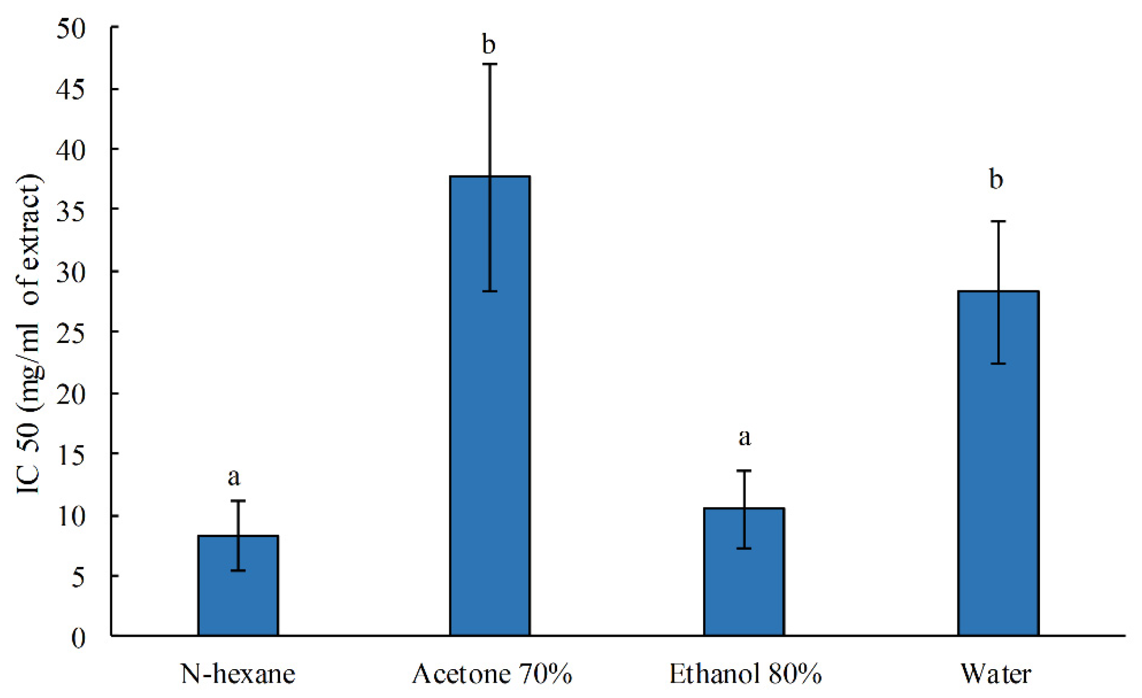
The G. gracilis extracts obtained by N-hexane and ethanol 80% showed higher antioxidant activity (DPPH IC50) (8.30 ± 2.80 mg DW/mL and 10.46 ± 3.17 mg DW/mL, respectively) (p < 0.05) than those obtained by water (28.25 ± 5.80 mg DW/mL) and acetone 70% (37.7 ± 9.34 mg DW/mL) (Figure 3).
Our results are in agreement with those of Chan et al. [51], who showed that the radical scavenging activity of wild-collected G. changii extracts decreased as the solvent polarity increased. The water and acetone extracts showed the lowest radical scavenging activity compared to ethanol and ethyl acetate extracts [51]. Vega et al. demonstrated that hydroethanolic extracts from marine red macroalgae cultivated in IMTA showed the lowest scavenger activity compared to wild-collected species [16].
Polyphenols produced by G. gracilis showed antibacterial and antioxidant activities, as reported by Afonso et al. [57], highlighting their possible use as an additive for functional fish feeds. This was confirmed by Passos et al. [24], whose study showed that supplementing compounds extracted from G. gracilis in the diet of a farmed fish species enhanced fish health and growth. These results suggest that red seaweeds cultivated in an IMTA system are able to adapt and to grow under different integrated culture conditions, as reported by Ashkenazi et al. [58], and to produce bioactive compounds that might have a significant antioxidant capacity and potential use in different sectors [16,23,58].

3.2. Cytoprotective Potential of Ethanol Extract of G. gracilis in SAF-1 Cells Exposed to BDE-47

The G. gracilis ethanol extract was selected to evaluate its protective role against oxidative stress induced by BDE-47 in vitro in the S. aurata fibroblast (SAF-1) cell line.
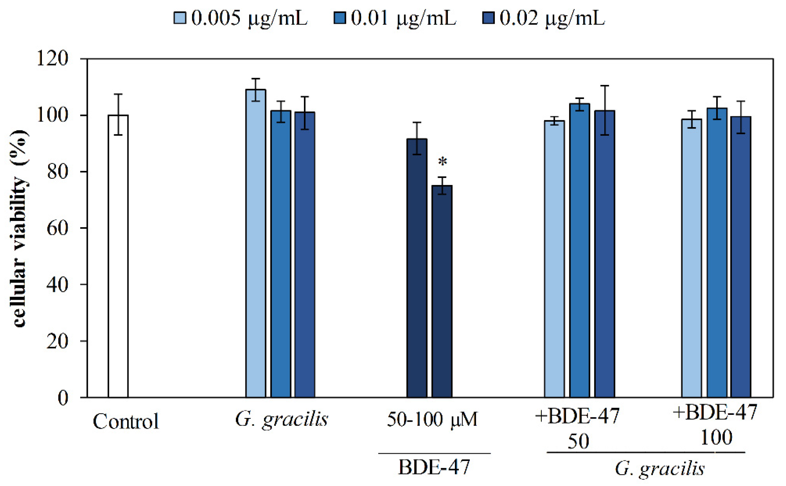
The ethanol extracts of G. gracilis at concentrations of 0.005–0.01–0.02 µg/mL tested in the SAF-1 cell line did not significantly affect the cell viability compared to the untreated cells (Figure 4). The SAF-1 cells untreated and exposed to BDE-47 (50 µM) did not affect the cell viability after 48 h of incubation, whereas the SAF-1 cells not pretreated and exposed to the highest BDE-47 concentration (100 μM) resulted in a significant decrease in cell viability (up to 30%) (p < 0.05) after 48h, compared to the control (Figure 4).
The SAF-1 cells pretreated with the ethanol extracts of G. gracilis (0.005–0.01–0.02 µg/mL) and then exposed to 50–100 μM of BDE-47 (G. gracilis + BDE-47) showed a protective effect against BDE-47 oxidative stress induction compared to the cells exposed to 100 μM of BDE-47 without any pretreatment (BDE-47) (Figure 4), suggesting that G. gracilis extract could prevent and counter cellular oxidative stress. The cytoprotective effect of G. gracilis extract against BDE-47 oxidative stress induction further confirms its antioxidant power. The G. gracilis extracts showed the same ability as synthetic antioxidants (β-carotene and gallic acid) to counteract emerging contaminant inducing oxidative stress in seabream fibroblast cell lines [26], highlighting that bioactive compounds extracted by macroalgae represent a bioavailable source of antioxidants [27,28,43,44,59,60].

Evaluation of Molecular Biomarkers Related to Cell Cycle and Oxidative Stress by Gene Expression Analysis

One of the main effects induced by exposure to chemical contaminants is the overproduction of ROS in cells, causing oxidative stress [26,61]. This stress condition influences cellular homeostasis and the antioxidant defense system, modifying some biochemical pathways that might induce cellular transformations [26,28,44]. Indeed, previous studies have suggested that BDE-47-induced oxidative stress led to cytotoxicity and apoptosis [26,28,44]. Since the molecular mechanisms by which BDE-47 affects the cell cycle, the cellular response to stress, and the antioxidant defense system remain unclear, the modulation of molecular markers related to the cell cycle (p53) and oxidative stress (nrf2, sod, and cat) was analyzed by gene expression analysis in SAF-1 cells (Figure 5).
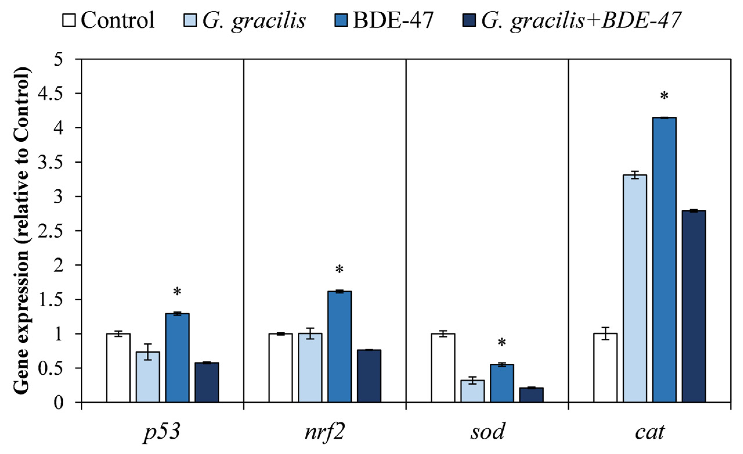
Tumor suppressor protein p53 plays a critical role in cell-cycle control, apoptosis, and in response to DNA damage [62]. After its activation by several stimuli, p53 regulates the expression of many target genes related to cell-cycle arrest and apoptosis induction, inhibiting cell proliferation [63,64]. In our study, the p53 mRNA levels were significantly up-regulated (p < 0.05) in cells exposed to BDE-47 (BDE-47) compared to the untreated cells (control) (Figure 5), suggesting that oxidative stress induced by BDE-47 caused DNA damage in SAF-1 cells [26,27]. The obtained results show that pre-incubation with G. gracilis extract before exposure to BDE-47 (G. gracilis + BDE-47) significantly decreased the p53 mRNA levels compared to the cells exposed to BDE-47 without any pretreatment (BDE-47) (p < 0.05) (Figure 5), highlighting that G. gracilis extract exhibited a strong cytoprotective activity against BDE-47-induced oxidative injury.
Nuclear factor erythroid 2-related factor 2 (Nrf2), a redox sensitive transcription factor, is activated in response to a wide range of oxidative stimuli [26,44,61]. After its activation, it translocates into the nucleus and activates the antioxidant responsive elements (AREs) of cytoprotective genes coding for antioxidant enzymes, stress protein, and phase II detoxification enzymes [61].
In this study, the up-regulation of the nrf2 gene observed in cells exposed to BDE-47 (BDE-47) (Figure 5) suggests a rapid antioxidant response in decreasing oxidative toxicity induced by environmental pollutants [26]. Persistent nuclear accumulation of Nrf2 may have dangerous effects, such as free radical damage, apoptosis, and tumorigenesis [65]. In the SAF-1 cells pretreated with G. gracilis extract and then exposed to BDE-47 (G. gracilis + BDE-47), the mRNA expression levels of nrf2 significantly decreased (p < 0.05) (Figure 5), showing a high protective capacity of the macroalga extract against BDE-47 oxidative stress induction. Furthermore, the decrease in nrf2 expression could be related to its signaling deactivation and to the restoration of cellular redox homeostasis [61].
Chen et al. [66], in accordance with our results, reported a decrease in nrf2 expression when Neuro-2a cells were pretreated with N-acetyl-l-cysteine (NAC) and then exposed to BDE-47, highlighting the ability of NAC to alleviate the oxidative stress and block apoptosis.
In addition, Nrf2 countered oxidative stress induced by environmental pollutants, through the transcription of cytoprotective genes or antioxidant proteins [61]. SOD and CAT are the major antioxidant enzymes to protect cells against oxidative stress. SOD converts superoxide anion (O2) produced by mitochondria to the less reactive hydrogen peroxide (H2O2) [67], while CAT detoxifies H2O2 to H2O and O2 [68]. High levels of SOD lead to an increase in intracellular H2O2, inducing DNA damage and tumor promotion [69]. A significant decrease in sod and an increase in cat mRNA levels (p < 0.05) were observed in cells exposed to BDE-47 (BDE-47) (Figure 5), suggesting that the overproduction of free radicals induced by BDE-47 affected the antioxidant system and led to oxidative cellular stress [27,61]. The pre-incubation of SAF-1 cells with G. gracilis extract and then exposed to BDE-47 (G. gracilis + BDE-47) significantly decreased the sod and cat mRNA levels (p < 0.05) compared to the cells exposed to BDE-47 without any pretreatment (BDE-47) (Figure 5). Furthermore, the cells pre-incubated with the ethanol extract and not exposed to BDE-47 further decreased the sod mRNA levels, suggesting that the ethanol extract of G. gracilis, could exert a preventive effect on BDE-47-induced oxidative stress, reducing the superoxide anion production and improving the antioxidant defense system [70]. The obtained results confirm that the pretreatment with G. gracilis extracts, before exposure to BDE-47, protected the SAF-1 cells against oxidative stress, improving the cellular antioxidant defense system and cell cycle control.

4. Conclusions

This study highlights that G. gracilis cultivated in IMTA could be a good source of antioxidants, as demonstrated by its antioxidant activity and determined by its polyphenol content. Hydroalcoholic solvent extraction (ethanol 80%) showed a higher extractive yield and polyphenol rate. Polyphenol compounds obtained from ethanol extract of G. gracilis showed protective capacity against oxidative stress induced by the pro-oxidant BDE-47 in the SAF-1 cell line. These results are supported by gene expression analysis, highlighting the antioxidant capacity of G. gracilis extracts against chemical contaminant inductors, which modulate molecular markers related to the cell cycle and oxidative stress. Furthermore, the biomarkers used as an early warning in this study to predict the effects of pro-oxidant contaminants at lower levels of biological organization could be adopted for environmental biomonitoring.
Given the high potential of bioactive compounds in counteracting the oxidative stress responsible for cell damage in all living organisms, including fish, induced by different environmental conditions, the extraction and isolation of bioactive compounds from marine matrices could be promoted for their potential applications in nutraceutical and aquaculture fields.

Author Contributions

Conceptualization, C.H., C.M.M. and A.S.; methodology, A.S., C.M.M., S.M. and E.C.; validation, C.H., C.M.M. and A.S.; formal analysis, S.M., R.A., E.C., G.R. and M.R.; resources, C.M.M. and A.S.; data curation, C.M.M. and A.S.; writing—original draft preparation, S.M., R.A. and E.C.; writing—review and editing, C.H., C.M.M. and A.S.; supervision, C.M.M. and A.S.; funding acquisition, C.M.M. and A.S. All authors have read and agreed to the published version of the manuscript.

Funding

This research received no external funding.

Data Availability Statement

The data contained within the article.

Acknowledgments

Simona Manuguerra is involved in the doctoral research program in Biochemistry and Molecular Biology at the Department of Biotechnology, Chemistry, and Pharmacy, University of Sienna (Italy). Rosaria Arena is supported by the PhD project “From waste to profit”, funded by PON RI FSE−FESR (2014/2020) Action IV.5 “PhD on Green deal”, University of Palermo (Italy). This work was developed in collaboration with the following projects: NBFC—National Biodiversity Future Center—Node 6 Biodiversity and Human Wellbeing, University of Palermo, grant code CN_00000033, CUP B73C22000790001; BLUEREV—Revitalisation of European local communities with innovative business models and social innovation in the blue bio-based sector; HORIZON EUROPE-CL6-2021-GOVERNANCE-01-09, proposal number 101059565.

Conflicts of Interest

The authors declare no conflicts of interest.

References

  1. Food and Agriculture Organization. In Brief to The State of World Fisheries and Aquaculture 2022; FAO: Rome, Italy, 2022; ISBN 978-92-5-136367-6. [Google Scholar]
  2. Lal, J.; Singh, S.K.; Pawar, L.; Biswas, P.; Meitei, M.M.; Meena, D.K. Integrated Multi-Trophic Aquaculture: A Balanced Ecosystem Approach to Blue Revolution. In Organic Farming: Global Perspectives and Methods, 2nd ed.; Woodhead Publishing: Sawston, UK, 2023; pp. 513–535. ISBN 9780323991452. [Google Scholar]
  3. Lothmann, R.; Sewilam, H. Potential of Innovative Marine Aquaculture Techniques to Close Nutrient Cycles. Rev. Aquac. 2023, 15, 947–964. [Google Scholar] [CrossRef]
  4. Food and Agriculture Organization. The State of Food Security and Nutrition in the World 2023; FAO: Rome, Italy, 2023; ISBN 9789251372265. [Google Scholar]
  5. Papageorgiou, N.; Dimitriou, P.D.; Chatzivasileiou, D.; Tsapakis, M.; Karakassis, I. Can IMTA Provide Added Ecosystem Value Services in the Fish Farms of Greece? Front. Mar. Sci. 2023, 9, 1083099. [Google Scholar] [CrossRef]
  6. Luo, G. Review of Waste Phosphorus from Aquaculture: Source, Removal and Recovery. Rev. Aquac. 2023, 15, 1058–1082. [Google Scholar] [CrossRef]
  7. Troell, M.; Joyce, A.; Chopin, T.; Neori, A.; Buschmann, A.H.; Fang, J.G. Ecological Engineering in Aquaculture—Potential for Integrated Multi-Trophic Aquaculture (IMTA) in Marine Offshore Systems. Aquaculture 2009, 297, 1–9. [Google Scholar] [CrossRef]
  8. Chopin, T. Aquaculture, Integrated Multi-Trophic (IMTA). In Sustainable Food Production; Springer: New York, NY, USA, 2013; pp. 184–205. [Google Scholar]
  9. Giangrande, A.; Licciano, M.; Arduini, D.; Borghese, J.; Pierri, C.; Trani, R.; Longo, C.; Petrocelli, A.; Ricci, P.; Alabiso, G.; et al. An Integrated Monitoring Approach to the Evaluation of the Environmental Impact of an Inshore Mariculture Plant (Mar Grande of Taranto, Ionian Sea). Biology 2022, 11, 617. [Google Scholar] [CrossRef]
  10. Vladkova, T.; Georgieva, N.; Staneva, A.; Gospodinova, D. Recent Progress in Antioxidant Active Substances from Marine Biota. Antioxidants 2022, 11, 439. [Google Scholar] [CrossRef] [PubMed]
  11. Faulkner, D.J. Highlights of Marine Natural Products Chemistry (1972–1999). Nat. Prod. Rep. 2000, 17, 1–6. [Google Scholar] [CrossRef]
  12. Lushchak, V.I. Environmentally Induced Oxidative Stress in Aquatic Animals. Aquat. Toxicol. 2011, 101, 13–30. [Google Scholar] [CrossRef]
  13. Herrero, M.; del Pilar Sánchez-Camargo, A.; Cifuentes, A.; Ibáñez, E. Plants, Seaweeds, Microalgae and Food by-Products as Natural Sources of Functional Ingredients Obtained Using Pressurized Liquid Extraction and Supercritical Fluid Extraction. TrAC Trends Anal. Chem. 2015, 71, 26–38. [Google Scholar] [CrossRef]
  14. Pangestuti, R.; Kim, S.K. Biological Activities and Health Benefit Effects of Natural Pigments Derived from Marine Algae. J. Funct. Foods 2011, 3, 255–266. [Google Scholar] [CrossRef]
  15. Ebrahimzadeh, M.A.; Khalili, M.; Dehpour, A.A. Antioxidant Activity of Ethyl Acetate and Methanolic Extracts of Two Marine Algae, Nannochloropsis Oculata and Gracilaria Gracilis—An in Vitro Assay. Brazilian J. Pharm. Sci. 2018, 54, 1–6. [Google Scholar] [CrossRef]
  16. Vega, J.; Álvarez-Gómez, F.; Güenaga, L.; Figueroa, F.L.; Gómez-Pinchetti, J.L. Antioxidant Activity of Extracts from Marine Macroalgae, Wild-Collected and Cultivated, in an Integrated Multi-Trophic Aquaculture System. Aquaculture 2020, 522, 735088. [Google Scholar] [CrossRef]
  17. Knowler, D.; Chopin, T.; Martínez-Espiñeira, R.; Neori, A.; Nobre, A.; Noce, A.; Reid, G. The Economics of Integrated Multi-Trophic Aquaculture: Where Are We Now and Where Do We Need to Go? Rev. Aquac. 2020, 12, 1579–1594. [Google Scholar] [CrossRef]
  18. Torres, P.; Santos, J.P.; Chow, F.; dos Santos, D.Y.A.C. A Comprehensive Review of Traditional Uses, Bioactivity Potential, and Chemical Diversity of the Genus Gracilaria (Gracilariales, Rhodophyta). Algal Res. 2019, 37, 288–306. [Google Scholar] [CrossRef]
  19. De Almeida, C.L.F.; De S. Falcão, H.; De M. Lima, G.R.; De A. Montenegro, C.; Lira, N.S.; De Athayde-Filho, P.F.; Rodrigues, L.C.; De Souza, M.d.F.V.; Barbosa-Filho, J.M.; Batista, L.M. Bioactivities from Marine Algae of the Genus Gracilaria. Int. J. Mol. Sci. 2011, 12, 4550–4573. [Google Scholar] [CrossRef]
  20. Silva-Brito, F.; Cardoso, A.; Machado, M.; Ramos-Pinto, L.; Hinzmann, M.; Abreu, H.; Costas, B.; Magnoni, L. Dietary Supplementation with Gracilaria Gracilis By-Products Modulates the Immune Status and Oxidative Stress Response of Gilthead Seabream (Sparus aurata) Stimulated with Photobacterium Damselae Subsp. Piscicida. Fish Shellfish Immunol. 2022, 126, 164–177. [Google Scholar] [CrossRef]
  21. Gioele, C.; Marilena, S.; Valbona, A.; Nunziacarla, S.; Andrea, S.; Antonio, M. Gracilaria Gracilis, Source of Agar: A Short Review. Curr. Org. Chem. 2017, 21, 380–386. [Google Scholar] [CrossRef]
  22. Kazir, M.; Abuhassira, Y.; Robin, A.; Nahor, O.; Luo, J.; Israel, A.; Golberg, A.; Livney, Y.D. Extraction of Proteins from Two Marine Macroalgae, Ulva Sp. and Gracilaria Sp., for Food Application, and Evaluating Digestibility, Amino Acid Composition and Antioxidant Properties of the Protein Concentrates. Food Hydrocoll. 2019, 87, 194–203. [Google Scholar] [CrossRef]
  23. Pradhan, B.; Bhuyan, P.P.; Patra, S.; Nayak, R.; Behera, P.K.; Behera, C.; Behera, A.K.; Ki, J.-S.; Jena, M. Beneficial Effects of Seaweeds and Seaweed-Derived Bioactive Compounds: Current Evidence and Future Prospective. Biocatal. Agric. Biotechnol. 2022, 39, 102242. [Google Scholar] [CrossRef]
  24. Passos, R.; Correia, A.P.; Ferreira, I.; Pires, P.; Pires, D.; Gomes, E.; do Carmo, B.; Santos, P.; Simões, M.; Afonso, C.; et al. Effect on Health Status and Pathogen Resistance of Gilthead Seabream (Sparus aurata) Fed with Diets Supplemented with Gracilaria Gracilis. Aquaculture 2021, 531, 735888. [Google Scholar] [CrossRef]
  25. Bonsignore, M.; Messina, C.; Bellante, A.; Manuguerra, S.; Arena, R.; Santulli, A.; Maricchiolo, G.; Del Core, M.; Sprovieri, M. Chemical and Biochemical Responses to Sub—Lethal Doses of Mercury and Cadmium in Gilthead Seabream (Sparus aurata). Chemosphere 2022, 307, 135822. [Google Scholar] [CrossRef]
  26. Ruiz, C.E.; Manuguerra, S.; Cuesta, A.; Esteban, M.A.; Santulli, A.; Messina, C.M. Sub-Lethal Doses of Polybrominated Diphenyl Ethers Affect Some Biomarkers Involved in Energy Balance and Cell Cycle, via Oxidative Stress in the Marine Fish Cell Line SAF-1. Aquat. Toxicol. 2019, 210, 1–10. [Google Scholar] [CrossRef] [PubMed]
  27. Espinosa Ruiz, C.; Manuguerra, S.; Curcuraci, E.; Santulli, A.; Messina, C.M. Carbamazepine, Cadmium Chloride and Polybrominated Diphenyl Ether-47, Synergistically Modulate the Expression of Antioxidants and Cell Cycle Biomarkers, in the Marine Fish Cell Line SAF-1. Mar. Environ. Res. 2020, 154, 104844. [Google Scholar] [CrossRef]
  28. Manuguerra, S.; Ruiz, C.E.; Santulli, A.; Messina, C.M. Sub-Lethal Doses of Polybrominated Diphenyl Ethers, in Vitro, Promote Oxidative Stress and Modulate Molecular Markers Related to Cell Cycle, Antioxidant Balance and Cellular Energy Management. Int. J. Environ. Res. Public Health 2019, 16, 588. [Google Scholar] [CrossRef] [PubMed]
  29. Sureda, A.; Box, A.; Terrados, J.; Deudero, S.; Pons, A. Antioxidant Response of the Seagrass Posidonia Oceanica When Epiphytized by the Invasive Macroalgae Lophocladia Lallemandii. Mar. Environ. Res. 2008, 66, 359–363. [Google Scholar] [CrossRef]
  30. Espinosa, C.; Pérez-Llamas, F.; Guardiola, F.A.; Esteban, M.A.; Arnao, M.B.; Zamora, S.; López-Jiménez, J.A. Molecular Mechanisms by Which White Tea Prevents Oxidative Stress. J. Physiol. Biochem. 2014, 70, 891–900. [Google Scholar] [CrossRef]
  31. Tanaka, M.; Kishimoto, Y.; Sasaki, M.; Sato, A.; Kamiya, T.; Kondo, K.; Iida, K. Terminalia bellirica (Gaertn.) Roxb. Extract and Gallic Acid Attenuate LPS-Induced Inflammation and Oxidative Stress via MAPK/NF-κ B and Akt/AMPK/Nrf2 Pathways. Oxid. Med. Cell. Longev. 2018, 2018, 9364364. [Google Scholar] [CrossRef]
  32. Yang, J.I.; Yeh, C.C.; Lee, J.C.; Yi, S.C.; Huang, H.W.; Tseng, C.N.; Chang, H.W. Aqueous Extracts of the Edible Gracilaria Tenuistipitata Are Protective against H2O2-Induced DNA Damage, Growth Inhibition, and Cell Cycle Arrest. Molecules 2012, 17, 7241–7254. [Google Scholar] [CrossRef]
  33. Zhao, Y.; Tang, X.; Quigg, A.; Lv, M.; Zhao, Y. The Toxic Mechanisms of BDE-47 to the Marine Diatom Thalassiosira Pseudonana-a Study Based on Multiple Physiological Processes. Aquat. Toxicol. 2019, 212, 20–27. [Google Scholar] [CrossRef]
  34. Wang, X.; Hales, B.F.; Robaire, B. Effects of Flame Retardants on Ovarian Function. Reprod. Toxicol. 2021, 102, 10–23. [Google Scholar] [CrossRef]
  35. Tornero, V.; Hanke, G. Chemical Contaminants Entering the Marine Environment from Sea-Based Sources: A Review with a Focus on European Seas. Mar. Pollut. Bull. 2016, 112, 17–38. [Google Scholar] [CrossRef] [PubMed]
  36. Álvarez-Muñoz, D.; Llorca, M.; Blasco, J.; Barceló, D. Contaminants in the Marine Environment. In Marine Ecotoxicology; Academic Press: Cambridge, MA, USA, 2016; pp. 1–34. [Google Scholar]
  37. Nasri, A.; Allouche, M.; Hannachi, A.; Barhoumi, B.; Wahbi, A.; Harrath, A.H.; Mahmoudi, E.; Beyrem, H.; Boufahja, F. Ecotoxicity of Polybrominated Diphenyl Ether (BDE-47) on a Meiobenthic Community with Special Emphasis on Nematodes: Taxonomic and Trophic Diversity Assessment. Environ. Pollut. 2021, 277, 116727. [Google Scholar] [CrossRef] [PubMed]
  38. Jiang, Y.; Tang, X.; Sun, T.; Wang, Y. BDE-47 Exposure Changed the Immune Function of Haemocytes in Mytilus Edulis: An Explanation Based on ROS-Mediated Pathway. Aquat. Toxicol. 2017, 182, 58–66. [Google Scholar] [CrossRef]
  39. Herrera, A.; Acosta-Dacal, A.; Luzardo, O.P.; Martínez, I.; Rapp, J.; Reinold, S.; Montesdeoca-Esponda, S.; Montero, D.; Gómez, M. Bioaccumulation of Additives and Chemical Contaminants from Environmental Microplastics in European Seabass (Dicentrarchus labrax). Sci. Total Environ. 2022, 822, 153396. [Google Scholar] [CrossRef]
  40. Harmelin-Vivien, M.; Bodiguel, X.; Charmasson, S.; Loizeau, V.; Mellon-Duval, C.; Tronczyński, J.; Cossa, D. Differential Biomagnification of PCB, PBDE, Hg and Radiocesium in the Food Web of the European Hake from the NW Mediterranean. Mar. Pollut. Bull. 2012, 64, 974–983. [Google Scholar] [CrossRef] [PubMed]
  41. Bodiguel, X.; Tronczynski, J.; Loizeau, V.; Munschy, C.; Guiot, N.; Le Guellec, A.M.; Olivier, N.; Roupsard, F.; Mellon, C. Classical and Novel Organohalogen Compounds (PCBs and PBDEs) in Hake (M. merluccius, L.) from the Mediterranean and Atlantic Coasts (France). WIT Trans. Ecol. Environ. 2008, 110, 157–166. [Google Scholar] [CrossRef]
  42. UNEP. Chemicals Listed in Annex A; Stockholm Convention for Persistent Organic Pollutants Chatelaine: Geneva, Switzerland, 2019. [Google Scholar]
  43. Geng, Q.; Guo, M.; Wu, H.; Peng, J.; Zheng, G.; Liu, X.; Zhai, Y.; Tan, Z. Effects of Single and Combined Exposure to BDE-47 and PFOA on Distribution, Bioaccumulation, and Toxicity in Blue Mussel (Mytilus galloprovincialis). Ecotoxicol. Environ. Saf. 2021, 228, 113014. [Google Scholar] [CrossRef]
  44. Espinosa Ruiz, C.; Manuguerra, S.; Cuesta, A.; Santulli, A.; Messina, C. Oxidative Stress, Induced by Sub-Lethal Doses of BDE 209, Promotes Energy Management and Cell Cycle Modulation in the Marine Fish Cell Line SAF-1. Int. J. Environ. Res. Public Health 2019, 16, 474. [Google Scholar] [CrossRef]
  45. Messina, C.M.; Troia, A.; Arena, R.; Manuguerra, S.; Ioannou, T.; Curcuraci, E.; Renda, G.; Hellio, C.; Santulli, A. Species-Specific Antioxidant Power and Bioactive Properties of the Extracts Obtained from Wild Mediterranean calendula Spp. (Asteraceae). Appl. Sci. 2019, 9, 4627. [Google Scholar] [CrossRef]
  46. Folin, O.; Ciocalteu, V. On Tyrosine and Tryptophane Determinations in Proteins. J. Biol. Chem. 1927, 73, 627–650. [Google Scholar] [CrossRef]
  47. Dhouibi, N.; Manuguerra, S.; Arena, R.; Mahdhi, A.; Messina, C.M.; Santulli, A.; Dhaouadi, H. Screening of Antioxidant Potentials and Bioactive Properties of the Extracts Obtained from Two Centaurea L. Species (C. kroumirensis Coss. and C. sicula L. Subsp Sicula). Appl. Sci. 2020, 10, 2267. [Google Scholar] [CrossRef]
  48. Bernatoniene, J.; Masteikova, R.; Davalgiene, J.; Peciura, R.; Gauryliene, R.; Bernatoniene, R.; Majiene, D.; Lazauskas, R.; Civinskiene, G.; Velziene, S.; et al. Topical Application of Calendula officinalis (L.): Formulation and Evaluation of Hydrophilic Cream with Antioxidant Activity. J. Med. Plants Res. 2011, 5, 868–877. [Google Scholar]
  49. Mosmann, T. Rapid Colorimetric Assay for Cellular Growth and Survival: Application to Proliferation and Cytotoxicity Assays. J. Immunol. Methods 1983, 65, 55–63. [Google Scholar] [CrossRef]
  50. Livak, K.J.; Schmittgen, T.D. Analysis of Relative Gene Expression Data Using Real-Time Quantitative PCR and the 2−ΔΔCT Method. Methods 2001, 25, 402–408. [Google Scholar] [CrossRef]
  51. Chan, P.T.; Matanjun, P.; Yasir, S.M.; Tan, T.S. Antioxidant Activities and Polyphenolics of Various Solvent Extracts of Red Seaweed, Gracilaria Changii. J. Appl. Phycol. 2015, 27, 2377–2386. [Google Scholar] [CrossRef]
  52. Messina, C.M.; Renda, G.; Laudicella, V.A.; Trepos, R.; Fauchon, M.; Hellio, C.; Santulli, A. From Ecology to Biotechnology, Study of the Defense Strategies of Algae and Halophytes (from Trapani Saltworks, NW Sicily) with a Focus on Antioxidants and Antimicrobial Properties. Int. J. Mol. Sci. 2019, 20, 881. [Google Scholar] [CrossRef]
  53. Hulkko, L.S.S.; Chaturvedi, T.; Thomsen, M.H. Extraction and Quantification of Chlorophylls, Carotenoids, Phenolic Compounds, and Vitamins from Halophyte Biomasses. Appl. Sci. 2022, 12, 840. [Google Scholar] [CrossRef]
  54. Foo, S.C.; Yusoff, F.M.; Ismail, M.; Basri, M.; Yau, S.K.; Khong, N.M.H.; Chan, K.W.; Ebrahimi, M. Antioxidant Capacities of Fucoxanthin-Producing Algae as Influenced by Their Carotenoid and Phenolic Contents. J. Biotechnol. 2017, 241, 175–183. [Google Scholar] [CrossRef]
  55. Arena, R.; Manuguerra, S.; Collins, E.; Mahdhi, A.; Renda, G.; Messina, C.M.; Santulli, A. Antioxidant Properties of a Supercritical Fluid Extract of the Halophyte Mesembryanthemum nodiflorum L. from Sicilian Coasts: Nutraceutical and Cosmeceutical Applications. Appl. Sci. 2020, 10, 2374. [Google Scholar] [CrossRef]
  56. Yang, J.; Chan, K.M. Evaluation of the Toxic Effects of Brominated Compounds (BDE-47, 99, 209, TBBPA) and Bisphenol a (BPA) Using a Zebrafish Liver Cell Line, ZFL. Aquat. Toxicol. 2015, 159, 138–147. [Google Scholar] [CrossRef]
  57. Afonso, C.; Correia, A.P.; Freitas, M.V.; Mouga, T.; Baptista, T. In Vitro Evaluation of the Antibacterial and Antioxidant Activities of Extracts of Gracilaria Gracilis with a View into Its Potential Use as an Additive in Fish Feed. Appl. Sci. 2021, 11, 6642. [Google Scholar] [CrossRef]
  58. Ashkenazi, D.Y.; Figueroa, F.L.; Korbee, N.; García-Sánchez, M.; Vega, J.; Ben-Valid, S.; Paz, G.; Salomon, E.; Israel, Á.; Abelson, A. Enhancing Bioproducts in Seaweeds via Sustainable Aquaculture: Antioxidant and Sun-Protection Compounds. Mar. Drugs 2022, 20, 767. [Google Scholar] [CrossRef] [PubMed]
  59. Klinčić, D.; Dvoršćak, M.; Jagić, K.; Mendaš, G.; Herceg Romanić, S. Levels and Distribution of Polybrominated Diphenyl Ethers in Humans and Environmental Compartments: A Comprehensive Review of the Last Five Years of Research. Environ. Sci. Pollut. Res. 2020, 27, 5744–5758. [Google Scholar] [CrossRef]
  60. De Oro-Carretero, P.; Landaluze, J.S. Bioaccumulation And Biotransformation of BDE-47 Using Zebrash Eleutheroembryos (Danio rerio). Environ Toxicol Chem. 2023, 42, 835–845. [Google Scholar] [CrossRef]
  61. Regoli, F.; Giuliani, M.E. Oxidative Pathways of Chemical Toxicity and Oxidative Stress Biomarkers in Marine Organisms. Mar. Environ. Res. 2014, 93, 106–117. [Google Scholar] [CrossRef]
  62. Lee, H.Y.; Oh, S.H. Autophagy-Mediated Cytoplasmic Accumulation of P53 Leads to Apoptosis through DRAM-BAX in Cadmium-Exposed Human Proximal Tubular Cells. Biochem. Biophys. Res. Commun. 2021, 534, 128–133. [Google Scholar] [CrossRef]
  63. Yee, K.S.; Vousden, K.H. Complicating the Complexity of P53. Carcinogenesis 2005, 26, 1317–1322. [Google Scholar] [CrossRef] [PubMed]
  64. Zhang, W.; Liu, N.; Wang, X.; Jin, X.; Du, H.; Peng, G.; Xue, J. Benzo(a)Pyrene-7,8-Diol-9,10-Epoxide Induced P53-Independent Necrosis via the Mitochondria-Associated Pathway Involving Bax and Bak Activation. Hum. Exp. Toxicol. 2015, 34, 179–190. [Google Scholar] [CrossRef]
  65. Giuliani, M.E.; Regoli, F. Identification of the Nrf2–Keap1 Pathway in the European Eel Anguilla Anguilla: Role for a Transcriptional Regulation of Antioxidant Genes in Aquatic Organisms. Aquat. Toxicol. 2014, 150, 117–123. [Google Scholar] [CrossRef]
  66. Chen, H.; Tang, X.; Zhou, B.; Zhou, Z.; Xu, N.; Wang, Y. A ROS-Mediated Mitochondrial Pathway and Nrf2 Pathway Activation Are Involved in BDE-47 Induced Apoptosis in Neuro-2a Cells. Chemosphere 2017, 184, 679–686. [Google Scholar] [CrossRef]
  67. Younus, H. Therapeutic Potentials of Superoxide Dismutase. Int. J. Health Sci. 2018, 12, 88. [Google Scholar]
  68. Sharma, I.; Ahmad, P. Catalase: A Versatile Antioxidant in Plants. In Oxidative Damage to Plants Antioxid. Networks Signal; Elsevier: Amsterdam, The Netherlands, 2014; pp. 131–148. [Google Scholar] [CrossRef]
  69. Khan, M.A.; Tania, M.; Zhang, D.Z.; Chen, H.C. Antioxidant Enzymes and Cancer. Chinese J. Cancer Res. 2010, 22, 87–92. [Google Scholar] [CrossRef]
  70. Fuchs, P.; Perez-Pinzon, M.A.; Dave, K.R. Cerebral Ischemia in Diabetics and Oxidative Stress. In Diabetes: Oxidative Stress and Dietary Antioxidants; Academic Press: Cambridge, MA, USA, 2013; pp. 15–23. ISBN 9780124058859. [Google Scholar]
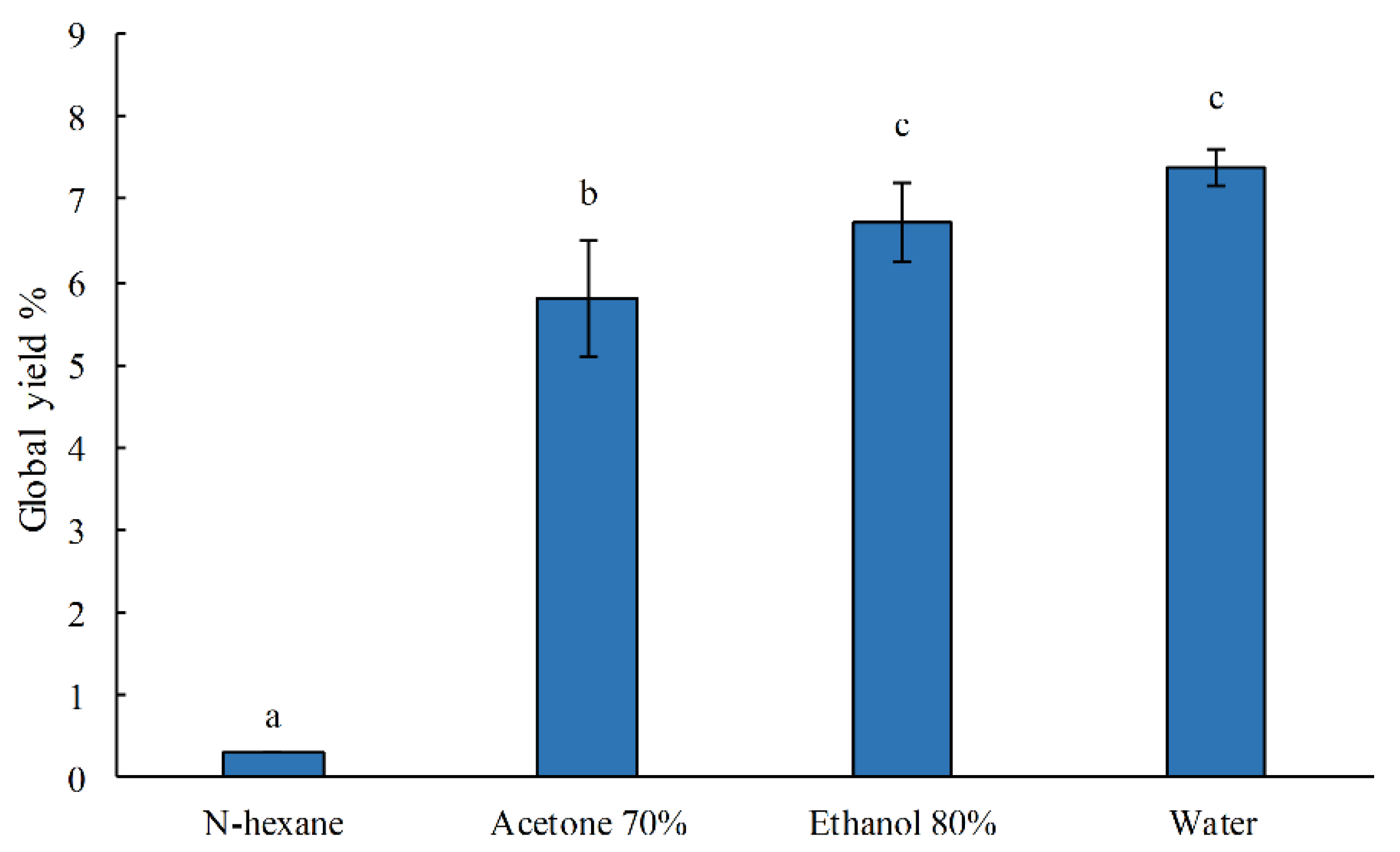
Figure 1. Extract yields (%) obtained with different solvents (N-hexane, acetone 70%, ethanol 80%, and water) from dried G. gracilis. Bars represent the mean ± SEM (n = 3). Lowercase letters indicate significant differences among different solvents (p < 0.05).
Water 16 02667 g001
Figure 2. Total polyphenol content (mg GAE/g DW) in G. gracilis extracted by N-hexane, acetone 70%, ethanol 80%, and water. Bars represent the mean ± SEM (n = 3). Lowercase letters indicate significant differences among the different solvents (p < 0.05).
Water 16 02667 g002
Figure 3. DPPH radical scavenging activity (IC 50, mg DW/mL) in G. gracilis by N-hexane, acetone 70%, ethanol 80%, and water. Bars represent the mean ± SEM (n = 3). Lowercase letters indicate significant differences among the different solvents (p < 0.05).
Water 16 02667 g003
Figure 4. Cellular viability (%) of SAF-1 cells measured, comparing the control cells and cells exposed to different concentrations of BDE-47, with the cells pretreated for 24 h with different concentrations of G. gracilis extracts (0.005–0.01–0.02 µg/mL) and then exposed to different concentrations of BDE-47 (50–100 µM) for 48 h. The bars represent the mean ± SEM (n = 6). Statistical differences (p < 0.05) among the groups are indicated by “*”.
Water 16 02667 g004
Figure 5. Relative gene expressions of genes related to p53, nrf2, sod, and cat in S. aurata SAF-1 cells not treated (Control), pretreated with G. gracilis (G. gracilis), and pretreated with G. gracilis extracts (0.02 µg/mL), and then exposed to BDE-47 (100 µM) (G. gracilis + BDE-47). The bars represent the mean ± SEM (n = 6). Statistical differences (p < 0.05) among the groups compared to the control are indicated by “*”.
Water 16 02667 g005
Table 1. Gilthead seabream primer sequences used for real-time PCR.
GenesF/R Primer Sequence (5′–3′)
p53CCTCATCCTCATCATCGCCT
AGCTCGTTGAATTTGCAGGG
nrf2GTTCAGTCGGTGCTTTGACA
CTCTGATGTGCGTCTCTCCA
sodCCATGGTAAGAATCATGGCGG
CGTGGATCACCATGGTTCTG
catTTCCCGTCCTTCATTCACTC
CTCCAGAAGTCCCACACCAT
β-actinGTCATGGATTCCGGTGATGG
TGGTGAAGGAGTAGCCACGC
18sTGTGCCGCTAGAGGTGAAATT
GCAAATGCTTTCGCTTTCG
Disclaimer/Publisher’s Note: The statements, opinions and data contained in all publications are solely those of the individual author(s) and contributor(s) and not of MDPI and/or the editor(s). MDPI and/or the editor(s) disclaim responsibility for any injury to people or property resulting from any ideas, methods, instructions or products referred to in the content.