Article

Bacterial Contamination in Health Care Centers: Differences between Urban and Rural Settings

1
H&TRC—Health & Technology Research Center, ESTeSL—Escola Superior de Tecnologia da Saúde, Instituto Politécnico de Lisboa, 1600-560 Lisbon, Portugal
2
Escola Nacional de Saúde Pública, Universidade NOVA de Lisboa, 1600-560 Lisbon, Portugal
3
Centro de Ciências e Tecnologias Nucleares (C2TN), Instituto Superior Técnico, Universidade de Lisboa, 1600-560 Lisbon, Portugal
4
Portugal & Centro Hospitalar São João, Faculdade de Medicina da Universidade de Porto, 4099-002 Porto, Portugal
5
Instituto de Ciência e Inovação em Engenharia Mecânica e Engenharia Industrial, 4200-465 Porto, Portugal
6
EPIUnit—Instituto de Saúde Pública, Universidade do Porto, 4050-600 Porto, Portugal
7
Centro de Investigação em Saúde Pública, Universidade NOVA de Lisboa, 1600-560 Lisbon, Portugal
8
Comprehensive Health Research Center (CHRC), 1600-560 Lisbon, Portugal
*
Author to whom correspondence should be addressed.
Atmosphere 2021, 12(4), 450; https://doi.org/10.3390/atmos12040450
Submission received: 19 March 2021 / Revised: 29 March 2021 / Accepted: 30 March 2021 / Published: 31 March 2021
(This article belongs to the Special Issue Indoor Air Quality—What Is Known and What Needs to Be Done)

Abstract

:
This study aims to assess the airborne bioburden of rural and urban Portuguese Primary Health Care Centers (PHCC) using active and passive sampling methods and identify the potential differences in airborne microbiota between both environments. The highest total aerobic mesophilic bacterial load in indoor air were found in the Vaccination Room (448 CFU.m−3) in the Rural PHCC and in the Waiting Room (420 CFU.m−3) for Urban PHCC. The total coliforms contamination level in indoor air was detected only in the Cleaning Supplies Room (4 CFU.m−3) in the Urban PHCC. The most frequent bacteria genera identified was Micrococcus (21% Rural PHCC; 31% Urban PHCC). The surface samples showed a highest total aerobic mesophilic bacterial contamination in the Treatment Room (86 × 103 CFU.m−2) from the Rural PHCC and in the Front Office (200 × 103 CFU.m−2) from the Urban PHCC. The electrostatic dust cloth (EDC) samples showed a highest bacterial load in the Urban PHCC. Total aerobic mesophilic bacterial load in settled dust and in the Heating, Ventilating and Air Conditioning (HVAC) filter samples in the Urban PHCC (8 CFU.g−1 and 6 × 10³ CFU.m−2) presented higher values compared with the Rural PHCC (1 CFU.g−1 and 2.5 × 10³ CFU.m−2). Urban PHCC presented higher bacterial airborne contamination compared with the Rural PHCC for the majority of sampling sites, and when compared with the indoor air quality (IAQ) Portuguese legislation it was the Rural PHCC in two sampling places who did not comply with the established criteria.

1. Introduction

Exposure to bioaerosols has become one of the major problems in indoor air quality (IAQ) for occupational and public health [1]. The importance of assessment and control of contamination in critical environments, such as the physical environment of health care organizations [2,3,4] is a requirement to protect patients and healthcare workers from hospital-acquired (nosocomial) infections and occupational diseases [5].
Hospital-acquired infections contracted by patients after admission to health care facilities are a concern not only in terms of patient health, as they have a high impact on patient morbidity and mortality [6,7], lengthened hospital stays, as well as increased health care costs resulting from failed treatments [8]. The scale of severity of the effects on human’s health, depends on their toxicity, exposure time, microbial load and also the immune system of the exposed individuals [9,10].
In the hospital context, the greatest concerns for airborne transmission are related to respiratory infections, including tuberculosis [11], measles [12], varicella [13], influenza [14], respiratory syncytial virus (RSV) [15], Bordetella pertussis [16], as well as non-respiratory infections, such as norovirus [17], methicillin-resistant Staphylococcus aureus (MRSA) [18] and Clostridium difficile [19,20].
Presently, one of the most serious bacterial pathogens associated with indoor environments is Legionella pneumophila that causes Legionnaire disease and Pontiac fever, which have become a leading public health concern [21]. The health care facilities have some medical equipment (i.e., respiratory devices, such as CPAP machines, hydrotherapy equipment and bronchoscopes) [22] that can originate the development of Legionella and, potentially, emit contaminated aerosols. In view of the number of patients who visit health care facilities, as well as their immune status, the likelihood of exposure and infection is increased in these institutions [23]. The rising incidences of Legionella-associated diseases [23], and the variety of sources of transmission, emphasize the need to understand the prevalence, survival and transmission of Legionella in diverse public settings, namely in health care facilities.
Moreover, antimicrobial resistance in human pathogens, such as Staphylococcus aureus, mostly associated with the inappropriate use of antibiotics, has led to emergence of antibiotic resistance strains (superbugs) at concerning rates worldwide [24]. During its evolution, S. aureus has acquired, via horizontal genetic transfer, the Staphylococcal Cassette Chromosome mec complex (SCCmec), in which is inserted the mecA gene which encodes the modified penicillin-binding protein 2 (PBP2), responsible for the resistance to β-lactam antibiotics (oxacillin, streptomycin, tetracycline, erythromycin, among others.), by reduction of the affinity of the antibiotics binding to their receptor [24,25]. MRSA is currently acknowledged as one of the most relevant human pathogens, particularly in health associated clinical settings, due to the fact that it is a major cause of infections worldwide [26] associated with extremely high mortality rates for invasive blood-stream and pneumonic infections [27]. Relevantly, the ability demonstrated by this particular pathogen to develop new clones with the capacity to invade community settings and infect people without predisposing risk factors and also to buildup MRSA reservoirs are certainly major concerns.
Moreover, for the past few years, Pseudomonas aeruginosa, an opportunistic pathogen, has also become a significant public health threat and one of the leading causes of nosocomial infections, particularly associated with immunocompromised patients [28]. Due to the worldwide dissemination of high-risk clones which are characterized as multidrug-resistant or extensively drug-resistant (MDR/XDR) strains [29] associated with severe hospital-acquired infections with very limited therapeutic options [30] more attention has been given to this human pathogen. Considering the fact that antimicrobial resistance is currently considered one of the most relevant health care challenges of this century [31], the identification of these particular strains, namely MRSA and Pseudomonas aeruginosa, in health care facilities is imperative not only to secure exposed individuals health but also to avoid the spread of resistant strains to the community.
The presence of dynamic and highly diversified microbial communities in the air of urban and rural environments assumes a major impact on Public Health due to their dispersion and health consequences [32,33,34,35].
The composition of bacteriota indoors is affected by ventilation, building design, environmental conditions (like temperature, humidity, presence of organic matter), ability to form biofilms, building occupancy, geographical location, outdoor environment [1,36,37,38,39] and also by the activities of human occupants [40,41]. In clinical environments, the building design, ventilation systems, medical activity, occupancy rate, clinical staff and types of patients, frequency of cleaning and hospital cleaning procedures, as well as outdoor air penetrating indoors [42,43,44,45,46], are decisive factors for the levels of aerial microbial concentration.
The airborne bioburden was previously identified in hospital environments [1,46,47,48,49,50], but until now, airborne bioburden assessment in Portuguese PHCCs has only been done in a few limited studies, although in this environment, patients may also present a vulnerable health. In a rural setting, located in the countryside, the population uses primary health centers due to the lack of other medical options and equipment available. On the other hand, in a city setting the population has a lot more options available to them. Likewise, the influence of urban versus rural locations on bioburden assessment has not been studied in these Primary Health Centers.
The World Health Organization publish air quality guidelines for specific pollutants, based on their health impacts [51]. In Europe, improvements in ambient air quality are driven by Community directives (2008/50/EC and 2004/107/C). In Portugal there is only legislation regarding IAQ that establishes limit values for microbiological contamination, being applicable in all indoor environments, without any specification for health care facilities, or occupational context [52]. Portuguese legal compliance differs in relation to the microorganism, for total bacteria, the indoor load should not exceed the outdoor load by 350 CFU.m−3, there is a second criterion when this condition is not satisfied [53]. Additionally, Portuguese legislation only considers air sampling to assess microbial contamination indoors that narrow the information regarding the exposure assessment [3,54]. As such, passive sampling methods such as surface swabs and electrostatics dust collectors should be included in the sampling approach to enrich the assessment and provide a more accurate risk characterization [3,54].
This study aims to assess the airborne bacteriota of rural and urban Portuguese Primary Health Care Centers, with a multi-approach protocol using active (air sampling by impaction) and passive (surface swabs, electrostatic dust cloth, settled dust and air-conditioning filters) sampling methods, while identifying the potential differences in airborne microbiota between both environments. The analysis of airborne microbiota in both PHCC consisted in the enumeration of mesophilic bacterial load, its phenotypic characterization and antibiotic resistance profile, and detection of Pseudomonas aeruginosa, Legionella sp. and Staphylococcus aureus.

2. Materials and Methods

This study was performed between May and September of 2018 in two selected Portuguese Primary Health Care Centers. One of the PHCC was in an urban setting and the other in a rural setting in Lisbon district. It is part of a larger exploratory study with financial support aiming to establish protocols to assess occupational exposure to bioburden in clinical environments.

2.1. PHCC Characterization and Measurement of Environmental Parameters

A walkthrough survey and checklist were applied to collect information regarding the characterization of the building, the procedures of the Health Center and sampling site characterization.
Both buildings were built as health units in 2016 and haven’t had modifications done in the last 12 months. However, during the walkthrough survey it was observed visible mold growth and cracks on some walls from the urban PHCC and cracks on the walls of the vaccination room in the rural PHCC. The urban PHCC has two floors above ground and the rural PHCC only has one.
The car parking, attached garage, direct access from basement car park, cooling towers and traffic in the urban PHCC and the outdoor car parking and waste management site (tip or garbage dumpsters) in the rural PHCC were identified as potential sources of air pollution that could influence the internal environment (up to 1 km).
The natural ventilation from the urban PHCC was only provided through the open door, while in the rural PHCC has natural ventilation from sliding windows, windows with tilting flag and full openness windows (usually open 1 h during the morning in the vaccination and treatments rooms, and after the cleaning procedures in all places). Both PHCC are provided with artificial ventilation provided by a Central HVAC system with air handing units and ductwork system, working from 8 am to 8 pm in the urban PHCC, whilst working all day in the rural PHCC. In Rural PHCC, these systems are located on the roof, while in Urban PHCC they are located next to the entrance, at 1.8 m from the ground level.
The cleaning procedures and waste collection are performed by an external company. In the urban PHCC the floors and surface are cleaned during patient service and working hours, while in the rural PHCC the cleaning is performed daily after patient service and after working hours. Considering the type of activity realized in the treatment room, it is cleaned several times a day, four times in the urban PHCC and two in the rural PHCC. A mop is used to clean the floor while cloths are used for surfaces.
The same areas were evaluated once in both PHCC and the measurements were carried out during the normal working hours (Table 1).
Measurements of temperature, relative humidity and carbon dioxide (CO2) were also performed in each sampling location in both PHCC. Temperature and relative humidity were measured by the Handheld Particle Counter from Lighthouse Worldwide Solutions —Model 3016/5016, and the CO2 was measured using the Graywolf equipment (Graywolf Sensing Solutions: Shelton, CT, USA). These measurements were performed in the same areas where bacteriota was evaluated and an average value for each place was obtained through a 5 min measurement.

2.2. Sampling and Characterization of Viable Bacteria

One indoor air sample in each evaluated area, and one outdoor sample (as reference), of 250 L were collected by impaction in both PHCC with a flow rate of 140 L/min (Millipore air Tester, Millipore, Billerica, MA, USA) onto each culture media plate according to the manufacturer’s instructions. Different culture media were used to ensure the selectivity for bacterial growth: Tryptic Soy Agar (TSA) supplemented with nystatin (0.2%) was used to assess the total aerobic mesophilic bacteria and Violet Red Bile Agar (VRBA) to total coliforms. For the air sampling was followed the guidelines of the national legislation [53].
Surface samples were collected using a swab method with a 10 cm × 10 cm square stencil, disinfected with a 70% alcohol solution between samplings [55]. Surface swab samples were extracted with 1 mL of 0.1% Tween™ 80 saline solution (NaCl 0.9%) for 30 min at 250 rpm on an orbital laboratory shaker (Edmund Bühler SM-30, Hechingen, Germany) and seeded on the selected culture media for bacterial growth (TSA and VRBA).
One piece of HVAC filter with approximately 2 cm2 (1.4 cm × 1.4 cm) was collected in each PHCC, refrigerated at 4 °C during transport before analysis, and washed with 10 mL of 0.1% Tween™ 80 saline solution (NaCl 0.9%) for 30 min at 250 rpm on an orbital laboratory shaker (Edmund Bühler SM-30, Hechingen, Germany) [56] and plated onto the selected media (TSA and VRBA) for bacterial growth.
All the inoculated plates from the different environmental parameters were incubated at 30 °C (TSA) and at 35 °C (VRBA) for 7 days, for bacterial growth. After the incubation period, quantitative (colony-forming units—CFU.m−3 and CFU.m−2) results for bacteria were obtained. Bacterial phenotyping was based primarily on morphology, Gram-staining, endospore formation, catalase activity (catalase test) and cytochrome oxidase presence (oxidase test). Bacteria was grouped into morphologic groups as Gram-positive cocci, Gram-negative cocci, Gram-positive rods and Gram-negative rods according to its phenotype based on Bergey manual of determinative bacteriology [57]. The most frequent bacterial isolates were identified using a miniaturized biochemical test (RapID, Remel; Santa Fe, Lenexa, KS, USA).

2.3. Electrostatic Dust Collector

One EDC with a surface exposure area of 0.00942 m2 was added in each sampled area at a minimum 0.93 m above floor level, and dust was left to settle for 15 days before analysis. EDC samples were submitted to extraction and bioburden characterized by culture-based methods described previously [58,59]. The EDC were weighed, before and after being exposed, and the mass of the collected dust was determined by subtracting the values. Each EDC cloth was then washed with 20 mL 0.9% NaCl with 0.05% Tween80™ (Merck S.A, Lisbon, Portugal) by orbital shaking (250 rpm, 60 min, at room temperature), and 150 μL of the wash suspension was inoculated onto two different culture media: TSA with 0.2% nystatin, and VRBA. After incubation of TSA and VRBA plates at 30 and 35 °C for 7 days, respectively, quantitative colony-forming units were obtained.

2.4. Settled Dust Sampling

Sampling of the settled dust was accomplished by a vacuum cleaner (HOOVER Brave BV71_BV10 A2, Brugherio (MB), Itália, USA) equipped with a 40-micron nylon mesh inserted in a DUSTREAM™ collector (Indoor Biotechnologies, Charlottesville, VA, USA). Vacuuming was performed for a period of 10 min in different selected areas [60] and the dust collected from several surfaces and according to previously described procedure [60]. Sampling duration was the same in all PHCCs in order to standardize sampling procedures and also to ensure a sufficient amount of dust for analysis [60]. A DUSTREAM™ filter tube was collected in each PHCC. The samples were measured and analyzed for bacterial load.
The samples were weighted and washed in a ratio of 1 g (dust) per 9.1 mL of NaCl 0.9% with 0.05% Tween 80 (10 μL) for 60 min at 250 rpm, and 0.15 mL of this suspension were inoculated onto two different media: TSA with 0.2% nystatin and VRBA. After incubation of TSA and VRBA at 30 °C and 35 °C for 7 days for mesophilic bacteria and coliforms, respectively, bioburden densities (CFU·g−1 of dust) were determined.

2.5. Antibiotic Susceptibility Testing

Antimicrobial susceptibility testing was performed on isolates based on the disk diffusion method on Mueller-Hinton agar [61,62]. The grades of susceptibility pattern were recognized as sensitive and resistant by comparison of the zone of inhibition as indicated in European Committee on Antimicrobial Susceptibility Testing standard 2020 [63]. The antibiotics tested for both gram-negative and gram-positive bacteria (Oxoid, Basingtone, UK) were aztreonam (30 μg), cefotaxime (30 μg), gentamicin (30 μg), imipenem (10 μg) and ticarcillin (75 μg). Antibiotics were selected based on literature, effectiveness and EUCAST Guidelines. In Rural PHCC, 130 isolates out of a total of 540 isolates were tested for antibiotic resistance and in Urban PHCC, 84 isolates out of a total of 440 isolates were tested.

2.6. Detection of the Presence of Pseudomonas aeruginosa, Staphylococcus aureus and Legionella sp.

The presence of P. aeruginosa was investigated in different environmental parameters such as surfaces, EDC, settled dust an HVAC filter, while S. aureus assessment was performed exclusively on surfaces. For P. aeruginosa and S. aureus detection, samples were inoculated in Tryptic Soy Broth for 24 h at 37 °C and seeded in non-selective Columbia blood agar and selective culture media plates Cetrimide Selective Agar and Chromogen MRSA, respectively for 24 h at 37 °C. After incubation presumptive colonies were isolated and MRSA isolates confirmed by immunologic assays.
Legionella samples were collected in December 2018 according to the guidelines from Portugal Health General Direction and the culture method described in ISO 11731:2017 [64] was applied for the isolation and quantification of Legionella in water samples. Water samples (1 L/PHCC) were collected from worker’s toilets showers and condensation water from the HVAC system tray (10 mL for PHCC), filtered and analyzed by the culture method for the detection of Legionella on the same day of sampling. The water sample (1 L) and condensation water (10 mL) were filtered through a 0.2 μm polycarbonate membrane filter followed by a washing procedure after added 10 mL of PBS and was divided into 3 portions. One portion was used for treatment with heat (5 mL of the concentrated sample in a sterile container and placed it in a water bath at 50 °C for 30 min), another portion used for treatment with acid solution (added 1 mL 0.2 M HCl-KCl (pH 2.2) at 1 mL of the concentrated sample in a sterile container) and the last portion untreated. Three aliquots of 0.5 mL water from each portion were inoculated to GVPC agar (Oxoid) and BCYE agar (Oxoid). The cultures were incubated at 37 ± 1 °C for 7 to 10 days in a humidified atmosphere and examined after two, three, four or five days, followed by a final inspection at the end of the incubation period. Identification was conducted by performing subcultures onto a plate of BCYE agar and a plate of BCYE–cys agar and incubate at 37 ± 1 °C for 2 to 5 days. If bacterial isolates grew on BCYE agar but fail to grow on the plate of BCYE–cys agar, biochemical confirmation/identification tests were performed.

2.7. Statistical Analysis
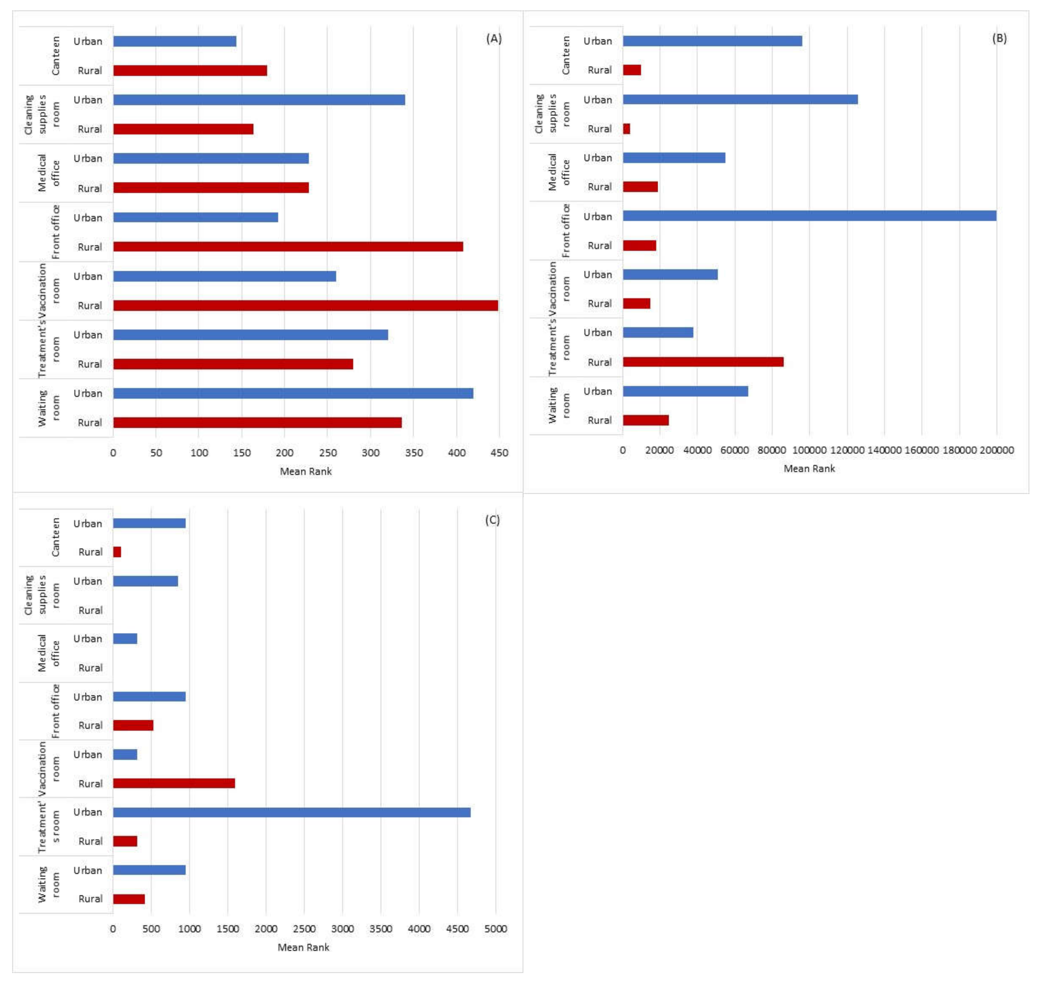
The data was analyzed in statistical software, SPSS 26.0 for Windows (Redmond, WA, USA). The results were considered significant at the 5% significance level. The Shapiro-Wilk test was used to test the normality of the data. Frequency analysis (n; %), and the calculation of the minimum, maximum and mean, were performed for the qualitative and quantitative data, respectively. Since the assumption of normality was not verified, the Mann-Whitney test was used to compare the rural and urban PHCC environment, regarding the total aerobic mesophilic bacteria and total coliforms. The Spearman correlation coefficient was used to study the relationship between total aerobic mesophilic bacteria, total coliforms and environmental parameters measured (temperature, relative humidity and CO2). To compare the settings, graphical representations were used, since the data did not allow any other type of analysis to be performed.

3. Results

3.1. Environmental Parameters

The indoor average values of CO2 and relative humidity were higher in the Rural PHCC. The Table 2 illustrates the results obtained for each of these parameters.

3.2. Culturable Bacteriota

Total aerobic mesophilic bacteria in air from the Rural PHCC ranged from 164 CFU.m−3 to 448 CFU.m−3 with a mean value of 292 CFU.m−3, while in the Urban PHCC ranged from 144 CFU.m−3 to 420 CFU.m−3 with a mean value of 272 CFU.m−3. The outdoor bacterial load was higher at the urban than in rural PHCC (164 CFU.m−3 vs. 12 CFU.m−3). Comparing the different PHCC areas, in the rural environment, the vaccination room and front office presented higher bacteria load, while in the urban environment, the waiting room, treatments room and cleaning supplies room were the ones presenting higher bacteria load (Figure 1A). Regarding the total coliforms load in the air, only the treatment room in the Rural PHCC and the cleaning supplies room in the Urban PHCC presented a reduced load (4 CFU.m−3).
In the surface samples of the Rural PHCC, the total aerobic mesophilic bacteria ranged from 4 × 10³ CFU.m−2 to 86 × 103 CFU.m−2 with a mean value of 25 × 103 CFU.m−2 and in the Urban PHCC ranged from 38 × 103 CFU.m−2 to 200 × 103 CFU.m−2 and with an average value of 90 × 103 CFU.m−2. In the Rural PHCC, the place with the highest aerobic mesophilic bacteria surface load was the treatment room. As for the Urban PHCC, the surfaces with the highest aerobic mesophilic bacteria load were in the front office, the cleaning supplies room and the canteen (Figure 1B). Regarding the total coliforms in surfaces it was only detected in the front office of the Rural PHCC and in the canteen of the Urban PHCC (1 × 10³ CFU.m−2 for both PHCC). Total aerobic mesophilic bacteria in HVAC filters were 2.5 × 10³ and 6 × 10³ CFU.m−2, from the Urban PHCC and the Rural PHCC, respectively. For this type of sample, no total coliforms contamination was observed.

3.3. Compliance of Bacteriota with IAQ Legal Criteria

Comparing the indoor air results with the Portuguese legal IAQ compliance criteria for bacteriota it was found that 29% (2 in 7 sampled areas analyzed, namely the vaccination room and the front office) of the Rural PHCC did not comply the first law criteria ((indoor) < (outdoor) + 350 CFU.m−3) and the second criteria (if (indoor) > (outdoor) + 350 CFU. m−3 and (CO2) < 1800 mg.m−3 than ratio between gram-negative bacteria and total bacteria should be ≤0.5) since the limits stipulated to CO2 value were exceeded. Concerning the Urban PHCC, all sampling sites analyzed were in total agreement with the IAQ law criteria.

3.4. Electrostatic Dust Collector Extraction

Total aerobic mesophilic bacteria from the Rural PHCC EDC samples ranged from <100 CFU.m−2 (detection limit) to 1592 CFU.m−2 with a mean value of 424 CFU.m−2, while in the Urban PHCC ranged from 318 CFU.m−2 to 4671 CFU.m−2 with an average value of 1289 CFU.m−2. In EDC samples, the areas with the highest aerobic mesophilic bacteria load were the vaccination room in the Rural PHCC and the treatment’s room in the Urban PHCC. Finally, in EDC, it was the urban environment that presented the highest bacterial loads in any of the settings (Figure 1C). As for the total coliforms, it was only detected in the Urban PHCC, in the waiting room and cleaning supplies room (106.16 CFU.m−2 and 530.79 CFU.m−2, respectively).

3.5. Settled Dust Sampling

Results for total aerobic mesophilic bacteria in settled dust samples were 1 CFU.g−1 in the Rural PHCC and 8 CFU.g−1 in the Urban PHCC. Total coliforms counts in settled dust samples was 1 CFU.g−1, the same value in both PHCC.

3.6. Characterization of Bacterial Isolates

Considering the different types of sample of this study, it was decided to only characterize the air samples due to their higher concentration and diversity, thus giving a general idea of the bacteriota existing in the indoor air of this kind of health care facilities.
Airborne Gram-positive cocci was the phenotype observed in more than half of the isolated bacterial population in the Rural and Urban PHCC, 53% and 63%, respectively. Micrococcus (21% Rural PHCC; 31% Urban PHCC), Kytococcus (22% Urban PHCC) and Staphylococcus (19% Rural PHCC) were dominant among the bacterial genera detected in both PHCC, as show in Table 3. The prevalent Gram-positive cocci included Staphylococcus (including S. aureus, S. auricularis, S. capitis subsp. capitis, S. cohnii ssp. cohnii, S. haemolyticus, S. hominis hominis, S. epidermidis, S. simulans, S. warner, S. uralyticus), Kytococcus (namely Kytococcus sedentarius) and Micrococcus. The treatment room, cleaning supplies room and vaccination room in the Rural PHCC and the treatment room, vaccination room and medical office from the Urban PHCC were the sampled areas with higher gram-positive cocci isolates.
Regarding the Heat-Map shown in Table 3, it presents a color gradation, which goes from green (lower frequency, lower risk) to red (higher frequency, higher risk).
Airborne Gram-negative cocci were also found with frequencies of 15% of the genus Moraxella (Moraxella catarrhalis and Moraxella osloensis) in the Rural and Urban PHCC air, and 13% of the genus Oligella (including Oligella ureolytica and Oligella urethralis) in Rural PHCC air. The medical office and the front office in the Rural PHCC and the waiting room and the cleaning supplies room from Urban PHCC were the sampled areas with higher gram-negative cocci isolates.
Gram-positive rods were only identified at lower frequency (1%) represented by the genus Brevibacterium in medical office in the Urban PHCC. The isolation rate of Gram-negative rods was slightly higher in the Rural PHCC compared to the Urban PHCC (4% and 3%, respectively). Yersinia pseudotuberculosis was detected in the waiting room in the Rural PHCC and Providencia alcalifaciens in the canteen of the Urban PHCC.

3.7. Antibiotic Susceptibility

The bacterial isolates tested for antibiotic susceptibility were selected considering its relative frequency among the detected airborne bacteria and the potential medical importance on healthcare associated infections [65,66]. Antibiotic resistance was detected among gram-positive cocci isolates. Staphylococcus (including S. capitis subsp. capitis, S. cohnii ssp. cohnii, S. haemolyticus, S. hominis hominis, S. epidermidis, S. simulans, S. warner, S. uralyticus) isolates showed resistance for Ticarcillin antibiotic in both PHCC. Neisseria gonorrhoeae isolates (from outdoor in Urban PHCC) and Yersinia pseudotuberculosis (from waiting room in the Rural PHCC) revealed resistance against Aztreonam. The antimicrobial resistance patterns of the isolates are shown in Table 4.

3.8. Detection of Targeted Bacteria Species

In both Rural and Urban PHCC it was not found isolates of Pseudomonas aeruginosa in the surfaces, EDC, dust and HVAC filters samples. Although isolates of S. aureus were detected in air samples, were not detected in the analyzed swab surfaces. Legionella sp. was not detected in the water from workers toilets showers and condensation water from the HVAC system tray.

3.9. Statistical Information

In the Rural PHCC, the following correlations were observed: (i) total aerobic mesophilic bacteria in air and total aerobic mesophilic bacteria in the EDC (rS = 0.937, p = 0.002), relative humidity (rS = −0.738, p = 0.037), and CO2 (rS = 0.857, p = 0.007), which suggests that higher aerobic mesophilic bacteria load in air is related to higher aerobic mesophilic bacteria contamination in the EDCs, with lower relative humidity and a higher concentration of CO2; (ii) total aerobic mesophilic bacteria in surface and relative humidity (rS = −0.821, p = 0.023), suggesting that higher aerobic mesophilic bacteria counts in surfaces is related to a lower relative humidity (Table 5).
Comparing rural and urban PHCC, statistically significant differences were detected in relation to: (i) total aerobic mesophilic bacteria in the surface (U = 4.000, p = 0.009), having presented in the Urban PHCC, higher aerobic mesophilic bacteria load; (ii) temperature (°C) (U = 0.000, p = 0.002), having the urban HPCC present higher temperature; (iii) carbon dioxide (U = 5.000, p = 0.013), having the Rural PHCC present higher concentration (Table 6).

4. Discussion

The most frequently available data on airborne microbial concentration refers to studies carried out in a hospital context, namely in operating rooms, lobbies [48], wards [49,67], operating theaters [42,68], surrounding areas, patient room and nursing care room [67,69,70]. However, there is a gap in evidence about microbiological assessment in primary health service facilities, albeit the potential impact on human health being comparable to that of hospital environment [71]. Both the characterization of microbial contamination on this type of health units, and the analysis of data in different geographical locations, were not yet subject of extensive investigation. Therefore, this study aims to highlight the bacterial profile in two types of health care centers, urban and rural. Health units serve the purpose of public health preservation, but they are also potential environments with negative effects and risks to human health and to the well-being of their users, workers and patients [3]. This effect is of particular concern for immunocompromised patients and other vulnerable populations, since those with a debilitated immune function are more susceptible to contract nosocomial infections [5,72].
The IAQ in healthcare facilities requires a higher degree of standards compared to other building types. Although the concern with indoor air was assumed in Portugal, with the creation of specific legislation for IAQ [52,53], it does not take in consideration the type of environment and the correlation between the clinical relevance/pathogenicity of the biological agent (bacteria) with the concentration observed in indoor air. Currently, the Portuguese legislation only defines a quantitative reference value for bacterial contamination in outdoor air quality, correlating it to all types of building and without reference to the kind of indoor environment. Furthermore, it appears that there are no adequate methodologies and reference standards for the exposure assessment of health professionals to biological risks [73].
The importance of applying different sampling methods allowing a more complete characterization of the exposure has already been mentioned in other studies [74,75,76], emphasizing the temporal variation as a factor to obtain a more accurate risk assessment. Thus, although there is a legal reference in Portugal on the assessment of IAQ through the use of air sampling as stand-alone method, passive methods (surface swabs, electrostatic dust cloth, vacuum settled dust and filters for HVAC systems) were also applied in this study, in order to assure a higher accuracy in the assessment of IAQ. Although passive sampling by vacuuming may be a limitation to assess the special and temporal variations, dust samples are easy to collect and may represent a long-term integrated sample of exposure to bacteria [77,78].
Our results show the same quantitative trend on results between active and passive methods. In fact, the vaccination room was the sampled area with the greatest bacterial contamination in Rural PHCC in both sampling methods (active-air and passive-EDC).
Although the treatment room presented the highest concentration in both passive methods, such as in the surfaces of Rural PHCC and in EDC of Urban PHCC, that was not the tendency in the active method. These results highlight the complementation of both types of sampling methods in IAQ assessment.
The indoor-to-outdoor bioaerosol concentration ratio (I/O) is used to estimate emissions sources of bioaerosol [79]. For situations in which I/O > 1.0, indoor air is considered the main source of indoor bioaerosols, and when I/O < 1.0, outdoor air is considered the main source of indoor bioaerosols. The I/O ratio in both Rural and Urban PHCC was superior to 1.0, only in the canteen from the Urban PHCC (0.8) was under, which suggests that air bacterial load may be derived from the indoor environment, following the trend of previous studies [1,3]. Indeed, the airborne microbial load from the Urban PHCC was under the limits recommended by IAQ Portuguese legislation, while in the Rural PHCC two sampled areas did not comply with the first and the second Portuguese legislation criteria. Moreover, the level of air bacteriota observed in both PHCC was like those described in the literature concerning similar sampled area from hospitals [1,9,48,80,81], highlighting the importance of assess bacteriota also in this type of health care facilities.
The identification of bacterial isolates was performed based on conventional methods through culture techniques and biochemical tests, following the same culturability assessment approach applied for the methodology of this study. Although having limitations (e.g., time consuming and lack to identify non-culturable microorganisms), culture and microscopy are still two of the most utilized techniques [82]. Molecular approaches (e.g., NGS, qPCR) are particularly important for non-culturable microorganisms, but need demanding equipment and resources. Considering the applied methodology, the bacterial phenotypic identification indicated that Gram-positive cocci were the most dominant group of genera (53% Rural PHCC and 63% Urban PHCC) identified in the air, followed by gram-negative cocci (37% Rural PHCC ad 20%/ Urban PHCC), gram-negative rods (4% Rural PHCC and 3% Urban PHCC) and gram-positive rods (1% Urban PHCC). Our results are consistent with previous studies [1,9,49,67,68,83], both in the rate of isolated bacterial species, and indicating Gram-positive cocci as the predominant microbial phenotype in hospital indoor air. The microorganisms found in hospital environments are diverse and can be defined based on their origin, some of these microorganisms can be traced to a human origin (skin and mucous membranes), including Staphylococcus aureus, Enterobacteriaceae or Enterococcus, whilst others are from environmental origin. From the latter, some have natural resistance to antibiotics, namely Gram-negative rods such as Pseudomonas aeruginosa, Acinetobacteriaceae, Burkholderia cepacia, Legionella pneumophilia, Stenotrophomonas maltophilia or atypical mycobacteria [80].
In fact, the airborne Gram-positive cocci (Staphylococcus and Micrococcus) were also the most detected bacterial genera in a sensitive ward of an academic pediatric hospital in Iran [84], in hospitals in India [85], in a hospital ward in India [67], in five educational Hospital wards air in Iran [86], in General Hospitals of Korea [87], in a regional Danish hospital [88] and in tertiary university hospital in NE Greece [89]. In our study, higher gram-positive cocci isolates were found in the treatment rooms and vaccination rooms of both PHCC, smaller spaces where they perform nursing treatments, shared between workers and patients, exposing them to both. The higher concentrations of Gram-positive cocci may be due to bioaerosol dispersal during skin shedding, their higher adaptability to environmental conditions, inadequate ventilation and occupancy rate [90,91].
In this study, even though the Urban PHCC has higher concentrations of bacteria in most types of samples, it was the Rural PHCC that presented the highest concentration of bacterial pathogens in the air. Different locations have their own unique bacterial communities, local sources of bacteria and the influence of other environmental factors that affect bacterial composition, diversity, abundance, and predominant genera [34]. The obtained results reflects that the compliance of quantitative results could not evidence the pathogenic potential of airborne bacterial population. Taking this in consideration, the legislation regarding Portuguese indoor air quality should be revised, not only to be focused on quantitative results, such as concentrations of bacteria, but also on the presence of specific bacterial species (e.g., Staphylococcus sp., Neisseria sp., Pseudomonas aeruginosa, Acinetobacter spp. Enterobacteriacea family members).
The most common indoor species documented are: Staphylococcus auricularius, S. capitis, S. epidermidis, S. haemolyticus, S. hominis, S. saprophyticus and S. warneri [92,93,94], all commonly found on human skin flora [95] and some with antibiotic resistant phenotypes and an opportunistic behavior as bacterial pathogens [96]. In our study, Staphylococcus (including S. capitis subsp. capitis, S. cohnii ssp cohnii, S. haemolyticus, S. hominis ssp hominis, S. epidermidis, S. simulans, S. warner, S. uralyticus) isolates showed resistance for Ticarcillin antibiotic in both PHCC. The ticarcillin antibiotic is used to treat infections of the skin and skin structure, infections of the lower respiratory tract, infections of the urinary tract and intra-abdominal. Ticarcillin is used combined with clavulanic acid, a beta-lactamase inhibitor, to eradicate some penicillin-resistant organisms [97]. Despite the fact that no MRSA were isolated in the analyzed samples, it is importance to notice that coagulase-negative staphylococci (CoNS) are opportunistic pathogens with the ability to produce several toxins, enzymes and enclose resistance genes, which can be further transferred to more virulent strains such as S. aureus [98]. It is relevant to acknowledge that, MRSA has acquired, via horizontal genetic transfer, the SCCmec, which is responsible for the resistance to β-lactam antibiotics [24,25]. In fact, molecular typing techniques have demonstrated that CoNS can enclose high levels of multidrug resistance that may be transferable to S. aureus [99] which is particularly concerning considering S. aureus ability to develop new clones with capacity to invade community settings [24].
Long-term care institutions were highlighted as important risk factors to emerging pathogens [100] in our study Pseudomonas aeruginosa it was not detected, probably due to the character more ambulatory use of this type of health facilities.
The absence of Legionella in the both PHCC, perhaps is due to the fact that in Portugal to minimize the spread of Legionella Pneumophila and the associated risk of Legionnaires’ Disease, it was established from 2017 the compliance with a strict program for the prevention and environmental control of the Legionella bacterium, in which measures for monitoring, surveillance, risk assessment and prevention and control (physical, chemical and microbiological) must be adopted to promote the adequate maintenance of the water and air systems in the health units [101].
It is recognized that contamination of surfaces with bacteria can increase air contamination by 15% [102]. Considering the obtained results, statistically significant differences were detected in relation to bacterial counts on the surface samples. Urban PHCC presented higher aerobic mesophilic bacteria counts than the Rural PHCC, which may be related to the cleaning procedures used for the correct disinfection of surfaces in Rural PHCC. Compared with previous studies, both PHCC results were higher than those assessed in other environments (86 CFU/25 cm2 in offices with synthetic floor, 120 CFU/25 cm2 in classrooms and 250 CFU/25 cm2 in training rooms) [103].
The higher values of contamination assessed by EDC were found in the vaccination room in Rural PHCC and in the treatment rooms of Urban PHCC. These two locations were also identified with higher indoor air contamination in both PHCC, showing a relationship in the sampling air at that time and in the long-term sampling since a single EDC analysis is equivalent to the sum of several air-impaction measurements [104].
Concerning settled dust samples, our results demonstrate a higher value for bacterial contamination in Urban PHCC, when compared with the Rural PHCC. However, these values were lower when comparing with other environments, namely in homes with a value averaging of 16 × 105 CFU.g−1 [105] and 2.9 × 10⁵ to 4.9 × 10⁷ CFU.g−1 [106], and in agricultural environments, with bacterial load ranging from 19 CFU.g−1 to 109 CFU.g−1 [107].
The positive correlation observed between temperature and aerobic mesophilic bacteria concentration in Urban PHCC, and the fact that higher temperatures may facilitate increased growth of bacteria in the environment, may be related to the higher aerobic mesophilic bacteria load in Urban PHCC [108]. Furthermore, the positive correlation detected between temperature, relative humidity and bacterial densities in air and surfaces are similar to the results observed in previous studies [3,109]. The use of the HVAC systems may increase the dispersion of all bacterial contamination that can be transmitted in the air. However, if efficient filters are used, 99.9% of all bacteria present in the air can be removed [108]. In our study, total mesophilic aerobic bacteria detected in the HVAC filters were higher in the urban PHCC compared to the rural PHCC, which may be related to the location of the outdoor air intakes. In the latter case, adjacent buildings and proximity to a roadway could be an increment of contamination. Therefore, it is crucial that the HVAC system in healthcare facilities maintains ideal temperature and humidity conditions for all its occupants, improving indoor air quality and decreasing the probability of infections [108].

5. Conclusions

In the present study, different PHCC geographic locations were compared and the environmental parameters that may influenced the levels of bacterial contamination were identified. Moreover, the Urban PHCC presented higher microbial airborne load than Rural PHCC. Our results demonstrated a higher concentration of bacterial pathogens in the air of Rural PHCC, with emphasis on the treatment room. The results also demonstrated that the vaccination rooms and front offices in Rural PHCC do not comply with the criteria established in the Portuguese IAQ legislation.
Our data emphasize the importance of a routine bacterial contamination assessment in Primary Health Care Centers, through a multi-approach sampling protocol that encompasses, besides air sampling, passive sampling methods for a more detailed exposure assessment and risk characterization. This will allow managing the risk aiming to reduce patients and healthcare workers exposure risk.

Author Contributions

Conceptualization, A.M., E.R., C.V., S.C.V.; Data curation, A.M.; Formal analysis, A.M., B.A., I.P., J.C.R., E.R., E.C., C.V., S.C.V.; Funding acquisition, C.V.; Investigation, A.M., C.V., S.C.V.; Methodology, A.M., I.P., J.C.R., E.R., C.V., S.C.V.; Project administration, C.V.; Resources, C.V., S.C.V.; Supervision, C.V., S.C.V.; Validation, A.M., E.R., S.C.V.; Writing—original draft, A.M.; Writing—review & editing, A.M., C.V., A.S.U., S.C.V. All authors have read and agreed to the published version of the manuscript.

Funding

This work was supported by FCT—Fundação para Ciência e Tecnologia for funding the project EXPOsE—Establishing protocols to assess occupational exposure to microbiota in clinical settings (02/SAICT/2016—Project No. 23222).

Institutional Review Board Statement

Not applicable.

Informed Consent Statement

Not applicable.

Data Availability Statement

Not applicable.

Acknowledgments

H&TRC authors gratefully acknowledge the FCT/MCTES national support through the UIDB/05608/2020 and UIDP/05608/2020. The FCT support is also acknowledged by C2TN/IST (UIDB/04349/2020þUIDP/04349/2020).

Conflicts of Interest

The authors declare no conflict of interest.

References

  1. Cabo Verde, S.; Almeida, S.M.; Matos, J.; Guerreiro, D.; Meneses, M.; Faria, T.; Botelho, D.; Santos, M.; Viegas, C. Microbiological assessment of indoor air quality at different hospital sites. Res. Microbiol. 2015, 166, 557–563. [Google Scholar] [CrossRef]
  2. Ferdyn-Grygierek, J. Monitoring of indoor air parameters in large museum exhibition halls with and without air-conditioning systems. Build. Environ. 2016, 107, 113–126. [Google Scholar] [CrossRef]
  3. Viegas, C.; Almeida, B.; Monteiro, A.; Aranha, L.; Carolino, E.; Gomes, A.Q.; Twarużek, M.; Kosicki, R.; Marchand, G.; Viegas, S. Bioburden in health care centers: Is the compliance with Portuguese legislation enough to prevent and control infection? Build. Environ. 2019, 160. [Google Scholar] [CrossRef]
  4. Zahar, J.R.; Jolivet, S.; Adam, H.; Dananché, C.; Lizon, J.; Alfandari, S.; Boulestreau, H.; Baghdadi, N.; Bay, J.-O.; Bénéteau, A.-M.; et al. Quelles mesures pour maîtriser le risque infectieux chez les patientsimmunodéprimés? Recommandations formalisées d’experts. J. Mycol. Med. 2017, 27, 449–456. [Google Scholar] [CrossRef]
  5. Leung, M.; Chan, A. Control and management of hospital indoor air quality. Med. Sci. Monit. 2006, 12, 17–23. [Google Scholar]
  6. Allegranzi, B.; Bagheri Nejad, S.; Combescure, C.; Graafmans, W.; Attar, H.; Donaldson, L.; Pittet, D. Burden of endemic health-care-associated infection in developing countries: Systematic review and meta-analysis. Lancet 2011, 377, 228–241. [Google Scholar] [CrossRef]
  7. Burke, J.P. Infection control—A problem for patient safety. N. Engl. J. Med. 2003, 348, 651–656. [Google Scholar] [CrossRef] [Green Version]
  8. McFee, R.B. Nosocomial or hospital-acquired infections: An overview. Dis. Mon. 2009, 55, 422–438. [Google Scholar] [CrossRef]
  9. Huff, W.E.; Kubena, L.F.; Harvey, R.B.; Doerr, J.A. Mycotoxin interactions in poultry and swine. J. Anim. Sci. 1988, 66, 2351–2355. [Google Scholar] [CrossRef] [Green Version]
  10. Kummer, V.; Thiel, W. Bioaerosols—Sources and control measures. Int. J. Hyg. Environ. Health 2008, 211, 299–307. [Google Scholar] [CrossRef]
  11. Dharmadhikari, A.S.; Mphahlele, M.; Stoltz, A.; Venter, K.; Mathebula, R.; Masotla, T.; Lubbe, W.; Pagano, M.; First, M.; Jensen, P.A.; et al. Surgical face masks worn by patients with multidrug-resistant tuberculosis: Impact on infectivity of air on a hospital ward. Am. J. Respir Crit. Care Med. 2012, 185, 1104–1109. [Google Scholar] [CrossRef] [PubMed]
  12. Bloch, A.B.; Orenstein, W.; Ewing, W.M.; Spain, W.H.; Mallison, G.F.; Herrmann, K.L.; Hinman, A. Measles outbreak in a pediatric practice: Airborne transmission in an office setting. Pediatrics 1985, 75, 676–683. [Google Scholar]
  13. Leclair, J.; Zaia, J.; Levin, M.; Congdon, R.; Goldmann, D. Airborne transmission of chickenpox in a hospital. N. Engl. J. Med. 1980, 302, 450–453. [Google Scholar] [CrossRef]
  14. Killingley, B.; Nguyen-Van-Tam, J. Routes of influenza transmission. Influenza Other Respir. Viruses 2003, 7, 42–51. [Google Scholar] [CrossRef] [PubMed]
  15. Kulkarni, H.; Smith, C.; Lee Ddo, H.; Hirst, R.; Easton, A.; O’Callaghan, C. Evidence of respiratory syncytial virus spread by aerosol. Time to revisit infection control strategies? Am. J. Respir. Crit. Care Med. 2016, 194, 308–316. [Google Scholar] [CrossRef]
  16. Warfel, J.M.; Beren, J.; Merkel, T.J. Airborne transmission of Bordetella pertussis. J. Infect. Dis. 2012, 206, 902–906. [Google Scholar] [CrossRef] [PubMed]
  17. Bonifait, L.; Charlebois, R.; Vimont, A.; Turgeon, N.; Veillette, M.; Longtin, Y.; Jean, J.; Duchaine, C. Detection and quantification of airborne norovirus during outbreaks in healthcare facilities. Clin. Infect. Dis. 2015, 61, 299–304. [Google Scholar] [CrossRef] [Green Version]
  18. Hara, S.; Yamamoto, H.; Kawabata, A.; Azuma, T.; Ishii, S.; Okumura, N.; Ito, Y. Airborne transmission from a neonate with Netherton syndrome during an outbreak of MRSA. Pediatr. Int. 2016, 58, 518–520. [Google Scholar] [CrossRef]
  19. Best, E.L.; Fawley, W.N.; Parnell, P.; Wilcox, M.H. The potential for airborne dispersal of Clostridium difficile from symptomatic patients. Clin. Infect. Dis. 2010, 50, 1450–1457. [Google Scholar] [CrossRef] [Green Version]
  20. Roberts, K.; Smith, C.F.; Snelling, A.M.; Kerr, K.G.; Banfield, K.R.; Sleigh, P.A.; Beggs, C.B. Aerial dissemination of Clostridium difficile spores. BMC Infect. Dis. 2008, 24. [Google Scholar] [CrossRef] [Green Version]
  21. Montagna, M.T.; De Giglio, O.; Napoli, C.; Cannova, L.; Cristina, M.L.; Deriu, M.G.; Delia, S.A.; Giuliano, A.; Guida, M.; Laganà, P.; et al. La contaminazione indoor da Legionella spp.: Risultati preliminari di una indagine multicentrica italiana [Legionella spp. contamination in indoor air: Preliminary results of an Italian multicenter study]. Epidemiol. Prev. 2014, 38, 62–65. [Google Scholar]
  22. Centers for Disease Control and Prevention. Available online: https://www.cdc.gov/legionella/wmp/overview/growth-and-spread.html (accessed on 28 March 2021).
  23. Herwaldt, L.A.; Marra, A.R. Legionella: A reemerging pathogen. Curr. Opin. Infect. Dis. 2018, 31, 325–333. [Google Scholar] [CrossRef] [PubMed]
  24. Lakhundi, S.; Zhang, K. Methicillin-resistant Staphylococcus aureus: Molecular characterization, evolution, and epidemiology. Clin. Microbiol. Rev. 2018, 31, e00020-18. [Google Scholar] [CrossRef] [Green Version]
  25. Peacock, S.; Paterson, G. Mechanisms of methicillin resistance in Staphylococcus aureus. Annu. Rev. Biochem. 2015, 84, 577–601. [Google Scholar] [CrossRef]
  26. Rolo, J.; Miragaia, M.; Turlej-Rogacka, A.; Empel, J.; Bouchami, O.; Faria, N.; Tavares, A.; Hryniewicz, W.; Fluit, A.; de Lencastre, H. High genetic diversity among community-associated Staphylococcus aureus in Europe: Results from a multicenter study. PLoS ONE 2012, 7, e34768. [Google Scholar] [CrossRef] [Green Version]
  27. Klevens, R.; Morrison, M.; Nadle, J.; Petit, S.; Gershman, K.; Ray, S.; Harrison, L.; Lynfield, R.; Dumyati, G.; Townes, J.; et al. Active bacterial core surveillance (ABCs) MRSA Investigators. Invasive methicillin-resistant Staphylococcus aureus infections in the United States. JAMA 2007, 298, 1763–1771. [Google Scholar] [CrossRef] [PubMed] [Green Version]
  28. Moradali, M.; Ghods, S.; Rehm, B. Pseudomonas aeruginosa lifestyle: A paradigm for adaptation, survival, and persistence. Front. Cell Infect. Microbiol. 2017, 7, 39. [Google Scholar] [CrossRef] [PubMed] [Green Version]
  29. Horcajada, J.P.; Montero, M.; Oliver, A.; Sorlí, L.; Luque, S.; Gómez-Zorrilla, S.; Benito, N.; Grau, S. Epidemiology and treatment of multidrug-resistant and extensively drug-resistant Pseudomonas aeruginosa infections. Clin. Microbiol. Rev. 2019, 32, e00031-19. [Google Scholar] [CrossRef] [PubMed]
  30. Bassetti, M.; Vena, A.; Russo, A.; Croxatto, A.; Calandra, T.; Guery, B. Rational approach in the management of Pseudomonas aeruginosa infections. Curr. Opin. Infect. Dis. 2018, 31, 578–586. [Google Scholar] [CrossRef] [PubMed]
  31. Ghosh, D.; Veeraraghavan, B.; Elangovan, R.; Vivekanandan, P. Antibiotic resistance and epigenetics: More to it than meets the eye. Antimicrob. Agents Chemother. 2020, 64. [Google Scholar] [CrossRef] [PubMed]
  32. Brągoszewska, E.; Biedroń, I. Indoor air quality and potential health risk impacts of exposure to antibiotic resistant bacteria in an office rooms in Southern Poland. Int. J. Environ. Res. Public Health 2018, 15, 2604. [Google Scholar] [CrossRef] [Green Version]
  33. Mbareche, H.; Morawska, L.; Duchaine, C. On the interpretation of bioaerosol exposure measurements and impacts on health. J. Air Waste Manag. Assoc. 2019, 69, 789–804. [Google Scholar] [CrossRef] [Green Version]
  34. Tanaka, D.; Fujiyoshi, S.; Maruyama, F.; Goto, M.; Koyama, S.; Kanatani, J.; Isobe, J.; Watahiki, M.; Sakatoku, A.; Kagaya, S.; et al. Size resolved characteristics of urban and suburban bacterial bioaerosols in Japan as assessed by 16S rRNA amplicon sequencing. Sci. Rep. 2020, 10, 12406. [Google Scholar] [CrossRef] [PubMed]
  35. Douglas, P.; Robertson, S.; Gay, H.A.; Gant, T. A systematic review of the public health risks of bioaerosols from intensive farming. Int. J. Hyg. Environ. Health. 2018, 221, 134–173. [Google Scholar] [CrossRef] [PubMed]
  36. Barberán, A.; Dunn, R.R.; Reich, B.J.; Pacifici, K.; Laber, E.B.; Menninger, H.L.; Morton, J.M.; Henley, J.B.; Leff, J.W.; Miller, S.L.; et al. The ecology of microscopic life in household dust. Proc. Biol. Sci. 2015, 282. [Google Scholar] [CrossRef] [PubMed]
  37. Carter, J. Enterically infecting viruses: Pathogenecity, transmission and significance for food and water borne infections. J. Appl. Microbiol. 2005, 98, 1354–1380. [Google Scholar] [CrossRef]
  38. National Academies of Sciences, Engineering and Medicine. Microbiomes of the Built Environment: A Research Agenda for Indoor Microbiology, Human Health, and Buildings; The National Academies Press: Washington, DC, USA, 2017; pp. 95–131. [Google Scholar]
  39. Kramer, A.; Schwebke, I.; Kampf, G. How long do nosocomial pathogens persist on inanimate surfaces? A systematic review. BMC Infect. Dis. 2006, 6. [Google Scholar] [CrossRef] [Green Version]
  40. Lax, S.; Smith, D.P.; Hampton-Marcell, J.; Owens, S.M.; Handley, K.M.; Scott, N.M.; Gibbons, S.M.; Larsen, P.; Shogan, B.D.; Weiss, S.; et al. Longitudinal analysis of microbial interaction between humans and the indoor environment. Science 2014, 345, 1048–1052. [Google Scholar] [CrossRef] [Green Version]
  41. Täubel, M.; Rintala, H.; Pitkäranta, M.; Paulin, L.; Laitinen, S.; Pekkanen, J.; Hyvärinen, A.; Nevalainen, A. The occupant as a source of house dust bacteria. J. Allergy Clin. Immunol. 2009, 124, 834–840.e47. [Google Scholar] [CrossRef] [PubMed]
  42. Scaltriti, S.; Cencetti, S.; Rovesti, S.; Marchesi, I.; Bargellini, A.; Borella, P. Risk factors for particulate and microbial contamination of air in operating theatres. J. Hosp. Infect. 2007, 66, 320–326. [Google Scholar] [CrossRef]
  43. Jung, C.; Wu, P.; Tseng, C.; Su, H. Indoor air quality varies with ventilation types and working areas in hospitals. Build. Environ. 2015, 85, 190–195. [Google Scholar] [CrossRef]
  44. Obbard, J.; Fang, L. Airborne concentrations of bacteria in a hospital environment in Singapore. Water Air Soil Pollut. 2003, 144, 333–341. [Google Scholar] [CrossRef]
  45. Qudiesat, K.; Abu-Elteen, K.; Elkarmi, A.; Hamad, M.; Abussaud, M. Assessment of airborne pathogens in healthcare settings. Afr. J. Microbiol. Res. 2009, 2, 66–76. [Google Scholar]
  46. Klánová, K.; Hollerová, J. Hospital indoor environment: Screening for micro-organisms and particulate matter. Indoor Built Environ. 2003, 12, 61–67. [Google Scholar] [CrossRef]
  47. Marchand, G.; Duchaine, C.; Lavoie, J.; Veillette, M.; Cloutier, Y. Bacteria emitted in ambient air during bronchoscopy-a risk to health care workers? Am. J. Infect. Control 2016, 44, 1634–1638. [Google Scholar] [CrossRef]
  48. Park, D.U.; Yeom, J.K.; Lee, W.J.; Lee, K.M. Assessment of the levels of airborne bacteria, Gram-negative bacteria, and fungi in hospital lobbies. Int. J. Environ. Res. Public Health 2013, 10, 541–555. [Google Scholar] [CrossRef]
  49. Tang, C.; Wan, G. Air quality monitoring of the post-operative recovery room and locations surrounding operating theaters in a medical center in Taiwan. PLoS ONE 2013, 8, e61093. [Google Scholar] [CrossRef]
  50. Viegas, C.; Ramos, C.; Almeida, M.; Sabino, R.; Veríssimo, C.; Rosado, L. Air fungal contamination in ten hospitals’ food units from Lisbon. WIT Trans. Ecol. Environ. 2011, 127–132. [Google Scholar] [CrossRef] [Green Version]
  51. World Health Organization. Air Quality Guidelines Global Update 2005; World Health Organization Regional Office for Europe: Copenhagen, Denmark, 2006. [Google Scholar]
  52. Ministério da Economia e do Emprego. Decreto-Lei No. 118/2013. Diário República 2013, 159, 4988–5005. [Google Scholar]
  53. Ministérios do Ambiente, Ordenamento do Território e Energia, da Saúde e da Solidariedade, Emprego e Segurança Social. Portaria No. 353-A/2013. In Diário da República; 1.a Serie—No. 235; República Portuguesa: Lisbon, Portugal, 2013. [Google Scholar]
  54. Viegas, C.; Almeida, B.; Monteiro, A.; Paciência, I.; Rufo, J.; Aguiar, L.; Lage, B.; Diogo Gonçalves, L.M.; Caetano, L.A.; Carolino, E.; et al. Exposure assessment in one central hospital: A multi-approach protocol to achieve an accurate risk characterization. Environ. Res. 2020, 181. [Google Scholar] [CrossRef] [PubMed]
  55. American National Standards Institute. ISO 18593: 2004, Microbiology of Food and Animal Feeding Stuffs, Horizontal Methods for Sampling Techniques from Surfaces Using Contact Plates and Swabs; American National Standards Institute: Washington, DC, USA, 2007. [Google Scholar]
  56. Viegas, C.; Faria, T.; Caetano, L.A.; Carolino, E.; Gomes, A.Q.; Viegas, S. Aspergillus spp. prevalence in different Portuguese occupational environments: What is the real scenario in high load settings? J. Occup. Environ. Hyg. 2017, 14, 771–785. [Google Scholar] [CrossRef] [PubMed]
  57. Bergey, D.; Krieg, N.; Holt, J. Bergey’s Manual of Systematic Bacteriology; Williams & Wilkins: Baltimore, MD, USA, 1984. [Google Scholar]
  58. Viegas, C.; Monteiro, A.; Aranha Caetano, L.; Faria, T.; Carolino, E.; Viegas, S. Electrostatic Dust Cloth: A passive screening method to assess occupational exposure to organic dust in bakeries. Atmosphere 2018, 9, 64. [Google Scholar] [CrossRef] [Green Version]
  59. Viegas, C.; Santos, P.; Almeida, B.; Monteiro, A.; Carolino, E.; Gomes, A.Q.; Viegas, S. Electrostatic dust collector: A passive screening method to assess occupational exposure to organic dust in primary health care centers. Air Qual. Atmos. Health 2019, 12, 573–583. [Google Scholar] [CrossRef]
  60. Viegas, C.; Almeida, B.; Monteiro, A.; Paciência, I.; Rufo, J.C.; Carolino, E.; Quintal-Gomes, A.; Twarużek, M.; Kosicki, R.; Marchand, G.; et al. Settled dust assessment in clinical environment: Useful for the evaluation of a wider bioburden spectrum. Int. J. Environ. Health Res. 2019, 26, 1–19. [Google Scholar] [CrossRef]
  61. Clinical and Laboratory Standards Institute (CLSI). Performance Standards for Antimicrobial Disk Susceptibility Tests, 13th ed.; CLSI Standard M02; Clinical and Laboratory Standards Institute: Wayne, PA, USA, 2018. [Google Scholar]
  62. Bauer, A.W.; Kirby, W.M.; Sherris, J.C.; Turck, M. Antibiotic susceptibility testing by a standardized single disk method. Am. J. Clin. Pathol. 1966, 45, 493–496. [Google Scholar] [CrossRef]
  63. EUCAST. Breakpoint Tables for Interpretation of MICs and Zone Diameters. Version 10.0. Available online: http://www.eucast.org (accessed on 10 November 2020).
  64. European Committee for Standardization. ISO 11731: 2017-Water Quality-Enumeration of Legionella; ISO: Geneva, Switzerland, 2017. [Google Scholar]
  65. Haque, M.; Sartelli, M.; McKimm, J.; Abu Bakar, M. Health care-associated infections—An overview. Infect. Drug Resist. 2018, 11, 2321–2333. [Google Scholar] [CrossRef] [PubMed] [Green Version]
  66. Yagupsky, P.; Katz, O.; Peled, N. Antibiotic susceptibility of Kingella kingae isolates from respiratory carriers and patients with invasive infections. J. Antimicrob. Chemother. 2001, 47, 191–193. [Google Scholar] [CrossRef] [Green Version]
  67. Sudharsanam, S.; Swaminathan, S.; Ramalingam, A.; Thangavel, G.; Annamalai, R.; Steinberg, R.; Balakrishnan, K.; Srikanth, P. Characterization of indoor bioaerosols from a hospital ward in a tropical setting. Afr. Health Sci. 2012, 12, 217–225. [Google Scholar] [CrossRef]
  68. Wan, G.H.; Chung, F.F.; Tang, C.S. Long-term surveillance of air quality in medical center operating rooms. Am. J. Infect. Control. 2011, 39, 302–308. [Google Scholar] [CrossRef]
  69. Baurès, E.; Blanchard, O.; Mercier, F.; Surget, E.; le Cann, P.; Rivier, A.; Gangneux, J.P.; Florentin, A. Indoor air quality in two French hospitals: Measurement of chemical and microbiological contaminants. Sci. Total Environ. 2018, 642, 168–179. [Google Scholar] [CrossRef]
  70. Dascalaki, E.; Lagoudi, A.; Balaras, C.; Gaglia, A. Air quality in hospital operating rooms. Build. Environ. 2008, 43, 1945–1952. [Google Scholar] [CrossRef]
  71. Karwowska, E.; Miaśkiewicz-Pęska, E.; Andrzejewska-Morzuch, D. Microbiological Air Contamination in Premises of the Primary Health-care. Arch. Environ. Prot. 2013, 39, 51–59. [Google Scholar] [CrossRef]
  72. Sobotova, L.; Noskova, T.; Volekova, J.; Aghova, L. Practical training on nosocomial infections in a hospital environment. Indoor Built Environ. 2006, 15, 73–76. [Google Scholar] [CrossRef]
  73. Bernasconi, C.; Rodolfi, M.; Picco, A.M.; Grisoli, P.; Dacarro, C.; Rembges, D. Pyrogenic activity of air to characterize bioaerosol exposure in public buildings: A pilot study. Lett. Appl. Microbiol. 2010, 50, 571–577. [Google Scholar] [CrossRef] [PubMed]
  74. Reponen, T. Sampling for microbial determinations. In Exposure to Microbiological Agents in Indoor and Occupational Environments, 1st ed.; Viegas, C., Viegas, S., Gomes, A., Täubel, M., Sabino, R., Eds.; Springer International Publishing: Berlin/Heidelberg, Germany, 2017; pp. 85–96. [Google Scholar]
  75. Viegas, C.; Faria, T.; Monteiro, A.; Caetano, L.A.; Carolino, E.; Quintal Gomes, A.; Viegas, S. A novel multi-approach protocol for the characterization of occupational exposure to organic dust-swine production case study. Toxics 2017, 6, 5. [Google Scholar] [CrossRef] [Green Version]
  76. Viegas, C.; Almeida, B.; Dias, M.; Caetano, L.A.; Carolino, E.; Gomes, A.Q.; Faria, T.; Martins, V.; Marta Almeida, S. Assessment of children’s potential exposure to bioburden in indoor environments. Atmosphere 2020, 11, 993. [Google Scholar] [CrossRef]
  77. Leppänen, H.; Täubel, M.; Jayaprakash, B.; Vepsäläinen, A.; Pasanen, P.; Hyvärinen, A. Quantitative assessment of microbes from samples of indoor air and dust. J. Expo. Sci. Environ. Epidemiol. 2018, 28, 231–241. [Google Scholar] [CrossRef] [PubMed]
  78. Giovannangelo, M.; Nordling, E.; Bellander, T.; Gerritsen, J.; de Jongste, J.; Smit, H.; Wichmann, H.; Wickman, M.; Brunekreef, B. Bacteria and mould components in house dust and children’s allergic sensitisation. Eur. Respir. J. 2007, 29, 1144–1153. [Google Scholar] [CrossRef] [Green Version]
  79. Viegas, C.; Dias, M.; Almeida, B.; Vicente, E.; Caetano, L.A.; Carolino, E.; Alves, C. Settleable dust and bioburden in portuguese dwellings. Microorganisms 2020, 8, 1799. [Google Scholar] [CrossRef] [PubMed]
  80. Kim, K.; Kim, C. Airborne microbiological characteristics in public buildings of Korea. Build. Environ. 2007, 5, 2188–2196. [Google Scholar] [CrossRef]
  81. Azimi, F.; Naddafi, K.; Nabizadeh, R.; Hassanvand, M.S.; Alimohammadi, M.; Afhami, S.; Musavi, S.N. Fungal air quality in hospital rooms: A case study in Tehran, Iran. J. Environ. Health Sci. Eng. 2013, 11, 30. [Google Scholar] [CrossRef] [Green Version]
  82. Franco-Duarte, R.; Černáková, L.; Kadam, S.; Kaushik, K.S.; Salehi, B.; Bevilacqua, A.; Corbo, M.R.; Antolak, H.; Dybka-Stępień, K.; Leszczewicz, M.; et al. Advances in chemical and biological methods to identify microorganisms-from past to present. Microorganisms 2019, 7, 130. [Google Scholar] [CrossRef] [PubMed] [Green Version]
  83. Asif, A.; Zeeshan, M.; Hashmi, I.; Zahid, U.B.M. Microbial quality assessment of indoor air in a large hospital building during winter and spring seasons. Build. Environ. 2018, 135, 68–73. [Google Scholar] [CrossRef]
  84. Mirhoseini, S.; Didehdar, M.; Akbari, M.; Moradzadeh, R.; Jarishidi, R.; Torabi, S. Indoor exposure to airborne bacteria and fungi in sensitive wards of an academic pediatric hospital. Aerobiologia 2020, 36, 225–232. [Google Scholar] [CrossRef]
  85. Sudharsanam, S.; Srikanth, P.; Sheela, M.; Steinberg, R. Study of the indoor air quality in hospital in South Chennai, India—Microbial profile. Indoor Built Environ. 2008, 5, 435–441. [Google Scholar] [CrossRef]
  86. Hoseinzadeh, E.; Samarghandie, M.; Ghiasian, S.; Alikhani, M.; Roshanaie, G. Evaluation of bioaerosols in five educational hospitals wards air in Hamedan, during 2011–2012. Jundishapur J. Microbiol. 2013, 6. [Google Scholar] [CrossRef] [Green Version]
  87. Kim, K.Y.; Kim, Y.S.; Kim, D. Distribution characteristics of airborne bacteria and fungi in the general hospitals of Korea. Ind. Health 2010, 48, 236–243. [Google Scholar] [CrossRef] [Green Version]
  88. Würtz, E.T.; Bønløkke, J.H.; Urth, T.R.; Larsen, J.; Islam, M.Z.; Sigsgaard, T.; Schlünssen, V.; Skou, T.; Madsen, A.M.; Feld, L.; et al. No apparent transmission of livestock-associated methicillin-resistant Staphylococcus aureus CC398 in a survey of staff at a regional Danish hospital. Antimicrob. Resist. Infect. Control 2017, 6, 1–8. [Google Scholar] [CrossRef] [Green Version]
  89. Selebonis, A.; Nena, E.; Panopoulou, M.; Kontogiorgis, C.; Bezirtzoglou, E.; Constantinidis, T. Air contamination in different departments of a tertiary hospital. Assessment of microbial load and of antimicrobial susceptibility. Biomedicines 2020, 8, 163. [Google Scholar] [CrossRef]
  90. Findley, K.; Oh, J.; Yang, J.; Conlan, S.; Deming, C.; Meyer, J.A.; Schoenfeld, D.; Nomicos, E.; Park, M.; Kong, H.H.; et al. Topographic diversity of fungal and bacterial communities in human skin. Nature 2013, 498, 367–370. [Google Scholar] [CrossRef]
  91. Mahy, B.W.J.; Meulen, V.T.; Borriello, P.S.; Murray, P.R.; Guido, F. Topley and Wilson’s Microbiology and Microbial Infections: Bacteriology-I, 10th ed.; American Society for Microbiology Press: Washington, DC, USA, 2005; pp. 185–194. [Google Scholar]
  92. Bonetta, S.; Bonetta, S.; Mosso, S.; Sampò, S.; Carraro, E. Assessment of microbiological indoor air quality in an Italian office building equipped with an HVAC system. Environ. Monit. Assess. 2010, 161, 473–483. [Google Scholar] [CrossRef] [PubMed]
  93. Kozajda, A.; Jeżak, K.; Kapsa, A. Airborne Staphylococcus aureus in different environments—A review. Environ. Sci. Pollut. Res. 2019, 26, 34741–34753. [Google Scholar] [CrossRef] [PubMed] [Green Version]
  94. Fraser, J.; Mwatondo, A.; Alimi, Y.; Varma, J.; Del Rio Vilas, V. Healthcare-associated outbreaks of bacterial infections in Africa, 2009–2018: A review. Int. J. Infect. Dis. 2021, 103, 469–477. [Google Scholar] [CrossRef]
  95. Robertson, C.E.; Baumgartner, L.K.; Harris, J.K.; Peterson, K.L.; Stevens, M.J.; Frank, D.N.; Pace, N.R. Culture-independent analysis of aerosol microbiology in a metropolitan subway system. Appl. Environ. Microbiol. 2013, 79, 3485–3493. [Google Scholar] [CrossRef] [PubMed] [Green Version]
  96. Takeuchi, F.; Watanabe, S.; Baba, T.; Yuzawa, H.; Ito, T.; Morimoto, Y.; Kuroda, M.; Cui, L.; Takahashi, M.; Ankai, A.; et al. Whole genome sequencing of Staphylococcus haemolyticus uncovers the extreme plasticity of its genome and the evolution of human-colonizing staphylococcal species. J. Bacteriol. 2005, 187, 7292–7308. [Google Scholar] [CrossRef] [PubMed] [Green Version]
  97. Castle, S. Ticarcillin in Pharm: The Comprehensive Pharmacology Reference. Enna, S.J., David, B., Eds.; Available online: https://www.sciencedirect.com/topics/pharmacology-toxicology-and-pharmaceutical-science/ticarcillin (accessed on 28 February 2021).
  98. Nasaj, M.; Saeidi, Z.; Asghari, B.; Roshanaei, G.; Arabestani, M.R. Identification of hemolysin encoding genes and their association with antimicrobial resistance pattern among clinical isolates of coagulase-negative Staphylococci. BMC Res. Notes 2020, 13, 68. [Google Scholar] [CrossRef] [PubMed] [Green Version]
  99. Ruiz-Ripa, L.; Feßler, A.T.; Hanke, D.; Sanz, S.; Olarte, C.; Mama, O.M.; Eichhorn, I.; Schwarz, S.; Torres, C. Coagulase-negative staphylococci carrying cfr and PVL genes, and MRSA/MSSA-CC398 in the swine farm environment. Vet. Microbiol. 2000, 243, 108631. [Google Scholar] [CrossRef] [PubMed]
  100. Tomczyk, S.; Zanichelli, V.; Grayson, M.; Twyman, A.; Abbas, M.; Pires, D.; Allegranzi, B.; Harbarth, S. Control of carbapenem-resistant Enterobacteriaceae, Acinetobacter baumannii, and Pseudomonas aeruginosa in healthcare facilities: A systematic review and reanalysis of quasi-experimental studies. Clin. Infect. Dis. 2019, 68, 873–884. [Google Scholar] [CrossRef] [PubMed] [Green Version]
  101. Jorge, R. Norma No. 24/2017, de 15 de Novembro; Direção Geral da Saúde e Instituto Nacional de Saúde Dr. Ricardo Jorge. Available online: http://www.aenfermagemeasleis.pt/2017/11/16/norma-dgs-insa-prevencao-e-controlo-ambiental-da-bacteria-legionella-em-unidades-de-saude/ (accessed on 28 February 2021).
  102. Hambraeus, A.; Bengtsson, S.; Laurell, G. Bacterial contamination in a modern operating suite. 3. Importance of floor contamination as a source of airborne bacteria. J. Hyg. 1978, 80, 169–174. [Google Scholar] [CrossRef] [PubMed] [Green Version]
  103. Bouillard, L.; Michel, O.; Dramaix, M.; Devleeschouwer, M. Bacterial contamination of indoor air, surfaces, and settled dust, and related dust endotoxin concentrations in healthy office buildings. Ann. Agric. Environ. Med. 2005, 12, 187–192. [Google Scholar]
  104. Institute of Medicine (US) Committee on Damp Indoor Spaces and Health. Damp Indoor Spaces and Health; National Academies Press: Washington, DC, USA, 2004; pp. 90–115. Available online: www.ncbi.nlm.nih.gov/books/NBK215643/ (accessed on 28 February 2021). [CrossRef]
  105. Horak, B.; Dutkiewicz, J.; Solarz, K. Microflora and acarofauna of bed dust from homes in Upper Silesia, Poland. Ann. Allergy Asthma Immunol. 1996, 76, 41–50. [Google Scholar] [CrossRef]
  106. Frankel, M.; Timm, M.; Hansen, E.W.; Madsen, A.M. Comparison of sampling methods for the assessment of indoor microbial exposure. Indoor Air 2012, 22, 405–414. [Google Scholar] [CrossRef] [PubMed]
  107. Shen, Y.E.; Sorenson, W.G.; Lewis, D.M.; Olenchock, S.A. Microbiological analyses and inflammatory effects of settled dusts from rice and hay. Biomed. Environ. Sci. 1990, 3, 353–363. [Google Scholar] [PubMed]
  108. Moscato, U.; Borghini, A.; Teleman, A. HVAC management in health facilities. In Indoor Air Quality in Healthcare Facilities; Capolongo, S., Settimo, G., Gola, M., Eds.; Springer: Berlin, Germany, 2017; pp. 95–106. [Google Scholar] [CrossRef]
  109. Eber, M.R.; Shardell, M.; Schweizer, M.L.; Laxminarayan, R.; Perencevich, E.N. Seasonal and temperature-associated increases in gram-negative bacterial bloodstream infections among hospitalized patients. PLoS ONE 2011, 6, e25298. [Google Scholar] [CrossRef] [PubMed] [Green Version]
Figure 1. Total aerobic mesophilic bacteria in air—CFU.m−3 (A), surfaces—CFU.m−2 (B) and EDC—CFU.m−2 (C) for sampled areas from rural and urban PHCC: Mann-Whitney test results.
Atmosphere 12 00450 g001
Table 1. Sampled performed and areas assessed in each PHCC.
PHCCAirSurfaceEDCHVAC Filters #Settled DustSampled Areas
urban77711vaccination and treatments rooms, medical office and waiting room, front office, cleaning supplies room and canteen
rural77711
# HVAC filters were collected after 6 months of use.
Table 2. Environmental parameters measured.
Environmental Parameters Measured
Sampled Areas | PHCCCarbon Dioxide (ppm)Temperature (°C)Relative Humidity (%)
RuralUrbanRuralUrbanRuralUrban
Waiting room1130.01731.8921.4425.6562.7557.03
Treatments room1120.87762.0622.0923.2559.9560.45
Vaccination room1442.24746.9222.0724.6361.4556.45
Front office1170.16658.4622.6324.1658.4559.18
Medical office1232.73787.7421.8525.3861.1656.54
Cleaning supplies room724.06844.4421.5725.2458.3859.18
Canteen905.67638.1522.1124.9059.2157.42
Outdoor398.48418.4318.5724.7060.2461.04
Table 3. Heat Map for frequency of airborne bacteria (%) in rural and urban PHCCs.
Bacterial Phenotype/GenusTotal RuralTotal Urban
Brevibacterium (Gp B) 1%
Hafnia alvei1%
Kingella kingae 2%
Kytococcus sedentarius7%22%
Macrococcus caseolyticus6%
Micrococcus sp.21%32%
Moraxella catarrhalis15%15%
Neisseria gonorrhoeae 1%
Neisseria havescen 0%
Neisseria meningitidis2%
Neisseria sicca/subflara2%
Oligella ureolytica1%
Oligella urethralis13%
Providencia alcalifaciens 3%
Psychrobacter phenylpyruvicus1%2%
Staphylococcus aureus 1%
Staphylococcus auricularis 1%
Staphylococcus capitis subs. capitis 5%
Staphylococcus cohnii ssp cohnii1%1%
Staphylococcus equorum1%
Staphylococcus haemolyticus1%
Staphylococcus hominis hominis5%
Staphylococcus simulans0.20%
Staphylococcus uralyticus10%
Staphylococcus warneri 2%
Yersinia pseudotuberculosis3%
Others10%11%
Table 4. Antimicrobial resistance of airborne bacterial isolates from Rural and Urban PHCC.
Bacterial Phenotype/GenusNo. Isolates (%) Antibiotics
ATMCTXGENIPMTICObservations
Gram-positive cocci
Staphylococcus capitis subs. capitis15 (2.8)NASSSRUrban PHCC, waiting room
Staphylococcus cohnii ss cohnii6 (1.2)NASSSRUrban PHCC, outdoor
5 (0.9)NASSSRRural PHCC, medical office
Staphylococcus equorum7 (1.3)NASSSRRural PHCC, treatment room
Staphylococcus haemolyticus4 (0.7)NASSSRRural PHCC, cleaning supplies room
Staphylococcus hominis hominis29 (5.4)NASSSRRural PHCC, canteen
Staphylococcus simulans1 (0.2)NASSSRRural PHCC, outdoor
Staphylococcus urealyticus55 (10.2)NASSSRRural PHCC, treatment room
Staphylococcus warneri9 (1.8)NASSSRUrban PHCC, vaccination room
Gram-negative cocci
Kingella kingae50 (10.2)NASNANANAUrban PHCC, waiting room
Neisseria gonorrhoeae5 (1.0)RNANANANAUrban PHCC, outdoor
Neisseria meningitidis11 (2.0)NASNANANARural PHCC, waiting room
Gram-negative rods
Yersinia pseudotuberculosis18 (3.3)RSSSSRural PHCC, waiting room
ATM Aztreonam, CTX Cefotaxime, GEN Gentamicin, IPM Imipenem, TIC Ticarcillin
NA = Not applicable, S = Sensitive, R = Resistant.
Table 5. Study of the relationship between total aerobic mesophilic bacteria, total coliforms (in air, surface and EDC) and environmental conditions (temperature, relative humidity and carbon dioxide concentration) in a rural and urban PHCC: results from Spearman’s correlation coefficient.
PHCCAirSurface EDC Indoor Environmental Conditions
Total ColiformsTotal Aerobic Mesophilic BacteriaTotal ColiformsTotal Aerobic Mesophilic BacteriaTotal ColiformsTemperature (°C)Relative Humidity (%)Carbon Dioxide (ppm—Mean)
AirTotal Aerobic Mesophilic BacteriaRural0.0820.3930.4080.937 **-0.214−0.738 *0.857 **
Urban0.412−0.250−0.6120.0560.757 *0.3570.0720.536
Total ColiformsRural0.612−0.1670.000-0.204−0.247−0.204
Urban0.408−0.167−0.2140.764 *0.2040.3090.612
SurfaceTotal Aerobic Mesophilic BacteriaRural 0.0000.216-−0.071−0.821 *0.321
Urban 0.2040.0190.3560.1790.234−0.286
Total ColiformsRural 0.412-0.612−0.4080.204
Urban 0.214−0.2550.0000.000−0.612
EDCTotal Aerobic Mesophilic BacteriaRural -0.306−0.4870.559
Urban −0.047−0.4300.774 *−0.412
Total ColiformsRural ---
Urban 0.5790.1350.401
Indoor Environmental ConditionsTemperature (°C)Rural 0.0000.071
Urban −0.5230.179
Relative Humidity (%)Rural −0.393
Urban 0.054
* Correlation is significant at the 0.05 level (2-tailed). ** Correlation is significant at the 0.01 level (2-tailed).
Table 6. Comparison of total aerobic mesophilic bacteria, total coliforms and environmental conditions (temperature, relative humidity and carbon dioxide concentration) between rural and urban PHCC: results of the Mann-Whitney test.
LocationNRanksTest Statistics
Mean RankSum of RanksMann-Whitney Up
AirTotal Aerobic Mesophilic BacteriaRural77.7954.5022.5000.798
Urban77.2150.50
Total14
Total ColiformsRural77.5052.5024.5001.000
Urban77.5052.50
Total14
SurfaceTotal Aerobic Mesophilic BacteriaRural74.5732.004.0000.009 *
Urban710.4373.00
Total14
Total ColiformsRural77.5052.5024.5001.000
Urban77.5052.50
Total14
EDCTotal Aerobic Mesophilic BacteriaRural75.5739.0011.0000.081
Urban79.4366.00
Total14
Total ColiformsRural76.5045.5017.5000.142
Urban78.5059.50
Total14
Indoor Environmental ConditionsTemperature (°C)Rural74.0028.000.0000.002 *
Urban711.0077.00
Total14
Relative Humidity (%)Rural77.3859.0023.0000.343
Urban79.6377.00
Total14
Carbon Dioxide (ppm—Mean)Rural710.2972.005.0000.013 *
Urban74.7133.00
14
* Statistically significant differences at 5% significance level.
Publisher’s Note: MDPI stays neutral with regard to jurisdictional claims in published maps and institutional affiliations.

