Article

Relationships between Exposure to Bioaerosols, Moldy Surface and Symptoms in French Mold-Damaged Homes

1
University of Caen Normandy, ABTE EA4651–ToxEMAC, Centre F. Baclesse, 14000 Caen, France
2
University of Rouen, CETAPS EA3832, 76000 Rouen, France
*
Author to whom correspondence should be addressed.
Atmosphere 2020, 11(3), 223; https://doi.org/10.3390/atmos11030223
Submission received: 6 February 2020 / Revised: 17 February 2020 / Accepted: 21 February 2020 / Published: 25 February 2020
(This article belongs to the Special Issue Detection and Monitoring of Bioaerosols)

Abstract

:
Air quality in homes is a major concern in Europe, where people spend most of their time indoors. According to the World Health Organization, numerous houses are subject to dampness that can lead to mold growth, with associated health and economic consequences. Our goal was to characterize the human exposure to bioaerosols in French mold-damaged houses but also to study the effects of these bioaerosols as suffered by the inhabitants of these houses. A global approach including both field study and laboratory experimentation was used to investigate 48 mold-damaged homes. Among a wide fungal diversity, 101 viable species, Aspergillus versicolor, Penicillium chrysogenum and P. crustosum were observed as recurrent species and could be used as microbial indicators of indoor air quality. Statistical analyses highlighted a relationship between the concentrations of these recurrent molds and the levels of surface contamination by molds in homes. Fever, cough, dyspnea, flu-like symptoms were observed with several fungal strains (A. versicolor, P. chrysogenum and P. crustosum) or in relation to moldy odor. Relationships between particles of 2 to 15 µm diameter and headaches and dizziness were also observed. In our study, we identified a cutaneous effect (itching) in relationship to the airborne concentration of A. versicolor.

1. Introduction

The quality of indoor air has recently become a concern in European countries, where people spend as much as 80% to 95% of their time indoors, and breathe around 10 m3 of air every day [1]. Amongst all the air pollutants, biological contaminants (molds, pollens, endotoxins) constitute a diversified group of airborne particles which is now referred to as bioaerosol [2].
Molds are filamentous fungi with focus of attention for health agencies, as high concentrations can be observed in damaged buildings [3]. A recent report estimated that 20% of European houses may be concerned by mold degradation causing health and economic damage [4,5,6]. Molds can produce high quantities of spores that can stay in the air over extended periods of time [7]. In addition, fungal fragments are also released and aerosolized from fungal growth. These mycelium fragments constitute smaller-diameter particles which can penetrate further in human airways. A recent study showed that these mycelium fragments may be produced in larger quantities than spores [8]. Both spores and mycelium fragments contain β-D-glucans, which are components of the cell wall that display pro-inflammatory effects [9]. Finally, molds are also known to produce some secondary metabolites named mycotoxins, such as sterigmatocystin, which is a carcinogenic and a cytotoxic mycotoxin known to be produced by Aspergillus versicolor [10].
Exposure to dampness and mold is associated with increased risk of asthma development and exacerbation, as well as dyspnea, cough, wheeze, respiratory infections, eczema and respiratory symptoms for allergic and non-allergic people [11]. Although some microbial species, such as bacteria, are suspected to have a protective effect against asthma development and exacerbation, it is not the case for mold exposure [12]. Inflammatory effects of fungal bioaerosols were also observed in experimental animal studies [13].
The study of human exposure in mold-damaged houses proves to be challenging both because of technical problems and of access to mold-damaged houses. In case of high concentration of molds, collection of bioaerosols by direct impaction may not be possible as the agar plates would be overloaded. Moreover, the short sampling time and the type of agar medium used for impaction do not allow analysis of the whole diversity of the species in these homes [14]. Improved metrics are needed to evaluate exposure to molds [15].
In our study, we applied an experimental approach based on the impingement of bioaerosols from mold-damaged dwellings into liquid, quantification of several microbial contaminants and collection of the symptoms felt by inhabitants of these mold-damaged houses.

2. Experiments

2.1. House Selection and Bioaerosol Collection

The study was based on 48 houses located in Normandy in which apparent mold growth was observed. The sampling frame took place between November 2015 and March 2018. Among the 48 houses of this study, 30 were apartments amongst which 21 of them were located in urban area and 9 in suburban area. The 18 other residences were individual houses which were located in urban areas (n = 2), suburban areas (n = 9) and rural areas (n = 7). These houses were selected by local partners: safety service of the city of Caen, health advisers of indoor environments and members of tenant associations.
The sampling took place in the center of the room the most contaminated by the mold growth, 1 meter above the ground. During sampling, no activities occurred indoor. Surface contamination level was evaluated by the investigator, by observation of visible molds and was classified as low (inferior to 0.2 m2), moderate (between 0.2 to 3 m2) or high (superior to 3 m2) as proposed by the French Agency for Food, Environmental and Occupational Health & Safety (ANSES) [3].
A cyclonic force sampler, Coriolis® µ (Bertin, France), was used to take air samples at a flow rate of 300 L min−1. A total of 4 samples was successively taken at the same place in each house. Each sample was collected during 10 min at 300 L min−1 with a starting volume of collection liquid of 15 mL of sterile water containing 0.02% of Tween 80 (Sigma-Aldrich, Darmstadt, Germany). At the end of the sampling, the 4 samples were pooled and the combined volume was measured.
This cyclonic force impinger was selected according to previous studies, in order to sample a large volume of air in mold-damaged homes [16]. Depositing the bioaerosols into a collection liquid allowed us to dilute this sampling liquid, avoiding the saturation of agar plates. It also allowed us to make different analyses from the bioaerosols sampled, such as β-D-glucans and endotoxins quantifications.
A Grimm particles counter (Model 1.108, Grimm Technologies, Inc., Douglasville, GA, USA) was also used to count particles from 0.3 to 20 µm in diameter, and to monitor the temperature and relative humidity during the sampling.

2.2. Quantification of Biological Contaminants

For each home, an aliquot of 1 mL of collection liquid and dilutions (10−1; 10−2; 10−3) were deposited into Petri plates, and malt extract agar (MEA) (Merck, Darmstadt, Germany) supplemented with chloramphenicol (0.02 g L−1) (Cooper, USA) was poured. Plates were incubated at 25 °C for 7 days, checked daily, and the number of colonies was counted and reported as Colony Forming Units per m3 of air (CFU m−3). Each different fungal colony was isolated and purified in MEA medium. Species belonging to Aspergillus and Penicillium genera were also cultured on Czapek yeast autolysate (CYA) and 25% glycerol nitrate (G25N) media.
Identifications through macroscopic and microscopic observations were based on various reference books [17,18,19,20,21,22,23,24] and comparison with fungal strains of the mold collection of the laboratory. When microscopic identifications were not sufficient, the DNA of pure fungal cultures was extracted using the Nucleospin® Plant II kit (Macherey-Nagel, France). Internal transcribed spacer (ITS) and beta-tubulin gene regions were then sequenced by GATC (Eurofins, Hamburg, Germany) using ITS1/ITS4 primers and Bt2a/Bt2b [25].
Endotoxins and β-glucans were quantified from the collection liquid using LAL test (Charles River Laboratories, Wilmington, MA, USA).

2.3. Questionnaire

Inhabitants were interviewed in order to complete a questionnaire about their health status (symptoms recorded in their homes) and the characteristics of their house (Table 1). This questionnaire was completed by the house investigator in the presence of the inhabitants. A total of 48 questionnaires were collected during this study.

2.4. Statistical Analysis

Descriptive statistics (frequency, mean and standard deviation, median, minimum and maximum) were used to determine the prevalence rates of microbial contaminants. Exploratory analyses were conducted to examine the relationship by using chi-square test, Fisher exact test, Mann–Whitney’s test, Kruskall–Wallis test and Spearman correlation coefficient as appropriate. Only results at p < 0.05 were considered statistically significant. All statistical analyses were executed using SAS system version 9.3 (SAS Institute Inc. Cary, NC, USA).

3. Results

3.1. Characteristics of Bioaerosols

The temperature, relative humidity and concentration of particles from 0.3 to 20 µm in these houses are shown in Table 2. The surface contaminated by visible molds was low (<0.2 m2) in 16.7% (n = 8) houses, moderate (between 0.2 to 3 m2) in 60.4% (n = 29) houses and high (>3 m2) in 22.9% (n = 11) houses.
Levels of contamination by viable molds ranged from 1.67 × 10 to 3.61 × 10⁵ CFU m−3 of air, with a mean value of 1.02 × 10⁴ CFU m−3 and a standard deviation of 5.33 × 10⁵ CFU m−3 (Figure 1).
A total of 101 different fungal species were identified and quantified from bioaerosols. Pielou evenness index ranged from 0.09 to 0.95, with a mean value of 0.69 and a standard deviation of 0.14. Table 3 presents the fungal species observed in bioaerosols at a frequency above 10%.
Other species belonging to different genera listed below were also identified in less than 10% of houses: Absidia, Acremonium, Actinomucor, Alternata, Aphanocladium, Arthrinium, Chaetomium, Chrysonilia, Chrysosporium, Cryptococcus, Emericella, Fusarium, Gliocladium, Hyphodermella, Mucor, Mycotypha, Naganishia, Paecilomyces, Phanerochaete, Phoma, Pithomyces, Rhizopus, Scedosporium, Stachybotrys, Talaromyces, Thamnidium, Trichoderma and Ulocladium.
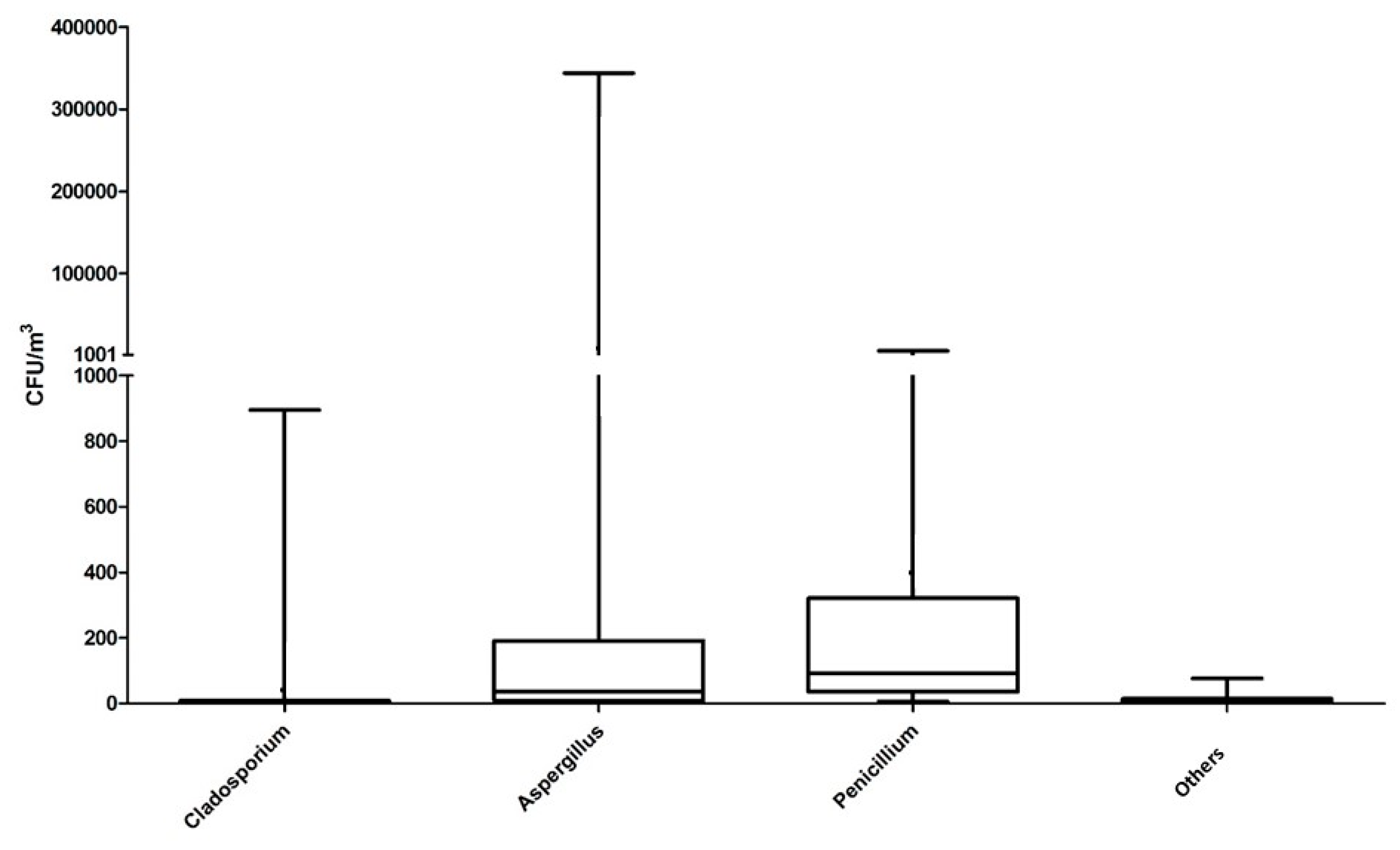
Three fungal groups were dominant in this study both for their recurrence and their concentration in bioaerosols: Aspergillus, Penicillium and Cladosporium (Figure 2).
The highest concentration of molds in bioaerosols was observed for A. versicolor (with a mean value of 9.3 × 10³ CFU m−3), which was also the dominant mold in the houses with high levels of airborne molds. The second highest concentration was observed for P. chrysogenum (2.4 × 10² CFU m−3). In the houses where P. chrysogenum was not identified, other species such as P. glabrum or P. expansum were observed. The P. brevicompactum, P. crustosum and C. cladosporioides molds were also present at non-negligible concentrations in most bioaerosols, with respective mean values of 3.8 × 10, 3.8 × 10 and 3.3 × 10 CFU m−3. All the others species we identified were observed at a lower mean concentration.
The detailed list of all quantified species with their mean and median concentrations in bioaerosols is presented in Table S1.
Endotoxin concentration in the air ranged from 1.45 to 911.25 Endotoxin Unit (EU) m−3 of air, with a mean value of 132.74 EU m−3, a median value of 57.88 EU m−3 and a standard deviation of 183.53 EU m−3.
(1-3)-β-D-glucans concentration ranged from 0.16 to 15.48 ng m−3 of air, with a mean value of 1.56 ng m−3, a median value of 1.05 ng m−3 and a standard deviation of 2.21 ng m−3. Glucans were not correlated with total levels of molds (p = 0.6901) and nor with A. versicolor (p = 0.5671), A. fumigatus (p = 0.9407), P. brevicompactum (p = 0.3207), P. chrysogenum (p = 0.6645) and P. crustosum (p = 0.7067).

3.2. Health Status of Inhabitants

The frequency and intensity of the symptoms mentioned by the inhabitants are presented in Table 4. The most frequently encountered symptoms were clogged nose and dyspnea.

3.3. Statistical Analyses from Microbial and Health Data Collected in Mold-Damaged Homes

3.3.1. Correlations

First, we searched correlation factors between biological contaminants, such as the total fungal concentration in air (total CFU) or the most recurrent molds in bioaerosols (A. versicolor, A. fumigatus, Penicillium brevicompactum, P. chrysogenum and P. crustosum), and different parameters measured like the level of contaminated surface, the endotoxins and β-D-glucans concentrations in air, and other physical data like the concentration of airborne particles between 0.3 to 20 µm and between 2 to 15 µm, and the temperature and relative humidity.
Some correlations were statistically significant:
Between the total fungal concentration in the air and the concentration of A. versicolor (rS = 0.603/p < 0.0001), as well as P. chrysogenum (rS = 0.469/p < 0.0008).
Between the concentration in the air of A. versicolor and P. chrysogenum (r = 0.476/p < 0.0006).
Between the concentration of A. versicolor in the air and the concentration of particles of 0.3 to 20 µm (rS = 0.333/p = 0.0205), the concentration of particles of 2 to 15 µm (r = 0.339/p= 0.0181) and the relative humidity (rS = 0.420/p = 0.0029).
Between the concentration of P. crustosum in the air and the concentration of particles of 2 to 15 µm (rS = 0.462/p = 0.0009).
Between the concentration of P. chrysogenum in the air and the relative humidity (rS = 0.343/p = 0.0168) as well as the temperature (rS = 0.299/p = 0.0385).

3.3.2. Explanatory Variables

To explain the airborne fungal contamination, we searched relations between the recurrent fungal species (A. versicolor, A. fumigatus, Penicillium brevicompactum, P. chrysogenum and P. crustosum) in bioaerosols and the different categories of mold-contaminated surfaces (Table 5).
A relationship was found between the different categories of mold-contaminated surfaces and several fungal constituents of bioaerosols: the total number of viable molds (total CFU), the concentration of A. versicolor and the concentration of P. chrysogenum in the air. In our study, higher contaminated surfaces corresponded to the higher median concentrations for these airborne molds.
We also searched for explanatory variables of the different symptoms as felt by inhabitants according to the different parameters measured in the houses like the concentrations of recurrent airborne molds, the surface of visible contaminated surface, the moldy odor, etc. Only significant results (p < 0.05) are shown in Table 6, Table 7 and Table 8.
We observed relationships between the symptoms felt by the inhabitants of mold-damaged homes and the concentrations in air of the most recurrent fungal species (Aspergillus versicolor, Penicillium chrysogenum and P. crustosum) except for P. brevicompactum. Relationships were also found between these symptoms and the physical parameters of bioaerosols, such as the relative humidity or the concentration of particles of 2 to 15 µm of diameter. The level of mold-contaminated surface and the moldy odor were also related to several symptoms like dyspnea and headache.

4. Discussion

We found different levels of fungal contamination during this field study. The median fungal concentration of viable spores in the air was 2.76 × 10² CFU m−3. Overall, this range of fungal contamination in air was similar to the levels already described in mold-damaged houses [16,26,27]. The levels observed in our study increased above 103 CFU m−3 of air in 9 homes. In a previous study, Trout et al. [28] reported a case of chronic lung disease in a worker at this concentration of culturable airborne molds. In comparison, a median level of 18.75 CFU m−3 has been observed in houses without visible molds (unpublished data). Chao et al. [29] investigated office building without mold degradation and observed similar fungal concentrations, with a median value of 21.53 CFU m−3.
We identified in bioaerosols the 3 recurrent fungal genera (Aspergillus, Penicillium and Cladosporium) which are known to be present in water-damaged houses, although in our study, Cladosporium was not the most prevalent fungal genus. This observation may not be due to the type of sampler used, as it was successfully used to collect Cladosporium in a previous study in indoor environments [16]. Alternaria species were also almost absent in our samples (identified in only one bioaerosol), although this genus has been described to be prevalent in other cultural studies on bioaerosols collected in houses of asthmatic patients [26,30]. A significant correlation between relative humidity and the concentration of some recurrent molds (A. versicolor and P. chrysogenum) was observed. Relationships between humidity and microbial contaminants has already been described from airborne dust [31] as well as settled dust [32], but the effect of relative humidity on sporulation and aerosolisation of fungal particles was different according to the fungal species [33]. As explained by Wolkoff [34], indoor air humidity appears as a meaningful indicator of air quality.
Penicillium chrysogenum was the most recurrent species, identified in 75% of the residences. This species was already known to be recurrent in indoor air [26], and showed cellulolytic properties [35] that could explain its growth in mold-damaged houses. Some other species belonging to the Penicillium genus were found to be recurrent in mold-damaged houses: P. crustosum was identified in 67% of our samples. This species was described in aerosols from houses [36], but was also observed in the air of other environments like composting plants and vegetal wastes [37,38], and was shown to be able to produce two mycotoxins: roquefortin C and penitrem A [39]. P. brevicompactum was also identified in 63% of our samples, and has been identified in several indoor environments and vegetal wastes. This species was known to produce several mycotoxins including, mycophenolic acid, an immunosuppressive mycotoxin [40]. P. glabrum was identified in 40% of the mold-damaged houses and constituted a fungal species which was able to degrade plants and fruits and could produce patulin. This mold has been previously identified in composting plants [18,37].
Among the Aspergillus genus, A. versicolor was the most recurrent species, identified in 73% of the mold-damaged houses, and represented the second most recurrent fungal species after P. chrysogenum, as well as the dominant species in the most heavily contaminated houses. This species was able to produce a potent mutagenic mycotoxin, named sterigmatocystin. Jussila et al. [41] have also showed that injection in mice of high concentration of spores of this species caused inflammatory responses. Among this genus, A. fumigatus and A. flavus were also somewhat recurrent, identified in 42% and 21%, respectively. These two species were known as the main cause for invasive respiratory aspergillosis in people with a weakened or compromised immune system, and were able to produce some mycotoxins [42].
Other species such as Botrytis cinerea, Chaetomium globosum, Paecilomyces variotii and Rhodotorula mucilaginosa were identified in more than 10% of mold-damaged houses. Different species belonging to Botrytis and Chaetomium genera have also been identified in houses located in Belgium [26] and have shown cellulolytic properties [18].
Some other species were known to be potentially infectious or toxic such as Cryptococcus albidus or Stachybotrys chartarum [43,44]. We also identified some molds that were able to degrade lignin and/or cellulose, and that might be involved in the degradation of the buildings, such as the Chaetomium or the Trichoderma species [45].
In regards to the technical aspects, a recent study showed that the use of the Coriolis® collector may underestimate the number of CFU for fungal spores [46]. This could mean that the fungal concentration recorded in our study may be below the real concentration of these fungal species in air. Several studies already documented the fungal content of bioaerosols in houses using cultural approach [16,26,27,30,47,48,49]. Most of these studies have used impaction onto agar, which are not suited to highly contaminated environments as agar plates will be quickly saturated by molds. The Coriolis® allows researchers to collect particles in a liquid medium used for serial dilutions and different analyses using various techniques.
In our study, we used a cultural method in order to investigate the viable fungal content of bioaerosols because this method allows to observe a large diversity of indoor molds. Other tools could be used to quantify molds, like quantitative polymerase chain reaction (qPCR) which allows a fast quantification for both lives and dead fungal spores and hyphae [50,51], but was restricted by the specificity of the primers to a fungal species or a fungal group. Studies have shown correlations between qPCR and cultural methods for evaluation of contamination by indoor fungi [52,53]. Recently, a next-generation sequencing study was used to quantify molds and bacteria in a waste composting facility and allowed the bacterial or fungal diversity of bioaerosols to be assessed and revealed genera that were missed by the cultural approach [54,55]. Although this approach seems promising, some disadvantages occur like difficulties in data analysis and low-abundance communities study.
The statistical analyses performed in this pilot study showed that several variables were found to be linked to the symptoms felt by the inhabitants. Most studies about mold exposure highlighted respiratory symptoms [4]. In our study, we also observed a cutaneous effect (itching) in relationship to the airborne concentration of A. versicolor, which has not been previously described to our knowledge. Although skin reactions have previously been described (atopic dermatitis, eczema), the association with a mold exposure was not demonstrated [56,57]. The relationship observed between itching and A. versicolor seems not to be monotonic (the concentration of A. versicolor only increased for strong symptoms). This could be explained by the existence of a threshold level of contamination above which the symptom could appear.
For A. versicolor, relationships with dizziness, fever, headache and expectorations were also found. In all cases, median concentrations of this mold were higher when the symptom was reported than in the absence of symptoms. Takigawa et al. [58] previously found a correlation between the concentration of Aspergillus species with the Sick Building Syndrome (SBS), which includes symptoms such as irritation of eyes, nose and throat as well as fatigue. Central nervous symptoms could be due to volatile organic chemicals emitted from mold and building materials which are known to cause headaches, inability to concentrate, or dizziness [59]. In our study, headaches were present in the analyses with several indicators of fungal contamination like A. versicolor, P. crustosum, mold-contaminated surface and moldy odor. We also found a relationship between the musty odor in house and the frequency of reports for muscular pain, expectorations, flu-like symptoms, dyspnea and anxiety. Herneberg et al. [60] previously observed a lower forced expiration volume and forced vital capacity for non-asthmatic adults exposed to moldy odor in comparison to non-asthmatic adults living in mold-free houses.
Fever, cough, dyspnea, flu-like symptoms were observed with several fungal strains (A. versicolor, P. chrysogenum and P. crustosum) or in relation to moldy odor. These symptoms can be observed in hypersensitivity pneumonitis which is associated with Immunoglobulin G (IgG) and T cell response [61].
The relationship between P. crustosum and insomnia could be explained by other studies which have found that mold or dampness was related to a high prevalence of sleep problems [62,63]. For the four symptoms (cough, fever, headache and insomnia) observed with P. crustosum, the median airborne concentrations were observed at higher extents when the symptoms were reported. Among the recurrent species of the Penicillium genus, no relationship was found between the concentrations of P. brevicompactum and the symptoms evaluated in this study.
Results on expectorations are difficult to interpret. On the one hand, the concentrations of A. versicolor and P. chrysogenum were higher when expectorations were reported. On the other hand expectorations were shown to be lower when the mold-contaminated surface was evaluated as moderate or high. This could be explained by the progressive decrease of expectoration capacities due to chronic exposure to molds [64].
For the evaluation of the mold-contaminated surface, a relationship was found with the frequency of report for headache, expectorations and dyspnea. All these symptoms, with the exception of dyspnea, were also found to be related to exposure to A. versicolor. This result was in agreement with the correlation factor that was found between the width of the contaminated surface and the concentration of Aspergillus versicolor in bioaerosols. Platt et al. [65] have examined the relation between damp and mold growth and symptomatic ill health. In their study, adult respondents living in damp and moldy homes reported more symptoms like blocked nose and dyspnea than respondents in homes without damp and mold. The children living in damp and moldy homes had a greater prevalence of respiratory symptoms (wheezing, sore throat, persistent cough) and other symptoms like headache and fever compared with those living in housing conditions without damp and mold. In our study we observed relationships between sore throats and relative humidity, and also between headaches and dyspnea with the mold-contaminated surface greater than 0.2 m2 and 3 m2 (respectively considered as moderate and high categories).
Relationships between particles of 2 to 15 µm diameter and headache and dizziness were observed. We noted that high concentrations of particles of 2 to 15 µm were associated with reports of this symptom by the inhabitants. We also found that these symptoms were also correlated with the concentrations of A. versicolor and P. crustosum in air, two species that were also correlated with this concentration of the particles of 2 to 15 µm. In a previous study, a relationship between the total fungal concentration and the concentration of particles of 2 to 15 µm in air had been observed concerning air samples collected from houses damaged by the wood-rotting fungi Serpula lacrymans [16].
In a meta-analysis, Sharpe et al. [66] identified a relationship between different fungal species of Aspergillus and Penicillium genera and asthma exacerbation. We did not find any association between molds and asthma aggravation in our study but this could probably be due to the low number of asthmatic patients (n = 11) observed in our study.
Concerning the concentration of endotoxins in air, relations were observed with the frequency of flu-like symptoms and dyspnea. Several studies have investigated the protective or detrimental effects of exposure to endotoxins in different places [67,68]. In our study, median concentrations of endotoxins in the air were lower when the symptom was present than when it was absent. This observation may suggest a protective effect of endotoxin exposure in mold-damaged houses as proposed by Douwes [67] for the exposure of children in farms.
Finally, we did not find relationships between the β-D-glucans concentration in air and the different health symptoms documented in our study. We did not observe important variations of the concentration of β-D-glucans in bioaerosols, only one high value at 15.48 ng m−3 of air was noted in a rural home located near a forest.

5. Conclusions

This study allowed us to provide the fungal profile of bioaerosols in mold-damaged buildings located in Normandy (France), a region characterized by a wet temperate oceanic climate. The identification of more than 100 species from bioaerosols showed a wide diversity of indoor fungi. Statistical analyses from the database developed in our study allowed us to identify relationships between these recurrent fungal species and various symptoms including one cutaneous symptom (itching) which was recurrent and not yet described in previous studies.
Because molds like Aspergillus versicolor stimulate the production of proinflammatory mediators [69], it would be interesting to evaluate the cytotoxic and inflammatory potential of the bioaerosols collected in our study. Thus, further works could focus on toxicological effects of bioaerosols on pulmonary cell lines and also cutaneous cell lines in order to explore and verify the relationship observed between Aspergillus versicolor and cutaneous symptom.

Supplementary Materials

The following are available online at https://www.mdpi.com/2073-4433/11/3/223/s1: Table S1: List of the different molds quantified in the bioaerosols of mold-damaged homes (n = 48).

Author Contributions

Conceptualization, D.G.; methodology, A.D., D.G., S.G., V.S.; software, N.H.; statistical analysis, N.H.; investigation, A.D.; writing—original draft preparation, A.D., D.G., V.S.; supervision, D.G.; project administration, D.G.; funding acquisition, D.G. All authors have read and agreed to the published version of the manuscript.

Funding

This research was funded by the CONSEIL REGIONAL DE NORMANDIE, the AGENCE REGIONALE DE SANTE DE NORMANDIE (ARS), and the AGENCE DE L’ENVIRONNEMENT ET DE LA MAITRISE DE L’ENERGIE (ADEME, program Aact-air).

Acknowledgments

We would like to acknowledge Anderson Dearing for improving the English of the manuscript.

Conflicts of Interest

The authors declare no conflict of interest.

References

  1. U.S. Environmental Protection Agency. Report to Congress on Indoor Air Quality; EPA/400/1-89/001C; National Service Center for Environmental Publications: Washington, DC, USA, 1989; Volume 2.
  2. Miller, D. Fungi as contaminants in indoor air. Atmos. Environ. 1992, 26, 2163–2172. [Google Scholar] [CrossRef]
  3. Lappalainen, H.; Salonen, H.; Lindroos, O.; Harju, R.; Reijula, K. Fungal species in mold-damaged and nondamaged office buildings in southern Finland. Scand. J. Work Environ. Health 2008, 4, 18–20. [Google Scholar]
  4. ANSES. Moisissures dans le bâti. Avis de l’ANSES, Rapport D’expertise Collective; ANSES: Maison-Alfort, France, 2016.
  5. Mudarri, D.H. Valuing the economic costs of allergic rhinitis, acute bronchitis, and asthma from exposure to indoor dampness and mold in the US. J. Environ. Public Health 2016, 2016, 2386596. [Google Scholar] [CrossRef] [Green Version]
  6. WHO Regional Office for Europe. WHO Guidelines for Indoor Air Quality: Dampness and Mould; WHO: Copenhagen, Denmark, 2009. [Google Scholar]
  7. Górny, R.L.; Reponen, T.; Willeke, K.; Schmechel, D.; Robine, E.; Boissier, M.; Grinshpun, S.A. Fungal fragments as indoor air biocontaminants. Environ. Microbiol. 2002, 68, 3522–3531. [Google Scholar] [CrossRef] [PubMed] [Green Version]
  8. Seo, S.; Ji, Y.G.; Yoo, Y.; Kwon, M.H.; Choung, J.T. Submicron fungal fragments as another indoor biocontaminant in elementary schools. Environ. Sci. Processes Impacts 2015, 17, 1164–1172. [Google Scholar] [CrossRef] [PubMed]
  9. Rylander, R.; Lin, R.H. (1→3)-β-d-glucan—Relationship to indoor air-related symptoms, allergy and asthma. Toxicology 2000, 152, 47–52. [Google Scholar] [CrossRef]
  10. Veršilovskis, A.; De Saeger, S. Sterigmatocystin: Occurrence in foodstuffs and analytical methods—An overview. Mol. Nutr. Food Res. 2010, 54, 136–147. [Google Scholar] [CrossRef]
  11. Mendell, M.; Mirer, A.; Cheung, K.; Tong, M.; Douwes, J. Respiratory and Allergic Health Effects of Dampness, Mold, and Dampness-Related Agents: A Review of the Epidemiologic Evidence. Environ. Health Perspect. 2011, 119, 748–756. [Google Scholar] [CrossRef]
  12. Valkonen, M.; Täubel, M.; Pekkanen, J.; Tischer, C.; Rintala, H.; Zock, J.P.; Casas, L.; Probst-Hensch, N.; Forsberg, B.; Holm, M.; et al. Microbial characteristics in homes of asthmatic and non-asthmatic adults in the ECRHS cohort. Indoor Air 2018, 28, 16–27. [Google Scholar] [CrossRef] [Green Version]
  13. Zamfir, M.; Gerstner, D.G.; Walser, S.M.; Bünger, J.; Eikmann, T.; Heinze, S.; Kolk, A.; Nowak, D.; Raulf, M.; Sagunski, H.; et al. A systematic review of experimental animal studies on microbial bioaerosols: Dose-response data for the derivation of exposure limits. Int. J. Hyg. Environ. Health 2019, 222, 249–259. [Google Scholar] [CrossRef]
  14. Cabral, J. Can we use indoor fungi as bioindicators of indoor air quality? Historical perspectives and open questions. Sci. Total Environ. 2010, 408, 4285–4295. [Google Scholar] [CrossRef] [PubMed]
  15. Mendell, M.; Kumagai, K. Observation-based metrics for residential dampness and mold with dose–response relationships to health: A review. Indoor Air 2017, 27, 506–517. [Google Scholar] [CrossRef] [PubMed]
  16. Pottier, D.; André, V.; Rioult, J.; Bourreau, A.; Duhamel, C.; Bouchart, V.; Richard, E.; Guibert, M.; Verité, P.; Garon, D. Airborne molds and mycotoxins in Serpula lacrymans–damaged homes. Atmos. Pollut. Res. 2014, 5, 325–334. [Google Scholar] [CrossRef] [Green Version]
  17. Booth, C. The Genus Fusarium; Commonwealth Mycological Institute: Kew, UK, 1966; pp. 1–237. [Google Scholar]
  18. Domsch, K.H.; Gams, W.; Anderson, T.H. Compendium of Soil Fungi; Academic Press: London, UK, 1980; Volume 1, pp. 1–672. [Google Scholar]
  19. Klich, M.A. Identification of Common Aspergillus Species; Centraalbureau voor Schimmelcultures: Utrecht, The Netherlands, 2002; pp. 1–116. [Google Scholar]
  20. Pitt, J.I. The Genus Penicillium and Its Teleomorphic States Eupenicillium and Talaromyces; Academic Press: London, UK, 1979; pp. 1–634. [Google Scholar]
  21. Pitt, J.I. A Laboratory Guide to Common Penicillium Species; Food Science Australia: North Ryde, Australia, 2000; pp. 1–187. [Google Scholar]
  22. Samson, R.A.; Frisvad, J.C. Penicillium Subgenus Penicillium: New Taxonomic Schemes. Mycotoxins and Other Extrolites; Centraalbureau voor Schimmelcultures: Utrecht, The Netherlands, 2004; pp. 1–260. [Google Scholar]
  23. Samson, R.A.; Hoekstra, E.S.; Frisvad, J.C.; Filtenborg, O. Introduction to Food and Airborne Fungi; Centraalbureau voor Schimmelcultures: Utrecht, The Netherlands, 2002; pp. 1–389. [Google Scholar]
  24. Von Arx, J.A. The Genus of Fungi Sporulating in Pure Culture; Cramer: Vaduz, Liechtenstein, 1981; pp. 1–315. [Google Scholar]
  25. Visagie, C.; Houbraken, J.; Frisvad, J.; Hong, S.; Klaassen, C.; Perrone, G.; Seifert, K.; Varga, J.; Yaguchi, T.; Samson, R. Identification and nomenclature of the genus Penicillium. Stud. Mycol. 2014, 78, 343–371. [Google Scholar] [CrossRef] [Green Version]
  26. Beguin, H.; Nolard, N. Mould biodiversity in homes I. Air and surface analysis of 130 dwellings. Aerobiologia 1994, 10, 157–166. [Google Scholar] [CrossRef]
  27. Jovanovic, S.; Felder-Kennel, A.; Gabrio, T.; Kouros, B.; Link, B.; Maisner, V.; Piechotowski, I.; Schick, K.H.; Schrimpf, M.; Weidner, U.; et al. Indoor fungi levels in homes of children with and without allergy history. Int. J. Hyg. Environ. Health 2004, 207, 369–378. [Google Scholar] [CrossRef]
  28. Trout, T.; Bernstein, J.; Martinez, K.; Biagini, R.; Wallingford, K. Bioaerosol lung damage in a worker with repeated exposure to fungi in a water-damaged building. Environ. Health Perspct. 2001, 109, 641–644. [Google Scholar] [CrossRef]
  29. Chao, J.; Schwartz, J.; Milton, D.; Burge, H. Populations and determinants of airborne fungi in large office buildings. Environ. Health Perspect 2002, 110, 777–782. [Google Scholar] [CrossRef] [Green Version]
  30. Hargreaves, M.; Parappukkaran, S.; Morawska, L.; Hitchins, J.; He, C.; Gilbert, D. A pilot investigation into associations between indoor airborne fungal and non-biological particle concentrations in residential houses in Brisbane, Australia. Sci. Total Environ. 2003, 312, 89–101. [Google Scholar] [CrossRef] [Green Version]
  31. Frankel, M.; Bekö, G.; Timm, M.; Gustavsen, S.; Hansen, E.W.; Madsen, A.M. Seasonal Variations of Indoor Microbial Exposures and Their Relation to Temperature, Relative Humidity, and Air Exchange Rate. Appl. Environ. Microbiol. 2012, 78, 8289–8297. [Google Scholar] [CrossRef] [Green Version]
  32. Cho, S.J.; Cox-Ganser, J.; Park, J.H. Observational scores of dampness and mold associated with measurements of microbial agents and moisture in three public schools. Indoor Air 2016, 26, 168–178. [Google Scholar] [CrossRef] [PubMed] [Green Version]
  33. Mensah-Attipoe, J.; Toyinbo, O. Fungal Growth and Aerosolization from Various Conditions and Materials. In Fungal Infection; Silva de Loreto, E., Ed.; IntechOpen: London, UK, 2019; pp. 1–10. [Google Scholar]
  34. Wolkoff, P. Indoor air humidity, air quality, and health—An overview. Int. J. Hyg. Environ. Health 2018, 221, 376–390. [Google Scholar] [CrossRef] [PubMed]
  35. Andersen, B.; Poulsen, R.; Hansen, G. Cellulolytic and xylanolytic activities of common indoor fungi. Int. Biodeterior. Biodegrad. 2016, 107, 111–116. [Google Scholar] [CrossRef] [Green Version]
  36. Flannigan, B.; Samson, R.A.; Miller, J.D. Microorganisms in Home and Indoor Work Environments: Diversity, Health Impacts, Investigation and Control, 1st ed.; CRC Press: London, UK, 2001; pp. 24, 62. [Google Scholar]
  37. Fischer, G.; Schwalbe, R.; Möller, M.; Ostrowski, R.; Dott, W. Species-specific production of microbial volatile organic compounds (MVOC) by airborne fungi from a compost facility. Chemosphere 1999, 39, 795–810. [Google Scholar] [CrossRef]
  38. Rundberget, T.; Skaar, I.; Flåøyen, A. The presence of Penicillium and Penicillium mycotoxins in food wastes. Int. J. Food Microbiol. 2004, 90, 181–188. [Google Scholar] [CrossRef]
  39. Kokkonen, M.; Jestoi, M.; Rizzo, A. The effect of substrate on mycotoxin production of selected Penicillium strains. Int. J. Food Microbiol. 2005, 99, 207–214. [Google Scholar] [CrossRef]
  40. Rand, T.; Giles, S.; Flemming, J.; Miller, D.; Puniani, E. Inflammatory and Cytotoxic Responses in Mouse Lungs Exposed to Purified Toxins from Building Isolated Penicillium brevicompactum Dierckx and P. chrysogenum Thom. Toxicol. Sci. 2005, 87, 213–222. [Google Scholar] [CrossRef]
  41. Jussila, J.; Komulainen, H.; Kosma, V.M.; Nevalainen, A.; Pelkonen, J.; Hirvonen, M.-R. Spores of Aspergillus versicolor isolated from indoor air of a moisture-damaged building provoke acute inflammation in mouse lungs. Inhal. Toxicol. 2002, 14, 1261–1277. [Google Scholar] [CrossRef]
  42. Jarvis, B.; Miller, D. Mycotoxins as harmful indoor air contaminants. Appl. Microbiol. Biotechnol. 2005, 66, 367–372. [Google Scholar] [CrossRef]
  43. Kurtzman, C.; Fell, J.; Boekhout, T. The Yeasts, 5th ed.; Elsevier Science: London, UK, 2010. [Google Scholar]
  44. Pollard, G.; Shaw, A.; Sowa, M.; Rand, T.; Thliveris, J.; Scott, J. Stachybotrys chartarum (atra) spore extract alters surfactant protein expression and surfactant function in isolated fetal rat lung epithelial cells, fibroblasts and human A549 cells. Open J. Pediatric 2013, 3, 243–256. [Google Scholar] [CrossRef] [Green Version]
  45. Beldman, G.; Searle van Leeuwen, M.; Rombouts, F.; Voragen, F. The cellulase of Trichoderma viride. Eur. J. Biochem. 1985, 146, 301–308. [Google Scholar] [CrossRef] [PubMed]
  46. Chang, C.W.; Ting, Y.T.; Horng, Y.J. Collection efficiency of liquid-based samplers for fungi in indoor air. Indoor Air 2019, 29, 380–389. [Google Scholar] [CrossRef]
  47. Holme, J.; Hägerhed-Engman, L.; Mattsson, J.; Sundell, J.; Bornehag, C.G. Culturable mold in indoor air and its association with moisture-related problems and asthma and allergy among Swedish children. Indoor Air 2010, 20, 329–340. [Google Scholar] [CrossRef] [Green Version]
  48. Jones, R.; Recer, G.; Hwang, S.; Lin, S. Association between indoor mold and asthma among children in Buffalo, New York. Indoor Air 2011, 21, 156–164. [Google Scholar] [CrossRef] [PubMed]
  49. Reponen, T.; Hyvärinen, A.; Ruuskanen, J.; Raunemaa, T.; Nevalainen, A. Comparison of concentrations and size distributions of fungal spores in buildings with and without mould problems. J. Aerosol. Sci. 1994, 25, 1595–1603. [Google Scholar] [CrossRef]
  50. Libert, X.; Chasseur, C.; Bladt, S.; Packeu, A.; Bureau, F.; Roosens, N.; De Keersmaecker, S. Development and performance assessment of a qualitative SYBR® green real-time PCR assay for the detection of Aspergillus versicolor in indoor air. Appl. Microbiol. Biotechnol. 2015, 99, 7267–7282. [Google Scholar] [CrossRef] [Green Version]
  51. Vesper, S.; Wymer, L.; Cox, D.; Dewalt, G. Populations of some molds in water-damaged homes may differ if the home was constructed with gypsum drywall compared to plaster. Sci. Total Environ. 2016, 562, 446–450. [Google Scholar] [CrossRef]
  52. Delanoë, A.; Guillamin, M.; Heutte, N.; Gente, S.; Séguin, V.; Garon, D. Interest of the qPCR method calibrated with flow cytometry to quantify Aspergillus versicolor in mold-damaged homes and comparison with the cultural approach. Atmospheric. Pollut. Res. 2018, 9, 871–876. [Google Scholar] [CrossRef]
  53. Lignell, U.; Meklin, T.; Rintala, H.; Hyvärinen, A.; Vepsäläinen, A.; Pekkanen, J.; Nevalainen, A. Evaluation of quantitative PCR and culture methods for detection of house dust fungi and streptomycetes in relation to moisture damage of the house. Lett. Appl. Microbiol. 2008, 47, 303–308. [Google Scholar] [CrossRef] [Green Version]
  54. Degois, J.; Clerc, F.; Simon, X.; Bontemps, C.; Leblond, P.; Duquenne, P. First Metagenomic Survey of the Microbial Diversity in Bioaerosols Emitted in Waste Sorting Plants. Ann. Work Expo. Health 2017, 61, 1076–1086. [Google Scholar] [CrossRef]
  55. Kettleson, E.; Adhikari, A.; Vesper, S.; Coombs, K.; Indugula, R.; Reponen, T. Key determinants of the fungal and bacterial microbiomes in homes. Environ. Res 2015, 138, 130–135. [Google Scholar] [CrossRef] [PubMed] [Green Version]
  56. Hurraß, J.; Heinzow, B.; Aurbach, U.; Bergmann, K.C.; Bufe, A.; Buzina, W.; Cornely, O.A.; Engelhart, S.; Fischer, G.; Gabrio, T.; et al. Medical diagnostics for indoor mold exposure. Int. J. Hyg. Environ. Health 2017, 220, 305–328. [Google Scholar] [CrossRef] [PubMed]
  57. Seltzer, J.M.; Fedoruk, M.J. Health effects of mold in children. Pediatric Clin. N. Am. 2007, 54, 309–333. [Google Scholar] [CrossRef] [PubMed]
  58. Takigawa, T.; Wang, B.L.; Sakano, N.; Wang, D.H.; Ogino, K.; Kishi, R. A longitudinal study of environmental risk factors for subjective symptoms associated with sick building syndrome in new dwellings. Sci. Total Environ. 2009, 407, 5223–5228. [Google Scholar] [CrossRef] [PubMed]
  59. Fungand, F.; Hughson, W.G. Health Effects of Indoor Fungal Bioaerosol Exposure. Appl. Occup. Environ. Hyg. 2003, 18, 535–544. [Google Scholar]
  60. Hernberg, S.; Sripaiboonkij, P.; Quansah, R.; Jaakkola, J.; Jaakkola, M. Indoor molds and lung function in healthy adults. Respir. Med. 2014, 108, 677–684. [Google Scholar] [CrossRef] [Green Version]
  61. Richerson, H.B.; Bernstein, I.L.; Fink, J.N.; Hunninghake, G.W.; Novey, H.S.; Reed, C.E.; Salvaggio, J.E.; Schuyler, M.R.; Schwartz, H.J.; Stechschulte, D.J. Guidelines for the Clinical Evaluation of Hypersensitivity Pneumonitis. Report of the Subcommittee on Hypersensitivity Pneumonitis. J. Allergy Clin. Immunol. 1989, 84, 839–844. [Google Scholar] [CrossRef]
  62. Packer, C.N.; Stewart-Brown, S.; Fowle, S.E. Damp housing and adult health: Results from a lifestyle study in Worcester, England. J. Epidem. Comm. Health 1994, 48, 555–559. [Google Scholar] [CrossRef] [Green Version]
  63. Tiesler, C.M.; Thiering, E.; Tischer, C.; Lehmann, I.; Schaaf, B.; von Berg, A.; Heinrich, J. Exposure to visible mould or dampness at home and sleep problems in children: Results from the LISAplus study. Environ. Res. 2015, 137, 357–363. [Google Scholar] [CrossRef] [Green Version]
  64. Liao, L.Y.; Chen, K.M.; Chung, W.S.; Chien, J.Y. Efficacy of a respiratory rehabilitation exercise training package in hospitalized elderly patients with acute exacerbation of COPD: A randomized control trial. Int. J. Chronic Obstr. Pulm. Dis. 2015, 10, 1703–1709. [Google Scholar]
  65. Platt, S.D.; Martin, C.J.; Hunt, S.M.; Lewis, C.W. Damp housing, mould growth, and symptomatic health state. Br. Med. J. 1989, 298, 1673–1678. [Google Scholar] [CrossRef] [PubMed] [Green Version]
  66. Sharpe, R.; Bearman, N.; Thornton, C.R.; Husk, K.; Osborne, N. Indoor fungal diversity and asthma: A meta-analysis and systematic review of risk factors. J. Allergy Clin. Immunol. 2015, 135, 110–122. [Google Scholar] [CrossRef] [PubMed]
  67. Douwes, J.; Pearce, N.; Heederik, D. Does environmental endotoxin exposure prevent asthma? Thorax 2002, 57, 86–90. [Google Scholar] [CrossRef] [PubMed] [Green Version]
  68. Liebers, V.; Raulf-Heimsoth, M.; Brüning, T. Health effects due to endotoxin inhalation (review). Arch. Toxicol. 2008, 82, 203–210. [Google Scholar] [CrossRef]
  69. Huttunen, K.; Pelkonen, J.; Nielsen, K.F.; Nuutinen, U.; Jussila, J.; Hirvonen, M.R. Synergistic Interaction in Simultaneous Exposure to Streptomyces californicus and Stachybotrys chartarum. Environ. Health Perspect. 2004, 112, 659–665. [Google Scholar] [CrossRef] [Green Version]
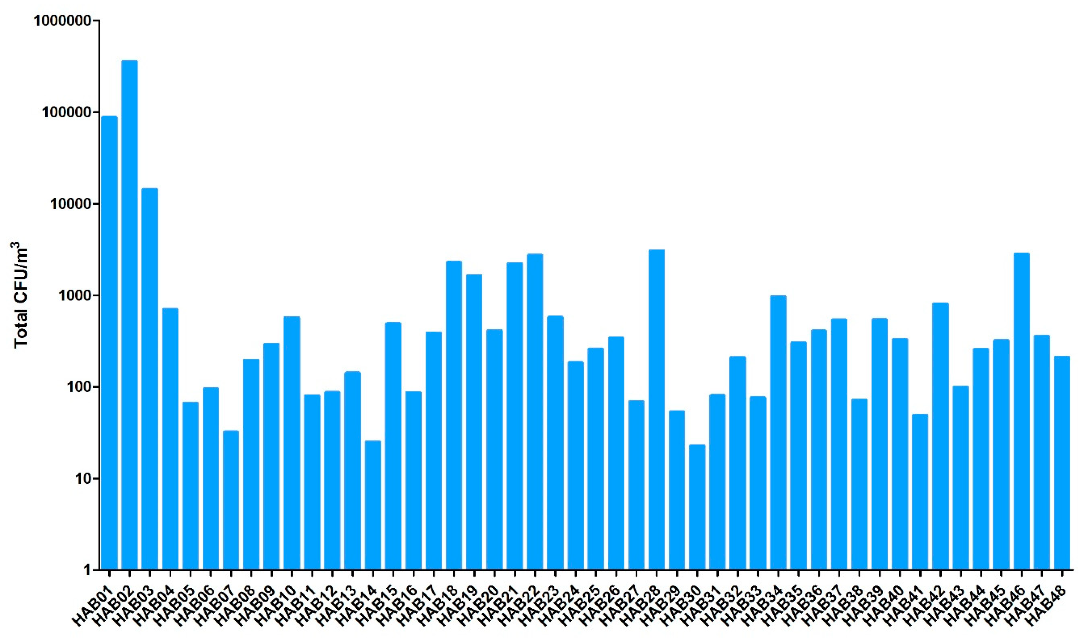
Figure 1. Total viable fungal particles concentration in the different houses (HAB01 to HAB48) included in this study.
Atmosphere 11 00223 g001
Figure 2. Representation of the fungal groups (Cladosporium, Aspergillus, Penicillium and others) quantified from bioaerosols. The box and whisker plots show the median percentage of fungal concentration (horizontal bars), their quartiles (box) and range (whiskers).
Atmosphere 11 00223 g002
Table 1. Main parameters documented in the questionnaire completed during home inspections.
Data DocumentedType of Answers
Habits of inhabitantsDaily time spent in the house; daily ventilation of room duration; ownership of a pet; presence of plants inside the house; Use of an absorber/dehumidifierYes/No
BuildingMold-contaminated surfaceCategories Low (<0.2 m2)/Moderate (between 0.2 to 3 m2)/High (>3 m2)
Type of ventilationType of ventilation (Natural/Mechanical)
Type of heating systemType of heater
(Electric/Gas/Heating oil)
Moldy odor; history of water damageYes/No
SymptomsAllergyYes/No
Anxiety; asthma aggravation; clogged nose; cough; dizziness; dyspnea; expectorations; eye irritation; fever; flu-like symptoms; headache; insomnia; itching; muscular pain; respiratory pain; skin rash; sinusitis; sore throatYes/No
and
Intensity
(Null/Weak/Moderate/Strong)
Table 2. Physical characteristics of bioaerosols from the 48 mold-damaged houses.
Physical CharacteristicsMeanMedianStandard DeviationMinMaxInterquartile Range
Temperature of room (°C)21.9922.252.5112.6426.103.28
Relative Humidity of room (%)60.6860.507.5544.8474.3013.30
Particles from 0.3 to 20 µm per m3 air1.16 × 1083.57 × 1073.07 × 1087.80 × 1062.01 × 1095.74 × 107
Table 3. Molds identified from bioaerosols in mold-damaged homes (n = 48) at a frequency above 10%.
Fungal SpeciesFrequency (n = 48)Mean Value (CFU m−3 of Air)
Penicillium chrysogenum36243.04
Aspergillus versicolor359321.47
Penicillium crustosum3237.68
Penicillium brevicompactum3038.36
Aspergillus fumigatus202.18
Penicillium glabrum1917.79
Penicillium expansum131.61
Penicillium olsonii1315.30
Aspergillus flavus100.94
Aspergillus niger100.26
Botrytis cinerea101.68
Cladosporium cladosporioides933.01
Penicillium nalgiovense91.90
Rhodotorula mucilaginosa90.68
Aspergillus sydowii89.83
Cladosporium herbarum86.60
Chaetomium globosum70.40
Paecilomyces variotii71.09
Penicillium citrinum72.51
Penicillium waksmanii71.93
Aspergillus pseudoglaucus61.67
Cladosporium sphaerospermum61.61
Penicillium italicum51.00
Penicillium piceum50.30
Penicillium purpurogenum56.45
Penicillium simplicissimum50.52
CFU: Colony Forming Unit.
Table 4. Symptoms occurrences (%) and intensities (weak/moderate/strong) in mold-damaged homes.
SymptomOccurrenceWeak IntensityModerate IntensityStrong Intensity
Clogged nose75.0011.5423.0840.38
Dyspnea75.0011.5432.6930.77
Insomnia69.239.6226.9232.69
Itching67.3117.3126.9223.08
Sore throat59.6221.1530.777.69
Headaches59.6226.9225.007.69
Eye irritation55.7711.5028.8515.38
Couch53.8515.3826.9211.54
Skin rash42.3115.3825.001.92
Respiratory pain40.3819.2317.313.85
Sinusitis34.629.6215.389.62
Expectorations32.691.9223.087.69
Muscular pain30.7723.085.771.92
Flu-like symptoms30.7721.159.620.00
Asthma aggravation25.001.929.6213.46
Anxiety23.087.6913.461.92
Dizziness19.2313.463.851.92
Fever17.319.623.853.85
Table 5. Mold-contaminated surfaces as explanatory variables used to explain fungal concentrations observed in bioaerosols.
Fungal ConcentrationMold-Contaminated Surface 1NMedianp-Value (Mann–Whitney Test)
Total CFU m−3Low8257.500.8681
Moderate29212.00
Moderate29212.000.0167 *
High11408.00
Low8257.500.0632
High11408.00
Aspergillus versicolor CFU m−3Low80.000.0155 *
Moderate2917.50
Moderate2917.500.0577
High11122.00
Low80.000.0035 *
High11122.00
Penicillium chrysogenum CFU m−3Low811.400.6947
Moderate297.78
Moderate297.780.0190 *
High1132.90
Low811.400.0465 *
High1132.90
Penicillium crustosum CFU m−3Low83.860.3934
Moderate290.96
Moderate290.960.0549
High115.07
Low83.860.4308
High115.07
1 Levels of contaminated surfaces: Low (<0.2 m2); Moderate (0.2 to 3 m2) and High (>0.3 m2). * Significant results (p < 0.05) are marked with an asterisk. CFU: Colony Forming Unit.
Table 6. Explanatory variables of the symptoms mentioned by inhabitants.
Explanatory Variable.SymptomNMedianp-Value (Mann–Whitney or Kruskal–Wallis Test)
A. versicolor (CFU m−3)DizzinessYes102.1 × 1020.0038 *
No399.46
FeverYes92.1 × 1020.0315 *
No4011.95
HeadacheYes2940.300.0154 *
No206.16
ItchingNull1717.500.0438 *
Weak75.65
Moderate144.68
Strong111.6 × 102
ExpectorationsYes1664.150.0489 *
No3311.10
P. chrysogenum (CFU m−3)FeverYes91.1 × 1020.0197 *
No407.92
ExpectorationsYes1631.550.0053 *
No333.78
P. crustosum (CFU m−3)FeverYes99.720.0344 *
No402.32
CoughYes284.950.0418 *
No210.00
HeadacheYes295.070.0163 *
No200.43
InsomniaYes364.360.0359 *
No130.00
Endotoxins (ng m−3)DyspneaYes363.750.0052 *
No1311.11
Flu-like symptomYes132.160.0149 *
No367.62
Particles of 2–15 µm diameter (per m3 of air)DizzinessYes106.1 × 1050.0136 *
No393.1 × 105
HeadacheYes294.4 × 1050.0419 *
No202.7 × 105
Relative humidity (%)Clogged noseYes3760.800.0315 *
No1255.28
ExpectorationsYes1668.500.0313 *
No3360.30
Sore throatYes2961.580.0451 *
No2055.68
* Significant results (p < 0.05) are marked with an asterisk. CFU: Colony Forming Unit.
Table 7. Contaminated surface as explanatory variable of the symptoms mentioned by inhabitants.
Explanatory VariableSymptomNLowModerateHighp-Value (chi² or Fisher Exact Test)
Contaminated surface 1DyspneaYes36323100.0354 *
No13571
HeadacheYes29316100.0385 *
No205141
ExpectorationsYes1610240.0001 *
No331626
1 Levels of contaminated surfaces: Low (<0.2 m2); Moderate (0.2 to 3 m2) and High (>3 m2). * Significant results (p < 0.05) are marked with an asterisk.
Table 8. Moldy odor as explanatory variable of the symptoms mentioned by inhabitants.
Explanatory VariableSymptomNYesNop-Value (chi² or Fisher Exact Test)
Moldy odorAnxietyYes171160.0131 *
No32923
DyspneaYes3618180.0295 *
No13211
Flu-like symptomYes13940.0150 *
No361125
HeadacheYes2913160.0138 *
No20164
Muscular painYes151050.0145 *
No341024
ExpectorationsYes161060.0315 *
No331023
* Significant results (p < 0.05) are marked with an asterisk.