Article

Generation of Viable Bacterial and Fungal Aerosols during Biomass Combustion

by
Ekaterina Mirskaya
and
Igor E. Agranovski
*
School of Engineering, Griffith University, Nathan 4111, Australia
*
Author to whom correspondence should be addressed.
Atmosphere 2020, 11(3), 313; https://doi.org/10.3390/atmos11030313
Submission received: 20 February 2020 / Revised: 6 March 2020 / Accepted: 22 March 2020 / Published: 24 March 2020
(This article belongs to the Special Issue Detection and Monitoring of Bioaerosols)

Abstract

:
Biomass combustion is known to be one of the main contributors to air pollution. However, the influence of biomass burning on the distribution of viable bacterial and fungal aerosols is uncertain. This study aimed to examine survivability of bacteria and fungi in the post-combustion products, and to investigate the aerosolization of viable cells during combustion of different types of organic materials. Laboratory experiments included a small-scale combustion of organic materials contaminated with microorganisms in order to determine the survivability of microbes in the combustion products and the potential aerosolization of viable cells during combustion. Field experiments were completed during intentional and prescribed biomass burning events in order to investigate the aerosolization mechanisms that are not available at the laboratory scale. Laboratory experiments did not demonstrate aerosolization of microorganisms during biomass combustion. However, the relatively high survival rate of bacteria in the combustion products ought to be accounted for, as the surviving microorganisms can potentially be aerosolized by high velocity natural air flows. Field investigations demonstrated significant increase in the bioaerosol concentration above natural background during and after biomass combustion.

1. Introduction

Bioaerosols are known to be highly sensitive to high temperatures. It was shown that common bacterial species such as Escherichia coli and Bacillus subtilis become more than 99.9% inactive when exposed to the temperature of 160 °C and 350 °C, respectively, for about 0.3 s [1]. However, a number of industrial high temperature processes can be responsible for microorganisms’ aerosolization. An exothermic process of lime slaking associated with a significant amount of heat causes aerosolization of bacteria at rates similar to other intensive mechanical processes, such as diffused aeration of wastewater [2]. In our previous work [3] we experimentally proved a possibility of aerosolization of viable microorganisms as a result of interaction of biologically contaminated liquid with hot surfaces that may occur in the processes of industrial cooling.
Biomass burning is a major source of aerosols which contributes up to 67% more carbon particle emissions compared to the combustion of fossil fuels [4]. At the same time, biomass burning is a process of interest as a renewable source for power and heat generation. Biomass-fueled electric generating facilities are characterized by a high level of particulate matter (PM), especially in boiler rooms and biomass storage rooms [5]. However, it was not confirmed that the combustion process itself represents a mechanism of bioaerosol generation, as the elevated levels of bioaerosols and biogenic organics are commonly related to the pre-combustion processes [6,7] that include storage or mechanical disturbance of biomass.
Some potential generation of biological aerosols can be related to biomass combustion in open fire at residential and industrial dwellings. Semple et al. [8] measured endotoxin levels within the living area of 69 houses while burning different biomass fuels in Malawi and Nepal. The results showed that median concentrations of total inhalable endotoxin were orders of magnitude higher than the level of 0.49 EU/m3 (geometric mean) which is linked to respiratory illnesses in children [9]. A very recent investigation reported some increase in airborne fungal concentration in the vicinity to forest fires at Madeira island [10]. It was suggested that the fire-induced convections are capable of promoting the release of fungal spores from their natural habitat.
Recent investigations of bacterial and fungal communities were conducted in China during summer harvest and biomass burning season [11]. The study showed that the total bacterial and fungal concentrations during biomass burning events are higher than the non-biomass burning events. However, the difference between the concentrations was not confirmed and the influence of harvesting activities was not investigated. It was suggested that bacterial cells and fungal spores can be carried: (a) by the turbulent air caused by combustion; or (b) attached to particulates released during combustion.
The aims of this study were: (1) to examine survivability of bacteria and fungi during combustion of different types of contaminated organic materials under controlled laboratory conditions; and (2) investigate microbial aerosolization during and post biomass combustion processes occurring in controlled and natural environments. Special attention will be given to comparison of microbial behavior in combustion processes of dramatically different scales in the laboratory and in the field.

2. Experiments

2.1. Laboratory Investigation-Microorganisms’ Survivability during Combustion

2.1.1. Bacterial Strains and Cultivation

Similar to our previous work [3], two common environmental bacterial strains, namely Gram-positive Bacillus subtilis (B. subtilis) American Type Culture Collection (ATCC) 6633 and Gram-negative Escherichia coli (E. coli) ATCC 27325 were obtained from Southern Biological (Nunawading, VIC, Australia) and used for the laboratory section of experiments. E. coli bacterial cells are known to be sensitive to pasteurization if present in a liquid material.
Stock cultures of B. subtilis and E. coli were grown in 1.3 g/100 mL of dry nutrient broth (OXOID Ltd., Basingstoke, Hampshire, England) in deionized and sterilized water for 18 h in an incubator shaker at a constant temperature of 37 °C. The microbial suspension was used directly after incubation in order to avoid sporulation of B. subtilis cells. The aliquots of the two microbial suspensions were mixed in a ratio of 1:1 (w/w) and the mixture was used for all of the experiments.

2.1.2. Samples Preparation

The entire experimental program was undertaken inside a 1200 mm wide Class II Biohazard Cabinet. The biohazard cabinet was used to meet the biosafety requirements, to prevent any escape of microbial materials to the laboratory air space and to ensure zero aerosol concentration eliminating chance for any potential alien microbial particles reaching experimental zone and interfere with the results.
The bacterial suspension was prepared as per the above description and spread undiluted over 60 mm diameter filter paper samples. The filters were then placed in the biohazard cabinet and air-dried overnight. The weight of each filter was measured with analytical scale ensuring even distribution of biological materials across all filters used. Then, the microbial materials from three filters were washed down with 50 mL of sterile deionized water and the aliquots were analyzed for culturable bacteria by commonly used plating technique as follows. An aliquot of 0.1 mL of an appropriate 10-fold dilution of the fluid was spread on the surface of the nutrient agar (NA) plates. The culture plates with bacteria were incubated at 37 °C for 1 day. Colony forming units (CFU) were counted after incubation with a colony counter (Biolab, Clayton, VIC, Australia), and the corresponding viable bacterial concentration in the liquid was determined. The results were expressed in CFU per g of paper.
Dry leaves were randomly collected from the soil organic horizon at the university surroundings at Nathan campus, mixed together, and delivered to the laboratory. Then, 1 g of the dry leaf matter was washed with 40 mL of sterile deionized water and the concentration of bacteria in the aliquot was analyzed according to the procedure described above for the paper filters and expressed in CFU/g of leaves.

2.1.3. Laboratory Set-Up

The laboratory setup consisted of a metal tray (40 cm × 30 cm), plastic funnel (3 0 cm diameter), and bioaerosol sampling equipment. The plastic funnel was strategically placed above the tray to ensure minimal escape of generated bioaerosols achieving maximum possible microbial collection by the bioaerosol sampling equipment during combustion experiments.
A personal bioaerosol sampler with the operational principle based on the passing of air through a porous medium submerged into a liquid [12] was used in the experiments. According to the previously described procedure [13], the personal sampler was sterilized before the experiments, drained, and filled with 40 mL of sterile distilled water. For all the experiments the air temperature (T) and the relative humidity (RH) were controlled at T = 24–25 °C and RH = 24–26%. The sampler was connected to the air sampling pump (Model PCXR8, SKC Inc., PA USA) and operated at a flowrate of 4 L/min over 60 s for all the experiments in order to cover entire duration of combustion process ensuring complete collection of released bioaerosols.
To analyze the collection fluid after sampling, the sampler was drained, and the porous media removed from the device and placed into container filled with 20 mL of distilled sterilized water. Then, the container was sonicated in an ultrasonic bath for 5 min to remove any microorganisms possibly remaining inside the porous medium and, on completion of the procedure mixed with the original collection fluid. The concentration was determined according to the previously described procedure [12]. The total volume of liquid samples was divided into three equal parts followed by filtration through a Nalgene cellulose nitrate membrane filter with 0.2 µm pore size (Nalge Co., Rochester, NY, USA). Then the filters were placed onto triplicate nutrient agar (NA) and malt extract agar (MEA) plates for bacteria and fungi respectively.
Then the NA plates were labelled and incubated at 37 °C for 2 days and the MEA plates were kept at room temperature until colonies had developed (all plates were observed daily). After incubation, the number of colonies was counted with the colony counter and the results were represented in CFU/mL for the bacterial and fungal concentration.

2.1.4. Combustion and Bioaerosol Collection

100 g of contaminated paper filters were placed in the metal tray and combusted. Bioaerosols were collected during the entire period of combustion. The materials remaining in the tray after combustion were collected, weighed, and diluted with 40 mL of sterilized deionized water. The concentration of bacteria was analyzed as described in the previous sections and expressed in CFU per g of ash. 100 g of the dry leaf matter were combusted in a similar way. The concentration of viable bacteria was analyzed following the same procedure.
Bacteria recovery was calculated using the following formula:
RR = C0/CASH
where C0 is the bacterial concentration on the contaminated paper filters or leaf matter in CFU/g and CASH is the bacterial concentration in the ash remaining after combustion in CFU/g.

2.1.5. Ash Aerosolization

Dry leaves was randomly collected from the ground at the university surroundings. Undiluted bacterial suspension was applied on the leaves. The leaves were air-dried in a biohazard cabinet overnight.
The dried leaves were ignited with a flexible gas lighter and combusted outdoors inside a metal cylinder with an open top. Bioaerosols were collected with a personal bioaerosol sampler over the entire period of combustion.
The remaining ash was left in the metal cylinder and covered with a metal lid with a hole in the middle utilized for placement of the bioaerosol sampler and bioaerosol collection. The set-up allows for minimization of the influence of background concentrations on the results of experiment.
Air flow, created with a manual pump, was passing from the bottom of the cylinder in order to aerosolize the ash. Bioaerosols were collected for two minutes and incubated at 37 °C over 48 h.

2.2. Field Investigation–Generation of Bioaerosols during Biomass Combustion

Four separate series of experiments were conducted in three different locations in order to use various burning strategies and sampling techniques.

2.2.1. 5 August 2018, Cainbable, Queensland

In order to characterize the bioaerosol from an open biomass burning, an experiment was conducted in a field near a farm at Cainbable. Branches from common Australian tree species including Angophora, Lophostemon, Syzygium, and Eucalyptus genera were collected and piled up on the field to dry the biomass for three months. The length of the branches was in the range from 30 cm up to 2 m. The pile was 1.7 m high with an estimated volume of 11 m3. There was no mechanical disturbance of the biomass pile at least two days before the experiment. Weather conditions remained consistent during all stages of the experiment; temperature was 23 °C, RH was 25%, the wind was below 10 km/h in the NE direction, and there was full sun.
All bioaerosol were collected with a personal bioaerosol sampler in a height of 1.5 m and distance of 2 m downwind from the pile. No closer sampling point location was possible due to very high air temperature and significant possibility of sampling equipment destruction in closer vicinity to the fire. The first series of samples had been collected before the pile was ignited to obtain natural bioaerosol background.
The pile was ignited with a lighter and combusted. The total combustion period lasted about 30 min. A number of bioaerosol samples was collected at the same point during the entire period of biomass flaming.
A series of bioaerosol samples was collected 30 min past combustion after biomass smoldering had been completed. There was no mechanical disturbance of the ash and remaining biomass prior to the bioaerosol sampling.
The bioaerosol samples were treated and incubated as per the procedure described above. The concentrations of viable microorganisms in the samples were analyzed and expressed in CFU/m3.
Three identically sized piles were burned to ensure statistically reliable and reproducible results.

2.2.2. 28 June 2019, Dirranbandi, Outback Queensland

A second series of experiments was completed in a remote arid area of Queensland in order to eliminate the influence of surrounding vegetation, utilize a different sampling technique, and analyze whether the microbe abundance differs with increasing distance from the fire. As in the first series of experiments, a variety of dry branches of common Australian tree species were collected in a pile and ignited with a lighter for combustion. Petri dishes with nutrient agar (NA) and malt extract agar (MEA) were placed at distances of 1.5, 2.7, 3.9, 5.1, 6.3 and 7.5 m downwind from the burning biomass pile (Figure 1) similar to the method described by Kobziar et al. [14]. Background samples were collected in the area not affected by the fire, however, to ensure similar ambient parameters and the abundance of vegetation. Samples were collected via passive deposition onto agar over 1.5 h. The samples were closed, sealed with parafilm and delivered to the laboratory. NA samples were incubated at 37 °C for 1 day, and MEA samples were stored at room temperature until colonies had developed. After incubation, the number of colonies was counted with the colony counter and the results were represented in CFU. The temperature during experiments was 21 °C, RH was 43%, wind speed was around 17 km/h in SW direction, and there was full sun.

2.2.3. 11 and 12 June 2019, Nathan Campus of Griffith University

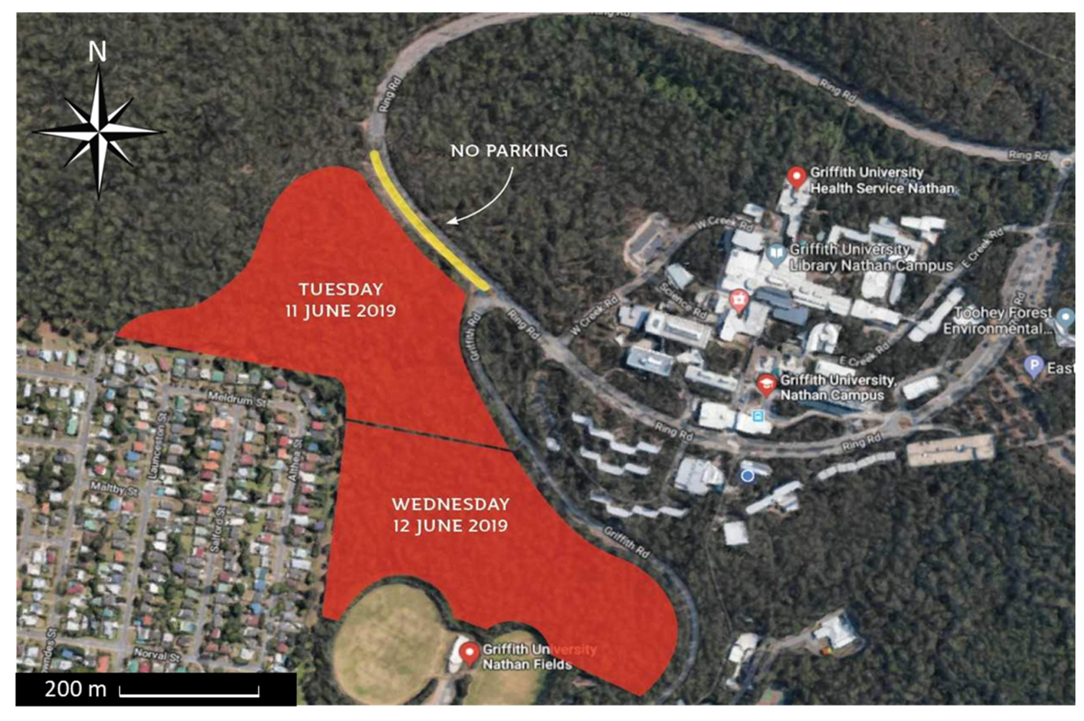
A third series of experiments was conducted during the hazard reduction burns at Nathan campus of Griffith University on 11 and 12 June 2019 (Figure 2). Fuel loads on the forest floor were assessed medium to high in places and needed to be managed for the safety of students, staff, motorists, and other people in the area. Controlled burning was lit in a mosaic pattern to have minimum impact on fauna in the area. The temperature was 23 °C, RH was 50%, wind speed was 11 km/h in the direction of NE, and it was slightly cloudy.
A number of samples were collected a day before the prescribed burning in order to evaluate the background concentration of microorganisms in the same environment. Twelve Petri dishes were suspended on the trees in six different parts of the forest approximately 2 m above ground. The samples were collected via passive deposition onto nutrient agar (NA) and malt extract agar (MEA) during 6 h. The samples were closed, sealed with parafilm and delivered to the laboratory immediately after the collection had been completed.
The same method was used to collect samples of bioaerosols during the prescribed burning. Petri dishes were suspended at the same locations 30 min before the fire was initiated with a drip torch. The dishes were collected in 6 h and at that time a remaining fire and smoldering were still observed in different parts of the forest. Weather conditions remained consistent during the two days of sampling.
The samples were delivered to the laboratory immediately after collection. NA samples were placed in the incubator at 37 °C for 48 h, and the MEA samples were kept at room temperature in the dark over 5 days. The plates were visually examined for microorganisms daily and CFUs were counted where possible.

2.2.4. 17 and 18 August 2019, Nathan Campus of Griffith University

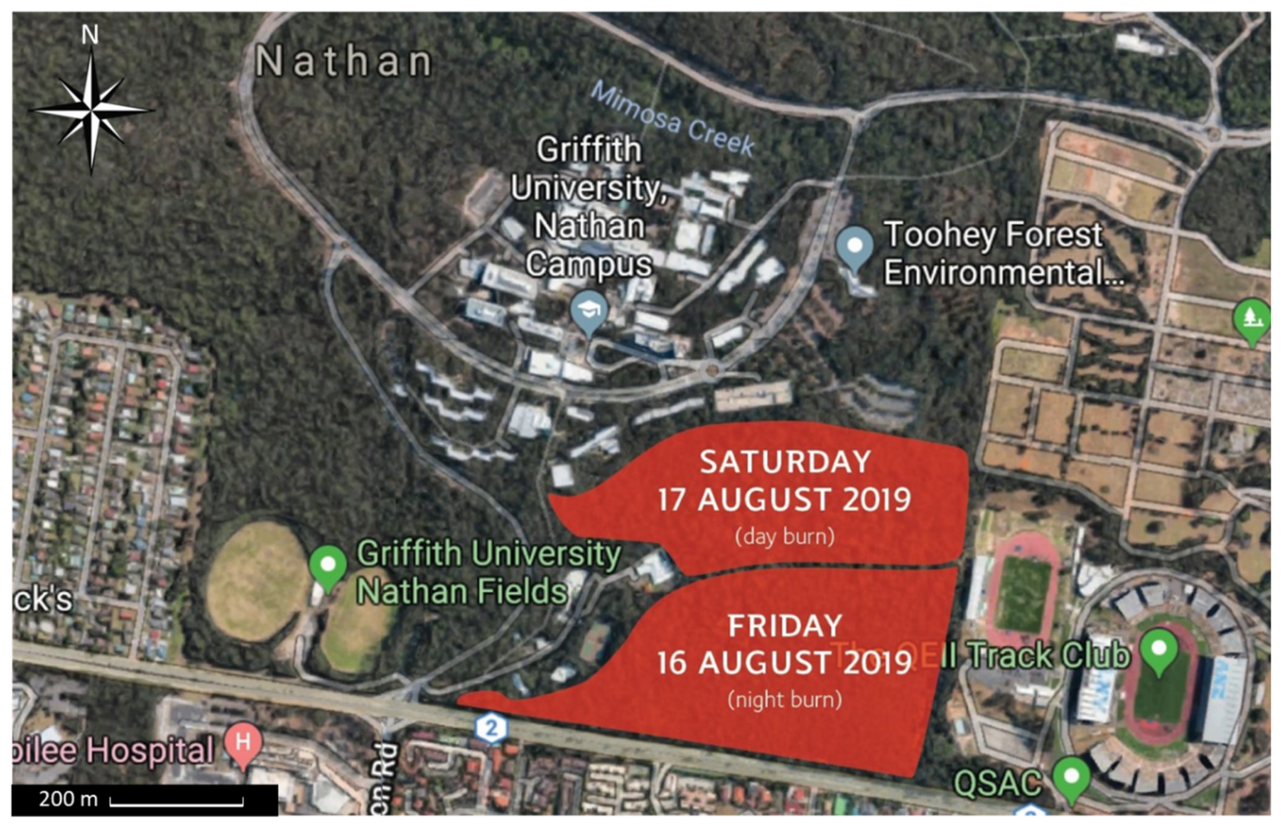
The final series of experiments was conducted during the next period of prescribed burning at Nathan campus of Griffith University (Figure 3) in order to utilize a different sampling strategy and technique. During the experiments, the temperature was 22 °C, RH was 47%, wind speed was 13 km/h in the direction of SW, and there was full sun.
Air samples were collected with a personal sampler over 15 min. Prior to the experiment, each device was charged with 40 mL of fresh collecting liquid, sealed in plastic bag and transported to the sampling location. On arrival, the samplers were unpacked, connected to the air sampling pump (Model PCXR8, SKC Inc., Eighty Four, PA, USA), and operated for 15 min at the flow rate of 4 L/min.
A number of samples had been collected: (a) before the prescribed burning started, in order to evaluate the background concentration; (b) in the hot areas of burning characterized by high smoke concentration and elevated temperature, in order to assess the potential of burning process itself to generate bioaerosols; and (c) at a distance of 30 m downwind from the most intensive burning, in order to assess the influence of other possible factors and mechanisms of aerosolization of microorganisms.
On completion, the sampler was disconnected from the pump, placed in the sealed plastic bags and carefully delivered to the laboratory for processing in accordance with the procedure described above.

3. Results

3.1. Contaminated Material Combustion and Bioaerosol Collection

The abundance of culturable bacteria on contaminated paper filters and leaf matter, as well as material remaining post combustion, were analyzed, and the results are summarized in Table 1. Despite significant mass reduction (94%), 28% of culturable bacteria were recovered from the paper filters post combustion. Leaf matter was characterized by lower mass reduction due to higher moisture content and complex nature of the material. However, the post combustion remainders of leaf mater showed a lower bacteria recovery rate (18%).
There were no viable microorganisms detected in the air samples collected during combustion of contaminated paper filters or leaf matter. The results allow for the possibility that the combustion process itself does not promote aerosolization of viable bacteria; however, culturable microorganisms were detected in the post combustion remainders as a result of incomplete combustion. Bioaerosols collected during ash aerosolization with manual pump demonstrated almost five times higher concentration of viable microorganisms (3244 CFU/m3) than the ambient concentration (650 CFU/m3).

3.2. Generation of Bioaerosols during Biomass Combustion

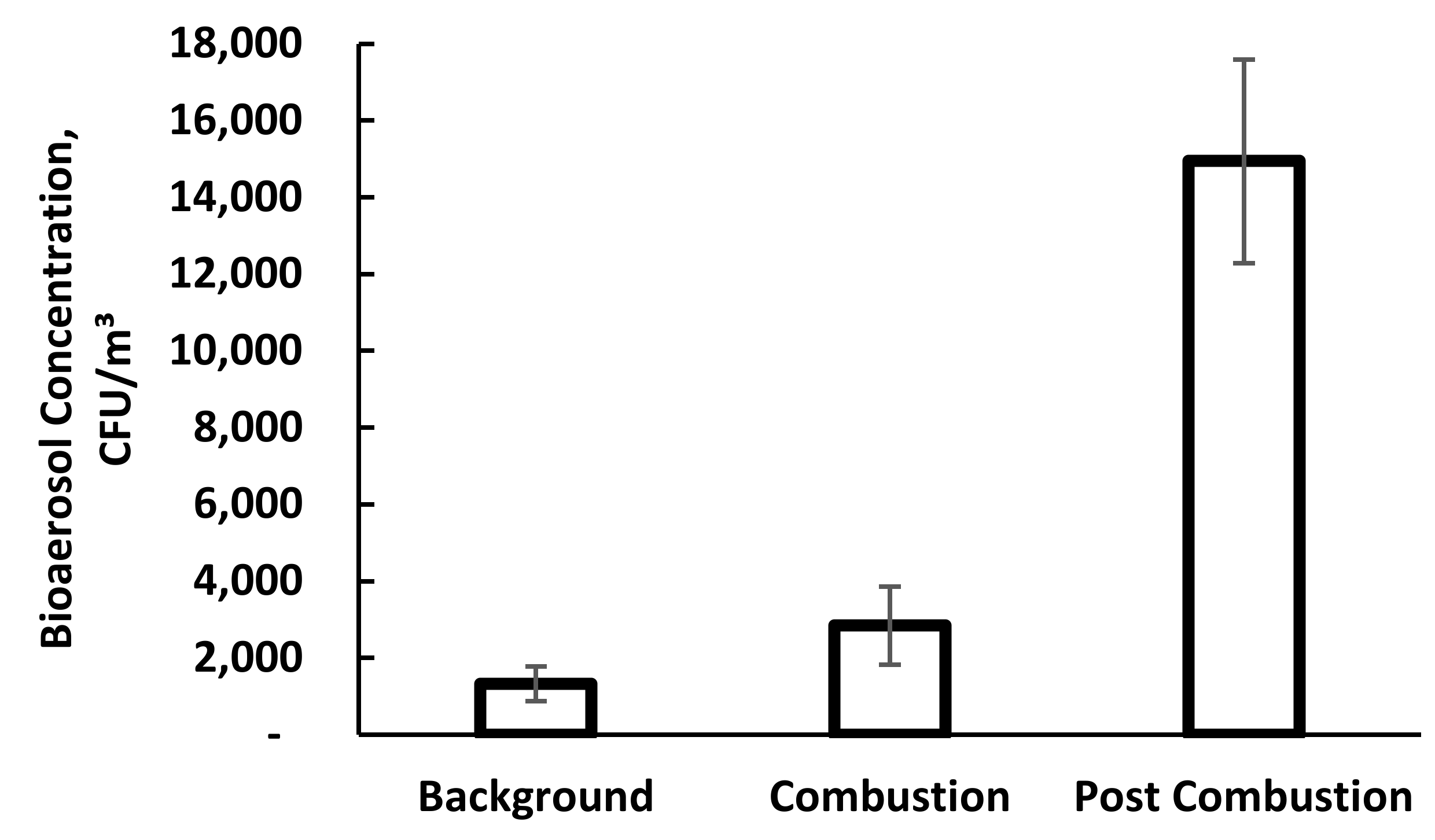
Bioaerosols collected during the first series of field investigation at a distance of two meters from the fire demonstrated higher concentrations of bacteria during biomass combustion (2.85 × 10³ CFU/m3) compared to the background concentrations measured prior to ignition (1.33 × 10³ CFU/m3). A significant increase in the total concentration of bacteria in the post combustion environment (1.49 × 10³ CFU/m3) can also be observed in Figure 4.
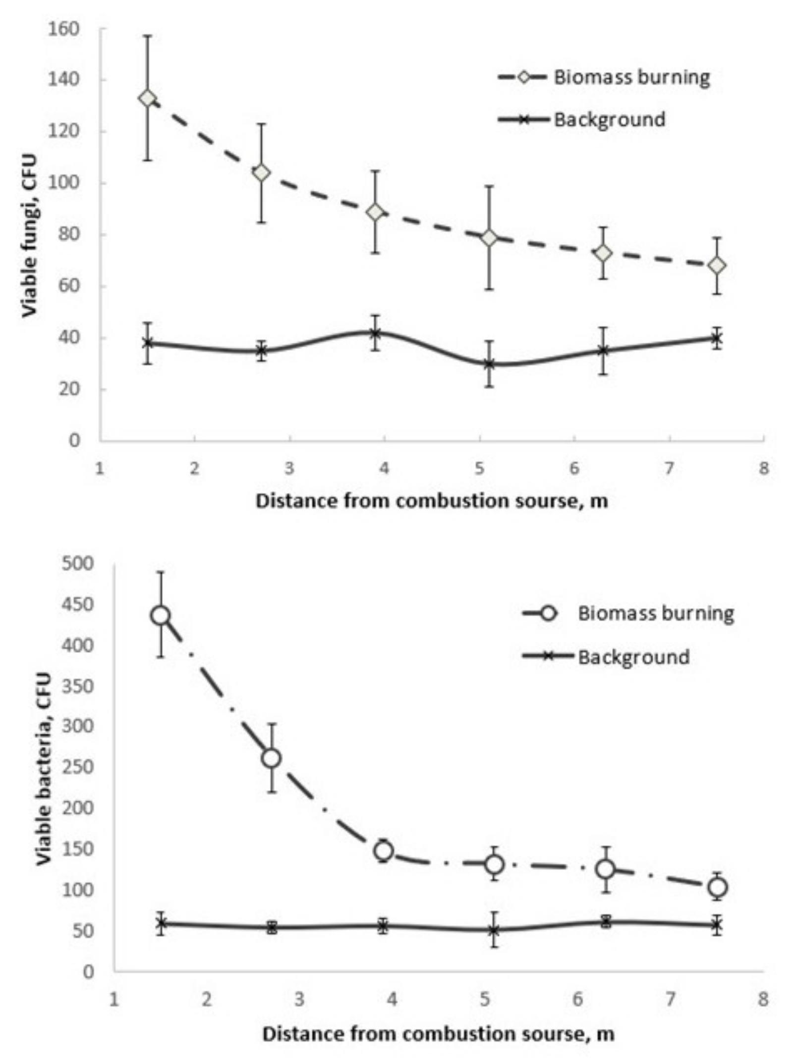
Bioaerosols collected via passive deposition in the arid area showed that the concentration of both bacteria and fungi in the air was reduced with increased distance from the fire (Figure 5). Visual assessment of the samples showed that the samples collected near the fire were characterized by a larger number of ash particles settling on the plates. The number of ash particles was reduced with increased distance from the fire. The background concentration was acquired by using the same plates placed at the same distances from the fire prior to the commencement of combustion process. To ensure sufficient integrity of the results, three repeats of the background concentration monitoring procedure were undertaken.

3.3. Prescribed Burning Experiment

Bioaerosol samples collected during prescribed burning via passive deposition on nutrient agar (NA) did not demonstrate any colony growth. The number of fungal colonies was higher in the background samples than in the samples collected in the hot area affected by the fire (temperature above 40 °C).
The samples collected with a personal sampler in August showed lower concentrations of microorganisms in the proximity and significantly higher concentrations of bioaerosols in the surrounding areas compared to the non-fire concentration (Figure 6). The difference in concentration was quite significant, reaching a concentration of bacterial aerosols almost 7 times higher compared to the fire front areas.

4. Discussion

Kobziar et al. [14] conducted a number of laboratory and field investigations to provide a foundational understanding of the capability of wildland fire to aerosolize viable microorganisms in smoke. As the investigation collaborated a few different disciplines, a new term, Pyroaerobiology, was introduced to integrate micro- and aerobiology, smoke and atmospheric sciences, and fire behavior and ecology.
The laboratory experiments did not demonstrate any significant differences between the concentration of culturable microorganisms during combustion and the ambient samples [14]. This study was conducted in a biohazard cabinet to eliminate the influence of ambient conditions. Similar to the current study, no culturable microorganisms were detected in the air during the laboratory experiments. This allows for the suggestion that a small-scale fire does not provide a mechanism of microorganism aerosolization. However, such conclusion can only be made for culturable microbes involved in the current investigation.
At the same time, the experiment showed a significant extent of microorganisms’ survivability in the post combustion material and possibility of their aerosolization where an additional factor such as wind is present. Survivability of microorganisms in these experiments can be explained by incomplete combustion of contaminated material that would also take place during biomass burning events and wildfires when the soil and vegetation biomass are affected by the fire. Biomass burning is known to have significant short-term effects on the abundance of soil microorganisms. A decrease in the abundance of total bacteria in fire-affected soil was observed three days after biomass burning, with the following restoration in the abundance of total bacteria nine days after burning [15]. Soil ecosystem stability is influenced by the burning frequencies and can sustain prescribed burning occurring with more than four-year intervals [16]. An investigation of wildfire effects on soil bacterial and fungal communities in an extreme fire season in the northwestern Canadian boreal forest showed that fire occurrence, as well as moisture regime, are among the significant predictors of post combustion soil microbial community composition [17].
Since the soil microorganisms, as well as microorganisms attached to the plants and trees, are capable of surviving during bushfires, they can be aerosolized if a suitable mechanism or conditions occur. There are two possible changes in the ambient conditions caused by the fire and relevant to variations in the bioerosol concentration: elevated level of pollutants in the air and turbulent air flow created by the burning process.
A number of laboratory biomass burning experiments were conducted to quantify emission factors of domestic heaters operating in different conditions and different types of biomass fuels. However, they are hardly capable of reflecting burning conditions similar to those of wide, open fires, due to the sensitivity of the combustion process to the burning conditions [18]. Small scale experiments conducted in remote areas as part of this study demonstrated significant dependence of both fungi and bacteria distribution upon the ash content in the air.
Wildfires and biomass burning events provide a significant contribution to the elevated concentrations of particulate matter with aerodynamic diameter <10 μm (PM10) [19,20]. PM10 is known to be the most significant factor that often shows positive correlation with the concentration of airborne microorganisms [21,22].
A comprehensive one-year study [23] showed a strong positive correlation of Gram-positive and gran-negative bacteria concentrations with organic carbon and biomass burning derived potassium that indicated their association with emissions from biomass burning. Wei et al. [11] have recently confirmed elevated concentrations total bacteria and fungi during the burning period in China. The critical factors for fungal and bacterial communities included organic carbon, magnesium and wind speed.
Elevated concentrations of hydroxy fatty acids were recently observed during biomass burning events in China [24]. Hydroxy fatty acids are potential tracers for soil microbes, plant pathogens, and higher plant waxes. Endotoxin concentrations were also higher than the health-based occupational guidance limit (~90 EU m−3). Emission factors for trace gases and aerosols, as well as their chemical speciation for the open fire burning of pruning residues were investigated in Portugal. Burning prunings emitted substantial amounts of gaseous and particulate pollutants that can have harmful impacts on human health and ecosystems, including polyols which might occur as constituents in bacteria or fungi [25]. Elevated concentrations of arabitol and mannitol, fungal molecular tracers, were detected in fine particle samples collected in during a biomass burning season in China [26].
Total bacteria in atmospheric aerosols depend on a number of factors, including geographical terrain and analytical methods, and may vary from 102 to 106 cells·m−3 [11]. The average concentration of microorganisms in the air before combustion was around 1.33 × 10³ CFU/m3 and 5.82 × 10² CFU/m3 for biomass combustion ad prescribed burning experiments respectively, which is in line with other studies. The concentration of microorganisms measured during combustion downwind from the fire in both experiments was two times higher than the background concentration and the concentration in the post combustion environment was more than an order of magnitude higher than the background concentration.
Wind speed is considered to be one of the main factors affecting bioaerosol and PM10-2.5 concentrations in the atmosphere. Winds with a mean speed of ≥5.5 m s−1 can mechanically resuspend surface dust, thus re-aerosolizing PM10-2.5 as well as bioaerosols settled on the surface or attached to dust particles [27]. Fire plumes are characterized by high velocities. Vertical velocity can reach 13 m/s, with downdrafts of ~8 m/s [28]. It is speculated that this high velocity acts a strong wind and causes a significant disturbance of plants therefore detaching microorganisms from biomass (leaves, trees etc.).
Future investigations are required in order to evaluate the relationship between the composition of microorganisms deposited on different types of biomass (soil layers, grass, trees) in the fire areas and the composition of microorganisms in the surrounding atmosphere. Laboratory investigations may focus on the aerosolization of microorganisms with vertical air flows, in order to imitate the process of biomass burning in the natural environment.

5. Conclusions

This study demonstrates the capability of viable microorganisms to survive in the material remaining after biomass combustion. Viable microorganisms are able to aerosolize if an additional source of aerosolization is present. Large-scale fires such as wildfires or prescribed biomass burnings significantly contribute to air microbial quality. Elevated concentrations of fungi and bacteria were observed during biomass combustion and prescribed burnings, as well as in the post combustion environment. The two possible changes in the ambient conditions caused by the fire and relevant to variations in the bioaerosol concentration include elevated levels of all types of combustion related air pollutants and turbulent air flow created by the burning process. Finally, it ought to be noticed that laboratory scale experiments might not always be representative for modeling bioaerosol release from natural bushfire events; special attention must be given to various simulated parameters, especially plume velocity and direction.

Author Contributions

Conceptualization, E.M. and I.E.A.; Methodology, E.M. and I.E.A.; investigation, E.M. and I.E.A.; writing—original draft preparation, E.M.; writing—review and editing, I.E.A.; visualization, E.M.; supervision, I.E.A. All authors have read and agreed to the published version of the manuscript.

Funding

This research received no external funding.

Conflicts of Interest

The authors declare no conflict of interest.

References

  1. Jung, J.H.; Lee, J.E.; Kim, S.S. Thermal effects on bacterial bioaerosols in continuous air flow. Sci. Total Environ. 2009, 407, 4723–4730. [Google Scholar] [CrossRef] [PubMed]
  2. Barth, E.F.; Reponen, T.; Succop, P. Evaluation of bioaerosol components, generation factors, and airborne transport associated with lime treatment of contaminated sediment. J. Air Waste Manag. Assoc. 2009, 59, 540–552. [Google Scholar] [CrossRef] [PubMed]
  3. Mirskaya, E.; Agranovski, I.E. Generation of bacterial aerosols by interaction of microbial suspension with hot surfaces. J. Aerosol. Sci. 2017, 107, 1–8. [Google Scholar] [CrossRef]
  4. Bond, T.C.; Streets, D.G.; Yarber, K.F.; Nelson, S.M.; Woo, J.H.; Klimont, Z. A technology-based global inventory of black and organic carbon emissions from combustion. J. Geophys. Res.-Atmos. 2004, 109, D14. [Google Scholar] [CrossRef] [Green Version]
  5. Cohn, C.A.; Lemieux, C.L.; Long, A.S.; Kystol, J.; Vogel, U.; White, P.A.; Madsen, A.M. Physical-chemical and microbiological characterization, and mutagenic activity of airborne PM sampled in a biomass-fueled electrical production facility. Environ. Mol. Mutagen 2011, 52, 319–330. [Google Scholar] [CrossRef]
  6. Rohr, A.; Campleman, S.; Long, C.; Peterson, M.; Weatherstone, S.; Quick, W.; Lewis, A. Potential occupational exposures and health risks associated with biomass-based power generation. Int. J. Environ. Res. Public Health 2015, 12, 8542–8605. [Google Scholar] [CrossRef] [Green Version]
  7. Laitinen, S.; Laitinen, J.; Fagernäs, L.; Korpijärvi, K.; Korpinen, L.; Ojanen, K.; Jokiniemi, J. Exposure to biological and chemical agents at biomass power plants. Biomass Bioenerg. 2016, 93, 78–86. [Google Scholar] [CrossRef] [Green Version]
  8. Semple, S.; Devakumar, D.; Fullerton, D.G.; Thorne, P.S.; Metwali, N.; Costello, A.; Ayres, J.G. Airborne endotoxin concentrations in homes burning biomass fuel. Environ. Health Perspect. 2010, 118, 988–991. [Google Scholar] [CrossRef] [Green Version]
  9. Dales, R.; Miller, D.; Ruest, K.; Guay, M.; Judek, S. Airborne endotoxin is associated with respiratory illness in the first 2 years of life. Environ. Health Perspect. 2005, 114, 610–614. [Google Scholar] [CrossRef]
  10. Camacho, I.; Góis, A.; Camacho, R.; Nóbrega, V.; Fernandez. The impact of urban and forest fires on the airborne fungal spore aerobiology. Aerobiologia 2018, 34, 585–592. [Google Scholar] [CrossRef]
  11. Wei, M.; Xu, C.; Xu, X.; Zhu, C.; Li, J.; Lv, G. Characteristics of atmospheric bacterial and fungal communities in PM2. 5 following biomass burning disturbance in a rural area of North China Plain. Sci. Total Environ. 2019, 651, 2727–2739. [Google Scholar] [CrossRef] [PubMed]
  12. Agranovski, I.E.; Agranovski, V.; Reponen, T.; Willeke, K.; Grinshpun, S.A. Development and evaluation of a new personal sampler for culturable airborne microorganisms. ATM Environ. 2002, 36, 889–898. [Google Scholar] [CrossRef]
  13. Agranovski, I.E.; Pyankov, O.V.; Altman, I.S. Bioaerosol contamination of ambient air as the result of opening envelopes containing microbial materials. Aerosol. Sci. Tech. 2005, 39, 1048–1055. [Google Scholar] [CrossRef]
  14. Kobziar, L.N.; Pingree, M.R.; Larson, H.; Dreaden, T.J.; Green, S.; Smith, J.A. Pyroaerobiology: The aerosolization and transport of viable microbial life by wildland fire. Ecosphere 2018, 9, e02507. [Google Scholar] [CrossRef] [Green Version]
  15. Li, Y.; Zhang, H. Short-term effects of biomass burning on soil ammonia-oxidizing bacteria and ammonia-oxidizing archaea. Afr. J. Microbiol. Res. 2013, 7, 2486–2496. [Google Scholar]
  16. Williams, R.J.; Hallgren, S.W.; Wilson, G.W. Frequency of prescribed burning in an upland oak forest determines soil and litter properties and alters the soil microbial community. For. Ecol. Manag. 2012, 265, 241–247. [Google Scholar] [CrossRef]
  17. Whitman, T.; Whitman, E.; Woolet, J.; Flannigan, M.D.; Thompson, D.K.; Parisien, M.A. Soil bacterial and fungal response to wildfires in the Canadian boreal forest across a burn severity gradient. Soil Biol. Biochem. 2019, 138, 107571. [Google Scholar] [CrossRef]
  18. Chen, J.; Li, C.; Ristovski, Z.; Milic, A.; Gu, Y.; Islam, M.S.; Guo, H. A review of biomass burning: Emissions and impacts on air quality, health and climate in China. Sci. Total Environ. 2017, 579, 1000–1034. [Google Scholar] [CrossRef] [Green Version]
  19. Popovicheva, O.; Kistler, M.; Kireeva, E.; Persiantseva, N.; Timofeev, M.; Kopeikin, V.; Kasper-Giebl, A. Physicochemical characterization of smoke aerosol during large-scale wildfires: Extreme event of August 2010 in Moscow. Atmos. Environ. 2014, 96, 405–414. [Google Scholar] [CrossRef]
  20. Nava, S.; Lucarelli, F.; Amato, F.; Becagli, S.; Calzolai, G.; Chiari, M.; Udisti, R. Biomass burning contributions estimated by synergistic coupling of daily and hourly aerosol composition records. Sci. Total Environ. 2015, 511, 11–20. [Google Scholar] [CrossRef]
  21. Hwang, S.H.; Park, W.M. Concentrations of PM 10 and airborne bacteria in daycare centers in Seoul relative to indoor environmental factors and daycare center characteristics. Air Qual. Atmos. Health 2017, 10, 139–145. [Google Scholar] [CrossRef]
  22. Liu, H.; Hu, Z.; Zhou, M.; Hu, J.; Yao, X.; Zhang, H.; Li, C. The distribution variance of airborne microorganisms in urban and rural environments. Environ. Pollut 2019, 247, 898–906. [Google Scholar] [CrossRef] [PubMed]
  23. Rajput, P.; Anjum, M.H.; Gupta, T. One year record of bioaerosols and particles concentration in Indo-Gangetic Plain: Implications of biomass burning emissions to high-level of endotoxin exposure. Environ Pollut 2017, 224, 98–106. [Google Scholar] [CrossRef]
  24. Tyagi, P.; Kawamura, K.; Fu, P.; Bikkina, S.; Kanaya, Y.; Wang, Z. Impact of biomass burning on soil microorganisms and plant metabolites: A view from molecular distributions of atmospheric hydroxy fatty acids over Mount Tai. J. Geophys. Res. Biogeosci. 2016, 121, 2684–2699. [Google Scholar] [CrossRef]
  25. Alves, C.A.; Vicente, E.D.; Evtyugina, M.; Vicente, A.; Pio, C.; Amado, M.F.; Mahía, P.L. Gaseous and speciated particulate emissions from the open burning of wastes from tree pruning. Atmos. Res. 2019, 226, 110–121. [Google Scholar] [CrossRef]
  26. Yang, Y.; Chan, C.Y.; Tao, J.; Lin, M.; Engling, G.; Zhang, Z.; Su, L. Observation of elevated fungal tracers due to biomass burning in the Sichuan Basin at Chengdu City, China. Sci. Total. Environ. 2012, 431, 68–77. [Google Scholar] [CrossRef]
  27. Chen, L.W.A.; Zhang, M.; Liu, T.; Fortier, K.; Chow, J.C.; Alonzo, F.; Cruz, P. Evaluation of epifluorescence methods for quantifying bioaerosols in fine and coarse particulate air pollution. Atmos. Environ. 2019, 213, 620–628. [Google Scholar] [CrossRef]
  28. Mueller, E.V.; Skowronski, N.; Thomas, J.C.; Clark, K.; Gallagher, M.R.; Hadden, R.; Simeoni, A. Local measurements of wildland fire dynamics in a field-scale experiment. Combust Flame 2018, 194, 452–463. [Google Scholar] [CrossRef] [Green Version]
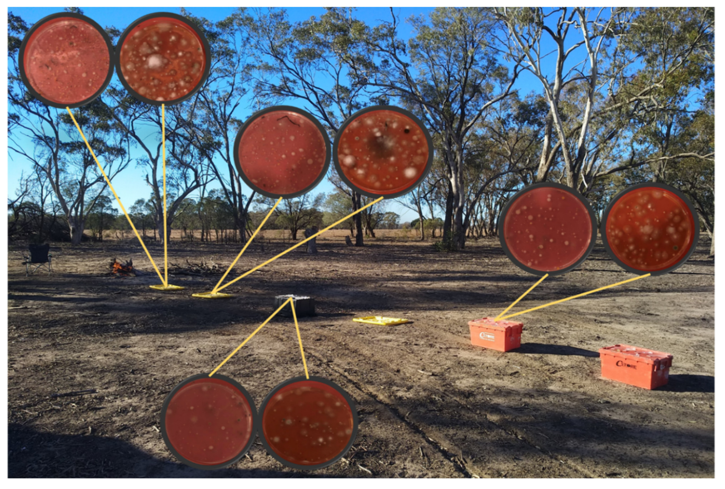
Figure 1. Positioning of Petri dishes in the Dirranbandi series of biomass combustion experiments.
Atmosphere 11 00313 g001
Figure 2. Map of hazard reduction burns carried out at Nathan campus of Griffith University on 11 and 12 June 2019.
Atmosphere 11 00313 g002
Figure 3. Map of hazard reduction burns carried out at Nathan campus of Griffith University on 17 and 18 August 2019.
Atmosphere 11 00313 g003
Figure 4. Concentration of culturable bacteria in the ambient air collected with a personal sampler in the Cainbable series of experiments. Error bars represent the standard deviation of three experimental runs.
Atmosphere 11 00313 g004
Figure 5. Relationships between the concentration of viable bacteria (a) and fungi (b), in the Dirranbandi series of experiments, and the distance from the fire. Error bars show the standard deviation of three experimental repeats for both, background and burning related monitoring.
Atmosphere 11 00313 g005
Figure 6. Concentration of viable microorganisms in the ambient air collected with a personal sampler during biomass combustion in the Griffith series of experiments. Error bars show the standard deviation of five experimental repeats.
Atmosphere 11 00313 g006
Table 1. Survivability of microorganisms as a result of incomplete combustion of contaminated organic materials. Bacteria recovery rate is calculated using formula (1).
ParametersUnitsBefore CombustionPost Combustion
Paper FilterLeaf Matter Paper FilterLeaf Matter
Massg0.65(±0.01) 1.89(±0.28)0.04(±0.00)0.71(±0.11)
ConcentrationCFU/g2.51(±0.38) × 1066.33(±0.95) × 1036.90(±1.04) × 1051.13(±0.17) × 103
Mass reduction%--94%62%
Bacteria recovery%--28%18%