Review

Linking Genotype to Clinical Features in SMC1A-Related Phenotypes: From Cornelia de Lange Syndrome to Developmental and Epileptic Encephalopathy, a Comprehensive Review

by
Maria Francesca Astorino
1,†,
Desirèe Speranza
2,†,
Giovanni Luppino
3,
Maria Angela La Rosa
4,
Silvana Briuglia
1,4,*,‡ and
Marco Calabrò
1,4,‡
1
Department of Biomedical and Dental Sciences and Morpho-Functional Imaging—BIOMORF, University of Messina, Via Consolare Valeria 1, 98125 Messina, Italy
2
Department of Chemical, Biological, Pharmaceutical and Environmental Sciences, University of Messina, Via Consolare Valeria 1, 98125 Messina, Italy
3
Department of Human Pathology of Adulthood and Childhood, University of Messina, Via Consolare Valeria 1, 98125 Messina, Italy
4
UOSD of Genetics and Pharmacogenetics, “Gaetano Martino” University Hospital, Via Consolare Valeria, 98125 Messina, Italy
*
Author to whom correspondence should be addressed.
These authors contributed equally to this work.
These authors also contributed equally to this work.
Genes 2025, 16(10), 1196; https://doi.org/10.3390/genes16101196
Submission received: 14 August 2025 / Revised: 27 September 2025 / Accepted: 2 October 2025 / Published: 13 October 2025
(This article belongs to the Special Issue Molecular Basis and Genetics of Intellectual Disability)

Abstract

Germline mutations in the X-linked cohesin subunit gene SMC1A have been increasingly recognized as a cause of developmental and epileptic encephalopathy (DEE); however, the underlying basis of its marked phenotypic heterogeneity remains elusive. In our narrative review, starting from all literature-reported clinical cases of SMC1A-related DEE, we propose an integrative framework summarizing all the clinical and genetic features, stratified by mutation type, mosaic fraction, and X-chromosome inactivation (XCI) patterns to provide valuable support for genetic diagnosis and variants, found to date. Also, we discuss how somatic mosaicism and epigenetic variability underlie the clinical diversity of SMC1A-associated epilepsy and systematically describe the entire phenotypic spectrum, from early-onset, therapy-resistant seizures to milder intellectual disability profiles. We further examine how SMC1A mutations perturb cohesin’s canonical roles in chromatin loop formation and sister-chromatid cohesion, leading to widespread transcriptional dysregulation of neurodevelopmental gene networks. Evidence that XCI skewing can ameliorate or exacerbate neuronal cohesin deficits and, thus modulate seizure threshold, is presented.

1. Introduction

The SMC1A (structural maintenance of chromosomes 1A) gene, also known as SMC1L1 (Mendelian inheritance in man MIM #300040) according to the revised Human Genome Organization HUGO Gene Nomenclature Committee (HGNC), is a member of the structural maintenance of chromosome (SMC) family that has a relevant role in DNA transactions. SMCs are implicated in the organization of the mitotic and meiotic macroscale chromosome, maintaining genetic stability during the duplication phases [1]. Of them, the SMC1A gene, which maps to Xp11.22 in a region that partially escapes X inactivation [2], is a core element of the cohesin multiprotein complex that participates in chromosomal segregation and gene regulation [3]. Specifically, the cohesin complex is a tripartite complex that mediates the cohesion of sister chromatids after DNA replication to ensure faithful chromosome segregation. Cohesin consists of four subunits, two SMC proteins or cohesion core subunits, SMC1A or SMC1B and SMC3, and two non-SMC subunits, RAD21 and either SA1 or SA2 [4]. SMC1A is linked also to cohesion-associated factors that permit cohesion loading in the chromatin such as nipped-B like protein (NIPBL) [5]. In addition, SMC1A also plays a role in the DNA damage repair (DDR) pathway, genome stability maintenance, and 3D genome organization [6,7]. SMC1A function in the DDR pathway is regulated by ataxia telangiectasia-mutated (ATM) and ataxia telangiectasia- and Rad3-related (ATR) threonine/serine kinases [8]. Additionally, SMC1A is also a component of the recombination protein complex (RC-1) that allows DNA repair [9].
Variants in the SMC1A gene have been associated with Cornelia de Lange syndrome (CdLS) (MIM #300590), in resembling the Rett syndrome (RTT) phenotype, intellectual disability, and developmental and epileptic encephalopathy (DEE) [10]. More than 100 SMC1A variants have been reported in the Human Genome Mutation Database [11], and several of them, mainly both missense and nonsense mutations, have been associated with CdLS. Variants in the SMC1A gene are associated with a relatively mild clinical presentation, characterized by the absence of major structural abnormalities commonly seen in CdLS [12], along with only a slight cognitive impairment [13]. In addition, in patients suffering from SMC1A-related CdLS, clinical manifestations are less marked compared to NIPBL-related CdLS cases, the growth rate is relatively preserved, craniofacial dysmorphisms are milder, and cognitive and adaptive functioning are generally higher in the case of SMC1A gene mutations [14,15].
SMC1A variants are also detected in patients with encephalopathy with epilepsy who do not resemble CdLS. Epileptic encephalopathy, intellectual disability of different grades, and stereotypic movements have also been reported in several patients with likely RTT due to SMC1A variants [14,16,17].
Notably, SMC1A variants are described in young patients with DEE. DEE is characterized by refractory seizures, developmental delay, or intellectual disability, which may be caused by gene mutation, also including the SMC1A gene. In addition, heterozygous truncation mutations of the SMC1A gene are detected in cases of severe early-onset epilepsy with cluster seizures in females [18,19].
Considering the role of SMC1A in DNA repair and genome stability maintenance, SMC1A variants have been only recently identified in human cancers, even if the role of these variants in oncogenesis is yet to be fully understood [20].
The aim of the narrative review is to evaluate the genetic and clinical features of the SMC1A gene mutation with a comprehensive analysis of the role of SMC1A variants in developmental and epileptic encephalopathy.

2. Materials and Methods

A rigorous search on PubMed and Google Scholar was performed with the terms genes (“SMC1A” OR “Cornelia de Lange Genes” OR “Developmental and Epileptic Encephalopathy Genes”), molecular pathways (“Cornelia de Lange Syndrome” OR “Developmental and Epileptic Encephalopathy”), and “Clinical” and “Diagnostic Approach” (last search 13 August 2025). All manuscripts including cases with clinical information were reviewed.
Outcome inclusion criteria focused on the influence of mutations of the SMC1A gene in CdLS or DEE. Data on the differences in gene expression or in SMC1A regulation were extracted from various publications.
For intervention, exclusion criteria included any study that did not involve SMC1A molecular pathways.

3. Genetic, Epigenetic, and Molecular Mechanisms

3.1. Molecular Mechanisms of Disease Pathogenesis

Cohesin is a multi-subunit ring complex built around the SMC ATPases SMC1A and SMC3 together with RAD21 and STAG proteins [21,22]. These components are encoded by the SMC1A, SMC3, RAD21, and STAG (STAG1/STAG2) genes. Cohesin and its regulatory factors, including the loader NIPBL–MAU2, the unloader WAPL, and the PDS5A/B proteins and HDAC8, cooperate to perform two canonical cellular functions: (1) topological entrapment of sister chromatids to ensure cohesion during and after DNA replication, and (2) active organization of interphase chromatin by loop extrusion, thereby shaping the three-dimensional genome architecture and influencing transcriptional programs [21,22,23,24].
Table 1 summarizes the biological pathways correlated to with SMC1A mutation.
These processes are essential for the correct distribution of genetic material and the regulation of gene expression. Numerous studies have investigated the roles of cohesion, including the specific requirements of subunits for DDR [30,31,32]. From a genetic perspective, impaired cohesin function can underline chromosomal instability and aneuploidy features that contribute to tumorigenesis and cancer progression [30,31,32]. Pathogenic variations in cohesin-related genes are heterogeneous and include missense substitutions, truncating loss-of-function (LoF) variants, splice-site changes, and copy-number alterations [30,31,32]. The molecular consequence of a given allele, dominant-negative versus haploinsufficiency versus hypomorphic effect, strongly influences the clinical phenotype [30,31,32].
Mutations in cohesin genes produce both classical developmental syndromes, exemplified by CdLS, and more recently recognized neurological presentations, including SMC1A-related developmental encephalopathies and SMC1A-DEE [30,32]. Notably, SMC1A is X-linked and displays sex-dependent phenotypic patterns: LoF SMC1A variants are reported predominantly in females, and mosaicism or skewed X-inactivation can modulate expressivity, complicating genotype–phenotype interpretation and genetic counseling [30,31,32].
Both missense and truncating SMC1A variants have been reported in patients with early-onset, therapy-refractory epilepsy and severe neurodevelopmental impairment [30]. In many cases genotype–phenotype correlations remain incomplete, with closely located SMC1A variants associated either with CdLS-like features or with isolated epileptic encephalopathy [30].
One or more cohesin functions may be compromised by SMC1A alterations. Mechanistic consequences include defective sister-chromatid cohesion and chromosome segregation, altered loop extrusion or reduced cohesin residence time on chromatin with consequent dysregulation of enhancer–promoter contacts and transcriptional programs, and sensitization to DNA damage through impaired recruitment or retention of cohesin at double-strand breaks [33,34]. Experimental modeling of CdLS-associated SMC1A alleles has demonstrated variable impacts on cohesion, mitotic progression, and DNA damage sensitivity, indicating that discrete molecular defects can underline overlapping clinical presentations [33,34].
Beyond these canonical roles, growing evidence implicates cohesin in additional regulatory processes that were less appreciated a decade ago. These include direct modulation of transcriptional elongation and pause-release of RNA polymerase II at specific loci, contributions to the cellular response to replication stress, and an active role in DNA double-strand break repair by promoting homologous recombination and sister-chromatid template usage [21,22]. Large-scale chromatin conformation studies have further clarified how dynamic cohesin loading and unloading shape loop domain landscapes in a cell-type-specific manner and thereby influence developmental gene regulation [21,22].
A particularly active area of inquiry is the interplay between cohesin-driven loop extrusion and phase-separated transcriptional condensates. Recent conceptual and experimental work has proposed that biomolecular condensates enriched for the mediator, BRD4, transcription factors, and RNA polymerase II can cooperate with or constrain cohesin extrusion, modulating enhancer clustering, and enhancer–promoter communication in a context-dependent manner [35,36]. This emerging intersection may explain how modest alterations in cohesin dynamics produce disproportionately large transcriptional effects during neurodevelopment [35,36].
Cohesin activity is tightly regulated by accessory factors that control loading, stabilization on chromatin, and release. NIPBL (with MAU2) is essential for establishing the loop-extruding activity of cohesin; WAPL promotes cohesin release, and limits loop extension; and PDS5 proteins together with HDAC8 modulate cohesin residence time, and post-translational modification [37,38]. Genetic or functional perturbation of these regulators alters cohesin distribution, loop length, and transcriptional outputs, and several regulators (notably NIPBL) are themselves disease genes in developmental syndromes [37,38]. Consequently, pathogenic effects attributed to SMC1A variants can be modified by the functional state of these partners, complicating simple one-gene/one-phenotype models [37,38].
Early studies, published in 2008 by independent groups [39], elucidated molecular links between cohesin function and developmental regulation. Given that cohesins are also expressed in post-mitotic cells, subsequent investigations explored their role in chromatin architecture. Genome-wide mapping of cohesin-binding sites revealed significant overlap with the CCCTC-binding factor (CTCF), highlighting a role for cohesin in transcriptional insulation and long-range regulatory interactions [40].
Being a core element of the cohesin complex, correct SMC1A functioning is pivotal for the physiological role for the SMC1A complex [41]. Any significant alteration of SMC1A structure or function due to mutations may profoundly impair the cohesin complex and cause several pathological consequences [41]. Indeed, SMC1A mutations disrupt interconnected pathways crucial for genome stability, cell division, DNA repair, epigenetic regulation, and neural development. These alterations can lead to chromosome segregation errors, aneuploidy, and increased genomic instability [26]. Also, alterations reduce efficiency of homologous recombination and other repair pathways, raising mutation rates and cancer risk [26], and perturb chromatin looping, producing widespread transcriptional dysregulation that disproportionately affects developmental and neural genes [8]. Clinically, these molecular defects manifest as disorders such as CdLS, characterized by growth delay, facial dysmorphism, limb anomalies, cognitive impairment [25], and, in some females, early-onset drug-resistant epilepsy and Rett-like encephalopathy, because of X-linked dosage sensitivity and disrupted neural circuitry [30]. In cancer, aberrant SMC1A expression or function can contribute to tumor proliferation, cell-cycle dysregulation, and apoptotic resistance, making cohesin a potential therapeutic target [42].
Functionally, SMC1A regulates clusters of developmental genes such as HOX and PCDHB that are important for neurogenesis and neuronal identity [25]. Pathogenic SMC1A variants also perturb transcriptional repressors and chromatin insulators, including CTCF, leading to epigenetic instability in a cell-type-specific manner and disrupting genes involved in neuronal differentiation and synaptic signaling [8,43]. CdLS patient-derived cell lines with NIPBL and SMC1A mutations exhibit altered expression of numerous genes [33,44,45]. Mutations in cohesin subunits are often missense changes or small in-frame deletions and are typically associated with milder forms of the disorder [33].

3.2. SMC1A Mutational Spectrum: Genotype–Phenotype Correlations and Clinical Implications

SMC1A is generally associated with CdLS and epileptic encephalopathy due to germline mutations. According to reported cases of CdLS children with apparently unaffected parents [46], it has been hypothesized that it could be the results of germline mutations, conventionally involved in gametocytes during oogenesis or spermatogenesis in meiosis [47]. These mutations include missense variants, small in-frame deletions, and, more recently, truncating mutations [48]. Unlike classical CdLS caused by NIPBL mutations, SMC1A-related CdLS phenotypes are often milder, lacking major limb anomalies and exhibiting more subtle facial dysmorphism and higher cognitive function [14]. Particularly, truncating variants have been linked to early-onset, drug-resistant epilepsy in females, consistent with X-linked dominant inheritance patterns [14]. Germline mutations in SMC1A cause X-linked neurodevelopmental syndromes, most notably a milder variant of CdLS and epileptic encephalopathies in females [14]. From reported cases, different types of germline mutations have been observed:
  • Missense and in-frame deletions, typically associated with classic or mild CdLS. They preserve the reading frame and allow partial protein function. Phenotypes include minor facial dysmorphisms and some developmental delay, but absence of major limb defects [14].
  • Nonsense and splice-site mutations. These often result in more severe neurological presentations like early-onset epileptic encephalopathy, with seizure onset before age 1 and features resembling the RTT (e.g., stereotypies, regression, profound intellectual disability) [19,49]. Notably, nonsense mutations cluster in regions crucial for cohesin function and have been shown to cause mRNA instability or altered splicing, resulting in loss of function [50].
Additionally, since SMC1A is located on the X chromosome and partially escapes X-inactivation, a possible influence of this process has been hypothesized in females, as it will be better described in the next section. Interestingly, most reported pathogenic germline mutations are seen in females [30,51]. These mutations have a functional impact on the role of SMC1A. Some mutations lead to aberrant splicing (e.g., intron retention), mRNA decay, and reduced SMC1A transcript levels, suggesting haploinsufficiency as a mechanism [49]. Also, the phenotype appears to depend on residual cohesin function. Current evidence suggests that the type and position of SMC1A mutations determine clinical outcome. Missense and in-frame deletions often correlate with CdLS-like phenotypes, while truncating mutations, especially those disrupting splicing or critical domains, are enriched in severe epileptic encephalopathies [51].
Although rare, somatic mutations in SMC1A have been identified in several cancers, implying a potential role in oncogenesis through impaired chromosome segregation and genome instability. Mostly these are mutations, missense, or small in-frame deletions that preserve the open reading frame. The presence of these alterations suggests the tumor requires a partially functional cohesin complex to survive and proliferate [25]. While no definitive mutational hotspots have been identified and variants are scattered across the gene, it was suggested that they may cluster in functionally significant domains [18].

3.3. X-Chromosome Inactivation Skewing and Other Epigenetic Modifiers

In females, one of the two X chromosomes is randomly inactivated in each somatic cell early in embryonic development, a process known as X-chromosome inactivation (XCI). This is crucial for dosage compensation between XX females and XY males. XCI is regulated by epigenetic mechanisms involving X-inactivation-specific transcript XIST RNA, DNA methylation, histone modifications, and chromatin remodeling [52].
Unlike many X-linked genes, SMC1A escapes XCI in humans, meaning both alleles are at least partially expressed in females [30,48]. However, the level of escape is incomplete and tissue-specific, which means that the impact of a mutation can vary depending on the degree of XCI and tissue context [30,48].
Skewed XCI occurs when one X chromosome is preferentially inactivated over the other [52]. This phenomenon can significantly alter the phenotypic impact of SMC1A mutations in females. If the mutated X chromosome is preferentially inactivated, the functional wild-type allele may dominate, resulting in a milder phenotype [53]. Conversely, if the wild-type X chromosome is preferentially inactivated, mutant SMC1A expression predominates, often leading to a more severe phenotype [2,30]. The mechanism of SMC1A’s partial escape from XCI and the associated phenotype are representated in Figure 1.
Studies show that XCI skewing correlates with clinical severity in female patients with early-onset epileptic encephalopathy due to SMC1A mutations [18]. Some females with the same mutation exhibit vastly different phenotypes, which supports the notion that XCI skewing is a critical modulator of disease expression [18,30]. Moreover, SMC1A’s partial escape from XCI introduces additional complexity: even when XCI is skewed, incomplete silencing of the mutant allele may permit residual RNA/protein expression from the inactivated X chromosome, which could contribute to pathogenic effects [18,30].
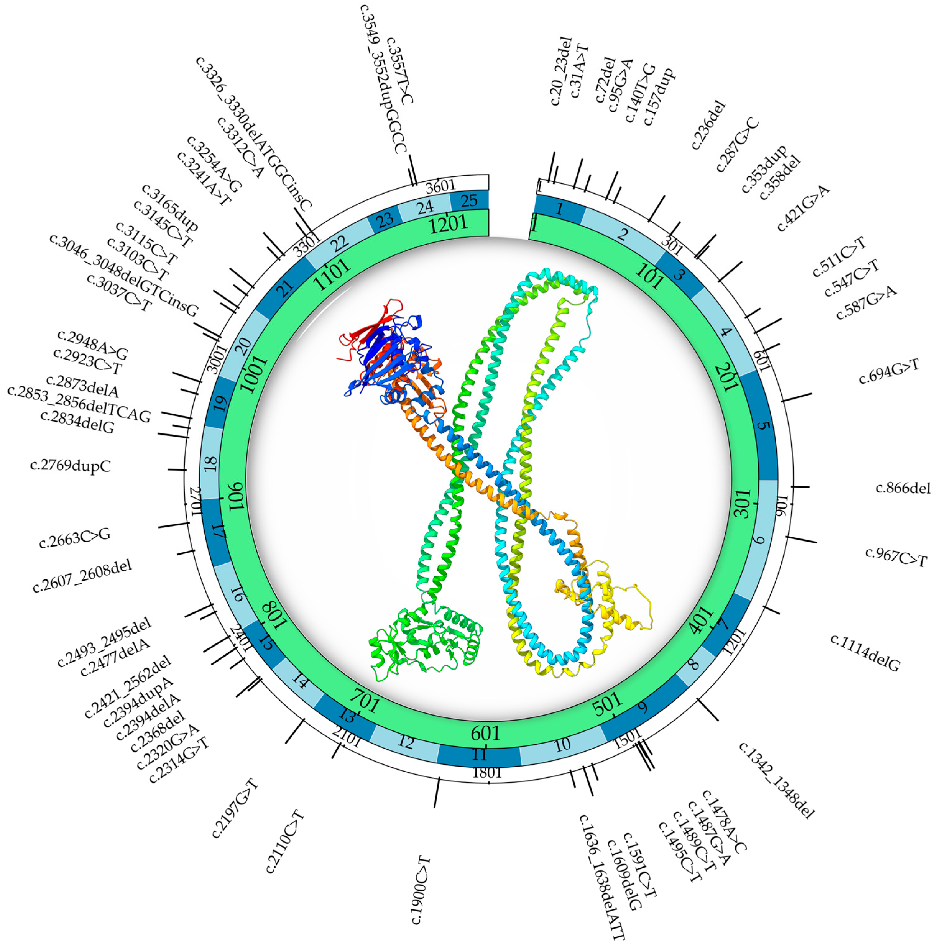
Beyond XCI, other epigenetic modifiers—such as histone acetylation, methylation, and chromatin accessibility—can influence SMC1A expression and cohesin function. These modifiers may interact with the mutational background to shape variable outcomes. Epigenetic differences between tissues (e.g., brain vs. blood) likely explain why some individuals with the same SMC1A mutation suffer from severe epilepsy while others only show mild cognitive impairment [48]. These findings align with the broader understanding that epigenetic plasticity contributes to phenotypic heterogeneity, especially in X-linked developmental disorders. Table 2 summarizes the clinical variants and phenotypes collected from three case series, while Figure 2 reports the variants and their position in the Certified Clinical Documentation Specialist (CCDS) of the SMC1A gene.

4. Clinical Phenotypes of SMC1A-Related Disorders

SMC1A-related disorders encompass a heterogeneous clinical spectrum, ranging from non-classical or attenuated CdLS to DEE and other neurodevelopmental phenotypes.
Genotype–phenotype correlations indicate that the molecular nature of the genetic variant (e.g., missense, in-frame, or LoF) is a major determinant [56]. Missense or small in-frame variants are typically associated with milder phenotypes—often within the CdLS spectrum—characterized by subtle craniofacial features, mild growth restriction, and moderate intellectual disability. Conversely, loss-of-function (LoF) variants include nonsense [56]. Nevertheless, the variant type alone does not fully explain the observed variability. Another factor that can substantially influence phenotypic severity is related to XCI. Since SMC1A resides on the X chromosome, the relative expression of wild-type and mutant alleles may alter the symptomatology in females.

4.1. X-Chromosome Inactivation and Dosage Effects

Skewed XCI may influence SMC1A-related phenotypes, when alterations are present in heterozygosis. When XCI is skewed toward the wild-type allele, functional SMC1A dosage may be preserved, leading to attenuated phenotypes even if a pathogenic variant is present. In contrast, skewing toward the mutant allele or a random XCI pattern can produce a higher proportion of cells with reduced functional SMC1A, driving more severe neurological manifestations, including early-onset, treatment-resistant epilepsy. Considering this, the proportion of cells expressing the mutant versus wild-type allele is critical in determining clinical outcome. This mechanism accounts for some of the variability observed even among individuals with the same variant. Moreover, cellular mosaicism in females may lead to cellular interference, where interactions between wild-type-expressing and mutant-expressing cells generate emergent tissue-level effects, a phenomenon also proposed in other X-linked epilepsies such as the PCDH19 clustering epilepsy [57]. SMC1A variants generally cause more consistent and severe phenotypes, as XCI does not occur to buffer the dosage effect.
Evidence from recent large-scale cohorts supports the clinical relevance of this model. Barañano et al. [10] reported that truncating SMC1A variants were frequently associated with skewed XCI, often correlating with severe DEE, whereas missense variants more often showed random XCI and a broader phenotypic range [19], confirming a predominance of LoF alleles (82.1%). These findings have been observed in a multinational cohort of 35 females, with early seizure onset, high drug resistance, and frequent neurodevelopmental regression [19]. Similarly, Fateh et al. described a patient carrying a novel missense variant who, despite profound developmental delay, exhibited no seizures—underscoring the interplay between variant type, XCI pattern, and clinical presentation [58].
Adding to the complexity, SMC1A is among the minority of X-linked genes that may escape complete inactivation, leading to biallelic expression in some tissues [44], thus further increasing phenotypic variance.

4.2. Cdls vs. SMC1A-DEE: Clinical and Molecular Distinctions

As previously introduced, SMC1A-related disorders exhibit a notable variance spanning from mild CdLS phenotypes to severe DEE cases. While both conditions share a molecular basis in cohesin complex dysfunction, their clinical presentations are distinct.
Notably, while the phenotype of SMC1A-DEE is clearly different from that of CdLS, it bears a strong resemblance to the MECP2-linked RTT, characterized by early-onset, intractable epilepsy and severe-to-profound intellectual disability. While no direct correlation between SMC1A and RTT has been described in the literature, variants in other chromatin regulators, such as HDAC8, SATB2, and HNRNPU, have been associated with RTT-like phenotypes [59]. This evidence underscores the dosage-sensitive role of chromatin-associated proteins in early brain development.
Table 3 summarizes the core phenotypic differences between SMC1A-related CdLS and SMC1A-DEE, highlighting the roles of variant type, XCI pattern, and dosage effects in shaping clinical expression.
The etiology of SMC1A-DEE remains poorly understood, partly due to the limited number of reported cases and the challenges in obtaining brain tissue for direct study. Nevertheless, genotype–phenotype correlations highlighted several variants within SMC1A that are correlated with this pathology, as will be better described in the next section. Literature data also suggested that an X chromosome-specific dosage effect, modulated by XCI, plays a major role in determining disease severity [61]. In unaffected females, wild-type alleles are expressed from both the active (Xa) and inactive (Xi) X chromosomes. In SMC1A-DEE, the impact depends on which X carries the loss-of-function variant and how XCI is patterned. Skewing toward the mutant Xi allows most cells to express the WT allele from Xa, resulting in a modest dosage reduction and milder phenotype. In contrast, skewing toward the mutant Xa produces the largest functional loss and the most severe manifestations. Random XCI leads to a mosaic pattern of WT and mutant allele expression, potentially producing an intermediate phenotype. Such mosaicism may also cause cellular interference, a mechanism also proposed for PCDH19 clustering epilepsy [57], another disorder with drug-resistant seizures and seizure clustering [18].

4.3. Case Series

In the Bozarth series [30], one patient exhibited skewed XCI toward the mutant Xi, resulting in a comparatively milder phenotype than those with random XCI—likely reflecting partial preservation of SMC1A dosage. Additional contributions from genes that escape XCI and are expressed from the Xi may also influence sex-specific differences in brain development and phenotypes associated with X-chromosome aneuploidy. Interestingly, the murine ortholog of SMC1A does not escape XCI in the brain [62], suggesting that mouse models may fail to capture the dosage effects observed in humans. For this reason, In vitro neuronal models derived from patient-specific induced pluripotent stem cells (iPSCs) carrying SMC1A-DEE variants on either the Xa or Xi may provide a more faithful platform for functional studies.
Gibellato [19] and colleagues conducted a multicentre study of 35 female patients with SMC1A-related developmental and DEE from 13 countries [19]. The median age at enrollment was 9.5 years (range, 6 months–34 years). No parental consanguinity was reported, and all pregnancies were spontaneous. Prenatal complications included preeclampsia (5.9%) and intrauterine growth restriction (5.7%). Fetal malformations were detected in 14.3% of cases, most commonly holoprosencephaly and arachnoid cysts. Vaginal delivery occurred in 62.9% of cases, with a mean gestational age of 39 weeks. Most patients had normal anthropometric measures at birth; however, 11.4% were small for gestational age in weight, and 8.6% had microcephaly. Neonatal complications occurred in 51.4% of patients, and 34.3% required neonatal intensive care unit (NICU) admission. Major congenital malformations were present in 40% of cases, affecting the cardiovascular system (20%), central nervous system (CNS) (20%), limbs, urinary tract, and genitalia. Postnatal growth was impaired in ~43% of cases, with persistent underweight or microcephaly. Neurodevelopmental milestones were globally delayed. Independent sitting was achieved by 82.4% at a mean age of 14.5 months, walking by 57.6% at 28.5 months, and first words by 56.7% at 26.9 months, while sentence formation was rare (28.6%). Developmental regression affected 51.4%, involving motor, language, and social skills. Behavioral comorbidities occurred in 37.1% cases, including autism spectrum disorder, hyperactivity, and self-injury. Epilepsy onset occurred at a mean age of 11.8 months, most often with generalized seizures (48.6%), frequent status epilepticus (48.6%), and high rates of drug resistance. Only 34.2% cases achieved good seizure control, typically with polytherapy. The ketogenic diet was used in 20% with variable benefit. Gastrointestinal and nutritional issues were common (62.9%), including feeding difficulties, gastroesophageal reflux disease (GERD) (28.6%), and constipation (74.3%). In total, 37.1% required nasogastric tube feeding, and 22.9% had a gastrostomy. Additional medical comorbidities affected 68.6% of patients. Dysmorphic features were observed in 40%, though none met clinical criteria for classical or non-classical CdLS. Genetic testing, predominantly whole-exome sequencing (88.6%), confirmed pathogenic SMC1A variants in all cases, of which 82.1% were loss-of-function alleles, while 17.9% were missense variants.
In 2024, Fateh and colleagues described three clinical cases, including a nine-year-old girl with a pathogenic SMC1A variant [58]. She was born at 36 weeks of gestation to non-consanguineous parents, with a birth weight of 2300 g and a head circumference of 29 cm. Delivery was by cesarean section, with no reported prenatal exposure to medications, tobacco, or alcohol. Dysmorphic features included micrognathia, synophrys, microcephaly, a low anterior hairline, long curly eyelashes, a thin upper lip, a long philtrum, a depressed nasal bridge, and a high-arched palate. Limb anomalies included syndactyly of the second and third toes, small hands with proximally positioned thumbs, and a single palmar crease. Echocardiography revealed a mild ventricular septal defect, while gastroesophageal function was normal. She had no history of seizures, and her electroencephalogram (EEG) was unremarkable. The patient exhibited failure to thrive and global developmental delay, walking independently at the age of four years with occupational therapy, and speaking only single words at age nine. Chromosome analysis showed a normal female karyotype (46,XX). Whole-exome sequencing identified a novel heterozygous SMC1A variant, NM_006306.4: c.2320G > A (p.Asp774Asn), absent in both parents and in population databases (gnomAD, Iranome). The variant was confirmed by Sanger sequencing and classified as likely pathogenic according to the American College of Medical Genetics and Genomics (ACMG) criteria.
In their study, Borck and colleagues [54] described six cases of CdLS associated with SMC1A mutations. Six male patients from four unrelated families were evaluated, and the following variants were identified: p.Asp831Glu/Gln832del, p.Glu493Ala [63], p.Tyr1085Cys, and p.Arg196His. At birth, the mean weight was 2608 g (±601), significantly lower than the population average (p < 0.05). The mean birth length was 47.0 cm (±2.8; p < 0.01), and the mean occipitofrontal circumference (OFC) was 31.9 cm (±0.5; p < 0.05). Postnatal growth parameters remained markedly reduced. The mean height standard deviation score (SDS) was −2.4 (±0.4; n = 2), the mean weight SDS was −2.3 (±0.7; n = 2), and the mean OFC SDS was −3.4 (±0.9; n = 2). Major congenital malformations were observed in 1 of 6 patients (p < 0.01), with an average of 1.0 malformation per affected individual. Seizures were reported in 5 of 6 cases (p < 0.001). These findings indicate a consistent phenotype characterized by intrauterine and postnatal growth restriction, microcephaly, and a high prevalence of seizures in patients with SMC1L1 missense variants.

5. Diagnostic Approaches in Cornelia de Lange Syndrome and SMC1A-Related Developmental and Epileptic Encephalopathy

CdLS is primarily a cohesinopathy caused by pathogenic variants in genes encoding cohesin subunits, regulatory factors, or proteins that modulate cohesin function. The most implicated genes are NIPBL, SMC1A, SMC3, RAD21, HDAC8, BRD4, and ANKRD11. De novo NIPBL variants account for ~60–65% of clinically ascertained cases while the remaining genes explain a smaller proportion and contribute to phenotypic heterogeneity [64]. International consensus criteria distinguish cardinal and suggestive features to guide clinical recognition. A diagnosis is established by characteristic clinical findings or identification of a pathogenic (or likely pathogenic) heterozygous variant in NIPBL, RAD21, SMC3, or BRD4, or a hemizygous pathogenic variant in HDAC8 or SMC1A [64].
For molecular testing, clinician-directed gene-targeted strategies that include multigene panels or serial single-gene analysis are appropriate when CdLS is strongly suspected. Recommended panels should include NIPBL, SMC1A, HDAC8, SMC3, RAD21, BRD4, and genes causing overlapping phenotypes (e.g., AFF4, ANKRD11, CREBBP, EP300), and should incorporate deletion/duplication analysis. Because somatic mosaicism is relatively frequent, NGS methods capable of detecting low-level mosaic variants—ideally applied to uncultured fibroblasts or other mosaic-enriched tissues (e.g., buccal epithelial cells)—are advised. Serial testing—considered in settings where multigene panels are unavailable—is the first step for individuals with high clinical suspicion. It typically begins with NIPBL (sequence ± deletion/duplication), followed by SMC1A in milder phenotypes, and then BRD4, SMC3, RAD21, and HDAC8 as indicated. When the phenotype is atypical or CdLS is not initially suspected, comprehensive genomic testing—such as exome or genome sequencing—is the preferred diagnostic route [64].
Molecular diagnosis of SMC1A-related DEE relies primarily on NGS strategies, including targeted epilepsy/CdLS gene panels and trio whole-exome or whole-genome sequencing, which have markedly increased detection of de novo SMC1A loss-of-function variants in females presenting with early-onset, drug-resistant seizures and severe neurodevelopmental impairment [10,65]. Because SMC1A is X-linked and can partially escape X-chromosome inactivation, allele-specific expression and X-inactivation analyses (and interpretation of variant dosage) are often informative when correlating genotype with phenotype [2,30]. Given the documented occurrence of post-zygotic mosaicism and the possibility that causal variants may be absent or at very low allele fractions in peripheral blood, diagnostic workups should include deep sequencing capable of mosaic detection and multi-tissue sampling (uncultured fibroblasts, buccal epithelial cells, or other mosaic-enriched tissues) when clinical suspicion remains high despite a negative blood test [66,67]. Complementary assays—copy-number analysis (array comparative genomic hybridization (CGH), multiplex ligation-dependent probe amplification (MLPA), or deletion/duplication algorithms applied to NGS data)—are recommended to exclude intragenic deletions or duplications not detected by standard sequencing [64]. For variants predicted to affect splicing, RNA-based studies (Reverse Transcription Polymerase Chain Reaction (RT-PCR) from appropriate tissues, RNA-seq, or minigene splicing assays) can demonstrate aberrant transcripts and help reclassify variants of uncertain significance. Functional characterization in cellular models (e.g., patient-derived or gene-edited iPSC-derived neurons) and targeted molecular assays (protein expression, allele-specific expression) are increasingly used to validate pathogenicity and to investigate mechanistic effects on neuronal function, although such assays are generally performed in research settings or specialized diagnostic laboratories [68]. Table 4 summarizes diagnostic techniques for SMC1A in DEE in particular the advantages and limitations in clinical practices.

5.1. Current Molecular Strategies

Diagnostic workflows for SMC1A in DEE must combine sensitive sequencing, copy-number interrogation, and strategic tissue selection to maximize detection of both germline and post-zygotic (mosaic) variants. When the clinical picture suggests SMC1A involvement (early-onset, drug-resistant seizures with developmental impairment or CdLS-like features), begin with a clinician-directed multigene NGS panel or trio WES/WGS; panels optimized for epilepsy/CdLS usually afford the best balance of coverage and interpretability, while exome/genome sequencing is preferable for atypical presentations. Multigene panels should explicitly include deletion/duplication analysis and parameters for mosaic calling [64].
Because pathogenic SMC1A variants can be mosaic- and tissue-restricted, peripheral blood alone may be insufficient. Empirical data show that buccal epithelial cells and uncultured skin fibroblasts frequently enrich for mosaic alleles missed in blood; therefore, collect a non-blood specimen (buccal swab or fibroblast biopsy) at the time of initial testing when clinically feasible or pursue reflex testing if blood is negative but suspicion remains high [76]. Multi-tissue sampling increases diagnostic yield and can reveal gonadosomatic mosaicism relevant to recurrence risk [64,76].
Analytical sensitivity depends on sequencing depth and the bioinformatic pipeline. Standard clinical exome/panel pipelines typically detect mosaic variants reliably at moderate variant allele fractions (VAFs)—often ≈5–10%—whereas Sanger sequencing generally fails below ~15–20% VAFs. Detecting VAFs approaching 1% requires targeted ultra-deep amplicon sequencing with robust error-correction and validated limit-of-detection (LLoD). Laboratories should define and report their LLoD and use mosaic-aware variant callers and filtering strategies validated against reference standards. Benchmarking studies recommend selection and parameter optimization specific to anticipated VAF ranges [77].
Complementary assays are essential: CNV analysis (array CGH/MLPA/NGS-based algorithms) to detect intragenic deletions/duplications, RNA studies (RT-PCR, RNA-seq, or minigene assays) for splice variants, and X-inactivation/allele-specific expression tests in females to contextualize variant impact given the X-linkage of the SMC1A gene and potential X-escape. Confirmatory testing of low-level variants should use an orthogonal method (e.g., independent ultra-deep amplicon sequencing). Finally, variant interpretation should follow ACMG-adapted rules for mosaic findings and incorporate phenotypic concordance, tissue distribution, parental testing for recurrence risk, and, when available, functional evidence from RNA or cellular models. Clear laboratory reporting of tissue source, VAF, LLoD, and interpretative caveats is critical for clinical decision-making [66,78]
Multiple studies investigating CdLS and SMC1A-related disorders have demonstrated that a multimodal diagnostic strategy combining conventional and high-resolution molecular methods across different tissues substantially improves variant detection and classification [13,79,80]. Traditional Sanger sequencing and chromosomal microarray analyses remain useful first-line tools but can fail to detect low-level post-zygotic variants or complex rearrangements. In contrast, next-generation sequencing (NGS) approaches, including targeted gene panels, exome/genome sequencing, and ultra-deep amplicon assays, increase sensitivity for single-nucleotide variants and permit mosaic-aware calling when coupled with appropriate bioinformatic pipelines. Complementary techniques such as copy-number interrogation, RNA-based splicing assays, and tissue-specific sampling (e.g., buccal cells, uncultured fibroblasts, bone marrow) further enhance diagnostic yield and enable functional interpretation, while cellular models provide mechanistic validation in selected cases. Collectively, these studies support an integrated, tissue-informed workflow for resolving diagnostically challenging cases.
To address these diverse clinical and economic demands, in their study, Balciuniene et al. [81] developed an exome sequencing (ES)-based test for pediatric epilepsy diagnosis, focusing on an initial analysis of 100 epilepsy genes. The ES-based strategy enables an easy reflex to analysis of the full exome for cases with a negative panel outcome and offers flexibility in modifying the panel’s gene content in response to new gene discoveries or clinical demands [81].
In another study including three patients, prior Sanger sequencing of peripheral blood DNA failed to identify pathogenic variants in any of the five canonical CdLS genes [82]. Subsequent analyses of blood DNA by exome sequencing (patient B) and chromosomal microarray (patients B and C) also did not reveal causative alterations. Consequently, a high-coverage NGS approach was pursued on DNA extracted from bone marrow. For this purpose, a custom AmpliSeq panel (AmpliSeq Designer, Life Technologies, Darmstadt, Germany) was designed for the Ion Torrent PGM platform that incorporated the five established CdLS genes alongside additional candidate loci. The final design was split into two libraries and covered >95% of an approximately 75 kb targeted genomic interval. In summary, these results support a substantial contribution of mosaic NIPBL mutations among individuals with classic CdLS phenotypes who were negative based on conventional Sanger sequencing of blood, or even bone marrow, DNA. Given the limited availability of patient fibroblasts in many diagnostic settings, these findings underscore the urgent need to integrate highly sensitive NGS-based methods into routine molecular diagnostics for CdLS. They therefore strongly recommend applying such mosaic-sensitive sequencing approaches for patients with a clinical diagnosis of CdLS who remain mutation-negative following standard sequencing of lymphocyte or bone marrow DNA [82].

5.2. Limits

While this review aimed to collect and summarize the available literature on the role of SMC1A, a limitation that should be acknowledged is related to the paucity of published studies, which restricts the evidence base. This inevitably narrows the scope of comparative analyses and limits the generalizability of the conclusions, underscoring the need for caution in interpreting the findings. Nevertheless, it should be noted that the pathologies discussed are rare and relatively understudied, which helps explain the limited amount of evidence available in the literature.

6. Conclusions

Variants of the SMC1A gene are associated with a broad clinical spectrum ranging from attenuated CdLS to SMC1A-related DEE. This reflects the diverse functions of the cohesin complex, which include sister-chromatid cohesion, damaged DNA repair, 3D chromatin structure, and gene expression control. Missense variants and small in-frame variants tend to have lower severity and are associated with CdLS-like features, while loss-of-function variants (nonsense, frameshift, and splicing-altering variants) are more frequent in individuals with severe neurodevelopmental disorders and early-onset refractory epilepsy. Sequence variants are not the only source of functional alteration in SMC1A. In females, the partial escape of SMC1A from XCI, together with skewed XCI, generates tissue-specific cellular mosaics that alter effective SMC1A dosage and thereby magnify interindividual phenotypic variability. From a diagnostic standpoint, the best results come from sequential multimodal analysis of XCI and allele-specific expression in women, integrated with NGS (targeted panels, exome/genome), detection of mosaic variants and copy number, and RNA-based tests involving splicing defects and XCI/allele expression dynamics. Due to the potentially tissue-limited nature of pathogenic SMC1A variants, multitissue sampling (e.g., blood plus buccal epithelium or fibroblasts) is often critical to identify low-level mosaicism, classify variants, and provide accurate counseling. In the clinical setting, supportive care remains the mainstay, with symptomatic treatment focused on seizure control, nutritional management, and multidisciplinary neurological development support. SMC1A disorders remain examples of a X-linked, dose-sensitive paradigm in which a combination of genetic lesions, epigenetic context, and tissue mosaicism converge to influence the phenotype, revealing the importance of personalized molecular diagnostics and precision in care pathways.

Author Contributions

Conceptualization, S.B., M.F.A., M.C. and D.S.; methodology, M.F.A., D.S., G.L. and M.C.; software, M.C. and D.S.; validation, M.A.L.R.; writing—original draft preparation M.F.A., D.S., G.L. and M.C. writing—review and editing, S.B. and M.A.L.R.; supervision, S.B. and M.A.L.R.; project administration, S.B. All authors have read and agreed to the published version of the manuscript.

Funding

This research received no external funding.

Institutional Review Board Statement

Not applicable.

Informed Consent Statement

Not applicable.

Data Availability Statement

No new data were created or analyzed in this study.

Conflicts of Interest

The authors declare no conflicts of interest.

Abbreviations

The following abbreviations are used in this manuscript:
ATMAtaxia telangiectasia-mutated
ATRAtaxia telangiectasia- and rad3-related
CdLSCornelia de Lange syndrome
CTCFCCCTC-binding factor
DEEDevelopmental and epileptic encephalopathy
iPSCsInduced pluripotent stem cells
LoFloss of function
NGSNext-generation sequencing
NIPBLNipped-B like protein
RC-1Recombination protein complex
RT-PCRReverse transcription polymerase chain reaction
RTTRett syndrome
SMCsStructural maintenance of chromosomes
SMC1AStructural maintenance of chromosomes 1A
WESWhole-exome sequencing
WGSWhole-genome sequencing
XaX active
XCIX-chromosome inactivation
XiX inactive

References

  1. Yatskevich, S.; Rhodes, J.; Nasmyth, K. Organization of Chromosomal DNA by SMC Complexes. Annu. Rev. Genet. 2019, 53, 445–482. [Google Scholar] [CrossRef]
  2. Parenti, I.; Rovina, D.; Masciadri, M.; Cereda, A.; Azzollini, J.; Picinelli, C.; Limongelli, G.; Finelli, P.; Selicorni, A.; Russo, S.; et al. Overall and allele-specific expression of the SMC1A gene in female Cornelia de Lange syndrome patients and healthy controls. Epigenetics 2014, 9, 973–979. [Google Scholar] [CrossRef]
  3. Yuen, K.C.; Gerton, J.L. Taking cohesin and condensin in context. PLoS Genet. 2018, 14, e1007118. [Google Scholar] [CrossRef] [PubMed]
  4. Mannini, L.; Cucco, F.; Quarantotti, V.; Amato, C.; Tinti, M.; Tana, L.; Frattini, A.; Delia, D.; Krantz, I.D.; Jessberger, R.; et al. SMC1B is present in mammalian somatic cells and interacts with mitotic cohesin proteins. Sci. Rep. 2015, 5, 18472. [Google Scholar] [CrossRef] [PubMed]
  5. Gligoris, T.G.; Scheinost, J.C.; Burmann, F.; Petela, N.; Chan, K.L.; Uluocak, P.; Beckouet, F.; Gruber, S.; Nasmyth, K.; Lowe, J. Closing the cohesin ring: Structure and function of its Smc3-kleisin interface. Science 2014, 346, 963–967. [Google Scholar] [CrossRef]
  6. Schwarzer, W.; Abdennur, N.; Goloborodko, A.; Pekowska, A.; Fudenberg, G.; Loe-Mie, Y.; Fonseca, N.A.; Huber, W.; Haering, C.H.; Mirny, L.; et al. Two independent modes of chromatin organization revealed by cohesin removal. Nature 2017, 551, 51–56. [Google Scholar] [CrossRef]
  7. Cukrov, D.; Newman, T.A.C.; Leask, M.; Leeke, B.; Sarogni, P.; Patimo, A.; Kline, A.D.; Krantz, I.D.; Horsfield, J.A.; Musio, A. Antioxidant treatment ameliorates phenotypic features of SMC1A-mutated Cornelia de Lange syndrome in vitro and in vivo. Hum. Mol. Genet. 2018, 27, 3002–3011. [Google Scholar] [CrossRef]
  8. Musio, A. The multiple facets of the SMC1A gene. Gene 2020, 743, 144612. [Google Scholar] [CrossRef] [PubMed]
  9. Stursberg, S.; Riwar, B.; Jessberger, R. Cloning and characterization of mammalian SMC1 and SMC3 genes and proteins, components of the DNA recombination complexes RC-1. Gene 1999, 228, 1–12. [Google Scholar] [CrossRef] [PubMed]
  10. Baranano, K.W.; Kimball, A.; Fong, S.L.; Egense, A.S.; Hudon, C.; Kline, A.D. Further Characterization of SMC1A Loss of Function Epilepsy Distinct From Cornelia de Lange Syndrome. J. Child. Neurol. 2022, 37, 390–396. [Google Scholar] [CrossRef]
  11. Cooper, D.N.; Ball, E.V.; Stenson, P.D.; Phillips, A.D.; Evans, K.; Heywood, S.; Chapman, M.; Hayden, M.J.; Shiel, J.; Beynon, S.; et al. The Human Gene Mutation Database. Available online: http://www.hgmd.cf.ac.uk/ac/index.php (accessed on 10 August 2025).
  12. Luppino, G.; Wasniewska, M.; Pepe, G.; Morabito, L.A.; Briuglia, S.; Moschella, A.; Franchina, F.; Lugarà, C.; Aversa, T.; Corica, D. Two Years of Growth Hormone Therapy in a Child with Severe Short Stature Due to Overlap Syndrome with a Novel SETD5 Gene Mutation: Case Report and Review of the Literature. Genes 2025, 16, 859. [Google Scholar] [CrossRef] [PubMed]
  13. Mannini, L.; Cucco, F.; Quarantotti, V.; Krantz, I.D.; Musio, A. Mutation spectrum and genotype-phenotype correlation in Cornelia de Lange syndrome. Hum. Mutat. 2013, 34, 1589–1596. [Google Scholar] [CrossRef] [PubMed]
  14. Huisman, S.; Mulder, P.A.; Redeker, E.; Bader, I.; Bisgaard, A.M.; Brooks, A.; Cereda, A.; Cinca, C.; Clark, D.; Cormier-Daire, V.; et al. Phenotypes and genotypes in individuals with SMC1A variants. Am. J. Med. Genet. A 2017, 173, 2108–2125. [Google Scholar] [CrossRef] [PubMed]
  15. Yang, Y.; Chen, L.; Wang, Z.; Ding, Y.; Liu, Y. A De Novo Frameshift Variant in SMC1A Causes Non-Classic Cornelia de Lange Syndrome with Epilepsy: A Case Report and Literature Review. Mol. Genet. Genomic Med. 2025, 13, e70058. [Google Scholar] [CrossRef]
  16. Pepe, G.; Coco, R.; Corica, D.; Luppino, G.; Morabito, L.A.; Lugara, C.; Abbate, T.; Zirilli, G.; Aversa, T.; Stagi, S.; et al. Endocrine disorders in Rett syndrome: A systematic review of the literature. Front. Endocrinol. 2024, 15, 1477227. [Google Scholar] [CrossRef]
  17. Parmeggiani, L.; Stanzial, F.; Menna, E.; Boni, E.; Manzoni, F.; Benedicenti, F.; Pellegrin, S. Early onset developmental and epileptic encephalopathy and Rett-like phenotype in a 15-year-old girl affected by Cornelia de Lange syndrome type 2 due to a SMC1A gene mutation. Epilepsy Behav. Rep. 2023, 24, 100634. [Google Scholar] [CrossRef]
  18. Symonds, J.D.; Joss, S.; Metcalfe, K.A.; Somarathi, S.; Cruden, J.; Devlin, A.M.; Donaldson, A.; DiDonato, N.; Fitzpatrick, D.; Kaiser, F.J.; et al. Heterozygous truncation mutations of the SMC1A gene cause a severe early onset epilepsy with cluster seizures in females: Detailed phenotyping of 10 new cases. Epilepsia 2017, 58, 565–575. [Google Scholar] [CrossRef]
  19. Gibellato, E.; Cianci, P.; Mariani, M.; Parma, B.; Huisman, S.; Smigiel, R.; Bisgaard, A.M.; Massa, V.; Gervasini, C.; Moretti, A.; et al. SMC1A epilepsy syndrome: Clinical data from a large international cohort. Am. J. Med. Genet. A 2024, 194, e63577. [Google Scholar] [CrossRef]
  20. De Koninck, M.; Losada, A. Cohesin Mutations in Cancer. Cold Spring Harb. Perspect. Med. 2016, 6, a026476. [Google Scholar] [CrossRef]
  21. Perea-Resa, C.; Wattendorf, L.; Marzouk, S.; Blower, M.D. Cohesin: Behind dynamic genome topology and gene expression reprogramming. Trends Cell Biol. 2021, 31, 760–773. [Google Scholar] [CrossRef]
  22. Grubert, F.; Srivas, R.; Spacek, D.V.; Kasowski, M.; Ruiz-Velasco, M.; Sinnott-Armstrong, N.; Greenside, P.; Narasimha, A.; Liu, Q.; Geller, B.; et al. Landscape of cohesin-mediated chromatin loops in the human genome. Nature 2020, 583, 737–743. [Google Scholar] [CrossRef]
  23. Sole-Ferran, M.; Losada, A. Cohesin in 3D: Development, differentiation, and disease. Genes. Dev. 2025, 39, 679–696. [Google Scholar] [CrossRef]
  24. Horsfield, J.A. Full circle: A brief history of cohesin and the regulation of gene expression. FEBS J. 2023, 290, 1670–1687. [Google Scholar] [CrossRef]
  25. Mannini, L.; Liu, J.; Krantz, I.D.; Musio, A. Spectrum and consequences of SMC1A mutations: The unexpected involvement of a core component of cohesin in human disease. Hum. Mutat. 2010, 31, 5–10. [Google Scholar] [CrossRef]
  26. Schar, P.; Fasi, M.; Jessberger, R. SMC1 coordinates DNA double-strand break repair pathways. Nucleic Acids Res. 2004, 32, 3921–3929. [Google Scholar] [CrossRef] [PubMed]
  27. Zhang, Y.F.; Jiang, R.; Li, J.D.; Zhang, X.Y.; Zhao, P.; He, M.; Zhang, H.Z.; Sun, L.P.; Shi, D.L.; Zhang, G.X.; et al. SMC1A knockdown induces growth suppression of human lung adenocarcinoma cells through G1/S cell cycle phase arrest and apoptosis pathways in vitro. Oncol. Lett. 2013, 5, 749–755. [Google Scholar] [CrossRef]
  28. Ma, Z.; Lin, M.; Li, K.; Fu, Y.; Liu, X.; Yang, D.; Zhao, Y.; Zheng, J.; Sun, B. Knocking down SMC1A inhibits growth and leads to G2/M arrest in human glioma cells. Int. J. Clin. Exp. Pathol. 2013, 6, 862–869. [Google Scholar] [PubMed]
  29. Di Nardo, M.; Astigiano, S.; Baldari, S.; Pallotta, M.M.; Porta, G.; Pigozzi, S.; Antonini, A.; Emionite, L.; Frattini, A.; Valli, R.; et al. The synergism of SMC1A cohesin gene silencing and bevacizumab against colorectal cancer. J. Exp. Clin. Cancer Res. 2024, 43, 49. [Google Scholar] [CrossRef]
  30. Bozarth, X.L.; Lopez, J.; Fang, H.; Lee-Eng, J.; Duan, Z.; Deng, X. Phenotypes and Genotypes in Patients with SMC1A-Related Developmental and Epileptic Encephalopathy. Genes 2023, 14, 852. [Google Scholar] [CrossRef] [PubMed]
  31. Kline, A.D.; Moss, J.F.; Selicorni, A.; Bisgaard, A.M.; Deardorff, M.A.; Gillett, P.M.; Ishman, S.L.; Kerr, L.M.; Levin, A.V.; Mulder, P.A.; et al. Diagnosis and management of Cornelia de Lange syndrome: First international consensus statement. Nat. Rev. Genet. 2018, 19, 649–666. [Google Scholar] [CrossRef]
  32. Gil-Salvador, M.; Latorre-Pellicer, A.; Lucia-Campos, C.; Arnedo, M.; Darnaude, M.T.; Diaz de Bustamante, A.; Villares, R.; Palma Milla, C.; Puisac, B.; Musio, A.; et al. Case report: A novel case of parental mosaicism in SMC1A gene causes inherited Cornelia de Lange syndrome. Front. Genet. 2022, 13, 993064. [Google Scholar] [CrossRef]
  33. Chen, J.; Floyd, E.N.; Dawson, D.S.; Rankin, S. Cornelia de Lange Syndrome mutations in SMC1A cause cohesion defects in yeast. Genetics 2023, 225, iyad159. [Google Scholar] [CrossRef]
  34. Costantino, L.; Hsieh, T.S.; Lamothe, R.; Darzacq, X.; Koshland, D. Cohesin residency determines chromatin loop patterns. eLife 2020, 9, e59889. [Google Scholar] [CrossRef]
  35. Selivanovskiy, A.V.; Molodova, M.N.; Khrameeva, E.E.; Ulianov, S.V.; Razin, S.V. Liquid condensates: A new barrier to loop extrusion? Cell Mol. Life Sci. 2025, 82, 80. [Google Scholar] [CrossRef]
  36. Stortz, M.; Presman, D.M.; Levi, V. Transcriptional condensates: A blessing or a curse for gene regulation? Commun. Biol. 2024, 7, 187. [Google Scholar] [CrossRef] [PubMed]
  37. Morales, C.; Ruiz-Torres, M.; Rodriguez-Acebes, S.; Lafarga, V.; Rodriguez-Corsino, M.; Megias, D.; Cisneros, D.A.; Peters, J.M.; Mendez, J.; Losada, A. PDS5 proteins are required for proper cohesin dynamics and participate in replication fork protection. J. Biol. Chem. 2020, 295, 146–157. [Google Scholar] [CrossRef] [PubMed]
  38. Alonso-Gil, D.; Losada, A. NIPBL and cohesin: New take on a classic tale. Trends Cell Biol. 2023, 33, 860–871. [Google Scholar] [CrossRef] [PubMed]
  39. Gause, M.; Webber, H.A.; Misulovin, Z.; Haller, G.; Rollins, R.A.; Eissenberg, J.C.; Bickel, S.E.; Dorsett, D. Functional links between Drosophila Nipped-B and cohesin in somatic and meiotic cells. Chromosoma 2008, 117, 51–66. [Google Scholar] [CrossRef]
  40. Faure, A.J.; Schmidt, D.; Watt, S.; Schwalie, P.C.; Wilson, M.D.; Xu, H.; Ramsay, R.G.; Odom, D.T.; Flicek, P. Cohesin regulates tissue-specific expression by stabilizing highly occupied cis-regulatory modules. Genome Res. 2012, 22, 2163–2175. [Google Scholar] [CrossRef]
  41. Brooker, A.S.; Berkowitz, K.M. The roles of cohesins in mitosis, meiosis, and human health and disease. Methods Mol. Biol. 2014, 1170, 229–266. [Google Scholar] [CrossRef] [PubMed]
  42. Sarogni, P.; Palumbo, O.; Servadio, A.; Astigiano, S.; D’Alessio, B.; Gatti, V.; Cukrov, D.; Baldari, S.; Pallotta, M.M.; Aretini, P.; et al. Overexpression of the cohesin-core subunit SMC1A contributes to colorectal cancer development. J. Exp. Clin. Cancer Res. 2019, 38, 108. [Google Scholar] [CrossRef] [PubMed]
  43. Remeseiro, S.; Cuadrado, A.; Gomez-Lopez, G.; Pisano, D.G.; Losada, A. A unique role of cohesin-SA1 in gene regulation and development. EMBO J. 2012, 31, 2090–2102. [Google Scholar] [CrossRef]
  44. Liu, J.; Zhang, Z.; Bando, M.; Itoh, T.; Deardorff, M.A.; Clark, D.; Kaur, M.; Tandy, S.; Kondoh, T.; Rappaport, E.; et al. Transcriptional dysregulation in NIPBL and cohesin mutant human cells. PLoS Biol. 2009, 7, e1000119. [Google Scholar] [CrossRef]
  45. Izumi, K.; Nakato, R.; Zhang, Z.; Edmondson, A.C.; Noon, S.; Dulik, M.C.; Rajagopalan, R.; Venditti, C.P.; Gripp, K.; Samanich, J.; et al. Germline gain-of-function mutations in AFF4 cause a developmental syndrome functionally linking the super elongation complex and cohesin. Nat. Genet. 2015, 47, 338–344. [Google Scholar] [CrossRef]
  46. Slavin, T.P.; Lazebnik, N.; Clark, D.M.; Vengoechea, J.; Cohen, L.; Kaur, M.; Konczal, L.; Crowe, C.A.; Corteville, J.E.; Nowaczyk, M.J.; et al. Germline mosaicism in Cornelia de Lange syndrome. Am. J. Med. Genet. A 2012, 158A, 1481–1485. [Google Scholar] [CrossRef]
  47. Gardner, R.M.; Sutherland, G.R.; Shaffer, L.G. Chromosome Abnormalities and Genetic Counseling; Oxford University Press (OUP): New York, NY, USA, 2012. [Google Scholar]
  48. Jansen, S.; Kleefstra, T.; Willemsen, M.H.; de Vries, P.; Pfundt, R.; Hehir-Kwa, J.Y.; Gilissen, C.; Veltman, J.A.; de Vries, B.B.; Vissers, L.E. De novo loss-of-function mutations in X-linked SMC1A cause severe ID and therapy-resistant epilepsy in females: Expanding the phenotypic spectrum. Clin. Genet. 2016, 90, 413–419. [Google Scholar] [CrossRef]
  49. Lebrun, N.; Lebon, S.; Jeannet, P.Y.; Jacquemont, S.; Billuart, P.; Bienvenu, T. Early-onset encephalopathy with epilepsy associated with a novel splice site mutation in SMC1A. Am. J. Med. Genet. A 2015, 167A, 3076–3081. [Google Scholar] [CrossRef] [PubMed]
  50. Ye, Y.Z.; Duan, J.; Hu, Z.Q.; Cao, D.Z.; Liao, J.X.; Chen, L. Developmental and epileptic encephalopathy 85 caused by SMC1A gene truncating variation: 4 cases report and literature review. Zhonghua Er Ke Za Zhi 2022, 60, 583–587. [Google Scholar] [CrossRef] [PubMed]
  51. Generoso, S.F.; Neguembor, M.V.; Hershberg, E.A.; Sadreyev, R.I.; Kurimoto, K.; Yabuta, Y.; Ricci, R.; Audergon, P.; Bauer, M.; Saitou, M.; et al. Cohesin controls X chromosome structure remodeling and X-reactivation during mouse iPSC-reprogramming. Proc. Natl. Acad. Sci. USA 2023, 120, e2213810120. [Google Scholar] [CrossRef]
  52. Schierding, W.; Vickers, M.H.; O’Sullivan, J.M.; Cutfield, W.S. 9-Epigenetics. In Fetal and Neonatal Physiology, 5th ed.; Polin, R.A., Abman, S.H., Rowitch, D.H., Benitz, W.E., Fox, W.W., Eds.; Elsevier: Amsterdam, The Netherlands, 2017; pp. 89–100.e103. [Google Scholar]
  53. Brand, B.A.; Blesson, A.E.; Smith-Hicks, C.L. The Impact of X-Chromosome Inactivation on Phenotypic Expression of X-Linked Neurodevelopmental Disorders. Brain Sci. 2021, 11, 904. [Google Scholar] [CrossRef] [PubMed]
  54. Borck, G.; Zarhrate, M.; Bonnefont, J.P.; Munnich, A.; Cormier-Daire, V.; Colleaux, L. Incidence and clinical features of X-linked Cornelia de Lange syndrome due to SMC1L1 mutations. Hum. Mutat. 2007, 28, 205–206. [Google Scholar] [CrossRef]
  55. Pettersen, E.F.; Goddard, T.D.; Huang, C.C.; Meng, E.C.; Couch, G.S.; Croll, T.I.; Morris, J.H.; Ferrin, T.E. UCSF ChimeraX: Structure visualization for researchers, educators, and developers. Protein Sci. 2021, 30, 70–82. [Google Scholar] [CrossRef]
  56. Moss, J.; Penhallow, J.; Ansari, M.; Barton, S.; Bourn, D.; FitzPatrick, D.R.; Goodship, J.; Hammond, P.; Roberts, C.; Welham, A.; et al. Genotype-phenotype correlations in Cornelia de Lange syndrome: Behavioral characteristics and changes with age. Am. J. Med. Genet. A 2017, 173, 1566–1574. [Google Scholar] [CrossRef] [PubMed]
  57. Moncayo, J.A.; Ayala, I.N.; Argudo, J.M.; Aguirre, A.S.; Parwani, J.; Pachano, A.; Ojeda, D.; Cordova, S.; Mora, M.G.; Tapia, C.M.; et al. Understanding Protein Protocadherin-19 (PCDH19) Syndrome: A Literature Review of the Pathophysiology. Cureus 2022, 14, e25808. [Google Scholar] [CrossRef]
  58. Tehrani Fateh, S.; Mohammad Zadeh, N.; Salehpour, S.; Hashemi-Gorji, F.; Omidi, A.; Sadeghi, H.; Mirfakhraie, R.; Moghimi, P.; Keyvanfar, S.; Mohammadi Sarvaleh, S.; et al. Comprehensive review and expanding the genetic landscape of Cornelia-de-Lange spectrum: Insights from novel mutations and skin biopsy in exome-negative cases. BMC Med. Genom. 2024, 17, 20. [Google Scholar] [CrossRef]
  59. Spagnoli, C.; Fusco, C.; Pisani, F. Rett Syndrome Spectrum in Monogenic Developmental-Epileptic Encephalopathies and Epilepsies: A Review. Genes 2021, 12, 1157. [Google Scholar] [CrossRef] [PubMed]
  60. Goldstein, J.H.; Tim-Aroon, T.; Shieh, J.; Merrill, M.; Deeb, K.K.; Zhang, S.; Bass, N.E.; Bedoyan, J.K. Novel SMC1A frameshift mutations in children with developmental delay and epilepsy. Eur. J. Med. Genet. 2015, 58, 562–568. [Google Scholar] [CrossRef] [PubMed]
  61. Fang, H.; Deng, X.; Disteche, C.M. X-factors in human disease: Impact of gene content and dosage regulation. Hum. Mol. Genet. 2021, 30, R285–R295. [Google Scholar] [CrossRef]
  62. Berletch, J.B.; Ma, W.; Yang, F.; Shendure, J.; Noble, W.S.; Disteche, C.M.; Deng, X. Escape from X inactivation varies in mouse tissues. PLoS Genet. 2015, 11, e1005079. [Google Scholar] [CrossRef]
  63. Musio, A.; Selicorni, A.; Focarelli, M.L.; Gervasini, C.; Milani, D.; Russo, S.; Vezzoni, P.; Larizza, L. X-linked Cornelia de Lange syndrome owing to SMC1L1 mutations. Nat. Genet. 2006, 38, 528–530. [Google Scholar] [CrossRef]
  64. Deardorff, M.A.; Noon, S.E.; Krantz, I.D. Cornelia de Lange Syndrome; University of Washington: Seattle, WA, USA, 2020. [Google Scholar]
  65. Vetri, L.; Cali, F.; Saccone, S.; Vinci, M.; Chiavetta, N.V.; Carotenuto, M.; Roccella, M.; Costanza, C.; Elia, M. Whole Exome Sequencing as a First-Line Molecular Genetic Test in Developmental and Epileptic Encephalopathies. Int. J. Mol. Sci. 2024, 25, 1146. [Google Scholar] [CrossRef]
  66. Gonzalez Garcia, A.; Malone, J.; Li, H. A novel mosaic variant on SMC1A reported in buccal mucosa cells, albeit not in blood, of a patient with Cornelia de Lange-like presentation. Cold Spring Harb. Mol. Case Stud. 2020, 6, a005322. [Google Scholar] [CrossRef]
  67. Gil-Salvador, M.; Trujillano, L.; Lucia-Campos, C.; Del Rincon, J.; Pamplona, P.; Arnedo, M.; Puisac, B.; Marcos-Alcalde, I.; Gomez-Puertas, P.; Ramos, F.J.; et al. Postzygotic mosaicism in SMC1A and the first reported case of a female with Cornelia de Lange syndrome. Sci. Rep. 2025, 15, 20772. [Google Scholar] [CrossRef]
  68. Simkin, D.; Kiskinis, E. Modeling Pediatric Epilepsy Through iPSC-Based Technologies. Epilepsy Curr. 2018, 18, 240–245. [Google Scholar] [CrossRef]
  69. Zou, H.; Zhang, Q.; Liao, J.; Zou, D.; Hu, Z.; Li, B.; Chen, L.; Wen, J.; Zhao, X.; Zhang, V.W.; et al. Diagnostic efficiency of exome-based sequencing in pediatric patients with epilepsy. Front. Genet. 2024, 15, 1496411. [Google Scholar] [CrossRef] [PubMed]
  70. Crowgey, E.L.; Mahajan, N.; Wong, W.H.; Gopalakrishnapillai, A.; Barwe, S.P.; Kolb, E.A.; Druley, T.E. Error-corrected sequencing strategies enable comprehensive detection of leukemic mutations relevant for diagnosis and minimal residual disease monitoring. BMC Med. Genom. 2020, 13, 32. [Google Scholar] [CrossRef] [PubMed]
  71. Clement, K.; Farouni, R.; Bauer, D.E.; Pinello, L. AmpUMI: Design and analysis of unique molecular identifiers for deep amplicon sequencing. Bioinformatics 2018, 34, i202–i210. [Google Scholar] [CrossRef] [PubMed]
  72. Royer-Bertrand, B.; Cisarova, K.; Niel-Butschi, F.; Mittaz-Crettol, L.; Fodstad, H.; Superti-Furga, A. CNV Detection from Exome Sequencing Data in Routine Diagnostics of Rare Genetic Disorders: Opportunities and Limitations. Genes 2021, 12, 1427. [Google Scholar] [CrossRef]
  73. Moreno-Cabrera, J.M.; Del Valle, J.; Castellanos, E.; Feliubadalo, L.; Pineda, M.; Brunet, J.; Serra, E.; Capella, G.; Lazaro, C.; Gel, B. Evaluation of CNV detection tools for NGS panel data in genetic diagnostics. Eur. J. Hum. Genet. 2020, 28, 1645–1655. [Google Scholar] [CrossRef]
  74. Zhang, X.; Cai, M.; Ma, Y.; Chen, J.; Huang, S.; Cai, M.; Ding, Y.; Ma, D.; Gao, Q.; Hu, X.; et al. Minigene Assay as an Effective Molecular Diagnostic Strategy in Determining the Pathogenicity of Noncanonical Splice-Site Variants in FLCN. J. Mol. Diagn. 2023, 25, 110–120. [Google Scholar] [CrossRef]
  75. Weng, O.Y.; Li, Y.; Wang, L.Y. Modeling Epilepsy Using Human Induced Pluripotent Stem Cells-Derived Neuronal Cultures Carrying Mutations in Ion Channels and the Mechanistic Target of Rapamycin Pathway. Front. Mol. Neurosci. 2022, 15, 810081. [Google Scholar] [CrossRef]
  76. Francis, D.I.; Stark, Z.; Scheffer, I.E.; Tan, T.Y.; Murali, K.; Gallacher, L.; Amor, D.J.; Goel, H.; Downie, L.; Stutterd, C.A.; et al. Comparing saliva and blood for the detection of mosaic genomic abnormalities that cause syndromic intellectual disability. Eur. J. Hum. Genet. 2023, 31, 521–525. [Google Scholar] [CrossRef]
  77. Kim, S.H.; Kwon, S.S.; Park, M.R.; Lee, H.A.; Kim, J.H.; Cha, J.; Kim, S.; Baek, S.T.; Kim, S.H.; Lee, J.S.; et al. Detecting Low-Variant Allele Frequency Mosaic Pathogenic Variants of NF1, TSC2, and AKT3 Genes from Blood in Patients with Neurodevelopmental Disorders. J. Mol. Diagn. 2023, 25, 583–591. [Google Scholar] [CrossRef]
  78. Rehm, H.L.; Bale, S.J.; Bayrak-Toydemir, P.; Berg, J.S.; Brown, K.K.; Deignan, J.L.; Friez, M.J.; Funke, B.H.; Hegde, M.R.; Lyon, E.; et al. ACMG clinical laboratory standards for next-generation sequencing. Genet. Med. 2013, 15, 733–747. [Google Scholar] [CrossRef]
  79. Barisic, I.; Tokic, V.; Loane, M.; Bianchi, F.; Calzolari, E.; Garne, E.; Wellesley, D.; Dolk, H.; Group, E.W. Descriptive epidemiology of Cornelia de Lange syndrome in Europe. Am. J. Med. Genet. A 2008, 146A, 51–59. [Google Scholar] [CrossRef] [PubMed]
  80. Gil-Rodriguez, M.C.; Deardorff, M.A.; Ansari, M.; Tan, C.A.; Parenti, I.; Baquero-Montoya, C.; Ousager, L.B.; Puisac, B.; Hernandez-Marcos, M.; Teresa-Rodrigo, M.E.; et al. De novo heterozygous mutations in SMC3 cause a range of Cornelia de Lange syndrome-overlapping phenotypes. Hum. Mutat. 2015, 36, 454–462. [Google Scholar] [CrossRef] [PubMed]
  81. Balciuniene, J.; DeChene, E.T.; Akgumus, G.; Romasko, E.J.; Cao, K.; Dubbs, H.A.; Mulchandani, S.; Spinner, N.B.; Conlin, L.K.; Marsh, E.D.; et al. Use of a Dynamic Genetic Testing Approach for Childhood-Onset Epilepsy. JAMA Netw. Open 2019, 2, e192129. [Google Scholar] [CrossRef] [PubMed]
  82. Braunholz, D.; Obieglo, C.; Parenti, I.; Pozojevic, J.; Eckhold, J.; Reiz, B.; Braenne, I.; Wendt, K.S.; Watrin, E.; Vodopiutz, J.; et al. Hidden mutations in Cornelia de Lange syndrome limitations of sanger sequencing in molecular diagnostics. Hum. Mutat. 2015, 36, 26–29. [Google Scholar] [CrossRef]
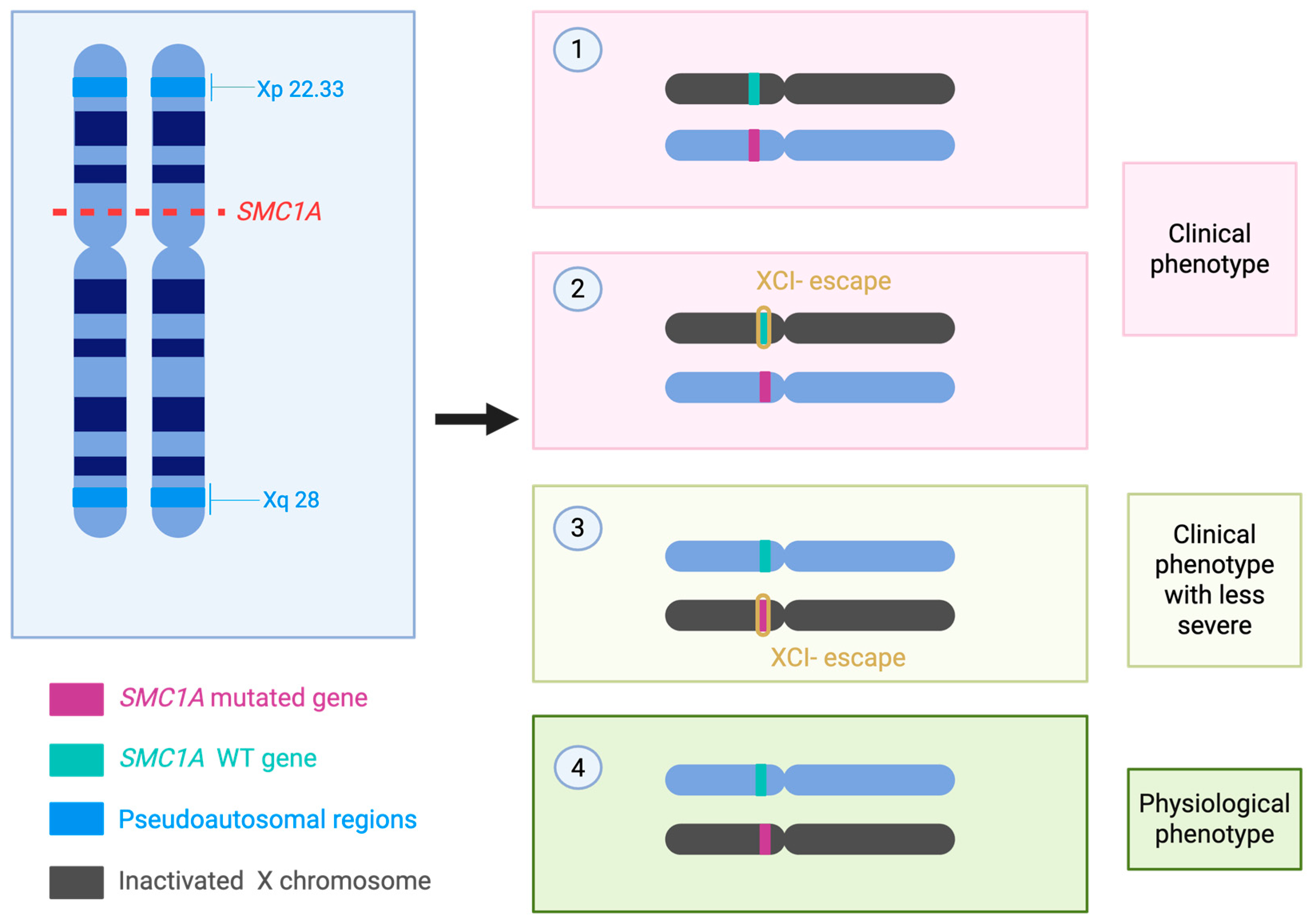
Figure 1. Schematic overview of the impact of SMC1A mutations and X-chromosome inactivation (XCI) escape on phenotypic expression. Mutant alleles may partially escape silencing, leading to variable gene dosage. This mechanism contributes to the wide clinical spectrum observed in affected individuals, ranging from physiological phenotypes—defined here as the absence of clinically detectable manifestations, due to sufficient dosage compensation—to milder clinical presentations and severe clinical phenotypes [30]. In the figure we illustrate the four possible events (1) WT SMC1A-carrying chromosome is silenced, while Mutated SMC1A-carrying chromosome is transcriptionally active. It leads to the production of the mutated protein and thus leads to the pathological phenotype. (2) WT SMC1A-carrying chromosome is silenced but SMC1A loci partially escapes X inactivation, while Mutated SMC1A-carrying chromosome is transcriptionally active. It leads to the production of both the mutated and WT (to a less extent) protein and thus leads to the pathological phenotype. (3) WT SMC1A-carrying chromosome is transcriptionally active, while the Mutated SMC1A-carrying chromosome is silenced with SMC1A loci partially escaping X inactivation. It leads to the production of both the WT and, to a lesser extent, the mutated protein. Being the WT SMC1A the protein mainly expresses, the phenotype is less severe, even though is still pathological. (4) the WT SMC1A-carrying chromosome is transcriptionally active, while the Mutated SMC1A-carrying chromosome is silenced. The complete inactivation of the Mutated SMC1A-carrying chromosome leads to a physiological phenotype. WT: wild type.
Genes 16 01196 g001
Figure 2. In Figure 2. We report the discussed variants and the positions relative to the CCDS of the SMC1A gene. From the outside to the inside, the first ring indicates the nucleotide positions, the second ring indicates the exons, and the third ring indicates the amino acid positions. At the center, we report the 3D structure of SMC1A encoded protein. CCDS data were retrieved from the NCBI database, CCDS ID: CCDS14352.1. The 3D structure was based on the crystallographic information of the cohesin complex from UniProt, PDB ID: 6WG3. Only chain A, which represents the SMC1A protein, is reported in the figure. The PDB file was opened and modified with the UCSF ChimeraX software version 1.10.1 [55].
Genes 16 01196 g002
Table 1. SMCA1 mutation and biological pathways.
Biological PathwayEffect of SMC1A MutationReferences
Chromatid cohesionChromosomal instability and segregation errors[25]
DNA damage repair
(Homologous recombination HR, Non homologous end joining NHEJ)
Reduced repair fidelity, genomic instability[26]
Gene transcription and chromatinMisregulation of developmental genes and epigenetic control[8]
Cell cycle controlG1/S or G2/M arrest, reduced proliferation, increased apoptosis[27,28,29]
Neural developmentEpilepsy, intellectual disability, Rett-like features in females[30]
Table 2. Mutations and associated phenotypes reported in the literature.
#Nucleotide ChangesAmino Acid ChangesTypeXCIAge of Seizure OnsetSpeechIDWalkingMRI
1c.20_23delIle7Argfs * 42FrameshiftSkewed 81:1915 moNoneYesNoN/A
2c.31A > TAsn11TyrMissenseN/A2.5 moN/AYes
Yes
N/ANormal
3c.140T > GPhe47CysMissenseRandom 7:263 moNoneYes
Yes
NoN/A
4c.157dupThr53AsnfsX34FrameshiftN/A5 moN/AYes
Yes
N/ACerebral volume loss
5c.287G > CArg96ProMissenseRandom 7:2618 moN/AYes
Yes
N/AN/A
6c.421G > AGlu141LysMissenseHighly skewed 100:02.5 moNoneYes
Yes
N/AN/A
7c.511C > TArg171TerNonsenseRandom4 wkNoneYes
Yes
NoNormal
8c.615G > AGlu2055 *Splice-siteN/AP3 (18) 4 mo; P12 (22): 13 moNoneYes
Yes
NoP3: small hemorrhage along with posterior falx and tentorium
9c.615G + 1G > CGlu2055 *Splice-siteModerately skewed 83:171 moN/AYes
Yes
NoN/A
10c.615 + 5G > AGlu2055 *Splice-siteRandom4 moNoneYesNoNormal
11c.694G > TGlu232TerNonsenseN/A4 moN/AYesN/AN/A
12c.1113 + 1G > AGln371fs *Splice-siteN/A7 wkNoneYesN/AN/A
13c.1114delGVal372TerNonsenseN/A7 wkNoneYesN/AN/A
14c.1487G > AArg496HisMissenseSkewed in patient P335 molimitedYesYesN/A
15c.1489C > TArg497TerNonsenseN/A4 moNoneYesN/AN/A
16c.1495C > TArg499TerNonsenseN/A40 moNoneYesYesMicroform of HPE, ventricular ectasia
17c.1591C > TGln531TerNonsenseN/A15 moNoneYesYesNormal
18c.1609delGVal537Phefs * 42FrameshiftN/A5 moNoneYesNoN/A
19c.1636_1638delATTIle546delIn-frameModerately skewed 86:14, Mosaic2 moNoneYesNoN/A
20c.1900C > TGln634TerNonsenseN/A3 moN/AYesN/AN/A
21c.1911 + 1G > TThr638Valfs * 48Splice-siteN/ANeonateNoneYesnoSmall frontal lobe, thin CC
22c.2197G > TGlu733TerNonsenseRandom5 moNoneYesNoVolume loss
23c.236delAsn788Lysfs * 10FrameshiftRandom9 moNoneYesNoSlightly enlarged ventricles, hypoplastic cerebellar vermis
24c.2394delALys798Asnfs * 31FrameshiftN/AN/AN/AYesN/ASemi-lobar HPE
25c.2394dupAArg799fsFrameshiftN/A4 moNoneYesN/AN/A
26c.2421_2562delLeu808Argfs * 6FrameshiftModerately skewed 85:152 moNoneYesN/AMild periventricular white matter abnormalities
27c.2477delAp825fsFrameshiftN/A28 mo in P8; <1 mo in P9P8: NoneP9: NoneP8: YesP9: normal; P8: hemi-lobar HPE
28c.2663C > GArg898GlyMissenseSkewed; P1/P225 mo; P1 NoneYesP1: Yes; P2: normal until onsetP2: cerebellar atrophy
29c.2769dupCSer924Glnfs * 2FrameshiftN/A24 moN/AYesN/AN/A
30c.2834delGGly945Lysfs * 19FrameshiftN/AN/AN/AYesN/ASemi-lobar HPE
31c.2853_2856delTCAGSer951Argfs * 12FrameshiftSkewed4 moN/AseverenoMild ventriculomegaly
32c.2873delAGln958Argfs * 6FrameshiftRandom3 moNoneYesNoN/A
33c.2923C > TArg975TerNonsense 5 moModerate to severenoYesNormal
34c.3046_3048delGTCinsGVal1016Alafs * 28FrameshiftN/ANeonateNoneYesNoThin abnormal CC and minimal cerebellar atrophy
35c.3115C > TGln1039TerNonsenseRandom 76:242 moNoneYesNoN/A
36c.3145C > TArg1049TerNonsenseN/A5–6 wkNoneYesnoCerebral volume loss
37c.3241A > TIle1081PheMissenseN/A4 yrNoneYesYesMildly prominent lateral ventricles
38c.3285 + 1G > Cp1095Splice-siteN/ANot reportedN/AYesN/AMiddle interhemispheric variant, HPE
39c.3312C > ATyr1107TerNonsenseN/A12 yrNormal before SEYesYesNormal
40c.3326_3330delATGGCinsCAsp1109Alafs * 102FrameshiftN/A6 moNoneYesnoSmall cavum septum vergae
41c.3549_3552dupGGCCIle1185glyfs * 23FrameshiftRandom in P1 (59)-N/A-P3-(22)17 mo P2-; 16 (59)mo P13-(22)N/A-Goldstein; P13-Baranano-limitedYesN/A-; P13(59)(22)-YesP2 (59): mild enlarged extra-axial spaces and slight thinning of CC
42c.2320G > AAsp774AsnMissenseN/AN/AdelayYesAt 4 yrN/A
43c.3103C > TArg1035TerNonsense
44c.1342_1348delSer448Lysfs * 6Frameshift/Truncating
44c.967C > TGln323TerNonsense
45c.2368delArg790Glyfs * 8Frameshift/Truncating
46c.866delSer289TerNonsense
473428bp Deletion-Microdeletion
48c.2110C > TGln704TerNonsense
49c.3557T > CVal1186AlaMissense
50c.358delGlu120Asnfs * 2Frameshift/Truncating
51c.72delGln25Argfs * 25Frameshift/Truncating
52c.3165dupLys1056Insfs * 13Frameshift/Truncating
53c.353dupSer118Argfs * 2Frameshift/Truncating
54c.3037C > TGln1013TerNonsense
55c.2314G > TVal772LeuMissense
56c.2948A > GTyr983CysMissense
57Ex1 del-Microdeletion
58c.95G > AGly326GluMissense
59c.2607_2608delGln869Hisfs * 17Frameshift/Truncating
60c.547C > TGln183TerNonsense
61c.2493_2496delAsp831Glu/Gln832delMissense/Del In-frame
62c.1478A > CGlu493AlaMissense
63c.3254A > GTyr1085CysMissense
64c.587G > AArg196HisMissense
Table 2 provides a list of variants and related clinic phenotypes. Patients from 1 to 41 were already reported in Bozarth et al. [30] (2023); patients from 42 to 58 were already reported in Gibellato et al. [19] (2024); and patients from 59 to 64 were already reported in Borck et al. [54] (2007). Variants were compared, and duplicated entries were removed. XCI: X-chromosome inactivation; ID: intellectual disability; MRI: magnetic resonance imaging; SE: status epilepticus; HPE: holoprosencephaly; CC: corpus callosum; P(N#): patient numbers from the original clinical reports or cohorts. *: stop-codon. N/A: data not available.
Table 3. Clinical DEE phenotypes summary [30,60].
Primary Clinical PresentationMultisystem Developmental DisorderSevere Neurodevelopmental Disorder with Epilepsy
Neurological featuresMild-to-moderate intellectual disability; variable motor delayProfound developmental impairment; hypotonia; regression possible
SeizuresRare or absentEarly-onset, often refractory epilepsy
Neurodevelopmental courseStable developmental impairmentProgressive decline related to epileptic activity
Facial dysmorphismDistinctive CdLS facial features (arched eyebrows, long eyelashes, short nose, thin upper lip)Absent or subtle facial dysmorphism
Growth parametersPrenatal and postnatal growth retardationNormal growth parameters in many cases
Other systemic involvementPossible limb anomalies, gastrointestinal and cardiac defectsLess frequent systemic malformations
Genetic cause (SMC1A context)Pathogenic variants in SMC1A (often missense or in-frame changes)Pathogenic variants in SMC1A (often LoF, splice-site, or truncating)
OnsetCongenitalInfancy (usually within the first year)
Table 4. Diagnostic techniques for SMC1A in DEE.
MethodAdvantagesLimitationsSpecimen/Practical NotesReferences
Multigene Next generation sequencing (NGS) panel (epilepsy/CdLS panels)High diagnostic yield when phenotype is suggestive; targeted content increases interpretability; panels can combine single-nucleotide variant (SNV) and copy number variation (CNV) detection.May miss very low-level mosaic variants if sequencing depth is limited; gene content and sensitivity vary between laboratories.DNA from peripheral blood; collect additional mosaic-enriched tissue (buccal swab, uncultured skin fibroblasts) if mosaicism is suspected. Panels should include validated CNV detection.[64]
Trio whole-exome sequencing (WES)/whole-genome sequencing (WGS)Broad coverage for atypical presentations; facilitates de novo variant detection and parental phasing.Standard WES/WGS pipelines often lack sensitivity for very low variant allele frequency (VAF) mosaicism; cost and analytic complexity are higher.DNA from proband + parents (trio) recommended. Follow-up targeted testing or deep sequencing may be required for mosaic detection.[65,69]
Ultra-deep targeted amplicon NGSSensitive detection of low VAF mosaic variants (with appropriate error-correction); ideal for confirmatory testing.Requires locus-specific assay design, rigorous error-correction and validation; not genome-wide.Multi-tissue sampling recommended (blood, buccal, fibroblasts). Typical target depths ≥ 500–1000× with validated limit of detection.[70,71]
Copy-number analysis (Array comparative genomic hybridization (a-CGH)/Multiple ligation-dependent probe (MLPA)/NGS-based CNV calling)Detects intragenic deletions/duplications not identified by SNV calling.May have reduced sensitivity for low-level mosaic CNVs and cannot detect single-nucleotide variants.DNA from blood or alternative tissues when mosaicism is suspected. Integrate with sequencing results.[72,73]
RNA-based assays (Reverse transcription (RT-PCR), RNA-seq, minigene splicing assays)Provide functional evidence for splice-altering variants and quantify transcript consequences; aid reclassification of variant of uncertain significance (VUS).Require expression of SMC1A in the sampled tissue; patient RNA may be unavailable or low expressing.Use tissue with relevant expression; if unavailable, deploy minigene constructs or fibroblast RNA where feasible.[74]
Functional cellular models (Induced pluripotent stem cells iPSC-derived neurons, gene-edited cellular assays)Directly assess cellular/neuronal consequences of variants and support pathogenicity assignments.Resource-intensive, time-consuming, and generally confined to research or specialized diagnostic labs.Derive iPSCs from patient fibroblasts or blood; used for mechanistic studies and advanced validation.[68,75]
X-inactivation/allele-specific expression assaysCritical for interpreting variant effect in heterozygous females given X-linkage and variable X-escape; refines genotype–phenotype correlation.Interpretation can be complex due to tissue-specific X-chromosome inactivation (XCI) patterns and mosaicism.Perform XCI assays on multiple tissues when possible; combine with allele-specific expression and phenotypic data.[66]
Disclaimer/Publisher’s Note: The statements, opinions and data contained in all publications are solely those of the individual author(s) and contributor(s) and not of MDPI and/or the editor(s). MDPI and/or the editor(s) disclaim responsibility for any injury to people or property resulting from any ideas, methods, instructions or products referred to in the content.