Article

Chitinase Chi 2 Positively Regulates Cucumber Resistance against Fusarium oxysporum f. sp. cucumerinum

1
School of Horticulture and Plant Protection, Yangzhou University, Yangzhou 225009, China
2
State Key Laboratory of Vegetable Germplasm Innovation, Tianjin 300192, China
*
Author to whom correspondence should be addressed.
Genes 2022, 13(1), 62; https://doi.org/10.3390/genes13010062
Submission received: 10 November 2021 / Revised: 17 December 2021 / Accepted: 24 December 2021 / Published: 27 December 2021
(This article belongs to the Special Issue Frontiers in Horticultural Genetics and Breeding)

Abstract

:
Cucumber (Cucumis sativus L.) is an important vegetable crop worldwide, and Fusarium wilt (FW), caused by Fusarium oxysporum f. sp. cucumerinum (Foc), severely restricts cucumber growth and yield. Accumulating lines of evidence indicate that chitinases play important roles in attacking the invading fungal pathogens through catalyzing their cell wall degradation. Here, we identified the chitinase (Chi) genes in cucumber and further screened the FW-responsive genes via a comparative transcriptome analysis and found that six common genes were predominantly expressed in roots but also significantly upregulated after Foc infection. Expression verification further conformed that Chi2 and Chi14 were obviously induced by Foc as well as by hormone treatments, compared with the controls. The purified Chi2 and Chi14 proteins significantly affected the growth of Foc in vitro, compared with the controls. Knockdown of Chi2 in cucumber by virus-induced gene silencing (VIGS) increased susceptibility to FW, compared with the Chi14-silenced and control plants, and silencing of Chi2 drastically impaired gene activation in the jasmonic acid pathway, suggesting that the Chi2 gene might play positive roles in cucumber FW defense and, therefore, can provide a gene resource for developing cucumber-FW-resistance breeding programs.

1. Introduction

Cucumber (Cucumis sativus L.) is an economically vegetable crop and cultivated worldwide [1]. However, the growth and production of cucumber are seriously constrained by cucumber Fusarium wilt (FW), which is a typical soil-borne and destructive fungal disease caused by Fusarium oxysporum f. sp. cucumerinum (Foc) [2,3,4,5,6].
Cucumber FW is usually considered one of the most significant biotic factors limiting global cucumber output and quality, resulting in the wilting of leaves, or even the entire plant, and ultimately plant death within several days or weeks after pathogen infection [7]. Fearfully, this disease can hazard the entire growth and development stages of cucumber [3]. Troublingly, FW is extremely difficult to control because Foc can survive in the soil, straw, or seeds for many years or even decades, executing the disease cycles for a primarily long term [8]; additionally, changes in pathogenicity have led to the ineffectiveness of certain fungicides [9,10]. Using resistant cultivars to mine more disease resistance genes and investigate disease resistance mechanisms is recognized as the most sustainable way to control FW disease.
Chitinases function in the hydrolysis of the β-1-4 linkage in the N-acetyl-D-glucosamine polymer of chitin, catalyzing fungal cell walls degradation, and are found and applied for defense by directly attacking invaded fungal pathogens in various plants [11,12]. Glycoside hydrolase (GH) families 18 and 19 are defined as chitinases based on the sequence similarity of the conserved catalytic domains [13]. Additionally, plant chitinases are recognized as pathogenesis-related (PR) proteins, determined as PR-3, PR-4, PR-8, or PR-11, respectively [14], which participate in plant immunity. Practically, plant chitinases are more propitious to inhibit the reproduction of fungi by generating hypersensitive reactions and defense responses [15]. Over-expression of chitinase genes from several plant species has successfully enhanced resistance to fungal pathogens in different transgenic plants [16,17,18,19]. However, to date, the functions of chitinase genes in cucumber have not been systematically analyzed, and the resistance of these transgenic lines against pathogens remains unclear.
An increasing number of studies focus on functional genomics to analyze the host immunity to restrict disease invasion [20]. Hence, the availability of data on the whole genome of cucumber species [1,21,22] made it possible to systematically investigate the functions of chitinase genes and identify the FW-resistance genes in cucumber. In this study, we performed a comparative transcriptome analysis and determined that six chitinase genes were predominantly expressed in cucumber roots and also significantly upregulated after Foc infection. Expression verification further found that Chi2 and Chi14 were obviously induced by Foc, as well as the hormone treatments, compared with the controls. The results of an anti-fungal assay in vitro and gene-silencing method in the cucumber confirmed that Chi2 genes play positive roles in cucumber FW defense. These findings might provide effective gene resources for improving cucumber FW resistance through breeding programs.

2. Materials and Methods

2.1. Plant Materials and Treatments

Cucumber inbred line “Rijiecheng” exhibited resistance to Foc used in this study. Then, the cucumber seedlings were grown in a growth chamber under 16 h/8 h with 25 °C/18 °C during day/night, respectively.
Foc was propagated on potato dextrose agar (PDA) medium at 28 °C for 4 days and then cultured in potato dextrose broth (PDB) medium on a shaker at 180 rpm at 28 °C for 3 days. Next, the spore suspension was adjusted to 106 spores/mL using sterile-distilled water for inoculation of the cucumber seedlings via the dip inoculation method [6]. Seedlings treated with sterile water were used as mock inoculation controls. The roots of cucumber seedling were harvested at different time points (0, 24, 48, 96, 192 h) after the Foc and mock treatment with three repeats. Moreover, the seedlings were, respectively, induced with 1 g/L ethephon, 100 µM/L methyl jasmonate (MeJA), and 100 µM/L salicylic acid (SA), at time points containing 0, 6, 12, and 24 h. Additionally, all samples were rapidly frozen in liquid nitrogen and stored at −80 °C until analysis.

2.2. Identification of the Chitinase Genes in Sequenced Cucumber

The available genomic database of sequenced cucumber was downloaded from http://cucurbitgenomics.org/, accessed on 17 December 2021. The Pfam protein family databases including the Glyco_hydro_18 (PF00704) and Glyco_hydro_19 (PF00182) domains [23], and the HMMER software version 3.0 were used to identify the chitinase genes in the cucumber species [24]. Further, SeqHunter software version 1.0 was used to isolate the chitinase genes from the cucumber databases. All the selective genes were further confirmed by the databases including the Pfam protein family, SMART, and National Center for Biotechnology Information (NCBI). The distribution of the identified chitinase genes in cucumber was visualized by MapInspect software, where the chromosome (Chr) orders from Chr1 to Chr7 referred to the map [22]. The chitinase genes in cucumber were named Chi 1 to Chi 27 based on the order of their chromosomes and location.

2.3. Transcriptome Analysis and Candidate Gene Identification

Our previous studies constructed the transcriptome of cucumber roots inoculated with Foc at 0, 24, 48, 96, and 192 h (NCBI Accession No. PRJNA472169). The differentially expressed genes (DEGs) were identified using the DESeq R package [25]. Additionally, the transcriptome of the cucumber with various tissues and organs was downloaded from the database (http://cucurbitgenomics.org/rnaseq/cu/3, accessed on 17 December 2021). The two transcriptomes were used for the identification and comparative analysis of chitinase genes, respectively.

2.4. Quantitative Reverse-Transcription PCR (qRT-PCR) Analysis

Total RNA of the cucumber roots inoculated with Foc at different time points (0, 24, 48, 96, and 192 h), and the samples with the hormone treatments, which were isolated using a TaKaRa MiniBEST Plant RNA Extraction Kit (TaKaRa, Dalian, China). Then, RNA was treated with DNase I to remove genomic DNA, and next, 2 μg RNA was used for the synthesis of the first-strand cDNA via the Superscript first-strand synthesis system (Invitrogen Life Technologies, Carlsbad, CA, USA). The AceQ SYBR Green Master (Vazyme, Nanjing, China) was used in accordance with the manufacturer’s instructions, with three technical replicates for each biological sample. The qRT-PCR was performed using an Iqtm5 Multicolor qPCR detection system (BioRad, Rockville, MD, USA). The Beacon Designer 7.0 software was used to design primer pairs according to the chitinase gene sequences in cucumber. The cucumber tubulin alpha chain gene (Csa4G000580) acted as the reference gene; all PCR primer data are available in Supplementary Materials, Table S1.

2.5. Chi2 and Chi14 Protein Purification

Chi2 and Chi14 were cloned from cDNA of cucumber roots and then constructed the pet28a vectors for sequencing verification. Subsequently, the Chi2-pet28a and Chi14-pet28a vectors were introduced into the Rosetta competent cells (DE3), and positive clones were cultured in 5 mL liquid LB medium containing 50 μg/mL kanamycin antibiotic on a shaking table with 160 rpm per minute until growing to an OD600 of 0.6 at 37 °C. A portion of bacterial cells was taken out as a control group, and the remaining bacteria solution was added to the IPTG inducer (final concentration 1 mM) shocking for 4 h at 37 °C. Then, 0.15 mL bacterial liquids were taken from two groups, and bacterial cells were re-suspended with 40 μL 1 × loading buffer after centrifugation and further detected with SDS–PAGE.
Next, 100 mL above bacterial cells was added to a 2000 mL LB medium containing 50 μg/mL kanamycin antibiotic, with 160 rpm per minute until growing to an OD600 of 0.6 at 37 °C. Subsequently, the IPTG inducers were added to the bacterial cells, with a final concentration of 0.1 mM, and shaken continuously for 8 h at 30 °C. After centrifugation at 8000× g for 3 min, the collected bacterial cells were resuspended in 50 mL pre-cooling NTA-0 buffer, and lysozyme was added on ice, with a final concentration of 0.1 mg /mL, for 30 min. The supernatant and precipitate were separated and collected after obtaining the sonicated cells by the ultrasonic crusher, and 10 μL supernatant and precipitate were taken for SDS–PAGE detection; the remaining supernatant and precipitate were stored at 4 °C until analysis. Finally, the above precipitates were purified and further tested by Western blot as described by Yakushiji et al. (2004) [26]. The commercialized protein TFPI (CUSABIO, Wuhan, China), added with His-tag, was 35 kDa and used as a control (CK), to distinguish the molecular weight of the Chi2-His and Chi14-His proteins.

2.6. Functional Characterization of Chi2 via Virus-Induced Gene Silencing (VIGS)

The VIGS vector (pV190) derived from cucumber green mottle mosaic virus (CGMMV) were used for functional verification of target genes generously provided by Dr. Qinsheng Gu of Zhengzhou Fruit Research Institute, Chinese Academy of Agricultural Sciences (Zhengzhou, China), and these vectors produced very mild viral symptoms and efficiently triggered gene silencing in the cucumber [27]. pV190: CsPDS, where CsPDS (Csa4G011080) encoded phytoene desaturase, was used as a control, to assess the silencing efficiency based on the photobleaching phenotypes.
pV190 vector was used to construct pV190: Chi2 and pV190: Chi14 to silence target genes, which contained 300 base pair (bp) gene-specific fragments of Chi2, and 340 bp gene-specific fragments of Chi14, respectively. These vectors were introduced into Agrobacterium tumefaciens strain GV3101 by freeze–thaw transformation, and then positive clones were transferred into LB liquid media containing the antibiotics 50 μg/mL kanamycin and 25 μg/mL rifampicin on a shaking table with 160 rpm per minute to grow to an OD600 of 0.5 at 28 °C. Following centrifugation, the Agrobacterium cells were resuspended by inducing a solution containing 10 mM/L MgCl2, 10 mM/L MES, and 100 µM/L acetosyringone to adjust the final OD600 value to 0.8–1 and maintained at room temperature (25 °C) for more than 2 h before inoculation. Subsequently, the Agrobacterium cells were infiltrated into the fully expanded cotyledons of cucumber seedlings. All plants were grown in the same growth chamber at 25/18 °C (day/night), with a16 h light/8 h dark cycle, and changes in plant phenotypes were observed.
After two weeks, the pV190: CsPDS-infiltrated plants showed highly uniform bleaching in newly emerged leaves. Meanwhile, we harvested the leaves from at least three seedlings per treatment to verify the silencing effect of these vectors via comparing expression levels of candidate genes in controls and gene-silencing plants. All VIGS cucumber seedlings were dip inoculated with Foc conidia suspension (1 × 10⁶ spores/mL), as described previously [6], to analyze disease phenotype for the identification of gene functions. Each treatment was applied to more than 20 plants, and the VIGS experiments were repeated at least three times to increase the liability of the results.

2.7. Histochemical Staining and Microscopic Observations

We also used the VIGS leaves to detect the pathogenesis after Foc infection. The histochemical detection of cells death in the Foc-infected leaves was also carried out through trypan blue staining, as described by Yoshioka et al. (2003) [28]. Infected leaves were transferred to a trypan blue solution containing 10 mL of lactic acid, 10 mL of glycerol, 10 g of phenol, 10 mL of H2O, and 10 mg of trypan blue, and further diluted in ethanol 1:1 and were boiled for 1 to 2 min. The Foc-infected leaves were then destained overnight in chloral hydrate to remove the green background followed by photographing with a stereoscopic microscope (SteREO Discovery.V8, ZEISS, Oberko, Germany).

2.8. Statistical Analysis

The statistics were analyzed using the SPSS Statistical 20.0 software, and the differences between different groups were calculated with Student’s t-tests.

3. Results

3.1. Expression Profiles of Chitinase Genes in Different Cucumber Tissues

We identified 27 chitinase genes (Chis) with verified domains, and these genes were matched to 6 scaffolds in cucumber, except in chromosome 7, and preferentially named from Chi1 to Chi27, based on the reordered cucumber chromosomes orders and location (Table S2). To further investigate the roles of chitinase genes in cucumber, transcriptome data from cucumber tissues containing the female flower tissue (female), leaf, male flower tissue (male), ovary, root, stem, and tendril were used to analyze the gene expression patterns [1]. In total, 27 Chis were found to be expressed in the different tissues and organs, indicating that they might play important roles in the diverse developmental and spatial regulation in cucumber (Figure 1). Among them, Chi4, Chi9, and Chi13 were predominantly expressed in female organs, and additionally, Chi10 and Chi15 were constitutively expressed at high levels in various tissues and organs. Moreover, 10 Chis were predominantly expressed in root tissue of cucumber, which is the first tissue that soil-borne pathogens contact and invade [29,30]. These results showed that these cucumber Chis possessed overlapping but diverse expression characteristics in various tissues and organs, indicating that they might have conserved structures but different functions.

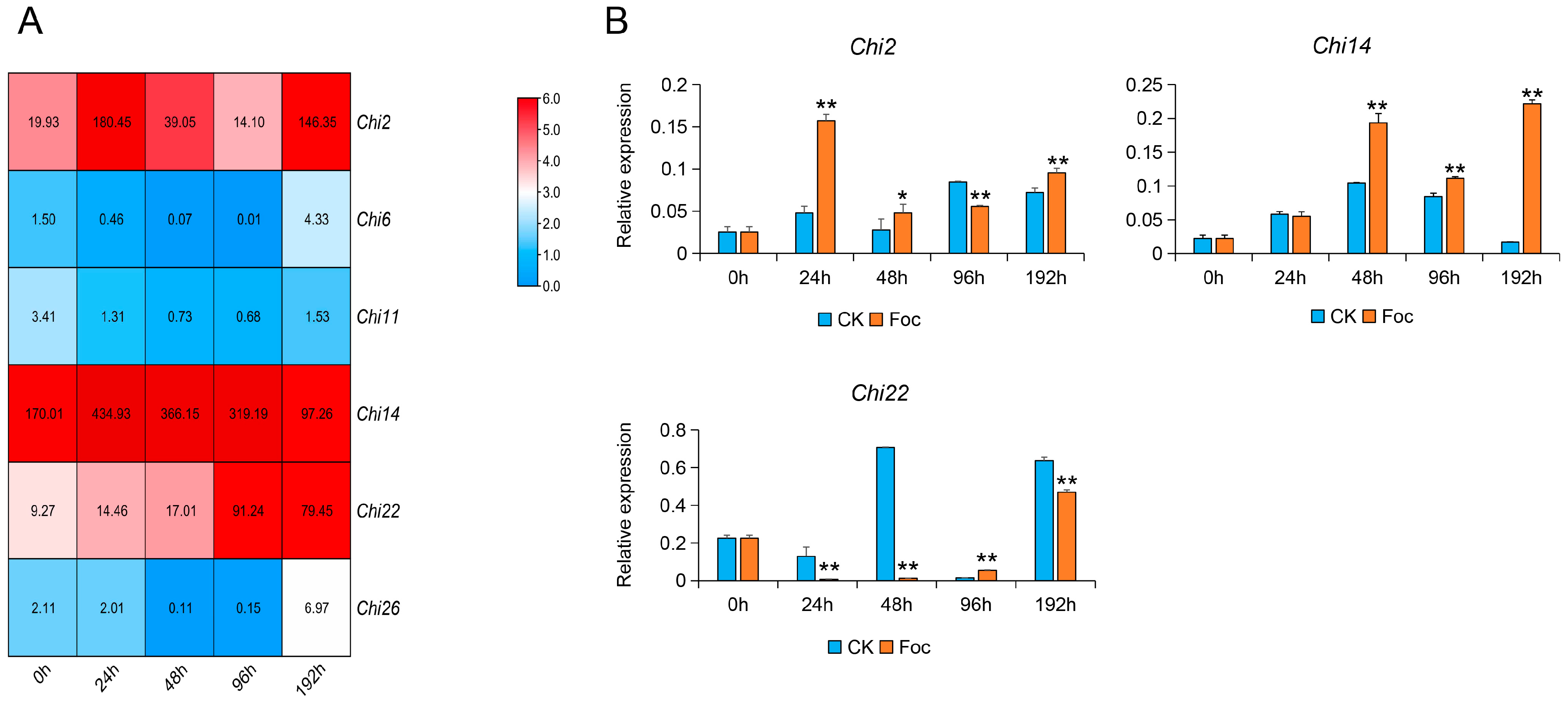
3.2. Transcriptome Analysis and Expression Verification of Chitinase Genes in Response to Foc

To obtain further evidence of the role of these chitinase genes in response to Foc, we used the transcriptome of cucumber roots inoculated with Foc at 0, 24, 48, 96, and 192 h to analyze the expression patterns of the chitinase genes. As a result, six chitinase genes were significantly induced by Foc, and among them, Chi2, Chi14, and Chi22 had higher expression levels (Figure 2A). To confirm the relationship between chitinase genes and Foc tolerance in cucumber, the candidate chitinase genes were verified in Rijiecheng cucumbers inoculated with Foc. It was observed that Chi2, Chi14, and Chi22 had significantly induced expression by Foc (Figure 2B), and Chi2 and Chi14 showed upregulated expression tendency, compared with the controls, indicating that they might play important roles in cucumber defense responses against FW.

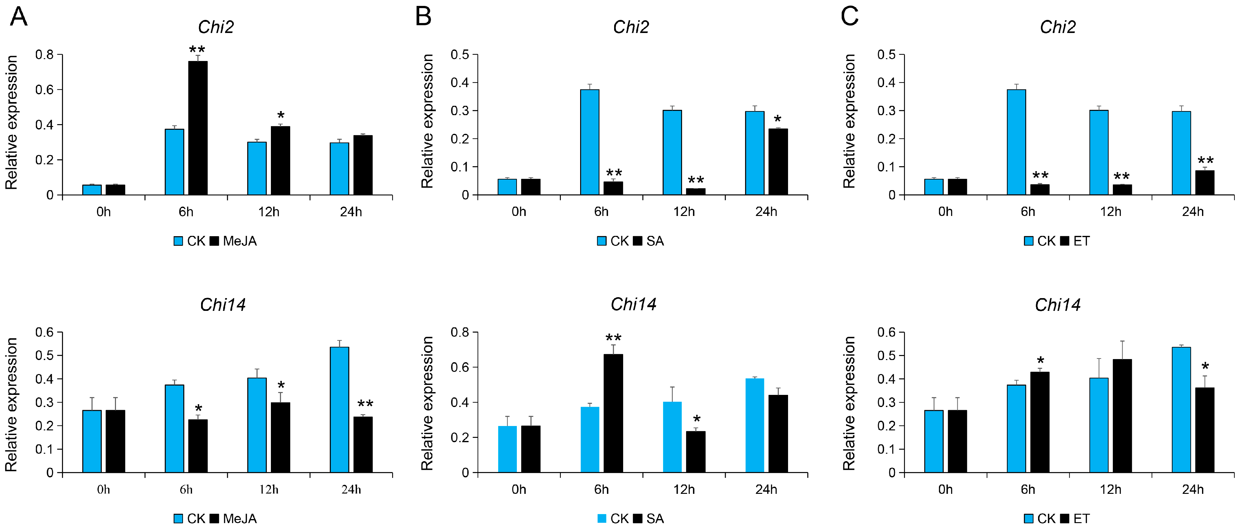
3.3. Expression Analysis of Chi2 and Chi14 in Response to Hormone Signals

Accumulated lines of evidence show that plant hormones played important roles in regulating developmental processes and signaling networks involved in plant responses to a wide range of biotic stresses [31]. The expression patterns of chitinase genes were also regulated by a variety of plant phytohormones including JA, SA, ET, etc., which contributed to the disease resistance in plants [32]. Additionally, based on the transcriptional analysis of cucumber roots inoculated with Foc, we found that many DEGs enriched in the plant hormone signal transduction mainly related to the JA, ET, and SA [6]. Hence, we analyzed the expression patterns of Chi2 and Chi14 in cucumber samples treated with SA, MeJA, and ethephon. As a result, the transcription levels of Chi2 and Chi14 were significantly induced by these three hormone treatments. Among them, Chi2 was also significantly upregulated and reached its peak 6 h after MeJA treatment but was downregulated after using SA and ET treatments. Likely, Chi14 was also affected by MeJA, SA, and ET; specifically, the expression levels of Chi14 was increased and accumulated at 6 h after SA and ET treatments, compared with the control; however, it was also significantly downregulated at 6–24 h after MeJA, as well as the ET treatment at 24 h (Figure 3). These results imply that Chi2 and Chi14 may participate in JA, SA, and ET signaling pathways to protect plants from various stresses.

3.4. Chi2 and Chi14 Suppresses Growth of Foc In Vitro

The entire open reading frame of Chi2 and Chi14 were cloned into a pet28a vector fused with a His-tag and further expressed in Escherichia coli. We used the 35 kDa protein TFPI (CUSABIO, Wuhan, China) added with the His-tag as the control. Subsequently, we detected the purified recombinant proteins by Western blot with anti-His and found that Chi2 and Chi14 were 44 KD and 37 KD, respectively (Figure 4A). To ascertain the functions of the Chi2 and Chi14 in cucumber FW defense, we further cultured the Foc strain on the PDA medium supplemented with sterile water (CK) and Chi2 and Chi14 recombinant protein solutions to observe phenotypic differences at the 3 and 7 days after inoculation (Figure 4B). The Foc growth on PDA supplemented with Chi2 and Chi14 protein solutions was obviously suppressed, compared with control samples (Figure 4C), indicating that Chi2 and Chi14 might play important roles in defense against Foc infection in cucumber.

3.5. Silencing Chi2 Impairs Cucumber Tolerance to Foc

To further investigate the functions of Chi2 and Chi14 in Foc resistance, we constructed pV190: Chi2, pV190: Chi14, and pV190: CsPDS vectors to silence endogenous genes in cucumber. All constructs were infiltrated into cotyledon, and two weeks later, all cucumber seedlings infiltrated with PV190: CsPDS showed bleached leaf phenotype in newly emerged leaves, suggesting that the PDS gene was silenced in these seedlings (Figure 5A). Subsequently, the VIGS seedlings infiltrated with pV190: Chi2, pV190: Chi14, and pV190 constructs were used for verification of gene-silencing effects via the qRT-PCR method. We found that the transcripts of Chi2 and Chi14 were significantly downregulated, compared with pV190 treatments (p < 0.01) (Figure 5B,C).
After Chi2 and Chi14 were silenced, we inoculated cucumber seedlings with Foc by dip infection, with a final concentration of 1 × 10⁶ spores per milliliter. Three weeks after inoculation, the Chi2-silenced seedlings showed discoloration of stem bases and possessed obviously leave wilting phenotype, compared with the cucumber samples infiltrated with pV190: Chi14 or pV190 vectors (Figure 5D). We also used the leaves of VIGS seedlings infiltrated with pV190: Chi2, pV190: Chi14, and pV190 constructs, to analyze the pathogenesis and death cells after Foc infection. We further found that the Chi2-silenced leaves were more susceptible to Foc infection, and also, the trypan blue staining showed that Chi2-silenced leaves had deep color and dead cells in the points inoculated with Foc (Figure 5E). Furthermore, we found that gene expression in the JA pathway included a basic helix-loop–helix-leucine zipper transcription factor (MYC2), jasmonate-associated VQ motif gene1(JAV1), and 2-oxophytodienoic acid reductases (OPR) 1 were significantly down-regulated in the Chi2-silenced leaves (Figure S1). These findings confirmed that down-regulated expression of Chi2 impaired cucumber tolerance to Foc, and Chi2 genes might play important roles in cucumber FW defense.

4. Discussion

FW acts as a destructive vascular disease; however, the related mechanisms of FW suppression are largely unknown. Chitinases functions by catalyzing the hydrolysis of chitin and damaging fungal cell walls, which are found and applied for defense against fungus pathogens in various plants. Although the genome-wide investigation of chitinase genes has been reported in many plant species, to date, the identification or functional analysis of chitinase genes involved in FW defense in cucumber remains limited. The number or distribution of chitinase genes shows obvious differences in different plants. For instance, 24 chitinase genes have been found and distributed between all 5 of the chromosomes in Arabidopsis, while rice chitinase genes are positioned on all chromosomes except chromosome 7 [33]. In cucumber, chitinase genes have been identified, and their evolutionary characteristics and expression patterns have also been analyzed in response to F. oxysporum [34]. Data on recent advances regarding the whole genome of cucumber species [1,21,22] made it possible to conveniently investigate the functions of chitinase genes. Here, we systematically identified 27 candidate chitinase genes in the sequenced cucumber species and further found that these genes were distributed on all chromosomes except chromosome 7 (Table S2).
An increasing number of studies found that the expression characteristics of chitinase genes exhibited both constitutive and induced patterns [35,36,37,38]. Among them, induced expression levels of chitinases have been recorded in response to numerous abiotic stresses containing wounding, drought, salinity, cold, heavy metals, ozone, frost, and UV light [37,38], as well as biotic stresses, including infectious fungi [17,39]. The availability of the whole genome of cucumber species made it convenient to screen candidate genes participating in plant growth and development, as well as abiotic and biotic stresses. For example, to investigate the development of mesocarp in cucumber, the expression profiles of mesocarp at 0, 3, 6, and 9 days after pollination (DAP) by RNA sequencing is used to identify functional genes [40]. An integrated analysis of the miRNA-seq and RNA-seq was performed to identify the FW-responsive miRNAs and their target genes in the FW-resistant cucumber cultivar Rijiecheng [41]. Here, we performed a comparative transcriptome analysis of the cucumber tissues (female, leaf, male, ovary, root, stem, and tendril) and roots of FW-resistant inbred line Rijiecheng infected with the Foc to screen the chitinase genes (Figure 1 and Figure 2). The expression verification found that Chi2 and Chi14 were significantly upregulated affected by Foc. An increasing number of studies show that plant hormones play important roles in defense against a wide range of biotic stresses [31], and the expression patterns of chitinase genes are also regulated by a variety of plant phytohormones, including JA, SA, ET, etc. [32]. Furthermore, transcriptional analysis of cucumber roots inoculated with Foc shows that many DEGs enriched on the plant hormone signal transduction mainly related to the JA, ET, and SA [6], suggesting that these hormones play important roles in FW defense. Here, we found that Chi2 was quickly upregulated, influenced by the Foc and MeJA in cucumber, therefore affecting disease resistance. Further research needs to define the precise underlying functions or molecular mechanisms of Chi2 in FW resistance through transgenic expression analysis in cucumber cultivars. Here, we found that Chi 2 and Chi14 were significantly upregulated, as a result of the effects of MeJA and SA, respectively (Figure 3). The diverse expression patterns of candidate genes might be affected by the concentration gradient of hormone and time points of treatments, which suggested the chitinase genes possessed diverse but overlapping functions.
Chitinases suppress fungal pathogens by affecting the fungal cell wall degradation. Several chitinase genes from different plants or fungi have been identified and successfully applied in different crops for disease defense. For instance, Cht-2 and Cht-3 are expressed in the different tissues and organs in the japonica rice, affecting the resistance to blast (Magnaporthe grisea) [17]. In pepper plants, co-expression of the ChitIV and receptor-like cytoplasmic protein kinase (CaPIK1) significantly enhances the CaPIK1-triggered cell death response, the abundance of reactive oxygen species (ROS), and resistance to Xanthomonas campestris pv. vesicatoria (Xcv) infection [42]. Taken together, these studies suggested that chitinase genes might play particular roles in the defense against fungal pathogens. Here, we also analyzed the inhibitory effect of Foc influenced by the Chi2 and Chi14 proteins with three repeats at different time points and found that Chi2 and Chi14 obviously suppress the growth of Foc, compared with the control (Figure 4). The function difference between Chi2 and Chi14 were further analyzed through the VIGS technology, and it was found that Chi2-silenced leaves were more susceptible to Foc infection (Figure 5). Although Chi2 and Chi14 are predominantly expressed in roots and obviously induced by the Foc, the expression of Chi2 plays a more considerable role in FW defense, compared with Chi14. The expression levels of Chi 2 are obviously upregulated after MeJA treatment, but Chi14 possesses the converse tendency and is upregulated after SA treatment. In other host plants, SA and JA participate in activating the complex defense signaling networks [31,43]. For instance, JA acts with ET to mediate resistance against necrotrophic pathogens, whereas SA affects defensive responses to effectively control hemi-biotrophs and biotrophs, as well as inducing the systemic acquired resistance [44,45]. Accumulated lines of evidence show that many induced genes related to JA, including CYP82D [46], GaWRKY1 [47], GhJAZ2, and GhPDF1.2 [48], positively contribute to the disease resistance of cotton to a soil-borne fungal pathogen Verticillium dahliae. Likely, we found that the MYC2, JAV1, and OPR1 genes in the JA pathway were significantly down-regulated in the Chi2-silenced leaves, and it is speculated that Chi2 may participate in regulating the JA pathway and have a positive role in regulating cucumber resistance against FW infection. Furthermore, although the VIGS method could deal with the phenotype of silent plants quickly, the silencing efficiency and lasting time of the phenotype were still limitations affecting the analysis of the functions of candidate genes in the later period of cucumber. In future studies, we will obtain the transgenic lines through over-expressing and knockdown of Chi2 and Chi14 in cucumber to better determine the molecular mechanisms or regulatory networks of FW defense and develop resistant cultivars.

5. Conclusions

Cucumber Fusarium wilt (FW) severely restricts cucumber yields and profits; however, the resistance genes and mechanisms underlying the FW tolerance in cucumber are poorly understood. An increasing number of studies found that chitinases played important roles in attacking the invading fungal pathogens through catalyzing their cell wall degradation. In this study, we performed a comparative transcriptome analysis and expression verification, and the results revealed that Chi2 and Chi14 were obviously induced by Foc, as well as hormone treatments, compared with the controls. We also found that Chi2 and Chi14 purification proteins significantly affected the growth of Foc in vitro compared with the control. Silencing of Chi2 resulted in increased susceptibility to FW, compared with the Chi14-silenced and control plants, suggesting that the Chi2 gene might play positive roles in cucumber FW defense and, therefore, can provide gene resources to develop cultivars resistant to cucumber FW.

Supplementary Materials

The following are available online at https://www.mdpi.com/article/10.3390/genes13010062/s1, Table S1: Information of the PCR primers, Table S2: The cucumber chitinase genes, Figure S1: Chi2 involved in activation of JA-related genes.

Author Contributions

Experiment design, X.C.; experiment execution, J.X., N.Z., K.W., Q.X., J.D. and X.Q.; writing—original draft preparation, writing—review and editing, J.X. and X.C. All authors have read and agreed to the published version of the manuscript.

Funding

This research was supported by Jiangsu Agricultural Science and Technology Innovation Fund (CX (19)3029) and Yangzhou City’s Green and Golden Phoenix Program.

Institutional Review Board Statement

Not applicable.

Informed Consent Statement

Not applicable.

Data Availability Statement

The RNA-Seq data of cucumber roots inoculated with Foc, and cucumber tissues and organs associated with this study were downloaded with NCBI Accession Number PRJNA472169 and PRJNA80169, respectively.

Acknowledgments

We thank Qinsheng Gu of Zhengzhou Fruit Research Institute, Chinese Academy of Agricultural Sciences (Zhengzhou, China) for providing the VIGS vector (pV190) to support our experiments in this study.

Conflicts of Interest

All the authors declare that they have no competing interests.

References

  1. Huang, S.W.; Li, R.Q.; Zhang, Z.H.; Li, L.; Gu, X.F.; Fan, W.; Lucas, W.J.; Wang, X.W.; Xie, B.Y.; Ni, P.X.; et al. The genome of the cucumber, Cucumis sativus L. Nat. Genet. 2009, 41, 1275–1281. [Google Scholar] [CrossRef] [Green Version]
  2. Hu, J.L.; Lin, X.G.; Wang, J.H.; Shen, W.S.; Wu, S.; Peng, S.P.; Mao, T.T. Arbuscular mycorrhizal fungal inoculation enhances suppression of cucumber Fusarium wilt in greenhouse soils. Pedosphere 2010, 20, 586–593. [Google Scholar] [CrossRef]
  3. Ahn, I.P.; Chung, H.S.; Lee, Y.H. Vegetative compatibility groups and pathogenicity among isolates of Fusarium oxysporum f. sp. cucumerinum. Plant Dis. 1998, 82, 244–246. [Google Scholar] [CrossRef] [PubMed] [Green Version]
  4. Zhou, X.G.; Wu, F.Z. Dynamics of the diversity of fungal and Fusarium communities during continuous cropping of cucumber in the greenhouse. FEMS Microbiol. Ecol. 2012, 80, 469–478. [Google Scholar] [CrossRef] [PubMed] [Green Version]
  5. Sun, Y.M.; Li, Y.R.; Li, Y.; Wang, M.; Mur, L.A.J.; Shen, Q.R.; Guo, S.W. Nitrate mediated resistance against Fusarium infection in cucumber plants acts via photorespiration. Plant Cell Environ. 2021, 44, 3412–3431. [Google Scholar] [CrossRef]
  6. Dong, J.P.; Wang, Y.A.; Xian, Q.Q.; Chen, X.H. Transcriptome analysis reveals ethylene-mediated defense responses to Fusarium oxysporum f. sp. cucumerinum infection in Cucumis sativus L. BMC Plant Biol. 2020, 20, 334. [Google Scholar] [CrossRef]
  7. Du, N.S.; Shi, L.; Du, L.T.; Yuan, Y.H.; Li, B.; Sang, T.; Sun, J.; Shu, S.; Guo, S.R. Effect of vinegar residue compost amendments on cucumber growth and Fusarium wilt. Environ. Sci. Pollut. Res. Int. 2015, 22, 19133–19141. [Google Scholar] [CrossRef]
  8. Moreno, A.; Alférez, A.; Avilés, M.; Diánez, F.; Blanco, R.; Santos, M.; Tello, J.C. First report of Fusarium wilt of cucumber caused by Fusarium oxysporum in Spain. Plant Pathol. 2003, 52, 410. [Google Scholar]
  9. Chen, L.H.; Huang, X.Q.; Zhang, F.G.; Zhao, D.K.; Yang, X.M.; Shen, Q.R. Application of Trichoderma harzianum SQR-T037 bioorganic fertiliser significantly controls Fusarium wilt and affects the microbial communities of continuously cropped soil of cucumber. J. Sci. Food Agric. 2012, 92, 2465–2470. [Google Scholar] [CrossRef]
  10. Huang, X.; Shi, D.; Sun, F.; Lu, H.; Liu, J.; Wu, W. Efficacy of sludge and manure compost amendments against Fusarium wilt of cucumber. Environ. Sci. Pollut. Res. Int. 2012, 19, 3895–3905. [Google Scholar] [CrossRef]
  11. Willie, L.; Anuratha, C.S.; Datta, K.; Potrykus, I.; Muthukrishnan, S. Genetic engineering of rice for resistance to sheath blight. Nat. Biotechnol. 1995, 13, 686–691. [Google Scholar]
  12. Xu, J.; Xu, X.; Tian, L.; Wang, G.; Zhang, X.; Wang, X.; Guo, W. Discovery and identification of candidate genes from the chitinase gene family for Verticillium dahliae resistance in cotton. Sci. Rep. 2016, 6, 29022. [Google Scholar] [CrossRef] [PubMed]
  13. Li, H.; Greene, L.H. Sequence and structural analysis of the chitinase insertion domain reveals two conserved motifs involved in chitin-binding. PLoS ONE 2010, 5, e8654. [Google Scholar] [CrossRef] [PubMed] [Green Version]
  14. Collinge, D.B.; Kragh, K.M.; Mikkelsen, J.D.; Nielsen, K.K.; Rasmussen, U.; Vad, K. Plant chitinases. Plant J. 1993, 3, 31–40. [Google Scholar] [CrossRef]
  15. Punja, Z.K.; Zhang, Y.Y. Plant chitinases and their roles in resistance to fungal diseases. J. Nematol. 1993, 25, 526–540. [Google Scholar]
  16. Kim, J.K.; Jang, I.C.; Wu, R.; Zuo, W.N.; Boston, R.S.; Lee, Y.H.; Ahn, I.P.; Nahm, B.H. Co-expression of a modified maize ribosome-inactivating protein and a rice basic chitinase gene in transgenic rice plants confers enhanced resistance to sheath blight. Transgenic Res. 2003, 12, 475–484. [Google Scholar] [CrossRef]
  17. Nishizawa, Y.; Nishio, Z.; Nakazono, K.; Soma, M.; Nakajima, E.; Ugaki, M.; Hibi, T. Enhanced resistance to blast (Magnaporthe grisea) in transgenic Japonica rice by constitutive expression of rice chitinase. Theor. Appl. Genet. 1999, 99, 383–390. [Google Scholar] [CrossRef]
  18. Kovács, G.; Sági, L.; Jacon, G.; Arinaitwe, G.; Busogoro, J.P.; Thiry, E.; Strosse, H.; Swennen, R.; Remy, S. Expression of a rice chitinase gene in transgenic banana (‘Gros Michel’, AAA genome group) confers resistance to black leaf streak disease. Transgenic Res. 2013, 22, 117–130. [Google Scholar] [CrossRef]
  19. Shah, M.R.; Mukherjee, P.K.; Eapen, S. Expression of a fungal endochitinase gene in transgenic tomato and tobacco results in enhanced tolerance to fungal pathogens. Physiol. Mol. Biol. Plants 2010, 16, 39–51. [Google Scholar] [CrossRef] [Green Version]
  20. Pétriacq, P.; López, A.; Luna, E. Fruit Decay to Diseases: Can Induced Resistance and Priming Help? Plants 2018, 7, 77. [Google Scholar] [CrossRef] [Green Version]
  21. Qi, J.J.; Liu, X.; Shen, D.; Miao, H.; Xie, B.; Li, X.; Zeng, P.; Wang, S.; Shang, Y.; Gu, X.F.; et al. A genomic variation map provides insights into the genetic basis of cucumber domestication and diversity. Nat. Genet. 2013, 45, 1510–1515. [Google Scholar] [CrossRef] [PubMed]
  22. Li, Q.; Li, H.; Huang, W.; Xu, Y.; Zhou, Q.; Wang, S.; Jue, R.; Huang, S.; Zhang, Z. A chromosome-scale genome assembly of cucumber (Cucumis sativus L.). Gigascience 2019, 8, giz072. [Google Scholar] [CrossRef] [Green Version]
  23. Finn, R.D.; Bateman, A.; Clements, J.; Coggill, P.; Eberhardt, R.Y.; Eddy, S.R.; Heger, A.; Hetherington, K.; Holm, L.; Mistry, J.; et al. Pfam: The protein families database. Nucleic Acids Res. 2014, 42, D222–D230. [Google Scholar] [CrossRef] [Green Version]
  24. Eddy, S.R. Accelerated profile HMM searches. PLoS Comput. Biol. 2011, 7, e1002195. [Google Scholar] [CrossRef] [PubMed] [Green Version]
  25. Wang, L.K.; Feng, Z.X.; Wang, X.; Wang, X.W.; Zhang, X.G. DEGseq: An R package for identifying differentially expressed genes from RNA-seq data. Bioinformatics 2010, 26, 136–138. [Google Scholar] [CrossRef] [PubMed]
  26. Yakushiji, Y.; Yamanaka, K.; Ogura, T. Identification of a cysteine residue important for the ATPase activity of C. elegans fidgetin homologue. FEBS Lett. 2004, 578, 191–197. [Google Scholar] [CrossRef] [Green Version]
  27. Liu, M.; Liang, Z.; Aranda, M.A.; Hong, N.; Liu, L.; Kang, B.; Gu, Q. A cucumber green mottle mosaic virus vector for virus-induced gene silencing in cucurbit plants. Plant Methods 2020, 16, 9. [Google Scholar] [CrossRef] [PubMed] [Green Version]
  28. Yoshioka, H.; Numata, N.; Nakajima, K.; Katou, S.; Kawakita, K.; Rowland, O.; Jones, J.D.; Doke, N. Nicotiana benthamiana gp91z homologs NbrbohA and NbrbohB participate in H2O2 accumulation and resistance to Phytophthora infestans. Plant Cell 2003, 15, 706–718. [Google Scholar] [CrossRef] [PubMed] [Green Version]
  29. Wang, Y.Y.; Wu, F.Z.; Liu, S.W. Allelopathic effects of root exudates from wheat, oat and soybean on seed germination and growth of cucumber. Allelopath. J. 2009, 24, 103–112. [Google Scholar]
  30. Baayen, R.P.; O’Donnell, K.; Bonants, P.J.; Cigelnik, E.; Kroon, L.P.; Roebroeck, E.J.; Waalwijk, C. Gene Genealogies and AFLP Analyses in the Fusarium oxysporum Complex Identify Monophyletic and Nonmonophyletic Formae Speciales Causing Wilt and Rot Disease. Phytopathology 2000, 90, 891–900. [Google Scholar] [CrossRef] [Green Version]
  31. Bari, R.; Jones, J.D. Role of plant hormones in plant defence responses. Plant Mol. Biol. 2009, 69, 473–488. [Google Scholar] [CrossRef]
  32. Kasprzewska, A. Plant chitinases-regulation and function. Cell. Mol. Biol. Lett. 2003, 8, 809–824. [Google Scholar]
  33. Xu, F.; Fan, C.; He, Y. Chitinases in Oryza sativas sp. Japonica and Arabidopsis thaliana. J. Genet. Genom. 2007, 34, 138–150. [Google Scholar] [CrossRef]
  34. Bartholomew, E.S.; Black, K.; Feng, Z.; Liu, W.; Shan, N.; Zhang, X.; Wu, L.; Bailey, L.; Zhu, N.; Qi, C.; et al. Comprehensive Analysis of the Chitinase Gene Family in Cucumber (Cucumis sativus L.): From Gene Identification and Evolution to Expression in Response to Fusarium oxysporum. Int. J. Mol. Sci. 2019, 20, 5309. [Google Scholar] [CrossRef] [PubMed] [Green Version]
  35. Passarinho, P.A.; Van Hengel, A.J.; Fransz, P.F.; de Vries, S.C. Expression pattern of the Arabidopsis thaliana AtEP3/AtchitIV endochitinase gene. Planta 2001, 212, 556–567. [Google Scholar] [CrossRef] [PubMed]
  36. Hamel, F.; Bellemare, G. Characterization of a class I chitinase gene and of wound-inducible, root and flower-specific chitinase expression in Brassica napus. Biochim. Biophys. Acta 1995, 1263, 212–220. [Google Scholar] [CrossRef]
  37. Cohen-Kupiec, R.; Chet, I. The molecular biology of chitin digestion. Curr. Opin. Biotechnol. 1998, 9, 270–277. [Google Scholar] [CrossRef]
  38. Hanfrey, C.; Fife, M.; Buchanan-Wollaston, V. Leaf senescence in Brassica naupus: Expression of genes encoding pathogenesis related proteins. Plant Mol. Biol. 1996, 30, 595–609. [Google Scholar] [CrossRef] [PubMed]
  39. Bishop, J.G.; Dean, A.M.; Mitchell-Olds, T. Rapid evolution in plant chitinases: Molecular targets of selection in plant-pathogen coevolution. Proc. Natl. Acad. Sci. USA 2000, 97, 5322–5327. [Google Scholar] [CrossRef] [Green Version]
  40. Qi, X.H.; Zhu, Y.M.; Li, S.F.; Zhou, H.X.; Xu, X.W.; Xu, Q.; Chen, X.H. Identification of genes related to mesocarp development in cucumber. Hortic. Plant J. 2020, 6, 293–300. [Google Scholar] [CrossRef]
  41. Xu, J.; Xian, Q.; Zhang, N.; Wang, K.; Zhou, X.; Li, Y.; Dong, J.; Chen, X. Identification of miRNA-Target Gene Pairs Responsive to Fusarium Wilt of Cucumber via an Integrated Analysis of miRNA and Transcriptome Profiles. Biomolecules 2021, 11, 1620. [Google Scholar] [CrossRef] [PubMed]
  42. Kim, D.S.; Kim, N.H.; Hwang, B.K. The Capsicum annuum class IV chitinase ChitIV interacts with receptor-like cytoplasmic protein kinase PIK1 to accelerate PIK1-triggered cell death and defence responses. J. Exp. Bot. 2015, 66, 1987–1999. [Google Scholar] [CrossRef] [PubMed] [Green Version]
  43. Pieterse, C.M.; Leon-Reyes, A.; Van der Ent, S.; Van Wees, S.C. Networking by small-molecule hormones in plant immunity. Nat. Chem. Biol. 2009, 5, 308–316. [Google Scholar] [CrossRef] [PubMed] [Green Version]
  44. Dempsey, D.A.; Klessig, D.F. SOS-toomany signals for systemic acquired resistance? Trends Plant Sci. 2012, 17, 538–545. [Google Scholar] [CrossRef] [PubMed]
  45. Gao, W.; Long, L.; Zhu, L.F.; Xu, L.; Gao, W.H.; Sun, L.Q.; Liu, L.L.; Zhang, X.L. Proteomic and virus-induced gene silencing (VIGS) Analyses reveal that gossypol, brassinosteroids, and jasmonic acid contribute to the resistance of cotton to Verticillium dahliae. Mol. Cell. Proteom. 2013, 12, 3690–3703. [Google Scholar] [CrossRef] [PubMed] [Green Version]
  46. Sun, L.; Zhu, L.; Xu, L.; Yuan, D.; Min, L.; Zhang, X. Cotton cytochrome P450 CYP82D regulates systemic cell death by modulating the octadecanoid pathway. Nat. Commun. 2014, 5, 5372. [Google Scholar] [CrossRef]
  47. Xu, Y.H.; Wang, J.W.; Wang, S.; Wang, J.Y.; Chen, X.Y. Characterization of GaWRKY1, a cotton transcription factor that regulates these squiterpene synthasegene(+)-delta-cadinenesynthase-A. Plant Physiol. 2004, 135, 507–515. [Google Scholar] [CrossRef] [Green Version]
  48. He, X.; Zhu, L.; Wassan, G.M.; Wang, Y.; Miao, Y.; Shaban, M.; Hu, H.; Sun, H.; Zhang, X. GhJAZ2 attenuates cotton resistance to biotic stresses via the inhibition of the transcriptional activity of GhbHLH171. Mol. Plant Pathol. 2018, 19, 896–908. [Google Scholar] [CrossRef]
Figure 1. Transcriptional analysis of chitinase genes in cucumber tissues and organs. Female (female flower tissue), leaf, male (male flower tissue), ovary, root, stem, and tendril were used for the expression analysis. The expression values were converted with Log2 (FPKM) to calculate the expression levels of Chis. The differences are displayed in colors indicated with a scale. This RNA-Seq data can be downloaded with NCBI Accession Number PRJNA80169.
Genes 13 00062 g001
Figure 2. Transcriptional analysis and verification of chitinase genes: (A) transcriptional analysis of the chitinase genes and visualized by hierarchical clustering. Fold difference was designated as Log2 (FPKM) to calculate Chis expression levels with the colors indicated in the scale. Sampling time points include 0 h, 24 h, 48 h, 96 h, and 192 h after Foc inoculation; (B) verification of candidate gene expression in response to Foc in cucumber. Induction of candidate DEGs after inoculation with Foc as assessed by qRT-PCR. The error bars with standard deviations were calculated from three biological replicates. * indicates significantly different at p ≤ 0.05; ** indicates significantly different at p ≤ 0.01.
Genes 13 00062 g002
Figure 3. The expression patterns of Chi2 and Chi14 in cucumber treated with hormones: (AC) the cucumber roots were inoculated with hormones including MeJA, SA, and ET, respectively, and were used to assess the expression of Chi2 and Chi14 by qRT-PCR. The error bars with standard deviations were calculated from three biological replicates. * indicates significantly different at p ≤ 0.05; ** indicates significantly different at p ≤ 0.01.
Genes 13 00062 g003
Figure 4. Chi2 and Chi14 affect the growth of Foc in vitro: (A) detection of these two purified proteins by Western blot with anti-His. Lane Chi2, recombinant His-Chi2; lane CK, the 35 kDa protein TFPI (CUSABIO, Wuhan, China) added with the His-tag; lane M, marker, molecular quantity standard (250/150/100/70/50/40/35/25/20/15 kDa); lane Chi14, recombinant His-Chi14; (B) the phenotypic differences between the Foc strain cultured on PDA medium supplemented with sterile water (CK), Chi2, and Chi14 protein solutions at 3 and 7 days after inoculation; (C,D) the diameter of Foc grown on CK, Chi2, and Chi14 protein solutions at 3 and 7 days after inoculation. The error bars with standard deviations were calculated from three biological replicates. ** indicates significantly different at p ≤ 0.01.
Genes 13 00062 g004
Figure 5. Silencing of Chi2 significantly impaired the resistance to Foc in cucumber: (A) silencing of the endogenous PDS gene in cucumber through VIGS. The leaf bleaching phenotype was observed three weeks after infiltration in pV190: CsPDS plants; (B,C) the transcription levels of Chi2 and Chi14 in the Chi2-silenced, Chi14-silenced, and pV190 leaves; the error bars with standard deviations were calculated from three biological replicates; ** indicates significantly different at p ≤ 0.01; (D) cucumber phenotypes at 14 days after Foc inoculation; the VIGS plants were dip inoculated with Foc conidia suspension (1 × 10⁶ conidia/mL); (E) the phenotypes of the Chi2-silenced, Chi14-silenced, and pV190 leaves inoculated with Foc and trypan blue staining. The leaves were infected with Foc for 5 days and performed the trypan blue staining and microscopic observation.
Genes 13 00062 g005
Publisher’s Note: MDPI stays neutral with regard to jurisdictional claims in published maps and institutional affiliations.

