Review

Immunotherapy in Myeloproliferative Diseases

by
Lukas M. Braun
1,2 and
Robert Zeiser
1,3,4,5,*
1
Department of Medicine I, Medical Center—University of Freiburg, Faculty of Medicine, University of Freiburg, 79106 Freiburg, Germany
2
Faculty of Biology, University of Freiburg, 79104 Freiburg, Germany
3
German Cancer Consortium (DKTK) Partner Site Freiburg, German Cancer Research Center (DKFZ), 69120 Heidelberg, Germany
4
Comprehensive Cancer Center Freiburg (CCCF), University of Freiburg, 79106 Freiburg, Germany
5
Centre for Biological Signalling Studies (BIOSS) and Centre for Integrative Biological Signalling Studies (CIBSS), University of Freiburg, 79104 Freiburg, Germany
*
Author to whom correspondence should be addressed.
Cells 2020, 9(6), 1559; https://doi.org/10.3390/cells9061559
Submission received: 9 June 2020 / Accepted: 23 June 2020 / Published: 26 June 2020
(This article belongs to the Special Issue Pathophysiology and Molecular Targets in Myeloid Neoplasia)

Abstract

:
Myeloproliferative diseases, including myeloproliferative neoplasms (MPN) and myelodysplastic syndromes (MDS), are driven by genetic abnormalities and increased inflammatory signaling and are at high risk to transform into acute myeloid leukemia (AML). Myeloid-derived suppressor cells were reported to enhance leukemia immune escape by suppressing an effective anti-tumor immune response. MPNs are a potentially immunogenic disease as shown by their response to interferon-α treatment and allogeneic hematopoietic stem-cell transplantation (allo-HSCT). Novel immunotherapeutic approaches such as immune checkpoint inhibition, tumor vaccination, or cellular therapies using target-specific lymphocytes have so far not shown strong therapeutic efficacy. Potential reasons could be the pro-inflammatory and immunosuppressive microenvironment in the bone marrow of patients with MPN, driving tumor immune escape. In this review, we discuss the biology of MPNs with respect to the pro-inflammatory milieu in the bone marrow (BM) and potential immunotherapeutic approaches.

1. Introduction

The term “Myeloproliferative Disorders” describes non-physiological changes of the myeloid compartment in the hematopoietic system leading to an overproduction of mature and functional myeloid blood cells [1]. The WHO classification for hematopoietic tumors and Myeloproliferative Neoplasms (MPN) distinguishes different forms of the disease into chronic myeloid leukemia, chronic neutrophilic leukemia, chronic eosinophilic leukemia-not otherwise specified, primary myelofibrosis (PMF), polycythemia vera (PV), essential thrombocythemia (ET), and MPN unclassifiable (MPN-U) [2]. The three classical BCR-ABL-negative forms of MPN, which are the most frequent disorders among all myeloproliferative diseases, comprise PV, PMF, and ET. Somatic mutations cause a constitutive activation of physiologic signaling pathways in hematopoietic stem and progenitor cells, leading to a clonal expansion of myeloid progenitor cells and single or multilineage hyperplasia [3,4,5]. MPNs are characterized by an increased production of fully differentiated and completely functional blood cells of the myeloid lineage [1,5,6]. ET, PV, and PMF are clinically classified by an overproduction of functional platelets, increased numbers of red blood cells and high counts of white blood cells and bone marrow and spleen fibrosis, respectively [1,5].
The discovery of a novel JAK2V617F mutation, which is found in 50–90% of all classical MPNs and results in a substitution of valine to phenylalanine in the JAK2 gene, significantly contributed to the discovery of the molecular pathogenesis of myeloproliferative neoplasms [5,7,8,9,10]. JAK2 is the most-frequently mutated gene in MPN and its mutant form encodes a constitutively active kinase. The JAK2V617F mutation usually arises in a multipotent hematopoietic progenitor clone and can be found in all myeloid lineages, but also in B-, T- and NK-cells [5]. Another mutation of JAK2 in exon 12 is found less frequently in MPNs and is mainly restricted to JAK2V617F negative PV [11]. Other more rarely seen genetic aberrations in MPN are mutations in the myeloproliferative leukemia virus (MPL; thrombopoietin receptor (TPOR)) gene resulting in a substitution of tryptophan at position W515 by leucine (MPLW515L) or lysine (MPLW515K) [12,13]. These mutations are not as common as JAK2 mutations and are only found in 3–5% of all ET and PMF cases [14,15]. More recent discoveries found frameshift mutations in exon 9 in the calretikulin (CALR) gene in the majority of JAK2- and MPL-negative PMF and ET, causing a 5-bp insertion or a 52-bp deletion [5,16,17,18]. Genetic analyses could elucidate that the major MPN driver mutations, JAK2V617F, CALR, and MPL515, play a pivotal role in increasing the risk of leukemic transformations [19].
The transformation of Philadelphia-chromosome negative MPN to acute myeloid leukemia (AML) is one of the major complications of MPN [5,20]. Such a transformation of MPN to secondary AML is seen in about 1.5%, 4–7% and 11–20% for ET, PV, and PMF, respectively [3,4,19,21]. Standard therapeutic options are not effective in secondary AML transformed from MPN and patients suffering from a post-MPN AML only have a dismal prognosis with a median survival below 6 months [3,22,23]. The mechanisms driving leukemic transformation have not been well understood yet but it was seen that the risk of developing a secondary AML is increased by additional factors, e.g., age and chemotherapy treatment [3,4]. Also, genetic instability and acquired mutations are known as a risk factor for the development of a post-MPN AML [3]. Frequently reported genetic aberrations include mutations in epigenetic modifiers such as the Ten-Eleven Translocation 2 (TET2), isocitrate dehydrogenase 1 and 2 (IDH1/2), additional sex combs like transcriptional regulator 1 (ASXL1) and enhancer of zeste 2 polycomb repressive complex 2 subunit (EZH2), mutations in spliceosome regulators such as serine and arginine rich splicing factor 2 (SRSF2), splicing factor 3B subunit 1 (SF3B1) and U2 small nuclear RNA auxiliary factor 1 (U2AF1) and genetic aberrations in the DNA damage control system, including TP53 [24,25,26,27,28,29,30,31,32,33,34]. Additional mutations were found in the protein tyrosine phosphatase non-receptor type 11 (PTPN11), MYC and the SET binding protein 1 (SETBP1) [3].

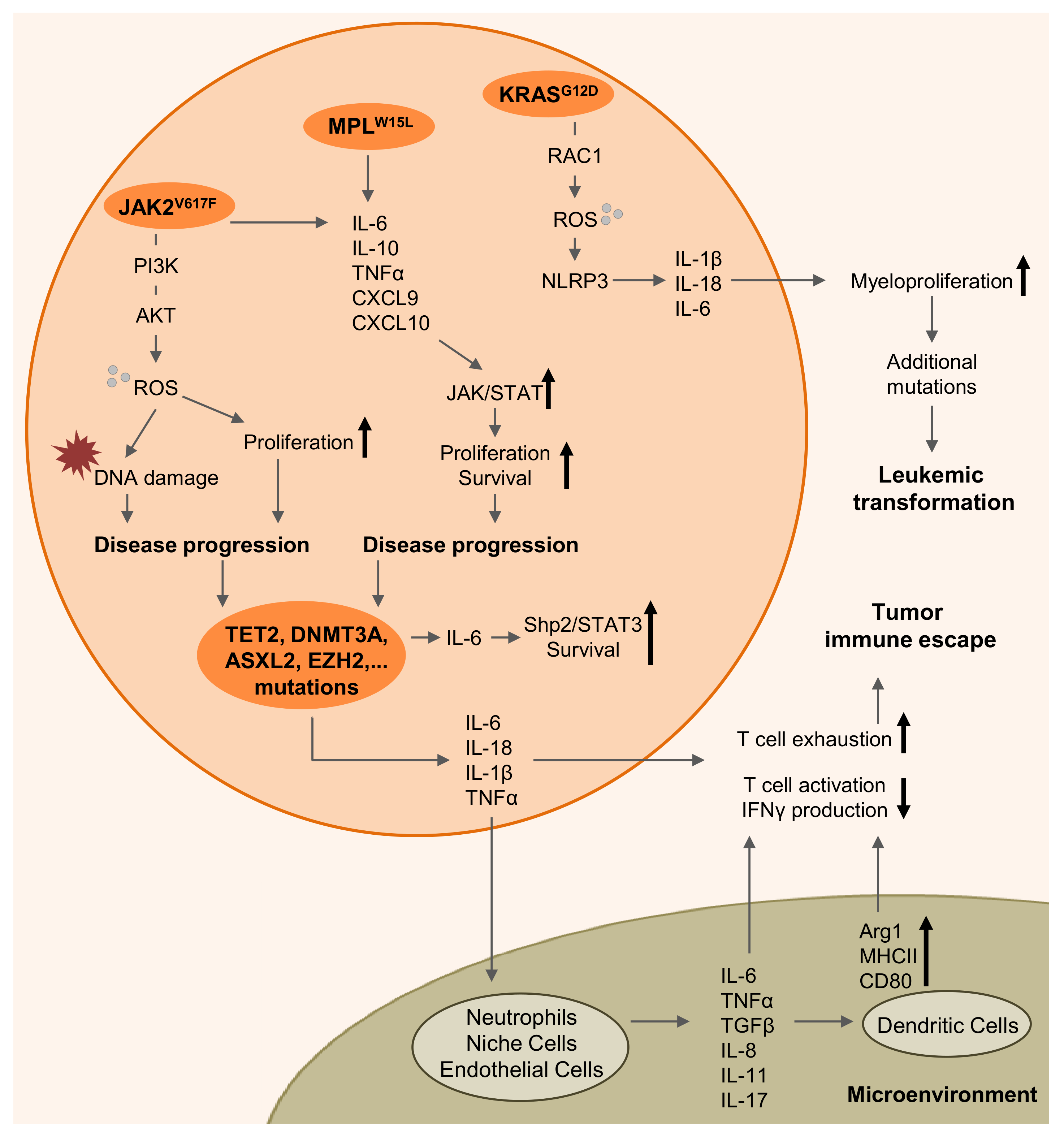
2. Myeloproliferative Diseases are Driven by Inflammation

Many publications described that myeloproliferative diseases are driven by activation of inflammatory pathways leading to increased levels of cytokines and accumulation of reactive oxygen species (ROS) [35]. The latter were reported to play an important role in JAK2-mutant MPN progression as ROS was seen to accumulate in the hematopoietic stem-cell compartment of JAK2V617F knock-in mice and was found increased in patients with JAK2V617F mutant MPN [36]. According to these findings, transcriptional profiling of peripheral blood samples from MPN patients revealed a significant deregulation of anti-oxidative stress genes, e.g., Nrf2, in MPN patient samples compared to controls [37]. Regarding the overproduction of ROS in the hematopoietic stem-cell compartment of JAK2V617F knock-in mice, the application of the anti-oxidant N-acetylcysteine (NAC) could restore the normal phenotype in these mice, normalize peripheral blood parameters, decrease splenomegaly, reduce the number of JAK2-mutant hematopoietic stem and progenitor cells in the spleen and bone marrow and reduce DNA double-strand breaks being increased in JAK2V617F mutant MPN. The authors claimed that the massive production of ROS in JAK2V617F-mutant MPN causes DNA damage, thereby driving disease progression and the development of MPN could be slowed down by specifically targeting ROS [36].
Regarding increased pro-inflammatory signaling, one publication reported the oncogenic KRASG12D mutation as a driver for elevated myeloproliferation and chronic myelomonocytic leukemia (CMML) through activation of the NLRP3 inflammasome and caspase-1-mediated cleavage of pro-inflammatory cytokines [38,39]. Underlining the notable role of inflammasome activation for driving myeloproliferation, a genetic deficiency of NLRP3 could ameliorate KRASG12D driven cytopenia in mice [39]. Moreover, additional studies could highlight that MPLW15L mutant mice showed high serum levels of pro-inflammatory cytokines including Interleukin-6 (IL-6), tumor necrosis factor (TNF) α, IL-10, CXCL9 and CXCL10 [40,41]. Comparable, the oncogenic JAK2V617F mutation caused high levels of IL-6 and TNFα in the serum of mice being transplanted with a JAK2V617F overexpressing cell line or carrying the mutation in the bone marrow [40,42]. Besides the major MPN mutations, also other genetic aberrations can increase the release of pro-inflammatory cytokines, thereby potentially driving the progress of the disease. One study highlighted the role of pro-inflammatory signaling pathways in driving the expansion of pre-leukemic hematopoietic stem and progenitor cells (HSPCs). It was shown that TET2-deficiency caused an increased IL-6 production which in turn activated the Shp2/STAT3 signaling axis leading to higher anti-apoptotic and pro-survival protein levels in HSPCs [43]. Moreover, additional studies described that TET2-deficiency drives the production of pro-inflammatory cytokines, including interleukin 1β (IL-1β) and interleukin 18 (IL-18) [44,45,46]. Besides TET2, mutations in other epigenetic modifiers, e.g., DNMT3A and ASXL1, which are frequently mutated in pre-leukemic hematopoiesis, were reported to drive an increased inflammatory signaling by secreting IL-6 and TNFα [46,47,48]. Further studies could prove a pivotal role of TNFα and interferon α (IFNα) secretion and the activation of pro-inflammatory signaling pathways for driving myelofibrosis in EZH2/JAK2V617F-mutant myeloid progenitor cells or increased TNFα signaling in DNMT3A/JAK2V617F-mutant myeloid stem and progenitor cells [49,50]. Activated TNFα pathways were also confirmed by RNA-seq analysis in patients suffering from EZH2- or DNMT3A-deficient myelofibrosis compared to non-mutant controls [49]. Hemmati et al. did propose the importance of an activated inflammatory signaling for driving pre-leukemic myeloproliferative diseases and leukemic transformations [46].
Most of the up-regulated inflammatory signaling molecules are reported to stimulate the JAK/STAT signaling axes, thereby increasing the viability, proliferation and survival of many different cell types, including malignant cancer cell clones [51,52,53]. However, pro-inflammatory signaling is not only up-regulated in pre-malignant and malignant MPN cell clones but also in niche cells, neutrophils, monocytes, and endothelial cells in the tumor microenvironment, thereby significantly contributing to tumor immune escape [40,42,54]. The stimulation of JAK2 signaling pathways again promotes the production of more inflammatory cytokines by activating nuclear factor-κ-B (NFκB) signaling [55,56].
Within the three major types of MPN, genetic abnormalities drive neutrophil gene expression causing an activation of inflammatory signaling pathways with elevated secretion of IL-6, IL-1β, interleukin-8 (IL-8), interleukin-11 (IL-11), interleukin-17 (IL-17), TNFα, transforming growth factor β (TGFβ) or granulocyte-macrophage colony-stimulating factor (GM-CSF) [4,43,57,58,59,60,61,62]. Therapeutic blockade of constitutively activated JAK1/2 signaling by Ruxolitinib treatment could decrease the inflammatory signature in the serum of MPN patients, showing that the underlying mutations cause and activation of pro-inflammatory signaling [63,64].
In 2018, Kaplanov et al. clearly demonstrated in a breast cancer model that increased levels of IL-1β in the tumor microenvironment are responsible for immunosuppression [65]. Although they did observe tumor progression and spontaneous metastasis in WT mice orthotopically transplanted with 4T1 breast cancer cells, tumors began to grow but regressed and did not form metastasis in IL-1β-deficient recipients. The mechanism is based on a reduced monocyte infiltration and a lower differentiation in IL-1β-deficient mice. Moreover, macrophages secrete immunosuppressive IL-10 in WT mice, whereas IL-12 secreted by dendritic cells in mice lacking IL-1β expression stimulates anti-tumor immunity. Following IL-1β loss or blockade, CD8+ T-cells are activated, express higher levels of the effector molecules IFNγ, TNFα and granzyme B and synergize with immune checkpoint blockade [65]. Also recent data indicate that oncogenic KRAS leads to NLRP3 activation and IL-1β production which promotes myeloproliferation [39]. Besides IL-1β signaling, increased levels of IL-6 are known to be a poor prognostic factor for a variety of tumors [66]. For a long time, IL-6 was thought to mediate its negative effects through the JAK/STAT, PI3K/Akt and Ras/MAPK signaling pathways, but it is also known that IL-6 has manifold immunomodulatory effects [66,67,68,69]. Increased levels of IL-6 were found responsible for impaired Th1 differentiation and responses and for causing an inadequate CD4+ helper T-cell activity for CD8+ T-cells, resulting in limited tumor elimination [70,71,72]. Regarding the myeloid compartment, increased IL-6 signaling could aid to enhance the expression of immunosuppressive arginase-1 or to diminish major histocompatibility complex II (MHCII) and CD80 expression in dendritic cells (DCs), thereby supporting tumor immune escape mechanisms [73,74,75]. Both cytokines are an example on how increased inflammatory signaling can not only stimulate immune responses, but also dampen an effective anti-tumor immune response. Figure 1 summarizes the inflammatory signaling cascades driving myeloproliferation, disease progression, leukemic transformation, and tumor immune escape.

3. Allogeneic Hematopoietic Stem-Cell Transplantation

For many different myeloid malignancies, including MPN, MDS, and AML, allogeneic hematopoietic stem-cell transplantation (allo-HSCT) is the only potentially curative therapy. Since many myeloid malignancies are clonal disorders, a removal of the diseased clone by a conditioning regimen can eliminate the malignant stimulus and cure fibrosis, pro-inflammatory signaling and disease progression which is driven by mutant cells [76]. Most importantly, for MPN patients being at high risk of progressing and transforming into AML, allo-HSCT is the only curative modality if carried out before transformation [77]. The 5-year survival rate after allo-HSCT ranges from 30% to 70% and the regimen was reported to have the potential to achieve a resolution of bone marrow fibrosis [77,78]. A graft-versus-myelofibrosis effect could be increased by donor lymphocyte infusion (DLI) for patients relapsing after allo-HSCT and the bone marrow fibrosis was reverted within 12 months after transplantation [76,79]. Although allo-HSCT can offer long-term relapse-free survival, it is associated with high mortality and morbidity in patients with myelofibrosis (MF) and the therapeutic approach is only available for about 30% of all patients [80,81,82]. For patients diagnosed with low-risk disease, allo-HSCT is not necessarily indicated as the best option and should rather be reserved for disease progression [83]. Patients undergoing allo-HSCT were more prone to engraftment failure if having severe bone marrow fibrosis, whereas the engraftment was good in patients with only mild or moderate marrow fibrosis. The rate of failure was 33% versus 6% in these cohorts, respectively [77,84]. Besides MPN, allo-HSCT is currently also the only curative treatment option for patients suffering from MDS with long-term survival rates ranging from 25% to 70% [85,86,87,88]. Comparable to allo-HSCT for the treatment of MPN and MF patients, the regimen does also come along with the risk of treatment-related mortality, severe toxicities, and fatal complications in MDS. These strong side effects must be carefully weighed against the potential benefit of transplantation [85]. It was reviewed that patients matching the appropriate performance status and donor criteria should directly undergo allo-HSCT rather than undergoing conventional treatment [77]. Moreover, postponing allogeneic transplantations until patients are in a more advanced disease state did result in a worse outcome and a treatment failure [77,89,90]. The selection of hematopoietic stem-cell donors is crucial for engraftment and the treatment success. Since only 25–30% of all patients have HLA-matched sibling donors, other stem-cell sources such as umbilical cord blood or HLA-haploidentical donors, or even HLA-mismatched donors have to be used. However, the use of alternative donor sources other than HLA-matched siblings is often accompanied by severe graft-versus-host disease (GvHD) and it needs to be further evaluated in terms of effectiveness and success rates to treat MPN patients [77]. Post-transplant relapse, graft failure and GvHD remain the major cause of treatment failure [91]. It is crucial to enhance the graft-versus-leukemia (GvL) effect while minimizing the risk of GvHD. The risk of development of severe GvHD mostly depends on HLA-matching, the stem-cell source, the conditioning regimen and GvHD prophylaxis following transplantation [85]. Patients relapsing after allo-HSCT have only very poor survival. About 32% of all AML patients receiving reduced intensity conditioning relapsed after allo-HSCT and the 2-year survival rate after relapse was only at 14% [92]. Additional DLI was reported to enhance patient survival, but did also lead to more severe GvHD [91]. Novel strategies to reduce severe GvHD while preserving the GvL effect include the application of hypomethylating agents (HMA), e.g., Azacitidine, as transplantation prophylaxis or together with DLI which was reported to reduce GvHD through higher regulatory T-cells (Treg) numbers. Besides an increase of regulatory T-cells, HMA treatment was reported to enhance cytotoxic T-cell reactivity against different tumor antigens, thereby increasing the anti-tumor immune response [93,94].
Besides allo-HSCT, JAK2 inhibition using Ruxolitinib is currently the only promising therapeutic option for MF patients and is also reviewed in the following chapters. Clinical studies could show that JAK2 inhibition is a promising strategy to reduce the spleen size and increase the survival of MPN patients [95]. It was therefore hypothesized that Ruxolitinib treatment before allo-HSCT could be a novel strategy to significantly improve patient survival after transplantation [77]. Jaekel et al. showed that JAK1/2 inhibition prior to HSCT significantly decreased MF-related symptoms and splenomegaly in most of all analyzed patients. Moreover, stem-cell engraftment was seen in 93% of all patients, whereas severe GvHD was only reported for 14% of all patients if treated with Ruxolitinib. It was concluded that JAK1/2 inhibition could improve the engraftment and outcome after allo-HSCT in MF patients through reduction of pro-inflammatory signaling and reduction of splenomegaly [96]. These findings clearly indicate that combinatorial treatments are promising and novel strategies to overcome the unmet need for successful therapies for MPN patients.
In summary, many different factors determine the outcome of patients suffering from hematopoietic malignancies after allo-HSCT. These include the age, disease state, symptom burden, genetic aberrations, and pre-treatments of the recipient, but also the timing of transplantation, the donor source, HLA-matching, and the conditioning regimen. Moreover, post-transplant and relapse handling is crucial for patient outcome [97]. Although the outcome of patients after allo-HSCT was continuously improved during recent decades, disease recurrence and severe GvHD remain the major problems which are tried to be controlled by additional therapeutic regimens [85]. Besides allo-HSCT, many more therapeutic concepts were arising during recent years trying to overcome the unmet need for novel and effective therapies for the treatment of patients suffering from myeloid malignancies.

4. MDSCs Mediate Leukemia Immune Escape

Myeloid-derived suppressor cells (MDSCs) were previously described as being able to suppress a strong anti-leukemia immune response in patients, thereby supporting tumor immune escape [98]. MDSCs are a heterogeneous population of myeloid cells which can suppress the activity and the anti-leukemia immune response of T-cells through a variety of mechanisms either via direct cell–cell contact or by the release of soluble factors [98,99,100]. The best understood mechanisms include increased production of ROS, an increased expression of arginase-1 and inducible nitric oxide synthase (iNOS), the secretion of peroxynitrite and the promotion of de novo development of regulatory T-cells [99,100,101,102,103]. L-arginine significantly contributes to the immunosuppressive effect of MDSCs, as it serves as a substrate for both arginase-1 and iNOS [100]. Nitric oxide (NO) was reported to limit T-cell functions through reduced MHCII surface expression and increased T-cell death [104,105]. A high expression of arginase-1 results in the depletion of L-arginine in the tumor microenvironment, thereby inhibiting the proliferation of NK-cells and the proliferation and IFNγ production of cytotoxic T-cells [106,107,108,109]. Steggerga et al. highlighted the pivotal role of arginase-1 in suppressing an effective anti-tumor immune response. Blockade of arginase-1 using a small molecule inhibitor did reverse the myeloid cell-mediated suppression of T-cell activity and proliferation and did reduce the growth of melanoma, lung cancer and breast tumors in vivo due to increased NK and T-cell infiltration into the tumor microenvironment [109].
Many studies could link increased numbers of MDSCs with the development of myeloid disorders, including MDS and MPN [110,111]. Chen et al. confirmed that MDSCs are significantly increased in the bone marrow of MDS patients compared to age- and gender-matched healthy control or non-MDS cancer specimens. Moreover, it was highlighted that increased CD33 expression on MDS-MDSCs contributes to the suppression of normal myeloid cell development. Knockdown of CD33 on MDSCs reduced the secretion of TGF-β, IL-10, and arginase-1 activity, thereby decreasing their immunosuppressive activity [110]. Besides MDS, MDSCs were also found to be significantly increased in MPN patient peripheral blood compared to healthy donor controls. There were no differences found between the different forms of MPN. Comparable with previously published results, MDSCs freshly isolated from MPN patients did inhibit the proliferation of CD3+ T-cells and showed an increased expression of arginase-1 [98].
One strategy to overcome MDSC-induced immune suppression is the application of the demethylating agent Decitabine which was shown to significantly deplete MDSCs in vivo through induction of apoptosis. Treatment of Decitabine did preferentially induce cell death in MDSCs, whereas healthy immune cells were only affected at higher doses. An in vitro mixed lymphocyte reaction did confirm that the T-cell proliferation is enhanced upon application of Decitabine and the findings were confirmed in an in vivo leukemia model [112]. A phase I/II study is aiming to evaluate the potential of a Ruxolitinib and Decitabine combination treatment in relapsed or refractory and post-MPN AML patients (ClinicalTrials.gov Identifier: NCT02257138). Since both compounds were reported to have single-agent activity and it was shown that the combination could suppress the colony formation of post-MPN AML cells in vitro, the investigators hypothesize that combining Decitabine and Ruxolitinib could be beneficial for post-MPN AML treatment in patients who represent a population with an unmet medical need [3,113]. An additional epigenetic targeting approach combining Ruxolitinib with bromodomain and extraterminal (BET) inhibitors was reported to be effective in pre-clinical studies using post-MPN AML cell lines and primary patient cells [114]. However, at time of review writing, no results of these studies were published yet.

5. Interferon Alpha

Myeloproliferative disorders (MPDs) are potentially immunogenic neoplasms, as demonstrated by their susceptibility to recombinant interferon-α-2a (rIFNα-2a) [115,116,117]. A first study could describe the application of rIFNα-2a to be an effective treatment strategy for controlling thrombocytosis in MPDs by decreasing platelet counts in patients within two to ten weeks after treatment [115]. More studies followed strengthening the outstanding role of long-term IFNα application in causing major molecular remission in JAK2V617F-mutant MPN patients [118]. Clinical studies found that pegylated IFNα is safe and well tolerated to use in PV and MF patients (ClinicalTrials.gov Identifier: NCT02910258, NCT00241241) [119,120]. All described clinical trials for IFNα-2a therapy in MPN are summarized in Table 1. The application of pegylated IFNα did cause a significant decrease of JAK2 allelic burden in JAK2V617F-mutant PV patients and after one year of treatment, all patients showed a hematologic and molecular response, including more than 90% of them being in complete remission with only mild adverse events (AEs) [119,120,121]. The mechanisms by which IFNα acts to control the disease are manifold and includes anti-proliferative, pro-apoptotic and immunomodulatory effects [122,123]. IFNα-2a binds to IFNα receptor chains and activates intracellular JAK signaling pathways, leading to a translocation of transcription factors into the nucleus [123]. Moreover, IFN application was shown to have an inhibitory effect on telomerase activity in leukemia cells [124]. Different studies of Riley et al. highlighted the immunomodulatory potential of IFNα by reducing the number of tumor cells through an increase of CD56bright NK-cells and a reduction of CD56dim NK-cells in the peripheral blood in patients with JAK2V617F-mutant MPN, therewith enhancing the anti-tumor immune response against JAK2-mutant MPN clones [118,125]. However, the treatment with IFNα also has limitations due to an inflammation-based degradation of IFNAR1, one of the type I IFN receptor chains necessary for IFNα binding. In a study on melanoma cells, the inflammatory cytokines interleukin 1α (IL-1α) and TNFα, secreted by melanoma cells, but no other cytokines, were reported to stimulate the phosphorylation, and subsequent ubiquitination and degradation of IFNAR1 [126]. Additionally, the induction of oxidative stress did block JAK/STAT signaling pathways in hepatitis patients, thereby reducing the antiviral gene expression which is normally induced upon INFα application [127]. As explained, MPDs are accompanied by an increased production and high serum levels of pro-inflammatory cytokines produced by the malignant MPN clone itself and by the tumor microenvironment [128]. One could now argue that interferon treatment is only a limited option in patients with late stage MPN, mostly in MF patients, due to an enhanced production of inflammatory cytokines in the bone marrow stroma cells [129]. A reduction of inflammatory cytokines might be a promising strategy to overcome the inflammation-induced limitation of IFNα treatment and a combination treatment with IFNα together with Ruxolitinib [129]. Ruxolitinib is a JAK1/2 inhibitor which has potent anti-inflammatory activity and is safe to combine with IFNα treatment, proven by clinical testing [130,131,132]. The RUXOPeg clinical study is currently recruiting patients with PMF, post-PV MF or post-ET MF to evaluate the combination of Ruxolitinib with pegylated IFN-alpha-2a (PEG-IFNα-2a) for its treatment safety and efficacy and the molecular response. However, the study is still recruiting and there were no results published yet (ClinicalTrials.gov Identifier: NCT02742324). Another clinical trial aimed to elucidate the potential of pegylated IFNα-2a compared to Hydroxyurea (HU) in PV and ET patients (ClinicalTrials.gov Identifier: NCT01259856) [133]. They recruited patients previously treated with HU and found that pegylated IFNα-2a achieves overall response rates of 69% and 60% in ET and PV patients, respectively. Moreover, they associated the presence of CALR mutations with better complete remission rates in ET patients upon treatment with pegylated IFNα-2a. Since the observed high rate of side effects was manageable in most patients, the investigators claimed that this therapy could be a promising therapy for ET and PV patients being refractory or resistant to HU treatment [133].

6. JAK2 Inhibition

Since the majority of all MPN is driven by JAK2V617F mutations, it was standing to reason to identify potent JAK2 inhibitors, of which Ruxolitinib is currently the only clinically approved compound [134]. Ruxolitinib is a tyrosine kinase inhibitor inhibiting both JAK1 and JAK2, and was approved for the treatment of intermediate and high-risk MF and as second-line therapy for PV refractory to HU treatment. Application of Ruxolitinib did proof its efficacy in reducing spleen volume, increasing overall survival (OS) and reducing symptom burden in general. A summary of the COMFORT-I and COMFORT-II clinical trial results highlighted that Ruxolitinib treatment significantly reduced splenomegaly, alleviated symptoms, reduced the death rate, and improved OS of MF patients. However, the treatment efficacy is variable throughout the different MPN phenotypes. For PV patients being resistant to HU treatment, the phase III trials RESPONSE I and II confirmed better disease control, splenomegaly reduction and a reduction of symptoms upon Ruxolitinib treatment [51]. Prolonged treatment with Ruxolitinib did reduce the risk of worsening fibrosis in some patients, did improve symptoms and bone marrow morphology, but curation from the underlying disease was rare [135,136,137,138]. The effects on how JAK2 inhibition contributes to a better disease control are manifold, but the best understood are the strong proliferation inhibition and the anti-inflammatory properties. Ruxolitinib treatment was proven to have a good efficacy to reduced inflammatory signaling in patients suffering from GvHD [130] and it did also reduce the concentration of pro-inflammatory cytokines in the serum of MPN patients through blockade of JAK/STAT signaling, thereby improving the clinical response of these patients [139]. One clinical trial did also link another JAK2 inhibitor with decreased inflammatory cytokines, reduced splenomegaly and the exertion of immunomodulatory effects (ClinicalTrials.gov Identifier: NCT01437787) [140]. Since Ruxolitinib does reduce the production of pro-inflammatory cytokines important for dendritic cell differentiation needed to activate T-cells, there were reports highlighting that Ruxolitinib causes an immunosuppressive effect and an increased risk of infections. Also, the proliferation of cytotoxic T-cells was reduced upon Ruxolitinib application [141,142]. The reduction of T-cell and NK cell numbers might be one reason for an increase of different infections, e.g., reactivation of toxoplasma retinitis, tuberculosis, and hepatitis B, seen in Ruxolitinib treated patients [143,144,145]. Nevertheless, the overall incidence of infections and infectious AEs was low and acceptable in MPN patients in most clinical trials and the effects of JAK2 inhibition in reducing MPN symptoms in patients were superior to possible AEs [146,147,148].
Studies were ongoing to evaluate if Ruxolitinib could be combined with any other known MPN treatment strategy to improve the treatment outcome. One study combined the previously described compound IFN-α2 with Ruxolitinib to overcome the inflammation-mediated toxicity which limits the use of IFN-α2 in 10–30% of all patients. The underlying phase II study did prove in a cohort with 50 MPN patients, of which 47 were resistant or intolerant to IFN-α2 monotherapy, that a combination therapy with pegylated IFN-α2 and Ruxolitinib could indeed reduce the JAKV617F allelic burden and cause complete or partial remission (9% in PV, 39% in MF) or sustained complete hematologic response (44% in PV, 58% in MF). AEs were seen in up to 50% of the patients, but were manageable by dose reduction. Treatment had to be discontinued in 20%. The investigators concluded that a combination therapy could be beneficial for patients with low-risk MF and some PV patients. However, more clinical studies are needed to support these findings [132].

7. Targeting CD123

Interleukin-3 is part of a discrete family of cytokines regulating growth, differentiation, and migration of hematopoietic cells and signals through heterodimeric cell surface receptors, dimerized from CD123 (alpha chain of the interleukin-3 receptor, IL-3R-α) and CD131 (common beta chain, βc) [149,150,151]. However, the overexpression of this cytokine family or its receptors can initiate excessive signaling resulting in pathological events like inflammatory diseases or MPN and myeloid leukemia [151]. IL-3 signaling is initiated by its binding to CD123, followed by the recruitment of CD131 and the assembly of the receptor complex, triggering downstream signaling through JAK2 [149]. The expression of CD123 in the hematopoietic compartment, particularly on the surface of stem and progenitor cells from healthy individuals and AML patients, was extensively studied [152]. CD123 expression was found to be highly expressed on CD34+ stem and progenitor cells and blasts derived from AML patients [150,153]. Although CD123 is expressed on CD34+CD38 AML cells, the normal CD34+CD38 bone marrow counterpart did not express CD123 [154]. Since these CD34+CD38CD123+ cells were able to engraft and recapitulate the leukemic disease in immunodeficient mice, these cells are seen as leukemic stem cells (LSCs) [154]. The overexpression of CD123 on AML cells is associated with a negative prognosis, increased cell numbers, higher cell-cycle activity, reduced apoptosis signaling and constitutive phosphorylation of STAT5 [155]. Based on these findings and the fact that CD123 is only expressed by malignant cell clones, CD123 was found as a promising suitable target molecule to attack AML cells without affecting normal hematopoietic cells.
One strategy to target leukemic stem cells via compounds binding to CD123, the immunotoxin Tagraxofusp (SL-401) was invented. SL-401 is a recombinant protein of IL-3 fused to the catalytic and translocation domains of diphtheria toxin and was FDA approved in December 2018 for the application in pediatric and adult blastic plasmacytoid dendritic cell neoplasm (BPDCN) [156]. The compound binds with high affinity to the IL-3 receptor (CD123) on malignant LSCs and is subsequently internalized via receptor-mediated endocytosis and transported to the endosome. The fragment containing the catalytic domain of diphtheria toxin is cleaved and translocated into the cytosol to inactivate elongation factor 2 (EF2) which is essential for protein synthesis. The protein synthesis is inhibited and the LSCs are thereby driven into apoptosis [156,157]. The combination of IL-3 and diphtheria toxin has shown robust activity in hematologic malignancies, e.g., BPDCN, in pre-clinical animal models and clinical trials [158,159]. A phase II clinical trial is currently ongoing to evaluate the potency of a targeted therapy to CD123 using the compound Tagraxofusp in patients with CMML or MF (ClinicalTrials.gov Identifier: NCT02268253). Stage 1 of the study is closed and evaluated the highest tolerated dose. Patients with CMML and MF are currently recruited in stage 2 of the study and treated with the dose of Tagraxofusp found in stage 1 (12 mcg/kg) to evaluate the response rate. In this study, 27 patients with MPN were treated, whereas 14 of these patients had previously received ≥3 lines of therapy. 53% of patients with evaluable baseline splenomegaly were reported with spleen size reduction, including 4 patients with reductions >45%. In summary, the symptom response rate was 45%. The most common treatment-related adverse events (TRAEs) included thrombocytopenia, anemia, headache, and hypoalbuminemia, whereas thrombocytopenia and anemia were the most common ≥ grade 3 TRAEs [153]. All trials about anti-CD123 therapies are summarized in Table 2. Two additional clinical trials about the use of Tagraxofusp in hematologic malignancies were launched in early 2020. One phase II trial is aiming to study the effects of the immunotoxin Tagraxofusp in treating patients with BPDCN after auto- or allo-HSCT (ClinicalTrials.gov Identifier: NCT04317781). The primary objective is to evaluate the safety of the treatment in this disease and secondary aims are to estimate progression-free survival and overall survival in patients with BPDCN receiving a maintenance therapy with Tagraxofusp after allo- or auto-HSCT. The second new phase II clinical trial is aiming to treat patients with relapsed/refractory CD123+ AML or with Blastic Plasmacytoid Dendritic Cell Neoplasm Immunophenotype-like (BPDCN-IPh-like) AML with Tagraxofusp (ClinicalTrials.gov Identifier: NCT04342962). At the time of this review writing, both of the above-mentioned clinical trials were recruiting patients and did not publish any results yet.
Besides the immunotoxin Tagraxofusp, CD123 can also be specifically targeted with therapeutic antibodies. In a pre-clinical murine xenograft model, Jin et al. could highlight the curative potential of a monoclonal antibody directed against CD123 to eliminate AML leukemia stem cells [160]. They reported that application of this antibody impairs the homing of leukemia stem cells to the bone marrow and their engraftment and that antibody treatment activates the innate immune system of the xenograft recipients. Mice treated with anti-CD123 showed reduced AML burden and reduced secondary transplantation capacity [160]. Nievergall et al. described in 2014 the potential of a humanized monoclonal antibody against CD123 (CSL362) for the treatment of chronic myeloid leukemia (CML) through depletion of CM progenitor and stem cells [161]. Although tyrosine kinase inhibitors (TKI) are effective in CML therapy, the remaining LSCs are hard to treat and may survive the treatment. Since CD123 was also found to be highly expressed on CD34+CD38 LSCs in blast crisis and chronic phase CML patients compared to healthy donors, these cells were targetable in CML patients. Indeed, infusion of anti-CD123 antibody in mice did diminish leukemia engraftment due to a selective antibody-dependent cell-mediated cytotoxicity (ADCC)-facilitated lysis of LSCs. The ADCC response was mainly promoted by allogeneic and autologous NK-cells. The authors did also nicely highlight the synergistic effects of TKIs together with anti-CD123 monoclonal antibodies to reduce CML progenitor cells without effecting normal HSPCs [161]. A second publication did underline the efficacy of the anti-CD123 monoclonal antibody CSL362 to reduce AML growth and to deplete LSCs and AML blasts in AML xenograft mouse models through a potent induction of NK cell-mediated ADCC. The therapy was also effective against plasmacytoid dendritic cells (pDCs) and basophils in cynomolgus monkeys [162]. The Immunologic effects behind CSL362-mediated ADCC against AML cells and LSCs are based on NK-cells. Busfield et al. described that CSL362 is engineered to bind with increased affinity to CD16 and they clearly demonstrated that NK-cells are the effector cells using PBMCs, purified NK-cells or NK cell depleted PBMCs as effector compartment in co-culture with AML cells [162]. Numerous clinical studies are currently ongoing to evaluate the potential of anti-CD123 antibodies for the treatment of myeloid malignancies. A first-in-human phase I clinical trial was launched in 2012 to investigate the safety, pharmacodynamics and -kinetics, as well as the immunogenicity of repeated doses of CSL362 in patients with CD123+ AML currently in complete remission or complete remission with incomplete platelet recovery at high risk of early relapse (ClinicalTrials.gov Identifier: NCT01632852). TRAEs against CSL362 in ≥10% of the patients were infusion reactions, hypertension, hypotension, and increase in C-reactive protein. Basophils and pDCs were depleted in patients within the first six hours after treatment application and depletion sustained for at least 15 days post-treatment and pro-inflammatory cytokines significantly increased after the first applied dose of CSL362. The authors did conclude that CSL362 is well tolerated and safe to use in AML patients with complete remission and complete remission with incomplete platelet recovery at high risk of early relapse [163]. Eleven patients were recruited into the trial with minimal residual disease (MRD+) status and CSL362 application could convert MRD+ to MRD status in 4 of these 11 patients at 24 weeks follow-up. These findings suggested that CSL362 did eradicate residual LSCs. Nevertheless, it is thought that treatment efficacy of CSL362 immunotherapy against AML is depending on many different factors including molecular mutations, cytogenetics, the patient’s immune system, target expression on LSCs and number and activity of NK-cells [164]. Another humanized monoclonal anti-CD123 antibody, JNJ-56022473 (Talacotuzumab), was derived from CSL362 and tested in vitro in AML cell lines and was found to potently mediate cytotoxic activity [165]. One phase II clinical trial evaluated an immunotherapy-based approach with JNJ-56022473 as a single agent in MDS and AML patients being refractory to hypomethylating agents (HMA). However, the study was terminated due to recommendations by FDA. A second trial was conducted to evaluate the efficacy and safety of Decitabine together with Talacotuzumab in AML patients who cannot undergo intensive chemotherapy treatment. However, the combination treatment did not show any superior effect over Decitabine alone (ClinicalTrials.gov Identifier: NCT02472145). SGN-CD123A is an antibody–drug conjugate (ADC) using a linker molecule together with a humanized anti-CD123 antibody. Upon application in vitro, SGN-CD123A induced DNA damage, cell-cycle alterations and apoptosis in CD123+ AML cell lines and primary samples from AML patients. Moreover, various in vivo studies did underline its efficacy against AML in xenograft models and its ability to reduce AML cell growth [166]. The safety profile, maximum tolerated dose and efficacy of this antibody was evaluated in a clinical trial in patients with refractory or relapsed AML. However, the study was terminated, and no results were published yet (ClinicalTrials.gov Identifier: NCT02848248). According to in vitro and pre-clinical in vivo data, one fully human anti-CD123 monoclonal antibody, KHK2823, was thought to be a promising anti-CD123 therapeutic therapy against AML, MDS and B-ALL [167]. However, a clinical trial investigating KHK2813 in patients with relapsed or refractory AML and MDS was conducted but recently terminated due to failed treatment response (ClinicalTrials.gov Identifier: NCT02181699). The last anti-CD123 antibody discussed in this review is the ADC IMGN632 which comprises a humanized anti-CD123 antibody linked to a DNA-alkylating agent. Pre-clinical studies showed its efficacy against AML cell lines, against primary human AML samples and in xenograft models. Notably, the compound demonstrated potent activity against AML samples at concentrations which were significantly lower than concentrations affecting healthy hematopoietic progenitor cells [168]. IMGN632 is currently evaluated in a clinical trial in patients with relapsed or refractory AML, BPDCN, MPN and ALL (ClinicalTrials.gov Identifier: NCT03386513).
Since clinical trial results with anti-CD123 antibodies do differ significantly, it remains to be investigated which anti-CD123 antibody and which formulation has the potential to be used in clinical applications or if other strategies to target CD123 might be superior.

8. Tumor Vaccination

The potency of typical MPN mutations, including JAK2V617F or CALR exon 9 mutations, for cancer vaccination therapy as a specific anti-cancer immunotherapy approach was pushed forward by findings of Holmström et al. who could identify a spontaneous T-cell response against a PD-L1-derived epitope in MPN patients [169,170,171]. Patients with mutated spliceosome regulators showed disease-specific splicing abnormalities and typical driver mutations in the CALR, JAK2 and MPL genes were hypothesized to be a rich and promising source for neoantigens [172]. Based on their findings, both JAK2V617F and CALR exon 9 mutation epitopes are recognized by T-cells, making these mutations promising targets for cancer immune therapy and anti-tumor vaccination [173]. They did claim that 71% of all analyzed MPN patients did display a strong immune response against PD-L1, whereas the PD-L1 specific T-cell response was stronger in patients with non-advanced MPN compared to patients with advanced MPN [169]. Following these findings, they initiated a phase I first-in-human study to monitor the safety, toxicity and Immunological response to vaccination in patients with CALR-mutant MPN vaccinated with a CALR exon 9 mutated peptide (ClinicalTrials.gov Identifier: NCT03566446). The outcome will be measured by AE grading and T-cell cytokine release towards the target antigens. So far, no results were published. The selected trials are summarized in Table 3. The idea of a vaccination therapy and strong immune response against mutant CALR is supported by recent findings about the potency of secreted mutant CALR acting as a cytokine to interact and bind to the thrombopoietin receptor on neighboring MPN-cells. It can thereby specifically stimulate JAK2/STAT5 signaling in these mutant cells. Pecquet et al. found that mutant CALR is secreted in MPN patients in high levels and that it can form complexes with the thrombopoietin receptor. Binding to the thrombopoietin receptor and subsequent activation of JAK/STAT signaling could stimulate the expansion of the mutant MPN clone [174]. Besides the finding about specific T-cell responses against these major MPN mutations, there was also one study proposing an activation of Arginase-1-specific T-cells could be a novel immunotherapeutic strategy to target immunosuppressive and malignant MPN-cells and to combine this strategy with further immunotherapeutic compounds [175]. Indeed, Arginase-1 does not only play a pivotal role in metabolic pathways but also regulates the immune response by depleting arginine in the microenvironment and thereby limiting the activation of T-cells and suppressing the immune response [175,176,177]. In line with its physiological immunosuppressive role, it could be shown that MPN patients have increased levels of Arginase-1 expressing MDSCs, thereby pathophysiologically suppressing the anti-leukemia immune response in these patients [98]. To the best of our knowledge, all the studies about reactive T-cells against mutant proteins frequently found in MPN patients were only done in vitro, so it is unclear how relevant these results are in vivo. Besides JAK2 and CALR-mutant epitopes, a recent study did elucidate the mutational landscape of Ph-negative MPNs with regard of putative novel targets for immunotherapy. Based on gene fusions, single nucleotide variants and insertions and deletions, the group generated a virtual peptide library of 149 unique neoantigens in 62% of all analyzed MPN patients. They proposed that splicing defects due to mutations of the splicing factor SF3B1 could even offer a broader repertoire of yet unknown neoantigens in MPN. The findings could be used as basis for the development of personalized anti-tumor vaccine therapies and a vaccination-based therapeutic intervention could therefore represent a potential new therapy to enhance the body’s own specific anti-tumor immune response [172]. Since PD-1 blockade was shown to expand mutation-associated, antigen-specific tumor-reactive T-cell clones in the peripheral blood in lung cancer patients, it was hypothesized that immune checkpoint blockade could be beneficial to combine with anti-tumor vaccines to enhance the anti-leukemia immune response [6,178].
In addition to vaccination therapy in MPN, it was recently discussed that cancer vaccination might be synergistically combined with HMAs to overcome the unmet need for a curative treatment in MDS patients. Although allo-HSCT is the only curative treatment option to date, the high treatment-related mortality often makes it not feasible [6]. Cancer testis antigens (CTAs) encode immunogenic proteins which are usually expressed by germ cells only, but not by healthy adult cells. It was found that tumor cells do also express these genes [179,180]. Application of HMAs, such as Azacitidine or Decitabine, did cause enhanced CTA expression on tumor and leukemia cells without affecting their expression on normal healthy cells [180,181,182,183,184]. It was proven previously in a patient study that treatment with HMAs increased the expression of CTAs, thereby stimulating a specific cytotoxic T-cell response [185]. Since CTAs are not found in healthy adult tissue and are highly immunogenic, they were hypothesized to be a new class of target molecules in cancer immunotherapeutic approaches and anti-tumor vaccination therapy, whereas the therapeutic response could even be enhanced upon combination with HMAs [6]. Besides an HMA-mediated increased inflammatory response in malignant cells, these compounds were also shown to deplete MDSCs which are found in high numbers in MDS patient bone marrow [112]. Clinical trials testing the potency of vaccination targeting CTAs either alone or in combination with HMAs are emerging, whereas most of them were designed to target solid tumors and not myeloproliferative diseases. Although the treatment regimen was well tolerated, the clinical response was only limited [186]. A phase 2 study did use a WT1 peptide vaccine for the treatment of AML and ALL patients and could highlight that this treatment increases a specific immune response and can increase the survival of patients (ClinicalTrials.gov Identifier: NCT01266083) [187]. With the aim to improve successful therapies for MDS patients, a phase I trial is currently evaluating the efficacy of a combination of HMAs and experimental peptide vaccination against the NY-ESO-1, PRAME, MAGE-A3, and WT-1 tumor antigens in patients with high-risk MDS and AML (ClinicalTrials.gov Identifier: NCT02750995). Since HMA treatment did increase the expression of immune checkpoint molecules PD-L1, PD-L2, PD-1, and CTLA4 in MDS and AML patients, a triple therapy combining tumor-specific vaccination peptides, HMAs and immune checkpoint blockade is hypothesized to significantly enhance the anti-leukemia immune reaction [188,189]. The concept is currently evaluated in a clinical trial (ClinicalTrials.gov Identifier: NCT03358719). To the best of our knowledge, there were no results published yet for the two last described trials.

9. Immune Checkpoint Blockade

Bozkus et al. described a novel concept of blocking immune checkpoints in patients with CALR-mutant MPN to increase the T-cell immune response against the myeloid disorder [190]. They could identify a specific T-cell reactivity against neoantigens in CALR-mutant MPN in some patients [190]. Additionally, naïve T-cells isolated from healthy donor peripheral blood mononuclear cells (PBMCs) did show effector functions after priming with mutant CALR peptides, whereas they did not exhibit any cytotoxic function against the corresponding WT peptide. Incubation of T-cells with the mutant peptide increased T-cell proliferation and production of IFNγ and TNFα in the effector cells [191]. The up-regulation of programmed cell death protein 1 (PD-1) and cytotoxic T lymphocyte antigen 4 (CTLA-4) on the cell surface did attenuate the specific immune response in other patients. After blockade of both immune checkpoint molecules ex vivo and an increased T-cell reactivity, they also applied immune checkpoint blockade (ICB) in patients with monoclonal antibodies which restored the specific T-cell response at least in some CALR-mutant MPN patients. Since the CALR neoantigen stimulates responses from CD4 and CD8 T-cells, they further hypothesized that this molecule could serve as a vaccine to improve MPN therapy [190]. A major concern for successful anti-PD-1 immunotherapy is T-cell exhaustion. The analysis of MPN patients revealed that 71% of MPN patients display a significant immune response against the programmed death-ligand 1 (PD-L1), whereas patients with advanced MPN have significantly fewer and weaker PD-L1 specific immune responses compared to patients with non-advanced MPN [169]. In naïve mice without exhausted T-cells, ICB reduced MPN disease burden and improved survival [192].
For many years, the molecular mechanism by which mutant HSPCs, as well as pre-leukemic cells can escape from the control through the body’s immune system remained unclear. In 2018, Prestipino et al. described that oncogenic JAK2V617F mutations cause the up-regulation of PD-L1 in myeloproliferative neoplasms, thereby enabling these cells to escape from the immune system [192]. They could show that mutant JAK2 caused the phosphorylation of STAT3 and STAT5, thereby enhancing PD-L1 promoter activity causing increased PD-L1 protein levels on JAK2V617F mutant myeloid cells. Up-regulated PD-L1 in turn attenuated cytotoxic T-cell activity and affected T-cell metabolism and cell-cycle activity which could be reversed by JAK2 inhibition or PD-1 blockade [192]. In general, PD-L1 binds to its receptor PD-1 which is located on T-cells, thereby stimulating T-cell attenuation, reduced cell-cycle progression and T-cell exhaustion [193,194]. It was reported before that tumor cells engage the PD-1 ligand on their surface to evade from the control through the body’s immune system [195]. Consistent with the high PD-L1 expression observed, JAK2V617F-MPN was susceptible to PD-1 blockade, which was dependent upon T-cells, in human MPN xenografts, in a JAK2V617F-driven mouse model and in one MPN patient who relapsed after allo-HSCT [196]. The findings described by Prestipino et al. summarize a novel immunotherapeutic concept for MPNs based on the oncogene-driven immune escape of JAK2V617F-mutant cells via the JAK/STAT/PD-L1 axis [192].
A phase II trial on Nivolumab for patients with PMF, post-ET MF, or post-PV MF has been performed (ClinicalTrials.gov Identifier: NCT02421354). Although this clinical trial on the efficacy of PD-1 blockade using the monoclonal antibody Nivolumab was prematurely terminated due to a lack of efficacy, more clinical studies are currently ongoing to evaluate if ICB could be a promising therapeutic option in MPN patients (Table 4). Based on pre-clinical findings, the role of PD-L1 blockade in patients with primary MF, post-PV MF or post-ET MF was planned to be evaluated using the PD-L1 antibody Durvalumab (ClinicalTrials.gov Identifier: NCT02871323). However, the study was withdrawn before any patients were enrolled. One ongoing phase II study is testing the effectiveness of PD-1 inhibition with the anti-PD1 antibody Pembrolizumab in advanced MPN, chronic phase (MF-CP), accelerated phase (MPN-AP), or blast phase (MPN-BP) (ClinicalTrials.gov Identifier: NCT03065400). Pembrolizumab is FDA approved for metastatic melanoma and is tested at a dose of 200 mg administered via intravenous infusion over 30 min, given every 3 weeks. One treatment cycle is 3 weeks and the study is planned for a time of 6 treatment cycles. Additional to the clinical outcome, exploratory biomarkers will be taken from patients at baseline, cycle 3, and cycle 7 and at 1 year of therapy. If patients show a clinical improvement after 6 cycles of therapy, they will continuously receive Pembrolizumab until evidence of disease progression, unacceptable toxicity, and patient or physician decision for a maximum of 2 years. To date of this review writing, no results were published in this study. Besides targeting the PD-1/PD-L1 axis in MPN, another clinical trial (phase I/Ib) aims to study the side effects, toxicity and best dose of the CTL-A4 inhibitor Ipilimumab or the PD-1 inhibitor Nivolumab in patients with hematologic malignancies. Different to other known studies, this trial only recruits patients who relapsed after allo-HSCT (ClinicalTrials.gov Identifier: NCT01822509). The investigators claim that immunotherapy with monoclonal blocking antibodies could help the patient’s own immune system to control cancer growth. To the best of our knowledge, there were no results published yet in this clinical trial. One phase I clinical trial was conducted to evaluate if CTLA-4 blockade using Ipilimumab is beneficial after HSCT for the treatment of patients with persistent, relapsed or progressive cancer (ClinicalTrials.gov Identifier: NCT00060372). Ipilimumab was found safe to use but might induce organ-specific immune-related adverse events (irAEs) at higher doses. The treatment did neither cause graft rejection, nor increased GvHD severity. However, the treatment efficacy was limited and further evaluation is necessary to find a suitable treatment schedule with maximal clinical efficacy and low irAEs [197]. To further characterize the efficacy of PD-1 blockade in relapsed/refractory AML, a phase II trial was conducted on the efficacy of standard high dose chemotherapy Cytarabine followed by Pembrolizumab (ClinicalTrials.gov Identifier: NCT02768792). The overall complete response rate was 35%, whereas 56% of these patients did not have any evidence of MRD. Grade II aGvHD and moderate cGvHD was seen in 50% of the enrolled and treated patients. Patients in CR did show a significantly increased T-cell receptor diversity in the peripheral blood. It could be concluded that high dose chemotherapy followed by PD-1 blockade is well tolerated and shows encouraging response rates in high-risk patients without any additive toxicity after HSCT. Moreover, the treatment did increase the B- and T-cell diversity in the peripheral blood. Three additional studies are ongoing to evaluate if the anti-PD-1 antibody Nivolumab has a beneficial effect for the treatment of AML, either as monotherapy or in combination with chemotherapy or HMAs. A phase II trial is studying the application of Nivolumab in AML patients currently being in remission but with a high risk of relapse (ClinicalTrials.gov Identifier: NCT02532231). The investigators hypothesize that ICB could help the body’s immune system to attack remaining tumor cells thereby preventing tumor growth and spread. The first update was published in 2018 and the authors concluded that Nivolumab is well tolerated, safe and feasible in high-risk AML to prevent relapse [198]. The second clinical trial (phase II) is evaluating the side effects and best dose of Nivolumab and Azacitidine with or without the anti-CTLA-4 antibody Ipilimumab in patients with treatment-refractory, relapsed or newly diagnosed AML (ClinicalTrials.gov Identifier: NCT02397720). The combination of Nivolumab and Azacitidine showed an ORR of 33%, whereas 22% of all enrolled patients were in CR. The addition of Ipilimumab had an encouraging CR (43%) but more patients need to be included into the trial. Nevertheless, a combination therapy could be superior over ICB monotherapy [199]. The third clinical trial which is testing ICB in combination with other compounds is combining the chemotherapeutics Idarubicin and Cytarabine with Nivolumab in patients with high-risk MDS and AML (ClinicalTrials.gov Identifier: NCT02464657). The results were published in 2019 and it was found that a combination therapy is feasible in patients with high-risk MDS or newly diagnosed AML and about 43% of all analyzed patients achieved a response and could proceed to allo-HSCT. However, the authors hypothesized that an earlier initiation of ICB could further improve the beneficial effects [200].

10. WT1-Specific T-Cells

The classical forms of MPN are mostly characterized by mutations of JAK2, CALR, and MPL, as described previously [201]. Moreover, Cottin et al. found that Wilms’ Tumor Antigen 1 (WT1) is overexpressed in MPN patients compared to healthy subjects [201]. A gene expression analysis of 152 patients at time of diagnosis revealed that WT1 expression was significantly higher in PMF patients compared to ET and PV patients or healthy controls. Moreover, the expression increased during myelofibrotic transformation and high WT1 transcript levels could be linked to splenomegaly and thrombocytopenia and overexpression of WT1 was hypothesized to play an important role in the leukemic transformation of MPN [201,202]. Comparable with the results reported about WT1 expression in MPN, its expression is also higher in leukemia cells and LSCs compared to normal healthy hematopoietic cells and is a possible prognostic factor to predict clinical outcome and to detect MRD [203,204,205,206]. WT1 is a possible antigen to specifically target in leukemia patients as it was demonstrated that WT1-reactive cytotoxic T-cells mediate a strong anti-tumor immune response in post-transplant patients [207]. The expansion of these cells was correlated with an increased GvL reaction in patients with ALL [208]. Further strategies, including the transfer of WT1-specific T-cells or autologous vaccination of AML patients with the WT1 peptide, did increase anti-leukemia immune responses in relapsed or high-risk leukemia patients [203,207,209]. These findings drew attention to the transfer of WT1-specific cytotoxic T-cells into AML patients. Chapuis et al. isolated a high-affinity WT1-specific T-cell receptor (TCR) from normal donor repertoires and inserted it into donor cytotoxic T-cells [206]. The engineered T-cells were prophylactically transferred into AML patients after HSCT and 100% relapse-free survival was observed at a median of 44 months following T-cell transfusion. The comparison group had only 54% relapse-free survival (ClinicalTrials.gov Identifier: NCT01640301) [206]. In a second study, Kim et al. demonstrated that in vitro generation of WT1-specific cytotoxic T lymphocytes and subsequent transfer is a feasible therapeutic approach for the treatment of AML patients being at high risk of relapse after allo-HSCT [203]. Summarizing these studies, the transfer of cytotoxic T-cells specifically targeting the WT1 antigen together with allogeneic or autologous T-cells in high-risk AML patients could be a promising strategy to enhance a strong and specific anti-leukemia immune reaction and to prevent AML recurrence in these patients [203,206].

11. Conclusions

During the last years, an impressive progress has been made in understanding the molecular mechanisms driving MPN development and leukemic transformation. Moreover, the role of pro-inflammatory cytokines and immunosuppressive myeloid cells in the bone marrow of MPN patients in mediating leukemia immune escape was elucidated in detail. Multiple studies are ongoing to evaluate novel treatment strategies aiming to overcome the immunosuppressive mechanisms and to enhance an anti-leukemia immune response. Although allo-HSCT is still the only potentially curative treatment for most patients suffering from myeloid malignancies, there are promising novel strategies effectively incorporating immunotherapeutic strategies to overcome the unmet need for an effective treatment for patients with hematologic malignancies. However, myeloid tumors were found to exhibit a variety of strategies to successfully undergo immune evasion making effective immunotherapy difficult. Nevertheless, target-specific cytotoxic T-cell transfer, hypomethylating agents, ICB, specific antibodies and tumor vaccination are promising novel immunotherapeutic approaches against myeloid disorders. The successful treatment will always depend on the detailed understanding of the underlying disease, its microenvironment, genetic aberrations, and potential immune escape mechanisms to successfully treat patients either as monotherapy or in a combination approach.

Author Contributions

L.M.B. and R.Z. collected and discussed the literature and wrote the manuscript together. All authors have read and agreed to the published version of the manuscript.

Funding

This study was supported by the DFG under Germany’s Excellence Strategy – EXC 2189 Project ID: 390939984, TRR167 to RZ, SFB1160 TP B09 to R.Z., the European Union: Proposal n 681012 GvHDCure (ERC consolidator grant to R.Z.), the Deutsche Krebshilfe (grant number 70113473), the Jose-Carreras Leukemia foundation (grant number DJCLS 01R/2019) (R.Z.).

Conflicts of Interest

The authors declare no conflicts of interest.

References

  1. Campbell, P.J.; Green, A.R. The myeloproliferative disorders. N. Engl. J. Med. 2006, 355, 2452–2466. [Google Scholar] [CrossRef]
  2. Barbui, T.; Thiele, J.; Gisslinger, H.; Kvasnicka, H.M.; Vannucchi, A.M.; Guglielmelli, P.; Orazi, A.; Tefferi, A. The 2016 who classification and diagnostic criteria for myeloproliferative neoplasms: Document summary and in-depth discussion. Blood Cancer J. 2018, 8, 15. [Google Scholar] [CrossRef] [PubMed]
  3. Rampal, R.; Ahn, J.; Abdel-Wahab, O.; Nahas, M.; Wang, K.; Lipson, D.; Otto, G.A.; Yelensky, R.; Hricik, T.; McKenney, A.S.; et al. Genomic and functional analysis of leukemic transformation of myeloproliferative neoplasms. Proc. Natl. Acad. Sci. USA 2014, 111, E5401–E5410. [Google Scholar] [CrossRef] [PubMed] [Green Version]
  4. Spivak, J.L. Myeloproliferative neoplasms. N. Engl. J. Med. 2017, 377, 895–896. [Google Scholar] [CrossRef] [PubMed] [Green Version]
  5. Vainchenker, W.; Kralovics, R. Genetic basis and molecular pathophysiology of classical myeloproliferative neoplasms. Blood 2017, 129, 667–679. [Google Scholar] [CrossRef] [Green Version]
  6. Klausen, U.; Holmberg, S.; Holmstrom, M.O.; Jorgensen, N.G.D.; Grauslund, J.H.; Svane, I.M.; Andersen, M.H. Novel strategies for peptide-based vaccines in hematological malignancies. Front. Immunol. 2018, 9, 2264. [Google Scholar] [CrossRef] [Green Version]
  7. James, C.; Ugo, V.; Le Couedic, J.P.; Staerk, J.; Delhommeau, F.; Lacout, C.; Garcon, L.; Raslova, H.; Berger, R.; Bennaceur-Griscelli, A.; et al. A unique clonal jak2 mutation leading to constitutive signalling causes polycythaemia vera. Nature 2005, 434, 1144–1148. [Google Scholar] [CrossRef]
  8. Kralovics, R.; Passamonti, F.; Buser, A.S.; Teo, S.S.; Tiedt, R.; Passweg, J.R.; Tichelli, A.; Cazzola, M.; Skoda, R.C. A gain-of-function mutation of jak2 in myeloproliferative disorders. N. Engl. J. Med. 2005, 352, 1779–1790. [Google Scholar] [CrossRef] [PubMed] [Green Version]
  9. Levine, R.L.; Wadleigh, M.; Cools, J.; Ebert, B.L.; Wernig, G.; Huntly, B.J.; Boggon, T.J.; Wlodarska, I.; Clark, J.J.; Moore, S.; et al. Activating mutation in the tyrosine kinase jak2 in polycythemia vera, essential thrombocythemia, and myeloid metaplasia with myelofibrosis. Cancer Cell 2005, 7, 387–397. [Google Scholar] [CrossRef] [PubMed] [Green Version]
  10. Baxter, E.J.; Scott, L.M.; Campbell, P.J.; East, C.; Fourouclas, N.; Swanton, S.; Vassiliou, G.S.; Bench, A.J.; Boyd, E.M.; Curtin, N.; et al. Acquired mutation of the tyrosine kinase jak2 in human myeloproliferative disorders. Lancet 2005, 365, 1054–1061. [Google Scholar] [CrossRef]
  11. Scott, L.M.; Tong, W.; Levine, R.L.; Scott, M.A.; Beer, P.A.; Stratton, M.R.; Futreal, P.A.; Erber, W.N.; McMullin, M.F.; Harrison, C.N.; et al. Jak2 exon 12 mutations in polycythemia vera and idiopathic erythrocytosis. N. Engl. J. Med. 2007, 356, 459–468. [Google Scholar] [CrossRef] [PubMed] [Green Version]
  12. Pardanani, A.D.; Levine, R.L.; Lasho, T.; Pikman, Y.; Mesa, R.A.; Wadleigh, M.; Steensma, D.P.; Elliott, M.A.; Wolanskyj, A.P.; Hogan, W.J.; et al. Mpl515 mutations in myeloproliferative and other myeloid disorders: A study of 1182 patients. Blood 2006, 108, 3472–3476. [Google Scholar] [CrossRef] [PubMed] [Green Version]
  13. Pikman, Y.; Lee, B.H.; Mercher, T.; McDowell, E.; Ebert, B.L.; Gozo, M.; Cuker, A.; Wernig, G.; Moore, S.; Galinsky, I.; et al. Mplw515l is a novel somatic activating mutation in myelofibrosis with myeloid metaplasia. PLoS Med. 2006, 3, e270. [Google Scholar] [CrossRef] [PubMed] [Green Version]
  14. Chaligne, R.; James, C.; Tonetti, C.; Besancenot, R.; Le Couedic, J.P.; Fava, F.; Mazurier, F.; Godin, I.; Maloum, K.; Larbret, F.; et al. Evidence for mpl w515l/k mutations in hematopoietic stem cells in primitive myelofibrosis. Blood 2007, 110, 3735–3743. [Google Scholar] [CrossRef] [PubMed]
  15. Beer, P.A.; Campbell, P.J.; Scott, L.M.; Bench, A.J.; Erber, W.N.; Bareford, D.; Wilkins, B.S.; Reilly, J.T.; Hasselbalch, H.C.; Bowman, R.; et al. Mpl mutations in myeloproliferative disorders: Analysis of the pt-1 cohort. Blood 2008, 112, 141–149. [Google Scholar] [CrossRef] [PubMed] [Green Version]
  16. Klampfl, T.; Gisslinger, H.; Harutyunyan, A.S.; Nivarthi, H.; Rumi, E.; Milosevic, J.D.; Them, N.C.; Berg, T.; Gisslinger, B.; Pietra, D.; et al. Somatic mutations of calreticulin in myeloproliferative neoplasms. N. Engl. J. Med. 2013, 369, 2379–2390. [Google Scholar] [CrossRef] [PubMed] [Green Version]
  17. Pietra, D.; Rumi, E.; Ferretti, V.V.; Di Buduo, C.A.; Milanesi, C.; Cavalloni, C.; Sant’Antonio, E.; Abbonante, V.; Moccia, F.; Casetti, I.C.; et al. Differential clinical effects of different mutation subtypes in calr-mutant myeloproliferative neoplasms. Leukemia 2016, 30, 431–438. [Google Scholar] [CrossRef] [Green Version]
  18. Nangalia, J.; Massie, C.E.; Baxter, E.J.; Nice, F.L.; Gundem, G.; Wedge, D.C.; Avezov, E.; Li, J.; Kollmann, K.; Kent, D.G.; et al. Somatic calr mutations in myeloproliferative neoplasms with nonmutated jak2. N. Engl. J. Med. 2013, 369, 2391–2405. [Google Scholar] [CrossRef] [Green Version]
  19. Li, B.; Mascarenhas, J.O.; Rampal, R.K. Leukemic transformation of myeloproliferative neoplasms: Therapeutic and genomic considerations. Curr. Hematol. Malig. Rep. 2018, 13, 588–595. [Google Scholar] [CrossRef]
  20. Spivak, J.L. The chronic myeloproliferative disorders: Clonality and clinical heterogeneity. Semin. Hematol. 2004, 41, 1–5. [Google Scholar] [CrossRef]
  21. Cervantes, F.; Tassies, D.; Salgado, C.; Rovira, M.; Pereira, A.; Rozman, C. Acute transformation in nonleukemic chronic myeloproliferative disorders: Actuarial probability and main characteristics in a series of 218 patients. Acta Haematol. 1991, 85, 124–127. [Google Scholar] [CrossRef] [PubMed]
  22. Kennedy, J.A.; Atenafu, E.G.; Messner, H.A.; Craddock, K.J.; Brandwein, J.M.; Lipton, J.H.; Minden, M.D.; Schimmer, A.D.; Schuh, A.C.; Yee, K.W.; et al. Treatment outcomes following leukemic transformation in philadelphia-negative myeloproliferative neoplasms. Blood 2013, 121, 2725–2733. [Google Scholar] [CrossRef] [PubMed]
  23. Mesa, R.A.; Li, C.Y.; Ketterling, R.P.; Schroeder, G.S.; Knudson, R.A.; Tefferi, A. Leukemic transformation in myelofibrosis with myeloid metaplasia: A single-institution experience with 91 cases. Blood 2005, 105, 973–977. [Google Scholar] [CrossRef] [Green Version]
  24. Abdel-Wahab, O.; Manshouri, T.; Patel, J.; Harris, K.; Yao, J.; Hedvat, C.; Heguy, A.; Bueso-Ramos, C.; Kantarjian, H.; Levine, R.L.; et al. Genetic analysis of transforming events that convert chronic myeloproliferative neoplasms to leukemias. Cancer Res. 2010, 70, 447–452. [Google Scholar] [CrossRef] [PubMed] [Green Version]
  25. Thoennissen, N.H.; Krug, U.O.; Lee, D.H.; Kawamata, N.; Iwanski, G.B.; Lasho, T.; Weiss, T.; Nowak, D.; Koren-Michowitz, M.; Kato, M.; et al. Prevalence and prognostic impact of allelic imbalances associated with leukemic transformation of philadelphia chromosome-negative myeloproliferative neoplasms. Blood 2010, 115, 2882–2890. [Google Scholar] [CrossRef] [PubMed] [Green Version]
  26. Zhang, S.J.; Rampal, R.; Manshouri, T.; Patel, J.; Mensah, N.; Kayserian, A.; Hricik, T.; Heguy, A.; Hedvat, C.; Gonen, M.; et al. Genetic analysis of patients with leukemic transformation of myeloproliferative neoplasms shows recurrent srsf2 mutations that are associated with adverse outcome. Blood 2012, 119, 4480–4485. [Google Scholar] [CrossRef] [PubMed]
  27. Beer, P.A.; Delhommeau, F.; LeCouedic, J.P.; Dawson, M.A.; Chen, E.; Bareford, D.; Kusec, R.; McMullin, M.F.; Harrison, C.N.; Vannucchi, A.M.; et al. Two routes to leukemic transformation after a jak2 mutation-positive myeloproliferative neoplasm. Blood 2010, 115, 2891–2900. [Google Scholar] [CrossRef] [Green Version]
  28. Harutyunyan, A.; Klampfl, T.; Cazzola, M.; Kralovics, R. P53 lesions in leukemic transformation. N. Engl. J. Med. 2011, 364, 488–490. [Google Scholar] [CrossRef] [Green Version]
  29. Vannucchi, A.M.; Lasho, T.L.; Guglielmelli, P.; Biamonte, F.; Pardanani, A.; Pereira, A.; Finke, C.; Score, J.; Gangat, N.; Mannarelli, C.; et al. Mutations and prognosis in primary myelofibrosis. Leukemia 2013, 27, 1861–1869. [Google Scholar] [CrossRef]
  30. Lundberg, P.; Karow, A.; Nienhold, R.; Looser, R.; Hao-Shen, H.; Nissen, I.; Girsberger, S.; Lehmann, T.; Passweg, J.; Stern, M.; et al. Clonal evolution and clinical correlates of somatic mutations in myeloproliferative neoplasms. Blood 2014, 123, 2220–2228. [Google Scholar] [CrossRef] [PubMed] [Green Version]
  31. Wang, L.; Swierczek, S.I.; Drummond, J.; Hickman, K.; Kim, S.J.; Walker, K.; Doddapaneni, H.; Muzny, D.M.; Gibbs, R.A.; Wheeler, D.A.; et al. Whole-exome sequencing of polycythemia vera revealed novel driver genes and somatic mutation shared by t cells and granulocytes. Leukemia 2014, 28, 935–938. [Google Scholar] [CrossRef] [PubMed] [Green Version]
  32. Delic, S.; Rose, D.; Kern, W.; Nadarajah, N.; Haferlach, C.; Haferlach, T.; Meggendorfer, M. Application of an ngs-based 28-gene panel in myeloproliferative neoplasms reveals distinct mutation patterns in essential thrombocythaemia, primary myelofibrosis and polycythaemia vera. Br. J. Haematol. 2016, 175, 419–426. [Google Scholar] [CrossRef] [PubMed]
  33. Lasho, T.L.; Jimma, T.; Finke, C.M.; Patnaik, M.; Hanson, C.A.; Ketterling, R.P.; Pardanani, A.; Tefferi, A. Srsf2 mutations in primary myelofibrosis: Significant clustering with idh mutations and independent association with inferior overall and leukemia-free survival. Blood 2012, 120, 4168–4171. [Google Scholar] [CrossRef] [PubMed] [Green Version]
  34. Courtier, F.; Carbuccia, N.; Garnier, S.; Guille, A.; Adelaide, J.; Cervera, N.; Gelsi-Boyer, V.; Mozziconacci, M.J.; Rey, J.; Vey, N.; et al. Genomic analysis of myeloproliferative neoplasms in chronic and acute phases. Haematologica 2017, 102, e11–e14. [Google Scholar] [CrossRef] [PubMed]
  35. Mantovani, A.; Allavena, P.; Sica, A.; Balkwill, F. Cancer-related inflammation. Nature 2008, 454, 436–444. [Google Scholar] [CrossRef] [PubMed]
  36. Marty, C.; Lacout, C.; Droin, N.; Le Couedic, J.P.; Ribrag, V.; Solary, E.; Vainchenker, W.; Villeval, J.L.; Plo, I. A role for reactive oxygen species in jak2 v617f myeloproliferative neoplasm progression. Leukemia 2013, 27, 2187–2195. [Google Scholar] [CrossRef] [Green Version]
  37. Hasselbalch, H.C.; Thomassen, M.; Riley, C.H.; Kjaer, L.; Larsen, T.S.; Jensen, M.K.; Bjerrum, O.W.; Kruse, T.A.; Skov, V. Whole blood transcriptional profiling reveals deregulation of oxidative and antioxidative defence genes in myelofibrosis and related neoplasms. Potential implications of downregulation of nrf2 for genomic instability and disease progression. PLoS ONE 2014, 9, e112786. [Google Scholar] [CrossRef]
  38. Basiorka, A.A.; McGraw, K.L.; Eksioglu, E.A.; Chen, X.; Johnson, J.; Zhang, L.; Zhang, Q.; Irvine, B.A.; Cluzeau, T.; Sallman, D.A.; et al. The nlrp3 inflammasome functions as a driver of the myelodysplastic syndrome phenotype. Blood 2016, 128, 2960–2975. [Google Scholar] [CrossRef]
  39. Hamarsheh, S.; Osswald, L.; Saller, B.S.; Unger, S.; De Feo, D.; Vinnakota, J.M.; Konantz, M.; Uhl, F.M.; Becker, H.; Lubbert, M.; et al. Oncogenic kras(g12d) causes myeloproliferation via nlrp3 inflammasome activation. Nat. Commun. 2020, 11, 1659. [Google Scholar] [CrossRef] [Green Version]
  40. Kleppe, M.; Kwak, M.; Koppikar, P.; Riester, M.; Keller, M.; Bastian, L.; Hricik, T.; Bhagwat, N.; McKenney, A.S.; Papalexi, E.; et al. Jak-stat pathway activation in malignant and nonmalignant cells contributes to mpn pathogenesis and therapeutic response. Cancer Discov 2015, 5, 316–331. [Google Scholar] [CrossRef] [Green Version]
  41. Kleppe, M.; Koche, R.; Zou, L.; van Galen, P.; Hill, C.E.; Dong, L.; De Groote, S.; Papalexi, E.; Hanasoge Somasundara, A.V.; Cordner, K.; et al. Dual targeting of oncogenic activation and inflammatory signaling increases therapeutic efficacy in myeloproliferative neoplasms. Cancer Cell 2018, 33, 29–43. [Google Scholar] [CrossRef] [PubMed] [Green Version]
  42. Boissinot, M.; Cleyrat, C.; Vilaine, M.; Jacques, Y.; Corre, I.; Hermouet, S. Anti-inflammatory cytokines hepatocyte growth factor and interleukin-11 are over-expressed in polycythemia vera and contribute to the growth of clonal erythroblasts independently of jak2v617f. Oncogene 2011, 30, 990–1001. [Google Scholar] [CrossRef] [PubMed] [Green Version]
  43. Cai, Z.; Kotzin, J.J.; Ramdas, B.; Chen, S.; Nelanuthala, S.; Palam, L.R.; Pandey, R.; Mali, R.S.; Liu, Y.; Kelley, M.R.; et al. Inhibition of inflammatory signaling in tet2 mutant preleukemic cells mitigates stress-induced abnormalities and clonal hematopoiesis. Cell Stem Cell 2018, 23, 833–849 e835. [Google Scholar] [CrossRef] [PubMed] [Green Version]
  44. Fuster, J.J.; MacLauchlan, S.; Zuriaga, M.A.; Polackal, M.N.; Ostriker, A.C.; Chakraborty, R.; Wu, C.L.; Sano, S.; Muralidharan, S.; Rius, C.; et al. Clonal hematopoiesis associated with tet2 deficiency accelerates atherosclerosis development in mice. Science 2017, 355, 842–847. [Google Scholar] [CrossRef] [Green Version]
  45. Jaiswal, S.; Natarajan, P.; Silver, A.J.; Gibson, C.J.; Bick, A.G.; Shvartz, E.; McConkey, M.; Gupta, N.; Gabriel, S.; Ardissino, D.; et al. Clonal hematopoiesis and risk of atherosclerotic cardiovascular disease. N. Engl. J. Med. 2017, 377, 111–121. [Google Scholar] [CrossRef] [PubMed]
  46. Hemmati, S.; Haque, T.; Gritsman, K. Inflammatory signaling pathways in preleukemic and leukemic stem cells. Front. Oncol. 2017, 7, 265. [Google Scholar] [CrossRef] [Green Version]
  47. Leoni, C.; Montagner, S.; Rinaldi, A.; Bertoni, F.; Polletti, S.; Balestrieri, C.; Monticelli, S. Dnmt3a restrains mast cell inflammatory responses. Proc. Natl. Acad. Sci. USA 2017, 114, E1490–E1499. [Google Scholar] [CrossRef] [Green Version]
  48. Jaiswal, S.; Fontanillas, P.; Flannick, J.; Manning, A.; Grauman, P.V.; Mar, B.G.; Lindsley, R.C.; Mermel, C.H.; Burtt, N.; Chavez, A.; et al. Age-related clonal hematopoiesis associated with adverse outcomes. N. Engl. J. Med. 2014, 371, 2488–2498. [Google Scholar] [CrossRef] [Green Version]
  49. Yang, Y.; Akada, H.; Nath, D.; Hutchison, R.E.; Mohi, G. Loss of ezh2 cooperates with jak2v617f in the development of myelofibrosis in a mouse model of myeloproliferative neoplasm. Blood 2016, 127, 3410–3423. [Google Scholar] [CrossRef] [Green Version]
  50. Jacquelin, S.; Straube, J.; Cooper, L.; Vu, T.; Song, A.; Bywater, M.; Baxter, E.; Heidecker, M.; Wackrow, B.; Porter, A.; et al. Jak2v617f and dnmt3a loss cooperate to induce myelofibrosis through activated enhancer-driven inflammation. Blood 2018, 132, 2707–2721. [Google Scholar] [CrossRef] [Green Version]
  51. Masarova, L.; Bose, P.; Verstovsek, S. The rationale for immunotherapy in myeloproliferative neoplasms. Curr. Hematol. Malig. Rep. 2019, 14, 310–327. [Google Scholar] [CrossRef] [PubMed]
  52. Le Bousse-Kerdiles, M.C.; Chevillard, S.; Charpentier, A.; Romquin, N.; Clay, D.; Smadja-Joffe, F.; Praloran, V.; Dupriez, B.; Demory, J.L.; Jasmin, C.; et al. Differential expression of transforming growth factor-beta, basic fibroblast growth factor, and their receptors in cd34+ hematopoietic progenitor cells from patients with myelofibrosis and myeloid metaplasia. Blood 1996, 88, 4534–4546. [Google Scholar] [CrossRef] [PubMed] [Green Version]
  53. Bock, O.; Hoftmann, J.; Theophile, K.; Hussein, K.; Wiese, B.; Schlue, J.; Kreipe, H. Bone morphogenetic proteins are overexpressed in the bone marrow of primary myelofibrosis and are apparently induced by fibrogenic cytokines. Am. J. Pathol. 2008, 172, 951–960. [Google Scholar] [CrossRef] [PubMed] [Green Version]
  54. Lataillade, J.J.; Pierre-Louis, O.; Hasselbalch, H.C.; Uzan, G.; Jasmin, C.; Martyre, M.C.; Le Bousse-Kerdiles, M.C.; French INSERM; the European EUMNET Networks on Myelofibrosis. Does primary myelofibrosis involve a defective stem cell niche? From concept to evidence. Blood 2008, 112, 3026–3035. [Google Scholar] [CrossRef]
  55. Flamant, L.; Toffoli, S.; Raes, M.; Michiels, C. Hypoxia regulates inflammatory gene expression in endothelial cells. Exp. Cell Res. 2009, 315, 733–747. [Google Scholar] [CrossRef]
  56. Levy, D.E.; Darnell, J.E., Jr. Stats: Transcriptional control and biological impact. Nat. Rev. Mol. Cell Biol. 2002, 3, 651–662. [Google Scholar] [CrossRef]
  57. Zhan, H.; Cardozo, C.; Yu, W.; Wang, A.; Moliterno, A.R.; Dang, C.V.; Spivak, J.L. Microrna deregulation in polycythemia vera and essential thrombocythemia patients. Blood Cells Mol. Dis. 2013, 50, 190–195. [Google Scholar] [CrossRef] [Green Version]
  58. Martyre, M.C.; Magdelenat, H.; Bryckaert, M.C.; Laine-Bidron, C.; Calvo, F. Increased intraplatelet levels of platelet-derived growth factor and transforming growth factor-beta in patients with myelofibrosis with myeloid metaplasia. Br. J. Haematol. 1991, 77, 80–86. [Google Scholar] [CrossRef]
  59. Hermouet, S.; Godard, A.; Pineau, D.; Corre, I.; Raher, S.; Lippert, E.; Jacques, Y. Abnormal production of interleukin (il)-11 and il-8 in polycythaemia vera. Cytokine 2002, 20, 178–183. [Google Scholar] [CrossRef]
  60. Panteli, K.E.; Hatzimichael, E.C.; Bouranta, P.K.; Katsaraki, A.; Seferiadis, K.; Stebbing, J.; Bourantas, K.L. Serum interleukin (il)-1, il-2, sil-2ra, il-6 and thrombopoietin levels in patients with chronic myeloproliferative diseases. Br. J. Haematol. 2005, 130, 709–715. [Google Scholar] [CrossRef]
  61. Allegra, A.; Alonci, A.; Bellomo, G.; D’Angelo, A.; Granata, A.; Russo, S.; Quartarone, E.; Musolino, C. Evaluation of interleukin-17 serum levels in patients with chronic myeloproliferative diseases. Tumori 2009, 95, 404–405. [Google Scholar] [CrossRef] [PubMed]
  62. Slezak, S.; Jin, P.; Caruccio, L.; Ren, J.; Bennett, M.; Zia, N.; Adams, S.; Wang, E.; Ascensao, J.; Schechter, G.; et al. Gene and microrna analysis of neutrophils from patients with polycythemia vera and essential thrombocytosis: Down-regulation of micro rna-1 and -133a. J. Transl. Med. 2009, 7, 39. [Google Scholar] [CrossRef] [PubMed] [Green Version]
  63. Verstovsek, S.; Kantarjian, H.; Mesa, R.A.; Pardanani, A.D.; Cortes-Franco, J.; Thomas, D.A.; Estrov, Z.; Fridman, J.S.; Bradley, E.C.; Erickson-Viitanen, S.; et al. Safety and efficacy of incb018424, a jak1 and jak2 inhibitor, in myelofibrosis. N. Engl. J. Med. 2010, 363, 1117–1127. [Google Scholar] [CrossRef] [Green Version]
  64. Jutzi, J.S.; Pahl, H.L. The hen or the egg: Inflammatory aspects of murine mpn models. Mediat. Inflamm. 2015, 2015, 101987. [Google Scholar] [CrossRef] [PubMed] [Green Version]
  65. Kaplanov, I.; Carmi, Y.; Kornetsky, R.; Shemesh, A.; Shurin, G.V.; Shurin, M.R.; Dinarello, C.A.; Voronov, E.; Apte, R.N. Blocking il-1beta reverses the immunosuppression in mouse breast cancer and synergizes with anti-pd-1 for tumor abrogation. Proc. Natl. Acad. Sci. USA 2019, 116, 1361–1369. [Google Scholar] [CrossRef] [Green Version]
  66. Tsukamoto, H.; Fujieda, K.; Senju, S.; Ikeda, T.; Oshiumi, H.; Nishimura, Y. Immune-suppressive effects of interleukin-6 on t-cell-mediated anti-tumor immunity. Cancer Sci. 2018, 109, 523–530. [Google Scholar] [CrossRef]
  67. Lesina, M.; Kurkowski, M.U.; Ludes, K.; Rose-John, S.; Treiber, M.; Kloppel, G.; Yoshimura, A.; Reindl, W.; Sipos, B.; Akira, S.; et al. Stat3/socs3 activation by il-6 transsignaling promotes progression of pancreatic intraepithelial neoplasia and development of pancreatic cancer. Cancer Cell 2011, 19, 456–469. [Google Scholar] [CrossRef] [Green Version]
  68. Ara, T.; Nakata, R.; Sheard, M.A.; Shimada, H.; Buettner, R.; Groshen, S.G.; Ji, L.; Yu, H.; Jove, R.; Seeger, R.C.; et al. Critical role of stat3 in il-6-mediated drug resistance in human neuroblastoma. Cancer Res. 2013, 73, 3852–3864. [Google Scholar] [CrossRef] [Green Version]
  69. Park, E.J.; Lee, J.H.; Yu, G.Y.; He, G.; Ali, S.R.; Holzer, R.G.; Osterreicher, C.H.; Takahashi, H.; Karin, M. Dietary and genetic obesity promote liver inflammation and tumorigenesis by enhancing il-6 and tnf expression. Cell 2010, 140, 197–208. [Google Scholar] [CrossRef] [Green Version]
  70. Fridman, W.H.; Pages, F.; Sautes-Fridman, C.; Galon, J. The immune contexture in human tumours: Impact on clinical outcome. Nat. Rev. Cancer 2012, 12, 298–306. [Google Scholar] [CrossRef]
  71. Tsukamoto, H.; Fujieda, K.; Hirayama, M.; Ikeda, T.; Yuno, A.; Matsumura, K.; Fukuma, D.; Araki, K.; Mizuta, H.; Nakayama, H.; et al. Soluble il6r expressed by myeloid cells reduces tumor-specific th1 differentiation and drives tumor progression. Cancer Res. 2017, 77, 2279–2291. [Google Scholar] [CrossRef] [PubMed] [Green Version]
  72. Tsukamoto, H.; Senju, S.; Matsumura, K.; Swain, S.L.; Nishimura, Y. Il-6-mediated environmental conditioning of defective th1 differentiation dampens antitumour immune responses in old age. Nat. Commun. 2015, 6, 6702. [Google Scholar] [CrossRef] [PubMed] [Green Version]
  73. Zhou, J.; Qu, Z.; Sun, F.; Han, L.; Li, L.; Yan, S.; Stabile, L.P.; Chen, L.F.; Siegfried, J.M.; Xiao, G. Myeloid stat3 promotes lung tumorigenesis by transforming tumor immunosurveillance into tumor-promoting inflammation. Cancer Immunol. Res. 2017, 5, 257–268. [Google Scholar] [CrossRef] [PubMed] [Green Version]
  74. Narita, Y.; Kitamura, H.; Wakita, D.; Sumida, K.; Masuko, K.; Terada, S.; Nakano, K.; Nishimura, T. The key role of il-6-arginase cascade for inducing dendritic cell-dependent cd4(+) t cell dysfunction in tumor-bearing mice. J. Immunol. 2013, 190, 812–820. [Google Scholar] [CrossRef] [Green Version]
  75. Kitamura, H.; Kamon, H.; Sawa, S.; Park, S.J.; Katunuma, N.; Ishihara, K.; Murakami, M.; Hirano, T. Il-6-stat3 controls intracellular mhc class ii alphabeta dimer level through cathepsin s activity in dendritic cells. Immunity 2005, 23, 491–502. [Google Scholar] [CrossRef] [Green Version]
  76. Deeg, H.J.; Gooley, T.A.; Flowers, M.E.; Sale, G.E.; Slattery, J.T.; Anasetti, C.; Chauncey, T.R.; Doney, K.; Georges, G.E.; Kiem, H.P.; et al. Allogeneic hematopoietic stem cell transplantation for myelofibrosis. Blood 2003, 102, 3912–3918. [Google Scholar] [CrossRef]
  77. Salit, R.B.; Deeg, H.J. Role of hematopoietic stem cell transplantation in patients with myeloproliferative disease. Hematol. Oncol. Clin. North. Am. 2014, 28, 1023–1035. [Google Scholar] [CrossRef] [Green Version]
  78. Keyzner, A.; Han, S.; Shapiro, S.; Moshier, E.; Schorr, E.; Petersen, B.; Najfeld, V.; Kremyanskaya, M.; Isola, L.; Hoffman, R.; et al. Outcome of allogeneic hematopoietic stem cell transplantation for patients with chronic and advanced phase myelofibrosis. Biol. Blood Marrow Transplant. J. Am. Soc. Blood Marrow Transplant. 2016, 22, 2180–2186. [Google Scholar] [CrossRef]
  79. Cervantes, F.; Rovira, M.; Urbano-Ispizua, A.; Rozman, M.; Carreras, E.; Montserrat, E. Complete remission of idiopathic myelofibrosis following donor lymphocyte infusion after failure of allogeneic transplantation: Demonstration of a graft-versus-myelofibrosis effect. Bone Marrow Transpl. 2000, 26, 697–699. [Google Scholar] [CrossRef] [PubMed] [Green Version]
  80. Ditschkowski, M.; Beelen, D.W.; Trenschel, R.; Koldehoff, M.; Elmaagacli, A.H. Outcome of allogeneic stem cell transplantation in patients with myelofibrosis. Bone Marrow Transpl. 2004, 34, 807–813. [Google Scholar] [CrossRef] [PubMed]
  81. Anderson, J.E.; Sale, G.; Appelbaum, F.R.; Chauncey, T.R.; Storb, R. Allogeneic marrow transplantation for primary myelofibrosis and myelofibrosis secondary to polycythaemia vera or essential thrombocytosis. Br. J. Haematol. 1997, 98, 1010–1016. [Google Scholar] [CrossRef] [PubMed]
  82. Sawyers, C.L. Chronic myeloid leukemia. N. Engl. J. Med. 1999, 340, 1330–1340. [Google Scholar] [CrossRef] [PubMed]
  83. Kroger, N.; Giorgino, T.; Scott, B.L.; Ditschkowski, M.; Alchalby, H.; Cervantes, F.; Vannucchi, A.; Cazzola, M.; Morra, E.; Zabelina, T.; et al. Impact of allogeneic stem cell transplantation on survival of patients less than 65 years of age with primary myelofibrosis. Blood 2015, 125, 3347–3350. [Google Scholar] [CrossRef] [PubMed] [Green Version]
  84. Rajantie, J.; Sale, G.E.; Deeg, H.J.; Amos, D.; Appelbaum, F.; Storb, R.; Clift, R.A.; Buckner, C.D. Adverse effect of severe marrow fibrosis on hematologic recovery after chemoradiotherapy and allogeneic bone marrow transplantation. Blood 1986, 67, 1693–1697. [Google Scholar] [CrossRef] [Green Version]
  85. Bartenstein, M.; Deeg, H.J. Hematopoietic stem cell transplantation for mds. Hematol. Oncol. Clin. North. Am. 2010, 24, 407–422. [Google Scholar] [CrossRef] [PubMed] [Green Version]
  86. Nachtkamp, K.; Kundgen, A.; Strupp, C.; Giagounidis, A.; Kobbe, G.; Gattermann, N.; Haas, R.; Germing, U. Impact on survival of different treatments for myelodysplastic syndromes (mds). Leuk. Res. 2009, 33, 1024–1028. [Google Scholar] [CrossRef] [PubMed]
  87. de Lima, M.; Anagnostopoulos, A.; Munsell, M.; Shahjahan, M.; Ueno, N.; Ippoliti, C.; Andersson, B.S.; Gajewski, J.; Couriel, D.; Cortes, J.; et al. Nonablative versus reduced-intensity conditioning regimens in the treatment of acute myeloid leukemia and high-risk myelodysplastic syndrome: Dose is relevant for long-term disease control after allogeneic hematopoietic stem cell transplantation. Blood 2004, 104, 865–872. [Google Scholar] [CrossRef]
  88. Sierra, J.; Perez, W.S.; Rozman, C.; Carreras, E.; Klein, J.P.; Rizzo, J.D.; Davies, S.M.; Lazarus, H.M.; Bredeson, C.N.; Marks, D.I.; et al. Bone marrow transplantation from hla-identical siblings as treatment for myelodysplasia. Blood 2002, 100, 1997–2004. [Google Scholar]
  89. Scott, B.L.; Gooley, T.A.; Sorror, M.L.; Rezvani, A.R.; Linenberger, M.L.; Grim, J.; Sandmaier, B.M.; Myerson, D.; Chauncey, T.R.; Storb, R.; et al. The dynamic international prognostic scoring system for myelofibrosis predicts outcomes after hematopoietic cell transplantation. Blood 2012, 119, 2657–2664. [Google Scholar] [CrossRef] [Green Version]
  90. Alchalby, H.; Yunus, D.R.; Zabelina, T.; Kobbe, G.; Holler, E.; Bornhauser, M.; Schwerdtfeger, R.; Bethge, W.; Kvasnicka, H.M.; Busche, G.; et al. Risk models predicting survival after reduced-intensity transplantation for myelofibrosis. Br. J. Haematol. 2012, 157, 75–85. [Google Scholar] [CrossRef]
  91. Magenau, J.; Couriel, D.R. Hematopoietic stem cell transplantation for acute myeloid leukemia: To whom, when, and how. Curr. Oncol. Rep. 2013, 15, 436–444. [Google Scholar] [CrossRef] [PubMed]
  92. Schmid, C.; Labopin, M.; Nagler, A.; Niederwieser, D.; Castagna, L.; Tabrizi, R.; Stadler, M.; Kuball, J.; Cornelissen, J.; Vorlicek, J.; et al. Treatment, risk factors, and outcome of adults with relapsed aml after reduced intensity conditioning for allogeneic stem cell transplantation. Blood 2012, 119, 1599–1606. [Google Scholar] [CrossRef] [PubMed] [Green Version]
  93. Choi, J.; Ritchey, J.; Prior, J.L.; Holt, M.; Shannon, W.D.; Deych, E.; Piwnica-Worms, D.R.; DiPersio, J.F. In vivo administration of hypomethylating agents mitigate graft-versus-host disease without sacrificing graft-versus-leukemia. Blood 2010, 116, 129–139. [Google Scholar] [CrossRef] [PubMed]
  94. Goodyear, O.C.; Dennis, M.; Jilani, N.Y.; Loke, J.; Siddique, S.; Ryan, G.; Nunnick, J.; Khanum, R.; Raghavan, M.; Cook, M.; et al. Azacitidine augments expansion of regulatory t cells after allogeneic stem cell transplantation in patients with acute myeloid leukemia (aml). Blood 2012, 119, 3361–3369. [Google Scholar] [CrossRef] [PubMed]
  95. Verstovsek, S.; Kantarjian, H.M.; Estrov, Z.; Cortes, J.E.; Thomas, D.A.; Kadia, T.; Pierce, S.; Jabbour, E.; Borthakur, G.; Rumi, E.; et al. Long-term outcomes of 107 patients with myelofibrosis receiving jak1/jak2 inhibitor ruxolitinib: Survival advantage in comparison to matched historical controls. Blood 2012, 120, 1202–1209. [Google Scholar] [CrossRef]
  96. Jaekel, N.; Behre, G.; Behning, A.; Wickenhauser, C.; Lange, T.; Niederwieser, D.; Al-Ali, H.K. Allogeneic hematopoietic cell transplantation for myelofibrosis in patients pretreated with the jak1 and jak2 inhibitor ruxolitinib. Bone Marrow Transpl. 2014, 49, 179–184. [Google Scholar] [CrossRef] [PubMed]
  97. McLornan, D.P.; Yakoub-Agha, I.; Robin, M.; Chalandon, Y.; Harrison, C.N.; Kroger, N. State-of-the-art review: Allogeneic stem cell transplantation for myelofibrosis in 2019. Haematologica 2019, 104, 659–668. [Google Scholar] [CrossRef] [Green Version]
  98. Wang, J.C.; Kundra, A.; Andrei, M.; Baptiste, S.; Chen, C.; Wong, C.; Sindhu, H. Myeloid-derived suppressor cells in patients with myeloproliferative neoplasm. Leuk. Res. 2016, 43, 39–43. [Google Scholar] [CrossRef]
  99. Draghiciu, O.; Lubbers, J.; Nijman, H.W.; Daemen, T. Myeloid derived suppressor cells-an overview of combat strategies to increase immunotherapy efficacy. OncoImmunology 2015, 4, e954829. [Google Scholar] [CrossRef] [Green Version]
  100. Gabrilovich, D.I.; Nagaraj, S. Myeloid-derived suppressor cells as regulators of the immune system. Nat. Rev. Immunol. 2009, 9, 162–174. [Google Scholar] [CrossRef]
  101. Kusmartsev, S.; Nefedova, Y.; Yoder, D.; Gabrilovich, D.I. Antigen-specific inhibition of cd8+ t cell response by immature myeloid cells in cancer is mediated by reactive oxygen species. J. Immunol. 2004, 172, 989–999. [Google Scholar] [CrossRef] [PubMed] [Green Version]
  102. Kusmartsev, S.; Nagaraj, S.; Gabrilovich, D.I. Tumor-associated cd8+ t cell tolerance induced by bone marrow-derived immature myeloid cells. J. Immunol. 2005, 175, 4583–4592. [Google Scholar] [CrossRef] [PubMed] [Green Version]
  103. Huang, B.; Pan, P.Y.; Li, Q.; Sato, A.I.; Levy, D.E.; Bromberg, J.; Divino, C.M.; Chen, S.H. Gr-1+cd115+ immature myeloid suppressor cells mediate the development of tumor-induced t regulatory cells and t-cell anergy in tumor-bearing host. Cancer Res. 2006, 66, 1123–1131. [Google Scholar] [CrossRef] [PubMed] [Green Version]
  104. Harari, O.; Liao, J.K. Inhibition of mhc ii gene transcription by nitric oxide and antioxidants. Curr. Pharm. Des. 2004, 10, 893–898. [Google Scholar] [CrossRef] [PubMed] [Green Version]
  105. Vig, M.; Srivastava, S.; Kandpal, U.; Sade, H.; Lewis, V.; Sarin, A.; George, A.; Bal, V.; Durdik, J.M.; Rath, S. Inducible nitric oxide synthase in t cells regulates t cell death and immune memory. J. Clin. Investig. 2004, 113, 1734–1742. [Google Scholar] [CrossRef] [PubMed] [Green Version]
  106. Rodriguez, P.C.; Hernandez, C.P.; Quiceno, D.; Dubinett, S.M.; Zabaleta, J.; Ochoa, J.B.; Gilbert, J.; Ochoa, A.C. Arginase i in myeloid suppressor cells is induced by cox-2 in lung carcinoma. J. Exp. Med. 2005, 202, 931–939. [Google Scholar] [CrossRef] [PubMed] [Green Version]
  107. Rodriguez, P.C.; Quiceno, D.G.; Ochoa, A.C. L-arginine availability regulates t-lymphocyte cell-cycle progression. Blood 2007, 109, 1568–1573. [Google Scholar] [CrossRef] [Green Version]
  108. Modolell, M.; Choi, B.S.; Ryan, R.O.; Hancock, M.; Titus, R.G.; Abebe, T.; Hailu, A.; Muller, I.; Rogers, M.E.; Bangham, C.R.; et al. Local suppression of t cell responses by arginase-induced l-arginine depletion in nonhealing leishmaniasis. PLoS Negl. Trop. Dis. 2009, 3, e480. [Google Scholar] [CrossRef] [Green Version]
  109. Steggerda, S.M.; Bennett, M.K.; Chen, J.; Emberley, E.; Huang, T.; Janes, J.R.; Li, W.; MacKinnon, A.L.; Makkouk, A.; Marguier, G.; et al. Inhibition of arginase by cb-1158 blocks myeloid cell-mediated immune suppression in the tumor microenvironment. J. Immunother. Cancer 2017, 5, 101. [Google Scholar] [CrossRef]
  110. Chen, X.; Eksioglu, E.A.; Zhou, J.; Zhang, L.; Djeu, J.; Fortenbery, N.; Epling-Burnette, P.; Van Bijnen, S.; Dolstra, H.; Cannon, J.; et al. Induction of myelodysplasia by myeloid-derived suppressor cells. J. Clin. Investig. 2013, 123, 4595–4611. [Google Scholar] [CrossRef]
  111. Barosi, G. An immune dysregulation in mpn. Curr. Hematol. Malig. Rep. 2014, 9, 331–339. [Google Scholar] [CrossRef] [PubMed]
  112. Zhou, J.; Yao, Y.; Shen, Q.; Li, G.; Hu, L.; Zhang, X. Demethylating agent decitabine disrupts tumor-induced immune tolerance by depleting myeloid-derived suppressor cells. J. Cancer Res. Clin. Oncol. 2017, 143, 1371–1380. [Google Scholar] [CrossRef] [PubMed]
  113. Bose, P.; Verstovsek, S.; Cortes, J.E.; Tse, S.; Gasior, Y.; Jain, N.; Jabbour, E.J.; Estrov, Z.; Alvarado, Y.; DiNardo, C.D.; et al. A phase 1/2 study of ruxolitinib and decitabine in patients with post-myeloproliferative neoplasm acute myeloid leukemia. Leukemia 2020. [Google Scholar] [CrossRef]
  114. Saenz, D.T.; Fiskus, W.; Manshouri, T.; Rajapakshe, K.; Krieger, S.; Sun, B.; Mill, C.P.; DiNardo, C.; Pemmaraju, N.; Kadia, T.; et al. Bet protein bromodomain inhibitor-based combinations are highly active against post-myeloproliferative neoplasm secondary aml cells. Leukemia 2017, 31, 678–687. [Google Scholar] [CrossRef] [PubMed] [Green Version]
  115. Tichelli, A.; Gratwohl, A.; Berger, C.; Lori, A.; Würsch, A.; Dieterle, A.; Thomssen, C.; Nissen, C.; Holdener, E.; Speck, B. Treatment of thrombocytosis in myeloproliferative disorders with interferon alpha-2a. Blut 1989, 58, 15–19. [Google Scholar] [CrossRef]
  116. Mullally, A.; Bruedigam, C.; Poveromo, L.; Heidel, F.H.; Purdon, A.; Vu, T.; Austin, R.; Heckl, D.; Breyfogle, L.J.; Kuhn, C.P.; et al. Depletion of jak2v617f myeloproliferative neoplasm-propagating stem cells by interferon-alpha in a murine model of polycythemia vera. Blood 2013, 121, 3692–3702. [Google Scholar] [CrossRef] [Green Version]
  117. Gowin, K.; Thapaliya, P.; Samuelson, J.; Harrison, C.; Radia, D.; Andreasson, B.; Mascarenhas, J.; Rambaldi, A.; Barbui, T.; Rea, C.J.; et al. Experience with pegylated interferon alpha-2a in advanced myeloproliferative neoplasms in an international cohort of 118 patients. Haematologica 2012, 97, 1570–1573. [Google Scholar] [CrossRef] [Green Version]
  118. Riley, C.H.; Brimnes, M.K.; Hansen, M.; Jensen, M.K.; Hasselbalch, H.C.; Kjaer, L.; Straten, P.T.; Svane, I.M. Interferon-alpha induces marked alterations in circulating regulatory t cells, nk cell subsets, and dendritic cells in patients with jak2v617f-positive essential thrombocythemia and polycythemia vera. Eur J. Haematol. 2016, 97, 83–92. [Google Scholar] [CrossRef]
  119. Kiladjian, J.J.; Cassinat, B.; Chevret, S.; Turlure, P.; Cambier, N.; Roussel, M.; Bellucci, S.; Grandchamp, B.; Chomienne, C.; Fenaux, P. Pegylated interferon-alfa-2a induces complete hematologic and molecular responses with low toxicity in polycythemia vera. Blood 2008, 112, 3065–3072. [Google Scholar] [CrossRef]
  120. Kiladjian, J.J.; Cassinat, B.; Turlure, P.; Cambier, N.; Roussel, M.; Bellucci, S.; Menot, M.L.; Massonnet, G.; Dutel, J.L.; Ghomari, K.; et al. High molecular response rate of polycythemia vera patients treated with pegylated interferon alpha-2a. Blood 2006, 108, 2037–2040. [Google Scholar] [CrossRef]
  121. Ianotto, J.C.; Chauveau, A.; Boyer-Perrard, F.; Gyan, E.; Laribi, K.; Cony-Makhoul, P.; Demory, J.L.; de Renzis, B.; Dosquet, C.; Rey, J.; et al. Benefits and pitfalls of pegylated interferon-alpha2a therapy in patients with myeloproliferative neoplasm-associated myelofibrosis: A french intergroup of myeloproliferative neoplasms (fim) study. Haematologica 2018, 103, 438–446. [Google Scholar] [CrossRef] [PubMed] [Green Version]
  122. Bracci, L.; Proietti, E.; Belardelli, F. Ifn-alpha and novel strategies of combination therapy for cancer. Ann. N. Y. Acad. Sci. 2007, 1112, 256–268. [Google Scholar] [CrossRef] [PubMed]
  123. Billiau, A. Interferon: The pathways of discovery i. Molecular and cellular aspects. Cytokine Growth Factor Rev. 2006, 17, 381–409. [Google Scholar] [CrossRef] [PubMed]
  124. Xu, D.; Erickson, S.; Szeps, M.; Gruber, A.; Sangfelt, O.; Einhorn, S.; Pisa, P.; Grander, D. Interferon alpha down-regulates telomerase reverse transcriptase and telomerase activity in human malignant and nonmalignant hematopoietic cells. Blood 2000, 96, 4313–4318. [Google Scholar] [CrossRef]
  125. Riley, C.H.; Hansen, M.; Brimnes, M.K.; Hasselbalch, H.C.; Bjerrum, O.W.; Straten, P.T.; Svane, I.M.; Jensen, M.K. Expansion of circulating cd56bright natural killer cells in patients with jak2-positive chronic myeloproliferative neoplasms during treatment with interferon-alpha. Eur J. Haematol. 2015, 94, 227–234. [Google Scholar] [CrossRef]
  126. Huangfu, W.C.; Qian, J.; Liu, C.; Liu, J.; Lokshin, A.E.; Baker, D.P.; Rui, H.; Fuchs, S.Y. Inflammatory signaling compromises cell responses to interferon alpha. Oncogene 2012, 31, 161–172. [Google Scholar] [CrossRef] [Green Version]
  127. Di Bona, D.; Cippitelli, M.; Fionda, C.; Camma, C.; Licata, A.; Santoni, A.; Craxi, A. Oxidative stress inhibits ifn-alpha-induced antiviral gene expression by blocking the jak-stat pathway. J. Hepatol. 2006, 45, 271–279. [Google Scholar] [CrossRef]
  128. Hasselbalch, H.C. The role of cytokines in the initiation and progression of myelofibrosis. Cytokine Growth Factor Rev. 2013, 24, 133–145. [Google Scholar] [CrossRef]
  129. Hasselbalch, H.C.; Holmstrom, M.O. Perspectives on interferon-alpha in the treatment of polycythemia vera and related myeloproliferative neoplasms: Minimal residual disease and cure? Semin. Immunopathol. 2019, 41, 5–19. [Google Scholar] [CrossRef] [Green Version]
  130. Zeiser, R.; von Bubnoff, N.; Butler, J.; Mohty, M.; Niederwieser, D.; Or, R.; Szer, J.; Wagner, E.M.; Zuckerman, T.; Mahuzier, B.; et al. Ruxolitinib for glucocorticoid-refractory acute graft-versus-host disease. N. Engl. J. Med. 2020, 382, 1800–1810. [Google Scholar] [CrossRef]
  131. Bjorn, M.E.; de Stricker, K.; Kjaer, L.; Ellemann, K.; Hasselbalch, H.C. Combination therapy with interferon and jak1-2 inhibitor is feasible: Proof of concept with rapid reduction in jak2v617f-allele burden in polycythemia vera. Leuk Res. Rep. 2014, 3, 73–75. [Google Scholar] [CrossRef] [PubMed] [Green Version]
  132. Mikkelsen, S.U.; Kjaer, L.; Bjorn, M.E.; Knudsen, T.A.; Sorensen, A.L.; Andersen, C.B.L.; Bjerrum, O.W.; Brochmann, N.; Fassi, D.E.; Kruse, T.A.; et al. Safety and efficacy of combination therapy of interferon-alpha2 and ruxolitinib in polycythemia vera and myelofibrosis. Cancer Med. 2018, 7, 3571–3581. [Google Scholar] [CrossRef]
  133. Yacoub, A.; Mascarenhas, J.; Kosiorek, H.; Prchal, J.T.; Berenzon, D.; Baer, M.R.; Ritchie, E.; Silver, R.T.; Kessler, C.; Winton, E.; et al. Pegylated interferon alfa-2a for polycythemia vera or essential thrombocythemia resistant or intolerant to hydroxyurea. Blood 2019, 134, 1498–1509. [Google Scholar] [CrossRef] [PubMed]
  134. Pettit, K.; Odenike, O. Novel therapies for myelofibrosis. Curr. Hematol. Malig. Rep. 2017, 12, 611–624. [Google Scholar] [CrossRef] [PubMed]
  135. Kvasnicka, H.M.; Thiele, J.; Bueso-Ramos, C.E.; Sun, W.; Cortes, J.; Kantarjian, H.M.; Verstovsek, S. Long-term effects of ruxolitinib versus best available therapy on bone marrow fibrosis in patients with myelofibrosis. J. Hematol. Oncol. 2018, 11, 42. [Google Scholar] [CrossRef] [Green Version]
  136. Vannucchi, A.M.; Verstovsek, S.; Guglielmelli, P.; Griesshammer, M.; Burn, T.C.; Naim, A.; Paranagama, D.; Marker, M.; Gadbaw, B.; Kiladjian, J.J. Ruxolitinib reduces jak2 p.V617f allele burden in patients with polycythemia vera enrolled in the response study. Ann. Hematol. 2017, 96, 1113–1120. [Google Scholar] [CrossRef] [Green Version]
  137. Deininger, M.; Radich, J.; Burn, T.C.; Huber, R.; Paranagama, D.; Verstovsek, S. The effect of long-term ruxolitinib treatment on jak2p.V617f allele burden in patients with myelofibrosis. Blood 2015, 126, 1551–1554. [Google Scholar] [CrossRef] [Green Version]
  138. Greenfield, G.; McPherson, S.; Mills, K.; McMullin, M.F. The ruxolitinib effect: Understanding how molecular pathogenesis and epigenetic dysregulation impact therapeutic efficacy in myeloproliferative neoplasms. J. Transl. Med. 2018, 16. [Google Scholar] [CrossRef]
  139. Bjorn, M.E.; Hasselbalch, H.C. The impact of ruxolitinib treatment on inflammation-mediated comorbidities in myelofibrosis and related neoplasms. Clin. Case Rep. 2015, 3, 499–503. [Google Scholar] [CrossRef]
  140. Pardanani, A.; Harrison, C.; Cortes, J.E.; Cervantes, F.; Mesa, R.A.; Milligan, D.; Masszi, T.; Mishchenko, E.; Jourdan, E.; Vannucchi, A.M.; et al. Safety and efficacy of fedratinib in patients with primary or secondary myelofibrosis: A randomized clinical trial. JAMA Oncol. 2015, 1, 643–651. [Google Scholar] [CrossRef]
  141. Heine, A.; Held, S.A.; Daecke, S.N.; Wallner, S.; Yajnanarayana, S.P.; Kurts, C.; Wolf, D.; Brossart, P. The jak-inhibitor ruxolitinib impairs dendritic cell function in vitro and in vivo. Blood 2013, 122, 1192–1202. [Google Scholar] [CrossRef] [PubMed]
  142. Rudolph, J.; Heine, A.; Quast, T.; Kolanus, W.; Trebicka, J.; Brossart, P.; Wolf, D. The jak inhibitor ruxolitinib impairs dendritic cell migration via off-target inhibition of rock. Leukemia 2016, 30, 2119–2123. [Google Scholar] [CrossRef]
  143. Schonberg, K.; Rudolph, J.; Vonnahme, M.; Parampalli Yajnanarayana, S.; Cornez, I.; Hejazi, M.; Manser, A.R.; Uhrberg, M.; Verbeek, W.; Koschmieder, S.; et al. Jak inhibition impairs nk cell function in myeloproliferative neoplasms. Cancer Res. 2015, 75, 2187–2199. [Google Scholar] [CrossRef] [PubMed] [Green Version]
  144. Parampalli Yajnanarayana, S.; Stubig, T.; Cornez, I.; Alchalby, H.; Schonberg, K.; Rudolph, J.; Triviai, I.; Wolschke, C.; Heine, A.; Brossart, P.; et al. Jak1/2 inhibition impairs t cell function in vitro and in patients with myeloproliferative neoplasms. Br. J. Haematol. 2015, 169, 824–833. [Google Scholar] [CrossRef] [PubMed]
  145. Massa, M.; Rosti, V.; Campanelli, R.; Fois, G.; Barosi, G. Rapid and long-lasting decrease of t-regulatory cells in patients with myelofibrosis treated with ruxolitinib. Leukemia 2014, 28, 449–451. [Google Scholar] [CrossRef] [PubMed]
  146. Wysham, N.G.; Sullivan, D.R.; Allada, G. An opportunistic infection associated with ruxolitinib, a novel janus kinase 1,2 inhibitor. Chest 2013, 143, 1478–1479. [Google Scholar] [CrossRef]
  147. Caocci, G.; Murgia, F.; Podda, L.; Solinas, A.; Atzeni, S.; La Nasa, G. Reactivation of hepatitis b virus infection following ruxolitinib treatment in a patient with myelofibrosis. Leukemia 2014, 28, 225–227. [Google Scholar] [CrossRef] [PubMed]
  148. Wathes, R.; Moule, S.; Milojkovic, D. Progressive multifocal leukoencephalopathy associated with ruxolitinib. N. Engl. J. Med. 2013, 369, 197–198. [Google Scholar] [CrossRef] [PubMed]
  149. Testa, U.; Pelosi, E.; Castelli, G. Cd123 as a therapeutic target in the treatment of hematological malignancies. Cancers 2019, 11, 1358. [Google Scholar] [CrossRef] [PubMed] [Green Version]
  150. Lasho, T.; Finke, C.; Kimlinger, T.K.; Zblewski, D.; Chen, D.; Patnaik, M.M.; Hanson, C.A.; Brooks, C.; Tefferi, A.; Pardanani, A. Expression of cd123 (il-3r-alpha), a therapeutic target of sl-401, on myeloproliferative neoplasms. Blood 2014, 124. [Google Scholar] [CrossRef]
  151. Broughton, S.E.; Dhagat, U.; Hercus, T.R.; Nero, T.L.; Grimbaldeston, M.A.; Bonder, C.S.; Lopez, A.F.; Parker, M.W. The gm-csf/il-3/il-5 cytokine receptor family: From ligand recognition to initiation of signaling. Immunol. Rev. 2012, 250, 277–302. [Google Scholar] [CrossRef] [PubMed]
  152. Testa, U.; Pelosi, E.; Frankel, A. Cd 123 is a membrane biomarker and a therapeutic target in hematologic malignancies. Biomark. Res. 2014, 2, 4. [Google Scholar] [CrossRef] [PubMed] [Green Version]
  153. Pemmaraju, N.; Gupta, V.; Ali, H.; Yacoub, A.; Wang, E.S.; Lee, S.; Schiller, G.J.; Sardone, M.; Wysowskyj, H.; Chen, J.; et al. Results from a phase 1/2 clinical trial of tagraxofusp (sl-401) in patients with intermediate, or high risk, relapsed/refractory myelofibrosis. Blood 2019, 134, 558. [Google Scholar] [CrossRef]
  154. Jordan, C.T.; Upchurch, D.; Szilvassy, S.J.; Guzman, M.L.; Howard, D.S.; Pettigrew, A.L.; Meyerrose, T.; Rossi, R.; Grimes, B.; Rizzieri, D.A.; et al. The interleukin-3 receptor alpha chain is a unique marker for human acute myelogenous leukemia stem cells. Leukemia 2000, 14, 1777–1784. [Google Scholar] [CrossRef] [Green Version]
  155. Testa, U.; Riccioni, R.; Militi, S.; Coccia, E.; Stellacci, E.; Samoggia, P.; Latagliata, R.; Mariani, G.; Rossini, A.; Battistini, A.; et al. Elevated expression of il-3ralpha in acute myelogenous leukemia is associated with enhanced blast proliferation, increased cellularity, and poor prognosis. Blood 2002, 100, 2980–2988. [Google Scholar] [CrossRef]
  156. Alkharabsheh, O.; Frankel, A.E. Clinical activity and tolerability of sl-401 (tagraxofusp): Recombinant diphtheria toxin and interleukin-3 in hematologic malignancies. Biomedicines 2019, 7, 6. [Google Scholar] [CrossRef] [Green Version]
  157. Hall, P.D.; Willingham, M.C.; Kreitman, R.J.; Frankel, A.E. Dt388-gm-csf, a novel fusion toxin consisting of a truncated diphtheria toxin fused to human granulocyte-macrophage colony-stimulating factor, prolongs host survival in a Sci.d mouse model of acute myeloid leukemia. Leukemia 1999, 13, 629–633. [Google Scholar] [CrossRef] [Green Version]
  158. Frankel, A.; Liu, J.S.; Rizzieri, D.; Hogge, D. Phase i clinical study of diphtheria toxin-interleukin 3 fusion protein in patients with acute myeloid leukemia and myelodysplasia. Leuk. Lymphoma 2008, 49, 543–553. [Google Scholar] [CrossRef]
  159. Black, J.H.; McCubrey, J.A.; Willingham, M.C.; Ramage, J.; Hogge, D.E.; Frankel, A.E. Diphtheria toxin-interleukin-3 fusion protein (dt(388)il3) prolongs disease-free survival of leukemic immunocompromised mice. Leukemia 2003, 17, 155–159. [Google Scholar] [CrossRef] [Green Version]
  160. Jin, L.; Lee, E.M.; Ramshaw, H.S.; Busfield, S.J.; Peoppl, A.G.; Wilkinson, L.; Guthridge, M.A.; Thomas, D.; Barry, E.F.; Boyd, A.; et al. Monoclonal antibody-mediated targeting of cd123, il-3 receptor alpha chain, eliminates human acute myeloid leukemic stem cells. Cell Stem Cell 2009, 5, 31–42. [Google Scholar] [CrossRef]
  161. Nievergall, E.; Ramshaw, H.S.; Yong, A.S.; Biondo, M.; Busfield, S.J.; Vairo, G.; Lopez, A.F.; Hughes, T.P.; White, D.L.; Hiwase, D.K. Monoclonal antibody targeting of il-3 receptor alpha with csl362 effectively depletes cml progenitor and stem cells. Blood 2014, 123, 1218–1228. [Google Scholar] [CrossRef] [PubMed] [Green Version]
  162. Busfield, S.J.; Biondo, M.; Wong, M.; Ramshaw, H.S.; Lee, E.M.; Ghosh, S.; Braley, H.; Panousis, C.; Roberts, A.W.; He, S.Z.; et al. Targeting of acute myeloid leukemia in vitro and in vivo with an anti-cd123 mab engineered for optimal adcc. Leukemia 2014, 28, 2213–2221. [Google Scholar] [CrossRef] [PubMed]
  163. Smith, B.D.; Roboz, G.J.; Walter, R.B.; Altman, J.K.; Ferguson, A.; Curcio, T.J.; Orlowski, K.F.; Garrett, L.; Busfield, S.J.; Barnden, M.; et al. First-in man, phase 1 study of csl362 (anti-il3r alpha/anti-cd123 monoclonal antibody) in patients with cd123+acute myeloid leukemia (aml) in cr at high risk for early relapse. Blood 2014, 124. [Google Scholar] [CrossRef]
  164. Smith, B.D.; Roberts, A.W.; Roboz, G.J.; DeWitte, M.; Ferguson, A.; Garrett, L.; Curcio, T.; Orlowski, K.F.; Dasen, S.; Bensen-Kennedy, D.M.; et al. Minimal residual disease (mrd) as exploratory endpoint in a phase 1 study of the anti-cd123 mab csl362 given as post-remission therapy in adult acute myeloid leukemia (aml). Blood 2015, 126. [Google Scholar] [CrossRef]
  165. Syed, K.; Pietsch, C.; Axel, A.; Forslund, A.; Sasser, K.; Salvati, M. Preclinical evaluation of csl362/jnj-56022473 in combination with decitabine or azacitidine in in vitro assays. Blood 2015, 126, 3819. [Google Scholar] [CrossRef]
  166. Li, F.; Sutherland, M.K.; Yu, C.; Walter, R.B.; Westendorf, L.; Valliere-Douglass, J.; Pan, L.; Cronkite, A.; Sussman, D.; Klussman, K.; et al. Characterization of sgn-cd123a, a potent cd123-directed antibody-drug conjugate for acute myeloid leukemia. Mol. Cancer Ther. 2018, 17, 554–564. [Google Scholar] [CrossRef] [Green Version]
  167. Akiyama, T.; Takayanagi, S.; Maekawa, Y.; Miyawaki, K.; Jinnouchi, F.; Jiromaru, T.; Sugio, T.; Daitoku, S.; Kusumoto, H.; Shimabe, M.; et al. First preclinical report of the efficacy and pd results of khk2823, a non-fucosylated fully human monoclonal antibody against il-3r alpha. Blood 2015, 126, 1349. [Google Scholar] [CrossRef]
  168. Kovtun, Y.; Jones, G.E.; Adams, S.; Harvey, L.; Audette, C.A.; Wilhelm, A.; Bai, C.; Rui, L.; Laleau, R.; Liu, F.; et al. A cd123-targeting antibody-drug conjugate, imgn632, designed to eradicate aml while sparing normal bone marrow cells. Blood Adv. 2018, 2, 848–858. [Google Scholar] [CrossRef] [Green Version]
  169. Holmstrom, M.O.; Riley, C.H.; Skov, V.; Svane, I.M.; Hasselbalch, H.C.; Andersen, M.H. Spontaneous t-cell responses against the immune check point programmed-death-ligand 1 (pd-l1) in patients with chronic myeloproliferative neoplasms correlate with disease stage and clinical response. OncoImmunology 2018, 7, e1433521. [Google Scholar] [CrossRef] [Green Version]
  170. Holmström, M.O.; Hjortsø, M.D.; Ahmad, S.M.; Met, Ö.; Martinenaite, E.; Riley, C.; Straten, P.; Svane, I.M.; Hasselbalch, H.C.; Andersen, M.H.; et al. The jak2v617f mutation is a target for specific t-cells in the jak2v617f positive myeloproliferative neoplasms. Leukemia 2017, 31, 495–498. [Google Scholar]
  171. Holmström, M.O.; Martinenaite, E.; Ahmad, S.M.; Met, Ö.; Friese, C.; Kjær, L.; Riley, C.H.; Thor Straten, P.; Svane, I.M.; Hasselbalch, H.C.; et al. The calreticulin (calr) exon 9 mutations are promising targets for cancer immune therapy. Leukemia 2018, 32, 429–437. [Google Scholar] [CrossRef] [PubMed]
  172. Schischlik, F.; Jager, R.; Rosebrock, F.; Hug, E.; Schuster, M.; Holly, R.; Fuchs, E.; Milosevic Feenstra, J.D.; Bogner, E.; Gisslinger, B.; et al. Mutational landscape of the transcriptome offers putative targets for immunotherapy of myeloproliferative neoplasms. Blood 2019, 134, 199–210. [Google Scholar] [CrossRef] [PubMed] [Green Version]
  173. Holmstrom, M.O.; Hasselbalch, H.C.; Andersen, M.H. The jak2v617f and calr exon 9 mutations are shared immunogenic neoantigens in hematological malignancy. OncoImmunology 2017, 6, e1358334. [Google Scholar] [CrossRef] [PubMed]
  174. Pecquet, C.; Balligand, T.; Chachoua, I.; Roy, A.; Vertenoeil, G.; Colau, D.; Fertig, E.; Marty, C.; Nivarthi, H.; Defour, J.P.; et al. Secreted mutant calreticulins as rogue cytokines trigger thrombopoietin receptor activation specifically in calr mutated cells: Perspectives for mpn therapy. Blood 2018, 132. [Google Scholar] [CrossRef]
  175. Jorgensen, M.A.; Holmstrom, M.O.; Martinenaite, E.; Riley, C.H.; Hasselbalch, H.C.; Andersen, M.H. Spontaneous t-cell responses against arginase-1 in the chronic myeloproliferative neoplasms relative to disease stage and type of driver mutation. OncoImmunology 2018, 7, e1468957. [Google Scholar] [CrossRef] [PubMed] [Green Version]
  176. Munder, M. Arginase: An emerging key player in the mammalian immune system. Br. J. Pharm. 2009, 158, 638–651. [Google Scholar] [CrossRef] [Green Version]
  177. Munder, M.; Mollinedo, F.; Calafat, J.; Canchado, J.; Gil-Lamaignere, C.; Fuentes, J.M.; Luckner, C.; Doschko, G.; Soler, G.; Eichmann, K.; et al. Arginase i is constitutively expressed in human granulocytes and participates in fungicidal activity. Blood 2005, 105, 2549–2556. [Google Scholar] [CrossRef]
  178. Forde, P.M.; Chaft, J.E.; Smith, K.N.; Anagnostou, V.; Cottrell, T.R.; Hellmann, M.D.; Zahurak, M.; Yang, S.C.; Jones, D.R.; Broderick, S.; et al. Neoadjuvant pd-1 blockade in resectable lung cancer. N. Engl. J. Med. 2018, 378, 1976–1986. [Google Scholar] [CrossRef]
  179. Salmaninejad, A.; Zamani, M.R.; Pourvahedi, M.; Golchehre, Z.; Hosseini Bereshneh, A.; Rezaei, N. Cancer/testis antigens: Expression, regulation, tumor invasion, and use in immunotherapy of cancers. Immunol. Invest. 2016, 45, 619–640. [Google Scholar] [CrossRef]
  180. Almstedt, M.; Blagitko-Dorfs, N.; Duque-Afonso, J.; Karbach, J.; Pfeifer, D.; Jager, E.; Lubbert, M. The DNA demethylating agent 5-aza-2’-deoxycytidine induces expression of ny-eso-1 and other cancer/testis antigens in myeloid leukemia cells. Leuk. Res. 2010, 34, 899–905. [Google Scholar] [CrossRef]
  181. Liang, G.; Gonzales, F.A.; Jones, P.A.; Orntoft, T.F.; Thykjaer, T. Analysis of gene induction in human fibroblasts and bladder cancer cells exposed to the methylation inhibitor 5-aza-2’-deoxycytidine. Cancer Res. 2002, 62, 961–966. [Google Scholar] [PubMed]
  182. Karpf, A.R.; Lasek, A.W.; Ririe, T.O.; Hanks, A.N.; Grossman, D.; Jones, D.A. Limited gene activation in tumor and normal epithelial cells treated with the DNA methyltransferase inhibitor 5-aza-2’-deoxycytidine. Mol. Pharm. 2004, 65, 18–27. [Google Scholar] [CrossRef] [PubMed] [Green Version]
  183. Siebenkas, C.; Chiappinelli, K.B.; Guzzetta, A.A.; Sharma, A.; Jeschke, J.; Vatapalli, R.; Baylin, S.B.; Ahuja, N. Inhibiting DNA methylation activates cancer testis antigens and expression of the antigen processing and presentation machinery in colon and ovarian cancer cells. PLoS ONE 2017, 12, e0179501. [Google Scholar] [CrossRef] [PubMed]
  184. Srivastava, P.; Paluch, B.E.; Matsuzaki, J.; James, S.R.; Collamat-Lai, G.; Blagitko-Dorfs, N.; Ford, L.A.; Naqash, R.; Lubbert, M.; Karpf, A.R.; et al. Induction of cancer testis antigen expression in circulating acute myeloid leukemia blasts following hypomethylating agent monotherapy. Oncotarget 2016, 7, 12840–12856. [Google Scholar] [CrossRef] [PubMed] [Green Version]
  185. Goodyear, O.; Agathanggelou, A.; Novitzky-Basso, I.; Siddique, S.; McSkeane, T.; Ryan, G.; Vyas, P.; Cavenagh, J.; Stankovic, T.; Moss, P.; et al. Induction of a cd8+ t-cell response to the mage cancer testis antigen by combined treatment with azacitidine and sodium valproate in patients with acute myeloid leukemia and myelodysplasia. Blood 2010, 116, 1908–1918. [Google Scholar] [CrossRef] [Green Version]
  186. Krishnadas, D.K.; Shusterman, S.; Bai, F.; Diller, L.; Sullivan, J.E.; Cheerva, A.C.; George, R.E.; Lucas, K.G. A phase i trial combining decitabine/dendritic cell vaccine targeting mage-a1, mage-a3 and ny-eso-1 for children with relapsed or therapy-refractory neuroblastoma and sarcoma. Cancer Immunol. Immunother. 2015, 64, 1251–1260. [Google Scholar] [CrossRef]
  187. Maslak, P.G.; Dao, T.; Bernal, Y.; Chanel, S.M.; Zhang, R.; Frattini, M.; Rosenblat, T.; Jurcic, J.G.; Brentjens, R.J.; Arcila, M.E.; et al. Phase 2 trial of a multivalent wt1 peptide vaccine (galinpepimut-s) in acute myeloid leukemia. Blood Adv. 2018, 2, 224–234. [Google Scholar] [CrossRef]
  188. Yang, H.; Bueso-Ramos, C.; DiNardo, C.; Estecio, M.R.; Davanlou, M.; Geng, Q.R.; Fang, Z.; Nguyen, M.; Pierce, S.; Wei, Y.; et al. Expression of pd-l1, pd-l2, pd-1 and ctla4 in myelodysplastic syndromes is enhanced by treatment with hypomethylating agents. Leukemia 2014, 28, 1280–1288. [Google Scholar] [CrossRef]
  189. Orskov, A.D.; Treppendahl, M.B.; Skovbo, A.; Holm, M.S.; Friis, L.S.; Hokland, M.; Gronbaek, K. Hypomethylation and up-regulation of pd-1 in t cells by azacytidine in mds/aml patients: A rationale for combined targeting of pd-1 and DNA methylation. Oncotarget 2015, 6, 9612–9626. [Google Scholar] [CrossRef] [Green Version]
  190. Cimen Bozkus, C.; Roudko, V.; Finnigan, J.P.; Mascarenhas, J.; Hoffman, R.; Iancu-Rubin, C.; Bhardwaj, N. Immune checkpoint blockade enhances shared neoantigen-induced t-cell immunity directed against mutated calreticulin in myeloproliferative neoplasms. Cancer Discov. 2019, 9, 1192–1207. [Google Scholar] [CrossRef]
  191. Bozkus, C.C.; Finnigan, J.P.; Mascarenhas, J.; Hoffman, R.; Bhardwaj, N.; Iancu-Rubin, C. Immune checkpoint blockade enhances mutated calreticulin-induced t cell immunity in myeloproliferative neoplasms. Blood 2017, 130. [Google Scholar]
  192. Prestipino, A.; Emhardt, A.J.; Aumann, K.; O’Sullivan, D.; Gorantla, S.P.; Duquesne, S.; Melchinger, W.; Braun, L.; Vuckovic, S.; Boerries, M.; et al. Oncogenic jak2(v617f) causes pd-l1 expression, mediating immune escape in myeloproliferative neoplasms. Sci. Transl. Med. 2018, 10. [Google Scholar] [CrossRef] [Green Version]
  193. Latchman, Y.; Wood, C.R.; Chernova, T.; Chaudhary, D.; Borde, M.; Chernova, I.; Iwai, Y.; Long, A.J.; Brown, J.A.; Nunes, R.; et al. Pd-l2 is a second ligand for pd-1 and inhibits t cell activation. Nat. Immunol. 2001, 2, 261–268. [Google Scholar] [CrossRef] [PubMed]
  194. Tsushima, F.; Yao, S.; Shin, T.; Flies, A.; Flies, S.; Xu, H.; Tamada, K.; Pardoll, D.M.; Chen, L. Interaction between b7-h1 and pd-1 determines initiation and reversal of t-cell anergy. Blood 2007, 110, 180–185. [Google Scholar] [CrossRef] [PubMed] [Green Version]
  195. McGranahan, N.; Furness, A.J.; Rosenthal, R.; Ramskov, S.; Lyngaa, R.; Saini, S.K.; Jamal-Hanjani, M.; Wilson, G.A.; Birkbak, N.J.; Hiley, C.T.; et al. Clonal neoantigens elicit t cell immunoreactivity and sensitivity to immune checkpoint blockade. Science 2016, 351, 1463–1469. [Google Scholar] [CrossRef] [PubMed] [Green Version]
  196. Prestipino, A.; Zeiser, R. Clinical implications of tumor-intrinsic mechanisms regulating pd-l1. Sci. Transl. Med. 2019, 11, 478. [Google Scholar] [CrossRef]
  197. Bashey, A.; Medina, B.; Corringham, S.; Pasek, M.; Carrier, E.; Vrooman, L.; Lowy, I.; Solomon, S.R.; Morris, L.E.; Holland, H.K.; et al. Ctla4 blockade with ipilimumab to treat relapse of malignancy after allogeneic hematopoietic cell transplantation. Blood 2009, 113, 1581–1588. [Google Scholar] [CrossRef]
  198. Kadia, T.M.; Cortes, J.E.; Ghorab, A.; Ravandi, F.; Jabbour, E.; Daver, N.G. Nivolumab (nivo) maintenance (maint) in high-risk (hr) acute myeloid leukemia (aml) patients. J. Clin. Oncol. 2018, 36, 7014. [Google Scholar] [CrossRef]
  199. Daver, N.G.; Garcia-Manero, G.; Basu, S.; Cortes, J.E.; Ravandi, F.; Kadia, T.M.; Konopleva, M.Y.; Jabbour, E.J.; DiNardo, C.D.; Assi, R.; et al. Safety, efficacy, and biomarkers of response to azacitidine (aza) with nivolumab (nivo) and aza with nivo and ipilimumab (ipi) in relapsed/refractory acute myeloid leukemia: A non-randomized, phase 2 study. Blood 2018, 132, 906. [Google Scholar] [CrossRef]
  200. Ravandi, F.; Assi, R.; Daver, N.; Benton, C.B.; Kadia, T.; Thompson, P.A.; Borthakur, G.; Alvarado, Y.; Jabbour, E.J.; Konopleva, M.; et al. Idarubicin, cytarabine, and nivolumab in patients with newly diagnosed acute myeloid leukaemia or high-risk myelodysplastic syndrome: A single-arm, phase 2 study. Lancet Haematol. 2019, 6, e480–e488. [Google Scholar] [CrossRef]
  201. Cottin, L.; Riou, J.; Boyer, F.; Bouvier, A.; Zannetti, A.; Blouet, A.; Truchan-Graczyk, M.; Jouanneau-Courville, R.; Beucher, A.; Ribourtout, B.; et al. Wt1 gene is overexpressed in myeloproliferative neoplasms, especially in myelofibrosis. Blood Cells Mol. Dis. 2019, 75, 35–40. [Google Scholar] [CrossRef] [PubMed]
  202. Tasdemir, S.; Sener, E.F.; Akalin, H.; Keklik, M.; Kaynar, L.; Ozkul, Y. Does the level of wt1 expression predict the outcome in philadelphia-negative myeloproliferative neoplasms? Genet. Test. Mol. Biomark. 2015, 19, 222–224. [Google Scholar] [CrossRef] [PubMed]
  203. Kim, H.J.; Sohn, H.J.; Hong, J.A.; Lee, H.J.; Sohn, D.H.; Shin, C.A.; Cho, H.I.; Min, W.S.; Kim, T.G. Post-transplant immunotherapy with wt1-specific ctls for high-risk acute myelogenous leukemia: A prospective clinical phase i/ii trial. Bone Marrow Transpl. 2019, 54, 903–906. [Google Scholar] [CrossRef] [PubMed]
  204. Inoue, K.; Sugiyama, H.; Ogawa, H.; Nakagawa, M.; Yamagami, T.; Miwa, H.; Kita, K.; Hiraoka, A.; Masaoka, T.; Nasu, K.; et al. Wt1 as a new prognostic factor and a new marker for the detection of minimal residual disease in acute leukemia. Blood 1994, 84, 3071–3079. [Google Scholar] [CrossRef] [PubMed] [Green Version]
  205. Yoon, J.H.; Kim, H.J.; Kwak, D.H.; Park, S.S.; Jeon, Y.W.; Lee, S.E.; Cho, B.S.; Eom, K.S.; Kim, Y.J.; Lee, S.; et al. High wt1 expression is an early predictor for relapse in patients with acute promyelocytic leukemia in first remission with negative pml-rara after anthracycline-based chemotherapy: A single-center cohort study. J. Hematol. Oncol. 2017, 10, 30. [Google Scholar] [CrossRef] [Green Version]
  206. Chapuis, A.G.; Egan, D.N.; Bar, M.; Schmitt, T.M.; McAfee, M.S.; Paulson, K.G.; Voillet, V.; Gottardo, R.; Ragnarsson, G.B.; Bleakley, M.; et al. T cell receptor gene therapy targeting wt1 prevents acute myeloid leukemia relapse post-transplant. Nat. Med. 2019, 25, 1064–1072. [Google Scholar] [CrossRef]
  207. Chapuis, A.G.; Ragnarsson, G.B.; Nguyen, H.N.; Chaney, C.N.; Pufnock, J.S.; Schmitt, T.M.; Duerkopp, N.; Roberts, I.M.; Pogosov, G.L.; Ho, W.Y.; et al. Transferred wt1-reactive cd8+ t cells can mediate antileukemic activity and persist in post-transplant patients. Sci. Transl. Med. 2013, 5, 174ra127. [Google Scholar] [CrossRef] [Green Version]
  208. Rezvani, K.; Yong, A.S.; Savani, B.N.; Mielke, S.; Keyvanfar, K.; Gostick, E.; Price, D.A.; Douek, D.C.; Barrett, A.J. Graft-versus-leukemia effects associated with detectable wilms tumor-1 specific t lymphocytes after allogeneic stem-cell transplantation for acute lymphoblastic leukemia. Blood 2007, 110, 1924–1932. [Google Scholar] [CrossRef] [Green Version]
  209. Mailander, V.; Scheibenbogen, C.; Thiel, E.; Letsch, A.; Blau, I.W.; Keilholz, U. Complete remission in a patient with recurrent acute myeloid leukemia induced by vaccination with wt1 peptide in the absence of hematological or renal toxicity. Leukemia 2004, 18, 165–166. [Google Scholar] [CrossRef]
Figure 1. Pro-inflammatory signaling processes driving myeloproliferation and leukemia immune escape in myeloid malignancies. Oncogenic mutations stimulate increased production of ROS and pro-inflammatory cytokines and interleukins. ROS causes DNA damage and favors proliferation of the mutant clone, thereby driving disease progression. Cytokines drive disease progression through elevated Shp2/STAT3 and JAK/STAT signaling. NLRP3-Inflammsome activation results in enhanced myeloproliferation, driving leukemic transformation of myeloproliferative diseases. Increased cytokine signaling in the tumor microenvironment contributes to T-cell exhaustion, reduced T-cell activation, and leukemia immune escape.
Cells 09 01559 g001
Table 1. Selected Clinical Trials of IFN-α2a therapy in MPN.
TrialTreatmentDiagnosisOutcome MeasuresStatus,
Response,
Comments
NCT02910258Pegylated interferon-α2aPMF, SMFPrimary: ORR;
Secondary: OS, AE
Completed;
observational study;
IFNα2a might improve OS, and leukemia free survival; IFNα2a increased risk of GvHD if given before allo-HSCT
NCT00241241
(Phase II)
Pegylated interferon-α2aPV; previously untreated patients or treated with phlebotomy or HUPrimary: ORR;
Secondary: safety, molecular response
Completed;
Decrease of JAK2 allele frequency in 89% of patients; peg-IFN-α2a targets mutant clone; molecular CR in 7 patients; low toxicity
NCT02742324
(Phase I/II)
Ruxolitinib;
Pegylated interferon-α2a
PMF, SMFPrimary: safety, DLT;
Secondary: Molecular response
Recruiting;
No DLT, well-tolerated combination therapy; decreased spleen size and JAK2 allele burden; improvement in blood counts; 63% with complete hematological response; decrease of other driver mutations
NCT01259856
(Phase III)
PEGASYS (peg-IFN-α2a);
Hydroxyurea;
Aspirin
High-risk PV and ETPrimary: CR, PR;
Secondary: AE, change in TSS, JAK2 allele burden, disease progression, death
Completed;
ORR at 12 months was 69.2% (ET) and 60% (PV); CR was higher in CALR-mutant ET compared to CALR non-mutant; significant rate of AE (manageable); PEG for patients being intolerant or resistant to HU
AE: adverse events; allo-HSCT: allogeneic hematopoietic stem-cell transplantation; CR: complete remission; DLT: dose-limiting toxicity; ET: essential thrombocythemia; GvHD: Graft-versus-Host Disease; ORR: overall response rate; OS: overall survival; PMF: primary myelofibrosis; PR: partial remission; PV: Polycythemia vera; SMF: secondary myelofibrosis.
Table 2. Selected Clinical Trials of CD123-directed therapy in myeloid malignancies.
TrialTreatmentDiagnosisOutcome MeasuresStatus,
Response,
Comments
NCT02268253
(Phase II)
Tagraxofusp (SL-401; CD123-directed cytotoxin)R/R MF, advanced MF, high-risk MF, CMMLPrimary: AE, ORRRecruiting;
Tagraxofusp has single-agent activity; well tolerated
NCT04317781
(Phase II)
Tagraxofusp (CD123-directed cytotoxin)BPDCN after autologous or allogeneic HSCTPrimary: AE;
Secondary: PFS, OS
Recruiting
NCT04342962
(Phase II)
Tagraxofusp (CD123-directed cytotoxin)R/R CD123+ AML, BPDCN-IFPrimary: ORR;
Secondary: AE, OS, EFS,
Not yet recruiting (estimated July 2020)
NCT01632852
(Phase I)
CSL362 (Anti-IL3Rα/Anti-CD123 Monoclonal Antibody)CD123+ AML in CR or CR with incomplete platelet recovery at high risk of early relapsePrimary: AE, DLT;
Secondary: PK, immunogenicity
Completed;
Study to generate dose and dosing schedule
NCT02472145
(Phase II/III)
Decitabine (HMA);
Talacotuzumab (anti-CD123)
R/R AML, de novo AML, patients not eligible for curative therapyPrimary: CRR, OS;
Secondary: EFS, ORR, DOR, CR
Completed;
Lack of efficacy and high toxicity of combination therapy
NCT02848248
(Phase I)
SGN-CD123A (anti-CD123 ADC)R/R AMLPrimary: AE, DLT, LA;
Secondary: PK, immunogenicity, OS, ORR
Terminated
NCT02181699
(Phase I)
KHK2823 (anti-CD123)R/R AML, R/R MDS, patients not eligible for curative therapyPrimary: AE;
Secondary: PK, ORR, OS, EFS, RFS, PFS, DFS, immunogenicity, PD
Terminated;
Failed treatment response
NCT03386513
(Phase I/II)
IMGN632 (anti-CD123, DGN549 ADC)R/R AML, R/R BPDCN, R/R ALL, high-risk MDS, MPN, CMMLPrimary: MTD, RP2D;
Secondary: AE, ORR, PK, Immunogenicity
Recruiting;
Objective responses in one third of patients during initial dose escalation; no DLT
AE: adverse events; ADC: antibody–drug conjugate; AML: Acute myeloid leukemia; ALL: Acute lymphoblastic leukemia; BM: bone marrow; BPDCN: blastic plasmacytoid dendritic cell neoplasm; CML: chronic myeloid leukemia; CMML: chronic myelomonocytic leukemia; CRR: complete response rate; DFS: disease-free survival; DLT: dose-limiting toxicity; EFS: event-free survival; MF: Myelofibrosis; MPN: myeloproliferative neoplasm; MTD: maximum tolerated dose; ORR: overall response rate; OS: overall survival; PB: peripheral blood; PFS: progression-free survival; PK: pharmacokinetics; PR: partial remission; RP2D: recommended phase 2 dose; R/R: relapsed/refractory.
Table 3. Selected Clinical Trials investigating tumor vaccination in myeloid malignancies.
TrialTreatmentDiagnosisOutcome MeasuresStatus,
Response,
Comments
NCT03566446
(Phase I)
CALRLong36 peptide (Ex 9 mut) vaccineCALR-mutant MPN (ET, PMF, MPN unclassifiable)Primary: AE;
Secondary: Immune response (T-cell cytokine release), mutation status, ORR
Active;
No trial results posted yet; CALRLong36 peptide did show prompt responses in vitro
NCT01266083
(Phase II)
WT1 peptide vaccineAML, ALL; patients being in CR; patients with WT1+ diseasePrimary: AE, OS;
Secondary: DFS, Immunol. ogic response, effects on MRD, OS
Completed;
Vaccine was well tolerated; AEs: injection site reaction, fatigue, skin induration; vaccine-stimulated specific immune response
NCT02750995
(Phase I)
NPMW-peptide vaccine (against long peptide sequences from NY-ESO-1, PRAME, MAGE-A3, WT-1);
Azacitidine
High-risk MDS, AML (<30% blasts)Primary: AE;
Secondary: specific T-cell reactivity, ORR
Recruiting
NCT03358719
(Phase I)
DEC-205/NY-ESO-1 Fusion Protein CDX-1401;
Decitabine;
Nivolumab
AML (<30% blasts), MDS, high-risk MDS, CMML, refractory anemiaPrimary: AE;
Secondary: immune profile, PB and BM response, CRR, PRR
Active; final data collection for primary outcome measure
AE: adverse events; AML: Acute myeloid leukemia; ALL: Acute lymphoblastic leukemia; BM: bone marrow; CMML: chronic myelomonocytic leukemia; CR: complete remission; CRR: complete response rate; DFS: disease-free survival; MDS: Myelodysplastic Syndrome; MPN: myeloproliferative neoplasm; ORR: overall response rate; OS: overall survival; PB: peripheral blood; PMF: primary myelofibrosis; PRR: partial remission rate.
Table 4. Selected Clinical Trials on ICB in myeloid malignancies.
TrialTreatmentDiagnosisOutcome MeasuresStatus,
Response,
Comments
NCT02421354
(Phase II)
Nivolumab
(anti-PD-1)
Hepatomegaly, MF transformation in ET, PV, PMF, SplenomegalyPrimary: efficacy in MF;
Secondary: AE;
Tertiary: time to response, symptom burden, BM fibrosis, JAK2 allele burden
Terminated;
8 patients enrolled; terminated due to serious AE (75%) and other AE (87.5%)
NCT02871323
(Phase I)
Durvalumab
(anti-PD-L1)
PMF, PVPrimary: AE;
Secondary: MF symptom burden, response in PB and BM, cytokine profile
Withdrawn before enrollment of patients
NCT03065400
(Phase II)
Pembrolizumab
(anti-PD-1)
Chronic phase MF, PMF, post-ET MF, PV, MPN-AP/BPPrimary: clinical improvement;
Secondary: MPN-AP/BP patients that achieve complete morphologic remission of blasts
Completed;
No results posted yet
NCT01822509
(Phase I/Ib)
Ipilimumab
(anti-CTLA-4), Nivolumab
(anti-PD-1)
Patients relapsed from hematologic malignancies after HSCT (MPN, ALL, AML, CLL, CML, MDS, Hodgkin lymphoma, Non-Hodgkin lymphoma)Primary: MTD, DLT, AE;
Secondary: clinical response, PFS, OS, immune cell numbers, cytokine production
Active;
21% AE; 14% Ipilimumab discontinuation (GvHD); 23% CR, 9% PR, 27% decreased tumor burden; infiltration of CD8 T-cells, decreased Treg activation; Nivolumab: 23% PFS, 56% OS; severe AE and GvHD
NCT00060372
(Phase I)
Ipilimumab
(anti-CTLA-4);
Donor lymphocytes
Patients with persistent or progressive cancer after allogeneic stem-cell transplantPrimary: incidence of aGvHD, graft rejection, immune reaction;
Secondary: cGvHD, DFS, OS, ORR, T-cell activation
Completed;
Induction of graft-versus-tumor effects after HSCT; Ipilimumab safe to use and causes anti-tumor response
NCT02768792
(Phase II)
Cytarabine (HiDAC);
Pembrolizumab
(anti-PD-1)
R/R AMLPrimary: CR;
Secondary: AE, PR, CR, RFS, PFS, OS
Active;
46% ORR, 38% CR/CRi, OS 8.9 months, DFS 5.7 months
NCT02532231
(Phase II)
Nivolumab
(anti-PD-1)
AML in remission at high risk of relapsePrimary: recurrence-free survival;
Secondary: Immunol. ogic response, OS, AE
Recruiting;
OS 86% (12 months) and 67% (18 months); therapy well tolerated; detectable MRD while on therapy
NCT02397720
(Phase II)
Azacitidine,
Ipilimumab
(anti-CTLA-4),
Nivolumab
(anti-PD-1)
R/R AML, newly diagnosed AMLPrimary: MTD, ORR, AE;
Secondary: DFS, OS, PFS
Recruiting;
21% CR/CRi, 26% BM blast reduction (>50%), 23% disease progression; 12–14% AE; pts with CR: higher CD8 Tc infiltration into BM; well tolerated
NCT02464657
(Phase I/II)
Idarubicin,
Cytarabine,
Nivolumab (anti-PD-1),
Solu-medrol,
Dexamethasone
High-risk MDS, AMLPrimary: MTD;
Secondary: EFS
Active;
Addition of Nivolumab to chemotherapy feasible in AML or high-risk MDS patients; GvHD needs to be improved
AE: adverse events; aGvHD: acute Graft-versus-Host Disease; allo-HSCT: allogeneic hematopoietic stem-cell transplantation; AML: Acute myeloid leukemia; ALL: Acute lymphoblastic leukemia; BM: bone marrow; cGvHD: chronic Graft-versus-Host Disease; CLL: Chronic lymphatic leukemia; CML: chronic myeloid leukemia; CMML: chronic myelomonocytic leukemia; CR: complete remission; DLT: dose-limiting toxicity; EFS: event-free survival; ET: Essential thrombocythemia; MDS: Myelodysplastic Syndrome; MPN: myeloproliferative neoplasm; MPN-AP/BP: accelerated/blast phase MPN; MRD: minimal residual disease; MTD: maximum tolerated dose; ORR: overall response rate; OS: overall survival; PFS: progression-free survival; PMF: primary myelofibrosis; R/R: relapsed/refractory.

