Article

An EPAC1/PDE1C-Signaling Axis Regulates Formation of Leading-Edge Protrusion in Polarized Human Arterial Vascular Smooth Muscle Cells

by
Paulina Brzezinska
and
Donald H. Maurice
*
Department of Biomedical and Molecular Sciences, Queen’s University, Kingston, ON K7L 3N6, Canada
*
Author to whom correspondence should be addressed.
Cells 2019, 8(12), 1473; https://doi.org/10.3390/cells8121473
Submission received: 25 October 2019 / Revised: 13 November 2019 / Accepted: 18 November 2019 / Published: 20 November 2019
(This article belongs to the Special Issue New Advances in Cyclic AMP Signalling)

Abstract

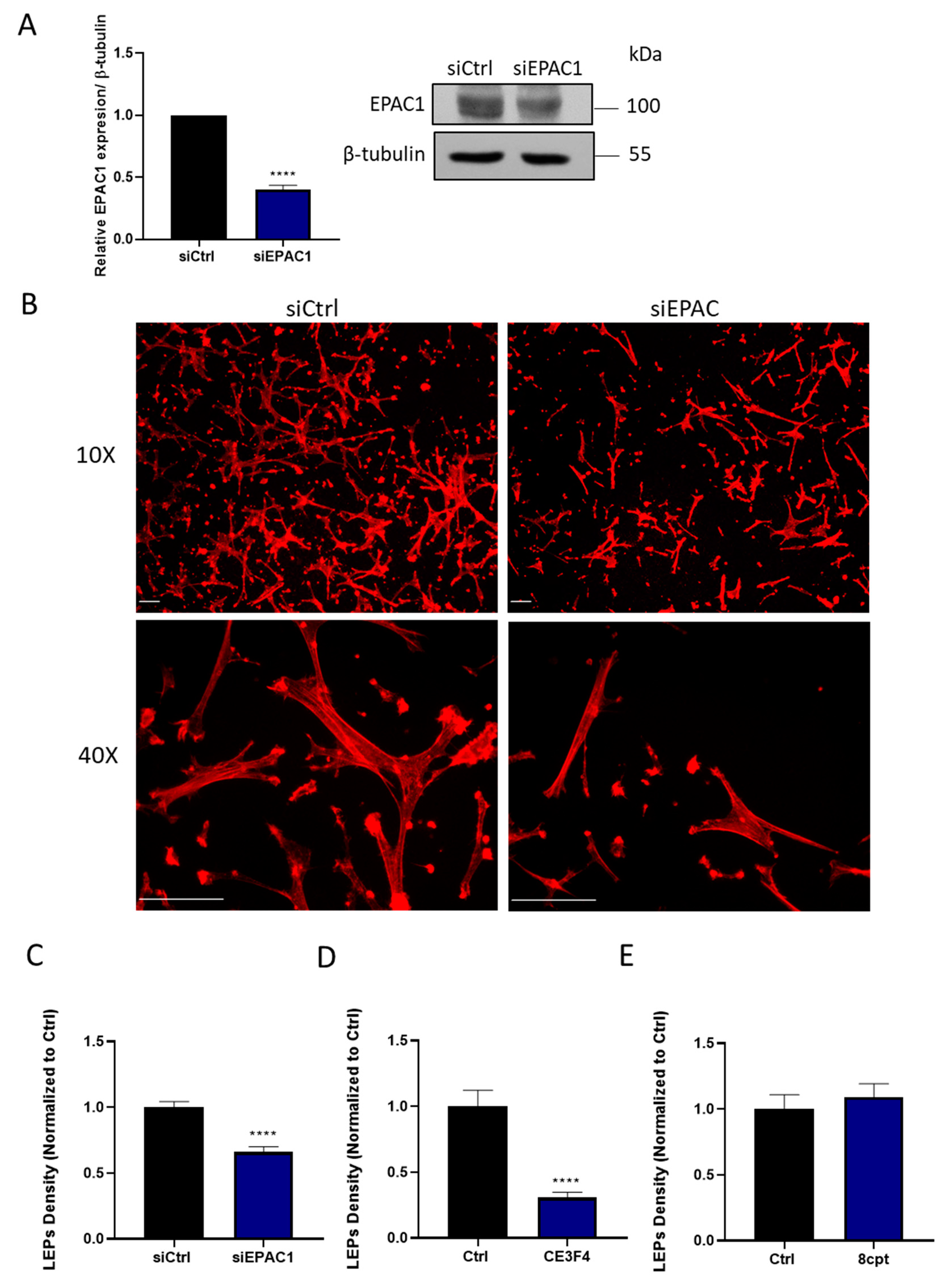
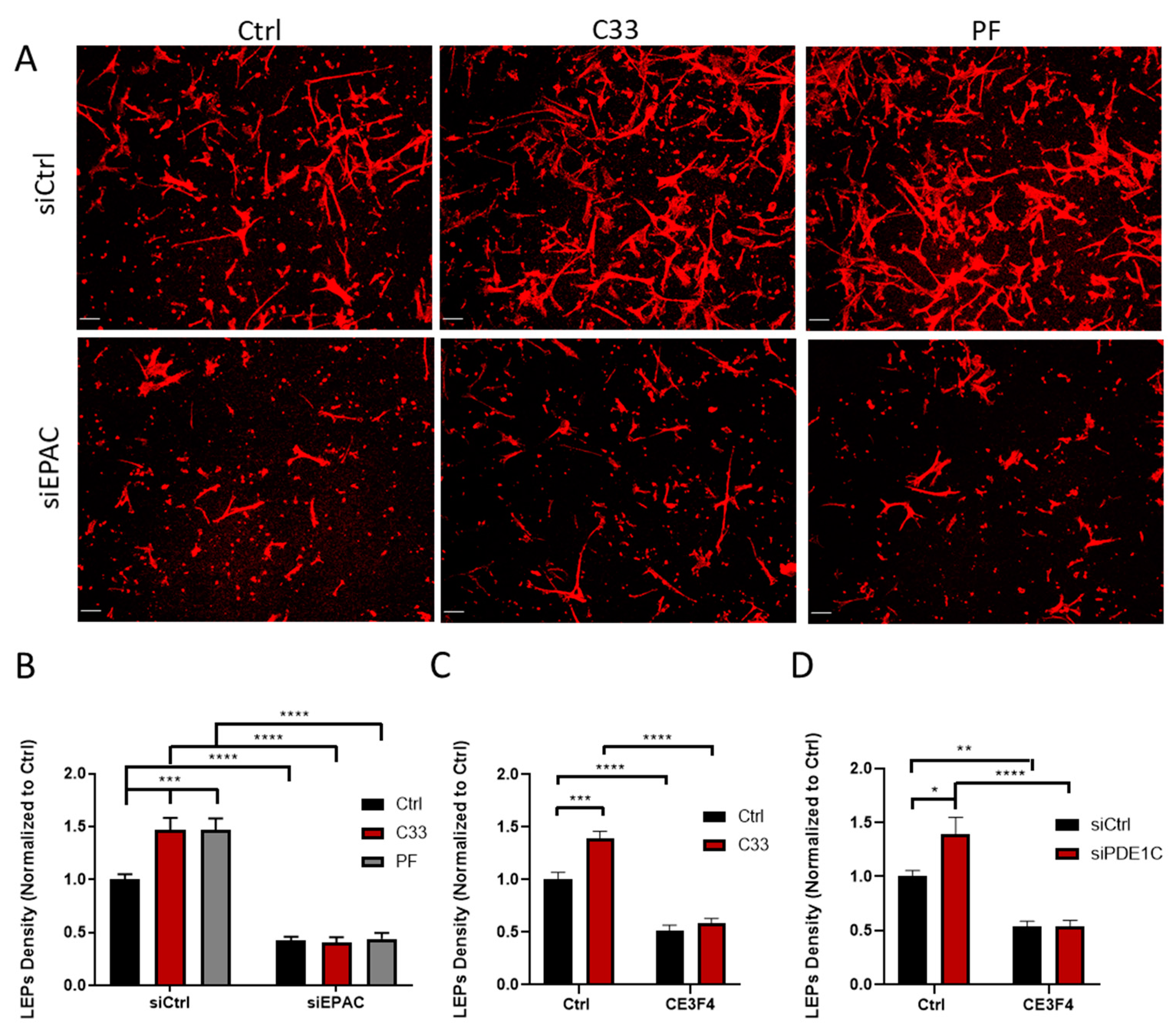
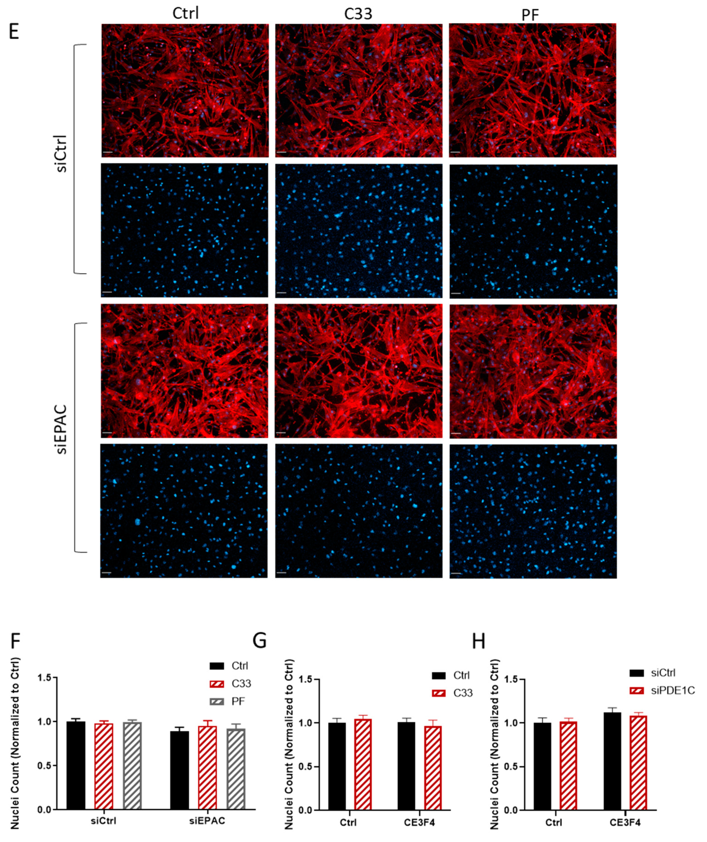
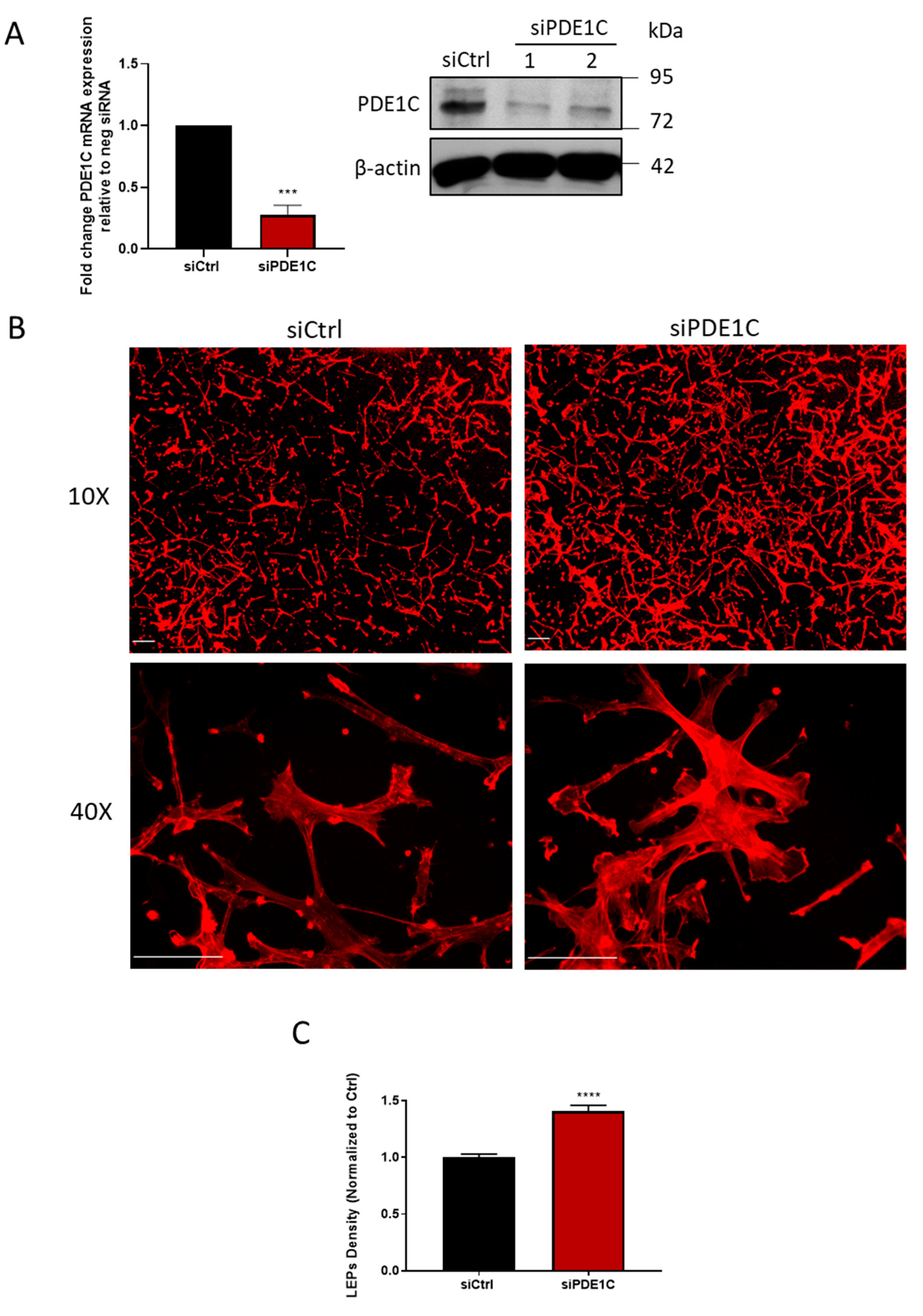
Pharmacological activation of protein kinase A (PKA) reduces migration of arterial smooth muscle cells (ASMCs), including those isolated from human arteries (HASMCs). However, when individual migration-associated cellular events, including the polarization of cells in the direction of movement or rearrangements of the actin cytoskeleton, are studied in isolation, these individual events can be either promoted or inhibited in response to PKA activation. While pharmacological inhibition or deficiency of exchange protein activated by cAMP-1 (EPAC1) reduces the overall migration of ASMCs, the impact of EPAC1 inhibition or deficiency, or of its activation, on individual migration-related events has not been investigated. Herein, we report that EPAC1 facilitates the formation of leading-edge protrusions (LEPs) in HASMCs, a critical early event in the cell polarization that underpins their migration. Thus, RNAi-mediated silencing, or the selective pharmacological inhibition, of EPAC1 decreased the formation of LEPs by these cells. Furthermore, we show that the ability of EPAC1 to promote LEP formation by migrating HASMCs is regulated by a phosphodiesterase 1C (PDE1C)-regulated “pool” of intracellular HASMC cAMP but not by those regulated by the more abundant PDE3 or PDE4 activities. Overall, our data are consistent with a role for EPAC1 in regulating the formation of LEPs by polarized HASMCs and show that PDE1C-mediated cAMP hydrolysis controls this localized event.
Keywords: exchange protein activated by cAMP; EPAC1; cyclic nucleotide phosphodiesterase; PDE1C; human arterial smooth muscle cells; HASMCs; migration; leading edge protrusion; cAMP; chemotaxis exchange protein activated by cAMP; EPAC1; cyclic nucleotide phosphodiesterase; PDE1C; human arterial smooth muscle cells; HASMCs; migration; leading edge protrusion; cAMP; chemotaxis