Review

MAP4K Family Kinases and DUSP Family Phosphatases in T-Cell Signaling and Systemic Lupus Erythematosus

Immunology Research Center, National Health Research Institutes, Zhunan 35053, Taiwan
*
Author to whom correspondence should be addressed.
Cells 2019, 8(11), 1433; https://doi.org/10.3390/cells8111433
Submission received: 3 October 2019 / Revised: 8 November 2019 / Accepted: 11 November 2019 / Published: 13 November 2019
(This article belongs to the Special Issue The Molecular and Cellular Basis for Lupus)

Abstract

:
T cells play a critical role in the pathogenesis of systemic lupus erythematosus (SLE), which is a severe autoimmune disease. In the past 60 years, only one new therapeutic agent with limited efficacy has been approved for SLE treatment; therefore, the development of early diagnostic biomarkers and therapeutic targets for SLE is desirable. Mitogen-activated protein kinase kinase kinase kinases (MAP4Ks) and dual-specificity phosphatases (DUSPs) are regulators of MAP kinases. Several MAP4Ks and DUSPs are involved in T-cell signaling and autoimmune responses. HPK1 (MAP4K1), DUSP22 (JKAP), and DUSP14 are negative regulators of T-cell activation. Consistently, HPK1 and DUSP22 are downregulated in the T cells of human SLE patients. In contrast, MAP4K3 (GLK) is a positive regulator of T-cell signaling and T-cell-mediated immune responses. MAP4K3 overexpression-induced RORγt–AhR complex specifically controls interleukin 17A (IL-17A) production in T cells, leading to autoimmune responses. Consistently, MAP4K3 and the RORγt–AhR complex are overexpressed in the T cells of human SLE patients, as are DUSP4 and DUSP23. In addition, DUSPs are also involved in either human autoimmune diseases (DUSP2, DUSP7, DUSP10, and DUSP12) or T-cell activation (DUSP1, DUSP5, and DUSP14). In this review, we summarize the MAP4Ks and DUSPs that are potential biomarkers and/or therapeutic targets for SLE.
Keywords:
SLE; DUSP; MAP4K; MAPK; MKP; T cells

1. Introduction

Both genetic and environmental factors contribute to the clinical heterogeneity of autoimmune diseases [1,2]. Innate immune responses cooperate with adaptive immune responses to induce autoimmune responses; therefore, multiple immune cells—including dendritic cells, neutrophils, macrophages, innate lymphoid cells, T helper cells, cytotoxic T cells, B cells, and Treg cells—are involved in the pathogenesis of autoimmune diseases [1]. Depending on the involvement of damaged tissues, autoimmune diseases are classified as either organ-specific diseases (e.g., multiple sclerosis, type I diabetes, and inflammatory bowel disease) or systemic diseases (e.g., systemic lupus erythematosus, rheumatoid arthritis, and Sjögren’s syndrome) [1].
Systemic lupus erythematosus (SLE) is a severe and even fatal autoimmune disease; SLE patients display pathogenic autoantibody production and multiple organ failures [3]. Inflammatory cytokines play an important role in the pathogenesis of autoimmune diseases. In particular, interleukin 17A (IL-17A) plays a critical role in SLE pathogenesis [4,5,6,7,8,9,10,11]. Several biologic agents have been used to treat autoimmune diseases [12,13,14,15,16,17]; however, the development of an effective therapeutic approach for SLE is very challenging due to the complexity and heterogeneity of the disease [4]. Over the past 60 years, only one therapeutic drug, belimumab/anti-BAFF antibody, has been approved for SLE treatment by the U.S. Food and Drug Administration (FDA) [13]. Even so, belimumab is useful only for SLE patients with modest symptoms, and its effect diminishes over the course of 72 weeks [18]. Thus, novel drug targets for effective treatment of SLE are needed [18]. Besides B cells, T cells also play pivotal roles in the pathogenesis of SLE [19]. Dysregulation of T-cell-mediated immune responses leads to enhanced production of pro-inflammation cytokines and autoantibodies, as well as chemokine-induced macrophage/neutrophil overactivation. Therefore, a better understanding of the T-cell-mediated SLE pathogenesis in T cells will be helpful in future developments of diagnostic biomarkers and effective treatments for SLE.
Signaling molecules (e.g., kinases and phosphatases) of immune cells play important roles in immune responses and autoimmune pathogenesis through induction of cytokines or chemokines [20,21,22,23,24]. Thus, signaling molecules in T cells are either potential biomarkers or therapeutic targets in the treatment of autoimmune diseases. For example, mitogen-activated protein kinases (MAPKs) are involved in the pathogenesis of autoimmune diseases, including SLE [25]; MAPK inhibitors have been developed for the attenuation of autoimmune responses [20,26]. To date, none of the MAPK inhibitors have progressed to phase III trials due to either lack of efficacy or adverse side effects [27,28]. Studies of these MAPK kinase inhibitors suggest that upstream signaling molecules may be more effective therapeutic targets than downstream signaling molecules [28,29,30]. Similarly, several upstream signaling molecules of MAPK are likely to be potential biomarkers or therapeutic targets for SLE. MAP kinase kinase kinase kinases (MAP4Ks) induce the MAPK c-Jun N-terminal kinase (JNK) through MAP3Ks and MAP2Ks [31,32]. Besides MAP4Ks, MAPK activities are also regulated by dual-specificity phosphatase (DUSP) family phosphatases, which comprise 25 members, including 9 MAPK phosphatases (MKPs) [33,34]. Several MAP4Ks and DUSPs are involved in the regulation of T-cell activation and human SLE. In this review, we summarize the potential utilization of MAP4Ks and DUSPs in T cells as biomarkers and/or therapeutic targets for SLE (Figure 1).

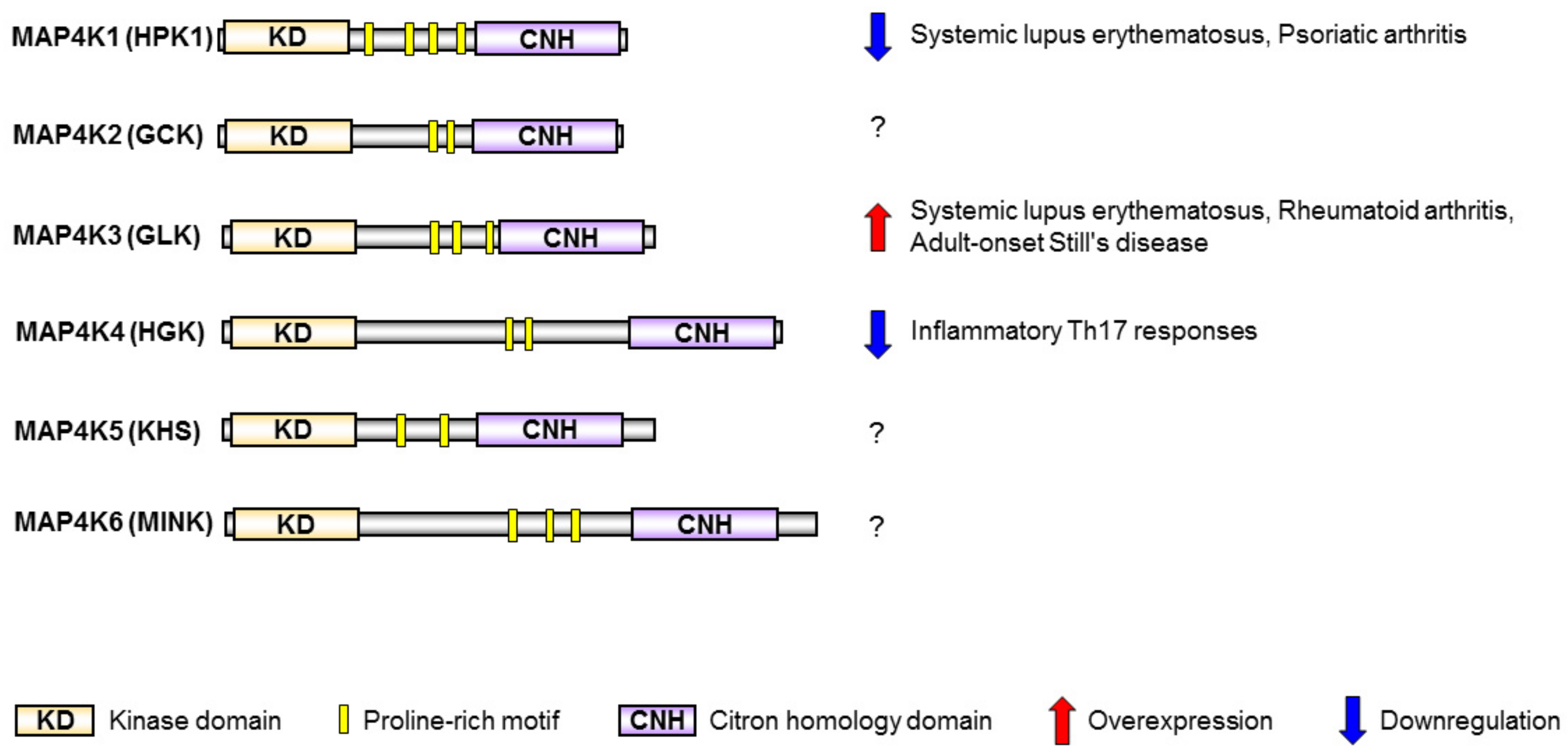
2. MAP4K Family Kinases Are Involved in T-Cell Activation and Human SLE

MAP4K family kinases—including MAP4K1/HPK1 [35,36,37], MAP4K2/GCK [38], MAP4K3/GLK [39], MAP4K4/HGK [40,41], MAP4K5/KHS [42], and MAP4K6/MINK [43]—are homologous to the mammalian STE20 family of serine/threonine protein kinases. MAP4K family kinases show a high-similarity protein structure, containing an N-terminal kinase domain, several proline-rich regions, and a C-terminal citron-homology domain [31] (Figure 2). MAP4K family kinases are initially identified as upstream molecules that activate MAP3Ks and MAP2Ks, leading to activation of the MAPK JNK [31,32,44,45]. MAP4Ks play important roles in the regulation of cell apoptosis, cell survival, cell autophagy, and cell migration [31,41,46]. Interestingly, several studies reported that MAP4Ks are involved in the regulation of immune-cell responses through JNK-independent pathways [21,22,47,48]. MAP4K1/HPK1 and MAP4K4/HGK play negative roles in T-cell activation and inflammatory responses [21,47]. In contrast, MAP4K3/GLK plays a positive role in T-cell activation and autoimmune responses [10,22]. Moreover, MAP4K1 downregulation and MAP4K3 overexpression in T cells are involved in human autoimmune diseases such as psoriatic arthritis, rheumatoid arthritis (RA), adult-onset Still’s disease, and SLE [22,49,50,51,52] (Figure 2).

2.1. HPK1 Transcription Is Reduced in CD4+ T Cells of Human SLE Patients

HPK1 (also known as MAP4K1) is a negative regulator of T-cell receptor signaling [21,36]. The HPK1 proteins are cleaved and activated by caspase 3 during apoptosis [53]. HPK1 is also activated by multiple adaptor proteins in mammalian cells, including T cells [36,54,55,56,57,58]. HPK1 directly interacts with and phosphorylates the adaptor protein SLP-76, leading to the inhibition of T-cell activation [21,59] (Figure 1). Notably, HPK1 also phosphorylates the adaptor protein BLNK, leading to the suppression of B-cell activation [48]. Overexpression of HPK1 inhibits T-cell proliferation, T-cell-secreted IFN-γ production, and T-cell-mediated antibody production [49]. Conversely, T-cell receptor (TCR)-induced T-cell proliferation, T-cell-secreted IFN-γ, and T-cell-mediated immune responses are significantly enhanced by HPK1 knockout [21]. Moreover, HPK1-knockout mice display enhanced autoimmune responses and increased CD4+ cell infiltration in the central nervous system during the induction of experimental autoimmune encephalomyelitis (EAE) [21]. Consistently, HPK1 mRNA and protein levels are decreased in the CD4+ T cells of SLE patients, compared to those of healthy controls [49] (Figure 2). Decreased binding of the Jumonji domain-containing protein 3 (JMJD3) to the HPK1 promoter results in increased H3K27me3 enrichment at the HPK1 promoter in SLE CD4+ T cells, leading to inhibition of HPK1 transcription [49]. Similarly, HPK1 is also downregulated in peripheral blood leukocytes in patients with another autoimmune disease—psoriatic arthritis [50] (Figure 2). Th17 cells are involved in the pathogenesis of SLE and psoriatic arthritis. It would be interesting to study the HPK1 inhibition of Th17 differentiation.
Besides HPK1, MAP4K4 (also known as HGK) is a negative regulator of Th17 differentiation [47]. HGK conditional T-cell deficiency results in the induction of inflammatory IL-6+ Th17 cells, leading to insulin resistance and systemic inflammation [47] (Figure 1 and Figure 2). Enhancement of HGK DNA methylation and subsequent downregulation of HGK in T cells are biomarkers of Asia-prevalent non-obese type 2 diabetes [60,61] (Figure 1). In addition, DNA methylation profiles of untreated SLE patients indicate that HGK methylation change is associated with SLE manifestations [62]. It is possible that HGK levels are also downregulated in the T cells of SLE patients, contributing to Th17-mediated inflammation.

2.2. GLK Is a Biomarker and Therapeutic Target for Human SLE

MAP4K3 (also known as GLK) is an activator of TCR signaling [11,22]. MAP4K3 directly interacts with and phosphorylates PKCθ upon TCR stimulation, resulting in IKK and NF-κB activation [22] (Figure 1). Like HPK1, GLK also interacts with the T-cell adaptor SLP-76, but GLK does not phosphorylate SLP-76. Moreover, SLP-76 is the upstream regulator for GLK kinase activity during TCR signaling [22]. In vitro T-cell proliferation, Th1 differentiation, Th2 differentiation, and Th17 differentiation are impaired by GLK deficiency [22]. GLK-deficient mice display decreased production of T-cell-mediated antigen-specific antibodies and cytokines [22]. Moreover, GLK-deficient mice are resistant to autoimmune disease induction in the experimental autoimmune encephalomyelitis (EAE) mouse model [22]. Consistently, GLK is overexpressed in the peripheral blood leukocytes (PBLs) of SLE patients; the activation of PKCθ and IKK are concomitantly induced in SLE PBLs compared to those of healthy controls [22] (Figure 2). The frequencies of GLK-overexpressing T cells, but not B cells, are increased in SLE patients, compared to those of healthy controls [9,22]. The GLK-overexpressing T cell population is correlated with the SLE disease activity index (SLEDAI) [9,22]. Besides SLE patients, GLK mRNA levels in T cells and GLK-overexpressing T cells are increased in patients with rheumatoid arthritis (RA) and adult-onset Still’s disease, compared to those of healthy controls [51,52] (Figure 2). Moreover, GLK overexpression also occurs in patients with other autoimmune diseases, such as Graves’ disease, Sjogren’s syndrome, and neuromyelitis optica, as well as in patients with cancer recurrence/metastasis [63,64,65,66].
The regulatory mechanisms of GLK overexpression in the T cells of SLE (or other autoimmune diseases) remain unknown. Three microRNAs (let-7c, miR-199-a-5p, and miR-206) have been reported to target GLK 3′UTR in cancer cells [11]; however, it is unclear whether these three microRNAs are decreased in SLE T cells. Enhancement of the long noncoding RNA NEAT induces the production of IL-6, CXCL10, and CCL8 through MAPKs in the monocytes of SLE patients [67]. It is possible that GLK overexpression in SLE T cells is also regulated by long noncoding RNAs. In addition, gene variants of the GLK gene may result in the induction of GLK mRNA levels in SLE T cells. Studying regulatory mechanisms of GLK overexpression in the T cells of SLE (or other autoimmune diseases) may help in the identification of additional therapeutic targets for SLE.
The pathogenic mechanism of GLK-induced autoimmune diseases has been revealed by the data derived from T-cell-specific GLK transgenic mice, plus several knockout mice for individual signaling molecules [10]. GLK overexpression in murine T cells specifically induces production of the inflammatory cytokine IL-17A through the AhR–RORγt complex (Figure 1). GLK signaling induces AhR nuclear translocation and AhR–RORγt complex formation through PKCθ and IKKβ, respectively [10]. In human SLE patients, the GLK+IL-17A+ CD4+ T cell population is drastically increased and is correlated with the SLEDAI [9]. The GLK+ Th17 cell population is also a biomarker for identifying active SLE [9]. T cells of SLE and RA patients display induction of GLK-induced AhR–RORγt complex, but healthy controls’ T cells do not [9]. Conversely, treatment of the GLK inhibitor verteporfin efficiently suppresses IL-17A production and AhR–RORγt complex in SLE T cells [9]. Verteporfin treatment also attenuates autoimmune responses in three autoimmune mouse models, including EAE, collagen-induced arthritis (CIA), and T-cell-specific GLK transgenic mice [9]. Collectively, GLK is a biomarker and therapeutic target for autoimmune diseases such as SLE.

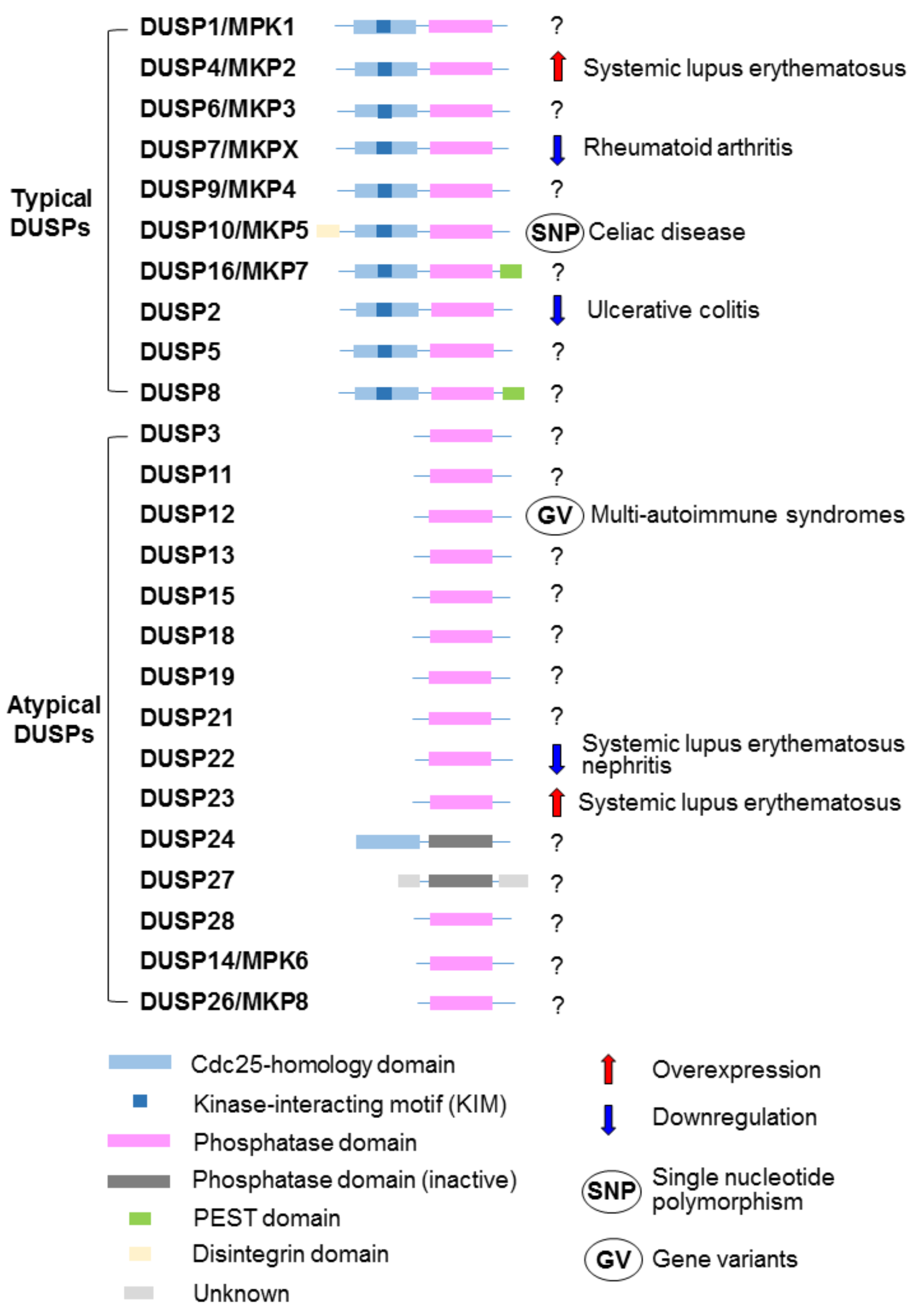
3. DUSP Family Phosphatases Are Involved in T-Cell Activation and Human SLE

The DUSP family contains 25 phosphatases, which dephosphorylate DUSPs’ substrates at threonine/serine residues and/or tyrosine residues [33,34]. All members of the DUSP family contain a common phosphatase domain [68] (Figure 3). Ten of 25 DUSPs contain the kinase-interacting motif (KIM) or the MAP-kinase-binding motif that interacts with MAPKs [34,69] (Figure 3). These 10 DUSPs are classified as typical DUSPs; 7 of 10 typical DUSPs are named as MAP kinase phosphatases (MKPs) [34] (Figure 3). Another 15 DUSPs do not have KIM and are classified as atypical DUSPs; however, 2 members (DUSP14 and DUSP26) of atypical DUSPs still dephosphorylate MAPKs [34]. Thus, 12 DUSPs have been reported to be MAPK phosphatases. One atypical DUSP, DUSP22, induces the MAPK JNK activation in a phosphatase activity-dependent manner [70]. DUSPs regulate various cellular functions, including cell survival, cell death, cell proliferation, and cell migration [34,71,72]. Several studies reported that DUSPs also regulate immune-cell responses [23,73,74,75,76]. DUSP2, DUSP4, DUSP7, DUSP10, DUSP12, DUSP22, and DUSP23 are involved in human autoimmune diseases, including SLE [77,78,79,80,81] (Figure 3).

3.1. DUSP22 Protein Level Is a Diagnostic and Prognostic Biomarker for SLE Nephritis

DUSP22 (also known as JKAP) is an atypical DUSP that activates the MAPK JNK [70]. Besides targeting JNK, JKAP dephosphorylates and inactivates focal adhesion kinase (FAK), leading to the inhibition of cell motility [82]. JKAP also inhibits prostate cancer cell proliferation by reducing EGFR- and androgen-receptor-dependent signaling [71]. Moreover, JKAP plays an inhibitory role in the turn-off stage of TCR signaling by dephosphorylating and inactivating the tyrosine kinase Lck [23] (Figure 1). JKAP-knockout mice display enhanced T-cell-secreted IFN-γ and IL-17A; JKAP-knockout mice are more susceptible to the autoimmune disease induction in the EAE model [23]. Aged DUSP22-knockout mice spontaneously display increased serum levels of pro-inflammatory cytokines (TNF-α, IFN-γ, IL-6, and IL-17A) and autoantibodies (antinuclear antibody and anti-dsDNA) [23]. Consistently, JKAP protein but not mRNA levels are decreased in the peripheral blood T cells of human SLE patients, compared to those of healthy controls [77]. JKAP downregulation in T cells is inversely correlated with daily urinary protein levels of SLE nephritis patients [77] (Figure 3). Moreover, the diagnostic power of JKAP downregulation for active lupus nephritis is higher than that of complements (C3 and C4) and anti-dsDNA antibody levels [77]. A longitudinal observational study further indicates that JKAP downregulation in T cells is correlated with the poor renal outcome of lupus nephritis patients [77]. These findings suggest that JKAP downregulation in T cells is a diagnostic and prognostic biomarker for SLE nephritis. The pathogenic role of DUSP22-deficient T cells in SLE nephritis has been demonstrated by characterizing T-cell-specific DUSP22 dominant-negative transgenic (Lck-DUSP22-C88S Tg) mice [77]. Lck-DUSP22-C88S Tg mice display inflammatory symptoms, including nephritis. Restoration of JKAP expression blocks the induction of IL-17A expression in the T cells of SLE patients [77]. These findings suggest that enhancing either JKAP protein levels or phosphatase activity may help the treatment and attenuation of SLE nephritis.
Protein levels of the tyrosine kinase Lck are decreased in the peripheral blood lymphocytes of SLE patients; however, phosphorylation and activation of Lck are still increased in the T cells of active SLE patients [83,84]. The enhancement of Lck activation is likely due to the JKAP downregulation in active SLE patients.
Besides DUSP22 downregulation in SLE patients, other DUSPs (DUSP2, DUSP7, DUSP10, and DUSP12) are also downregulated or mutated in human autoimmune diseases (Figure 3). The mRNA levels of the Th17 modulator DUSP2 are decreased in the PBMCs of ulcerative colitis patients [75]. DUSP7 mRNA levels are decreased in RA patients [80]. Single-nucleotide polymorphisms on the DUSP10 loci are associated with human celiac disease [85]. DUSP12 gene variants have been identified in patients with multi-autoimmune syndromes, such as the coincidence of Sjögren’s syndrome, RA, and either psoriasis or autoimmune thyroid disease [78]. It would be interesting to study whether DUSP2, DUSP7, DUSP10, and DUSP12 are also involved in human SLE.

3.2. DUSP4 mRNA Level Is Increased in CD4+ T Cells of Human Juvenile-Onset SLE

DUSP4 (also known as MKP2) is a typical DUSP that inactivates JNK, p38, and ERK [86,87]. DUSP4 overexpression also inhibits STAT5 phosphorylation, whereas DUSP4 deficiency results in enhanced STAT5 phosphorylation/activation in T cells [73]. Moreover, DUSP4-deficient mice show enhanced population of CD4+CD25+ T (Treg) cells [73]. DUSP4-deficient mice display decreased T-cell-secreted IL-17A; DUSP4-deficient mice are resistant to autoimmune disease induction in the EAE model [88]. Consistently, DUSP4 mRNA levels in differentiated human Th17 cells from healthy donors are higher than those of naïve T cells [79]. Furthermore, DUSP4 mRNA levels of the CD4+ T cells from 14 juvenile-onset SLE patients were significantly higher than those of healthy controls [79] (Figure 3). DUSP4 overexpression was associated with high disease activity in 14 SLE patients [79]. The DUSP4 overexpression in SLE T cells is likely due to the enhancement of CREMα/p300-mediated histone acetylation at the DUSP4 gene locus [79]. The data suggest that DUSP4 may be a potential biomarker for juvenile-onset SLE.

3.3. DUSP23 mRNA Levels Are Increased in CD4+ T Cells of Human SLE

Several genetic variants on human chromosome 1 (1q21–23) have been found to be associated with SLE; these polymorphic genes express inflammation-associated molecules such as C-reactive protein and FasL [89]. Interestingly, DUSP23 is also a gene with polymorphisms on chromosome 1q23. DUSP23 mRNA levels are increased in the CD4+ T cells of SLE patients (Figure 3); however, the DUSP23 mRNA levels are not correlated with any SLE clinical parameters [81]. Nevertheless, DUSP23 mRNA levels are correlated with mRNA levels of DNA-methylation enzyme, including DNMT1, DNMT3A, DNMD3B, MBD2, and MBD4 in the CD4+ T cells of SLE patients [81]. To date, the role of DUSP23 in the pathogenesis of human SLE remains unclear. Furthermore, the role of DUSP23 in autoimmune responses needs to be validated using DUSP23 knockout and transgenic mice.

3.4. DUSP1, DUSP5, and DUSP14 Also Regulate T Cell-Mediated Autoimmune Responses in Mice

DUSP1 (also known as MKP1)-deficient mice display impaired T-cell-mediated immune responses [90]. DUSP1-deficient mice are also resistant to EAE induction; the infiltrating Th17 and Th1 populations are decreased compared to those of wild-type mice [90]. In addition, the induction of CIA was attenuated by DUSP5 overexpression using an electroporation approach in mice [91]. DUSP5-overexpressing mice displayed reduced pro-inflammatory cytokines (IL-1β, IL-6, and TNF-α) in joint tissues and decreased Th17 cells in draining lymph nodes during CIA induction. The attenuation of CIA symptoms may be due to inactivation of STAT3 and ERK by DUSP5 overexpression in CD4+ T cells [91]. To date, it remains unknown whether DUSP1 is overexpressed/activated, and if DUSP5 is downregulated/inactivated in the T cells of SLE patients. Nevertheless, the inhibition of DUSP1 and overexpression of DUSP5 may be potential therapeutic approaches for autoimmune diseases, including SLE.
DUSP14 (also known as MKP6) directly dephosphorylates the adaptor TAB1 and inactivates the kinase complex TAB1/TAK1, leading to inactivation of T-cell activation [92]. DUSP14-knockout mice display enhanced T-cell-mediated immune responses; DUSP14-knockout mice are more susceptible to the EAE model than wild-type mice [92]. The phosphatase activity of DUSP14 is induced by the arginine methyltransferase PRMT5-induced methylation and the E3 ubiquitin ligase TRAF2-induced K63-linked ubiquitination of DUSP14 during T-cell receptor signaling [93,94]. Therefore, enhancement/activation of DUSP14 or DUSP14 upstream molecules may lead to the treatment or attenuation of autoimmune diseases.

4. Conclusions

Signaling molecules in T cells are dysregulated in SLE patients; downregulation or overexpression of these signaling molecules may be useful diagnostic biomarkers for SLE. Among MAP4Ks and DUPSs, the roles of MAP4K1 (HPK1), MAP4K3 (GLK), and DUSP22 (JKAP) in SLE pathogenesis have been validated using both clinical samples and gene-knockout mice (Figure 1). HPK1 downregulation and knockout result in T-cell hyperactivation enhanced autoimmune phenotypes. JKAP downregulation in T cells is a non-invasive diagnostic biomarker for SLE nephritis and is also a prognostic biomarker for poor outcome in SLE nephritis. Moreover, GLK+ Th17 population is a biomarker for active SLE. This GLK+ Th17 population will help in the selection of SLE patients that are responsive to GLK inhibitors (e.g., verteporfin), which block RORγt–AhR-complex-induced IL-17A production. Besides inhibition of GLK, activation or overexpression of T-cell signaling suppressors such as DUSP14 and DUSP5 may attenuate inflammatory and autoimmune responses of SLE patients. In addition, the overexpression of DUSP4 and DUSP23 in human SLE T cells as well as the reduction of inflammation in DUSP1-deficient mice suggest that inhibition of DUSP4, DUSP23, or DUSP1 may provide therapeutic benefits for SLE patients. Monitoring the knockout mice for the above potential therapeutic targets of SLE may help to identify any adverse effects caused by inhibiting these targets. A better understanding of additional signaling molecules that regulate T-cell signaling may lead to the identification of novel therapeutic targets for SLE.

Author Contributions

Conceptualization, T.-H.T.; writing—original draft preparation, H.-C.C.; writing—review and editing, H.-C.C. and T.-H.T.

Funding

This work was supported by grants from the National Health Research Institutes, Taiwan (IM-107-PP-01 and IM-107-SP-01, to T.-H.T.) and the Ministry of Science and Technology, Taiwan (107-2314-B-400-027 and 107-2321-B-400-013 to T.-H.T.; 107-2628-B-400-001 to H.-C.C.).

Acknowledgments

T.-H.T. is a recipient of the Taiwan Bio-Development Foundation (TBF) Chair in Biotechnology.

Conflicts of Interest

The authors declare no conflict of interest. The funders had no role in the design of the study; in the collection, analyses, or interpretation of data; in the writing of the manuscript; or in the decision to publish the results.

Abbreviations

MAP4KMAP Kinase Kinase Kinase Kinase
DUSPDual-Specificity Phosphatase
HPK1Hematopoietic Progenitor Kinase 1
GCKGerminal Center Kinase
GLKGCK-Like Kinase
HGKHPK1/GCK-Like Kinase
KHSKinase Homologous to Sps1/Ste20
MINKMisshapen/Nck-Related Kinase
TCRT-Cell Receptor
PKCθProtein Kinase C-theta
IKKIκB Kinase
JMJD3Jumonji Domain-Containing Protein 3
H3K27me3Histone H3 Lysine 27 Trimethylation
PRMT5Protein Arginine Methyltransferase 5
CREMαCamp Response Element Modulator α
CIACollagen-Induced Arthritis
EAEExperimental Autoimmune Encephalomyelitis
SLESystemic Lupus Erythematosus
RARheumatoid Arthritis
AOSDAdult-Onset Still’s Disease
SLEDAISLE Disease Activity Index
SNPSingle-Nucleotide Polymorphism

References

  1. Theofilopoulos, A.N.; Kono, D.H.; Baccala, R. The multiple pathways to autoimmunity. Nat. Immunol. 2017, 18, 716–724. [Google Scholar] [CrossRef]
  2. Gutierrez-Arcelus, M.; Rich, S.S.; Raychaudhuri, S. Autoimmune diseases–connecting risk alleles with molecular traits of the immune system. Nat. Rev. Genet. 2016, 17, 160–174. [Google Scholar] [CrossRef]
  3. Tsokos, G.C. Systemic lupus erythematosus. N. Engl. J. Med. 2011, 365, 2110–2121. [Google Scholar] [CrossRef]
  4. Martin, J.C.; Baeten, D.L.; Josien, R. Emerging role of IL-17 and Th17 cells in systemic lupus erythematosus. Clin. Immunol. 2014, 154, 1–12. [Google Scholar] [CrossRef] [PubMed]
  5. Yang, J.; Chu, Y.; Yang, X.; Gao, D.; Zhu, L.; Yang, X.; Wan, L.; Li, M. Th17 and natural Treg cell population dynamics in systemic lupus erythematosus. Arthritis Rheum. 2009, 60, 1472–1483. [Google Scholar] [CrossRef] [PubMed]
  6. Henriques, A.; Ines, L.; Couto, M.; Pedreiro, S.; Santos, C.; Magalhaes, M.; Santos, P.; Velada, I.; Almeida, A.; Carvalheiro, T.; et al. Frequency and functional activity of Th17, Tc17 and other T-cell subsets in systemic lupus erythematosus. Cell. Immunol. 2010, 264, 97–103. [Google Scholar] [CrossRef] [PubMed]
  7. Lopez, P.; Rodriguez-Carrio, J.; Caminal-Montero, L.; Mozo, L.; Suarez, A. A pathogenic IFNα, BLyS and IL-17 axis in systemic lupus erythematosus patients. Sci. Rep. 2016, 6, 20651. [Google Scholar] [CrossRef]
  8. Shah, K.; Lee, W.W.; Lee, S.H.; Kim, S.H.; Kang, S.W.; Craft, J.; Kang, I. Dysregulated balance of Th17 and Th1 cells in systemic lupus erythematosus. Arthritis Res. Ther. 2010, 12, R53. [Google Scholar] [CrossRef]
  9. Chuang, H.C.; Chen, Y.M.; Chen, M.H.; Hung, W.T.; Yang, H.Y.; Tseng, Y.H.; Tan, T.H. AhR-RORγt complex is a therapeutic target for MAP4K3/GLKhighIL-17Ahigh subpopulation of systemic lupus erythematosus. FASEB J. 2019, 33, 11469–11480. [Google Scholar] [CrossRef]
  10. Chuang, H.C.; Tsai, C.Y.; Hsueh, C.H.; Tan, T.H. GLK-IKKβ signaling induces dimerization and translocation of AhR-RORγt complex in IL-17A induction and autoimmune disease. Sci. Adv. 2018, 4, eaat5401. [Google Scholar] [CrossRef]
  11. Chuang, H.C.; Tan, T.H. MAP4K3/GLK in autoimmune disease, cancer and aging. J. Biomed. Sci. 2019, 26, 82. [Google Scholar] [CrossRef] [PubMed]
  12. Corbett, M.; Soares, M.; Jhuti, G.; Rice, S.; Spackman, E.; Sideris, E.; Moe-Byrne, T.; Fox, D.; Marzo-Ortega, H.; Kay, L.; et al. Tumour necrosis factor-α inhibitors for ankylosing spondylitis and non-radiographic axial spondyloarthritis: A systematic review and economic evaluation. Health Technol. Assess. 2016, 20, 1–334. [Google Scholar] [CrossRef] [PubMed]
  13. Manzi, S.; Sanchez-Guerrero, J.; Merrill, J.T.; Furie, R.; Gladman, D.; Navarra, S.V.; Ginzler, E.M.; D’Cruz, D.P.; Doria, A.; Cooper, S.; et al. Effects of belimumab, a B lymphocyte stimulator-specific inhibitor, on disease activity across multiple organ domains in patients with systemic lupus erythematosus: Combined results from two phase III trials. Ann. Rheum. Dis. 2012, 71, 1833–1838. [Google Scholar] [CrossRef]
  14. Smolen, J.S.; Aletaha, D.; Koeller, M.; Weisman, M.H.; Emery, P. New therapies for treatment of rheumatoid arthritis. Lancet 2007, 370, 1861–1874. [Google Scholar] [CrossRef]
  15. Breedveld, F.C.; Combe, B. Understanding emerging treatment paradigms in rheumatoid arthritis. Arthritis Res. Ther. 2011, 13 (Suppl. 1), S3. [Google Scholar]
  16. Gaffen, S.L.; Jain, R.; Garg, A.V.; Cua, D.J. The IL-23-IL-17 immune axis: From mechanisms to therapeutic testing. Nat. Rev. Immunol. 2014, 14, 585–600. [Google Scholar] [CrossRef] [PubMed]
  17. Patel, D.D.; Kuchroo, V.K. Th17 cell pathway in human immunity: Lessons from genetics and therapeutic interventions. Immunity 2015, 43, 1040–1051. [Google Scholar] [CrossRef]
  18. Mahieu, M.A.; Strand, V.; Simon, L.S.; Lipsky, P.E.; Ramsey-Goldman, R. A critical review of clinical trials in systemic lupus erythematosus. Lupus 2016, 25, 1122–1140. [Google Scholar] [CrossRef]
  19. Bluestone, J.A.; Bour-Jordan, H.; Cheng, M.; Anderson, M. T cells in the control of organ-specific autoimmunity. J. Clin. Invest. 2015, 125, 2250–2260. [Google Scholar] [CrossRef]
  20. Gorelik, G.; Richardson, B. Key role of ERK pathway signaling in lupus. Autoimmunity 2010, 43, 17–22. [Google Scholar] [CrossRef]
  21. Shui, J.W.; Boomer, J.S.; Han, J.; Xu, J.; Dement, G.A.; Zhou, G.; Tan, T.H. Hematopoietic progenitor kinase 1 negatively regulates T cell receptor signaling and T cell-mediated immune responses. Nat. Immunol. 2007, 8, 84–91. [Google Scholar] [CrossRef] [PubMed]
  22. Chuang, H.C.; Lan, J.L.; Chen, D.Y.; Yang, C.Y.; Chen, Y.M.; Li, J.P.; Huang, C.Y.; Liu, P.E.; Wang, X.; Tan, T.H. The kinase GLK controls autoimmunity and NF-κB signaling by activating the kinase PKC-θ in T cells. Nat. Immunol. 2011, 12, 1113–1118. [Google Scholar] [CrossRef] [PubMed]
  23. Li, J.P.; Yang, C.Y.; Chuang, H.C.; Lan, J.L.; Chen, D.Y.; Chen, Y.M.; Wang, X.; Chen, A.J.; Belmont, J.W.; Tan, T.H. The phosphatase JKAP/DUSP22 inhibits T-cell receptor signalling and autoimmunity by inactivating Lck. Nat. Commun. 2014, 5, 3618. [Google Scholar] [CrossRef] [PubMed]
  24. Perl, A. Overview of signal processing by the immune system in systemic lupus erythematosus. Autoimmun. Rev. 2009, 8, 177–178. [Google Scholar] [CrossRef] [PubMed]
  25. Molad, Y.; Amit-Vasina, M.; Bloch, O.; Yona, E.; Rapoport, M.J. Increased ERK and JNK activities correlate with disease activity in patients with systemic lupus erythematosus. Ann. Rheum. Dis. 2010, 69, 175–180. [Google Scholar] [CrossRef] [PubMed]
  26. Mavropoulos, A.; Orfanidou, T.; Liaskos, C.; Smyk, D.S.; Billinis, C.; Blank, M.; Rigopoulou, E.I.; Bogdanos, D.P. p38 mitogen-activated protein kinase (p38 MAPK)-mediated autoimmunity: Lessons to learn from ANCA vasculitis and pemphigus vulgaris. Autoimmun. Rev. 2013, 12, 580–590. [Google Scholar] [CrossRef] [PubMed]
  27. Schett, G.; Zwerina, J.; Firestein, G. The p38 mitogen-activated protein kinase (MAPK) pathway in rheumatoid arthritis. Ann. Rheum. Dis. 2008, 67, 909–916. [Google Scholar] [CrossRef]
  28. Poulikakos, P.I.; Solit, D.B. Resistance to MEK inhibitors: Should we co-target upstream? Sci. Signal. 2011, 4, pe16. [Google Scholar] [CrossRef]
  29. Hammaker, D.; Firestein, G.S. “Go upstream, young man”: Lessons learned from the p38 saga. Ann. Rheum. Dis. 2010, 69 (Suppl. 1), i77–i82. [Google Scholar] [CrossRef]
  30. Ghoreschi, K.; Laurence, A.; O’Shea, J.J. Selectivity and therapeutic inhibition of kinases: To be or not to be? Nat. Immunol. 2009, 10, 356–360. [Google Scholar] [CrossRef]
  31. Chuang, H.C.; Wang, X.; Tan, T.H. MAP4K family kinases in immunity and inflammation. Adv. Immunol. 2016, 129, 277–314. [Google Scholar] [PubMed]
  32. Chen, Y.R.; Tan, T.H. The c-Jun N-terminal kinase pathway and apoptotic signaling. Int. J. Oncol. 2000, 16, 651–662. [Google Scholar] [CrossRef] [PubMed]
  33. Chen, H.F.; Chuang, H.C.; Tan, T.H. Regulation of dual-specificity phosphatase (DUSP) ubiquitination and protein stability. Int. J. Mol. Sci. 2019, 20, 2668. [Google Scholar] [CrossRef] [PubMed] [Green Version]
  34. Huang, C.Y.; Tan, T.H. DUSPs, to MAP kinases and beyond. Cell Biosci. 2012, 2, 24. [Google Scholar] [CrossRef] [PubMed] [Green Version]
  35. Hu, M.C.; Qiu, W.R.; Wang, X.; Meyer, C.F.; Tan, T.H. Human HPK1, a novel human hematopoietic progenitor kinase that activates the JNK/SAPK kinase cascade. Genes Dev. 1996, 10, 2251–2264. [Google Scholar] [CrossRef] [Green Version]
  36. Boomer, J.S.; Tan, T.H. Functional interactions of HPK1 with adaptor proteins. J. Cell. Biochem. 2005, 95, 34–44. [Google Scholar] [CrossRef]
  37. Ling, P.; Meyer, C.F.; Redmond, L.P.; Shui, J.W.; Davis, B.; Rich, R.R.; Hu, M.C.; Wange, R.L.; Tan, T.H. Involvement of hematopoietic progenitor kinase 1 in T cell receptor signaling. J. Biol. Chem. 2001, 276, 18908–18914. [Google Scholar] [CrossRef] [Green Version]
  38. Pombo, C.M.; Kehrl, J.H.; Sanchez, I.; Katz, P.; Avruch, J.; Zon, L.I.; Woodgett, J.R.; Force, T.; Kyriakis, J.M. Activation of the SAPK pathway by the human STE20 homologue germinal centre kinase. Nature 1995, 377, 750–754. [Google Scholar] [CrossRef]
  39. Diener, K.; Wang, X.S.; Chen, C.; Meyer, C.F.; Keesler, G.; Zukowski, M.; Tan, T.H.; Yao, Z. Activation of the c-Jun N-terminal kinase pathway by a novel protein kinase related to human germinal center kinase. Proc. Natl. Acad. Sci. USA 1997, 94, 9687–9692. [Google Scholar] [CrossRef] [Green Version]
  40. Yao, Z.; Zhou, G.; Wang, X.S.; Brown, A.; Diener, K.; Gan, H.; Tan, T.H. A novel human STE20-related protein kinase, HGK, that specifically activates the c-Jun N-terminal kinase signaling pathway. J. Biol. Chem. 1999, 274, 2118–2125. [Google Scholar] [CrossRef] [Green Version]
  41. Fiedler, L.R.; Chapman, K.; Xie, M.; Maifoshie, E.; Jenkins, M.; Golforoush, P.A.; Bellahcene, M.; Noseda, M.; Faust, D.; Jarvis, A.; et al. MAP4K4 inhibition promotes survival of human stem cell-derived cardiomyocytes and reduces infarct size in vivo. Cell Stem Cell 2019, 24, 579–591. [Google Scholar] [CrossRef] [PubMed] [Green Version]
  42. Tung, R.M.; Blenis, J. A novel human SPS1/STE20 homologue, KHS, activates Jun N-terminal kinase. Oncogene 1997, 14, 653–659. [Google Scholar] [CrossRef] [PubMed] [Green Version]
  43. Dan, I.; Watanabe, N.M.; Kobayashi, T.; Yamashita-Suzuki, K.; Fukagaya, Y.; Kajikawa, E.; Kimura, W.K.; Nakashima, T.M.; Matsumoto, K.; Ninomiya-Tsuji, J.; et al. Molecular cloning of MINK, a novel member of mammalian GCK family kinases, which is up-regulated during postnatal mouse cerebral development. FEBS Lett 2000, 469, 19–23. [Google Scholar] [CrossRef] [Green Version]
  44. Chen, Y.R.; Meyer, C.F.; Tan, T.H. Persistent activation of c-Jun N-terminal kinase 1 (JNK1) in γ radiation-induced apoptosis. J. Biol. Chem. 1996, 271, 631–634. [Google Scholar] [CrossRef] [Green Version]
  45. Chen, Y.R.; Wang, X.; Templeton, D.; Davis, R.J.; Tan, T.H. The role of c-Jun N-terminal kinase (JNK) in apoptosis induced by ultraviolet C and γ radiation: Duration of JNK activation may determine cell death and proliferation. J. Biol. Chem. 1996, 271, 31929–31936. [Google Scholar] [CrossRef] [Green Version]
  46. Hsu, C.L.; Lee, E.X.; Gordon, K.L.; Paz, E.A.; Shen, W.C.; Ohnishi, K.; Meisenhelder, J.; Hunter, T.; La Spada, A.R. MAP4K3 mediates amino acid-dependent regulation of autophagy via phosphorylation of TFEB. Nat. Commun. 2018, 9, 942. [Google Scholar] [CrossRef]
  47. Chuang, H.C.; Sheu, W.H.; Lin, Y.T.; Tsai, C.Y.; Yang, C.Y.; Cheng, Y.J.; Huang, P.Y.; Li, J.P.; Chiu, L.L.; Wang, X.; et al. HGK/MAP4K4 deficiency induces TRAF2 stabilization and Th17 differentiation leading to insulin resistance. Nat. Commun. 2014, 5, 4602. [Google Scholar] [CrossRef] [Green Version]
  48. Wang, X.; Li, J.P.; Kuo, H.K.; Chiu, L.L.; Dement, G.A.; Lan, J.L.; Chen, D.Y.; Yang, C.Y.; Hu, H.; Tan, T.H. Down-regulation of B cell receptor signaling by hematopoietic progenitor kinase 1 (HPK1)-mediated phosphorylation and ubiquitination of activated B cell linker protein (BLNK). J. Biol. Chem. 2012, 287, 11037–11048. [Google Scholar] [CrossRef] [Green Version]
  49. Zhang, Q.; Long, H.; Liao, J.; Zhao, M.; Liang, G.; Wu, X.; Zhang, P.; Ding, S.; Luo, S.; Lu, Q. Inhibited expression of hematopoietic progenitor kinase 1 associated with loss of jumonji domain containing 3 promoter binding contributes to autoimmunity in systemic lupus erythematosus. J. Autoimmun. 2011, 37, 180–189. [Google Scholar] [CrossRef]
  50. Stoeckman, A.K.; Baechler, E.C.; Ortmann, W.A.; Behrens, T.W.; Michet, C.J.; Peterson, E.J. A distinct inflammatory gene expression profile in patients with psoriatic arthritis. Genes Immun. 2006, 7, 583–591. [Google Scholar] [CrossRef]
  51. Chen, D.Y.; Chuang, H.C.; Lan, J.L.; Chen, Y.M.; Hung, W.T.; Lai, K.L.; Tan, T.H. Germinal center kinase-like kinase (GLK/MAP4K3) expression is increased in adult-onset Still’s disease and may act as an activity marker. BMC Med. 2012, 10, 84. [Google Scholar] [CrossRef] [PubMed] [Green Version]
  52. Chen, Y.M.; Chuang, H.C.; Lin, W.C.; Tsai, C.Y.; Wu, C.W.; Gong, N.R.; Hung, W.T.; Lan, T.H.; Lan, J.L.; Tan, T.H.; et al. Germinal center kinase-like kinase overexpression in T cells as a novel biomarker in rheumatoid arthritis. Arthritis Rheum. 2013, 65, 2573–2582. [Google Scholar] [PubMed] [Green Version]
  53. Chen, Y.R.; Meyer, C.F.; Ahmed, B.; Yao, Z.; Tan, T.H. Caspase-mediated cleavage and functional changes of hematopoietic progenitor kinase 1 (HPK1). Oncogene 1999, 18, 7370–7377. [Google Scholar] [CrossRef] [PubMed] [Green Version]
  54. Han, J.; Shui, J.W.; Zhang, X.; Zheng, B.; Han, S.; Tan, T.H. HIP-55 is important for T-cell proliferation, cytokine production, and immune responses. Mol. Cell. Biol. 2005, 25, 6869–6878. [Google Scholar] [CrossRef] [Green Version]
  55. Han, J.; Kori, R.; Shui, J.W.; Chen, Y.R.; Yao, Z.; Tan, T.H. The SH3 domain-containing adaptor HIP-55 mediates c-Jun N-terminal kinase activation in T cell receptor signaling. J. Biol. Chem. 2003, 278, 52195–52202. [Google Scholar] [CrossRef] [Green Version]
  56. Ma, W.; Xia, C.; Ling, P.; Qiu, M.; Luo, Y.; Tan, T.H.; Liu, M. Leukocyte-specific adaptor protein Grap2 interacts with hematopoietic progenitor kinase 1 (HPK1) to activate JNK signaling pathway in T lymphocytes. Oncogene 2001, 20, 1703–1714. [Google Scholar] [CrossRef] [Green Version]
  57. Ling, P.; Yao, Z.; Meyer, C.F.; Wang, X.S.; Oehrl, W.; Feller, S.M.; Tan, T.H. Interaction of hematopoietic progenitor kinase 1 with adapter proteins Crk and CrkL leads to synergistic activation of c-Jun N-terminal kinase. Mol. Cell. Biol. 1999, 19, 1359–1368. [Google Scholar] [CrossRef] [Green Version]
  58. Ensenat, D.; Yao, Z.; Wang, X.S.; Kori, R.; Zhou, G.; Lee, S.C.; Tan, T.H. A novel src homology 3 domain-containing adaptor protein, HIP-55, that interacts with hematopoietic progenitor kinase 1. J. Biol. Chem. 1999, 274, 33945–33950. [Google Scholar] [CrossRef] [Green Version]
  59. Wang, X.; Li, J.P.; Chiu, L.L.; Lan, J.L.; Chen, D.Y.; Boomer, J.; Tan, T.H. Attenuation of T cell receptor signaling by serine phosphorylation-mediated lysine 30 ubiquitination of SLP-76 protein. J. Biol. Chem. 2012, 287, 34091–34100. [Google Scholar] [CrossRef] [Green Version]
  60. Chuang, H.C.; Tan, T.H. MAP4K4 and IL-6+ Th17 cells play important roles in non-obese type 2 diabetes. J. Biomed. Sci. 2017, 24, 4. [Google Scholar] [CrossRef] [Green Version]
  61. Chuang, H.C.; Wang, J.S.; Lee, I.T.; Sheu, W.H.; Tan, T.H. Epigenetic regulation of HGK/MAP4K4 in T cells of type 2 diabetes patients. Oncotarget 2016, 7, 10976–10989. [Google Scholar] [CrossRef] [PubMed] [Green Version]
  62. Imgenberg-Kreuz, J.; Carlsson Almlof, J.; Leonard, D.; Alexsson, A.; Nordmark, G.; Eloranta, M.L.; Rantapaa-Dahlqvist, S.; Bengtsson, A.A.; Jonsen, A.; Padyukov, L.; et al. DNA methylation mapping identifies gene regulatory effects in patients with systemic lupus erythematosus. Ann. Rheum. Dis. 2018, 77, 736–743. [Google Scholar] [CrossRef] [PubMed] [Green Version]
  63. Tan, T.H.; Chuang, H.C. MAP Kinase Kinase Kinase Kinase 3 (MAP4K3) as a Biomarker and Therapeutic Target for Autoimmune Disease, Cancer, Inflammation and IL-17-Associated Disease. U.S. Patent 8,846,311 B2, 2014. [Google Scholar]
  64. Hsu, C.P.; Chuang, H.C.; Lee, M.C.; Tsou, H.H.; Lee, L.W.; Li, J.P.; Tan, T.H. GLK/MAP4K3 overexpression associates with recurrence risk for non-small cell lung cancer. Oncotarget 2016, 7, 41748–41757. [Google Scholar] [CrossRef] [PubMed] [Green Version]
  65. Ho, C.H.; Chuang, H.C.; Wu, I.C.; Tsai, H.W.; Lin, Y.J.; Sun, H.Y.; Young, K.C.; Chiu, Y.C.; Cheng, P.N.; Liu, W.C.; et al. Prediction of early hepatocellular carcinoma recurrence using germinal center kinase-like kinase. Oncotarget 2016, 7, 49765–49776. [Google Scholar] [CrossRef] [PubMed]
  66. Chuang, H.C.; Chang, C.C.; Teng, C.F.; Hsueh, C.H.; Chiu, L.L.; Hsu, P.M.; Lee, M.C.; Hsu, C.P.; Chen, Y.R.; Liu, Y.C.; et al. MAP4K3/GLK promotes lung cancer metastasis by phosphorylating and activating IQGAP1. Cancer Res. 2019, 79, 4978–4993. [Google Scholar] [CrossRef] [PubMed] [Green Version]
  67. Zhang, F.; Wu, L.; Qian, J.; Qu, B.; Xia, S.; La, T.; Wu, Y.; Ma, J.; Zeng, J.; Guo, Q.; et al. Identification of the long noncoding RNA NEAT1 as a novel inflammatory regulator acting through MAPK pathway in human lupus. J. Autoimmun. 2016, 75, 96–104. [Google Scholar] [CrossRef]
  68. Farooq, A.; Zhou, M.M. Structure and regulation of MAPK phosphatases. Cell. Signal. 2004, 16, 769–779. [Google Scholar] [CrossRef]
  69. MacCorkle, R.A.; Tan, T.H. Mitogen-activated protein kinases in cell-cycle control. Cell Biochem. Biophys. 2005, 43, 451–461. [Google Scholar] [CrossRef]
  70. Chen, A.J.; Zhou, G.; Juan, T.; Colicos, S.M.; Cannon, J.P.; Cabriera-Hansen, M.; Meyer, C.F.; Jurecic, R.; Copeland, N.G.; Gilbert, D.J.; et al. The dual specificity JKAP specifically activates the c-Jun N-terminal kinase pathway. J. Biol. Chem. 2002, 277, 36592–36601. [Google Scholar] [CrossRef] [Green Version]
  71. Lin, H.P.; Ho, H.M.; Chang, C.W.; Yeh, S.D.; Su, Y.W.; Tan, T.H.; Lin, W.J. DUSP22 suppresses prostate cancer proliferation by targeting the EGFR-AR axis. FASEB J. 2019. [Google Scholar] [CrossRef] [Green Version]
  72. Chen, Y.R.; Chou, H.C.; Yang, C.H.; Chen, H.Y.; Liu, Y.W.; Lin, T.Y.; Yeh, C.L.; Chao, W.T.; Tsou, H.H.; Chuang, H.C.; et al. Deficiency in VHR/DUSP3, a suppressor of focal adhesion kinase, reveals its role in regulating cell adhesion and migration. Oncogene 2017, 36, 6509–6517. [Google Scholar] [CrossRef] [PubMed]
  73. Huang, C.Y.; Lin, Y.C.; Hsiao, W.Y.; Liao, F.H.; Huang, P.Y.; Tan, T.H. DUSP4 deficiency enhances CD25 expression and CD4+ T-cell proliferation without impeding T-cell development. Eur. J. Immunol. 2012, 42, 476–488. [Google Scholar] [CrossRef] [PubMed] [Green Version]
  74. Hsu, W.C.; Chen, M.Y.; Hsu, S.C.; Huang, L.R.; Kao, C.Y.; Cheng, W.H.; Pan, C.H.; Wu, M.S.; Yu, G.Y.; Hung, M.S.; et al. DUSP6 mediates T cell receptor-engaged glycolysis and restrains TFH cell differentiation. Proc. Natl. Acad. Sci. USA 2018, 115, E8027–E8036. [Google Scholar] [CrossRef] [PubMed] [Green Version]
  75. Lu, D.; Liu, L.; Ji, X.; Gao, Y.; Chen, X.; Liu, Y.; Liu, Y.; Zhao, X.; Li, Y.; Li, Y.; et al. The phosphatase DUSP2 controls the activity of the transcription activator STAT3 and regulates TH17 differentiation. Nat. Immunol. 2015, 16, 1263–1273. [Google Scholar] [CrossRef] [PubMed]
  76. Lang, R.; Raffi, F.A.M. Dual-specificity phosphatases in immunity and infection: An update. Int. J. Mol. Sci. 2019, 20. [Google Scholar] [CrossRef] [Green Version]
  77. Chuang, H.C.; Chen, Y.M.; Hung, W.T.; Li, J.P.; Chen, D.Y.; Lan, J.L.; Tan, T.H. Downregulation of the phosphatase JKAP/DUSP22 in T cells as a potential new biomarker of systemic lupus erythematosus nephritis. Oncotarget 2016, 7, 57593–57605. [Google Scholar] [CrossRef] [Green Version]
  78. Johar, A.S.; Mastronardi, C.; Rojas-Villarraga, A.; Patel, H.R.; Chuah, A.; Peng, K.; Higgins, A.; Milburn, P.; Palmer, S.; Silva-Lara, M.F.; et al. Novel and rare functional genomic variants in multiple autoimmune syndrome and Sjogren’s syndrome. J. Transl. Med. 2015, 13, 173. [Google Scholar] [CrossRef] [Green Version]
  79. Hofmann, S.R.; Mabert, K.; Kapplusch, F.; Russ, S.; Northey, S.; Beresford, M.W.; Tsokos, G.C.; Hedrich, C.M. cAMP response element modulator α induces dual specificity protein phosphatase 4 to promote effector T cells in juvenile-onset lupus. J. Immunol. 2019. [Google Scholar] [CrossRef]
  80. Castro-Sanchez, P.; Ramirez-Munoz, R.; Lamana, A.; Ortiz, A.; Gonzalez-Alvaro, I.; Roda-Navarro, P. mRNA profiling identifies low levels of phosphatases dual-specific phosphatase-7 (DUSP7) and cell division cycle-25B (CDC25B) in patients with early arthritis. Clin. Exp. Immunol. 2017, 189, 113–119. [Google Scholar] [CrossRef] [Green Version]
  81. Balada, E.; Felip, L.; Ordi-Ros, J.; Vilardell-Tarres, M. DUSP23 is over-expressed and linked to the expression of DNMTs in CD4+ T cells from systemic lupus erythematosus patients. Clin. Exp. Immunol. 2017, 187, 242–250. [Google Scholar] [CrossRef] [Green Version]
  82. Li, J.P.; Fu, Y.N.; Chen, Y.R.; Tan, T.H. JNK pathway-associated phosphatase dephosphorylates focal adhesion kinase and suppresses cell migration. J. Biol. Chem. 2010, 285, 5472–5478. [Google Scholar] [CrossRef] [PubMed] [Green Version]
  83. Matache, C.; Stefanescu, M.; Onu, A.; Tanaseanu, S.; Matei, I.; Frade, R.; Szegli, G. p56lck activity and expression in peripheral blood lymphocytes from patients with systemic lupus erythematosus. Autoimmunity 1999, 29, 111–120. [Google Scholar] [CrossRef] [PubMed]
  84. Jury, E.C.; Isenberg, D.A.; Mauri, C.; Ehrenstein, M.R. Atorvastatin restores Lck expression and lipid raft-associated signaling in T cells from patients with systemic lupus erythematosus. J. Immunol. 2006, 177, 7416–7422. [Google Scholar] [CrossRef] [Green Version]
  85. Ostensson, M.; Monten, C.; Bacelis, J.; Gudjonsdottir, A.H.; Adamovic, S.; Ek, J.; Ascher, H.; Pollak, E.; Arnell, H.; Browaldh, L.; et al. A possible mechanism behind autoimmune disorders discovered by genome-wide linkage and association analysis in celiac disease. PLoS ONE 2013, 8, e70174. [Google Scholar] [CrossRef] [PubMed]
  86. Chu, Y.; Solski, P.A.; Khosravi-Far, R.; Der, C.J.; Kelly, K. The mitogen-activated protein kinase phosphatases PAC1, MKP-1, and MKP-2 have unique substrate specificities and reduced activity in vivo toward the ERK2 sevenmaker mutation. J. Biol. Chem. 1996, 271, 6497–6501. [Google Scholar] [CrossRef] [PubMed] [Green Version]
  87. Al-Mutairi, M.; Al-Harthi, S.; Cadalbert, L.; Plevin, R. Over-expression of mitogen-activated protein kinase phosphatase-2 enhances adhesion molecule expression and protects against apoptosis in human endothelial cells. Br. J. Pharmacol. 2010, 161, 782–798. [Google Scholar] [CrossRef] [PubMed] [Green Version]
  88. Hsiao, W.Y.; Lin, Y.C.; Liao, F.H.; Chan, Y.C.; Huang, C.Y. Dual-specificity phosphatase 4 regulates STAT5 protein stability and helper T cell polarization. PLoS ONE 2015, 10, e0145880. [Google Scholar] [CrossRef]
  89. Moser, K.L.; Neas, B.R.; Salmon, J.E.; Yu, H.; Gray-McGuire, C.; Asundi, N.; Bruner, G.R.; Fox, J.; Kelly, J.; Henshall, S.; et al. Genome scan of human systemic lupus erythematosus: Evidence for linkage on chromosome 1q in African-American pedigrees. Proc. Natl. Acad. Sci. USA 1998, 95, 14869–14874. [Google Scholar] [CrossRef] [Green Version]
  90. Zhang, Y.; Reynolds, J.M.; Chang, S.H.; Martin-Orozco, N.; Chung, Y.; Nurieva, R.I.; Dong, C. MKP-1 is necessary for T cell activation and function. J. Biol. Chem. 2009, 284, 30815–30824. [Google Scholar] [CrossRef] [Green Version]
  91. Moon, S.J.; Lim, M.A.; Park, J.S.; Byun, J.K.; Kim, S.M.; Park, M.K.; Kim, E.K.; Moon, Y.M.; Min, J.K.; Ahn, S.M.; et al. Dual-specificity phosphatase 5 attenuates autoimmune arthritis in mice via reciprocal regulation of the Th17/Treg cell balance and inhibition of osteoclastogenesis. Arthritis Rheumatol. 2014, 66, 3083–3095. [Google Scholar] [CrossRef] [Green Version]
  92. Yang, C.Y.; Li, J.P.; Chiu, L.L.; Lan, J.L.; Chen, D.Y.; Chuang, H.C.; Huang, C.Y.; Tan, T.H. Dual-specificity phosphatase 14 (DUSP14/MKP6) negatively regulates TCR signaling by inhibiting TAB1 activation. J. Immunol. 2014, 192, 1547–1557. [Google Scholar] [CrossRef] [PubMed] [Green Version]
  93. Yang, C.Y.; Chiu, L.L.; Tan, T.H. TRAF2-mediated Lys63-linked ubiquitination of DUSP14/MKP6 is essential for its phosphatase activity. Cell. Signal. 2016, 28, 145–151. [Google Scholar] [CrossRef] [PubMed]
  94. Yang, C.Y.; Chiu, L.L.; Chang, C.C.; Chuang, H.C.; Tan, T.H. Induction of DUSP14 ubiquitination by PRMT5-mediated arginine methylation. FASEB J. 2018, 32, 6760–6770. [Google Scholar] [CrossRef] [PubMed] [Green Version]
Figure 1. MAP4K1, MAP4K3, MAP4K4, and DUSP22 in T-cell signaling and systemic lupus erythematosus (SLE). The roles of MAP4K1 (HPK1), MAP4K3 (GLK), and DUSP22 (JKAP) in T-cell receptor (TCR) signaling and SLE pathogenesis have been validated using both gene-knockout mice and clinical samples. HPK1 phosphorylates SLP-76 at the serine 376 (S376) residue upon TCR stimulation, resulting in ubiquitin-mediated degradation of SLP-76. HPK1 downregulation in the T cells of human SLE patients leads to the enhancement of T-cell-mediated autoimmune responses. Moreover, DUSP22 (JKAP) dephosphorylates the tyrosine kinase Lck at the tyrosine 394 (Y394) residue, leading to inactivation of Lck and inhibition of T-cell activation. JKAP knockout or deficiency induces T-cell hyperactivation. Consistently, JKAP downregulation in T cells is highly correlated with SLE nephritis and thus is a prognostic biomarker for poor outcome. Furthermore, GCK-Like Kinase (GLK) phosphorylates PKCθ at the threonine 538 (T538) residue, resulting in the activation of the IKK kinase complex and NF-κB. GLK overexpression in T cells further induces interleukin 17A (IL-17A) transcription through the RORγt–AhR complex. IKKβ-induced RORγt serine 489 (S489) phosphorylation and PKCθ-induced AhR serine 36 (S36) phosphorylation result in IL-17A overproduction, leading to autoimmune responses. The GLK-induced SLE pathogenesis has been verified using T-cell-specific GLK transgenic mice and human SLE T cells. In addition, HGK phosphorylates TRAF2 at the serine 35 (S35) residue, resulting in lysosomal degradation of TRAF2. DNA hypermethylation on the HGK promoter results in HGK downregulation and TRAF2 overexpression in T cells of human non-obese type II diabetes patients. DNA methylation of HGK is also changed in human SLE peripheral blood mononuclear cells (PBMCs). HGK levels might also be downregulated in SLE T cells, contributing to autoimmunity. Red residue denotes activating phosphorylation site; blue residue denotes inhibitory phosphorylation site. Arrows denote activation; T bar denotes inhibition. Dashed rectangle denotes potential molecular mechanism for SLE pathogenesis.
Cells 08 01433 g001
Figure 2. The structural domains and autoimmune-disease involvement of MAP4K family kinases.
Cells 08 01433 g002
Figure 3. The structural domains and autoimmune-disease involvement of DUSP family phosphatases.
Cells 08 01433 g003

