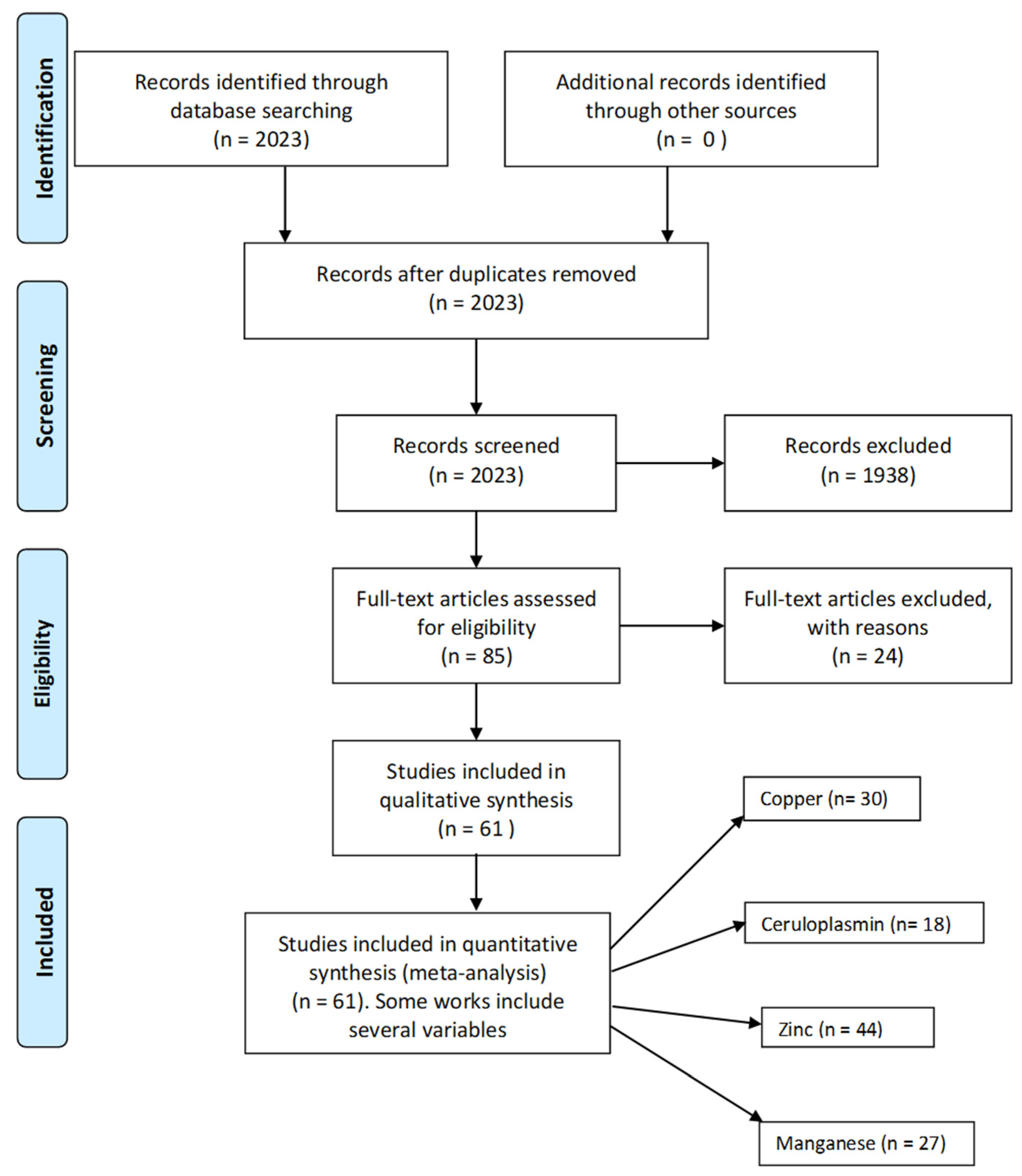
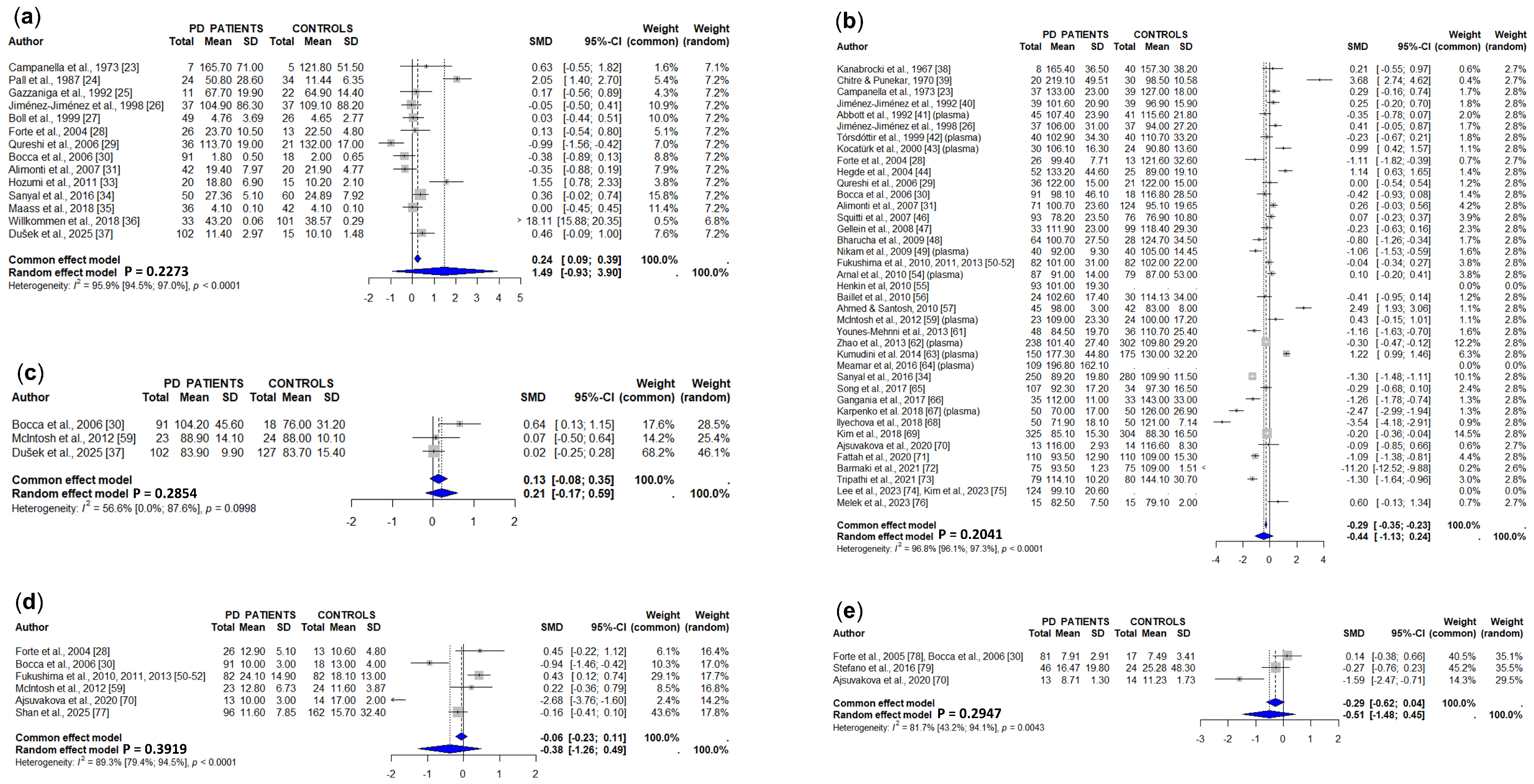
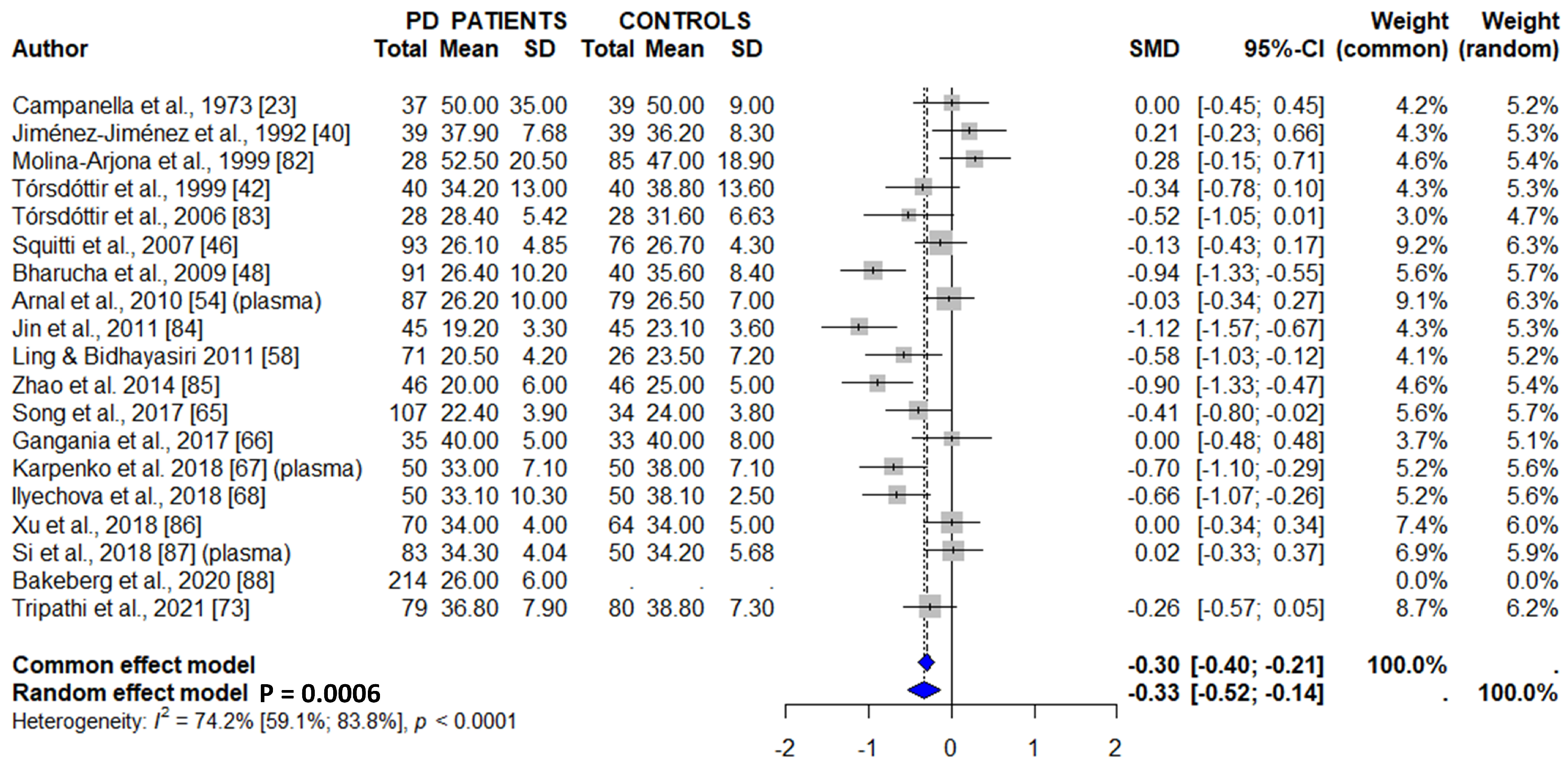
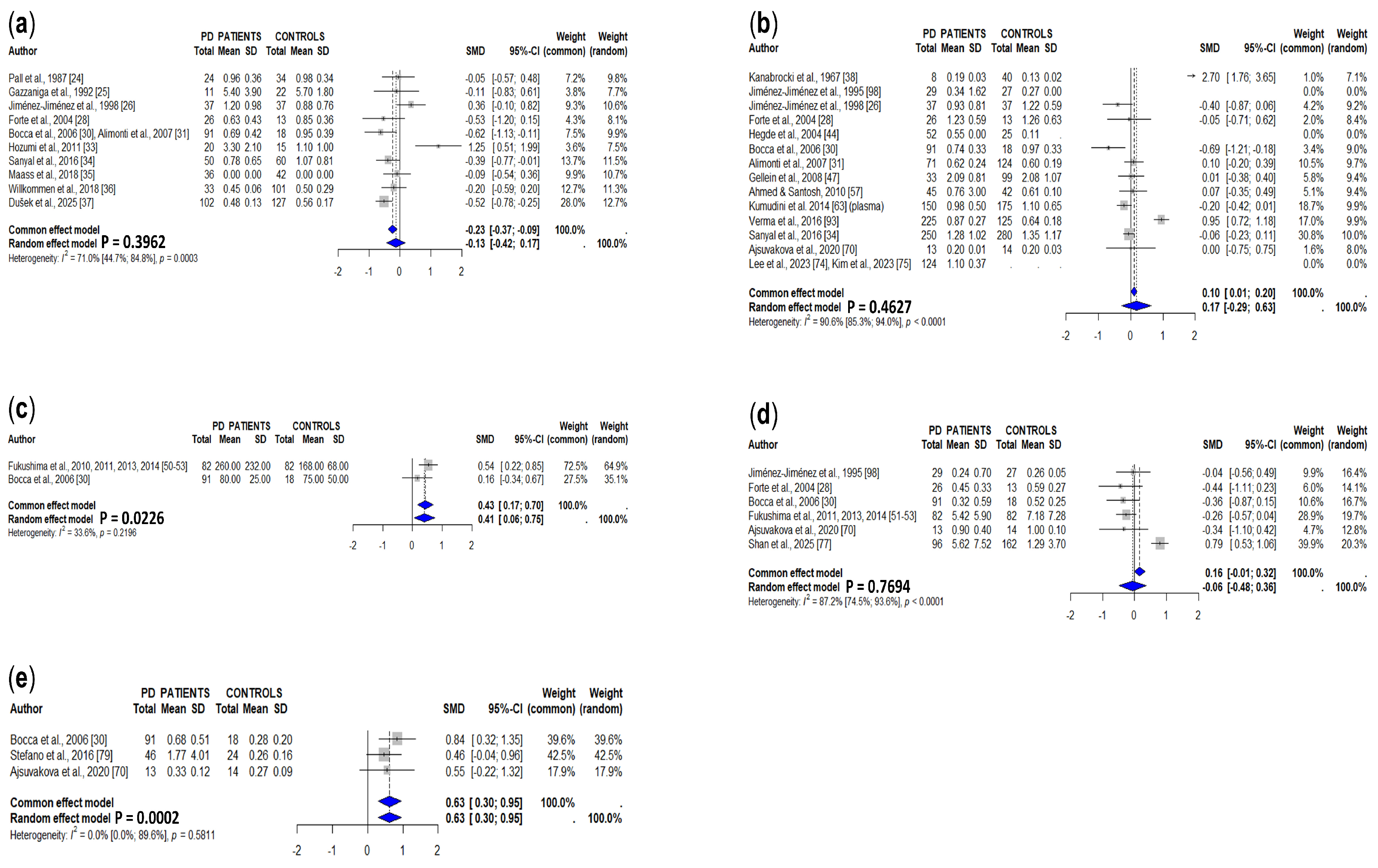
Copper, Ceruloplasmin, Zinc, and Manganese Levels in Brain and Biological Fluids from Parkinson’s Disease Patients: Systematic Review and Meta-Analysis

 ,
,  , , , , ,
Abstract
1. Introduction
2. Methods
2.1. Search Strategy
2.2. Criteria for Eligibility and Exclusion of Studies from the Meta-Analyses
2.3. Statistical Analysis
3. Results
3.1. Copper
3.1.1. Brain
3.1.2. Cerebrospinal Fluid
3.1.3. Serum/Plasma
3.1.4. Whole Blood
3.1.5. Urine
3.1.6. Hair
3.2. Ceruloplasmin
3.2.1. Brain
3.2.2. Cerebrospinal Fluid
3.2.3. Serum/Plasma
3.3. Zinc
3.3.1. Brain
3.3.2. Cerebrospinal Fluid
3.3.3. Serum/Plasma
3.3.4. Whole Blood
3.3.5. Urine
3.3.6. Hair
3.4. Manganese
3.4.1. Brain
3.4.2. Cerebrospinal Fluid
3.4.3. Serum/Plasma
3.4.4. Whole Blood
3.4.5. Urine
3.4.6. Hair
4. Discussion
5. Conclusions
Supplementary Materials
Author Contributions
Funding
Data Availability Statement
Acknowledgments
Conflicts of Interest
References
- Hamidpour, S.K.; Amiri, M.; Ketabforoush, A.H.M.E.; Saeedi, S.; Angaji, A.; Tavakol, S. Unraveling Dysregulated Cell Signaling Pathways, Genetic and Epigenetic Mysteries of Parkinson’s Disease. Mol. Neurobiol. 2024, 61, 8928–8966. [Google Scholar] [CrossRef]
- Walecha, V.; Luthra, P.M. The Mitochondrial-Astrocyte-Neuron Triad Hypothesis in Parkinson’s Disease: A Toxic Feedback Loop of Metabolism, Aggregation, and Oxidative Stress. Neurochem. Res. 2025, 50, 317. [Google Scholar] [CrossRef]
- Wei, Z.; Li, X.; Li, X.; Liu, Q.; Cheng, Y. Oxidative Stress in Parkinson’s Disease: A Systematic Review and Meta-Analysis. Front. Mol. Neurosci. 2018, 11, 236. [Google Scholar] [CrossRef]
- Jomova, K.; Alomar, S.Y.; Valko, R.; Nepovimova, E.; Kuca, K.; Valko, M. The Role of Redox-Active Iron, Copper, Manganese, and Redox-Inactive Zinc in Toxicity, Oxidative Stress, and Human Diseases. EXCLI J. 2025, 24, 880–954. [Google Scholar] [CrossRef] [PubMed]
- Mezzaroba, L.; Alfieri, D.F.; Colado Simão, A.N.; Vissoci Reiche, E.M. The Role of Zinc, Copper, Manganese and Iron in Neurodegenerative Diseases. Neurotoxicology 2019, 74, 230–241. [Google Scholar] [CrossRef] [PubMed]
- Jiménez-Jiménez, F.J.; Alonso-Navarro, H.; García-Martín, E.; Agúndez, J.A.G. Biological Fluid Levels of Iron and Iron-Related Proteins in Parkinson’s Disease: Review and Meta-Analysis. Eur. J. Neurol. 2021, 28, 1041–1055. [Google Scholar] [CrossRef] [PubMed]
- Genoud, S.; Senior, A.M.; Hare, D.J.; Double, K.L. Meta-Analysis of Copper and Iron in Parkinson’s Disease Brain and Biofluids. Mov. Disord. 2020, 35, 662–671. [Google Scholar] [CrossRef]
- Scolari Grotto, F.; Glaser, V. Are High Copper Levels Related to Alzheimer’s and Parkinson’s Diseases? A Systematic Review and Meta-Analysis of Articles Published between 2011 and 2022. Biometals 2024, 37, 3–22. [Google Scholar] [CrossRef]
- Adani, G.; Filippini, T.; Michalke, B.; Vinceti, M. Selenium and Other Trace Elements in the Etiology of Parkinson’s Disease: A Systematic Review and Meta-Analysis of Case-Control Studies. Neuroepidemiology 2020, 54, 1–23. [Google Scholar] [CrossRef]
- Sun, H.; Liu, X.; Ge, H.; Wang, T.; Wang, Y.; Li, W. Association between Serum Zinc Levels and the Risk of Parkinson’s Disease: A Meta-Analysis. Biol. Trace Elem. Res. 2017, 179, 45–51. [Google Scholar] [CrossRef]
- Du, K.; Liu, M.Y.; Zhong, X.; Wei, M.J. Decreased Circulating Zinc Levels in Parkinson’s Disease: A Meta-Analysis Study. Sci. Rep. 2017, 7, 3902. [Google Scholar] [CrossRef] [PubMed]
- Riederer, P.; Sofic, E.; Rausch, W.D.; Schmidt, B.; Reynolds, G.P.; Jellinger, K.; Youdim, M.B. Transition Metals, Ferritin, Glutathione, and Ascorbic Acid in Parkinsonian Brains. J. Neurochem. 1989, 52, 515–520. [Google Scholar] [CrossRef] [PubMed]
- Dexter, D.T.; Wells, F.R.; Lees, A.J.; Agid, F.; Agid, Y.; Jenner, P.; Marsden, C.D. Increased Nigral Iron Content and Alterations in Other Metal Ions Occurring in Brain in Parkinson’s Disease. J. Neurochem. 1989, 52, 1830–1836. [Google Scholar] [CrossRef]
- Uitti, R.J.; Rajput, A.H.; Rozdilsky, B.; Bickis, M.; Wollin, T.; Yuen, W.K. Regional Metal Concentrations in Parkinson’s Disease, Other Chronic Neurological Diseases, and Control Brains. Can. J. Neurol. Sci. 1989, 16, 310–314. [Google Scholar] [CrossRef] [PubMed]
- Dexter, D.T.; Carayon, A.; Javoy-Agid, F.; Agid, Y.; Wells, F.R.; Daniel, S.E.; Lees, A.J.; Jenner, P.; Marsden, C.D. Alterations in the Levels of Iron, Ferritin and Other Trace Metals in Parkinson’s Disease and Other Neurodegenerative Diseases Affecting the Basal Ganglia. Brain 1991, 114, 1953–1975. [Google Scholar] [CrossRef]
- Loeffler, D.A.; LeWitt, P.A.; Juneau, P.L.; Sima, A.A.; Nguyen, H.U.; DeMaggio, A.J.; Brickman, C.M.; Brewer, G.J.; Dick, R.D.; Troyer, M.D.; et al. Increased Regional Brain Concentrations of Ceruloplasmin in Neurodegenerative Disorders. Brain Res. 1996, 738, 265–274. [Google Scholar] [CrossRef]
- Wypijewska, A.; Galazka-Friedman, J.; Bauminger, E.R.; Wszolek, Z.K.; Schweitzer, K.J.; Dickson, D.W.; Jaklewicz, A.; Elbaum, D.; Friedman, A. Iron and Reactive Oxygen Species Activity in Parkinsonian Substantia Nigra. Park. Relat. Disord. 2010, 16, 329–333. [Google Scholar] [CrossRef]
- Szczerbowska-Boruchowska, M.; Krygowska-Wajs, A.; Adamek, D. Elemental Micro-Imaging and Quantification of Human Substantia Nigra Using Synchrotron Radiation Based X-ray Fluorescence in Relation to Parkinson’s Disease. J. Phys. Condens. Matter 2012, 24, 244104. [Google Scholar] [CrossRef]
- Davies, K.M.; Bohic, S.; Carmona, A.; Ortega, R.; Cottam, V.; Hare, D.J.; Finberg, J.P.; Reyes, S.; Halliday, G.M.; Mercer, J.F.; et al. Copper Pathology in Vulnerable Brain Regions in Parkinson’s Disease. Neurobiol. Aging 2014, 35, 858–866. [Google Scholar] [CrossRef]
- Gardner, B.; Dieriks, B.V.; Cameron, S.; Mendis, L.H.S.; Turner, C.; Faull, R.L.M.; Curtis, M.A. Metal Concentrations and Distributions in the Human Olfactory Bulb in Parkinson’s Disease. Sci. Rep. 2017, 7, 10454. [Google Scholar] [CrossRef]
- Genoud, S.; Roberts, B.R.; Gunn, A.P.; Halliday, G.M.; Lewis, S.J.G.; Ball, H.J.; Hare, D.J.; Double, K.L. Subcellular Compartmentalisation of Copper, Iron, Manganese, and Zinc in the Parkinson’s Disease Brain. Metallomics 2017, 9, 1447–1455. [Google Scholar] [CrossRef] [PubMed]
- Mindadse, A.A.; Tschikowani, T.I. Über die Verteilung von Spurenelementen (Mangan, Kupfer, Zink und Gold) im Serum und Liquor bei Epilepsie und Parkinsonsyndrom. Dtsch. Gesundheitsw. 1967, 22, 1746–1748. [Google Scholar]
- Campanella, G.; Carrieri, P.; Romito, D.; Pasqual-Marsettin, E. Ferro, Transferrina, Rame e Ceruloplasmina del Siero e del Liquor nelle Malattie Extrapiramidali e nelle Miopatie Primitive. Acta Neurol. 1973, 28, 1–34. [Google Scholar]
- Pall, H.S.; Williams, A.C.; Blake, D.R.; Lunec, J.; Gutteridge, J.M.; Hall, M.; Taylor, A. Raised Cerebrospinal-Fluid Copper Concentration in Parkinson’s Disease. Lancet 1987, 2, 238–241. [Google Scholar] [CrossRef]
- Gazzaniga, G.C.; Ferraro, B.; Camerlingo, M.; Casto, L.; Viscardi, M.; Mamoli, A. A Case–Control Study of CSF Copper, Iron and Manganese in Parkinson Disease. Ital. J. Neurol. Sci. 1992, 13, 239–243. [Google Scholar] [CrossRef] [PubMed]
- Jiménez-Jiménez, F.J.; Molina, J.A.; Aguilar, M.V.; Meseguer, I.; Mateos-Vega, C.J.; González-Muñoz, M.J.; de Bustos, F.; Martínez-Salio, A.; Ortí-Pareja, M.; Zurdo, M.; et al. Cerebrospinal Fluid Levels of Transition Metals in Patients with Parkinson’s Disease. J. Neural Transm. 1998, 105, 497–505. [Google Scholar] [CrossRef] [PubMed]
- Boll, M.C.; Sotelo, J.; Otero, E.; Alcaraz-Zubeldia, M.; Rios, C. Reduced Ferroxidase Activity in the Cerebrospinal Fluid from Patients with Parkinson’s Disease. Neurosci. Lett. 1999, 265, 155–158. [Google Scholar] [CrossRef]
- Forte, G.; Bocca, B.; Senofonte, O.; Petrucci, F.; Brusa, L.; Stanzione, P.; Zannino, S.; Violante, N.; Alimonti, A.; Sancesario, G. Trace and Major Elements in Whole Blood, Serum, Cerebrospinal Fluid and Urine of Patients with Parkinson’s Disease. J. Neural Transm. 2004, 111, 1031–1040. [Google Scholar] [CrossRef]
- Qureshi, G.A.; Qureshi, A.A.; Memon, S.A.; Parvez, S.H. Impact of Selenium, Iron, Copper and Zinc in On/Off Parkinson’s Patients on L-Dopa Therapy. J. Neural Transm. Suppl. 2006, 71, 229–236. [Google Scholar] [CrossRef]
- Bocca, B.; Alimonti, A.; Senofonte, O.; Pino, A.; Violante, N.; Petrucci, F.; Sancesario, G.; Forte, G. Metal Changes in CSF and Peripheral Compartments of Parkinsonian Patients. J. Neurol. Sci. 2006, 248, 23–30. [Google Scholar] [CrossRef]
- Alimonti, A.; Bocca, B.; Pino, A.; Ruggieri, F.; Forte, G.; Sancesario, G. Elemental Profile of Cerebrospinal Fluid in Patients with Parkinson’s Disease. J. Trace Elem. Med. Biol. 2007, 21, 234–241. [Google Scholar] [CrossRef]
- Boll, M.C.; Alcaraz-Zubeldia, M.; Montes, S.; Rios, C. Free Copper, Ferroxidase and SOD1 Activities, Lipid Peroxidation and NO(x) Content in the CSF: A Different Marker Profile in Four Neurodegenerative Diseases. Neurochem. Res. 2008, 33, 1717–1723. [Google Scholar] [CrossRef]
- Hozumi, I.; Hasegawa, T.; Honda, A.; Ozawa, K.; Hayashi, Y.; Hashimoto, K.; Yamada, M.; Koumura, A.; Sakurai, T.; Kimura, A.; et al. Patterns of Levels of Biological Metals in CSF Differ among Neurodegenerative Diseases. J. Neurol. Sci. 2011, 303, 95–99. [Google Scholar] [CrossRef]
- Sanyal, J.; Ahmed, S.S.; Ng, H.K.; Naiya, T.; Ghosh, E.; Banerjee, T.K.; Lakshmi, J.; Guha, G.; Rao, V.R. Metallomic Biomarkers in Cerebrospinal Fluid and Serum in Patients with Parkinson’s Disease in Indian Population. Sci. Rep. 2016, 6, 35097. [Google Scholar] [CrossRef] [PubMed]
- Maass, F.; Michalke, B.; Leha, A.; Boerger, M.; Zerr, I.; Koch, J.C.; Tönges, L.; Bähr, M.; Lingor, P. Elemental Fingerprint as a Cerebrospinal Fluid Biomarker for the Diagnosis of Parkinson’s Disease. J. Neurochem. 2018, 145, 342–351. [Google Scholar] [CrossRef]
- Willkommen, D.; Lucio, M.; Schmitt-Kopplin, P.; Gazzaz, M.; Schroeter, M.; Sigaroudi, A.; Michalke, B. Species Fractionation in a Case–Control Study Concerning Parkinson’s Disease: Cu–Amino Acids Discriminate CSF of PD from Controls. J. Trace Elem. Med. Biol. 2018, 49, 164–170. [Google Scholar] [CrossRef] [PubMed]
- Dušek, P.; Subramanian, R.G.; Serranová, T.; Šonka, K.; Růžička, E.; Kuta, J. Blood and Cerebrospinal Fluid Metallomics Uncover Mercury, Chromium, and Iron Alterations in De Novo Parkinson’s Disease. J. Park. Dis. 2025, in press. [Google Scholar] [CrossRef]
- Kanabrocki, E.L.; Case, L.F.; Graham, L.; Fields, T.; Miller, E.B.; Oester, Y.T.; Kaplan, E. Non-Dialyzable Manganese and Copper Levels in Serum of Patients with Various Diseases. J. Nucl. Med. 1967, 8, 166–172. [Google Scholar] [PubMed]
- Chitre, V.S.; Punekar, B.D. Changes in Serum Copper and PPD-Oxidase in Different Diseases. II. Comparative Studies in Wilson’s Disease, Schizophrenia and Parkinsonism. Indian J. Med. Res. 1970, 58, 563–573. [Google Scholar]
- Jiménez-Jiménez, F.J.; Fernández-Calle, P.; Martínez-Vanaclocha, M.; Herrero, E.; Molina, J.A.; Vázquez, A.; Codoceo, R. Serum Levels of Zinc and Copper in Patients with Parkinson’s Disease. J. Neurol. Sci. 1992, 112, 30–33. [Google Scholar] [CrossRef]
- Abbott, R.A.; Cox, M.; Markus, H.; Tomkins, A. Diet, Body Size and Micronutrient Status in Parkinson’s Disease. Eur. J. Clin. Nutr. 1992, 46, 879–884. [Google Scholar]
- Tórsdóttir, G.; Kristinsson, J.; Sveinbjörnsdóttir, S.; Snaedal, J.; Jóhannesson, T. Copper, Ceruloplasmin, Superoxide Dismutase and Iron Parameters in Parkinson’s Disease. Pharmacol. Toxicol. 1999, 85, 239–243. [Google Scholar] [CrossRef]
- Kocatürk, P.A.; Akbostanci, M.C.; Tan, F.; Kavas, G.O. Superoxide Dismutase Activity and Zinc and Copper Concentrations in Parkinson’s Disease. Pathophysiology 2000, 7, 63–67. [Google Scholar] [CrossRef] [PubMed]
- Hegde, M.L.; Shanmugavelu, P.; Vengamma, B.; Rao, T.S.; Menon, R.B.; Rao, R.V.; Rao, K.S. Serum Trace Element Levels and the Complexity of Inter-Element Relations in Patients with Parkinson’s Disease. J. Trace Elem. Med. Biol. 2004, 18, 163–171. [Google Scholar] [CrossRef]
- Alimonti, A.; Ristori, G.; Giubilei, F.; Stazi, M.A.; Pino, A.; Visconti, A.; Brescianini, S.; Sepe Monti, M.; Forte, G.; Stanzione, P.; et al. Serum Chemical Elements and Oxidative Status in Alzheimer’s Disease, Parkinson Disease and Multiple Sclerosis. Neurotoxicology 2007, 28, 450–456. [Google Scholar] [CrossRef] [PubMed]
- Squitti, R.; Gorgone, G.; Binetti, G.; Ghidoni, R.; Pasqualetti, P.; Draicchio, F.; Albini, E.; Benedetti, L.; Lucchini, R.; Rossini, P.M. Dismetabolismo dei Metalu e Stress Ossidativo in Pazienti con Parkinson’s Disease di un’Area Industriale Esposta ad Inquinamento da Metalli. G. Ital. Med. Lav. Ergon. 2007, 29, 294–296. [Google Scholar]
- Gellein, K.; Syversen, T.; Steinnes, E.; Nilsen, T.I.; Dahl, O.P.; Mitrovic, S.; Duraj, D.; Flaten, T.P. Trace Elements in Serum from Patients with Parkinson’s Disease—A Prospective Case–Control Study: The Nord-Trøndelag Health Study (HUNT). Brain Res. 2008, 1219, 111–115. [Google Scholar] [CrossRef] [PubMed]
- Bharucha, K.J.; Friedman, J.K.; Vincent, A.S.; Ross, E.D. Lower Serum Ceruloplasmin Levels Correlate with Younger Age of Onset in Parkinson’s Disease. J. Neurol. 2009, 255, 1957–1962. [Google Scholar] [CrossRef]
- Nikam, S.; Nikam, P.; Ahaley, S.K.; Sontakke, A.V. Oxidative Stress in Parkinson’s Disease. Indian J. Clin. Biochem. 2009, 24, 98–101. [Google Scholar] [CrossRef]
- Fukushima, T.; Tan, X.; Luo, Y.; Kanda, H. Relationship between Blood Levels of Heavy Metals and Parkinson’s Disease in China. Neuroepidemiology 2010, 34, 18–24. [Google Scholar] [CrossRef]
- Fukushima, T.; Tan, X.; Luo, Y.; Kanda, H. Serum Vitamins and Heavy Metals in Blood and Urine, and the Correlations among Them in Parkinson’s Disease Patients in China. Neuroepidemiology 2011, 36, 240–244. [Google Scholar] [CrossRef]
- Fukushima, T.; Tan, X.; Luo, Y.; Wang, P.; Song, J.; Kanda, H.; Hayakawa, T.; Kumagai, T.; Kakamu, T.; Tsuji, M.; et al. Heavy Metals in Blood and Urine and Its Relation to Depressive Symptoms in Parkinson’s Disease Patients. Fukushima J. Med. Sci. 2013, 59, 76–80. [Google Scholar] [CrossRef]
- Fukushima, T.; Tan, X.; Luo, Y.; Wang, P.; Song, J.; Kanda, H.; Hayakawa, T.; Kumagai, T.; Kakamu, T.; Tsuji, M.; et al. Correlations among Heavy Metals in Blood and Urine and Their Relations to Depressive Symptoms in Parkinson’s Disease Patients. Fukushima J. Med. Sci. 2014, 60, 108–115. [Google Scholar] [CrossRef] [PubMed]
- Arnal, N.; Cristalli, D.O.; de Alaniz, M.J.; Marra, C.A. Clinical Utility of Copper, Ceruloplasmin, and Metallothionein Plasma Determinations in Human Neurodegenerative Patients and Their First-Degree Relatives. Brain Res. 2010, 1319, 118–130. [Google Scholar] [CrossRef]
- Henkin, R.I.; Potolicchio, S.J.; Levy, L.M.; Moharram, R.; Velicu, I.; Martin, B.M. Carbonic Anhydrase I, II, and VI, Blood Plasma, Erythrocyte and Saliva Zinc and Copper Increase after Repetitive Transcranial Magnetic Stimulation. Am. J. Med. Sci. 2010, 339, 249–257. [Google Scholar] [CrossRef] [PubMed]
- Baillet, A.; Chanteperdrix, V.; Trocmé, C.; Casez, P.; Garrel, C.; Besson, G. The Role of Oxidative Stress in Amyotrophic Lateral Sclerosis and Parkinson’s Disease. Neurochem. Res. 2010, 35, 1530–1537. [Google Scholar] [CrossRef]
- Ahmed, S.S.; Santosh, W. Metallomic Profiling and Linkage Map Analysis of Early Parkinson’s Disease: A New Insight to Aluminum Marker for the Possible Diagnosis. PLoS ONE 2010, 5, e11252. [Google Scholar] [CrossRef]
- Ling, H.; Bhidayasiri, R. Reduced Serum Caeruloplasmin Levels in Non-Wilsonian Movement Disorders. Eur. Neurol. 2011, 66, 123–127. [Google Scholar] [CrossRef] [PubMed]
- McIntosh, K.G.; Cusack, M.J.; Vershinin, A.; Chen, Z.W.; Zimmerman, E.A.; Molho, E.S.; Celmins, D.; Parsons, P.J. Evaluation of a Prototype Point-of-Care Instrument Based on Monochromatic X-Ray Fluorescence Spectrometry: Potential for Monitoring Trace Element Status of Subjects with Neurodegenerative Disease. J. Toxicol. Environ. Health A 2012, 75, 1253–1268. [Google Scholar] [CrossRef]
- Mariani, S.; Ventriglia, M.; Simonelli, I.; Donno, S.; Bucossi, S.; Vernieri, F.; Melgari, J.M.; Pasqualetti, P.; Rossini, P.M.; Squitti, R. Fe and Cu Do Not Differ in Parkinson’s Disease: A Replication Study plus Meta-Analysis. Neurobiol. Aging 2013, 34, 632–633. [Google Scholar] [CrossRef]
- Younes-Mhenni, S.; Aissi, M.; Mokni, N.; Boughammoura-Bouatay, A.; Chebel, S.; Frih-Ayed, M.; Kerkeni, A.; Bost, M.; Chazot, G.; Sfar, M.T.; et al. Serum Copper, Zinc and Selenium Levels in Tunisian Patients with Parkinson’s Disease. Tunis. Med. 2013, 91, 402–405. [Google Scholar]
- Zhao, H.W.; Lin, J.; Wang, X.B.; Cheng, X.; Wang, J.Y.; Hu, B.L.; Zhang, Y.; Zhang, X.; Zhu, J.H. Assessing Plasma Levels of Selenium, Copper, Iron and Zinc in Patients of Parkinson’s Disease. PLoS ONE 2013, 8, e83060. [Google Scholar] [CrossRef] [PubMed]
- Kumudini, N.; Uma, A.; Devi, Y.P.; Naushad, S.M.; Mridula, R.; Borgohain, R.; Kutala, V.K. Association of Parkinson’s Disease with Altered Serum Levels of Lead and Transition Metals among South Indian Subjects. Indian J. Biochem. Biophys. 2014, 51, 121–126. [Google Scholar]
- Meamar, R.; Nikyar, H.; Dehghani, L.; Basiri, K.; Ghazvini, M.R. Assessing Plasma Levels of Iron, Zinc and Copper in Iranian Parkinson’s Disease Patients. Adv. Biomed. Res. 2016, 5, 31. [Google Scholar] [CrossRef]
- Song, Y.S.; Kim, J.M.; Kim, K.J.; Yun, J.Y.; Kim, S.E. Serum Ceruloplasmin and Striatal Dopamine Transporter Density in Parkinson Disease: Comparison with 123I-FP-CIT SPECT. Clin. Nucl. Med. 2017, 42, 675–679. [Google Scholar] [CrossRef]
- Gangania, M.K.; Batra, J.; Kushwaha, S.; Agarwal, R. Role of Iron and Copper in the Pathogenesis of Parkinson’s Disease. Indian J. Clin. Biochem. 2017, 32, 353–356. [Google Scholar] [CrossRef]
- Karpenko, M.N.; Ilyicheva, E.Y.; Muruzheva, Z.M.; Milyukhina, I.V.; Orlov, Y.A.; Puchkova, L.V. Role of Copper Dyshomeostasis in the Pathogenesis of Parkinson’s Disease. Bull. Exp. Biol. Med. 2018, 164, 596–600. [Google Scholar] [CrossRef] [PubMed]
- Ilyechova, E.Y.; Miliukhina, I.V.; Orlov, I.A.; Muruzheva, Z.M.; Puchkova, L.V.; Karpenko, M.N. A Low Blood Copper Concentration Is a Co-Morbidity Burden Factor in Parkinson’s Disease Development. Neurosci. Res. 2018, 135, 54–62. [Google Scholar] [CrossRef] [PubMed]
- Kim, M.J.; Oh, S.B.; Kim, J.; Kim, K.; Ryu, H.S.; Kim, M.S.; Ayton, S.; Bush, A.I.; Lee, J.Y.; Chung, S.J. Association of Metals with the Risk and Clinical Characteristics of Parkinson’s Disease. Park. Relat. Disord. 2018, 55, 117–121. [Google Scholar] [CrossRef]
- Ajsuvakova, O.P.; Tinkov, A.A.; Willkommen, D.; Skalnaya, A.A.; Danilov, A.B.; Pilipovich, A.A.; Aschner, M.; Skalny, A.V.; Michalke, B.; Skalnaya, M.G. Assessment of Copper, Iron, Zinc and Manganese Status and Speciation in Patients with Parkinson’s Disease: A Pilot Study. J. Trace Elem. Med. Biol. 2020, 59, 126423. [Google Scholar] [CrossRef]
- Fattah, A.; Amiri, F.; Mohammadian, M.; Alipourfard, I.; Valilo, M.; Taheraghdam, A.; Hemmati-Dinarvand, M. Dysregulation of Body Antioxidant Content Is Related to Initiation and Progression of Parkinson’s Disease. Neurosci. Lett. 2020, 736, 135297. [Google Scholar] [CrossRef] [PubMed]
- Barmaki, H.; Morovati, A.; Eydivandi, Z.; Jafari Naleshkenani, F.; Saedi, S.; Musavi, H.; Abbasi, M.; Hemmati-Dinarvand, M. The Association between Serum Oxidative Stress Indexes and Pathogenesis of Parkinson’s Disease in the Northwest of Iran. Iran. J. Public Health 2021, 50, 606–615. [Google Scholar] [CrossRef] [PubMed]
- Tripathi, C.B.; Gangania, M.; Kushwaha, S.; Agarwal, R. Evidence-Based Discriminant Analysis: A New Insight into Iron Profile for the Diagnosis of Parkinson’s Disease. Ann. Indian Acad. Neurol. 2021, 24, 234–238. [Google Scholar] [CrossRef]
- Lee, J.; Park, S.; Jang, W. Serum Zinc Deficiency Could Be Associated with Dementia Conversion in Parkinson’s Disease. Front. Aging Neurosci. 2023, 15, 1132907. [Google Scholar] [CrossRef]
- Kim, J.E.; Lee, H.S.; Jang, W. Serum Zinc Deficiency Is a Potential Risk Factor for the Occurrence of Levodopa-Induced Dyskinesia in Drug-Naïve Parkinson’s Disease. Front. Aging Neurosci. 2023, 15, 1282367. [Google Scholar] [CrossRef]
- Melek, İ.M.; Kuş, B.; Kaptan, Z.; Petekkaya, E. Correlation of Metal Ions with Specific Brain Region Volumes in Neurodegenerative Diseases. Turk. J. Med. Sci. 2023, 53, 1465–1475. [Google Scholar] [CrossRef]
- Shan, C.; Zheng, Q.; Li, M.; Ge, Y.; Bian, J.; Huang, Z.; Guo, Z.; Jiang, Y.; Xu, P.; Lu, S.; et al. Exploring the Interactions of Urinary Metals and the Mediating Role of Oxidative Stress in Parkinson’s Disease Risk: An Epidemiological Study in the Elderly. Environ. Sci. Process. Impacts 2025, 27, 1439–1447. [Google Scholar] [CrossRef]
- Forte, G.; Alimonti, A.; Violante, N.; Di Gregorio, M.; Senofonte, O.; Petrucci, F.; Sancesario, G.; Bocca, B. Calcium, Copper, Iron, Magnesium, Silicon and Zinc Content of Hair in Parkinson’s Disease. J. Trace Elem. Med. Biol. 2005, 19, 195–201. [Google Scholar] [CrossRef]
- Stefano, F.; Cinzia, N.; Marco, P.; Marco, G.; Rita, G.; Augusto, F.; Rosita, G. Hair Microelement Profile as a Prognostic Tool in Parkinson’s Disease. Toxics 2016, 4, 27. [Google Scholar] [CrossRef]
- Loeffler, D.A.; Sima, A.A.; LeWitt, P.A. Ceruloplasmin Immunoreactivity in Neurodegenerative Disorders. Free Radic. Res. 2001, 35, 111–118. [Google Scholar] [CrossRef]
- Loeffler, D.A.; DeMaggio, A.J.; Juneau, P.L.; Brickman, C.M.; Mashour, G.A.; Finkelman, J.H.; Pomara, N.; LeWitt, P.A. Ceruoplasmin Is Increased in Cerebrospinal Fluid in Alzheimer’s Disease but Not Parkinson’s Disease. Alzheimer Dis. Assoc. Disord. 1994, 8, 190–197. [Google Scholar] [CrossRef]
- Molina-Arjona, J.A.; de Bustos, F.; Benito-León, J.; Jiménez-Jiménez, F.J.; Rodríguez, J.; Trincado, R.; Porta-Etessan, J.; Vega, S.; Bermejo, F. Serum Pro-Oxidant and Antioxidant Factors and Risk of Parkinson’s Disease: A Population Study. Rev. Neurol. 1999, 29, 12–15. [Google Scholar]
- Tórsdóttir, G.; Sveinbjörnsdóttir, S.; Kristinsson, J.; Snaedal, J.; Jóhannesson, T. Ceruloplasmin and Superoxide Dismutase (SOD1) in Parkinson’s Disease: A Follow-Up Study. J. Neurol. Sci. 2006, 241, 53–58. [Google Scholar] [CrossRef]
- Jin, L.; Wang, J.; Zhao, L.; Jin, H.; Fei, G.; Zhang, Y.; Zeng, M.; Zhong, C. Decreased Serum Ceruloplasmin Levels Characteristically Aggravate Nigral Iron Deposition in Parkinson’s Disease. Brain 2011, 134, 50–58. [Google Scholar] [CrossRef]
- Zhao, N.; Jin, L.; Fei, G.; Zheng, Z.; Zhong, C. Serum microRNA-133b Is Associated with Low Ceruloplasmin Levels in Parkinson’s Disease. Park. Relat. Disord. 2014, 20, 1177–1180. [Google Scholar] [CrossRef]
- Xu, W.; Zhi, Y.; Yuan, Y.; Zhang, B.; Shen, Y.; Zhang, H.; Zhang, K.; Xu, Y. Correlations between Abnormal Iron Metabolism and Non-Motor Symptoms in Parkinson’s Disease. J. Neural Transm. 2018, 125, 1027–1032. [Google Scholar] [CrossRef]
- Si, Q.Q.; Yuan, Y.S.; Zhi, Y.; Tong, Q.; Zhang, L.; Zhang, K. Plasma Transferrin Level Correlates with the Tremor-Dominant Phenotype of Parkinson’s Disease. Neurosci. Lett. 2018, 684, 42–46. [Google Scholar] [CrossRef] [PubMed]
- Bakeberg, M.C.; Riley, M.; Byrnes, M.; Jefferson, A.; Ghosh, S.; Horne, M.K.; McGregor, S.; Stell, R.; Walters, S.; Evans, T.; et al. Elevated Serum Ceruloplasmin Levels Are Associated with Higher Impulsivity in People with Parkinson’s Disease. Park. Dis. 2020, 2020, 8296203. [Google Scholar] [CrossRef] [PubMed]
- Hirsch, E.C.; Brandel, J.P.; Galle, P.; Javoy-Agid, F.; Agid, Y. Iron and Aluminum Increase in the Substantia Nigra of Patients with Parkinson’s Disease: An X-Ray Microanalysis. J. Neurochem. 1991, 56, 446–451. [Google Scholar] [CrossRef] [PubMed]
- Mann, V.M.; Cooper, J.M.; Daniel, S.E.; Srai, K.; Jenner, P.; Marsden, C.D.; Schapira, A.H. Complex I, Iron, and Ferritin in Parkinson’s Disease Substantia Nigra. Ann. Neurol. 1994, 36, 876–881. [Google Scholar] [CrossRef]
- Popescu, B.F.; George, M.J.; Bergmann, U.; Garachtchenko, A.V.; Kelly, M.E.; McCrea, R.P.; Lüning, K.; Devon, R.M.; George, G.N.; Hanson, A.D.; et al. Mapping Metals in Parkinson’s and Normal Brain Using Rapid-Scanning X-Ray Fluorescence. Phys. Med. Biol. 2009, 54, 651–663. [Google Scholar] [CrossRef]
- Brewer, G.J.; Kanzer, S.H.; Zimmerman, E.A.; Molho, E.S.; Celmins, D.F.; Heckman, S.M.; Dick, R. Subclinical Zinc Deficiency in Alzheimer’s Disease and Parkinson’s Disease. Am. J. Alzheimers Dis. Other Demen. 2010, 25, 572–575. [Google Scholar] [CrossRef]
- Verma, A.K.; Keshari, A.K.; Raj, J.; Kumari, R.; Kumar, T.; Sharma, V.; Singh, T.B.; Srivastava, S.; Srivastava, R. Prolidase-Associated Trace Elements (Mn, Zn, Co, and Ni) in Patients with Parkinson’s Disease. Biol. Trace Elem. Res. 2016, 171, 48–53. [Google Scholar] [CrossRef] [PubMed]
- Matsuyama, H.; Matsuura, K.; Ishikawa, H.; Hirata, Y.; Kato, N.; Niwa, A.; Narita, Y.; Tomimoto, H. Correlation between Serum Zinc Levels and Levodopa in Parkinson’s Disease. Nutrients 2021, 13, 4114. [Google Scholar] [CrossRef] [PubMed]
- Dos Santos, A.B.; Kohlmeier, K.A.; Rocha, M.E.; Barreto, G.E.; Barreto, J.A.; de Souza, A.C.A.; Bezerra, M.A. Hair in Parkinson’s Disease Patients Exhibits Differences in Calcium, Iron and Zinc Concentrations Measured by Flame Atomic Absorption Spectrometry. J. Trace Elem. Med. Biol. 2018, 47, 134–139. [Google Scholar] [CrossRef] [PubMed]
- Larsen, N.A.; Pakkenberg, H.; Damsgaard, E.; Heydorn, K.; Wold, S. Distribution of Arsenic, Manganese, and Selenium in the Human Brain in Chronic Renal Insufficiency, Parkinson’s Disease, and Amyotrophic Lateral Sclerosis. J. Neurol. Sci. 1981, 51, 437–446. [Google Scholar] [CrossRef]
- Dos Santos, A.B.; Bezerra, M.A.; Rocha, M.E.; Barreto, G.E.; Kohlmeier, K.A. Higher Zinc Concentrations in Hair of Parkinson’s Disease Are Associated with Psychotic Complications and Depression. J. Neural Transm. 2019, 126, 1291–1301. [Google Scholar] [CrossRef]
- Jiménez-Jiménez, F.J.; Molina, J.A.; Aguilar, M.V.; Arrieta, F.J.; Jorge-Santamaría, A.; Cabrera-Valdivia, F.; Ayuso-Peralta, L.; Rabasa, M.; Vázquez, A.; García-Albea, E.; et al. Serum and Urinary Manganese Levels in Patients with Parkinson’s Disease. Acta Neurol. Scand. 1995, 91, 317–320. [Google Scholar] [CrossRef]
- Balduzzi, S.; Rücker, G.; Schwarzer, G. How to Perform a Meta-Analysis with R: A Practical Tutorial. Evid.-Based Ment. Health 2019, 22, 153–160. [Google Scholar] [CrossRef]
- Moher, D.; Liberati, A.; Tetzlaff, J.; Altman, D.G.; PRISMA Group. Preferred Reporting Items for Systematic Reviews and Meta-Analyses: The PRISMA Statement. PLoS Med. 2009, 6, e1000097. [Google Scholar] [CrossRef]
- Stroup, D.F.; Berlin, J.A.; Morton, S.C.; Olkin, I.; Williamson, G.D.; Rennie, D.; Moher, D.; Becker, B.J.; Sipe, T.A.; Thacker, S.B. Meta-Analysis of Observational Studies in Epidemiology: A Proposal for Reporting. JAMA 2000, 283, 2008–2012. [Google Scholar] [CrossRef] [PubMed]
- Jiménez-Jiménez, F.J.; Ayuso, P.; Alonso-Navarro, H.; Calleja, M.; Díez-Fairén, M.; Álvarez, I.; Pastor, P.; Plaza-Nieto, J.F.; Navarro-Muñoz, S.; Turpín-Fenoll, L.; et al. Serum Trace Elements Concentrations in Patients with Restless Legs Syndrome. Antioxidants 2022, 11, 272. [Google Scholar] [CrossRef]
- Auclair, S.; Feillet-Coudray, C.; Coudray, C.; Schneider, S.; Muckenthaler, M.U.; Mazur, A. Mild Copper Deficiency Alters Gene Expression of Proteins Involved in Iron Metabolism. Blood Cells Mol. Dis. 2006, 36, 15–20. [Google Scholar] [CrossRef]
- Walke, G.; Kumar, R.; Wittung-Stafshede, P. Copper Ion Incorporation in α-Synuclein Amyloids. Protein Sci. 2024, 33, e4956. [Google Scholar] [CrossRef]
- Genchi, G.; Catalano, A.; Carocci, A.; Sinicropi, M.S.; Lauria, G. Copper, Cuproptosis, and Neurodegenerative Diseases. Int. J. Mol. Sci. 2025, 26, 9173. [Google Scholar] [CrossRef]
- Zecca, L.; Bellei, C.; Costi, P.; Albertini, A.; Monzani, E.; Casella, L.; Gallorini, M.; Bergamaschi, L.; Moscatelli, A.; Turro, N.J.; et al. New Melanic Pigments in the Human Brain That Accumulate in Aging and Block Environmental Toxic Metals. Proc. Natl. Acad. Sci. USA 2008, 105, 17567–17572. [Google Scholar] [CrossRef] [PubMed]
- Kastner, A.; Hirsch, E.C.; Lejeune, O.; Javoy-Agid, F.; Rascol, O.; Agid, Y. Is the Vulnerability of Neurons in the Substantia Nigra of Patients with Parkinson’s Disease Related to Their Neuromelanin Content? J. Neurochem. 1992, 59, 1080–1089. [Google Scholar] [CrossRef]
- Zecca, L.; Stroppolo, A.; Gatti, A.; Tampellini, D.; Toscani, M.; Gallorini, M.; Giaveri, G.; Arosio, P.; Santambrogio, P.; Fariello, R.G.; et al. The Role of Iron and Copper Molecules in the Neuronal Vulnerability of Locus Coeruleus and Substantia Nigra during Aging. Proc. Natl. Acad. Sci. USA 2004, 101, 9843–9848. [Google Scholar] [CrossRef]
- Kristinsson, J.; Snaedal, J.; Tórsdóttir, G.; Jóhannesson, T. Ceruloplasmin and Iron in Alzheimer’s Disease and Parkinson’s Disease: A Synopsis of Recent Studies. Neuropsychiatr. Dis. Treat. 2012, 8, 515–521. [Google Scholar] [CrossRef]
- Popescu, C.; Munteanu, C.; Spînu, A.; Andone, I.; Bistriceanu, R.; Postoiu, R.; Suciu, A.; Giuvara, S.; Vlădulescu-Trandafir, A.I.; Aurelian, S.M.; et al. Actual Data on Essential Trace Elements in Parkinson’s Disease. Nutrients 2025, 17, 1852. [Google Scholar] [CrossRef] [PubMed]
- Byrd, E.J.; Rowlinson, B.; Crossley, J.A.; Brockwell, D.J.; Ross, J.F.; Radford, S.E.; Sobott, F. Zn2+ Binding Shifts the Conformational Ensemble of α-Synuclein Monomers toward Accelerated Amyloid Formation. J. Am. Chem. Soc. 2025, 147, 36464–36477. [Google Scholar] [CrossRef] [PubMed]
- Du, K.; Liu, M.Y.; Pan, Y.Z.; Zhong, X.; Wei, M.J. Association of Circulating Manganese Levels with Parkinson’s Disease: A Meta-Analysis. Neurosci. Lett. 2018, 665, 92–98. [Google Scholar] [CrossRef]
- Tarohda, T.; Ishida, Y.; Kawai, K.; Yamamoto, M.; Amano, R. Regional Distributions of Manganese, Iron, Copper, and Zinc in the Brains of 6-Hydroxydopamine-Induced Parkinsonian Rats. Anal. Bioanal. Chem. 2005, 383, 224–234. [Google Scholar] [CrossRef] [PubMed]
- Wang, J.; Bi, M.; Xie, J. Ceruloplasmin Is Involved in the Nigral Iron Accumulation of 6-OHDA-Lesioned Rats. Cell. Mol. Neurobiol. 2015, 35, 661–668. [Google Scholar] [CrossRef] [PubMed]
- Matusch, A.; Depboylu, C.; Palm, C.; Wu, B.; Höglinger, G.U.; Schäfer, M.K.; Becker, J.S. Cerebral Bioimaging of Cu, Fe, Zn, and Mn in the MPTP Mouse Model of Parkinson’s Disease Using Laser Ablation ICP-MS. J. Am. Soc. Mass Spectrom. 2010, 21, 161–171. [Google Scholar] [CrossRef]
- Youdim, M.B.; Grünblatt, E.; Mandel, S. The Copper Chelator D-Penicillamine Does Not Attenuate MPTP-Induced Dopamine Depletion in Mice. J. Neural Transm. 2007, 114, 205–209. [Google Scholar] [CrossRef]
- Taylor, A.; Bryant, T.N. Comparison of procedures for determination of copper and zinc in serum by atomic absorption spectroscopy. Clin. Chim. Acta 1981, 110, 83–90. [Google Scholar] [CrossRef] [PubMed]
- van Beek, H.; Greefkes, H.C.; Baars, A.J. Determination of copper, iron, manganese, lead and cadmium in automatically wet-digested animal tissue by graphite-furnace atomic-absorption spectrometry with Zeeman background correction. Talanta 1987, 34, 580–582. [Google Scholar] [CrossRef]
- Dipietro, E.S.; Bashor, M.M.; Stroud, P.E.; Smarr, B.J.; Burgess, B.J.; Turner, W.E.; Neese, J.W. Comparison of an inductively coupled plasma–atomic emission spectrometry method for the determination of calcium, magnesium, sodium, potassium, copper and zinc with atomic absorption spectroscopy and flame photometry methods. Sci. Total Environ. 1988, 74, 249–262. [Google Scholar] [CrossRef]
- Cerchiaro, G.; Manieri, T.M.; Bertuchi, F.R. Analytical methods for copper, zinc and iron quantification in mammalian cells. Metallomics 2013, 5, 1336–1345. [Google Scholar] [CrossRef]
- Gordon, S.J.V.; Xiao, Y.; Paskavitz, A.L.; Navarro-Tito, N.; Navea, J.G.; Padilla-Benavides, T. Atomic absorbance spectroscopy to measure intracellular zinc pools in mammalian cells. J. Vis. Exp. 2019, 147, e59519. [Google Scholar] [CrossRef]
- El Hosry, L.; Sok, N.; Richa, R.; Al Mashtoub, L.; Cayot, P.; Bou-Maroun, E. Sample preparation and analytical techniques in the determination of trace elements in food: A review. Foods 2023, 12, 895. [Google Scholar] [CrossRef] [PubMed]
- Shaikh, R.; Uzgare, A.S. Tracing elements with precision: A review of inductively coupled plasma mass spectrometry (ICP-MS). Int. J. Chem. Res. 2026, 10, 26–34. [Google Scholar] [CrossRef]
- Cheng, P.; Yu, J.; Huang, W.; Bai, S.; Zhu, X.; Qi, Z.; Shao, W.; Xie, P. Dietary Intake of Iron, Zinc, Copper, and Risk of Parkinson’s Disease: A Meta-Analysis. Neurol. Sci. 2015, 36, 2269–2275. [Google Scholar] [CrossRef] [PubMed]

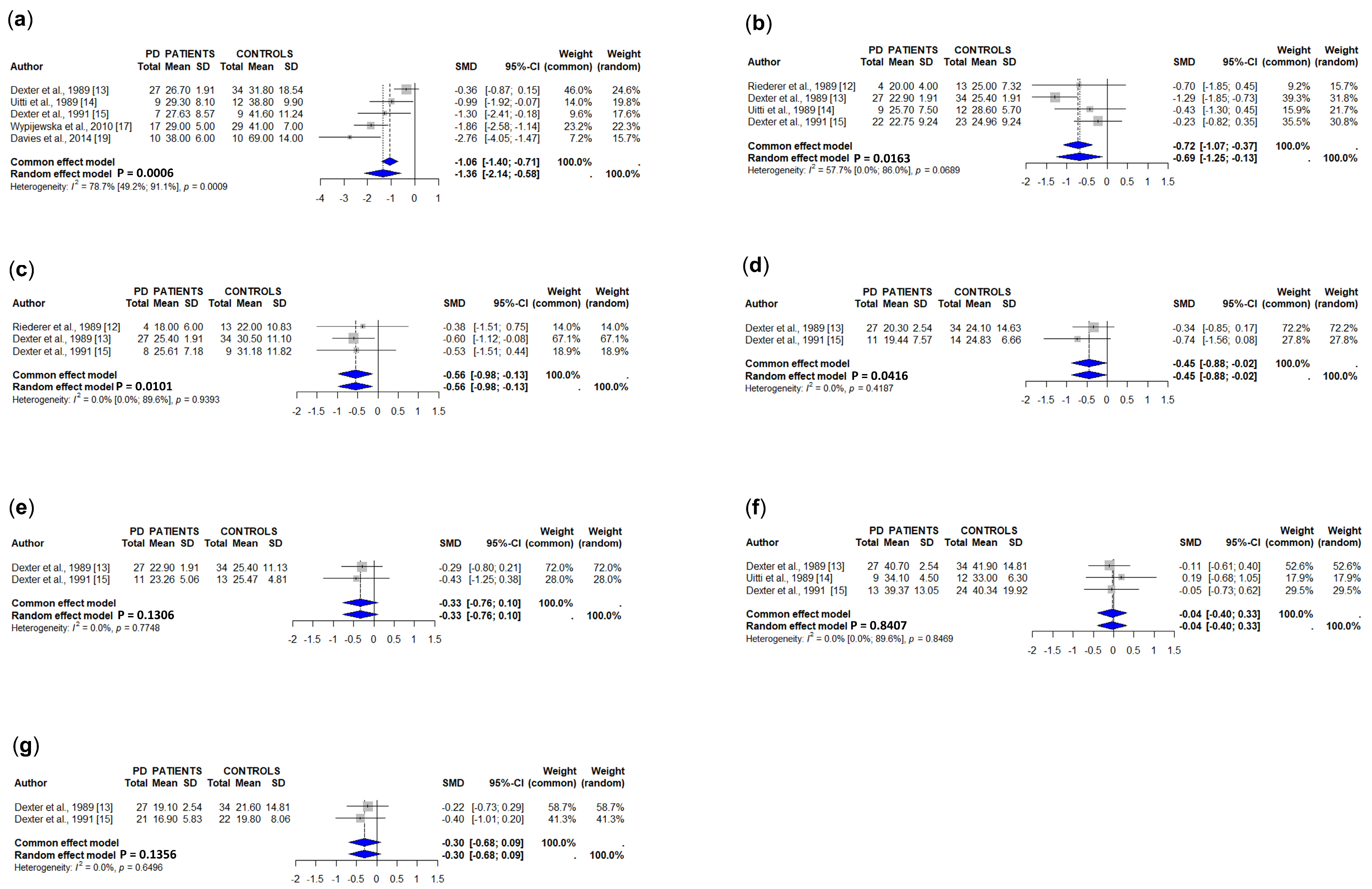


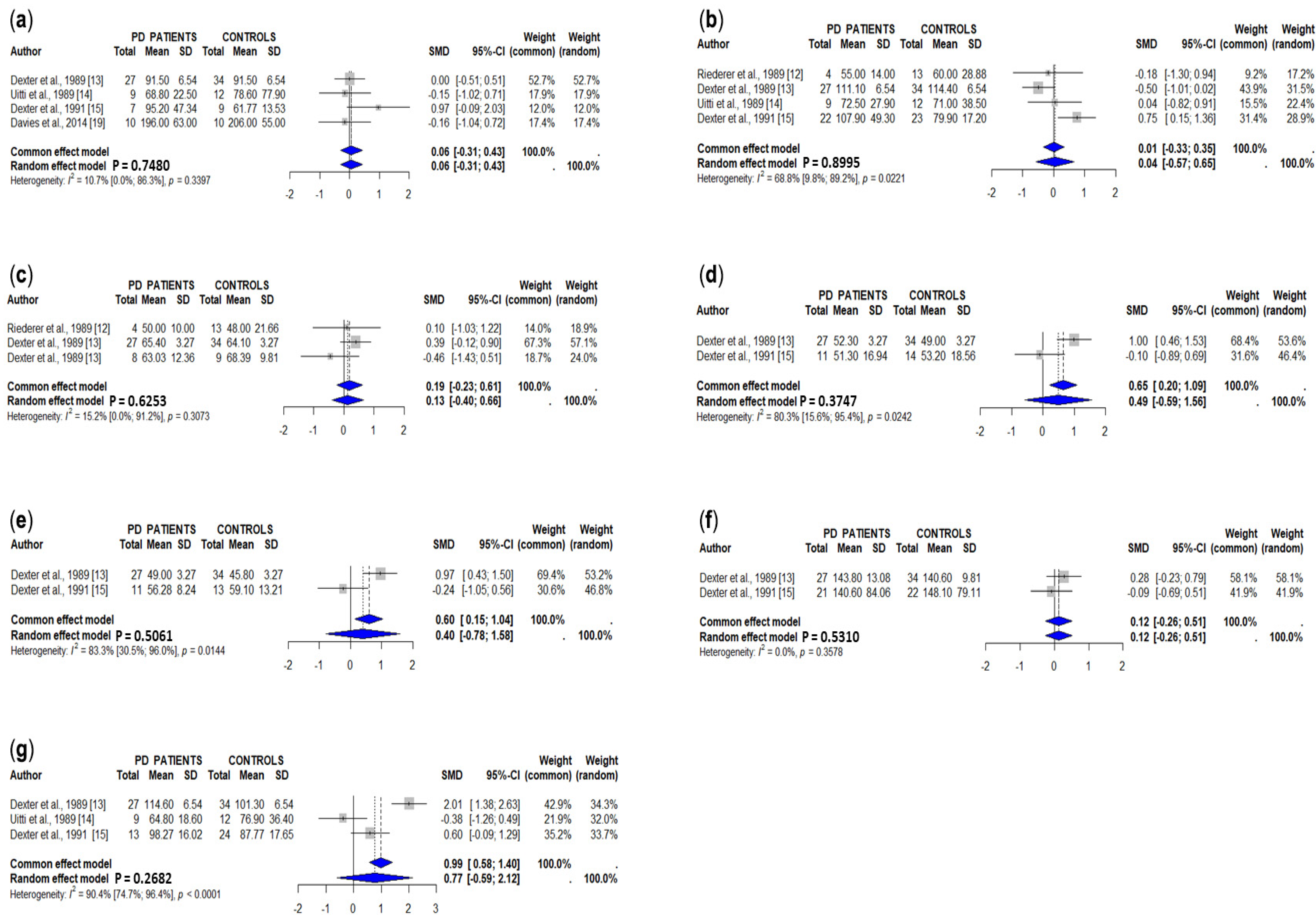
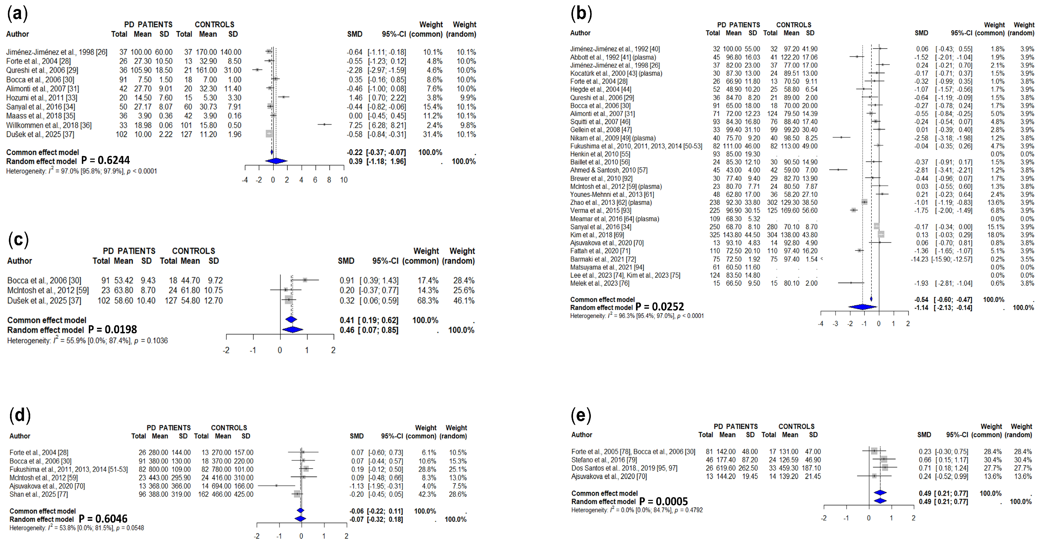


| Copper | Ceruloplasmin | Zinc | Manganese | |
|---|---|---|---|---|
| Brain | 346 | 93 | 298 | 485 |
| Cerebrospinal fluid | 30 | 23 | 19 | 22 |
| Serum | 86 | 59 | 56 | 50 |
| Plasma | 70 | 22 | 49 | 67 |
| Blood | 117 | 48 | 95 | 132 |
| Urine | 20 | 7 | 12 | 30 |
| Hair | 14 | 1 | 7 | 13 |
| Copper | |||||
|---|---|---|---|---|---|
| Tissue | Author, Year [Ref] | Method | Main Results | Main Results (2) | Comments/ Observations |
| Brain | Riederer et al., 1989 [12] | Atomic absorption spectrophotometry | Significant increase in the raphe plus reticular formation and red nucleus in PD brains, and non-significant differences in other brain areas, including substantia nigra, caudate, putamen, and globus pallidus. | 3− for raphe, reticular formation and red nucleus | Data obtained as estimated from a graphic. |
| Dexter et al., 1989 [13] | Inductively coupled plasma spectroscopy | Significant decrease in substantia nigra from PD brains (34–45%). Non-significant differences between PD and controls in cerebellum, cerebral cortex, caudate nucleus, putamen, and globus pallidus. | 3− for substantia nigra | Data obtained as estimated from a graphic. | |
| Uitti et al., 1989 [14] | Atomic emission spectroscopy and atomic absorption spectrophotometry | Significant decrease in the substantia nigra from PD brains, and non-significant differences with controls in the frontal cortex, caudate nucleus, and cerebellum. | 3− for substantia nigra | ||
| Dexter et al., 1991 [15] | Inductively coupled plasma spectroscopy | Significant decrease in the substantia nigra from PD brains. Non-significant differences between PD and controls in cerebellum, cerebral cortex (Brodmann area 10), caudate nucleus, putamen, and globus pallidus. | 3− for substantia nigra | ||
| Loeffler et al., 1996 [16] | Flame atomic absorption spectrophotometry | Significant decrease in caudate from PD brains. Non-significant differences between PD and elderly controls in the substantia nigra, putamen, and frontal cortex. | 2− for caudate | Data expressed in ng/μg of protein. Not included in meta-analysis. | |
| Wypijewska et al., 2010 [17] | Electrothermal atomic absorption spectrometry | Significant decrease in the substantia nigra from PD patients. | 3− for caudate | ||
| Szczerbowska-Boruchoswska et al., 2012 [18] | Synchrotron radiation-based X-ray fluorescence (SRXRF) | Non-significant differences between PD and controls. | 0 | Data expressed in Cu mass fraction in the substantia nigra. Not included in the meta-analysis. | |
| Davies et al., 2014 [19] | Synchrotron radiation x-ray fluorescence microscopy (SRXFM) and particle-induced x-ray emission (PIXE) microscopy | Significant decrease in the substantia nigra and locus ceruleus from PD patients. Non-significant differences between PD and controls in the occipital cortex. | 3− for substantia nigra and locus ceruleus | ||
| Gardner et al., 2017 [20] | Inductively coupled plasma spectroscopy | Non-significant differences between PD and controls in the olfactory bulb and olfactory tract. | 0 | ||
| Genoud et al., 2017 [21] | Inductively coupled plasma spectroscopy | Significant decrease in the substantia nigra from PD patients (54%). Non-significant differences between PD and controls in the occipital cortex and fusiform gyrus. | 3− in substantia nigra | Full data not available. Not included in the meta-analysis. | |
| CSF | Mindadse & Tschikowani, 1967 [22] | Not available | Not available | Unable to get this article; abstract not available. | |
| Campanella et al., 1973 [23] | Colorimetric method | Non-significant differences between PD patients and controls. | 0 | ||
| Pall et al., 1987 [24] | Electrochemical atomization/atomic absorption spectrophotometry | Significant increase in PD patients compared to controls. | 3+ | Controls were subjects with neurological diseases who had a clinical indication for lumbar puncture. Estimation from a graphic. | |
| Gazzaniga et al., 1992 [25] | Atomic absorption spectrophotometry with electrothermal atomization | Non-significant differences between PD patients and controls. | 0 | ||
| Jiménez-Jiménez et al., 1998 [26] | Flame atomic absorption spectrophotometry | Non-significant differences between PD patients and controls. | 0 | Controls were subjects with neurological diseases who had a clinical indication for lumbar puncture. | |
| Boll et al., 1999 [27] | Atomic absorption spectrophotometry | Non-significant differences between PD patients and controls. | 0 | ||
| Forte et al., 2004 [28] | Inductively coupled plasma atomic emission spectrometry | Non-significant differences between PD patients and controls. | 0 | ||
| Qureshi et al., 2006 [29] | Atomic absorption spectrophotometry with an electrothermal atomizer | Non-significant differences between PD patients and controls. | 0 | ||
| Bocca et al., 2006 [30] | Inductively coupled plasma atomic emission spectrometry | Non-significant differences between PD patients and controls. | 0 | Data obtained as estimated from a graphic. | |
| Alimonti et al., 2007 [31] | Inductively coupled plasma mass spectrometry and sector field inductively coupled Plasma Mass Spectrometry | Non-significant differences between PD patients and controls. | 0 | ||
| Boll et al., 2008 [32] | Graphite furnace atomic absorption spectrophotometry | Significant increase in PD patients compared to controls. | 3+ | Measurement of free (and not total) copper. This article was excluded from meta-analysis. | |
| Hozumi et al., 2011 [33] | Inductively coupled plasma spectroscopy | Significant increase in PD patients. | 3+ | ||
| Sanyal et al., 2016 [34] | Atomic absorption spectrophotometry and flame atomic absorption spectrophotometry | Non-significant differences between PD patients and controls. | 0 | ||
| Maass et al., 2018 [35] | Inductively coupled plasma-sector field mass spectrometry | Non-significant differences between PD patients and controls. | 0 | ||
| Willkommen et al., 2018 [36] | Size-exclusion chromatography hyphenated to inductively coupled plasma mass spectrometry (SEC-ICP-MS) | Non-significant differences between PD patients and controls. | 0 | ||
| Dušek et al., 2025 [37] | Inductively coupled plasma spectroscopy | Significant decrease in PD patients. | 1− | ||
| Serum/Plasma | Mindadse & Tschikowani, 1967 [22] | Not available | Not available | Unable to get this article; abstract not available. | |
| Kanabrocki et al., 1967 [38] | RCL 512-Channel Analyzer (an instrument used in nuclear and radiation spectroscopy) | Non-significant differences between PD patients and controls. | 0 | ||
| Chitre & Punekar, 1970 [39] | Iodometric redox titration | Significant increase in PD patients compared to controls. | 3+ | ||
| Campanella et al., 1973 [23] | Colorimetric methods | Non-significant differences between PD patients and controls. | 0 | ||
| Jiménez-Jiménez et al., 1992 [40] | Flame atomic absorption spectrophotometry | Non-significant differences between PD patients and controls. | 0 | ||
| Abbott et al., 1992 [41] (plasma) | Atomic absorption spectrophotometry | Non-significant differences between PD patients and controls. | |||
| Jiménez-Jiménez et al., 1998 [26] | Flame atomic absorption spectrophotometry | Non-significant differences between PD patients and controls. | 0 | Controls were subjects with neurological diseases who had a clinical indication for lumbar puncture. PD patients and controls were not the same as in [CMZ-14]. | |
| Tórsdóttir et al., 1999 [42] (plasma) | Atomic absorption spectrophotometry | Non-significant differences between PD patients and controls. | 0 | ||
| Kocatürk et al., 2000 [43] (plasma) | Atomic absorption spectrophotometry | Significant increase in PD patients compared to controls. | |||
| Forte et al., 2004 [28] | Inductively Coupled Plasma Atomic Emission Spectrometry | Significant decrease in PD patients compared to controls. | 2+ | ||
| Hegde et al., 2004 [44] | Inductively Coupled Plasma Atomic Emission Spectrometry | Significant increase in PD patients compared to controls. | 2− | ||
| Qureshi et al., 2006 [29] | Atomic absorption spectrophotometry with an electrothermal atomizer | Non-significant differences between PD patients and controls. | 0 | ||
| Bocca et al., 2006 [30] | Inductively Coupled Plasma Atomic Emission Spectrometry | Significant decrease in PD patients compared to controls. | 1− | ||
| Alimonti et al., 2007 [45] | Inductively Coupled Plasma Atomic Emission Spectrometry | Non-significant differences between PD patients and controls. | 0 | ||
| Squitti et al., 2007 [46] | Graphite furnace atomic absorption. | Non-significant differences between PD patients and controls. | 0 | ||
| Gellein et al., 2008 [47] | High Resolution Inductively Coupled Plasma Atomic Emission Spectrometry | Non-significant differences between PD patients and controls. | 0 | ||
| Bharucha et al., 2009 [48] | Not specified | Significant decrease in PD patients compared to controls. | 2− | ||
| Nikam et al., 2009 [49] (plasma) | Atomic absorption spectrophotometry | Significant decrease in PD patients compared to controls. | 2− | ||
| Fukushima et al., 2010, 2011, 2013, 2014 [50,51,52,53] | Inductively Coupled Plasma Atomic Emission Spectrometry | Non-significant differences between PD patients and controls. | 0 | ||
| Arnal et al., 2010 [54] (plasma) | Atomic absorption spectrophotometry | Non-significant differences between PD patients and controls. | 0 | ||
| Henkin et al., 2010 [55] | Atomic absorption spectrophotometry | Lack of a control group. | Lack of a control group | ||
| Baillet et al., 2010 [56] | Atomic absorption Spectrophotometry | Non-significant differences between PD patients and controls. | 0 | ||
| Ahmed & Santosh 2010 [57] | Inductively Coupled Plasma Atomic Emission Spectrometry and atomic absorption spectroscopy | Significant increase in PD patients compared to controls. | 2+ | ||
| Ling & Bidhayasiri 2011 [58] | Not specified | Non-significant differences between PD patients and controls. | 0 | Data not given | |
| McIntosh et al., 2012 [59] (plasma) | Monochromatic X-ray fluorescence spectrometry | Non-significant differences between PD patients and controls. | 0 | ||
| Mariani et al.,2013 [60] | Not specified | Non-significant differences between PD patients and controls. | 0 | Data not available in the Supplementary Material. | |
| Younes-Mehnni et al., 2013 [61] | Plasma atomic absorption spectrophotometry | Significant decrease in PD patients compared to controls. | 2− | ||
| Zhao et al., 2013 [62] (plasma) | Zeeman atomic absorption spectroscopy with a graphite tube atomizer | Significant decrease in PD patients compared to controls. | 2− | ||
| Kumudini et al. 2014 [63] (plasma) | Inductively Coupled Plasma Atomic Emission Spectrometry | Significant increase in PD patients compared to controls. | 2+ | ||
| Meamar et al., 2016 [64] (plasma) | Colorimetric method | Lack of a control group. | Lack of a control group. | ||
| Sanyal et al., 2016 [34] | Atomic absorption spectrophotometry and flame atomic absorption spectrophotometry | Significant decrease in PD patients compared to controls. | 2− | ||
| Song et al., 2017 [65] | Immunoassay | Non-significant differences between PD patients and controls. | 0 | ||
| Gangania et al., 2017 [66] | Colorimetric method | Significant decrease in PD patients compared to controls. | 2− | ||
| Karpenko et al., 2018 [67] (plasma) | Atomic absorption spectrophotometry | Significant decrease in PD patients compared to controls. | 3− | ||
| Ilyechova et al., 2018 [68] | Graphite furnace atomic absorption spectrometry | Significant decrease in PD patients compared to controls. | 3− | ||
| Kim et al., 2018 [69] | Inductively Coupled Plasma Atomic Emission Spectrometry | Significant decrease in PD patients compared to controls. | 1− | ||
| Ajsuvakova et al., 2020 [70] | Inductively Coupled Plasma Atomic Emission Spectrometry | Non-significant differences between PD patients and controls. | |||
| Fattah et al., 2020 [71] | Graphite furnace atomic absorption spectrophotometry | Significant decrease in PD patients compared to controls. | 2− | ||
| Barmaki et al., 2021 [72] | Graphite furnace atomic absorption spectrophotometry | Significant decrease in PD patients compared to controls. | 2− | ||
| Tripathi et al., 2021 [73] | Spectrophotometery immunoturbidimetry | Significant decrease in PD patients compared to controls. | 3− | ||
| Lee et al., 2023 [74] | Inductively coupled plasma atomic emission spectrometry | Lack of a control group. Non-significant difference between PD patients with and without dementia. | Lack of a control group. | ||
| Kim et al., 2023 [75] | Inductively coupled plasma atomic emission spectrometry | Lack of a control group. Non-significant difference between PD patients with and without levodopa-induced dyskinesia. | Lack of a control group. | ||
| Melek et al., 2023 [76] | Atomic emission spectrometry | Non-significant differences between PD patients and controls. | 0 | ||
| Whole Blood | Bocca et al., 2006 [30] | Inductively coupled plasma atomic emission spectrometry | Significant increase in PD patients compared to controls. | 2+ | |
| McIntosh et al., 2012 [59] | Monochromatic X-ray fluorescence spectrometry | Non-significant differences between PD patients and controls. | 0 | ||
| Dušek et al., 2025 [37] | Inductively coupled plasma spectroscopy | Non-significant differences between PD patients and controls. | 0 | ||
| Urine | McIntosh et al., 2012 [59] | Monochromatic X-ray fluorescence spectrometry | Non-significant differences between PD patients and controls. | 0 | |
| Forte et al., 2004 [28] | Inductively coupled plasma atomic emission spectrometry | Non-significant differences between PD patients and controls. | 0 | ||
| Bocca et al., 2006 [30] | Inductively coupled plasma atomic emission spectrometry | Non-significant differences between PD patients and controls. | 0 | Data obtained as estimated from a graphic. | |
| Fukushima et al., 2011, 2013, 2014 [51,52,53] | Atomic absorption spectrometry | Significant increase in PD patients compared to controls. | 1+ | ||
| Ajsuvakova et al., 2020 [70] | Inductively coupled plasma atomic emission spectrometry | Non-significant differences between PD patients and controls. | 0 | ||
| Shan et al., 2025 [77] | Inductively coupled plasma atomic emission spectrometry | Non-significant differences between PD patients and controls. | 0 | ||
| Hair | Forte et al., 2005 [78] | Inductively coupled plasma atomic emission spectrometry | Non-significant differences between PD patients and controls. | 0 | |
| Bocca et al., 2006 [30] | Inductively coupled plasma atomic emission spectrometry | Non-significant differences between PD patients and controls | 0 | Data obtained as estimated from a graphic. | |
| Stefano et al., 2016 [79] | Inductively coupled plasma atomic emission spectrometry | Non-significant differences between PD patients and controls. | 0 | ||
| Ajsuvakova et al., 2020 [70] | Inductively coupled plasma atomic emission spectrometry | Significant increase in PD patients compared to controls. | 2+ | ||
| Ceruloplasmin | |||||
| Tissue | Author, Year [Ref] | Method | Main Results | Main Results (2) | Comments/ Observations |
| Brain | Loeffler et al., 1996 [16] | Enzyme-linked immunosorbent assay (ELISA) | Significant increase in PD substantia nigra, frontal, temporal, and parietal cortices, and hippocampus; and non-significant differences with elderly controls in caudate, putamen, and cerebellum. | 1+ in mentioned brain areas | Data given as corrected by proteins instead of g of tissue. |
| Loeffler et al., 2001 [80] | Immunocytochemical staining with avidin-biotin-peroxidase complex | Non-significant differences in ceruloplasmin immunoreactivity in substantia nigra, caudate, frontal and parietal cortices, parahippocampus, subiculum, and CA1 region. | 0 | Data given in immunoreactivity for ceruloplasmins, not in concentrations, data not included. | |
| CSF | Campanella et al., 1973 [23] | Colorimetric methods | Non-significant differences between PD patients and controls. | 0 | |
| Loeffler et al., 1994 [81] | Enzyme-linked immunosorbent assay (ELISA) | Non-significant differences between PD patients and controls. | 0 | ||
| Serum | Campanella et al., 1973 [23] | Colorimetric methods | Non-significant differences between PD patients and controls. | 0 | |
| Jiménez-Jiménez et al., 1992 [40] | Nephelometry immunoassay | Non-significant differences between PD patients and controls. | 0 | ||
| Molina-Arjona et al., 1999 [82] | Nephelometry immunoassay | Non-significant differences between PD patients and controls. | 0 | ||
| Tórsdóttir et al., 1999 [42] (plasma) | Nephelometry immunoassay | Significant decrease in PD patients compared to controls. | 0 | Non-significant changes when recalculated. | |
| Tórsdóttir et al., 2006 [83] | Nephelometry immunoassay | Significant decrease in PD patients compared to controls. | 0 | Non-significant changes when recalculated. | |
| Squitti et al., 2007 [46] | Immunoturbidimetric assay | Non-significant differences between PD patients and controls. | 0 | ||
| Bharucha et al., 2009 [48] | Nephelometric immu-neturbidimetric assay | Significant decrease in PD patients compared to controls. | 2− | ||
| Nikam et al., 2009 [49] (plasma) | Colorimetric method | Significant decrease in PD patients compared to controls. | Concentrations expressed as Units/L, Conversion to mg/dL not available. Data not included. | ||
| Arnal et al., 2010 [54] (plasma) | Atomic absorption spectrophotometry | Non-significant differences between PD patients and controls. | 0 | ||
| Jin et al., 2011 [84] | Nephelometry immunoassay | Significant decrease in PD patients compared to controls. | 1− | ||
| Ling & Bidhayasiri 2011 [58] | Immunoturbidimetric assay with specific antiserum | Significant decrease in PD patients compared to controls. | 0 | Non-significant changes when recalculated. | |
| Zhao et al. 2014 [85] | Nephelometry immunoassay | Significant decrease in PD patients compared to controls. | 2− | ||
| Song et al., 2017 [65] | Immunoassay | Significant decrease in PD patients compared to controls. | 1− | ||
| Gangania et al., 2017 [66] | Turbidimetric assay | Non-significant differences between PD patients and controls. | 0 | ||
| Karpenko et al., 2018 [67] (plasma) | Atomic absorption spectrophotometry | Significant decrease in PD patients compared to controls. | 1− | ||
| Ilyechova et al., 2018 [68] | Spectrophotometric method with p-phenylenediamine | Significant decrease in PD patients compared to controls. | 1− | ||
| Xu et al., 2018 [86] | Immunoturbidimetric assay | Non-significant differences between PD patients and controls. | 0 | ||
| Si et al., 2018 [87] (plasma) | Immunoturbidimetric assay | Non-significant differences between PD patients and controls. | 0 | ||
| Bakeberg et al., 2020 [88] | Immunoturbidimetric assay | Lack of a control group. High impulsivity in PD patients with high compared with those with low ceruloplasmin levels. | Lack of a control group. | ||
| Tripathi et al., 2021 [73] | Spectrophotometery immunoturbidimetry | Non-significant differences between PD patients and controls. | 0 | ||
| Zinc | |||||
| Tissue | Author, Year [Ref] | Method | Main Results | Main Results (2) | Comments/ Observations |
| Brain | Riederer et al., 1989 [12] | Atomic absorption spectrophotometry | Significant increase in the raphe plus reticular formation in PD brains, and non-significant differences in other brain areas, including substantia nigra, caudate, putamen, and globus pallidus. | 0 | Data obtained as estimated from a graphic. Non-significant changes when recalculated. |
| Dexter et al., 1989 [13] | Inductively coupled plasma spectroscopy | Significant increase in substantia nigra from PD brains (50–54%) and in lateral putamen (18–35%). Non-significant differences between PD and controls in cerebellum, cerebral cortex, caudate nucleus, medial putamen, and globus pallidus. | 3+ for substantia nigra, 2+ for lateral putamen | Data obtained as estimated from a graphic. | |
| Uitti et al., 1989 [14] | Atomic emission spectroscopy and atomic absorption spectrophotometry | Non-significant differences between PD and controls in the frontal cortex, caudate nucleus, and cerebellum. | 0 | ||
| Hirsch et al., 1991 [89] | X-ray microanalysis | Non-significant differences between PD and controls in the substantia nigra (cells positive or negative for neuromelanin) and central grey substance. | Data from the control group not available. | ||
| Dexter et al., 1991 [15] | Inductively coupled plasma spectroscopy | Significant increase in substantia nigra from PD brains. Non-significant differences between PD and controls in cerebellum, cerebral cortex (Brodmann area 10), caudate nucleus, putamen, and globus pallidus. | 0 | Non-significant changes when recalculated. | |
| Mann et al., 1994 [90] | Inductively coupled plasma spectroscopy | Non-significant differences between PD patients and controls in the substantia nigra. | 0 | ||
| Popescu et al., 2009 [91] | Rapid-scanning X-ray fluorescence mapping | Decrease fluorescence in substantia nigra, caudate, putamen, internal and external globus pallidus, inferior colliculus, white matter, internal capsule, grey matter, and optic tract. | 1− for all mentioned brain areas | Data given in normalized fluorescence | |
| Szczerbowska-Boruchoswska et al., 2012 [18] | Synchrotron radiation-based X-ray fluorescence | Significant increase in Zn mass fraction in the substantia nigra from PD patients. | 1+ | ||
| Davies et al., 2014 [19] | Particle-induced X-ray emission (PIXE) microscopy | Non-significant differences between PD and controls in the substantia nigra, locus ceruleus, and occipital cortex. | 0 | ||
| Gardner et al., 2017 [20] | Inductively coupled plasma spectroscopy | Non-significant differences between PD and controls in the olfactory bulb and olfactory tract. | 0 | ||
| Genoud et al., 2017 [21] | Inductively coupled plasma spectroscopy | Non-significant differences between PD and controls in the substantia nigra, occipital cortex, and fusiform gyrus. | 0 | Full data not available | |
| CSF | Mindadse & Tschikowani, 1967 [22] | Not available | Not available | Unable to get this article; abstract not available. | |
| Jiménez-Jiménez et al., 1998 [26] | Flame atomic absorption spectrophotometry | Significant decrease in PD patients. | 3− | Controls were subjects with neurological diseases who had a clinical indication for lumbar puncture. | |
| Forte et al., 2004 [28] | Inductively coupled plasma atomic emission spectrometry | Significant decrease in PD patients. | 1− | ||
| Qureshi et al., 2006 [29] | Atomic absorption spectrophotometry with an electrothermal atomizer | Non-significant differences between PD patients and controls. | 0 | ||
| Bocca et al., 2006 [30] | Inductively coupled plasma atomic emission spectrometry | Non-significant differences between PD patients and controls. | 0 | Data obtained as estimated from a graphic. | |
| Alimonti et al., 2007 [31] | Inductively Coupled Plasma Mass Spectrometry and Sector Field Inductively Coupled Plasma Mass Spectrometry | Non-significant differences between PD patients and controls. | 0 | ||
| Hozumi et al., 2011 [33] | Inductively coupled plasma spectroscopy | Significant increase in PD patients. | 3+ | ||
| Sanyal et al., 2016 [34] | Atomic absorption spectrophotometry and flame atomic absorption spectrophotometry | Significant decrease in PD patients compared to controls. | 1− | ||
| Maass et al., 2018 [35] | Inductively coupled plasma-sector field mass spectrometry | Non-significant differences between PD patients and controls. | 0 | ||
| Willkommen et al., 2018 [36] | Size-exclusion chromatography hyphenated to inductively coupled plasma mass spectrometry (SEC-ICP-MS) | Non-significant differences between PD patients and controls. | 0 | ||
| Dušek et al., 2025 [37] | Inductively coupled plasma spectroscopy | Non-significant differences between PD patients and controls. | 0 | ||
| Serum | Mindadse & Tschikowani, 1967 [22] | Not available | Not available | Unable to get this article; abstract not available. | |
| Jiménez-Jiménez et al., 1992 [40] | Flame atomic absorption spectrophotometry | Non-significant differences between PD patients and controls. | 0 | ||
| Abbott et al., 1992 [41] (plasma) | Atomic absorption spectrophotometry | Significant decrease in PD patients. | 3− | ||
| Jiménez-Jiménez et al., 1998 [26] | Flame atomic absorption spectrophotometry | Non-significant differences between PD patients and controls. | 0 | Controls were subjects with neurological diseases who had a clinical indication for lumbar puncture. PD patients and controls were not the same as in [40]. | |
| Kocatürk et al., 2000 [43] (plasma) | Atomic absorption spectrophotometry | Non-significant differences between PD patients and controls. | 0 | ||
| Forte et al., 2005 [78] | Inductively coupled plasma atomic emission spectrometry | Non-significant differences between PD patients and controls. | 0 | ||
| Hegde et al., 2004 [44] | Inductively coupled plasma atomic emission spectrometry | Significant decrease in PD patients compared to controls. | 2− | ||
| Qureshi et al., 2006 [29] | Atomic absorption spectrophotometry with an electrothermal atomizer | Non-significant differences between PD patients and controls. | 0 | ||
| Bocca et al., 2006 [30] | Inductively coupled plasma atomic emission spectrometry | Non-significant differences between PD patients and controls. | 0 | Data obtained as estimated from a graphic. | |
| Alimonti et al., 2007 [31] | Inductively coupled plasma atomic emission spectrometry | Non-significant differences between PD patients and controls. | 0 | ||
| Squitti et al., 2007 [46] | Graphite furnace atomic absorption. | Non-significant differences between PD patients and controls. | 0 | ||
| Gellein et al., 2008 [47] | High resolution inductively coupled plasma atomic emission spectrometry | Non-significant differences between PD patients and controls. | 0 | ||
| Nikam et al., 2009 [49] | Atomic absorption spectrophotometry | Significant decrease in PD patients compared to controls. | 2− | ||
| Fukushima et al., 2010, 2011, 2013, 2014 [50,51,52,53] | Inductively coupled plasma atomic emission spectrometry | Non-significant differences between PD patients and controls. | 0 | ||
| Henkin et al., 2010 [55] | Atomic absorption spectrophotometry | Lack of a control group. | Lack of a control group. | ||
| Baillet et al., 2010 [56] | Atomic absorption spectrophotometry | Significant decrease in PD patients. | 0 | ||
| Ahmed & Santosh 2010 [57] | Inductively coupled plasma atomic emission spectrometry and atomic absorption spectroscopy | Significant decrease in PD patients compared to controls. | 0 | ||
| Brewer et al., 2010 [92] | Atomic absorption spectrophotometry | Significant decrease in PD patients. | 1− | ||
| McIntosh et al., 2012 [59] (plasma) | Monochromatic X-ray fluorescence spectrometry | Non-significant differences between PD patients and controls. | 0 | ||
| Younes-Mehnni et al., 2013 [61] | Plasma atomic absorption spectrophotometry | Significant decrease in PD patients compared to controls. | 0 | Non-significant differences when recalculated. | |
| Zhao et al., 2013 [62] (plasma) | Fast sequential atomic absorption spectroscopy | Significant decrease in PD patients compared to controls. | 2− | ||
| Verma et al., 2015 [93] | Inductively coupled plasma atomic emission spectrometry | Significant decrease in PD patients. | 3− | ||
| Meamar et al., 2016 [64] (plasma) | Colorimetric method | Lack of a control group. | |||
| Sanyal et al., 2016 [34] | Atomic absorption spectrophotometry and flame atomic absorption spectrophotometry | Non-significant differences between PD patients and controls. | 0 | ||
| Kim et al., 2018 [69] | Inductively coupled plasma atomic emission spectrometry | Non-significant differences between PD patients and controls. | 0 | ||
| Ajsuvakova et al., 2020 [70] | Inductively coupled plasma atomic emission spectrometry | Non-significant differences between PD patients and controls. | 0 | ||
| Fattah et al., 2020 [71] | Graphite furnace atomic absorption spectrophotometry | Significant decrease in PD patients compared to controls. | 2− | ||
| Barmaki et al., 2021 [72] | Graphite furnace atomic absorption spectrophotometry | Significant decrease in PD patients compared to controls. | 2− | ||
| Matsuyama et al., 2021 [94] | Not specified | Lack of a control group. | |||
| Lee et al., 2023 [74] | Inductively coupled plasma atomic emission spectrometry | Lack of a control group. Increase in patients with PD who developed dementia compared to PD patients without dementia during the follow-up period. | Lack of a control group. | ||
| Kim et al., 2023 [75] | Inductively coupled plasma atomic emission spectrometry | Lack of a control group. Decrease in patients with PD who developed levodopa-induced dyskinesia compared to PD patients without this adverse effect during the follow-up period. | Lack of a control group. | ||
| Melek et al., 2023 [76] | Atomic emission spectrometry | Significant decrease in PD patients. | 2− | ||
| Whole Blood | Bocca et al., 2006 [30] | Inductively coupled plasma atomic emission spectrometry | Significant increase in PD patients. | 1+ | |
| McIntosh et al., 2012 [59] | Monochromatic X-ray fluorescence spectrometry | Non-significant differences between PD patients and controls. | 0 | ||
| Dušek et al., 2025 [37] | Inductively coupled plasma spectroscopy | Significant increase in PD patients. | 1+ | ||
| Urine | McIntosh et al., 2012 [59] | Monochromatic X-ray fluorescence spectrometry | Non-significant differences between PD patients and controls. | 0 | |
| Forte et al., 2004 [28] | Inductively coupled plasma atomic emission spectrometry | Non-significant differences between PD patients and controls. | 0 | ||
| Bocca et al., 2006 [30] | Inductively Coupled Plasma Atomic Emission Spectrometry | Non-significant differences between PD patients and controls. | 0 | Data obtained as estimated from a graphic. | |
| Brewer et al., 2010 [92] | Atomic absorption spectrophotometry | Significant increase in PD patients. | 2+ | Standard deviation values not available. Not included in the meta-analysis. | |
| Fukushima et al., 2011, 2013, 2024 [51,52,53] | Atomic absorption spectrometry | Non-significant differences between PD patients and controls. | 0 | ||
| Ajsuvakova et al., 2020 [79] | Inductively coupled plasma atomic emission spectrometry | Non-significant differences between PD patients and controls. | 0 | ||
| Shan et al., 2025 [77] | Inductively coupled plasma atomic emission spectrometry | Non-significant differences between PD patients and controls. | 0 | ||
| Hair | Forte et al., 2004 [28] | Inductively coupled plasma atomic emission spectrometry | Non-significant differences between PD patients and controls. | 0 | |
| Bocca et al., 2006 [30] | Inductively coupled plasma atomic emission spectrometry | Non-significant differences between PD patients and controls. | 0 | Data obtained as estimated from a graphic. | |
| Stefano et al., 2016 [79] | Inductively coupled plasma atomic emission spectrometry | Non-significant differences between PD patients and controls. | 0 | ||
| Dos Santos et al. 2018, 2019 [95,97] | Flame atomic absorption spectrophotometry | Significant increase in PD patients. | 3+ | ||
| Ajsuvakova et al., 2020 [70] | Inductively coupled plasma atomic emission spectrometry | Non-significant differences between PD patients and controls. | 0 | ||
| Manganese | |||||
| Tissue | Author, Year [Ref] | Method | Main Results | Main Results (2) | Comments/ Observations |
| Brain | Larsen et al., 1981 [96] | Neutron activation analysis with radiochemical separation | Non-significant differences between PD patients (n = 2) and controls (n = 5) for 24 brain areas. | 0 | Not included in the meta-analysis. There are no statistical data. |
| Dexter et al., 1989 [13] | Inductively coupled plasma spectroscopy | Significant decrease in medial putamen from PD brains (20%). Non-significant differences between PD and controls in the substantia nigra, cerebellum, cerebral cortex, caudate nucleus, lateral putamen, and globus pallidus. | 1− | Data obtained as estimated from a graphic. | |
| Uitti et al., 1989 [14] | Atomic emission spectroscopy and atomic absorption spectrophotometry | Non-significant differences between PD and controls in the frontal cortex, caudate nucleus, and cerebellum. | 0 | ||
| Dexter et al., 1991 [15] | Inductively coupled plasma spectroscopy | Non-significant differences between PD and controls in the substantia nigra, cerebellum, cerebral cortex (Brodmann area 10), caudate nucleus, putamen, and globus pallidus. There was a 20% decrease in the medial putamen of PD. | 0 | ||
| Gardner et al., 2017 [20] | Inductively coupled plasma spectroscopy | Non-significant differences between PD and controls in the olfactory bulb and olfactory tract. | 0 | ||
| Genoud et al., 2017 [21] | Inductively coupled plasma spectroscopy | Non-significant differences between PD and controls in the substantia nigra, occipital cortex, and fusiform gyrus. | 0 | Full data not available. | |
| CSF | Mindadse & Tschikowani, 1967 [22] | Not available | Not available | Unable to get this article; abstract not available. | |
| Pall et al., 1987 [24] | Electrochemical atomization/atomic absorption spectrophotometry | Non-significant differences between PD patients and controls. | 0 | Controls were subjects with neurological diseases who had a clinical indication for lumbar puncture. Estimation from a graphic. | |
| Gazzaniga et al., 1992 [25] | Atomic absorption spectrophotometry with electrothermal atomization | Non-significant differences between PD patients and controls. | 0 | ||
| Jiménez-Jiménez et al., 1998 [26] | Flame atomic absorption spectrophotometry | Non-significant differences between PD patients and controls. | 0 | Controls were subjects with neurological diseases who had a clinical indication for lumbar puncture. | |
| Forte et al., 2004 [28] | Inductively coupled plasma atomic emission spectrometry | Non-significant differences between PD patients and controls. | 0 | ||
| Bocca et al., 2006 [30] | Sector field inductively coupled plasma mass spectrometry | Non-significant differences between PD patients and controls. | 0 | ||
| Hozumi et al., 2011 [33] | Inductively coupled plasma spectroscopy | Significant increase in PD patients. | 3+ | ||
| Alimonti et al., 2007 [31] | Inductively coupled plasma mass spectrometry and sector field inductively coupled plasma mass spectrometry | Non-significant differences between PD patients and controls. | 0 | ||
| Sanyal et al., 2016 [34] | Atomic absorption spectrophotometry and flame atomic absorption spectrophotometry | Non-significant differences between PD patients and controls. | 0 | ||
| Maass et al., 2018 [35] | Inductively coupled plasma-sector field mass spectrometry | Non-significant differences between PD patients and controls. | 0 | ||
| Willkommen et al., 2018 [36] | Size-exclusion chromatography hyphenated to inductively coupled plasma mass spectrometry (SEC-ICP-MS) | Non-significant differences between PD patients and controls. | 0 | ||
| Dušek et al., 2025 [37] | Inductively coupled plasma spectroscopy | Non-significant differences between PD patients and controls. | 0 | ||
| Serum | Mindadse & Tschikowani, 1967 [22] | Not available | Not available | Unable to get this article; abstract not available | |
| Kanabrocki et al., 1967 [38] | RCL 512-Channel Analyzer (an instrument used in nuclear and radiation spectroscopy) | Non-significant differences between PD patients and controls. | 0 | ||
| Jiménez-Jiménez et al., 1992 [98] | Flame atomic absorption spectrophotometry | Non-significant differences between PD patients and controls. | 0 | ||
| Jiménez-Jiménez et al., 1998 [26] | Flame atomic absorption spectrophotometry | Non-significant differences between PD patients and controls. | 0 | Controls were subjects with neurological diseases who had a clinical indication for lumbar puncture. PD patients and controls were not the same as in [CMZ-14]. | |
| Forte et al., 2004 [28] | Inductively coupled plasma atomic emission spectrometry | Non-significant differences between PD patients and controls. | 0 | ||
| Hegde et al., 2004 [44] | Inductively coupled plasma atomic emission spectrometry | Significant increase in PD patients compared to controls. | 3+ | Mean ± SD for controls not given. Not included in the meta-analysis. | |
| Bocca et al., 2006 [30] | Sector field inductively coupled plasma mass spectrometry | Non-significant differences between PD patients and controls. | 0 | ||
| Alimonti et al., 2007 [45] | Inductively coupled plasma atomic emission spectrometry | Non-significant differences between PD patients and controls. | 0 | ||
| Gellein et al., 2008 [47] | High resolution inductively coupled plasma atomic emission spectrometry | Non-significant differences between PD patients and controls. | 0 | ||
| Ahmed & Santosh 2010 [57] | Inductively coupled plasma atomic emission spectrometry and atomic absorption spectroscopy | Significant increase in PD patients compared to controls. | 1+ | ||
| Kumudini et al. 2014 [63] (plasma) | Inductively coupled plasma atomic emission spectrometry | Non-significant differences between PD patients and controls. | 0 | ||
| Verma et al., 2016 [93] | Inductively Coupled Plasma Atomic Emission Spectrometry | Significant increase in PD patients. | 2+ | ||
| Sanyal et al., 2016 [34] | Atomic absorption spectrophotometry and flame atomic absorption spectrophotometry | Non-significant differences between PD patients and controls. | 0 | ||
| Ajsuvakova et al., 2020 [70] | Inductively coupled plasma atomic emission spectrometry | Non-significant differences between PD patients and controls. | 0 | ||
| Lee et al., 2023 [74] | Inductively coupled plasma atomic emission spectrometry | Lack of a control group. Non-significant difference between PD patients with and without dementia. | Lack of a control group. | ||
| Kim et al., 2023 [75] | Inductively coupled plasma atomic emission spectrometry | Lack of a control group. Non-significant difference between PD patients with and without levodopa-induced dyskinesia. | Lack of a control group. | ||
| Whole Blood | Bocca et al., 2006 [30] | Sector field inductively coupled plasma mass spectrometry | Non-significant differences between PD patients and controls. | 0 | Data obtained as estimated from a graphic. |
| Fukushima et al., 2010, 2011, 2013 [50,51,52] | Atomic absorption spectrometry | Significant increase in PD patients. | 3+ | ||
| Dušek et al., 2025 [37] | Inductively coupled plasma spectroscopy | Non-significant differences between PD patients and controls. | 0 | ||
| Urine | Jiménez-Jiménez et al., 1992 [98] | Flame atomic absorption spectrophotometry | Non-significant differences between PD patients and controls. | 0 | |
| Forte et al., 2004 [28] | Inductively coupled plasma atomic emission spectrometry | Non-significant differences between PD patients and controls. | 0 | ||
| Bocca et al., 2006 [30] | Sector field inductively coupled plasma mass spectrometry | Significant decrease in PD patients. | 2− | ||
| Fukushima et al., 2010, 2011, 2013 [50,51,52] | Atomic absorption spectrometry | Non-significant differences between PD patients and controls. | 0 | ||
| Ajsuvakova et al., 2020 [70] | Inductively coupled plasma atomic emission spectrometry | Non-significant differences between PD patients and controls. | 0 | ||
| Shan et al., 2025 [77] | Inductively coupled plasma atomic emission spectrometry | Significant increase in PD patients. | 3+ | ||
| Hair | Bocca et al., 2006 [30] | Sector field inductively coupled plasma mass spectrometry | Significant increase in PD patients. | 3+ | |
| Stefano et al., 2016 [79] | Inductively coupled plasma atomic emission spectrometry | Non-significant differences between PD patients and controls. | 0 | ||
| Ajsuvakova et al., 2020 [70] | Inductively coupled plasma atomic emission spectrometry | Non-significant differences between PD patients and controls. | 0 | ||
| Technique | Principle | Sensitivity for Cu, Zn, Mn | Advantages | Limitations |
|---|---|---|---|---|
| Atomic Absorption Spectrophotometry (AAS) | Absorption of element-specific radiation by free atoms | Moderate | Reliable, relatively low cost | Single-element analysis |
| Flame Atomic Absorption Spectrophotometry (FAAS) | AAS with flame atomization | Moderate for Cu, Zn; low for Mn | Simple, rapid, inexpensive | Insufficient sensitivity for low Mn |
| Electrothermal Atomization/AAS (ETAAS) | Electrical heating atomization | High | High sensitivity, small sample volume | Matrix interferences |
| Graphite Furnace AAS (GFAAS) | AAS using graphite furnace | Very high | Excellent sensitivity | Low throughput, single-element |
| Inductively Coupled Plasma Spectroscopy (ICP-OES) | Optical emission from plasma-excited atoms | High | Multi-element capability | Higher cost than AAS |
| ICP–Sector Field Mass Spectrometry (ICP-SFMS) | Plasma ionization and high-resolution MS | Extremely high | Outstanding sensitivity/selectivity | Very high cost, complex |
| Monochromatic X-ray Fluorescence (XRF) | Characteristic X-ray emission | Moderate | Non-destructive | Limited sensitivity in soft tissues |
| Immunoturbidimetry | Antigen–antibody turbidity measurement | Indirect | Automated, clinical use | Does not measure metals directly |
Disclaimer/Publisher’s Note: The statements, opinions and data contained in all publications are solely those of the individual author(s) and contributor(s) and not of MDPI and/or the editor(s). MDPI and/or the editor(s) disclaim responsibility for any injury to people or property resulting from any ideas, methods, instructions or products referred to in the content. |
© 2026 by the authors. Licensee MDPI, Basel, Switzerland. This article is an open access article distributed under the terms and conditions of the Creative Commons Attribution (CC BY) license.
Share and Cite
Jiménez-Jiménez, F.J.; Alonso-Navarro, H.; García-Martín, E.; Martín-Gómez, M.A.; Salgado-Cámara, P.; Cárcamo-Fonfría, A.; Arroyo-Solera, M.; Agúndez, J.A.G. Copper, Ceruloplasmin, Zinc, and Manganese Levels in Brain and Biological Fluids from Parkinson’s Disease Patients: Systematic Review and Meta-Analysis. Cells 2026, 15, 288. https://doi.org/10.3390/cells15030288
Jiménez-Jiménez FJ, Alonso-Navarro H, García-Martín E, Martín-Gómez MA, Salgado-Cámara P, Cárcamo-Fonfría A, Arroyo-Solera M, Agúndez JAG. Copper, Ceruloplasmin, Zinc, and Manganese Levels in Brain and Biological Fluids from Parkinson’s Disease Patients: Systematic Review and Meta-Analysis. Cells. 2026; 15(3):288. https://doi.org/10.3390/cells15030288
Chicago/Turabian StyleJiménez-Jiménez, Félix Javier, Hortensia Alonso-Navarro, Elena García-Martín, Miguel Angel Martín-Gómez, Paula Salgado-Cámara, Alba Cárcamo-Fonfría, Margarita Arroyo-Solera, and José A. G. Agúndez. 2026. "Copper, Ceruloplasmin, Zinc, and Manganese Levels in Brain and Biological Fluids from Parkinson’s Disease Patients: Systematic Review and Meta-Analysis" Cells 15, no. 3: 288. https://doi.org/10.3390/cells15030288
APA StyleJiménez-Jiménez, F. J., Alonso-Navarro, H., García-Martín, E., Martín-Gómez, M. A., Salgado-Cámara, P., Cárcamo-Fonfría, A., Arroyo-Solera, M., & Agúndez, J. A. G. (2026). Copper, Ceruloplasmin, Zinc, and Manganese Levels in Brain and Biological Fluids from Parkinson’s Disease Patients: Systematic Review and Meta-Analysis. Cells, 15(3), 288. https://doi.org/10.3390/cells15030288