Versatile hiPSC Models and Bioengineering Platforms for Investigation of Atrial Fibrosis and Fibrillation
 , , ,
, , ,  , , ,
, , ,
Highlights
- This review identifies a critical gap: existing atrial fibrillation (AF) models (animal, 2D) fail to replicate the complex, 3D interplays between human atrial cells and the fibrotic extracellular matrix.
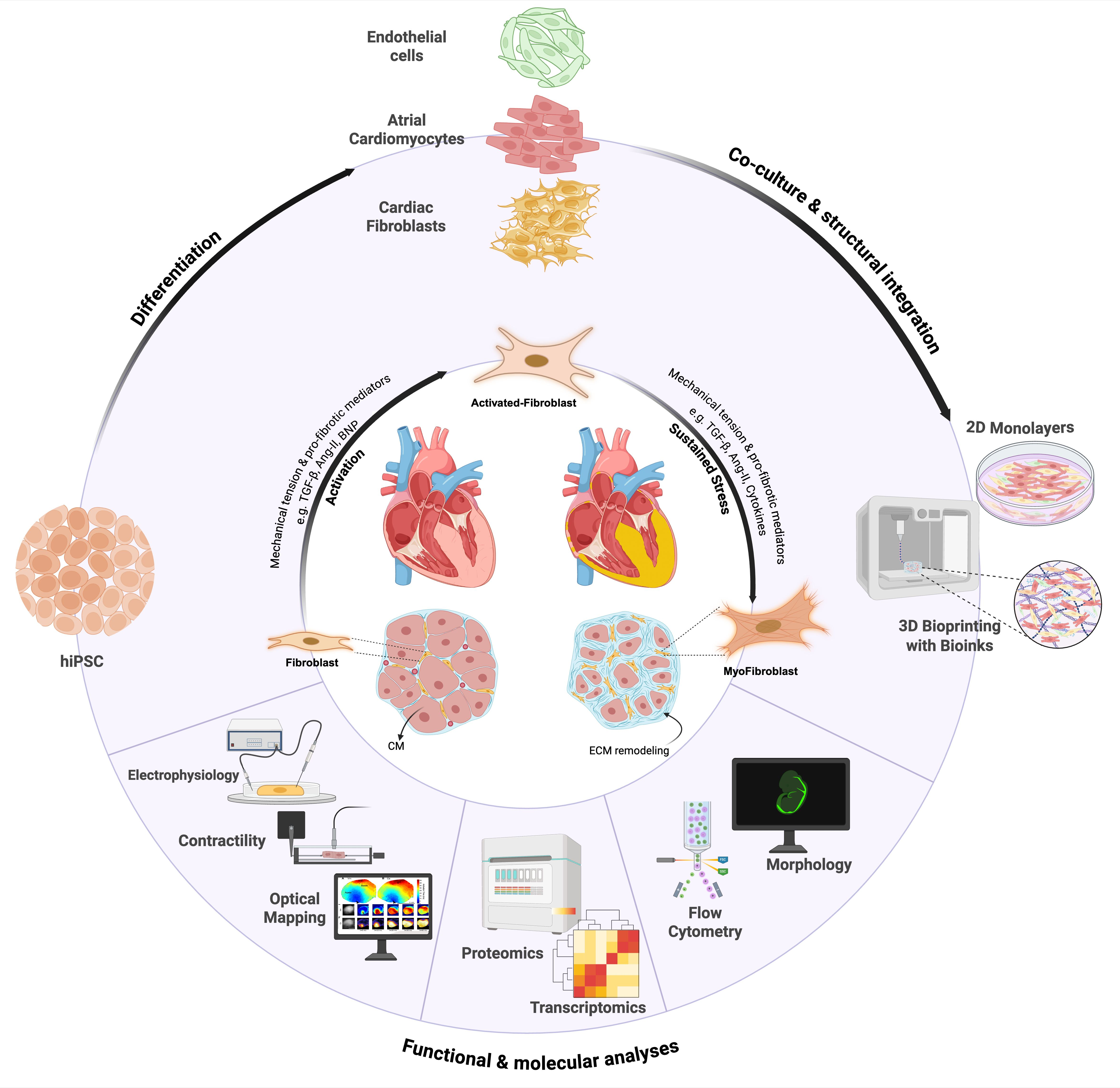
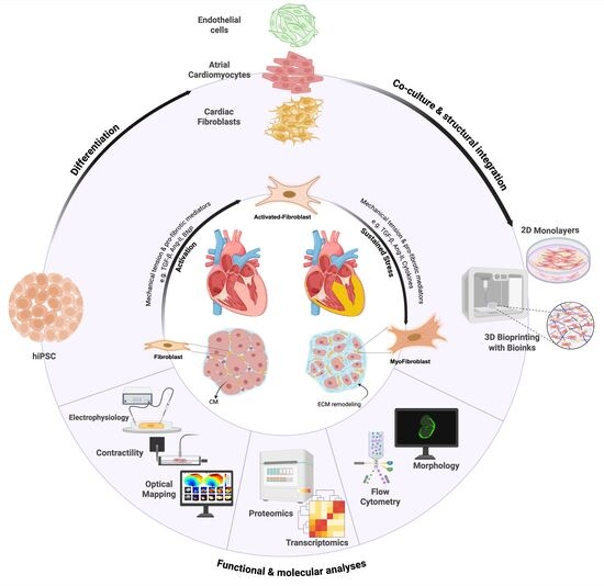
- We present a conceptual roadmap to address this gap by integrating human-induced pluripotent stem cell (hiPSC)-derived atrial cardiomyocytes and fibroblasts with 3D bioengineering techniques to build functional, human-specific models of atrial fibrosis.
- The bioengineered models developed from this roadmap will allow researchers to mechanistically connect specific fibrotic patterns to the arrhythmogenic electrical conduction changes that drive AF.
- These high-fidelity in vitro platforms will accelerate the discovery of novel anti-fibrotic drugs and enable patient-specific testing to advance personalized medicine for atrial fibrillation.
Abstract
1. Introduction: Atrial Fibrillation and ECM Remodeling
2. Limitations of Current AF Models and Therapies
2.1. Animal Models
2.2. Ex Vivo and 2D Cell Models
2.3. Clinical Imaging and Surrogates
2.4. Current Therapies
3. Advances in hiPSC-Derived Cardiac Cells for Modeling AF
3.1. hiPSC-Derived Atrial Cardiomyocytes (hiPSC-aCMs)
3.2. hiPSC-Derived Cardiac Fibroblasts (hiPSC-CFs)
4. Generating Atrial Fibrotic Tissue
4.1. 2D Monolayer Approach
4.2. 3D Bioprinting Technologies
4.2.1. Bioinks for Cardiac Tissue
4.2.2. Bioprinting Techniques
4.2.3. Vascularization and Maturation
5. Characterization of In Vitro Atrial Fibrosis Models
5.1. Bioink Optimization for Atrial Matrix
5.2. 3D Bioprinting of Functional Atrial Tissue Constructs
5.3. Controlled Fibroblast Activation (Inducing Fibrosis in Construct)
5.4. Fibrosis Induction and Assessment
5.5. Architectural and Cellular Patterning to Mimic Fibrotic Atria
5.6. Pathophysiological Insights into AF and Fibrosis
5.6.1. Conduction and Electrophysiology
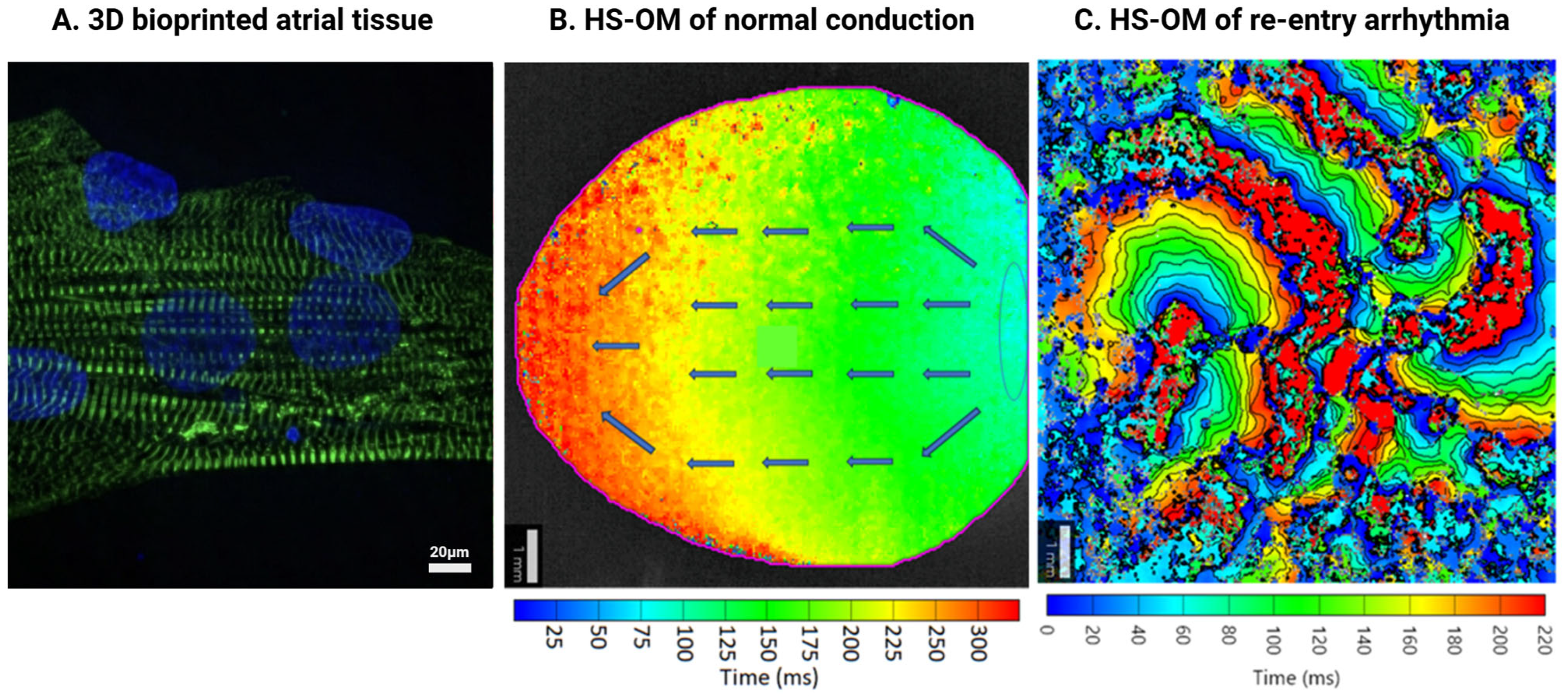
5.6.2. MEA and HS-OM Application in Fibrotic Cardiac Tissue Models
5.6.3. Assessment in Fibrotic Cardiac Tissue Models Using Patch-Clamp Measurements
5.6.4. Fibroblast-Cardiomyocyte Crosstalk
5.6.5. Characterizing Fibrotic Features Using ICC, Flow Cytometry, and RNA-Seq
5.6.6. RNA Extraction, Reverse Transcription, and Real-Time Quantitative PCR Analysis
5.6.7. Flow Cytometry
5.6.8. Immunocytochemistry (ICC)
5.6.9. Matrix Biology in AF
5.6.10. Ca2+ Handling
5.6.11. Mitochondrial and Metabolic Assessment
5.6.12. Validation Against Native Tissue
6. Future Perspectives and Conclusions
6.1. Applications of Engineered Fibrotic Atrial Tissue Models
6.2. Personalized Cardiac Models
6.3. Integration of Additional Cell Types and Complexity
6.4. Maturation and Scaling
6.5. Therapeutic Development
6.6. Challenges and Next Steps
Funding
Data Availability Statement
Conflicts of Interest
References
- Brown, G.E.; Han, Y.D.; Michell, A.R.; Ly, O.T.; Vanoye, C.G.; Spanghero, E.; George, A.L.; Darbar, D.; Khetani, S.R. Engineered cocultures of iPSC-derived atrial cardiomyocytes and atrial fibroblasts for modeling atrial fibrillation. Sci. Adv. 2024, 10, eadg1222. [Google Scholar] [CrossRef]
- Li, X.; Li, Z.; He, H.; Wang, S.; Su, H.; Kang, G. Global burden and health inequality of atrial fibrillation/atrial flutter from 1990 to 2021. Front. Cardiovasc. Med. 2025, 12, 1585980. [Google Scholar] [CrossRef] [PubMed]
- Rush, K.L.; Seaton, C.L.; Burton, L.; Loewen, P.; O’cOnnor, B.P.; Moroz, L.; Corman, K.; Smith, M.A.; Andrade, J.G. Quality of life among patients with atrial fibrillation: A theoretically-guided cross-sectional study. PLoS ONE 2023, 18, e0291575. [Google Scholar] [CrossRef] [PubMed]
- Schotten, U.; Goette, A.; Verheule, S. Translation of pathophysiological mechanisms of atrial fibrosis into new diagnostic and therapeutic approaches. Nat. Rev. Cardiol. 2024, 22, 225–240. [Google Scholar] [CrossRef]
- Nesheiwat, Z.; Goyal, A.; Jagtap, M. StatPearls; StatPearls Publishing LLC: Orlando, FL, USA, 2025. [Google Scholar]
- BaniHani, H.A.; Khaled, L.H.; Al Sharaa, N.M.; Al Saleh, R.A.; Ghalaita, A.K.B.; Sulaiman, A.S.B.; Holeihel, A. Causes, Diagnosis, Treatment, and Prognosis of Cardiac Fibrosis: A Systematic Review. Cureus 2025, 17, e81264. [Google Scholar] [CrossRef]
- Aguilar, M.; Rose, R.A.; Takawale, A.; Nattel, S.; Reilly, S. New aspects of endocrine control of atrial fibrillation and possibilities for clinical translation. Cardiovasc. Res. 2021, 117, 1645–1661. [Google Scholar] [CrossRef] [PubMed]
- Jansen, H.J.; Bohne, L.J.; Gillis, A.M.; Rose, R.A. Atrial remodeling and atrial fibrillation in acquired forms of cardiovascular disease. Heart Rhythm. O2 2020, 1, 147–159. [Google Scholar] [CrossRef]
- Karakasis, P.; Theofilis, P.; Vlachakis, P.K.; Korantzopoulos, P.; Patoulias, D.; Antoniadis, A.P.; Fragakis, N. Atrial Fibrosis in Atrial Fibrillation: Mechanistic Insights, Diagnostic Challenges, and Emerging Therapeutic Targets. Int. J. Mol. Sci. 2024, 26, 209. [Google Scholar] [CrossRef]
- Pellman, J.; Sheikh, F. Atrial Fibrillation: Mechanisms, Therapeutics, and Future Directions. In Comprehensive Physiology; John Wiley & Sons, Inc.: Hoboken, NJ, USA, 2015; Volume 5, pp. 649–665. [Google Scholar]
- Jafari, A.; Ajji, Z.; Mousavi, A.; Naghieh, S.; Bencherif, S.A.; Savoji, H. Latest Advances in 3D Bioprinting of Cardiac Tissues. Adv. Mater. Technol. 2022, 7, 2101636. [Google Scholar] [CrossRef]
- Koenig, A.L.; Shchukina, I.; Amrute, J.; Andhey, P.S.; Zaitsev, K.; Lai, L.; Bajpai, G.; Bredemeyer, A.; Smith, G.; Jones, C.; et al. Single-cell transcriptomics reveals cell-type-specific diversification in human heart failure. Nat. Cardiovasc. Res. 2022, 1, 263–280. [Google Scholar] [CrossRef]
- Shi, S.Y.; Luo, X.; Yamawaki, T.M.; Li, C.-M.; Ason, B.; Furtado, M.B. Recent Advances in Single-Cell Profiling and Multispecific Therapeutics: Paving the Way for a New Era of Precision Medicine Targeting Cardiac Fibroblasts. Curr. Cardiol. Rep. 2021, 23, 82. [Google Scholar] [CrossRef]
- Zeng, Z.; Wang, X.; Pei, J.; Wang, P.; Li, J.; Li, F.; Wang, J.; Zhao, Y.; Yu, C.; Liu, H.; et al. Endothelial plasticity in atrial fibrosis by integrating single-cell sequencing and genetic lineage tracing. Eur. Heart J. 2025, 46, 3470–3482. [Google Scholar] [CrossRef]
- Tallquist, M.D. Cardiac Fibroblast Diversity. Annu. Rev. Physiol. 2020, 82, 63–78. [Google Scholar] [CrossRef] [PubMed]
- Zhou, P.; Pu, W.T. Recounting Cardiac Cellular Composition. Circ. Res. 2017, 118, 368–370. [Google Scholar] [CrossRef]
- Gibb, A.A.; Lazaropoulos, M.P.; Elrod, J.W. Myofibroblasts and Fibrosis: Mitochondrial and Metabolic Control of Cellular Differentiation. Circ. Res. 2020, 127, 427–447. [Google Scholar] [CrossRef]
- Goette, A.; Jentsch-Ullrich, K.; Lendeckel, U.; Röcken, C.; Agbaria, M.; Auricchio, A.; Mohren, M.; Franke, A.; Klein, H.U. Effect of Atrial Fibrillation on Hematopoietic Progenitor Cells. Circulation 2003, 108, 2446–2449. [Google Scholar] [CrossRef]
- Gong, Q.; Le, X.; Yu, P.; Zhuang, L. Therapeutic advances in atrial fibrillation based on animal models. J. Zhejiang Univ. B 2024, 25, 135–152. [Google Scholar] [CrossRef] [PubMed]
- Schüttler, D.; Bapat, A.; Kääb, S.; Lee, K.; Tomsits, P.; Clauss, S.; Hucker, W.J. Animal Models of Atrial Fibrillation. Circ. Res. 2020, 127, 91–110. [Google Scholar] [CrossRef] [PubMed]
- Peter, A.K.; Bjerke, M.A.; Leinwand, L.A. Biology of the cardiac myocyte in heart disease. Mol. Biol. Cell 2016, 27, 2149–2160. [Google Scholar] [CrossRef]
- Schmidt, K.; Fuchs, M.; Weber, N.; Werlein, C.; Schmitto, J.D.; Ius, F.; Ruhparwar, A.; Bär, C.; Fiedler, J.; Thum, T. Single-nucleus RNA sequencing identifies cell-type–specific effects of sodium-glucose co-transporter 2 inhibitors in human myocardial slices. Eur. Heart J. 2024, 45, 3292–3295. [Google Scholar] [CrossRef]
- Abbas, N.; Haas, J.A.; Xiao, K.; Fuchs, M.; Just, A.; Pich, A.; Perbellini, F.; Werlein, C.; Ius, F.; Ruhparwar, A.; et al. Inhibition of miR-21: Cardioprotective effects in human failing myocardium ex vivo. Eur. Heart J. 2024, 45, 2016–2018. [Google Scholar] [CrossRef] [PubMed]
- Zhang, Y.; Li, T.-S.; Lee, S.-T.; Wawrowsky, K.A.; Cheng, K.; Galang, G.; Malliaras, K.; Roselle Abraham, M.; Wang, C.; Marbán, E. Dedifferentiation and Proliferation of Mammalian Cardiomyocytes. PLoS ONE 2010, 5, e12559. [Google Scholar] [CrossRef]
- Klumm, M.J.; Heim, C.; Fiegle, D.J.; Weyand, M.; Volk, T.; Seidel, T. Long-Term Cultivation of Human Atrial Myocardium. Front. Physiol. 2022, 13, 839139. [Google Scholar] [CrossRef]
- Woodley, J.P.; Lambert, D.W.; Asencio, I.O. Understanding Fibroblast Behavior in 3D Biomaterials. Tissue Eng. Part B Rev. 2022, 28, 569–578. [Google Scholar] [CrossRef]
- Marrouche, N.F.; Wazni, O.; McGann, C.; Greene, T.; Dean, J.M.; Dagher, L.; Kholmovski, E.; Mansour, M.; Marchlinski, F.; Wilber, D.; et al. Effect of MRI-Guided Fibrosis Ablation vs Conventional Catheter Ablation on Atrial Arrhythmia Recurrence in Patients with Persistent Atrial Fibrillation. JAMA 2022, 327, 2296–2305. [Google Scholar] [CrossRef]
- Mannhardt, I.; Breckwoldt, K.; Letuffe-Brenière, D.; Schaaf, S.; Schulz, H.; Neuber, C.; Benzin, A.; Werner, T.; Eder, A.; Schulze, T.; et al. Human Engineered Heart Tissue: Analysis of Contractile Force. Stem Cell Rep. 2016, 7, 29–42. [Google Scholar] [CrossRef]
- Sakata, K.; Bradley, R.P.; Prakosa, A.; Yamamoto, C.A.P.; Ali, S.Y.; Loeffler, S.; Tice, B.M.; Boyle, P.M.; Kholmovski, E.G.; Yadav, R.; et al. Assessing the arrhythmogenic propensity of fibrotic substrate using digital twins to inform a mechanisms-based atrial fibrillation ablation strategy. Nat. Cardiovasc. Res. 2024, 3, 857–868. [Google Scholar] [CrossRef]
- Sakata, K.; Yamamoto, C.A.P.; Prakosa, A.; Tice, B.M.; Ali, S.Y.; Loeffler, S.; Kholmovski, E.G.; Sinha, S.K.; Marine, J.E.; Calkins, H.; et al. Digital twins enable stratification of persistent atrial fibrillation patients for ablation diminishing unnecessary heart damage. npj Digit. Med. 2025, 8, 256. [Google Scholar] [CrossRef] [PubMed]
- Trayanova, N.A.; Prakosa, A. Up digital and personal: How heart digital twins can transform heart patient care. Heart Rhythm. 2023, 21, 89–99. [Google Scholar] [CrossRef]
- Andrade, J.G.; Wells, G.A.; Deyell, M.W.; Bennett, M.; Essebag, V.; Champagne, J.; Roux, J.-F.; Yung, D.; Skanes, A.; Khaykin, Y.; et al. Cryoablation or Drug Therapy for Initial Treatment of Atrial Fibrillation. N. Engl. J. Med. 2021, 384, 305–315. [Google Scholar] [CrossRef] [PubMed]
- An, J.; Bansal, N.; Zheng, C.; Lee, M.-S.; Wei, R.; Harrison, T.N.; Fan, D.; Garcia, E.; Lidgard, B.; Zelnick, L.R.; et al. Clinical and socioeconomic factors associated with recurrent atrial fibrillation after catheter ablation or antiarrhythmic drug therapy. Heart Rhythm. O2 2025, 6, 631–640. [Google Scholar] [CrossRef]
- Ganesan, A.N.; Nandal, S.; Lüker, J.; Pathak, R.K.; Mahajan, R.; Twomey, D.; Lau, D.H.; Sanders, P. Catheter Ablation of Atrial Fibrillation in Patients with Concomitant Left Ventricular Impairment: A Systematic Review of Efficacy and Effect on Ejection Fraction. Heart Lung Circ. 2015, 24, 270–280. [Google Scholar] [CrossRef]
- Noujaim, C.; Assaf, A.; Lim, C.; Feng, H.; Younes, H.; Mekhael, M.; Chouman, N.; Shamaileh, G.; El Hajjar, A.H.; Ayoub, T.; et al. Comprehensive atrial fibrillation burden and symptom reduction post-ablation: Insights from DECAAF II. Europace 2024, 26, euae104. [Google Scholar] [CrossRef]
- Dretzke, J.; Chuchu, N.; Agarwal, R.; Herd, C.; Chua, W.; Fabritz, L.; Bayliss, S.; Kotecha, D.; Deeks, J.J.; Kirchhof, P.; et al. Predicting recurrent atrial fibrillation after catheter ablation: A systematic review of prognostic models. EP Eur. 2020, 22, 748–760. [Google Scholar] [CrossRef] [PubMed]
- Iseoka, H.; Miyagawa, S.; Sakai, Y.; Sawa, Y. Cardiac fibrosis models using human induced pluripotent stem cell-derived cardiac tissues allow anti-fibrotic drug screening in vitro. Stem Cell Res. 2021, 54, 102420. [Google Scholar] [CrossRef] [PubMed]
- Cardona-Timoner, M.; Gomes, R.N.; Nascimento, D.S. Dressed in Collagen: 2D and 3D Cardiac Fibrosis Models. Int. J. Mol. Sci. 2025, 26, 3038. [Google Scholar] [CrossRef] [PubMed]
- Giacomelli, E.; Meraviglia, V.; Campostrini, G.; Cochrane, A.; Cao, X.; van Helden, R.W.; Garcia, A.K.; Mircea, M.; Kostidis, S.; Davis, R.P.; et al. Human-iPSC-Derived Cardiac Stromal Cells Enhance Maturation in 3D Cardiac Microtissues and Reveal Non-cardiomyocyte Contributions to Heart Disease. Cell Stem Cell 2020, 26, 862–879.e11. [Google Scholar] [CrossRef]
- Zhang, H.; Thai, P.N.; Shivnaraine, R.V.; Ren, L.; Wu, X.; Siepe, D.H.; Liu, Y.; Tu, C.; Shin, H.S.; Caudal, A.; et al. Multiscale drug screening for cardiac fibrosis identifies MD2 as a therapeutic target. Cell 2024, 187, 7143–7163.e22. [Google Scholar] [CrossRef]
- Niro, F.; Fernandes, S.; Cassani, M.; Apostolico, M.; La Cruz, J.O.-D.; Pereira-Sousa, D.; Pagliari, S.; Vinarsky, V.; Zdráhal, Z.; Potesil, D.; et al. Fibrotic extracellular matrix impacts cardiomyocyte phenotype and function in an iPSC-derived isogenic model of cardiac fibrosis. Transl. Res. 2024, 273, 58–77. [Google Scholar] [CrossRef]
- Beauchamp, P.; Jackson, C.B.; Ozhathil, L.C.; Agarkova, I.; Galindo, C.L.; Sawyer, D.B.; Suter, T.M.; Zuppinger, C. 3D Co-culture of hiPSC-Derived Cardiomyocytes with Cardiac Fibroblasts Improves Tissue-Like Features of Cardiac Spheroids. Front. Mol. Biosci. 2020, 7, 14. [Google Scholar] [CrossRef]
- Lee, J.H.; Protze, S.I.; Laksman, Z.; Backx, P.H.; Keller, G.M. Human Pluripotent Stem Cell-Derived Atrial and Ventricular Cardiomyocytes Develop from Distinct Mesoderm Populations. Cell Stem Cell 2017, 21, 179–194.e4. [Google Scholar] [CrossRef]
- Gunawan, M.G.; Sangha, S.S.; Shafaattalab, S.; Lin, E.; Heims-Waldron, D.A.; Bezzerides, V.J.; Laksman, Z.; Tibbits, G.F. Drug screening platform using human induced pluripotent stem cell-derived atrial cardiomyocytes and optical mapping. Stem Cells Transl. Med. 2020, 10, 68–82. [Google Scholar] [CrossRef]
- Sweat, M.E.; Pu, W.T. Genetic and molecular underpinnings of atrial fibrillation. npj Cardiovasc. Health 2024, 1, 35. [Google Scholar] [CrossRef]
- Babini, H.; Jiménez-Sábado, V.; Stogova, E.; Arslanova, A.; Butt, M.; Dababneh, S.; Asghari, P.; Moore, E.D.W.; Claydon, T.W.; Chiamvimonvat, N.; et al. hiPSC-derived cardiomyocytes as a model to study the role of small-conductance Ca2+-activated K+ (SK) ion channel variants associated with atrial fibrillation. Front. Cell Dev. Biol. 2024, 12, 1298007. [Google Scholar] [CrossRef] [PubMed]
- Nagalingam, R.S.; Jayousi, F.; Hamledari, H.; Dababneh, S.; Hosseini, D.; Lindsay, C.; Geltink, R.K.; Lange, P.F.; Dixon, I.M.; Rose, R.A.; et al. Molecular and metabolomic characterization of hiPSC-derived cardiac fibroblasts transitioning to myofibroblasts. Front. Cell Dev. Biol. 2024, 12, 1496884. [Google Scholar] [CrossRef] [PubMed]
- Zhang, H.; Tian, L.; Shen, M.; Tu, C.; Wu, H.; Gu, M.; Paik, D.T.; Wu, J.C. Generation of Quiescent Cardiac Fibroblasts From Human Induced Pluripotent Stem Cells for In Vitro Modeling of Cardiac Fibrosis. Circ. Res. 2019, 125, 552–566. [Google Scholar] [CrossRef]
- Protze, S.I.; Lee, J.H.; Keller, G.M. Human Pluripotent Stem Cell-Derived Cardiovascular Cells: From Developmental Biology to Therapeutic Applications. Cell Stem Cell 2019, 25, 311–327. [Google Scholar] [CrossRef]
- Cumberland, M.J.; Euchner, J.; Azad, A.J.; Vo, N.T.N.; Kirchhof, P.; Holmes, A.P.; Denning, C.; Gehmlich, K. Generation of a human iPSC-derived cardiomyocyte/fibroblast engineered heart tissue model. F1000Research 2024, 12, 1224. [Google Scholar] [CrossRef] [PubMed]
- Rother, J.; Richter, C.; Turco, L.; Knoch, F.; Mey, I.; Luther, S.; Janshoff, A.; Bodenschatz, E.; Tarantola, M. Crosstalk of cardiomyocytes and fibroblasts in co-cultures. Open Biol. 2015, 5, 150038. [Google Scholar] [CrossRef]
- Karakikes, I.; Ameen, M.; Termglinchan, V.; Wu, J.C. Human Induced Pluripotent Stem Cell–Derived Cardiomyocytes. Circ. Res. 2015, 117, 80–88. [Google Scholar] [CrossRef]
- Engler, A.J.; Carag-Krieger, C.; Johnson, C.P.; Raab, M.; Tang, H.-Y.; Speicher, D.W.; Sanger, J.W.; Sanger, J.M.; Discher, D.E. Embryonic cardiomyocytes beat best on a matrix with heart-like elasticity: Scar-like rigidity inhibits beating. J. Cell Sci. 2008, 121, 3794–3802. [Google Scholar] [CrossRef]
- Mohr, E.; Thum, T.; Bär, C. Accelerating cardiovascular research: Recent advances in translational 2D and 3D heart models. Eur. J. Heart Fail. 2022, 24, 1778–1791. [Google Scholar] [CrossRef]
- Prondzynski, M.; Berkson, P.; Trembley, M.A.; Tharani, Y.; Shani, K.; Bortolin, R.H.; Sweat, M.E.; Mayourian, J.; Yucel, D.; Cordoves, A.M.; et al. Efficient and reproducible generation of human iPSC-derived cardiomyocytes and cardiac organoids in stirred suspension systems. Nat. Commun. 2024, 15, 5929. [Google Scholar] [CrossRef]
- Zhao, Y.; Rafatian, N.; Feric, N.T.; Cox, B.J.; Aschar-Sobbi, R.; Wang, E.Y.; Aggarwal, P.; Zhang, B.; Conant, G.; Ronaldson-Bouchard, K.; et al. A Platform for Generation of Chamber-Specific Cardiac Tissues and Disease Modeling. Cell 2019, 176, 913–927.e18. [Google Scholar] [CrossRef] [PubMed]
- Burnham, M.P.; Harvey, R.; Sargeant, R.; Fertig, N.; Haddrick, M. A Scalable Approach Reveals Functional Responses of iPSC Cardiomyocyte 3D Spheroids. SLAS Discov. Adv. Sci. Drug Discov. 2021, 26, 352–363. [Google Scholar] [CrossRef]
- Bliley, J.M.; Stang, M.A.; Behre, A.; Feinberg, A.W. Advances in 3D Bioprinted Cardiac Tissue Using Stem Cell-Derived Cardiomyocytes. Stem Cells Transl. Med. 2024, 13, 425–435. [Google Scholar] [CrossRef]
- Du, B.; Dai, Z.; Wang, H.; Ren, Z.; Li, D. Advances and Prospects in Using Induced Pluripotent Stem Cells for 3D Bioprinting in Cardiac Tissue Engineering. Rev. Cardiovasc. Med. 2025, 26, 26697. [Google Scholar] [CrossRef]
- Esser, T.U.; Anspach, A.; Muenzebrock, K.A.; Kah, D.; Schrüfer, S.; Schenk, J.; Heinze, K.G.; Schubert, D.W.; Fabry, B.; Engel, F.B. Direct 3D-Bioprinting of hiPSC-Derived Cardiomyocytes to Generate Functional Cardiac Tissues. Adv. Mater. 2023, 35, e2305911. [Google Scholar] [CrossRef] [PubMed]
- Lu, T.-Y.; Xiang, Y.; Tang, M.; Chen, S. 3D Printing Approaches to Engineer Cardiac Tissue. Curr. Cardiol. Rep. 2023, 25, 505–514. [Google Scholar] [CrossRef]
- Stankey, P.P.; Kroll, K.T.; Ainscough, A.J.; Reynolds, D.S.; Elamine, A.; Fichtenkort, B.T.; Uzel, S.G.; Lewis, J.A. Embedding Biomimetic Vascular Networks via Coaxial Sacrificial Writing into Functional Tissue. Adv. Mater. 2024, 36, e2401528. [Google Scholar] [CrossRef] [PubMed]
- Noor, N.; Shapira, A.; Edri, R.; Gal, I.; Wertheim, L.; Dvir, T. 3D Printing of Personalized Thick and Perfusable Cardiac Patches and Hearts. Adv. Sci. 2019, 6, 1900344. [Google Scholar] [CrossRef]
- Yeung, E.; Fukunishi, T.; Bai, Y.; Bedja, D.; Pitaktong, I.; Mattson, G.; Jeyaram, A.; Lui, C.; Ong, C.S.; Inoue, T.; et al. Cardiac regeneration using human-induced pluripotent stem cell-derived biomaterial-free 3D-bioprinted cardiac patch in vivo. J. Tissue Eng. Regen. Med. 2019, 13, 2031–2039. [Google Scholar] [CrossRef]
- Ronaldson-Bouchard, K.; Ma, S.P.; Yeager, K.; Chen, T.; Song, L.; Sirabella, D.; Morikawa, K.; Teles, D.; Yazawa, M.; Vunjak-Novakovic, G. Advanced maturation of human cardiac tissue grown from pluripotent stem cells. Nature 2018, 556, 239–243. [Google Scholar] [CrossRef]
- Liu, H.; Fan, P.; Jin, F.; Huang, G.; Guo, X.; Xu, F. Dynamic and static biomechanical traits of cardiac fibrosis. Front. Bioeng. Biotechnol. 2022, 10, 1042030. [Google Scholar] [CrossRef]
- Lane, K.V.; Dow, L.P.; Castillo, E.A.; Boros, R.; Feinstein, S.D.; Pardon, G.; Pruitt, B.L. Cell Architecture and Dynamics of Human Induced Pluripotent Stem Cell-Derived Cardiomyocytes (hiPSC-CMs) on Hydrogels with Spatially Patterned Laminin and N-Cadherin. ACS Appl. Mater. Interfaces 2024, 17, 174–186. [Google Scholar] [CrossRef]
- Schussler, O.; Falcoz, P.E.; Chachques, J.C.; Alifano, M.; Lecarpentier, Y. Possible Treatment of Myocardial Infarct Based on Tissue Engineering Using a Cellularized Solid Collagen Scaffold Functionalized with Arg-Glyc-Asp (RGD) Peptide. Int. J. Mol. Sci. 2021, 22, 12563. [Google Scholar] [CrossRef] [PubMed]
- Han, Y.; Shao, Z.; Zhang, Y.; Zhao, H.; Sun, Z.; Yang, C.; Tang, H.; Han, Y.; Gao, C. 3D matrix stiffness modulation unveils cardiac fibroblast phenotypic switching. Sci. Rep. 2024, 14, 17015. [Google Scholar] [CrossRef]
- Kupfer, M.E.; Lin, W.-H.; Ravikumar, V.; Qiu, K.; Wang, L.; Gao, L.; Bhuiyan, D.B.; Lenz, M.; Ai, J.; Mahutga, R.R.; et al. In Situ Expansion, Differentiation, and Electromechanical Coupling of Human Cardiac Muscle in a 3D Bioprinted, Chambered Organoid. Circ. Res. 2020, 127, 207–224. [Google Scholar] [CrossRef]
- Maiullari, F.; Costantini, M.; Milan, M.; Pace, V.; Chirivì, M.; Maiullari, S.; Rainer, A.; Baci, D.; Marei, H.E.S.; Seliktar, D.; et al. A multi-cellular 3D bioprinting approach for vascularized heart tissue engineering based on HUVECs and iPSC-derived cardiomyocytes. Sci. Rep. 2018, 8, 13532. [Google Scholar] [CrossRef]
- Dai, X.; Liu, L.; Ouyang, J.; Li, X.; Zhang, X.; Lan, Q.; Xu, T. Coaxial 3D bioprinting of self-assembled multicellular heterogeneous tumor fibers. Sci. Rep. 2017, 7, 1457. [Google Scholar] [CrossRef]
- Zhang, Y.S.; Arneri, A.; Bersini, S.; Shin, S.-R.; Zhu, K.; Goli-Malekabadi, Z.; Aleman, J.; Colosi, C.; Busignani, F.; Dell’Erba, V.; et al. Bioprinting 3D microfibrous scaffolds for engineering endothelialized myocardium and heart-on-a-chip. Biomaterials 2016, 110, 45–59. [Google Scholar] [CrossRef]
- Shadrin, I.Y.; Allen, B.W.; Qian, Y.; Jackman, C.P.; Carlson, A.L.; Juhas, M.E.; Bursac, N. Cardiopatch platform enables maturation and scale-up of human pluripotent stem cell-derived engineered heart tissues. Nat. Commun. 2017, 8, 1825. [Google Scholar] [CrossRef]
- Devalla, H.D.; Schwach, V.; Ford, J.W.; Milnes, J.T.; El-Haou, S.; Jackson, C.; Gkatzis, K.; Elliott, D.; Lopes, S.M.C.D.S.; Mummery, C.L.; et al. Atrial-like cardiomyocytes from human pluripotent stem cells are a robust preclinical model for assessing atrial-selective pharmacology. EMBO Mol. Med. 2015, 7, 394–410. [Google Scholar] [CrossRef]
- Laksman, Z.; Wauchop, M.; Lin, E.; Protze, S.; Lee, J.; Yang, W.; Izaddoustdar, F.; Shafaattalab, S.; Gepstein, L.; Tibbits, G.F.; et al. Modeling Atrial Fibrillation using Human Embryonic Stem Cell-Derived Atrial Tissue. Sci. Rep. 2017, 7, 5268. [Google Scholar] [CrossRef]
- Goldfracht, I.; Efraim, Y.; Shinnawi, R.; Kovalev, E.; Huber, I.; Gepstein, A.; Arbel, G.; Shaheen, N.; Tiburcy, M.; Zimmermann, W.H.; et al. Engineered heart tissue models from hiPSC-derived cardiomyocytes and cardiac ECM for disease modeling and drug testing applications. Acta Biomater. 2019, 92, 145–159. [Google Scholar] [CrossRef]
- Walker, M.; Godin, M.; Pelling, A.E. Mechanical stretch sustains myofibroblast phenotype and function in microtissues through latent TGF-β1 activation. Integr. Biol. 2020, 12, 199–210. [Google Scholar] [CrossRef]
- Aujla, P.K.; Kassiri, Z. Diverse origins and activation of fibroblasts in cardiac fibrosis. Cell. Signal. 2021, 78, 109869. [Google Scholar] [CrossRef]
- Zhou, Y.; Richards, A.M.; Wang, P. Characterization and Standardization of Cultured Cardiac Fibroblasts for Ex Vivo Models of Heart Fibrosis and Heart Ischemia. Tissue Eng. Part C Methods 2017, 23, 422–433. [Google Scholar] [CrossRef]
- Rupert, C.E.; Kim, T.Y.; Choi, B.-R.; Coulombe, K.L.K. Human Cardiac Fibroblast Number and Activation State Modulate Electromechanical Function of hiPSC-Cardiomyocytes in Engineered Myocardium. Stem Cells Int. 2020, 2020, 9363809. [Google Scholar] [CrossRef]
- Trager, L.E.; Lyons, M.; Kuznetsov, A.; Sheffield, C.; Roh, K.; Freeman, R.; Rhee, J.; Guseh, J.S.; Li, H.; Rosenzweig, A. Beyond cardiomyocytes: Cellular diversity in the heart’s response to exercise. J. Sport Health Sci. 2022, 12, 423–437. [Google Scholar] [CrossRef]
- Zhang, J.; Tao, R.; Campbell, K.F.; Carvalho, J.L.; Ruiz, E.C.; Kim, G.C.; Schmuck, E.G.; Raval, A.N.; da Rocha, A.M.; Herron, T.J.; et al. Functional cardiac fibroblasts derived from human pluripotent stem cells via second heart field progenitors. Nat. Commun. 2019, 10, 2238. [Google Scholar] [CrossRef]
- Seguret, M.; Davidson, P.; Robben, S.; Jouve, C.; Pereira, C.; Lelong, Q.; Deshayes, L.; Cerveau, C.; Le Berre, M.; Ribeiro, R.S.R.; et al. A versatile high-throughput assay based on 3D ring-shaped cardiac tissues generated from human induced pluripotent stem cell-derived cardiomyocytes. eLife 2024, 12, RP87739. [Google Scholar] [CrossRef]
- Munawar, S.; Turnbull, I.C. Cardiac Tissue Engineering: Inclusion of Non-cardiomyocytes for Enhanced Features. Front. Cell Dev. Biol. 2021, 9, 653127. [Google Scholar] [CrossRef]
- Hall, C.; Law, J.P.; Reyat, J.S.; Cumberland, M.J.; Hang, S.; Vo, N.T.N.; Raniga, K.; Weston, C.J.; O’shea, C.; Townend, J.N.; et al. Chronic activation of human cardiac fibroblasts in vitro attenuates the reversibility of the myofibroblast phenotype. Sci. Rep. 2023, 13, 12137. [Google Scholar] [CrossRef]
- Jang, Y.; Kang, M.; Kang, Y.G.; Lee, D.; Jung, H.G.; Yoon, D.S.; Kim, J.; Park, Y. Cardiac fibroblast-mediated ECM remodeling regulates maturation in an in vitro 3D engineered cardiac tissue. J. Tissue Eng. 2025, 16, 1–17. [Google Scholar] [CrossRef]
- Wang, E.Y.; Rafatian, N.; Zhao, Y.; Lee, A.; Lai, B.F.L.; Lu, R.X.; Jekic, D.; Huyer, L.D.; Knee-Walden, E.J.; Bhattacharya, S.; et al. Biowire Model of Interstitial and Focal Cardiac Fibrosis. ACS Cent. Sci. 2019, 5, 1146–1158. [Google Scholar] [CrossRef]
- Riccio, J.; Alcaine, A.; Rocher, S.; Martinez-Mateu, L.; Saiz, J.; Invers-Rubio, E.; Guillem, M.S.; Martínez, J.P.; Laguna, P. Atrial fibrosis identification with unipolar electrogram eigenvalue distribution analysis in multi-electrode arrays. Med. Biol. Eng. Comput. 2022, 60, 3091–3112. [Google Scholar] [CrossRef]
- Huang, K.; Ashraf, M.; Rohani, L.; Luo, Y.; Sacayanan, A.; Huang, H.; Haegert, A.; Volik, S.; Sar, F.; LeBihan, S.; et al. Atrial Fibrillation Related Titin Truncation Is Associated with Atrial Myopathy in Patient-Derived Induced Pluripotent Stem Cell Disease Models. Circ. Genom. Precis. Med. 2025, 18, e004412. [Google Scholar] [CrossRef]
- Jansen, H.J.; Moghtadaei, M.; Rafferty, S.A.; Rose, R.A. Atrial Fibrillation in Aging and Frail Mice. Circ. Arrhythmia Electrophysiol. 2021, 14, e010077. [Google Scholar] [CrossRef]
- Laughner, J.I.; Ng, F.S.; Sulkin, M.S.; Arthur, R.M.; Efimov, I.R. Processing and analysis of cardiac optical mapping data obtained with potentiometric dyes. Am. J. Physiol. Circ. Physiol. 2012, 303, H753–H765. [Google Scholar] [CrossRef]
- Baines, O.; Sha, R.; Kalla, M.; Holmes, A.P.; Efimov, I.R.; Pavlovic, D.; O’sHea, C. Optical mapping and optogenetics in cardiac electrophysiology research and therapy: A state-of-the-art review. Europace 2024, 26, euae017. [Google Scholar] [CrossRef]
- Vigmond, E.; Pashaei, A.; Amraoui, S.; Cochet, H.; Hassaguerre, M. Percolation as a mechanism to explain atrial fractionated electrograms and reentry in a fibrosis model based on imaging data. Heart Rhythm. 2016, 13, 1536–1543. [Google Scholar] [CrossRef]
- Zaman, J.A.; Grace, A.A.; Narayan, S.M. Future Directions for Mapping Atrial Fibrillation. Arrhythmia Electrophysiol. Rev. 2022, 11, e08. [Google Scholar] [CrossRef]
- van Gorp, P.R.R.; Trines, S.A.; Pijnappels, D.A.; de Vries, A.A.F. Multicellular In vitro Models of Cardiac Arrhythmias: Focus on Atrial Fibrillation. Front. Cardiovasc. Med. 2020, 7, 43. [Google Scholar] [CrossRef] [PubMed]
- Haïssaguerre, M.; Jaïs, P.; Shah, D.C.; Takahashi, A.; Hocini, M.; Quiniou, G.; Garrigue, S.; Le Mouroux, A.; Le Métayer, P.; Clémenty, J. Spontaneous Initiation of Atrial Fibrillation by Ectopic Beats Originating in the Pulmonary Veins. N. Engl. J. Med. 1998, 339, 659–666. [Google Scholar] [CrossRef]
- Yang, P.; Zhu, L.; Wang, S.; Gong, J.; Selvaraj, J.N.; Ye, L.; Chen, H.; Zhang, Y.; Wang, G.; Song, W.; et al. Engineered model of heart tissue repair for exploring fibrotic processes and therapeutic interventions. Nat. Commun. 2024, 15, 7996. [Google Scholar] [CrossRef] [PubMed]
- Song, M.; Bin Choi, D.; Im, J.S.; Na Song, Y.; Kim, J.H.; Lee, H.; An, J.; Kim, A.; Choi, H.; Kim, J.-C.; et al. Modeling acute myocardial infarction and cardiac fibrosis using human induced pluripotent stem cell-derived multi-cellular heart organoids. Cell Death Dis. 2024, 15, 308. [Google Scholar] [CrossRef] [PubMed]
- Lou, X.; Tang, Y.; Ye, L.; Pretorius, D.; Fast, V.G.; Kahn-Krell, A.M.; Zhang, J.; Zhang, J.; Qiao, A.; Qin, G.; et al. Cardiac muscle patches containing four types of cardiac cells derived from human pluripotent stem cells improve recovery from cardiac injury in mice. Cardiovasc. Res. 2023, 119, 1062–1076. [Google Scholar] [CrossRef]
- Røsand, Ø.; Wang, J.; Scrimgeour, N.; Marwarha, G.; Høydal, M.A. Exosomal Preconditioning of Human iPSC-Derived Cardiomyocytes Beneficially Alters Cardiac Electrophysiology and Micro RNA Expression. Int. J. Mol. Sci. 2024, 25, 8460. [Google Scholar] [CrossRef]
- Xing, C.; Bao, L.; Li, W.; Fan, H. Progress on role of ion channels of cardiac fibroblasts in fibrosis. Front. Physiol. 2023, 14, 1138306. [Google Scholar] [CrossRef]
- Heijman, J.; Voigt, N.; Nattel, S.; Dobrev, D. Cellular and Molecular Electrophysiology of Atrial Fibrillation Initiation, Maintenance, and Progression. Circ. Res. 2014, 114, 1483–1499. [Google Scholar] [CrossRef]
- Tani, H.; Tohyama, S. Human Engineered Heart Tissue Models for Disease Modeling and Drug Discovery. Front. Cell Dev. Biol. 2022, 10, 855763. [Google Scholar] [CrossRef]
- Scuderi, G.J.; Butcher, J. Naturally Engineered Maturation of Cardiomyocytes. Front. Cell Dev. Biol. 2017, 5, 50. [Google Scholar] [CrossRef]
- Lemme, M.; Ulmer, B.M.; Lemoine, M.D.; Zech, A.T.; Flenner, F.; Ravens, U.; Reichenspurner, H.; Rol-Garcia, M.; Smith, G.; Hansen, A.; et al. Atrial-like Engineered Heart Tissue: An In Vitro Model of the Human Atrium. Stem Cell Rep. 2018, 11, 1378–1390. [Google Scholar] [CrossRef] [PubMed]
- Shinde, A.V.; Humeres, C.; Frangogiannis, N.G. The role of α-smooth muscle actin in fibroblast-mediated matrix contraction and remodeling. Biochim. Biophys. Acta (BBA)-Mol. Basis Dis. 2017, 1863, 298–309. [Google Scholar] [CrossRef] [PubMed]
- Archer, C.R.; Sargeant, R.; Basak, J.; Pilling, J.; Barnes, J.R.; Pointon, A. Characterization and Validation of a Human 3D Cardiac Microtissue for the Assessment of Changes in Cardiac Pathology. Sci. Rep. 2018, 8, 10160. [Google Scholar] [CrossRef]
- Min, S.; Kim, S.; Sim, W.-S.; Choi, Y.S.; Joo, H.; Park, J.-H.; Lee, S.-J.; Kim, H.; Lee, M.J.; Jeong, I.; et al. Versatile human cardiac tissues engineered with perfusable heart extracellular microenvironment for biomedical applications. Nat. Commun. 2024, 15, 2564. [Google Scholar] [CrossRef]
- Li, W.; Lou, X.; Zha, Y.; Qin, Y.; Zha, J.; Hong, L.; Xie, Z.; Yang, S.; Wang, C.; An, J.; et al. Single-cell RNA-seq of heart reveals intercellular communication drivers of myocardial fibrosis in diabetic cardiomyopathy. eLife 2023, 12, e80479. [Google Scholar] [CrossRef]
- Biendarra-Tiegs, S.M.; Clemens, D.J.; Secreto, F.J.; Nelson, T.J. Human Induced Pluripotent Stem Cell-Derived Non-Cardiomyocytes Modulate Cardiac Electrophysiological Maturation Through Connexin 43-Mediated Cell-Cell Interactions. Stem Cells Dev. 2020, 29, 75–89. [Google Scholar] [CrossRef]
- Min, S.; Cho, S.-W. Engineered human cardiac tissues for modeling heart diseases. BMB Rep. 2022, 56, 32–42. [Google Scholar] [CrossRef] [PubMed]
- Ronaldson-Bouchard, K.; Vunjak-Novakovic, G. Organs-on-a-Chip: A Fast Track for Engineered Human Tissues in Drug Development. Cell Stem Cell 2018, 22, 310–324. [Google Scholar] [CrossRef] [PubMed]
- Liu, T.; Xiong, F.; Qi, X.-Y.; Xiao, J.; Villeneuve, L.; Abu-Taha, I.; Dobrev, D.; Huang, C.; Nattel, S. Altered calcium handling produces reentry-promoting action potential alternans in atrial fibrillation–remodeled hearts. J. Clin. Investig. 2020, 5, e133754. [Google Scholar] [CrossRef] [PubMed]
- Abilez, O.J.; Yang, H.; Guan, Y.; Shen, M.; Yildirim, Z.; Zhuge, Y.; Venkateshappa, R.; Zhao, S.R.; Gomez, A.H.; El-Mokahal, M.; et al. Gastruloids enable modeling of the earliest stages of human cardiac and hepatic vascularization. Science 2025, 388, eadu9375. [Google Scholar] [CrossRef] [PubMed]





| Model Type | Advantages | Limitations | Key Applications | Key References |
|---|---|---|---|---|
| Animal Models (e.g., mouse, canine, porcine) |
|
| Studying systemic contributors to AF and fibrosis progression. | [1,9,19,20] |
| Ex Vivo Tissue Slices (e.g., human atrial trabeculae) |
|
| Acute drug testing and electrophysiological studies on intact human tissue. | [21,22,23,24,25,26] |
| 2D Monolayer Cell Culture |
|
| High-throughput screening and basic mechanistic studies of single cells. | [37,38,39,40,41] |
| 3D Bioprinted Tissues |
|
| Patient-specific disease modeling, mechanistic studies of fibrosis–arrhythmia links, and preclinical therapeutic testing. | [38,42] |
| Induction Method | Mechanism | Advantages | Limitations |
|---|---|---|---|
| Biochemical (e.g., TGF-β, Ang-II) | Soluble factors like growth factors, cytokines, and hormones activate intracellular signaling cascades that promote myofibroblast activation and collagen synthesis. |
| May not fully capture the chronic, low-grade inflammation or mechanical stress that drives fibrosis in vivo. |
| Mechanical (e.g., substrate stiffness, cyclic stretch) | Fibroblasts sense and respond to mechanical cues like stiffness, topography, or dynamic strain, which can promote their activation to myofibroblasts. |
|
|
| Cellular (e.g., increasing CF:CM ratio) | A higher proportion of cardiac fibroblasts (CFs) mimics profibrotic conditions, resulting in their enhanced proliferation, activation, and secretion of collagen and pro-fibrotic signals like TGF-β. |
| Does not model the activation of resident fibroblasts in response to a stimulus but rather starts with a pre-determined pro-fibrotic cell density. |
| Context | Reported Cardiomyocyte (CM): Fibroblast (CF) Ratio | Implication/Rationale for Modeling | Citation |
|---|---|---|---|
| Native Human Heart Tissue | Highly variable and controversial; estimates range from 1:10 to 4:1 (CM:CF). Varies by species, cardiac region, and measurement technique. | The lack of a single “correct” ratio in native tissue means that in vitro models should treat the ratio as a key experimental variable to be systematically tested, rather than a fixed parameter. | [81,82,83] |
| In Vitro “Healthy” Model | Commonly ~3:1 to 4:1 (CM:CF). | This ratio mimics a myocyte-rich environment while still including fibroblasts to support cardiomyocyte maturation, ECM production, and electrical function. | [84,85] |
| In Vitro “Fibrotic” Model | Commonly inverted to ~1:3 (CM:CF) or uses even higher fibroblast densities (e.g., ~70% CFs). | A higher proportion of fibroblasts simulates profibrotic conditions, promoting excessive ECM deposition, increased tissue stiffness, and altered electrophysiology characteristic of disease. | [41,86,87] |
Disclaimer/Publisher’s Note: The statements, opinions and data contained in all publications are solely those of the individual author(s) and contributor(s) and not of MDPI and/or the editor(s). MDPI and/or the editor(s) disclaim responsibility for any injury to people or property resulting from any ideas, methods, instructions or products referred to in the content. |
© 2026 by the authors. Licensee MDPI, Basel, Switzerland. This article is an open access article distributed under the terms and conditions of the Creative Commons Attribution (CC BY) license.
Share and Cite
Panahi, B.; Dababneh, S.; Fadaei, S.; Babini, H.; Singh, S.; Prondzynski, M.; Akbari, M.; Backx, P.H.; Andrade, J.G.; Rose, R.A.; et al. Versatile hiPSC Models and Bioengineering Platforms for Investigation of Atrial Fibrosis and Fibrillation. Cells 2026, 15, 187. https://doi.org/10.3390/cells15020187
Panahi B, Dababneh S, Fadaei S, Babini H, Singh S, Prondzynski M, Akbari M, Backx PH, Andrade JG, Rose RA, et al. Versatile hiPSC Models and Bioengineering Platforms for Investigation of Atrial Fibrosis and Fibrillation. Cells. 2026; 15(2):187. https://doi.org/10.3390/cells15020187
Chicago/Turabian StylePanahi, Behnam, Saif Dababneh, Saba Fadaei, Hosna Babini, Sanjana Singh, Maksymilian Prondzynski, Mohsen Akbari, Peter H. Backx, Jason G. Andrade, Robert A. Rose, and et al. 2026. "Versatile hiPSC Models and Bioengineering Platforms for Investigation of Atrial Fibrosis and Fibrillation" Cells 15, no. 2: 187. https://doi.org/10.3390/cells15020187
APA StylePanahi, B., Dababneh, S., Fadaei, S., Babini, H., Singh, S., Prondzynski, M., Akbari, M., Backx, P. H., Andrade, J. G., Rose, R. A., & Tibbits, G. F. (2026). Versatile hiPSC Models and Bioengineering Platforms for Investigation of Atrial Fibrosis and Fibrillation. Cells, 15(2), 187. https://doi.org/10.3390/cells15020187