1. Introduction
Herpesviruses are a large family of enveloped double-stranded DNA viruses that establish lifelong infections in humans, characterized by latency and periodic reactivation. Among them, herpes simplex virus type 1 (HSV-1) and type 2 (HSV-2), as well as varicella–zoster virus (VZV), are of major clinical importance. HSV-1 and HSV-2 are responsible for oral and genital lesions, keratitis, encephalitis, and severe complications in immunocompromised individuals, whereas VZV causes primary infection manifesting as varicella (chickenpox) and may later reactivate as herpes zoster (shingles) [
1,
2]. The global burden of these infections remains substantial, particularly in aging and immunocompromised populations, where reactivation can lead to severe neurological and systemic complications [
2,
3].
A hallmark of alphaherpesviruses, including HSV and VZV, is their ability to establish latency in sensory neurons and reactivate under conditions such as stress or immune suppression. In the case of VZV, reactivation is often associated with postherpetic neuralgia, a debilitating chronic pain condition that significantly affects quality of life [
3,
4]. Despite the availability of vaccines against VZV, breakthrough infections and reactivation still occur, underscoring the need for effective antiviral therapies targeting both primary infection and reactivation phases [
4].
Current antiviral therapy for herpes simplex virus (HSV) and varicella–zoster virus (VZV) infections relies predominantly on nucleoside and nucleotide analogs, including acyclovir, valacyclovir, famciclovir, and related agents, which interfere with viral DNA synthesis. Although these drugs are generally effective in reducing viral replication and disease severity, they do not eliminate latent viral reservoirs, and their therapeutic efficacy may be compromised by delayed administration, incomplete viral suppression, and the emergence of drug-resistant variants, particularly in immunocompromised patients [
4,
5,
6,
7]. Recent advances in HSV antiviral development have therefore focused on agents with mechanisms distinct from those of conventional DNA polymerase inhibitors, including helicase–primase inhibitors and other compounds targeting alternative stages of the viral replication cycle. These developments underscore the ongoing need to identify antiviral agents with novel or multitarget mechanisms of action that may complement or overcome the limitations of existing therapies [
8].
Natural products continue to represent an important source of structurally diverse antiviral compounds and potential lead molecules for the development of new therapeutic strategies. Recent studies and reviews have highlighted the ability of plant-, marine-, and microorganism-derived compounds to interfere with multiple stages of herpesvirus infection, including viral attachment and entry, intracellular genome replication, viral gene expression, protein synthesis, virion assembly, and host signaling pathways [
9,
10,
11,
12,
13,
14,
15]. Such multitarget activity is particularly attractive in the context of herpesvirus infections because it may provide opportunities to develop agents with mechanisms distinct from conventional nucleoside analogues and potentially reduce selective pressure on a single viral target. Recent experimental studies have further demonstrated that crude botanical extracts and their major isolated constituents can selectively inhibit HSV replication, supporting continued efforts to identify and characterize bioactive antiviral compounds from natural sources [
11,
16].
Cinnamomum cassia (Chinese cinnamon) is a medicinal plant widely used in traditional medicine and known for its antimicrobial, antioxidant, anti-inflammatory, and antiviral properties [
16,
17].
C. cassia contains a variety of bioactive compounds, and cinnamaldehyde is widely recognized as a characteristic constituent of cinnamon and has been associated with many of its biological effects [
18]. However, the composition and cinnamaldehyde content of the ethanolic extract tested in the present study were not determined. Cinnamaldehyde, a phenylpropanoid compound and a major component of cinnamon oil, is used in traditional Chinese medicine and has been extensively studied for its antimicrobial and antiviral activities against a broad range of pathogens, including enveloped viruses [
17,
19,
20,
21]. However, the antiviral properties of cinnamon-derived preparations may depend substantially on the botanical species, extraction method, chemical composition, formulation, and virus examined.
Several studies have demonstrated that cinnamaldehyde exhibits antiviral activity through multiple mechanisms, including disruption of viral envelope integrity, inhibition of viral protein synthesis, and modulation of host cellular pathways [
20,
21,
22]. In vitro studies have shown that
cinnamaldehyde and related phenolic compounds can inhibit viral replication and reduce viral infectivity. Notably, essential oils and extracts derived from cinnamon species have demonstrated virucidal activity against HSV, particularly by interfering with early stages of viral infection [
22]. Given that both HSV and VZV are enveloped viruses with similar entry and replication strategies, these findings suggest that cinnamaldehyde and cinnamon-derived extracts may also exhibit activity against VZV, although this remains underexplored.
Recent investigations have further supported the antiviral potential of
cinnamaldehyde and related compounds, demonstrating their ability to interact with viral or host targets involved in infection and replication [
11,
15,
16,
17,
18,
19,
20,
21,
22,
23,
24,
25]. Cinnamaldehyde has been reported to exhibit antiviral activity against several enveloped and non-enveloped viruses through mechanisms that may involve both direct effects on viral replication and modulation of host–cell processes [
18]. In particular, cinnamaldehyde and preparations derived from
Cinnamomum species have demonstrated inhibitory activity against HSV-1 in vitro. Zhou et al. [
26] reported that cinnamaldehyde, as well as several
Cinnamomi ramulus extracts, inhibited HSV-1 replication, supporting the presence of antiviral activity associated with this compound and cinnamon-derived preparations. In addition, studies in other viral systems have shown that cinnamaldehyde can inhibit viral replication when administered after infection and may interfere with viral protein synthesis, suggesting that its antiviral activity is not necessarily restricted to direct virucidal effects or inhibition of viral entry [
27].
Cinnamaldehyde may also influence host–cell pathways that are relevant to productive viral infection. Experimental studies have demonstrated that cinnamaldehyde and related cinnamaldehyde derivatives can inhibit NF-κB activation and modulate MAPK-associated signaling pathways. These observations are of potential relevance to HSV-1 infection because activation of NF-κB has been associated with efficient HSV-1 replication and the regulation of cellular survival responses in infected cells [
28,
29,
30]. Thus, although the precise molecular targets responsible for the anti-HSV-1 activity of cinnamaldehyde remain incompletely defined, the available literature supports the possibility that its antiviral effects may involve both interference with viral replication processes and modulation of host pathways required for efficient infection.
These findings highlight the potential of cinnamaldehyde as a multitarget antiviral agent. However, the antiviral activity of Cinnamomum cassia ethanolic extract and cinnamaldehyde, tested separately as a purified compound, against herpesviruses, particularly in a comparative context between HSV and VZV, remains incompletely characterized. Despite growing evidence supporting the broader antiviral potential of cinnamaldehyde and other cinnamon-derived constituents, their activity against alpha herpesviruses remains insufficiently characterized. In particular, relatively little is known about whether C. cassia ethanolic extract and cinnamaldehyde can inhibit both HSV-1 and VZV through comparable mechanisms or whether their activity primarily affects viral entry, direct virion infectivity, or intracellular stages of the replication cycle. This knowledge gap provides a rationale for directly comparing the antiviral activity and timing-dependent effects of C. cassia extract and cinnamaldehyde against these two clinically important alpha herpesviruses.
Therefore, in the present study, we evaluate the antiviral activity of an ethanolic extract of Cinnamomum cassia and cinnamaldehyde against HSV-1 and VZV. By investigating their effects on viral infectivity and replication, this work aims to advance the development of novel plant-based antiviral agents with broader therapeutic applicability.
2. Materials and Methods
2.1. Preparation of Cinnamomum Ethanolic Extract
Commercially obtained dried cinnamon bark marketed as Cinnamomum cassia was used for extract preparation. The material was obtained from a local commercial source and was not supplied with formal botanical certification. Taxonomic authentication and a herbarium voucher specimen were therefore not available, and the exact botanical identity of the commercial plant material could not be independently verified. Two grams of finely ground dried bark were extracted with 95% ethanol at 4 °C under continuous agitation for 48 h, centrifuged at 2000 rpm for 10 min, and the supernatant was collected. The collected supernatant was dried by evaporation and then dissolved in 95% ethanol to obtain a 10 mg/mL stock solution. The extract was diluted with medium containing 2% newborn calf serum (NBCS) to the required concentrations.
Cinnamaldehyde was purchased from Sigma-Aldrich, Rehovot, Israel (Cat. No. W228613; >95% purity).
2.2. Cells and Viruses
African green monkey kidney (Vero 76) cells were purchased from the American Type Culture Collection (ATCC), Rockville, MD, USA and grown [
31]. Vero cells are highly permissive to herpesvirus infection and represent a well-established model for antiviral screening.
HSV-1 was obtained from ATCC (VR-735), and VZV was obtained from the virology laboratory at Soroka University Medical Center, Beer-Sheva, Israel.
2.3. Cytotoxicity Examination
Vero cells were plated at 4 × 10
5 cells/well in 24-well culture plates and grown in an RPMI medium with 10% fetal calf serum, 1% glutamine, and 50 µg/mL antibiotic mixture (penicillin and streptomycin), and incubated at 37 °C in humidified air containing 5% CO
2. All the supplements for the Vero cultivation were purchased from Biological Industries (Beit-Haemek, Israel). The cells were treated with various doses of either
Cinnamomum ethanolic extract, Cinnamaldehyde, or
Acyclovir (
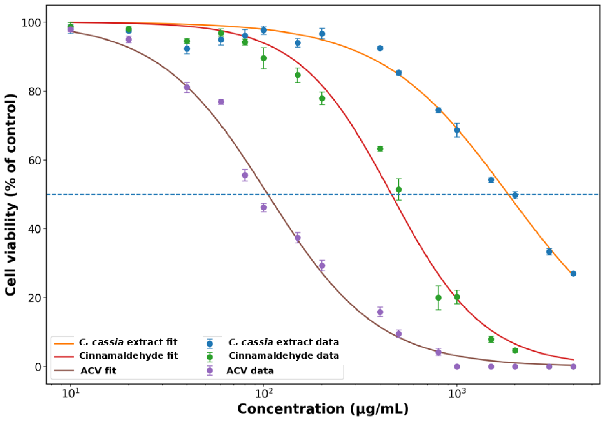
ACV), and their cytotoxicity was tested by two methods during a three-day period: (1) daily morphological observations using an inverted optical microscope; and (2) the (sodium 3′-[1-(phenylaminocarbonyl)-3,4-tetrazolium]-bis (4-methoxy6-nitro) benzene sulfonic acid hydrate) colorimetric assay (XTT Cell Proliferation Assay Kit, Sigma-Aldrich) for the quantification of cellular proliferation, viability, and cytotoxicity, as described previously [
32]. Untreated cells were used as controls, and the percentage of cell viability was calculated as the absorbance of treated wells divided by the absorbance of untreated control wells, multiplied by 100.
2.4. Viral Adsorption
Vero cell monolayers were inoculated with HSV-1 or VZV at 1 PFU/cell in RPMI medium containing 2% NBCS and incubated at 37 °C for a 2 h viral adsorption period. Following adsorption, the inoculum was removed and subsequent procedures were performed as described below. Infection development was evaluated by plaque assay as previously described [
31].
2.5. Plaque Assay
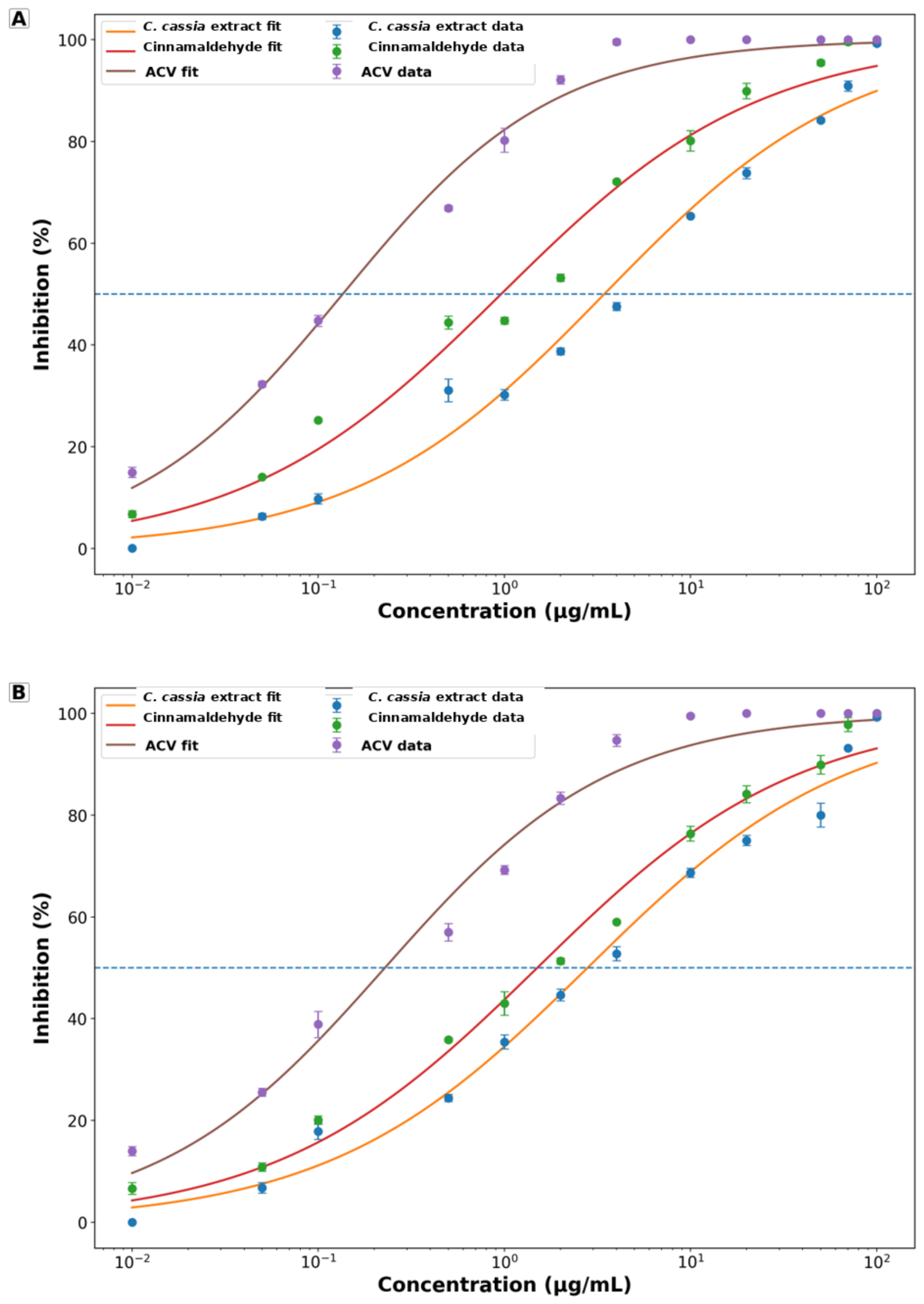
Following the 2 h viral adsorption period, unadsorbed virus particles were removed and the cells were covered with medium containing carboxymethylcellulose (CMC), in the presence or absence of the treatment solution, and incubated for 2 days. Then the CMC overlay was removed, and the cell monolayers were fixed with 10% formaldehyde in saline, stained with crystal violet, and the plaques counted. The antiviral effect (%) was calculated as the number of plaques in untreated cells minus the number of plaques in treated cells, divided by the number of plaques in untreated cells, and multiplied by 100.
2.6. Time-of-Addition Assay
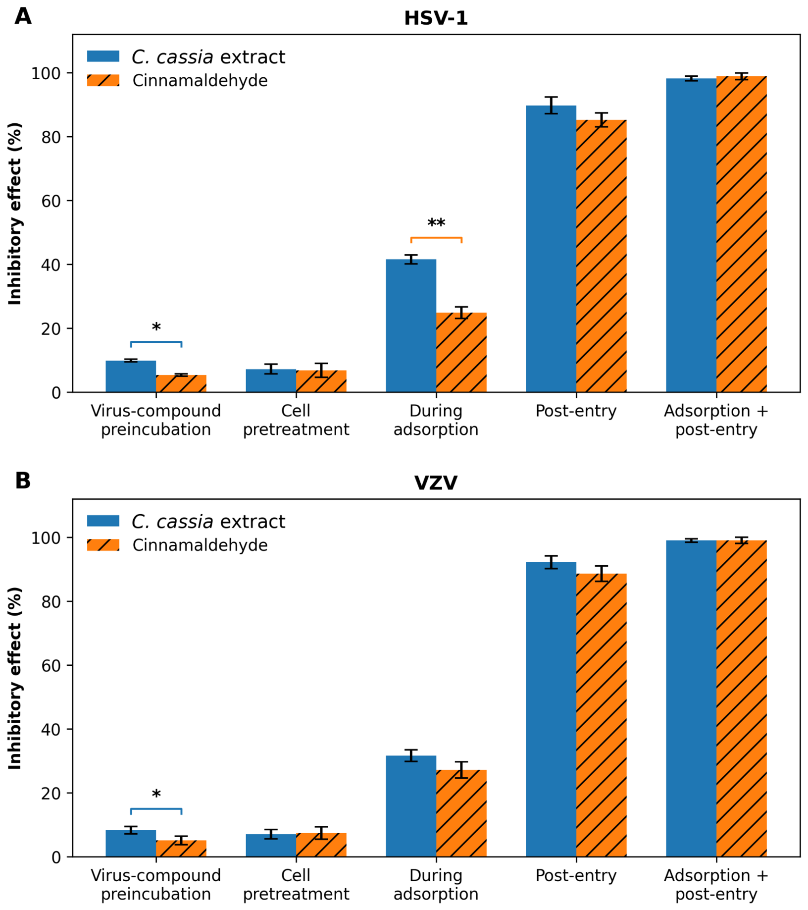
Vero cells were treated with 100 µg/mL of either C. cassia extract or cinnamaldehyde at different stages relative to infection, as follows: (1) Cell pretreatment: cells were pretreated with the tested material for 2 h before infection and then washed twice with 0.9% NaCl solution. (2) During adsorption: cells were treated with the tested material during the 2 h virus adsorption period and then washed twice with 0.9% NaCl solution. (3) Post-entry: after the 2 h adsorption period, cells were incubated with the tested material throughout the subsequent 2-day post-infection period. (4) Adsorption + post-entry: cells were treated during the 2 h adsorption period, and treatment was maintained throughout the subsequent 2-day post-infection period. (5) Virus-compound preincubation: to assess a possible direct effect of the tested material on viral particle infectivity, viral particles were pre-incubated with the tested material at room temperature for 1 h. The mixtures were then diluted 104-fold with fresh medium to minimize the test-material concentration during infection, and the diluted mixtures were used to infect cells.
2.7. Assessment of Intracellular Virus Production
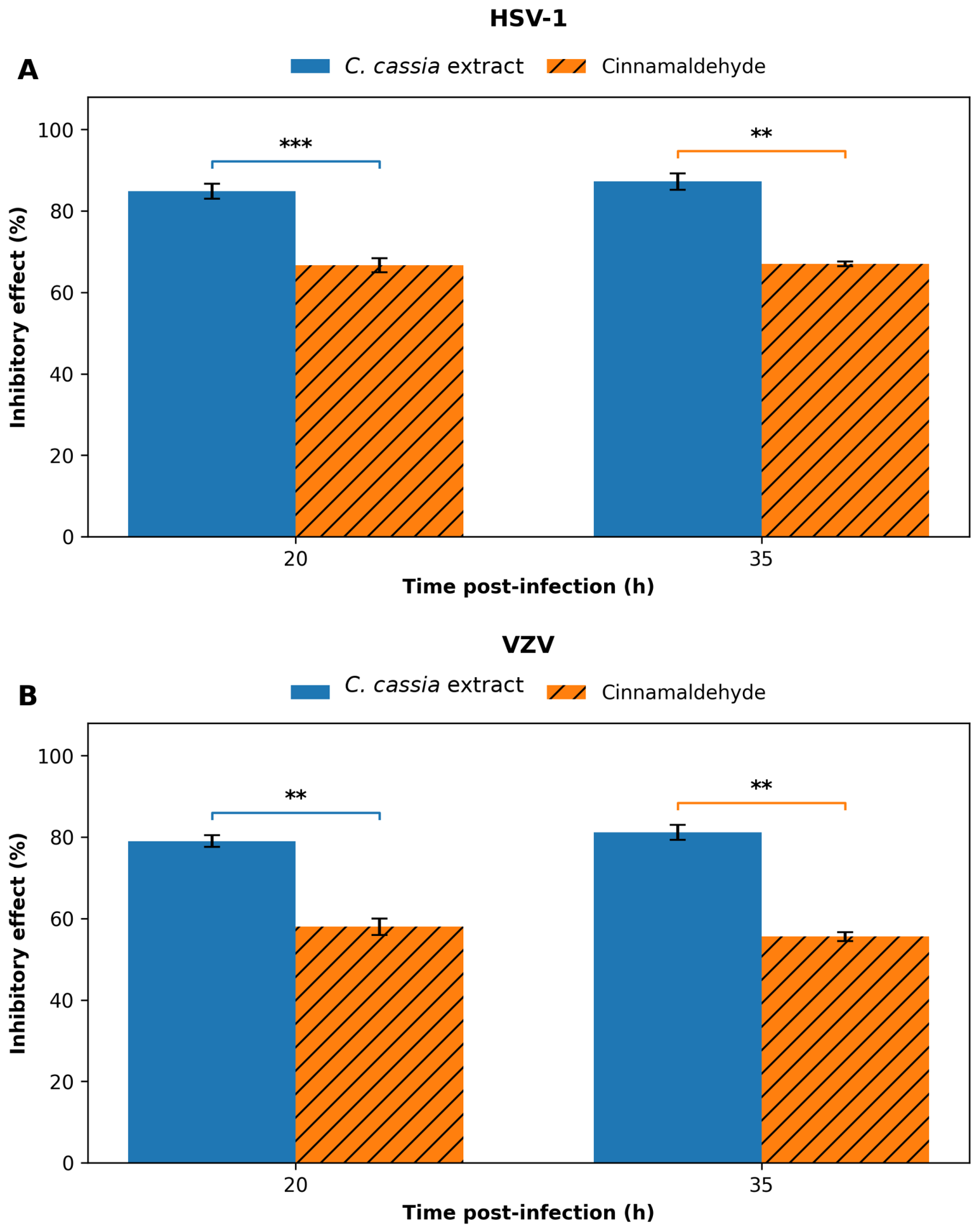
Cells were infected with HSV-1 or VZV for 2 h without treatment. The medium was then replaced with fresh medium with or without the tested product, and cells were harvested at 20 or 35 h post-infection using trypsin. After centrifugation (1500 rpm, 5 min) and three saline washes, cell pellets were resuspended in 100 µL saline, lysed by freeze–thaw cycles, and centrifuged again to remove debris. The resulting virus-containing supernatant was used to infect cell monolayers.
2.8. DNA Analysis
For viral DNA analysis, infected cells were harvested at 24 h post-infection. Viral DNA extraction was performed using the Genomic DNA Purification Kit (Thermo Fisher Scientific, Waltham, MA, USA) according to the manufacturer’s instructions. Viral DNA was quantified by SYBR Green-based real-time PCR using virus-specific primer sets. HSV-1 DNA was amplified using 5′-CGTACCTGCGGCTCGTGAAGT-3′ and 5′-AGCAGGGTGCTCGTGTATGGGC-3′, generating a 271 bp HSV-1-specific amplicon as previously described [
33]. VZV DNA was amplified using 5′-CATTACCGCACCCAAAGTGAA-3′ and 5′-CTAACGCTTCCACCTCGGGT-3′, generating an 89 bp VZV-specific amplicon as previously described [
34]. Cellular β-actin DNA was used as the endogenous reference for normalization using the primers 5′-GCACCCAGCACAATGAAGA-3′ and 5′-CGATCCACACGGAGTACTTG-3′. Relative viral DNA levels were calculated using the 2
−ΔΔCt method [
35], where ΔCt was calculated as Ct(viral target) − Ct(β-actin). Infected untreated cells were used as the calibrator and assigned a relative viral DNA level of 100%. No absolute viral copy-number determination or standard curve was used.
2.9. RNA Analysis
For RNA analysis, HSV-1-infected cells were harvested at 4 h post-infection and VZV-infected cells at 14 h post-infection. Total RNA was extracted from HSV-1- or VZV-infected Vero cells using the GenElute Mammalian Total RNA Miniprep Kit (Merck, Darmstadt, Germany) according to the manufacturer’s instructions. RNA samples were treated with DNase to remove contaminating DNA prior to reverse transcription. RNA was reverse-transcribed to cDNA using the qScript cDNA Synthesis Kit (Quantabio, Beverly, MA, USA). Viral transcript levels were determined by SYBR Green-based quantitative real-time PCR. For VZV, the immediate-early ORF63 (IE63) transcript was amplified using the primers 5′-TCGGACGGGGAAGACTTTAT-3′ and 5′-CGTCTGGTTCACAAGAATCG-3′ [
36]. For HSV-1, the ICP4/RS1 immediate-early (α) transcript was amplified using the primers 5′-CGGTGATGAAGGAGCTGCTGTTGC-3′ and 5′-CTGATCACGCGGCTGCTGTACA-3′ [
37]. β-actin was used as the endogenous reference gene using the primers mentioned above. Relative viral transcript levels were calculated using the 2
−ΔΔCt method [
35], with infected untreated cells serving as the calibrator and assigned a relative expression level of 100%.
2.10. Statistical Analyses
All experiments were independently repeated three times on different days, and each condition within each independent experiment was assayed in technical triplicate. Technical triplicates were averaged to obtain a single value for each independent experiment; therefore, n = 3 represents three independent experiments. Data are expressed as the mean ± SD of the three independent experiments. For concentration-response analyses, the technical triplicates within each independent experiment were averaged first, and the resulting values were analyzed by nonlinear least-squares regression using a normalized variable-slope sigmoidal (Hill) dose–response model. For the normalized curves, the lower and upper plateaus were constrained to 0% and 100%, respectively. The 50% cytotoxic concentration (CC
50) and 50% inhibitory concentration (IC
50) were obtained from the fitted curves, and goodness of fit was assessed using the coefficient of determination (R
2). The selectivity index (SI) was calculated as CC
50/IC
50. For the time-of-addition and progeny-virus experiments shown in
Figure 4 and
Figure 5, technical triplicates were averaged within each independent experiment before statistical analysis. For each virus, data were analyzed by two-way repeated-measures ANOVA, with tested material and treatment timing (
Figure 4) or sampling time (
Figure 5) as within-experiment factors. Where appropriate, Šídák-adjusted pairwise comparisons were performed between
C. cassia extract and cinnamaldehyde within each treatment condition or time point. A two-tailed adjusted
p-value < 0.05 was considered statistically significant. Nonlinear regression and statistical analyses were performed using GraphPad Prism 7.02 (San Diego, CA, USA). For the relative qPCR data shown in
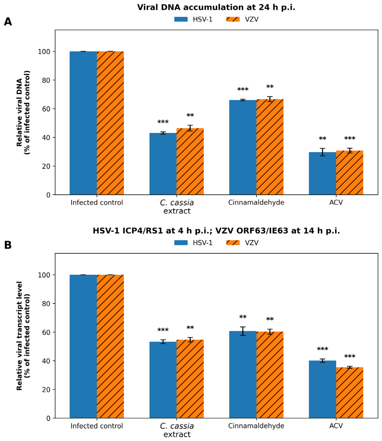
Figure 6, technical triplicates were first averaged within each independent experiment. Because the untreated infected group served as the calibrator and was defined as 100%, treatment values from the three independent experiments were compared with the reference value of 100% using two-sided one-sample
t-tests against 100%, with Šídák correction for the three treatment comparisons within each virus and panel. A two-tailed adjusted
p-value < 0.05 was considered statistically significant.
4. Discussion
In the present study, we demonstrated that the ethanolic extract of
Cinnamomum cassia and cinnamaldehyde exhibit marked antiviral activity against the clinically important alphaherpesviruses HSV-1 and VZV in vitro [
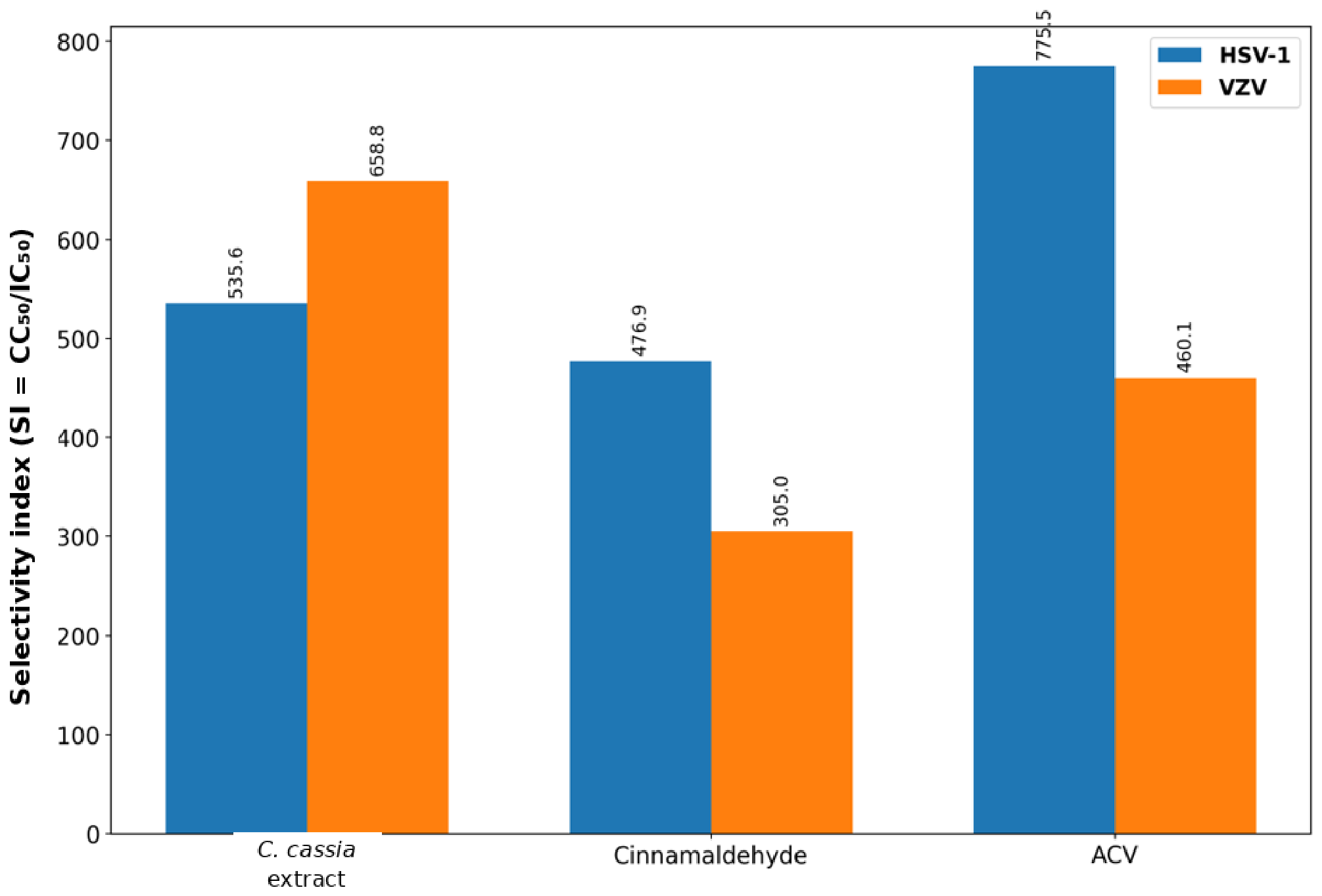
18]. Both agents inhibited viral replication in a dose-dependent manner while exhibiting relatively low cytotoxicity, resulting in favorable selectivity indices. Nonlinear regression-based analysis showed that the selectivity index of the extract and cinnamaldehyde was similar against HSV-1 (SI 535.6 and 476.9, respectively), whereas the extract showed a substantially higher selectivity index than cinnamaldehyde against VZV (SI 658.8 and 305.0, respectively). These findings suggest that the relative therapeutic window of the crude extract compared with cinnamaldehyde may differ between the two viruses. Compared with acyclovir, the
C. cassia extract showed a lower selectivity index against HSV-1 (535.6 vs. 775.5) but a higher selectivity index against VZV under the present in vitro assay conditions (658.8 vs. 460.1). These comparisons should be interpreted in the context of the present cell-based assay and do not by themselves establish comparative clinical efficacy. Cinnamon extracts contain multiple phytochemicals (polyphenols, flavonoids, coumarins, procyanidins, and terpenoids) that could potentially exert additive, complementary, or synergistic biological effects. However, because the present study did not characterize the complete chemical composition of the extract or formally evaluate interactions among individual constituents, the contribution of specific compounds and potential synergistic effects remains to be determined. Similar interactions have been described for other medicinal plant extracts, in which the whole extract exhibits greater biological activity than isolated constituents [
38,
39].
The comparable susceptibility of HSV-1 and VZV to both treatments suggests that these results are consistent with the possibility of interference with conserved intracellular processes, but do not identify the molecular target. HSV and VZV share similar replication strategies, including nuclear DNA replication, expression of conserved immediate-early and early genes, and dependence on highly conserved viral DNA polymerases. Therefore, compounds interfering with these conserved intracellular processes may exhibit broad-spectrum activity against multiple alphaherpesviruses.
These results for the extracts and cinnamaldehyde are consistent with previous studies reporting significant antiviral activity of ethanolic plant extracts and essential oils against various viruses, including poliovirus, coxsackievirus B1, adenovirus type 3, and herpes viruses [
40,
41,
42,
43,
44].
The results obtained in this study suggest that the antiviral activity of the extract and cinnamaldehyde is unlikely to result from stable interactions with host cell surface receptors or direct inactivation of viral particles. Instead, any interaction occurring before infection appears to be weak and reversible, as antiviral activity was largely lost after the compounds were removed prior to viral challenge. These findings agree with previous reports indicating that certain plant-derived ethanolic extracts exert limited effects on viral adsorption or receptor binding [
41,
42,
43]. However, they differ from studies proposing that many natural antiviral compounds primarily act by directly inactivating virions or preventing viral attachment and penetration into host cells [
45,
46]. In fact, previous studies have shown that the antiviral effects of both plant ethanolic extracts and essential oils are attributed to multiple complementary mechanisms that interfere with different stages of the viral life cycle. Due to the hydrophobic character of many essential oils constituents, they may associate with viral lipid membranes, resulting in disruption of viral envelope integrity and impairment of viral attachment, fusion, and entry into host cells [
47].
In the present study, all treatments markedly reduced the production of infectious progeny viruses within infected cells (
Figure 5), indicating that the antiviral activity extends beyond inhibition of viral entry and predominantly targets intracellular events essential for productive viral replication. The sustained reduction in virus production at both time points suggests that the antiviral effect is maintained throughout the replication cycle rather than representing only a transient delay in viral replication.
The marked reduction in progeny virus production is consistent with inhibition of intracellular viral replication. Also, reduced viral DNA accumulation and reduced abundance of the specific immediate-early transcripts examined were associated with this effect. Although these nucleic-acid changes did not fully account for the pronounced reduction in infectious virus production (
Figure 5), the findings suggest that additional intracellular processes, including viral protein expression, genome replication, virion assembly, maturation, or egress, may also be affected. These observations support the concept that cinnamon-derived compounds exert multitarget antiviral activity, a property that may reduce the likelihood of antiviral resistance. If confirmed, compounds acting independently of viral thymidine kinase may retain activity against acyclovir-resistant herpesvirus isolates.
It is worth noting that the decrease in the specific immediate-early transcript abundance observed after treatment with the extract or cinnamaldehyde could be a concomitant result of interference with other intracellular replication processes rather than a direct effect on transcription itself.
The identification of agents that interfere with intracellular stages of herpesvirus replication is particularly relevant in the context of recent efforts to develop antiviral drugs with mechanisms distinct from those of conventional nucleoside analogues. Current research on HSV therapeutics has increasingly focused on alternative viral and host targets, including the helicase–primase complex and other processes required for productive viral replication. In this context, the post-entry activity observed for both C. cassia extract and cinnamaldehyde is of interest because it suggests that their antiviral effects are not limited to interference with viral attachment or direct virion inactivation. Although the precise molecular target was not identified in the present study, the time-of-addition experiments together with reduced viral DNA accumulation and reduced abundance of the specific immediate-early transcripts examined support interference with intracellular events occurring after viral entry. These findings are consistent with interference at one or more intracellular stages of the viral replication cycle. Further studies should therefore determine whether these effects involve viral DNA replication, regulation of viral gene expression, protein synthesis, virion assembly, or modulation of host pathways required for efficient HSV and VZV replication. Possible mechanisms include inhibition of viral DNA polymerase activity, suppression of immediate-early viral gene expression, modulation of host signaling pathways required for efficient viral replication, or impairment of viral protein synthesis. Alternatively, cinnamaldehyde may influence host–cell signaling pathways, including NF-κB and MAPK signaling, that contribute to efficient herpesvirus replication. These possibilities warrant future investigation using viral protein expression analyses, transcriptomic approaches, and enzymatic assays.
Collectively, these findings support the growing potential of ethanolic extracts and essential oils as multifunctional antiviral agents and underscore the need for further studies to elucidate their molecular targets and optimize their integration into antiviral treatment strategies. In addition, isolating and purifying the bioactive antiviral constituent(s) of the extract may enhance its antiviral efficacy and improve its therapeutic potential against herpesvirus infections.
Finally, the present study has several limitations. First, all experiments were performed in Vero cells, which do not fully recapitulate the complexity of human epithelial or neuronal infection. Future studies should therefore include primary human fibroblasts and keratinocytes, which more closely resemble the natural target cells of HSV and VZV infection. Second, only in vitro antiviral activity was evaluated, and pharmacokinetic properties, bioavailability, and systemic toxicity remain unknown. Third, the precise molecular targets responsible for antiviral activity were not identified. Although the HSV-1 ICP4/RS1 and VZV ORF63/IE63 transcripts were examined at 4 h and 14 h post-infection, respectively, only one immediate-early transcript was analyzed for each virus. Therefore, these data do not establish a generalized inhibition of viral transcription and do not identify the precise intracellular target of the tested materials. The observed reduction in transcript abundance may reflect a direct or indirect consequence of interference with one or more intracellular replication processes. Future kinetic studies examining representative immediate-early, early, and late genes at multiple time points will be required to determine more precisely which stage(s) of the viral replication cycle are affected. Fourth, the dried cinnamon bark was obtained as a commercial product marketed as C. cassia and was not taxonomically authenticated through a certified botanical supplier or voucher specimen. Consequently, its exact botanical identity cannot be independently verified. The findings obtained with the crude extract should therefore be interpreted as specific to the commercial preparation tested and should not be generalized without qualification to taxonomically authenticated C. cassia material. Future studies should use voucher-documented plant material together with chromatographic profiling and quantitative standardization. Finally, the ethanolic extract used in the present study was not chemically profiled or standardized. Therefore, the abundance of cinnamaldehyde in the tested extract and the identity and contribution of other constituents cannot be determined from the present data. The antiviral findings obtained with the crude extract should consequently be regarded as specific to the preparation tested. Future studies should include chromatographic profiling and quantitative standardization of the extract before mechanistic attribution to individual constituents.
5. Conclusions
The present study shows that the ethanolic extract prepared from commercial cinnamon bark marketed as Cinnamomum cassia and cinnamaldehyde exhibit in vitro antiviral activity against HSV-1 and VZV in Vero cells. Both treatments inhibited viral replication in a dose-dependent manner and reduced the production of infectious progeny viruses. The time-of-addition experiments further suggest that their antiviral effects are mainly associated with events occurring after viral entry, rather than with direct inactivation of viral particles or inhibition of viral attachment.
Treatment of infected cells was also associated with reduced viral DNA accumulation and reduced abundance of the specific immediate-early transcripts examined. However, the present experiments do not establish the precise molecular target(s) responsible for these effects, and it remains unclear which specific intracellular step(s) of the viral replication cycle are affected.
Overall, these findings provide preliminary evidence that C. cassia extract and cinnamaldehyde may interfere with HSV-1 and VZV replication in vitro. Further studies using additional relevant cell models, chemically characterized preparations, and in vivo systems will be required to define their molecular mechanisms, evaluate their antiviral efficacy and safety, and determine their potential relevance as candidates for future antiviral development.