Review

Immunological Reprogramming in Cardiomyopathies: From Cardiomyocyte Injury to Disease of the Cardiac Immune Ecosystem


by
Tomasz Urbanowicz
1,* and
Krzysztof J. Filipiak
2
1
Cardiac Surgery and Transplantology Department, Poznan University of Medical Sciences, ½ Długa, 61-848 Poznań, Poland
2
The Centre of Postgraduate Medical Education, 99/103 Marymoncka Street, 01-813 Warsaw, Poland
*
Author to whom correspondence should be addressed.
Cells 2026, 15(14), 1308; https://doi.org/10.3390/cells15141308
Submission received: 28 June 2026 / Revised: 17 July 2026 / Accepted: 20 July 2026 / Published: 22 July 2026
(This article belongs to the Special Issue Cellular Mechanisms and Molecular Signaling in Heart Failure)

Highlights

  • This review proposes a conceptual shift from a cardiomyocyte-centered view of cardiomyopathies to a disease model grounded in dysregulation of the cardiac immune ecosystem.
  • Diverse immune mechanisms—including innate immune activation, immunometabolic remodeling, trained immunity, cGAS–STING signaling, and fibroblast–immune interactions—are integrated into a unified framework of chronic cardiac remodeling.
  • Emerging single-cell, spatial transcriptomic, and systems biology approaches support immunophenotyping as a complementary strategy to conventional phenotypic and genetic classification of cardiomyopathies.
  • Understanding cardiomyopathies as disorders of the cardiac immune ecosystem may facilitate mechanism-based patient stratification and support the future development of precision immunomodulatory therapies.

Abstract
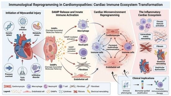
Cardiomyopathies have traditionally been regarded as disorders driven primarily by cardiomyocyte injury resulting from genetic defects, infection, metabolic stress, or toxic exposure. This paradigm has substantially advanced diagnosis and treatment. Still, it does not fully account for the marked heterogeneity in disease progression, persistent fibrosis, or variable therapeutic responses among patients with similar phenotypes. Increasing evidence indicates that immune remodeling is not merely a secondary consequence of myocardial injury but a dynamic process that actively shapes disease evolution. In this review, we integrate recent advances in cardiovascular immunology, single-cell and spatial transcriptomics, immunometabolism, and systems biology to propose a unified framework of immunological reprogramming in cardiomyopathies. We discuss how danger-associated molecular patterns, inflammasome activation, trained immunity, the cGAS–STING pathway, fibroblast–immune interactions, and the cardio–bone marrow axis converge to establish chronic inflammatory circuits that promote fibrosis, electrical remodeling, and progressive ventricular dysfunction. We further examine the emerging concept of immunotypes, emphasizing that distinct immune programs may underlie the biological heterogeneity of cardiomyopathies beyond conventional phenotypic or genetic classification. Finally, we discuss the translational potential of immune profiling, advancing a shift toward viewing cardiomyopathies as disorders of a dysregulated cardiac immune ecosystem. We propose that immune ecosystem organization constitutes an additional biological dimension that complements traditional phenotypic and genetic classifications of cardiomyopathies.

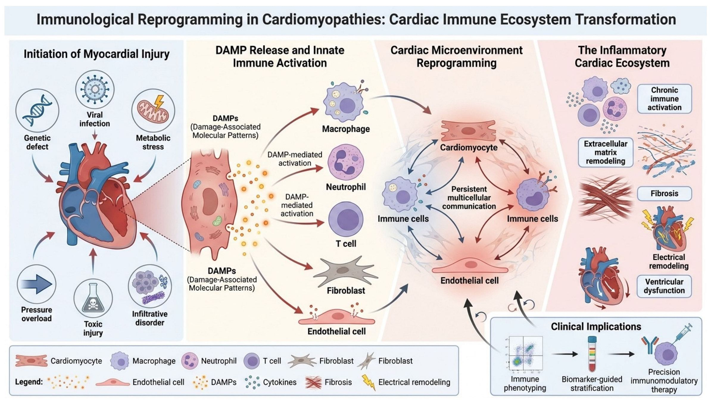
Graphical Abstract

1. Introduction

Cardiomyopathies represent a heterogeneous group of myocardial disorders characterized by structural and functional abnormalities that frequently progress to heart failure, arrhythmias, and premature death [1,2]. Traditionally, their pathogenesis has been interpreted through a cardiomyocyte-centered paradigm, in which genetic mutations, viral injury, toxic exposure, or metabolic stress directly damage contractile cells and initiate myocardial remodeling [3,4]. This framework has been highly successful in identifying causal genes, defining disease phenotypes, and guiding contemporary therapies [5]. Nevertheless, it incompletely explains several fundamental clinical observations, including the remarkable heterogeneity of disease penetrance among carriers of identical pathogenic variants, the persistence of myocardial fibrosis despite optimal guideline-directed therapy, and the variable progression of heart failure in patients with apparently similar structural phenotypes [6,7].
Over the last decade, advances in single-cell transcriptomics, spatial transcriptomics, proteomics, and systems immunology have fundamentally changed our understanding of cardiac biology [8,9]. These technologies have revealed that the failing heart is not simply a collection of injured cardiomyocytes but a complex multicellular ecosystem composed of immune cells, fibroblasts, endothelial cells, stromal populations, and hematopoietic-derived cells that continuously interact through dynamic signaling networks [10,11]. Within this framework, disease progression is increasingly recognized as the consequence not only of the initiating injury but also of how the immune system interprets and responds to it.
A growing body of evidence indicates that innate immune activation is a central driver of cardiac remodeling across diverse cardiomyopathies [12,13]. Damage-associated molecular patterns (DAMPs) released from stressed or injured cardiomyocytes activate pattern-recognition pathways, including Toll-like receptors, the NLRP3 inflammasome, and the cGAS–STING axis [14,15,16]. These pathways promote recruitment and reprogramming of monocyte–macrophage populations, activation of fibroblasts, production of pro-inflammatory cytokines, and ultimately the development of fibrosis and electrical remodeling. Importantly, these mechanisms appear to converge across seemingly distinct etiologies, including dilated cardiomyopathy, arrhythmogenic cardiomyopathy, laminopathies, myocarditis, and metabolic heart disease [17,18,19].
Concurrently, emerging concepts such as immunometabolism, trained immunity, and clonal hematopoiesis of indeterminate potential (CHIP) have expanded the scope of cardiovascular immunology beyond the heart itself [20,21,22]. Immune cells can undergo long-lasting functional reprogramming via metabolic and epigenetic mechanisms, creating persistent pro-inflammatory states that continue to influence disease progression even after the original insult has resolved. Furthermore, bidirectional communication between the heart and the hematopoietic system has established the concept of a cardio–bone marrow axis, suggesting that chronic heart failure should be viewed as a systemic immunohematologic disorder rather than an isolated cardiac disease [23].
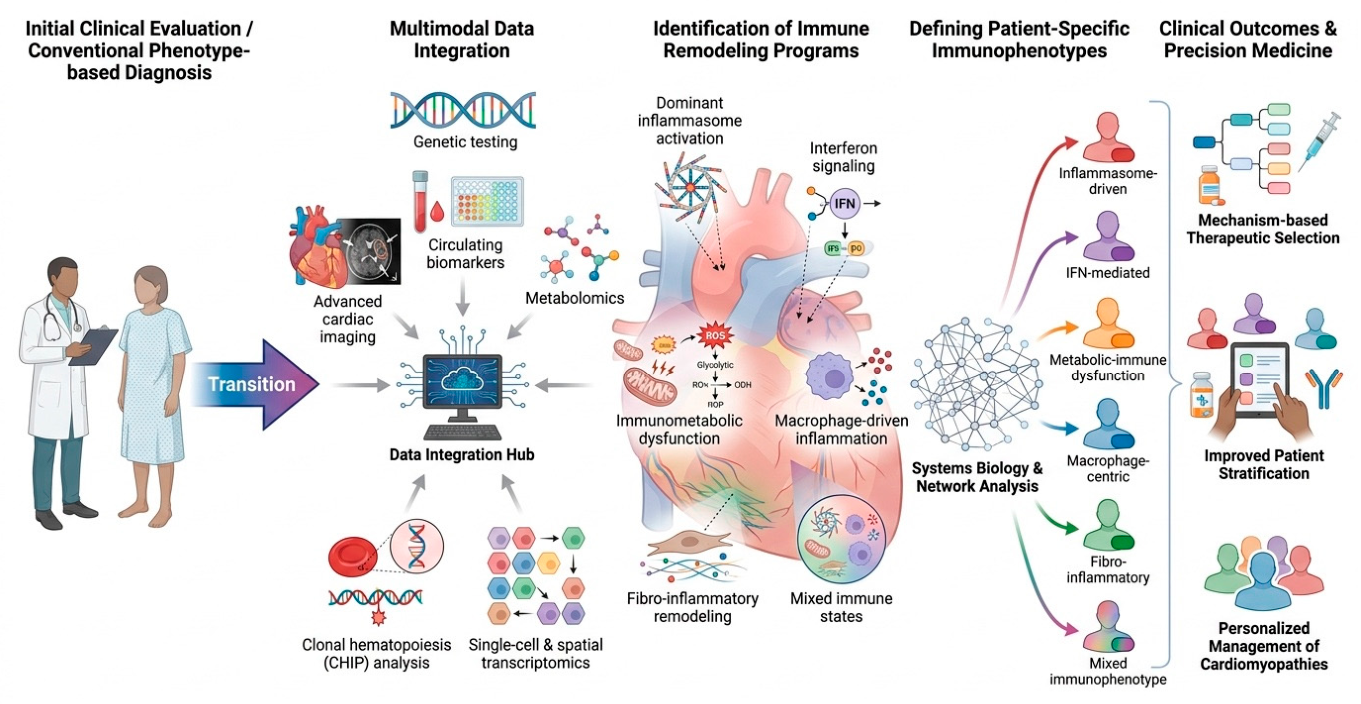
These discoveries challenge conventional classifications based solely on phenotype or genotype and support the emergence of a new conceptual framework centered on immunological remodeling. In this model, the disease trajectory is determined not only by the nature of myocardial injury but also by the dominant immune programs activated within the cardiac microenvironment. Such programs may define distinct “immunotypes” characterized by inflammasome activation, interferon signaling, macrophage-driven inflammation, fibroblast activation, or immunometabolic dysfunction. Recognition of these immunotypes may ultimately enable precision immunomodulatory therapies tailored to the biological mechanisms driving remodeling in individual patients.
In this review, we discuss the evolving concept of immunological reprogramming in cardiomyopathies and heart failure. We examine the cellular and molecular pathways linking cardiomyocyte injury to chronic immune activation, including DAMP signaling, inflammasome activation, trained immunity, immunometabolism, the cGAS–STING pathway, and cardio–bone marrow interactions. We further explore how recent advances in single-cell and spatial technologies are redefining our understanding of myocardial remodeling and discuss the potential transition from traditional phenotyping toward immunophenotyping as a foundation for future precision cardiovascular medicine.
Central hypothesis of this review:
We propose that the biological heterogeneity of cardiomyopathies is determined not only by the initiating myocardial injury but also by the manner in which the cardiac immune ecosystem interprets and sustains that injury. Although cardiomyocyte damage initiates disease, persistent immune reprogramming progressively becomes an independent determinant of remodeling, fibrosis, arrhythmogenesis, and heart failure progression. Consequently, individual differences in immune-cell composition, activation state, metabolic programming, and intercellular communication may explain why patients with apparently similar genetic or structural phenotypes often experience markedly different clinical trajectories. We therefore hypothesize that immunophenotyping may complement conventional phenotypic and genetic classification by identifying biologically distinct immune programs that support precision therapeutic strategies.
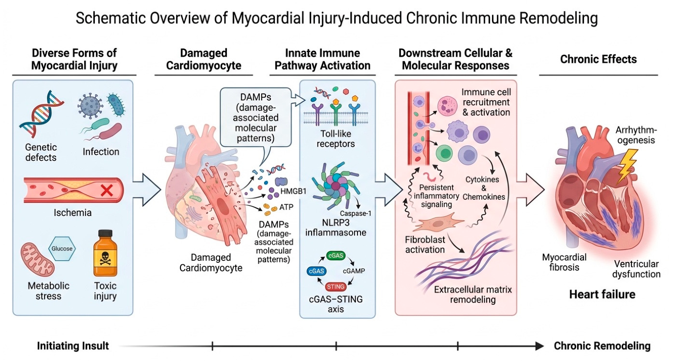
The mechanisms discussed throughout this review are traditionally studied in separate experimental frameworks, yet disease progression arises from their combined activity rather than from any single pathway. To provide a unified conceptual perspective, Figure 1 summarizes the proposed sequence of events linking diverse forms of myocardial injury to chronic immune remodeling and its clinical consequences.

Methodological Considerations

This review was developed through a comprehensive narrative analysis of the contemporary literature addressing the role of immune remodeling in cardiomyopathies and heart failure. Literature retrieval was performed primarily using the PubMed/MEDLINE database, with particular emphasis on studies published in the last decade. At the same time, seminal earlier investigations were included when essential to establishing mechanistic concepts or historical context.
The search strategy was designed to capture the evolving intersection between cardiovascular biology, immunology, and systems medicine. Search terms included, among others, cardiomyopathy, heart failure, cardiac inflammation, immune remodeling, immunological reprogramming, damage-associated molecular patterns (DAMPs), NLRP3 inflammasome, cGAS–STING, trained immunity, immunometabolism, cardiac macrophages, fibroblast–immune interactions, cardiac fibrosis, clonal hematopoiesis, cardio–bone marrow axis, single-cell transcriptomics, spatial transcriptomics, arrhythmogenesis, and immunophenotyping. These terms were queried individually and in multiple Boolean combinations to identify mechanistic, translational, and clinical studies relevant to immune-mediated cardiac remodeling.
Representative search strategies included combinations such as “cardiomyopathy AND immune remodeling”, “heart failure AND inflammation”, “NLRP3 inflammasome AND cardiac remodeling”, “cGAS–STING AND cardiomyopathy”, “trained immunity AND heart failure”, “immunometabolism AND cardiac inflammation”, “cardiac macrophages AND fibrosis”, “clonal hematopoiesis AND heart failure”, and “spatial transcriptomics AND cardiac remodeling”.
Priority was given to original experimental investigations, translational studies, landmark clinical observations, and high-impact reviews that contributed substantially to the current understanding of immune mechanisms in cardiovascular disease. Additional relevant publications were identified through manual examination of reference lists from key articles. Rather than providing an exhaustive, systematic synthesis, this review aimed to integrate emerging evidence from traditionally distinct fields—including innate immunity, immunometabolism, epigenetic reprogramming, hematopoietic biology, and spatial systems biology—into a unified conceptual framework for immunological remodeling in cardiomyopathies.

2. The Heart as an Immune Organ: Homeostasis and the Origins of Immune Remodeling

For many years, the immune system was considered relevant to cardiac pathology primarily in the context of infectious myocarditis, acute myocardial infarction, or systemic inflammatory disorders. This view has changed substantially with the recognition that the healthy heart contains a highly specialized immune network that contributes to tissue homeostasis under physiological conditions. Rather than functioning solely as responders to injury, resident immune cells actively participate in maintaining myocardial structure, metabolism, vascular integrity, and electrical function [24,25].
Among the most important components of this network are tissue-resident macrophages. In the healthy myocardium, resident CCR2-negative macrophages, many of which originate during embryonic development, perform a wide range of homeostatic functions [26,27]. These cells remove apoptotic debris through efferocytosis, support angiogenesis, contribute to extracellular matrix maintenance, and interact closely with cardiomyocytes and fibroblasts. Experimental studies have further demonstrated that resident macrophages participate in electrical conduction within the atrioventricular node, highlighting a previously unrecognized link between immunity and cardiac electrophysiology.
Cardiac immune homeostasis is additionally maintained by regulatory T lymphocytes (Tregs), which suppress excessive inflammatory activation and promote tissue repair [28,29]. Through the production of anti-inflammatory mediators, including interleukin-10 and transforming growth factor-β, Tregs contribute to immune tolerance within the myocardium and limit maladaptive remodeling following injury [30]. The importance of these populations is increasingly supported by experimental models demonstrating accelerated fibrosis and ventricular dysfunction following their depletion.
Equally important is the recognition that fibroblasts are not merely structural cells responsible for collagen production [31,32]. Contemporary single-cell studies have revealed that cardiac fibroblasts possess substantial immunological capabilities [33]. They express pattern-recognition receptors, including Toll-like receptors, that respond directly to cytokines and danger signals and actively shape leukocyte recruitment by secreting chemokines such as CCL2 and inflammatory mediators such as IL-6 [34,35]. Consequently, fibroblasts occupy a central position at the interface between tissue injury and immune activation.
Profound alterations in the cardiac immune landscape often accompany the transition from physiological homeostasis to pathological remodeling. One of the earliest detectable changes is the replacement of resident macrophage populations by inflammatory CCR2-positive monocyte-derived macrophages recruited from the circulation. This shift fundamentally alters the myocardium’s biological behavior. Whereas resident macrophages promote repair and tissue maintenance, infiltrating populations are characterized by increased production of pro-inflammatory cytokines, fibroblast activation, and amplification of extracellular matrix remodeling.
Importantly, these alterations frequently precede overt structural changes detectable by conventional imaging techniques. Experimental and translational studies suggest that immune reprogramming may occur long before the development of extensive fibrosis or measurable declines in ventricular function. Consequently, loss of immune homeostasis should be regarded not merely as a consequence of myocardial injury but as an early and potentially decisive event in the progression of cardiomyopathy.
These observations support a broader conceptual shift in cardiovascular biology. Rather than viewing the heart as an organ that becomes inflamed only after injury, it may be more appropriate to consider it a highly regulated immunological ecosystem in which disease emerges when physiological repair programs become chronically dysregulated. Understanding the mechanisms governing this transition is therefore essential for developing future therapeutic strategies to prevent maladaptive remodeling before irreversible structural damage occurs.
The cellular landscape of the myocardium extends far beyond cardiomyocytes, encompassing multiple immune and stromal populations that continuously interact under physiological and pathological conditions. Because these cells differ substantially in origin, function, and contribution to remodeling, a structured overview of their respective roles within the cardiac immune ecosystem is presented in Table 1.
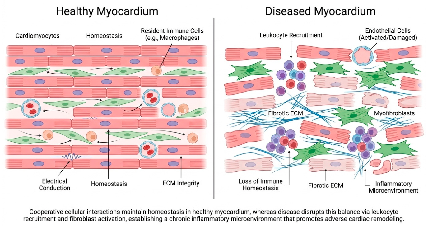
Understanding cardiomyopathy requires consideration not only of individual cell populations but also of the relationships among them. The transition from physiological immune surveillance to pathological remodeling involves coordinated changes across multiple cellular compartments. Figure 2 illustrates this shift within the myocardial ecosystem, emphasizing that alterations in cellular balance may precede overt structural disease.
Although this review primarily focuses on innate immune mechanisms, adaptive immunity also contributes to remodeling in selected cardiomyopathies. T-cell subsets, B-cell activation, and autoantibody-mediated responses have been implicated particularly in inflammatory cardiomyopathies and post-myocarditis remodeling. Interactions between adaptive and innate immune pathways likely influence disease chronicity, although their relative importance varies among cardiomyopathy phenotypes.

3. Damage-Associated Molecular Patterns: Translating Cardiomyocyte Injury into Innate Immune Activation

A central question in contemporary cardiovascular immunology is how the immune system detects myocardial injury in the absence of infection. Most cardiomyopathies develop without an identifiable pathogen, yet they exhibit many hallmarks of innate immune activation, including cytokine production, monocyte recruitment, inflammasome signaling, and progressive fibrosis [60]. The answer lies in the ability of injured cells to release endogenous danger signals collectively known as damage-associated molecular patterns (DAMPs) [61].
DAMPs represent intracellular molecules that are normally concealed from immune surveillance but become exposed or released following cellular stress, necrosis, mechanical injury, or mitochondrial dysfunction [62]. In the myocardium, these molecules include extracellular ATP, high-mobility group box 1 protein (HMGB1), mitochondrial DNA, nuclear DNA fragments, heat-shock proteins, extracellular RNA, and extracellular matrix degradation products. Although structurally diverse, these signals share a common function: they inform the immune system that tissue integrity has been compromised [63,64].
Among the various DAMPs identified in cardiac disease, mitochondrial components appear particularly important. Because mitochondria evolved from ancestral bacteria, mitochondrial DNA retains molecular characteristics resembling those of microbial nucleic acids [65]. When released into the extracellular space or cytoplasm, mitochondrial DNA is recognized by innate immune receptors as a potent danger signal. Similarly, extracellular ATP serves as an indicator of membrane disruption and cellular stress, whereas HMGB1 is released during necrosis and is a molecular marker of tissue damage.
An interconnected network of pattern-recognition receptors mediates recognition of DAMPs. Toll-like receptors, particularly TLR2, TLR4, and TLR9, play a prominent role in sensing extracellular and endosomal danger signals [66,67]. Cytoplasmic DNA activates the cGAS–STING pathway, while multiple forms of cellular stress converge on the NLRP3 inflammasome [68]. Together, these systems translate tissue injury into inflammatory signaling, leading to activation of nuclear factor-κB (NF-κB), production of cytokines, recruitment of monocytes, and stimulation of fibroblast activity.
An important consequence of this framework is that diverse etiologies may activate remarkably similar downstream pathways. Genetic cardiomyopathies caused by variants in TTN, LMNA, DSP, or FLNC, viral myocarditis, anthracycline-induced cardiotoxicity, ischemic injury, and metabolic stress all generate cellular damage capable of releasing DAMPs [60,69]. Consequently, the immune system responds not to the specific cause of injury but to its molecular consequences. This concept helps explain why distinct forms of cardiomyopathy frequently converge toward common pathological features, including chronic inflammation, fibrosis, and ventricular dysfunction.
Under physiological circumstances, DAMP-mediated signaling is transient and contributes to effective tissue repair. However, persistent or repetitive release of danger signals fundamentally alters this response. Continuous activation of innate immune pathways promotes sustained recruitment of inflammatory cells, prolonged fibroblast activation, and progressive extracellular matrix remodeling. Over time, the repair process becomes self-sustaining and partially independent of the initiating insult. This phenomenon may explain why many cardiomyopathies continue to progress even after viral clearance, stabilization of hemodynamic stress, or control of the original disease trigger.

4. NLRP3 Inflammasome: A Central Hub Linking Cellular Stress to Cardiac Remodeling

Among the innate immune pathways implicated in cardiovascular disease, the NLRP3 inflammasome has emerged as one of the most extensively studied molecular platforms connecting tissue injury with chronic inflammation and adverse remodeling. Although initially characterized as a key component of host defense against pathogens, increasing evidence suggests that NLRP3 also functions as a sensor of sterile tissue injury, making it particularly relevant to the pathogenesis of cardiomyopathies and heart failure.
The NLRP3 inflammasome is a multiprotein signaling complex composed of the sensor molecule NLRP3 (NOD-like receptor family pyrin domain-containing 3), the adaptor protein ASC (apoptosis-associated speck-like protein containing a CARD), and pro-caspase-1. Its activation is typically described as a two-step process [70,71]. In the diseased myocardium, multiple upstream triggers capable of activating NLRP3 coexist simultaneously. Mechanical stress, mitochondrial injury, ischemia, metabolic overload, and genetic defects that compromise cardiomyocyte integrity collectively create a cellular environment conducive to inflammasome activation. Importantly, NLRP3 signaling is not restricted to infiltrating immune cells. Cardiomyocytes, fibroblasts, endothelial cells, and resident macrophages have all been shown to participate in inflammasome-mediated signaling, underscoring the multicellular nature of myocardial inflammation [72,73]. One of the most significant consequences of inflammasome activation is the induction of pyroptosis, a highly inflammatory form of programmed cell death [74]. Unlike apoptotic cells, which generally do not elicit substantial immune activation, pyroptotic cells release intracellular contents, including cytokines and additional danger-associated molecular patterns, into the extracellular space. As a result, inflammasome activation not only responds to tissue injury but also amplifies it through self-reinforcing feed-forward mechanisms. This process may contribute to the transition from transient injury to persistent inflammation and progressive fibrosis observed in many forms of cardiomyopathy.
The translational relevance of these findings has been reinforced by clinical studies targeting downstream components of the inflammasome pathway. The CANTOS trial [75] provided the first large-scale evidence that selective inhibition of IL-1β can improve cardiovascular outcomes independently of lipid lowering, highlighting the pathogenic importance of inflammatory signaling in cardiovascular disease. Similarly, smaller studies using anakinra, an interleukin-1 receptor antagonist, have demonstrated favorable effects on inflammatory biomarkers and selected clinical endpoints in patients with heart failure and myocarditis [76,77]. Although these interventions do not directly inhibit NLRP3 itself, they provide indirect support for the concept that inflammasome-driven inflammation contributes to disease progression.
Despite considerable enthusiasm, several important questions remain unresolved. NLRP3 activation is a common downstream response to diverse forms of cardiac injury. Yet, the extent to which it functions as a primary driver versus a secondary amplifier of disease likely varies across cardiomyopathy subtypes. Moreover, chronic inhibition of innate immune pathways may carry unintended consequences, including impaired host defense and altered tissue repair. Consequently, future therapeutic strategies will likely require precise identification of patients in whom inflammasome signaling represents a dominant pathogenic mechanism.
Taken together, current evidence positions the NLRP3 inflammasome as a critical interface between cellular stress and maladaptive remodeling. Rather than acting as an isolated inflammatory pathway, NLRP3 integrates metabolic, mechanical, and immunological signals into a coordinated response that influences fibrosis, ventricular dysfunction, and arrhythmogenesis. Its central location within the network of cardiac immune remodeling makes it one of the most attractive yet also most challenging targets for future precision immunomodulatory therapies.

5. Immunometabolism: Metabolic Control of Cardiac Immune Responses

Rather than being passive consequences of activation, metabolic pathways actively determine immune cell phenotype and function. Over the past decade, immunometabolism has emerged as a fundamental regulator of innate and adaptive immune responses, providing a mechanistic link between cellular energetics and inflammation [78,79]. This concept is particularly relevant in cardiomyopathies and heart failure, where metabolic stress and immune activation frequently coexist.
Under physiological conditions, tissue-resident macrophages rely predominantly on oxidative phosphorylation and fatty-acid oxidation to support homeostatic functions, including efferocytosis, angiogenesis, and maintenance of tissue integrity. In contrast, inflammatory activation induces a profound metabolic shift toward aerobic glycolysis, a phenomenon analogous to the Warburg effect observed in cancer cells [80,81]. Although energetically less efficient, glycolysis enables rapid ATP generation and provides biosynthetic intermediates required for cytokine production and cellular proliferation.
Several interconnected signaling pathways, including mTOR, AMPK, HIF-1α, and NF-κB, orchestrate this metabolic reprogramming. Among these, HIF-1α has emerged as a particularly important regulator of inflammatory macrophage function [82]. Stabilization of HIF-1α promotes transcription of pro-inflammatory mediators, including IL-1β, while simultaneously reinforcing glycolytic metabolism. Consequently, metabolic and inflammatory signaling become tightly coupled processes that sustain one another through positive feedback mechanisms [83,84]. Elevated intracellular succinate stabilizes HIF-1α and enhances IL-1β production, effectively transforming a metabolic disturbance into an inflammatory signal. Such observations have fundamentally altered traditional views of metabolism, demonstrating that metabolites can function as signaling molecules with direct immunological consequences.
The clinical relevance of immunometabolism is supported by therapies originally developed for metabolic disease. Sodium–glucose cotransporter-2 inhibitors (SGLT2i) and glucagon-like peptide-1 receptor agonists (GLP-1RA) reduce cardiovascular events and heart-failure outcomes through mechanisms that extend beyond glycemic control, including effects on inflammation, oxidative stress, endothelial function, mitochondrial signaling, and immune-cell activation. These observations suggest that modulation of cellular metabolism may be an indirect yet clinically effective strategy to attenuate pathological immune activation [85,86,87]. These observations suggest that targeting cellular metabolism is an indirect but highly effective strategy for controlling pathological immune activation.
Collectively, accumulating evidence indicates that cellular metabolism is not simply a downstream consequence of immune activation but a fundamental determinant of immune-cell identity, functional polarization, and inflammatory behavior. Consequently, metabolic reprogramming is now recognized as a central mechanism regulating both innate and adaptive immune responses [88,89,90].

Master Transcriptional Programs Orchestrating Immune Remodeling

Although individual inflammatory pathways are often discussed in isolation, they operate under the control of a relatively small set of transcriptional programs that integrate environmental stress into coordinated cellular responses [91]. Rather than functioning as isolated signaling cascades, innate immune activation, metabolic adaptation, oxidative stress, and tissue repair converge on master transcription factors that determine the magnitude, duration, and biological consequences of myocardial remodeling [92]. These regulatory networks translate diverse forms of cardiac injury into shared molecular programs, providing a plausible explanation for the remarkable convergence of genetically, metabolically, ischemically, and inflammation-distinct cardiomyopathies toward similar pathological phenotypes [93].
Among these regulators, hypoxia-inducible factor-1α (HIF-1α) occupies a central position. Although initially characterized as a sensor of oxygen deprivation, HIF-1α is now recognized as a broader integrator of cellular stress [94]. Persistent myocardial hypoxia resulting from microvascular dysfunction, increased wall stress, mitochondrial impairment, or extracellular matrix expansion promotes HIF-1α stabilization even in the absence of severe ischemia [95]. This phenomenon appears relevant across multiple cardiomyopathy phenotypes, including hypertrophic cardiomyopathy, diabetic cardiomyopathy, storage disorders, infiltrative diseases such as transthyretin amyloidosis, and cardiomyopathies associated with coronary microvascular dysfunction. Sustained HIF-1α activation promotes glycolytic reprogramming, mitochondrial dysfunction, angiogenic signaling, fibroblast activation, and inflammatory cytokine production, thereby linking metabolic adaptation with progressive structural remodeling [96].
An equally important regulator is nuclear factor-κB (NF-κB), which serves as a central coordinator of inflammatory gene expression [97]. Activation of Toll-like receptors, DAMP signaling, reactive oxygen species, inflammatory cytokines, and mechanical stress converges on NF-κB, resulting in transcription of numerous mediators involved in innate immunity, leukocyte recruitment, endothelial activation, and extracellular matrix remodeling [98]. Because NF-κB integrates signals originating from multiple upstream pathways—including NLRP3 inflammasome activation, cGAS–STING signaling, and oxidative stress—it represents one of the principal molecular hubs sustaining chronic myocardial inflammation [99]. Persistent activation of NF-κB further amplifies oxidative stress and inflammatory signaling, establishing self-reinforcing feed-forward circuits that promote fibrosis, electrical remodeling, and ventricular dysfunction [100].
Signal transducer and activator of transcription 3 (STAT3) illustrates the complexity of transcriptional regulation within the injured myocardium [101]. Unlike purely pro-inflammatory regulators, STAT3 exerts context-dependent effects that encompass cardiomyocyte survival, mitochondrial homeostasis, immune modulation, and tissue repair [102]. Transient STAT3 activation may support adaptive responses following acute injury, whereas chronic dysregulation has been associated with persistent inflammatory signaling, fibroblast activation, and maladaptive remodeling [103]. These observations emphasize that therapeutic intervention should not indiscriminately suppress inflammatory pathways but rather restore the physiological balance between protective and pathological transcriptional programs.
Additional regulatory systems further expand this network. Activator protein-1 (AP-1) contributes to inflammatory gene expression downstream of mitogen-activated protein kinase signaling. In contrast, SMAD transcription factors mediate transforming growth factor-β–dependent profibrotic responses that ultimately determine extracellular matrix accumulation and scar formation [104]. Rather than acting independently, HIF-1α, NF-κB, STAT3, AP-1, and SMAD signaling continuously interact through extensive transcriptional cross-talk, allowing metabolic stress, oxidative injury, immune activation, and mechanical loading to converge on a coordinated remodeling response [91]. Consequently, alterations in one regulatory pathway frequently influence multiple downstream biological processes simultaneously. Activation of these transcription factors ultimately regulates extensive networks of downstream effector genes controlling cytokine production, extracellular matrix turnover, mitochondrial adaptation, angiogenesis, and cellular survival.
Recognition of these master transcriptional programs has important therapeutic implications. Current guideline-directed therapies already modulate several of these pathways indirectly. Sodium-glucose cotransporter-2 inhibitors attenuate inflammatory activation by favorably affecting cellular metabolism, oxidative stress, and inflammasome activity. At the same time, glucagon-like peptide-1 receptor agonists influence immune-cell metabolism, endothelial function, and cytokine production [105]. Experimental strategies targeting HIF-1α, NF-κB, STAT3, or their downstream effector networks are also being actively investigated as potential approaches to interrupt maladaptive remodeling before irreversible fibrosis becomes established [106]. Although clinical translation remains challenging because these pathways regulate numerous physiological processes, targeting upstream transcriptional regulators offers the theoretical advantage of simultaneously modifying multiple interconnected mechanisms rather than inhibiting individual inflammatory mediators.
These observations suggest that transcriptional regulation represents a fundamental organizational level of cardiac immune remodeling. Master transcription factors do not merely control isolated signaling pathways but orchestrate the integrated biological response to myocardial injury. Understanding how these regulatory networks interact is therefore essential for the development of future precision immunomodulatory strategies that limit disease progression while preserving the physiological mechanisms of tissue repair.

6. Trained Immunity: Persistent Inflammatory Memory Beyond the Initial Injury

One of the most intriguing observations in cardiovascular medicine is the persistence of inflammatory activity long after the initiating insult has resolved. Patients recovering from viral myocarditis may continue to develop progressive ventricular dysfunction despite clearance of the infectious virus, reflecting the persistence of immune-mediated myocardial inflammation, adverse remodeling, and autoimmune mechanisms [107,108]. Similarly, Individuals carrying pathogenic variants associated with inherited cardiomyopathies frequently undergo prolonged periods of apparent clinical stability before experiencing age-dependent and sometimes abrupt disease progression characterized by ventricular dysfunction, arrhythmias, or heart failure [5,109]. These observations challenge the traditional view that immune activation is merely a transient response to tissue injury and suggest the existence of mechanisms capable of sustaining inflammation independently of the original trigger.
The concept of trained immunity has emerged as a potential explanation for this phenomenon [110,111]. Unlike adaptive immune memory, which relies on antigen-specific responses mediated by T and B lymphocytes, trained immunity describes a long-lasting functional reprogramming of innate immune cells and their progenitors. Following exposure to inflammatory stimuli, monocytes, macrophages, and hematopoietic stem cells undergo metabolic and epigenetic changes that enhance their responsiveness to subsequent challenges. As a result, future inflammatory responses become quantitatively and qualitatively different, even when triggered by unrelated stimuli [112,113]. Exposure to inflammatory mediators induces persistent modifications in chromatin accessibility, including histone acetylation and methylation events that facilitate transcription of pro-inflammatory genes. Among the best-characterized changes are increased H3K4 trimethylation and H3K27 acetylation at promoters and enhancers controlling cytokine production [114]. These alterations create a permissive transcriptional landscape that allows rapid reactivation of inflammatory programs upon subsequent stimulation.
Although direct evidence in human cardiomyopathies remains limited, several observations support the relevance of trained immunity in heart failure [115,116]. Patients with chronic heart failure frequently exhibit persistent elevations of inflammatory mediators despite the absence of active infection or overt tissue necrosis [117]. Moreover, circulating inflammatory biomarkers often remain elevated despite stabilization of hemodynamic status and optimization of guideline-directed medical therapy, suggesting that immune activation may become partially self-sustaining rather than simply reflecting ongoing hemodynamic stress [118,119]. Experimental studies further demonstrate that prior inflammatory stimulation induces innate immune memory, resulting in exaggerated inflammatory responses and accelerated adverse cardiac remodeling following subsequent cardiovascular insults [120,121]. The concepts of trained immunity provide a conceptual framework linking transient myocardial injury to sustained immune activation. It offers a mechanistic explanation for why inflammation frequently persists after the resolution of the initiating insult and suggests that the progression of cardiomyopathy may be determined not only by the extent of cardiac damage but also by the immune system’s capacity to remember and perpetuate that damage.

Epigenetic Remodeling: From Adaptive Plasticity to Irreversible Cardiac Remodeling

One of the most fundamental yet incompletely understood questions in cardiomyopathy concerns the transition from reversible myocardial injury to irreversible structural disease. Clinical observations consistently demonstrate that some patients recover ventricular function following removal of the initiating insult. In contrast, others continue to progress toward diffuse fibrosis, electrical instability, and end-stage heart failure despite elimination of the original trigger and optimal medical therapy [122]. These divergent trajectories suggest that disease progression cannot be explained solely by the magnitude of myocardial injury but instead reflects the establishment of stable biological programs that progressively lose their capacity for physiological adaptation [123].
Epigenetic remodeling has emerged as a plausible mechanism underlying this transition. Unlike genetic mutations, epigenetic modifications alter gene expression without changing the underlying DNA sequence, allowing cells to acquire persistent functional phenotypes in response to environmental stimuli. DNA methylation, histone modifications, chromatin remodeling, and non-coding RNAs collectively determine transcriptional accessibility, thereby shaping cellular identity over prolonged periods [124]. Initially, these mechanisms facilitate adaptive responses to mechanical stress, oxidative injury, inflammation, and metabolic disturbance [125]. However, persistent activation of these regulatory programs may ultimately stabilize pathological cellular states that continue to drive remodeling independently of the initiating stimulus [126].
Although the concept of trained immunity illustrates this phenomenon in innate immune cells, similar principles appear to govern multiple cellular populations within the failing myocardium. Cardiomyocytes exposed to sustained inflammatory and metabolic stress undergo widespread transcriptional reprogramming characterized by altered calcium-handling proteins, mitochondrial dysfunction, impaired energy metabolism, and activation of fetal gene programs [127]. Increasing evidence further suggests that mitochondrial epigenetic regulation contributes to persistent metabolic dysfunction, limiting the ability of injured cardiomyocytes to restore normal bioenergetic homeostasis even after hemodynamic conditions improve [128].
Fibroblasts provide the most compelling example of persistent epigenetic reprogramming during cardiac remodeling [129]. Under physiological conditions, fibroblast activation is tightly regulated and resolves following completion of tissue repair. Chronic inflammatory signaling, however, promotes stable epigenetic changes that maintain myofibroblasts in a matrix-producing, contractile, and highly secretory phenotype [130]. Once established, these cells continue to synthesize extracellular matrix proteins, profibrotic cytokines, and matricellular mediators despite progressive separation from the initiating injury. Such epigenetic stabilization may explain why myocardial fibrosis frequently progresses even after effective treatment of the underlying disease.
Endothelial cells likewise develop persistent phenotypic alterations under chronic inflammatory conditions [131]. Sustained exposure to oxidative stress and inflammatory cytokines promotes endothelial dysfunction characterized by impaired nitric oxide bioavailability, increased leukocyte adhesion, and persistent activation of inflammatory signaling pathways [132]. This phenomenon, often referred to as endothelial memory, may contribute to persistent microvascular dysfunction and recruitment of inflammatory cells despite partial restoration of systemic cardiovascular homeostasis.
Importantly, these epigenetic programs do not develop independently within individual cell populations. Rather, reciprocal communication among cardiomyocytes, fibroblasts, endothelial cells, and immune cells continuously reinforces pathological transcriptional states through cytokines, growth factors, extracellular vesicles, metabolic intermediates, and matrix-derived signaling molecules [133]. As a consequence, the myocardium progressively transitions from a biologically plastic tissue capable of adaptive repair to a self-sustaining pathological ecosystem in which multiple cell populations become epigenetically synchronized in maintaining chronic inflammation and fibrosis [134,135].
Recognition of these mechanisms has important therapeutic implications. Conventional heart failure therapies primarily attenuate neurohormonal activation and hemodynamic stress but may only partially influence the epigenetic programs that perpetuate disease progression. Future therapeutic strategies may therefore require interventions that restore cellular plasticity rather than suppress individual inflammatory mediators [136]. Advances in epigenetic pharmacology, chromatin-targeted therapies, RNA-based therapeutics, and precision immunomodulation raise the possibility that pathological transcriptional programs may eventually become reversible before extensive structural remodeling has become permanently established [137].
The epigenetic remodeling may represent a critical biological interface between transient myocardial injury and irreversible cardiomyopathy. Rather than serving as passive consequences of chronic inflammation, epigenetic mechanisms progressively stabilize pathological cellular phenotypes throughout the cardiac immune ecosystem, ultimately determining whether myocardial injury resolves through adaptive repair or progresses to permanent fibrosis, electrical instability, and end-stage heart failure.

7. The cGAS–STING Axis: Interferon Signaling as a Driver of Chronic Cardiac Remodeling

While inflammasome activation primarily links tissue injury to interleukin-mediated inflammation, an additional innate immune pathway has emerged as a critical regulator of chronic sterile inflammation in the heart. The cyclic GMP–AMP synthase (cGAS)–stimulator of interferon genes (STING) pathway functions as a cytosolic DNA-sensing system that evolved to detect viral and bacterial nucleic acids [138,139]. However, increasing evidence indicates that this pathway also plays a central role in non-infectious cardiovascular disease by recognizing endogenous DNA released during cellular stress and injury.
Under physiological conditions, DNA is compartmentalized within the nucleus and mitochondria. The appearance of DNA within the cytoplasm is therefore interpreted by the cell as a danger signal. Cytoplasmic DNA is detected by cGAS, which catalyzes the production of cyclic GMP–AMP (cGAMP), a second messenger that activates STING localized within the endoplasmic reticulum membrane [140]. Activated STING recruits and activates TBK1, leading to phosphorylation of IRF3 and subsequent transcription of type I interferons and interferon-stimulated genes. In parallel, STING activates the NF-κB signaling pathway, thereby amplifying the expression of pro-inflammatory cytokines, chemokines, and other inflammatory mediators [141].
Among inherited cardiomyopathies, laminopathies provide the most compelling example of this pathway’s pathological relevance. The LMNA gene encodes nuclear lamins that maintain the structural integrity of the nuclear envelope. Pathogenic variants disrupt nuclear architecture, resulting in increased susceptibility to mechanical stress and the formation of transient nuclear envelope ruptures. Experimental studies have demonstrated that LMNA deficiency promotes nuclear envelope rupture and cytoplasmic DNA accumulation, which can activate cGAS–STING signaling. In several experimental models, this response has been associated with type I interferon signaling, inflammatory cell recruitment, myocardial fibrosis, and ventricular dysfunction. However, the contribution of cGAS–STING signaling to disease progression appears to be model-dependent and remains an area of active investigation [142,143].
The cGAS–STING pathway serves as a critical molecular bridge between cellular damage and chronic immune activation. Its ability to translate structural abnormalities, mitochondrial dysfunction, and tissue stress into sustained interferon signaling positions it as a central component of the emerging concept of immunological remodeling. Furthermore, the pathway provides a compelling example of how distinct cardiomyopathy phenotypes may be driven by specific immune programs, supporting the broader notion that future classification systems may need to incorporate immunological mechanisms alongside conventional phenotypic and genetic descriptors.
Future myocardial tissue characterization will likely integrate conventional histopathology, immunohistochemistry, ultrastructural electron microscopy, laser-capture microdissection, single-cell and spatial transcriptomics, proteomics, and multi-omic profiling to generate biologically informed disease classifications and guide individualized immunomodulatory therapies. Rather than replacing existing diagnostic approaches, these complementary technologies are expected to provide progressively higher-resolution characterization of myocardial biology, enabling identification of disease-defining cellular interactions, dominant immune programs, and patient-specific therapeutic targets. Such integrated tissue profiling may ultimately transform endomyocardial biopsy from a predominantly diagnostic procedure into a comprehensive platform for biological classification, prognostic stratification, and precision therapeutic decision-making.

8. The Cardio–Bone Marrow Axis and Clonal Hematopoiesis: Expanding the Boundaries of Cardiac Remodeling

For decades, research in heart failure and cardiomyopathy focused almost exclusively on the myocardium. Cardiomyocytes, fibroblasts, extracellular matrix remodeling, and neurohormonal activation were viewed as the principal determinants of disease progression.
Recent studies suggest that chronic cardiac remodeling cannot be understood solely as a local myocardial process. Instead, the failing heart appears to engage in continuous communication with the hematopoietic system, particularly the bone marrow, creating a bidirectional network that sustains inflammation long after the initiating injury [23]. In this model, the heart is not merely the recipient of immune responses but an active participant in shaping them.
Experimental studies have demonstrated that injured myocardium releases a diverse repertoire of cytokines, chemokines, damage-associated molecular patterns (DAMPs), growth factors, and neurohumoral mediators that influence hematopoietic stem and progenitor cell activity, promoting emergency myelopoiesis and sustained inflammatory responses within the bone marrow [144,145]. Cytokines, chemokines, damage-associated molecular patterns (DAMPs), and neurohumoral mediators released from the injured myocardium reach the bone marrow, where they alter the behavior of hematopoietic stem and progenitor cells. This signaling promotes emergency myelopoiesis, expands inflammatory myeloid progenitors, and increases the production and mobilization of pro-inflammatory monocytes that subsequently infiltrate the myocardium, thereby reinforcing chronic immune activation and adverse cardiac remodeling [146,147]. These cells subsequently traffic back to the myocardium, where they contribute to cytokine production, fibroblast activation, extracellular matrix remodeling, and progressive ventricular dysfunction. Once established, this circuit can maintain pathological remodeling even after the original trigger has diminished or disappeared.
Clinical observations support this interpretation. Imaging studies have identified increased metabolic activity within the bone marrow of individuals with advanced cardiovascular disease [148,149]. It may represent a previously underappreciated component of the disease process itself.
The importance of hematopoietic biology became even more apparent with the recognition of clonal hematopoiesis of indeterminate potential (CHIP) [150,151]. Initially regarded as an age-related hematologic phenomenon with uncertain clinical significance, CHIP has emerged as one of the most compelling links between immune biology and cardiovascular disease. Large population studies consistently demonstrated that individuals carrying CHIP-associated mutations experience substantially higher rates of cardiovascular events, heart failure, and cardiovascular mortality, independent of traditional risk factors [152,153].
The implications extend far beyond cardiovascular risk prediction. CHIP provides direct evidence that the biological characteristics of the hematopoietic system can influence the progression of cardiac disease. Two patients with comparable myocardial injury may experience markedly different outcomes because their immune systems respond differently to the same stimulus. From this perspective, part of the heterogeneity traditionally attributed to chance, environmental exposure, or incomplete penetrance may instead reflect differences in immune programming.
This concept aligns naturally with emerging ideas surrounding trained immunity. Both frameworks suggest that cardiovascular disease progression is shaped not only by the magnitude of injury but also by the state of the immune system responding to that injury. The myocardium and the hematopoietic compartment, therefore, become components of a single adaptive network rather than independent biological entities.
Although the pathways discussed in the preceding sections are often presented individually, they operate as components of an interconnected network linking cellular injury to chronic remodeling. Table 2 provides an integrated overview of the principal immune mechanisms implicated in cardiomyopathies, highlighting their key triggers, biological consequences, and potential relevance to disease progression.
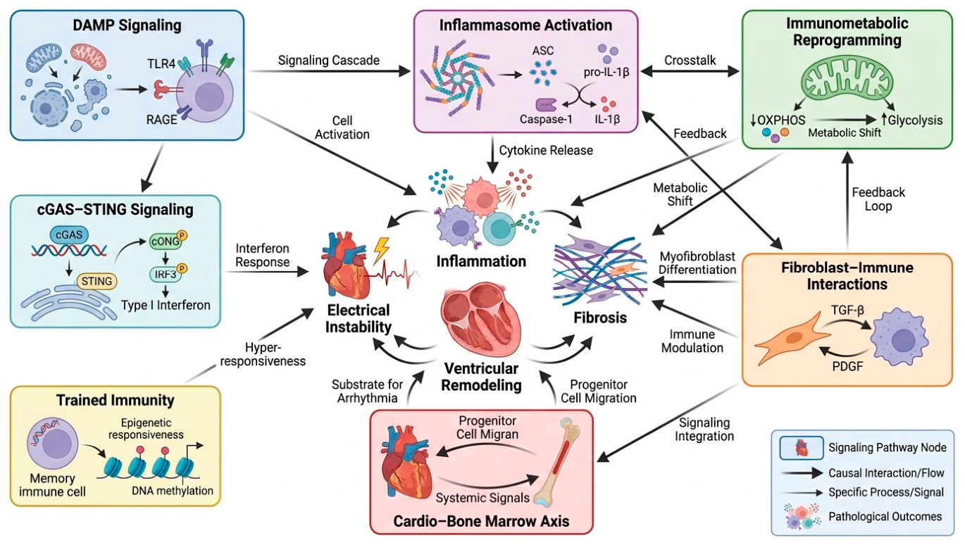
Individual mechanisms are often discussed separately for clarity, but substantial biological overlap exists between inflammatory, metabolic, and hematopoietic pathways involved in cardiac remodeling. Figure 3 highlights the interconnected nature of these processes and illustrates how apparently distinct mechanisms converge on common downstream remodeling programs.
Whether this paradigm will ultimately reshape clinical practice remains uncertain. Nevertheless, it compels a broader view of cardiomyopathy and heart failure. The biological drivers of disease may reside not only within the myocardium but also within distant tissues that continuously influence the composition and function of the immune response. Recognizing this possibility may prove essential for understanding why patients with seemingly similar cardiac phenotypes often follow remarkably different clinical courses.

9. Fibroblasts as Integrators of Immune Remodeling in Cardiomyopathy

Myocardial fibrosis is one of the most powerful determinants of clinical outcome across the spectrum of cardiomyopathies [164,165]. Regardless of etiology, the extent of fibrotic remodeling is closely associated with ventricular dysfunction, arrhythmia susceptibility, progression to heart failure, and mortality [166,167]. Yet despite its clinical importance, fibrosis has often been viewed primarily as a structural consequence of injury rather than as an active biological process. This perspective is increasingly difficult to reconcile with contemporary insights into cardiac remodeling.
Historically, fibroblasts were regarded as relatively passive cells responsible for maintaining extracellular matrix integrity and producing collagen in response to tissue damage. Recent advances in single-cell transcriptomics, spatial biology, and lineage-tracing studies have fundamentally altered this view. Fibroblasts are now recognized as highly dynamic cellular populations capable of sensing environmental signals, interacting with immune cells, and orchestrating tissue responses to injury [168,169]. In many respects, they function as integrators of information derived from mechanical stress, metabolic disturbance, and immune activation.
The relationship between fibroblasts and the immune system is a central determinant of chronic cardiac remodeling. Cardiac fibroblasts express a broad repertoire of pattern-recognition receptors, including Toll-like receptors, as well as cytokine and chemokine receptors that enable them to detect inflammatory and danger-associated signals within the injured myocardium [170]. Exposure to IL-1β, TNF-α, interferons, and transforming growth factor-β induces profound phenotypic reprogramming, promoting extracellular matrix synthesis and the secretion of chemokines, cytokines, and other inflammatory mediators that amplify local immune responses [171]. Consequently, fibroblasts should no longer be regarded as passive structural cells; rather, they function as active immunoregulatory cells that both respond to and perpetuate chronic myocardial inflammation through reciprocal communication with resident and infiltrating immune populations.
From a pathophysiological perspective, fibroblasts occupy a unique position within the remodeling cascade. Unlike immune cells, they persist within the tissue for prolonged periods and directly determine the structural consequences of inflammation. They therefore serve as the final common pathway through which diverse upstream signals—including inflammasome activation, interferon signaling, metabolic stress, and trained immunity—translate into permanent alterations in myocardial architecture. In this sense, fibroblasts may be viewed as the principal executors of immunological remodeling. Understanding how fibroblast populations interact with immune and stromal cells may therefore prove essential for developing future therapies that interrupt the transition from adaptive repair to irreversible myocardial fibrosis.
Persistent immune remodeling influences not only the severity but also the spatial distribution of myocardial injury [172]. Rather than remaining confined to the site of the initiating insult, chronic inflammatory signaling progressively reshapes the surrounding tissue through reciprocal interactions among activated fibroblasts, immune cells, endothelial cells, and cardiomyocytes. Local production of cytokines, chemokines, reactive oxygen species, and profibrotic mediators establishes inflammatory niches that extend beyond the original area of damage, promoting gradual expansion of extracellular matrix remodeling and regional alterations in tissue architecture [173]. These changes are accompanied by disruption of gap junction integrity, heterogeneous slowing of conduction, impaired electrical coupling, and increasing anisotropy, thereby creating a substrate favorable for both atrial and ventricular arrhythmias [174,175]. Consequently, inflammatory remodeling should not be regarded solely as a mechanism of fibrosis but also as a dynamic process governing the progressive propagation of electrical instability throughout the myocardium [176]. This concept provides a mechanistic explanation for the development of atrial fibrillation and ventricular tachyarrhythmias, as well as the increasing complexity of arrhythmogenic substrates observed during disease progression, even in the absence of new primary myocardial injury. Thus, persistent innate immune activation not only intensifies local injury but also drives the regional expansion of pathological substrates that promote atrial and ventricular arrhythmogenesis.
Ultimately, prospective clinical studies will be required to determine whether immune profiling provides incremental prognostic information beyond existing clinical, imaging, and genetic risk models.

10. Beyond Phenotype and Genotype: Toward an Immunological Classification of Cardiomyopathy

The remarkable heterogeneity of cardiomyopathies remains one of the most difficult problems in contemporary cardiovascular medicine. Despite extraordinary advances in molecular genetics, imaging, and heart failure therapeutics, our ability to predict disease behavior in individual patients remains limited. This is particularly evident in inherited cardiomyopathies, where carriers of the same pathogenic variant may exhibit profoundly different clinical trajectories. Some remain asymptomatic throughout life, whereas others develop early heart failure, malignant ventricular arrhythmias, or require transplantation. Similar observations can be made in acquired disease. Patients with apparently comparable degrees of ventricular dysfunction often demonstrate markedly different rates of progression despite receiving identical medical therapy.
Traditionally, inflammation has been viewed as a secondary consequence of myocardial injury. However, findings from single-cell transcriptomics, spatial biology, and experimental immunology increasingly challenge this interpretation [177,178]. Rather than representing a uniform reaction to tissue damage, immune activation appears highly heterogeneous, both between and within cardiomyopathy subtypes. Distinct inflammatory programs can be identified despite similar structural phenotypes, suggesting that the biological consequences of injury depend not only on the initiating insult but also on how the immune system interprets it.
This concept is most apparent in dilated cardiomyopathy. Although DCM is commonly classified as a single disease entity, it likely represents a final common phenotype arising from multiple biological processes [33,179]. Such findings suggest that DCM should not be viewed as a single disease but rather as a spectrum of pathological repair responses converging on a common anatomical phenotype.
Increasing experimental and translational evidence indicates that innate immune activation is an integral component of disease progression in arrhythmogenic cardiomyopathy [33,180]. Recurrent inflammatory episodes, activation of innate immune signaling pathways, and infiltration of inflammatory cells have been documented in both experimental models and human myocardial tissue, suggesting that immune dysregulation contributes to ongoing myocardial injury and fibrofatty remodeling [181,182]. Whether inflammation represents a primary pathogenic mechanism or a secondary amplifier of desmosomal dysfunction remains an area of active investigation. Nevertheless, the close temporal relationship between inflammatory activity and disease progression supports the concept that immunological mechanisms contribute substantially to the clinical phenotype and may represent future therapeutic targets.
At the opposite end of the spectrum lies HFpEF [183]. Unlike inherited cardiomyopathies, HFpEF rarely originates from a single dominant myocardial insult. Instead, it emerges from the cumulative effects of obesity, hypertension, diabetes, chronic kidney disease, aging, and systemic metabolic dysfunction [184]. What unites these diverse conditions is chronic low-grade inflammation [185]. In many respects, HFpEF may represent the clearest example of a disease in which immune activation is not a consequence of cardiac pathology but one of its principal drivers [186,187]. The myocardium becomes the target organ of a broader systemic process involving immune cells, adipose tissue, the microvasculature, and altered cellular metabolism.
These observations suggest that the future classification of cardiomyopathies may require expansion beyond conventional phenotypic and genetic frameworks. This does not imply that existing classifications are obsolete. On the contrary, genotype and phenotype remain essential determinants of diagnosis and management. However, they provide only a partial description of disease biology. Similar structural phenotypes may emerge from fundamentally different biological processes. Comparing cardiomyopathies through an immunological lens reveals important differences in dominant inflammatory programs, cellular responses, and remodeling pathways that are not readily apparent from conventional phenotypic classifications alone. Table 3 illustrates these emerging disease-specific patterns.
The proposed immunotypes should be viewed as heuristic frameworks rather than discrete disease categories. In practice, inflammatory, interferon-driven, immunometabolic, and profibrotic programs frequently coexist within the same myocardium, and their relative contribution may change during disease progression. Consequently, immunotypes are unlikely to represent fixed biological states. Instead, they may reflect dominant immune programs operating within a dynamic remodeling continuum. Future integration of single-cell, spatial, and longitudinal clinical datasets will be required to determine whether these proposed categories possess sufficient biological stability and prognostic value to support clinical implementation.
Importantly, the proposed framework does not imply that all cardiomyopathies originate from primary immune dysregulation. Rather, immune activation may represent either the initiating pathogenic mechanism, as in inflammatory cardiomyopathies, or a secondary process that progressively becomes an autonomous driver of disease progression after genetically, metabolically, mechanically, or toxic-induced myocardial injury.
Different immunotypes will probably require distinct combinations of circulating biomarkers, imaging characteristics, endomyocardial biopsy findings, spatial transcriptomics, and multi-omic profiling. Integration of these complementary datasets, rather than reliance on any single biomarker, will likely be necessary to accurately identify the dominant biological processes operating in individual patients and to support personalized immunomodulatory therapy.
An additional dimension may be required—one that captures the dominant immune processes operating within the myocardium. Whether such processes ultimately prove robust enough to justify formal immunological classification remains uncertain. Nevertheless, emerging technologies are increasingly capable of identifying distinct cellular and molecular signatures within diseased hearts. It is therefore conceivable that future patient stratification will incorporate not only genotype and ventricular phenotype but also immune state, fibrotic activity, and cellular network architecture.
The contribution of innate immunity differs substantially among cardiomyopathy subtypes. In inflammatory cardiomyopathies, including viral myocarditis and immune-mediated myocardial diseases, innate immune activation frequently initiates the pathogenic cascade leading to subsequent tissue injury [205,206]. In contrast, inherited, metabolic, toxic, pressure-overload, and infiltrative cardiomyopathies primarily arise from non-immunological insults, whereas innate immune activation develops secondarily in response to cardiomyocyte stress and progressively evolves into a self-sustaining amplifier of myocardial remodeling [207]. Consequently, although the temporal sequence differs across disease entities, persistent innate immune activation ultimately converges on common biological pathways that promote fibrosis, electrical remodeling, and ventricular dysfunction [208].
The most important implication of this shift is conceptual rather than terminological. Cardiomyopathies may differ not only because the myocardium is injured in different ways, but also because the immune system responds to that injury differently. Understanding those differences may ultimately prove as important as identifying the initiating cause itself.
We therefore propose that variability in immune-cell composition, immune activation, stromal remodeling, and intercellular communication contributes substantially to the clinical heterogeneity observed among patients with apparently similar cardiomyopathy phenotypes.
Different immunotypes will probably require distinct combinations of circulating biomarkers, advanced imaging, endomyocardial biopsy findings, spatial transcriptomics, proteomics, and other multi-omic approaches. Integration of these complementary datasets, rather than reliance on any single biomarker, will likely be necessary to accurately identify the dominant biological processes operating within individual patients and support personalized immunomodulatory therapy. Ultimately, prospective clinical studies will be required to determine whether immune profiling provides incremental prognostic information beyond existing clinical, imaging, and genetic risk models.

11. Spatial Biology of Cardiac Remodeling: From Cellular Composition to Cellular Geography

For many years, our understanding of myocardial remodeling was constrained by a fundamental limitation. We could identify the cells present within diseased myocardium, quantify fibrosis, characterize inflammatory infiltrates, and measure gene expression. What remained largely inaccessible was the spatial organization of these processes. Yet remodeling is inherently a spatial phenomenon. Fibrosis develops in specific regions rather than uniformly throughout the ventricle. Inflammatory infiltrates accumulate around particular structures. Arrhythmogenic substrates emerge in discrete anatomical locations. The critical question was never which cells are present within the failing heart, but where they are located and with whom they interact. Recent advances in spatial transcriptomics have begun to address this question and, in doing so, have substantially altered our understanding of cardiac pathology. Classical histology and bulk transcriptomic analyses provided valuable information regarding tissue composition but inevitably averaged biological signals across heterogeneous cellular populations [209,210]. Even single-cell sequencing, despite its transformative impact on cardiovascular biology, largely dissociates cells from their native anatomical context. Spatial approaches bridge this gap by integrating gene expression profiles with tissue architecture, enabling direct visualization of biological processes within intact myocardial microenvironments [211].
One of the most important insights emerging from these studies is that remodeling does not occur diffusely throughout the myocardium. Instead, it appears to be concentrated within highly organized cellular neighborhoods characterized by recurrent interactions among immune cells, fibroblasts, endothelial cells, and cardiomyocytes. These microenvironments are not random accumulations of cells but rather structured biological units in which local signaling networks shape disease progression.
Importantly, spatial biology provides an experimental framework for the concept of immunotypes. Much of the discussion surrounding immune heterogeneity in cardiomyopathies has remained inferential, based on circulating biomarkers, animal models, or dissociated-cell analyses. Spatial transcriptomics offers the opportunity to visualize these biological programs directly within the diseased myocardium. Rather than defining immunotypes solely through systemic measurements, future studies may identify them as distinct spatially organized ecosystems characterized by specific cellular compositions, signaling pathways, and patterns of tissue interaction.
Viewed in this context, the future challenge is no longer limited to identifying the molecules responsible for remodeling. Equally important will be understanding where these signals originate, how they propagate through tissue networks, and why certain cellular neighborhoods evolve toward fibrosis, arrhythmogenesis, or ventricular dysfunction while others remain relatively protected. Spatial biology has begun to reveal this previously hidden dimension of myocardial disease and, in doing so, has provided one of the strongest arguments for rethinking cardiomyopathies as disorders of a dysregulated cellular ecosystem rather than isolated abnormalities of cardiomyocytes alone.

12. Inflammation and Arrhythmogenesis: The Immunological Substrate of Electrical Remodeling

For much of the modern era of electrophysiology, ventricular arrhythmias were viewed primarily as structural diseases. Fibrosis, scar formation, chamber dilatation, and abnormalities of impulse conduction constituted the principal framework through which arrhythmogenesis was interpreted. This paradigm remains fundamentally valid and continues to explain a substantial proportion of clinical observations. Nevertheless, it leaves several important phenomena insufficiently explained.
Patients with active myocarditis may develop sustained ventricular arrhythmias before the appearance of extensive fibrosis [212,213]. Individuals with arrhythmogenic cardiomyopathy frequently experience periods of heightened arrhythmic activity that appear disproportionate to structural progression [214]. Similar clinicopathological dissociation has been observed in laminopathies and inflammatory cardiomyopathies, where electrical instability and malignant ventricular arrhythmias may precede substantial deterioration in ventricular function or the development of advanced structural remodeling [215]. Temporal changes in inflammatory biomarkers frequently coincide with periods of increased arrhythmic activity, suggesting that active myocardial inflammation may better reflect the dynamic arrhythmogenic substrate than static measures of structural remodeling [216,217]. These observations suggest that inflammation is not simply creating the substrate for future arrhythmias. In many circumstances, it may constitute part of the substrate itself.
This concept represents an important shift in perspective. The traditional model assumes that inflammation contributes to fibrosis and that fibrosis subsequently generates conduction abnormalities. Emerging evidence suggests a more direct relationship. Cytokines, immune cells, and inflammatory signaling pathways can modify electrophysiological behavior independently of structural remodeling. Consequently, electrical instability may arise not only from the architecture of diseased myocardium but also from its immunological state.
Equally important are the effects of inflammation on intercellular communication. Efficient electrical conduction depends on tightly coordinated coupling between adjacent cardiomyocytes through gap junctions, predominantly composed of connexin-43 (Cx43). Inflammatory signaling disrupts Cx43 expression, phosphorylation, and localization, promoting electrical uncoupling, conduction slowing, and increased susceptibility to ventricular arrhythmias [218,219]. Inflammatory signaling has been shown to disrupt connexin-43 organization and distribution, producing electrical uncoupling and localized conduction slowing even during the early stages of myocardial remodeling, before extensive fibrotic scar formation is established [220]. Such changes may help explain why arrhythmias can emerge during periods of active inflammation before irreversible structural remodeling becomes evident.
From a therapeutic perspective, this interpretation remains largely untested but potentially transformative. Current strategies focus predominantly on antiarrhythmic drugs, catheter ablation, implantable defibrillators, and treatment of underlying structural disease. Yet if inflammation directly contributes to the generation of arrhythmogenic substrates, immunomodulatory interventions may eventually be relevant in carefully selected patient populations. The goal would not be suppression of inflammation per se, but interruption of specific biological pathways responsible for electrical destabilization.

13. Current Limitations and Unresolved Questions in Immune Remodeling of Cardiomyopathies

Despite substantial advances in cardiovascular immunology, several fundamental questions remain unresolved. Much of the current framework linking immune remodeling to cardiomyopathy progression is derived from experimental models, whereas direct evidence in human disease remains comparatively limited. Consequently, distinguishing causal mechanisms from secondary biological responses represents one of the major challenges facing the field.
Similarly, the concept of immune-driven heterogeneity in cardiomyopathy requires further validation. Throughout this review, we discuss the possibility that distinct inflammatory programs may define biologically meaningful immunotypes. Although emerging data from single-cell transcriptomics, spatial biology, and translational studies support this concept, these categories should currently be regarded as conceptual rather than established clinical entities. Individual patients are likely to exhibit substantial overlap between multiple immune programs, and the dominant mechanisms may evolve during different stages of disease progression. Prospective studies integrating molecular profiling with clinical outcomes will be necessary before immunotype-based classification can be incorporated into routine practice.
Important uncertainties also surround therapeutic translation. Previous experience with anti-inflammatory strategies in cardiovascular disease illustrates the complexity of immune modulation. While selective targeting of pathways such as IL-1 signaling has shown promise, broader suppression of inflammatory responses has often yielded neutral or disappointing clinical outcomes. These observations suggest that effective intervention will require greater biological precision than has previously been possible. Future therapies may need to identify not only the appropriate molecular target but also the appropriate patient population and disease stage.
Another major challenge lies in bridging experimental discoveries with human pathology. Single-cell and spatial transcriptomic technologies have transformed our understanding of cellular interactions within diseased myocardium. Yet, most available datasets remain relatively small and are often derived from advanced-stage disease. Whether the cellular networks identified in explanted hearts accurately reflect earlier stages of remodeling remains uncertain. Longitudinal studies capable of tracking immune remodeling throughout disease evolution are still largely lacking.
Current evidence strongly supports the importance of immune remodeling in cardiomyopathies, but many mechanistic and translational questions remain open. The next phase of research must move beyond pathway identification toward rigorous determination of causality, validation of immune phenotypes in large patient cohorts, and development of clinically actionable biomarkers to guide precision immunomodulatory therapies.

14. From Mechanism to Medicine: Can Immune Biology Change Clinical Practice?

The history of cardiovascular medicine is marked by recurring attempts to translate biological insight into therapeutic progress. Some have transformed clinical practice; others, despite compelling experimental rationale, have failed to improve patient outcomes. The growing interest in immune mechanisms of cardiomyopathy must therefore be viewed within this broader historical context.
Contemporary management of cardiomyopathies continues to rely on a framework that has proved remarkably successful. Imaging, genetics, electrophysiology, and neurohormonal modulation remain the cornerstones of clinical decision-making. The emergence of immune biology does not invalidate these approaches. Rather, it highlights dimensions of disease that they do not adequately capture.
The central premise underlying much of the work discussed in this review is that patients with apparently similar cardiac phenotypes may differ substantially in the biological mechanisms driving disease progression. This concept is attractive because it offers a potential explanation for one of the most persistent observations in clinical cardiology: the extraordinary heterogeneity of patient outcomes. Yet acknowledging biological heterogeneity and translating it into clinical utility are fundamentally different challenges.
The notion of immunophenotyping represents one possible path forward. Advances in transcriptomics, proteomics, molecular imaging, and circulating biomarker analysis increasingly allow characterization of biological processes that were previously inaccessible in living patients. Such approaches may eventually permit the identification of dominant inflammatory programs within individual disease states; whether these programs prove stable, reproducible, and clinically meaningful remains to be determined.
To be clinically meaningful, immunophenotyping will require biomarkers that identify dominant immune remodeling programs in individual patients. Although no validated framework currently exists, emerging data suggest that distinct inflammatory pathways may be associated with characteristic molecular signatures detectable in blood, tissue, or advanced imaging studies. Although these candidate biomarkers remain largely investigational, they illustrate how integrating molecular profiling with conventional phenotypic assessment may enable the identification of biologically distinct patient subsets and facilitate the selection of mechanism-based therapeutic strategies, as shown in Table 4.
Importantly, the objective should not be to create increasingly complex classifications for their own sake. The value of any biological framework ultimately depends on its ability to improve clinical decision-making. An immunophenotype that does not influence prognosis, therapeutic selection, or disease monitoring remains an interesting observation rather than a useful clinical tool.
To facilitate translation of the concepts discussed throughout this review into a clinically relevant framework, Figure 4 summarizes a potential strategy for integrating conventional cardiovascular assessment with emerging immune profiling technologies. Rather than representing an established clinical algorithm, the proposed model illustrates how multimodal biological characterization may ultimately support individualized immunophenotyping and guide the development of mechanism-based therapeutic approaches.
Growing mechanistic insight into immune remodeling has generated numerous potential therapeutic targets across the inflammatory cascade. While most remain under investigation and their clinical utility has yet to be established, these approaches illustrate how advances in cardiovascular immunology may eventually translate into more biologically tailored treatment strategies. Table 5 summarizes selected targets and their current translational status.
Advances in molecular profiling, single-cell technologies, and spatial biology increasingly reveal dimensions of disease that are not captured by conventional clinical classifications. Rather than proposing a replacement for established diagnostic frameworks, the emerging concept of immunophenotyping may provide an additional layer of biological information to refine patient stratification and therapeutic decision-making.
The experience of other disciplines may nevertheless be instructive. Oncology has undergone a gradual transition from morphology-based classification to molecular characterization. Similar developments have occurred in rheumatology and hematology. In each case, deeper biological understanding preceded therapeutic innovation. Cardiovascular medicine may be approaching a comparable moment, although the complexity of myocardial disease and the relative inaccessibility of cardiac tissue create unique challenges.
The most realistic expectation is not the replacement of existing paradigms but their refinement. Future patient assessment may incorporate immune activity alongside genotype, imaging phenotype, fibrosis burden, and electrophysiological risk. Such integration would not represent a departure from precision medicine but rather its natural extension.
Whether this vision ultimately becomes clinical reality will depend less on the sophistication of emerging technologies than on their ability to answer practical clinical questions. The challenge for the coming decade is therefore not simply to describe immune remodeling with increasing resolution, but to determine when and how this knowledge can meaningfully alter patient care.

15. Critical Appraisal and Remaining Challenges

As interest in cardiovascular immunology continues to expand, it is important to maintain a balanced perspective regarding both the strengths and limitations of the current evidence. Many of the concepts discussed throughout this review are supported by substantial experimental data and increasingly sophisticated analytical approaches. At the same time, several remain at an early stage of clinical validation.
Perhaps the most important limitation concerns the relationship between association and causation. Inflammatory signatures are consistently observed in failing myocardium, but their presence alone does not establish a causal role in disease progression. The heart responds to injury through a complex series of adaptive and maladaptive mechanisms, many of which involve immune activation. Distinguishing protective responses from pathogenic ones remains one of the central challenges in the field.
This issue is particularly relevant when interpreting findings generated through modern omics technologies. Single-cell sequencing, spatial transcriptomics, and large-scale molecular profiling have dramatically improved our ability to describe biological systems. However, these approaches primarily identify associations. They reveal which cells are present, which pathways are activated, and which interactions are occurring, but they do not necessarily establish which processes are driving disease. As a consequence, increasing biological resolution does not automatically translate into mechanistic certainty.
A second limitation relates to the predominance of experimental evidence. Much of our current understanding of inflammasome activation, trained immunity, cGAS–STING signalling, and immune–fibroblast interactions originates from animal models. These systems have been invaluable for hypothesis generation, yet cardiovascular research has repeatedly demonstrated that findings derived from experimental models may not fully recapitulate human disease. Differences in lifespan, genetics, environmental exposures, and disease chronicity complicate direct translation.
The concept of immunotypes warrants particular caution. The existence of biologically distinct inflammatory states is increasingly plausible, but the boundaries separating such states remain poorly defined. It is entirely possible that immune responses exist along continuous spectra rather than within discrete categories. Furthermore, immune activity is dynamic and may evolve during disease progression, raising questions regarding the stability of any proposed classification system.
Therapeutic translation presents an additional challenge. The history of cardiovascular inflammation research contains numerous examples of promising biological targets that failed to deliver meaningful clinical benefit. Broad immunosuppression has generally produced disappointing results, while more selective approaches have yielded mixed outcomes. These experiences underscore a recurring lesson: biological plausibility alone is insufficient to guarantee therapeutic success.
Finally, there is a risk that enthusiasm for immune mechanisms may inadvertently promote a new form of reductionism. Cardiomyopathies are multifactorial disorders shaped by genetic architecture, biomechanical stress, metabolism, neurohormonal activation, environmental influences, and immune responses. None of these processes operates in isolation. The goal should therefore not be to replace cardiomyocyte-centred models with immune-centred models, but rather to develop a more integrated understanding of disease biology.
Despite these limitations, the field has reached a point at which immune mechanisms can no longer be considered peripheral to the study of cardiomyopathies. Whether they ultimately redefine disease classification or simply enrich existing frameworks remains uncertain. What is clear, however, is that future progress will require careful integration of mechanistic insight, translational research, and clinical validation. The most important advances are likely to arise not from any single pathway or technology, but from a more comprehensive understanding of how cellular, immunological, and structural processes interact throughout the course of disease.

16. Conclusions

The history of cardiomyopathy research can be viewed as a progressive expansion of biological perspective. Early models focused primarily on the cardiomyocyte as the principal determinant of disease. Subsequent advances highlighted the importance of neurohormonal activation, extracellular matrix remodeling, and genetic architecture. Each of these developments contributed substantially to our understanding of myocardial disease and continues to shape contemporary clinical practice. Yet none fully explains the remarkable heterogeneity that characterizes cardiomyopathies in the clinic.
A recurring theme throughout this review is that myocardial injury alone is often insufficient to explain disease progression. Equally important is the manner in which that injury is interpreted by the surrounding cellular environment. The diseased heart is not merely a collection of dysfunctional cardiomyocytes but a dynamic multicellular system in which immune cells, fibroblasts, endothelial cells, vascular structures, and hematopoietic organs continuously interact. Within this framework, inflammation is no longer viewed solely as a secondary consequence of tissue damage. Instead, it emerges as an active participant in the processes that determine whether injury resolves, stabilizes, or progresses toward fibrosis, electrical instability, and heart failure.
Recent advances in cardiovascular immunology have revealed a remarkable degree of complexity within these responses. Innate immune sensing pathways, inflammasome activation, interferon signalling, immunometabolic reprogramming, trained immunity, and cardio–bone marrow communication collectively influence the biological trajectory of disease. At the same time, single-cell and spatial technologies have demonstrated that remodeling is not a diffuse phenomenon but rather a highly organized process occurring within specific cellular microenvironments. These observations increasingly challenge the notion that cardiomyopathies can be fully understood through structural or genetic classifications alone.
Importantly, the emerging role of immune mechanisms should not be interpreted as a replacement for established paradigms. Cardiomyopathies remain multifactorial disorders shaped by genetic predisposition, biomechanical forces, metabolic disturbances, neurohormonal activation, environmental exposures, and immune responses. The contribution of immunology lies not in superseding these factors but in providing a framework through which their interactions can be better understood. The future of the field will likely depend on integrating these dimensions rather than prioritizing any one of them in isolation.
Whether immune biology will ultimately transform clinical management remains an open question. The field is still in a transitional phase in which mechanistic discovery has advanced more rapidly than therapeutic translation. Many proposed pathways remain incompletely validated in human disease, and the clinical relevance of emerging concepts such as immunophenotyping has yet to be established. Nevertheless, the direction of current evidence is difficult to ignore. The growing ability to characterize cellular states, tissue architecture, and molecular interactions within diseased myocardium is steadily revealing aspects of disease biology that were previously inaccessible.
Perhaps the most important consequence of these developments is conceptual. Cardiomyopathies are increasingly being recognized not simply as diseases of contractile dysfunction, fibrosis, or genetic abnormalities, but as disorders of a dysregulated cellular ecosystem. Disease progression reflects not only the nature of the initial insult but also the biological response that follows it. Understanding this response—and the factors that govern its transition from adaptive repair to maladaptive remodeling—may represent one of the most important challenges in cardiovascular research over the coming decade.
The ultimate value of this evolving perspective will not be measured by the number of pathways identified or technologies developed, but by its ability to improve patients’ lives. If the growing understanding of immune remodeling leads to more accurate risk stratification, more precise therapeutic selection, and a deeper understanding of disease heterogeneity, then cardiovascular immunology may ultimately contribute not merely a new layer of biological complexity, but a meaningful advance in the care of patients with cardiomyopathies and heart failure.

Author Contributions

Conceptualization, T.U.; investigation, T.U. and K.J.F.; writing—original draft preparation, T.U. and K.J.F.; writing—review and editing, T.U. and K.J.F.; visualization, T.U.; supervision, K.J.F.; project administration, T.U. All authors have read and agreed to the published version of the manuscript.

Funding

This research received no external funding.

Institutional Review Board Statement

Not applicable.

Informed Consent Statement

Not applicable.

Data Availability Statement

No new data were created for this manuscript.

Acknowledgments

During the preparation of this manuscript/study, the authors used [FigureLabs] for graphics creation. The graphical abstract was created using www.figurelabs.ai by T. Urbanowicz (17 July 2026), certificate ID: FL-PUB-20260717-FUO5DA.

Conflicts of Interest

The authors declare no conflicts of interest.

Abbreviations

The following abbreviations are used in this manuscript:
ACMArrhythmogenic cardiomyopathy
AMPKAMP-activated protein kinase
ASCApoptosis-associated speck-like protein containing a CARD
ATPAdenosine triphosphate
CCL2C-C motif chemokine ligand 2
CCR2C-C chemokine receptor type 2
cGAMPCyclic guanosine monophosphate–adenosine monophosphate
cGASCyclic GMP–AMP synthase
CHIPClonal hematopoiesis of indeterminate potential
DAMPsDamage-associated molecular patterns
DCMDilated cardiomyopathy
DNADeoxyribonucleic acid
DSPDesmoplakin
DSC2Desmocollin-2
DSG2Desmoglein-2
ECMExtracellular matrix
FLNCFilamin C
GLP-1RAGlucagon-like peptide-1 receptor agonist
HFHeart failure
HFpEFHeart failure with preserved ejection fraction
HIF-1αHypoxia-inducible factor-1 alpha
HMGB1High-mobility group box 1
HSPCsHematopoietic stem and progenitor cells
IFN-γInterferon gamma
ILInterleukin
IL-1βInterleukin-1 beta
IL-6Interleukin-6
IL-10Interleukin-10
IL-18Interleukin-18
IRF3Interferon regulatory factor 3
JAK2Janus kinase 2
LMNALamin A/C gene
mTORMechanistic target of rapamycin
NETsNeutrophil extracellular traps
NF-κBNuclear factor kappa B
NLRP3NOD-like receptor family pyrin domain-containing 3
PKP2Plakophilin-2
PRRsPattern-recognition receptors
RNARibonucleic acid
ROSReactive oxygen species
SGLT2iSodium–glucose cotransporter-2 inhibitors
STINGStimulator of interferon genes
TBK1TANK-binding kinase 1
TET2Tet methylcytosine dioxygenase 2
TGF-βTransforming growth factor beta
TLRToll-like receptor
TNF-αTumor necrosis factor alpha
TregsRegulatory T cells
TTNTitin

References

  1. Heidenreich, P.A.; Bozkurt, B.; Aguilar, D.; Allen, L.A.; Byun, J.J.; Colvin, M.M.; Deswal, A.; Drazner, M.H.; Dunlay, S.M.; Evers, L.R.; et al. 2022 AHA/ACC/HFSA Guideline for the Management of Heart Failure: A Report of the American College of Cardiology/American Heart Association Joint Committee on Clinical Practice Guidelines. Circulation 2022, 145, e895–e1032. [Google Scholar] [CrossRef] [Scilit] [PubMed]
  2. McKenna, W.J.; Maron, B.J.; Thiene, G. Classification, Epidemiology, and Global Burden of Cardiomyopathies. Circ. Res. 2017, 121, 722–730. [Google Scholar] [CrossRef] [Scilit] [PubMed]
  3. McNally, E.M.; Golbus, J.R.; Puckelwartz, M.J. Genetic mutations and mechanisms in dilated cardiomyopathy. J. Clin. Investig. 2013, 123, 19–26. [Google Scholar] [CrossRef] [Scilit] [PubMed]
  4. Schultheiss, H.P.; Fairweather, D.; Caforio, A.L.P.; Escher, F.; Hershberger, R.E.; Lipshultz, S.E.; Liu, P.P.; Matsumori, A.; Mazzanti, A.; McMurray, J.; et al. Dilated cardiomyopathy. Nat. Rev. Dis. Prim. 2019, 5, 32. [Google Scholar] [CrossRef] [Scilit] [PubMed]
  5. Yigit, G.; Kaulfuß, S.; Wollnik, B. Understanding inherited cardiomyopathies: Clinical aspects and genetic determinants. Med. Genet. 2025, 37, 103–111. [Google Scholar] [CrossRef] [Scilit] [PubMed]
  6. Shah, R.A.; Asatryan, B.; Sharaf Dabbagh, G.; Aung, N.; Khanji, M.Y.; Lopes, L.R.; van Duijvenboden, S.; Holmes, A.; Muser, D.; Landstrom, A.P.; et al. Frequency, Penetrance, and Variable Expressivity of Dilated Cardiomyopathy-Associated Putative Pathogenic Gene Variants in UK Biobank Participants. Circulation 2022, 146, 110–124. [Google Scholar] [CrossRef] [Scilit] [PubMed]
  7. Jin, Y.; Che, W.; Yang, J.; Chang, S.; Bao, W.; Ren, X.; Yu, P.; Hou, A. Classification, Diagnosis, and Prognosis of Cardiomyopathy: A Comprehensive Narrative Review. Rev. Cardiovasc. Med. 2025, 26, 36280. [Google Scholar] [CrossRef] [Scilit] [PubMed]
  8. Long, X.; Yuan, X.; Du, J. Single-cell and spatial transcriptomics: Advances in heart development and disease applications. Comput. Struct. Biotechnol. J. 2023, 21, 2717–2731. [Google Scholar] [CrossRef] [Scilit] [PubMed]
  9. Lee, S.E.; Joo, J.H.; Hwang, H.S.; Chen, S.F.; Evans, D.; Lee, K.Y.; Kim, K.H.; Hyun, J.; Kim, M.S.; Jung, S.H.; et al. Spatial transcriptional landscape of human heart failure. Eur. Heart J. 2025, 46, 3098–3114. [Google Scholar] [CrossRef] [Scilit] [PubMed]
  10. Taylor, J.; Lal, S.; Braet, F.; McLachlan, C.S.; Li, A. The molecular and cellular landscape of hypertrophic cardiomyopathy phenotypes: Transition from obstructive to end-stage heart failure. J. Mol. Med. 2025, 103, 113–123. [Google Scholar] [CrossRef] [Scilit] [PubMed]
  11. Farah, E.N.; Hu, R.K.; Kern, C.; Zhang, Q.; Lu, T.Y.; Ma, Q.; Tran, S.; Zhang, B.; Carlin, D.; Monell, A.; et al. Spatially organized cellular communities form the developing human heart. Nature 2024, 627, 854–864. [Google Scholar] [CrossRef] [Scilit] [PubMed]
  12. Dick, S.A.; Epelman, S. Chronic Heart Failure and Inflammation: What Do We Really Know? Circ. Res. 2016, 119, 159–176. [Google Scholar] [CrossRef] [Scilit] [PubMed]
  13. Tschöpe, C.; Ammirati, E.; Bozkurt, B.; Caforio, A.L.P.; Cooper, L.T.; Felix, S.B.; Hare, J.M.; Heidecker, B.; Heymans, S.; Hübner, N.; et al. Myocarditis and inflammatory cardiomyopathy: Current evidence and future directions. Nat. Rev. Cardiol. 2021, 18, 169–193. [Google Scholar] [PubMed]
  14. Zhang, Q.; Shen, L.; Ruan, H.; Huang, Z. CGAS-STING signaling in cardiovascular diseases. Front. Immunol. 2024, 15, 1402817. [Google Scholar] [CrossRef] [Scilit] [PubMed]
  15. Mou, J.; Chen, Y.; Zhu, X.; Xu, B.; Wang, M.; Xie, J.; Lin, T.; Gu, Q.; Wu, Q.; Che, Z.; et al. Emerging role of the cGAS-STING pathway in cardiovascular diseases: Biologic function, mechanisms and targeted therapy. Mol. Med. 2025, 31, 218. [Google Scholar] [CrossRef] [Scilit] [PubMed]
  16. Wiger, C.W.; Ranheim, T.; Arnesen, H.; Vaage, J.; Pischke, S.E.; Yndestad, A.; Stensløkken, K.O.; Torp, M.K. TLR4 Inhibition Attenuated LPS-Induced Proinflammatory Signaling and Cytokine Release in Mouse Hearts and Cardiomyocytes. Immun. Inflamm. Dis. 2025, 13, e70133. [Google Scholar] [CrossRef] [Scilit] [PubMed]
  17. Zhang, X.Z.; Li, Q.L.; Tang, T.T.; Cheng, X. Emerging Role of Macrophage-Fibroblast Interactions in Cardiac Homeostasis and Remodeling. JACC Basic Transl. Sci. 2024, 10, 113–127. [Google Scholar] [CrossRef] [Scilit] [PubMed]
  18. Shrestha, S.; Dick, S.A. Cardiac Macrophages Across Space and Time: Roles in Homeostasis, Disease, and Remodeling. Can. J. Cardiol. 2026. epub ahead of print. [Google Scholar] [CrossRef] [Scilit] [PubMed]
  19. Ren, H.; Lai, H.; Chen, Z. Inflammatory and fibrotic signaling pathways mediated by cardiac macrophages in atrial fibrillation. Front. Cardiovasc. Med. 2026, 12, 1692638. [Google Scholar] [CrossRef] [Scilit] [PubMed]
  20. Mitroulis, I.; Hajishengallis, G.; Chavakis, T. Bone marrow inflammatory memory in cardiometabolic disease and inflammatory comorbidities. Cardiovasc. Res. 2024, 119, 2801–2812. [Google Scholar] [PubMed]
  21. Chavakis, T.; Wielockx, B.; Hajishengallis, G. Inflammatory Modulation of Hematopoiesis: Linking Trained Immunity and Clonal Hematopoiesis with Chronic Disorders. Annu. Rev. Physiol. 2022, 84, 183–207. [Google Scholar] [CrossRef] [Scilit] [PubMed]
  22. Hajishengallis, G.; Li, X.; Divaris, K.; Chavakis, T. Maladaptive trained immunity and clonal hematopoiesis as potential mechanistic links between periodontitis and inflammatory comorbidities. Periodontology 2000 2022, 89, 215–230. [Google Scholar]
  23. Mann, D.L.; Schafer, A.I. Cardio-Hematopoietic Axis in Cardiac Injury and Repair: From Adaptation to Maladaptation. Arter. Thromb. Vasc. Biol. 2026, 46, 85–96. [Google Scholar] [CrossRef] [Scilit]
  24. Zaman, R.; Epelman, S. Resident cardiac macrophages: Heterogeneity and function in health and disease. Immunity 2022, 55, 1549–1563. [Google Scholar] [CrossRef] [Scilit] [PubMed]
  25. Isidoro, C.A.; Deniset, J.F. The role of macrophage subsets in and around the heart in modulating cardiac homeostasis and pathophysiology. Front. Immunol. 2023, 14, 1111819. [Google Scholar] [CrossRef] [Scilit] [PubMed]
  26. Kopecky, B.J.; Lavine, K.J. Cardiac macrophage metabolism in health and disease. Trends Endocrinol. Metab. 2024, 35, 249–262. [Google Scholar] [CrossRef] [Scilit] [PubMed]
  27. Yang, S.; Penna, V.; Lavine, K.J. Functional diversity of cardiac macrophages in health and disease. Nat. Rev. Cardiol. 2025, 22, 431–442. [Google Scholar] [CrossRef] [Scilit] [PubMed]
  28. Wang, X.; Zhou, H.; Liu, Q.; Cheng, P.; Zhao, T.; Yang, T.; Zhao, Y.; Sha, W.; Zhao, Y.; Qu, H. Targeting regulatory T cells for cardiovascular diseases. Front. Immunol. 2023, 14, 1126761. [Google Scholar] [CrossRef] [Scilit] [PubMed]
  29. Fung, T.H.W.; Yang, K.Y.; Lui, K.O. An emerging role of regulatory T-cells in cardiovascular repair and regeneration. Theranostics 2020, 10, 8924–8938. [Google Scholar] [CrossRef] [Scilit] [PubMed]
  30. Qin, Y.; Li, M.; Liu, H. Regulatory T cells: A promising new therapeutic target in ventricular remodeling after myocardial infarction. Front. Immunol. 2025, 16, 1514335. [Google Scholar] [CrossRef] [Scilit] [PubMed]
  31. Amrute, J.M.; Luo, X.; Penna, V.; Yang, S.; Yamawaki, T.; Hayat, S.; Bredemeyer, A.; Jung, I.H.; Kadyrov, F.F.; Heo, G.S.; et al. Targeting immune-fibroblast cell communication in heart failure. Nature 2024, 635, 423–433. [Google Scholar] [CrossRef] [Scilit] [PubMed]
  32. Mouton, A.J.; Ma, Y.; Rivera Gonzalez, O.J.; Daseke, M.J., II; Flynn, E.R.; Freeman, T.C.; Garrett, M.R.; DeLeon-Pennell, K.Y.; Lindsey, M.L. Fibroblast polarization over the myocardial infarction time continuum shifts roles from inflammation to angiogenesis. Basic Res. Cardiol. 2019, 114, 6. [Google Scholar] [CrossRef] [Scilit] [PubMed]
  33. Russell-Hallinan, A.; Cappa, O.; Kerrigan, L.; Tonry, C.; Edgar, K.; Glezeva, N.; Ledwidge, M.; McDonald, K.; Collier, P.; Simpson, D.A.; et al. Single-Cell RNA Sequencing Reveals Cardiac Fibroblast-Specific Transcriptomic Changes in Dilated Cardiomyopathy. Cells 2024, 13, 752. [Google Scholar] [CrossRef] [Scilit] [PubMed]
  34. Li, Z.; Nguyen, T.T.; Valaperti, A. Human cardiac fibroblasts produce pro-inflammatory cytokines upon TLRs and RLRs stimulation. Mol. Cell. Biochem. 2021, 476, 3241–3252. [Google Scholar] [CrossRef] [Scilit] [PubMed]
  35. Chaudhari, A.; Axelsson, C.; Mattsson Hultén, L.; Rotter Sopasakis, V. Toll-like Receptors 1, 3 and 7 Activate Distinct Genetic Features of NF-κB Signaling and γ-Protocadherin Expression in Human Cardiac Fibroblasts. Inflammation 2025, 48, 2896–2907. [Google Scholar] [CrossRef] [Scilit] [PubMed]
  36. Silvis, M.J.M.; Kaffka Genaamd Dengler, S.E.; Odille, C.A.; Mishra, M.; van der Kaaij, N.P.; Doevendans, P.A.; Sluijter, J.P.G.; de Kleijn, D.P.V.; de Jager, S.C.A.; Bosch, L.; et al. Damage-Associated Molecular Patterns in Myocardial Infarction and Heart Transplantation: The Road to Translational Success. Front. Immunol. 2020, 11, 599511. [Google Scholar] [CrossRef] [Scilit] [PubMed]
  37. Nakayama, H.; Otsu, K. Mitochondrial DNA as an inflammatory mediator in cardiovascular diseases. Biochem. J. 2018, 475, 839–852. [Google Scholar] [CrossRef] [Scilit] [PubMed]
  38. Chen, R.; Kang, R.; Tang, D. The mechanism of HMGB1 secretion and release. Exp. Mol. Med. 2022, 54, 91–102. [Google Scholar] [CrossRef] [Scilit] [PubMed]
  39. Grazioli, S.; Pugin, J. Mitochondrial Damage-Associated Molecular Patterns: From Inflammatory Signaling to Human Diseases. Front. Immunol. 2018, 9, 832. [Google Scholar] [CrossRef] [Scilit] [PubMed]
  40. Ma, Y.; Mouton, A.J.; Lindsey, M.L. Cardiac macrophage biology in the steady-state heart, the aging heart, and following myocardial infarction. Transl. Res. 2018, 191, 15–28. [Google Scholar] [CrossRef] [Scilit] [PubMed]
  41. Hu, S.; Yang, M.; Huang, S.; Zhong, S.; Zhang, Q.; Ding, H.; Xiong, X.; Hu, Z.; Yang, Y. Different Roles of Resident and Non-resident Macrophages in Cardiac Fibrosis. Front. Cardiovasc. Med. 2022, 9, 818188. [Google Scholar] [CrossRef] [Scilit] [PubMed]
  42. Chen, R.; Zhang, H.; Tang, B.; Luo, Y.; Yang, Y.; Zhong, X.; Chen, S.; Xu, X.; Huang, S.; Liu, C. Macrophages in cardiovascular diseases: Molecular mechanisms and therapeutic targets. Signal Transduct. Target. Ther. 2024, 9, 130. [Google Scholar] [CrossRef] [Scilit] [PubMed]
  43. Xia, Y.; Gao, D.; Wang, X.; Liu, B.; Shan, X.; Sun, Y.; Ma, D. Role of Treg cell subsets in cardiovascular disease pathogenesis and potential therapeutic targets. Front. Immunol. 2024, 15, 1331609. [Google Scholar] [CrossRef] [Scilit] [PubMed]
  44. Lu, Y.; Xia, N.; Cheng, X. Regulatory T Cells in Chronic Heart Failure. Front. Immunol. 2021, 12, 732794. [Google Scholar] [CrossRef] [Scilit] [PubMed]
  45. Zhang, Y.; Bauersachs, J.; Langer, H.F. Immune mechanisms in heart failure. Eur. J. Heart Fail. 2017, 19, 1379–1389. [Google Scholar] [CrossRef] [Scilit] [PubMed]
  46. Andreadou, I.; Ghigo, A.; Nikolaou, P.E.; Swirski, F.K.; Thackeray, J.T.; Heusch, G.; Vilahur, G. Immunometabolism in heart failure. Nat. Rev. Cardiol. 2025, 22, 751–772. [Google Scholar] [CrossRef] [Scilit] [PubMed]
  47. Li, G.; Ni, C.; Wang, J.; Zhang, F.; Fu, Z.; Wang, L.; Wang, B.; Liu, Y.; Zhao, J.; Li, M.; et al. Dynamic molecular atlas of cardiac fibrosis at single-cell resolution shows CD248 in cardiac fibroblasts orchestrates interactions with immune cells. Nat. Cardiovasc. Res. 2025, 4, 380–396. [Google Scholar] [CrossRef] [Scilit] [PubMed]
  48. Ke, D.; Cao, M.; Ni, J.; Yuan, Y.; Deng, J.; Chen, S.; Dai, X.; Zhou, H. Macrophage and fibroblast trajectory inference and crosstalk analysis during myocardial infarction using integrated single-cell transcriptomic datasets. J. Transl. Med. 2024, 22, 560. [Google Scholar] [CrossRef] [Scilit] [PubMed]
  49. Henderson, N.C.; Rieder, F.; Wynn, T.A. Fibrosis: From mechanisms to medicines. Nature 2020, 587, 555–566. [Google Scholar] [CrossRef] [Scilit] [PubMed]
  50. Tallquist, M.D. Cardiac Fibroblast Diversity. Annu. Rev. Physiol. 2020, 82, 63–78. [Google Scholar] [CrossRef] [Scilit] [PubMed]
  51. Fu, X.; Khalil, H.; Kanisicak, O.; Boyer, J.G.; Vagnozzi, R.J.; Maliken, B.D.; Sargent, M.A.; Prasad, V.; Valiente-Alandi, I.; Blaxall, B.C.; et al. Specialized fibroblast differentiated states underlie scar formation in the infarcted mouse heart. J. Clin. Investig. 2018, 128, 2127–2143. [Google Scholar] [CrossRef] [Scilit] [PubMed]
  52. Jakubowski, H.; Witucki, Ł. Homocysteine Metabolites, Endothelial Dysfunction, and Cardiovascular Disease. Int. J. Mol. Sci. 2025, 26, 746. [Google Scholar] [CrossRef] [Scilit] [PubMed]
  53. Cahill, P.A.; Redmond, E.M. Vascular endothelium—Gatekeeper of vessel health. Atherosclerosis 2016, 248, 97–109. [Google Scholar] [CrossRef] [Scilit] [PubMed]
  54. Saleh, D.; Jones, R.T.L.; Schroth, S.L.; Thorp, E.B.; Feinstein, M.J. Emerging Roles for Dendritic Cells in Heart Failure. Biomolecules 2023, 13, 1535. [Google Scholar] [CrossRef] [Scilit] [PubMed]
  55. Li, X.; Li, J.; Li, G.; Zhu, L.; Cheng, G.; Li, H.; Lin, H.; Jia, N.; Hong, X.; Liu, Y.; et al. Engineered immunosuppressive dendritic cells protect against cardiac remodelling. Nature 2026, 653, 1205–1215. [Google Scholar] [CrossRef] [Scilit] [PubMed]
  56. Swirski, F.K.; Nahrendorf, M. Cardioimmunology: The immune system in cardiac homeostasis and disease. Nat. Rev. Immunol. 2018, 18, 733–744. [Google Scholar] [CrossRef] [Scilit] [PubMed]
  57. Filipp, M.; Ge, Z.D.; DeBerge, M.; Lantz, C.; Glinton, K.; Gao, P.; Smolgovsky, S.; Dai, J.; Zhao, Y.Y.; Yvan-Charvet, L.; et al. Myeloid Fatty Acid Metabolism Activates Neighboring Hematopoietic Stem Cells to Promote Heart Failure with Preserved Ejection Fraction. Circulation 2025, 151, 1451–1466. [Google Scholar] [CrossRef] [Scilit] [PubMed]
  58. Wang, H.; Kim, S.J.; Lei, Y.; Wang, S.; Wang, H.; Huang, H.; Zhang, H.; Tsung, A. Neutrophil extracellular traps in homeostasis and disease. Signal Transduct. Target. Ther. 2024, 9, 235. [Google Scholar] [CrossRef] [Scilit] [PubMed]
  59. Shahzad, A.; Ni, Y.; Yang, Y.; Liu, W.; Teng, Z.; Bai, H.; Liu, X.; Sun, Y.; Xia, J.; Cui, K.; et al. Neutrophil Extracellular Traps (NETs) in health and disease. Mol. Biomed. 2025, 6, 130. [Google Scholar] [CrossRef] [Scilit] [PubMed]
  60. Peretto, G.; Villatore, A.; Cooper, L.T., Jr. Inflammation and Immunogenetics in Cardiomyopathies: From Molecular Mechanisms to Therapeutic Perspectives. Immunol. Rev. 2026, 338, e70105. [Google Scholar] [CrossRef] [Scilit] [PubMed]
  61. Lin, H.; Xiong, W.; Fu, L.; Yi, J.; Yang, J. Damage-associated molecular patterns (DAMPs) in diseases: Implications for therapy. Mol. Biomed. 2025, 6, 60. [Google Scholar] [CrossRef] [Scilit] [PubMed]
  62. Kim, H.G.; Rincon, J.C.; Efron, P.A.; Maile, R. DAMP-driven trained immunity: Metabolic and epigenetic reprogramming in critical illness and chronic inflammation. Front. Immunol. 2025, 16, 1669054. [Google Scholar] [CrossRef] [Scilit] [PubMed]
  63. Foglio, E.; Pellegrini, L.; Russo, M.A.; Limana, F. HMGB1-Mediated Activation of the Inflammatory-Reparative Response Following Myocardial Infarction. Cells 2022, 11, 216. [Google Scholar] [CrossRef] [Scilit] [PubMed]
  64. Koenig, A.; Buskiewicz-Koenig, I.A. Redox Activation of Mitochondrial DAMPs and the Metabolic Consequences for Development of Autoimmunity. Antioxid. Redox Signal. 2022, 36, 441–461. [Google Scholar] [CrossRef] [Scilit] [PubMed]
  65. Antonello, J.; Roy, P. Damage-associated molecular patterns (DAMPs) in vascular diseases. J. Biol. Chem. 2025, 301, 110241. [Google Scholar] [CrossRef] [Scilit] [PubMed]
  66. West, A.P. Mitochondrial dysfunction as a trigger of innate immune responses and inflammation. Toxicology 2017, 391, 54–63. [Google Scholar] [CrossRef] [Scilit] [PubMed]
  67. Becher, P.M.; Hinrichs, S.; Fluschnik, N.; Hennigs, J.K.; Klingel, K.; Blankenberg, S.; Westermann, D.; Lindner, D. Role of Toll-like receptors and interferon regulatory factors in different experimental heart failure models of diverse etiology: IRF7 as novel cardiovascular stress-inducible factor. PLoS ONE 2018, 13, e0193844. [Google Scholar] [CrossRef] [Scilit] [PubMed]
  68. Tong, Y.; Wang, Z.; Cai, L.; Lin, L.; Liu, J.; Cheng, J. NLRP3 Inflammasome and Its Central Role in the Cardiovascular Diseases. Oxid. Med. Cell Longev. 2020, 2020, 4293206. [Google Scholar] [CrossRef] [Scilit] [PubMed]
  69. Gigli, M.; Stolfo, D.; Merlo, M.; Sinagra, G.; Taylor, M.R.G.; Mestroni, L. Pathophysiology of dilated cardiomyopathy: From mechanisms to precision medicine. Nat. Rev. Cardiol. 2025, 22, 183–198. [Google Scholar] [PubMed]
  70. Liu, X.; Li, M.; Chen, Z.; Yu, Y.; Shi, H.; Yu, Y.; Wang, Y.; Chen, R.; Ge, J. Mitochondrial calpain-1 activates NLRP3 inflammasome by cleaving ATP5A1 and inducing mitochondrial ROS in CVB3-induced myocarditis. Basic Res. Cardiol. 2022, 117, 40. [Google Scholar] [CrossRef] [Scilit] [PubMed]
  71. Duan, Y.; Li, Q.; Wu, J.; Zhou, C.; Liu, X.; Yue, J.; Chen, X.; Liu, J.; Zhang, Q.; Zhang, Y.; et al. A detrimental role of endothelial S1PR2 in cardiac ischemia-reperfusion injury via modulating mitochondrial dysfunction, NLRP3 inflammasome activation, and pyroptosis. Redox Biol. 2024, 75, 103244. [Google Scholar] [CrossRef] [Scilit] [PubMed]
  72. Mo, B.; Ding, Y.; Ji, Q. NLRP3 inflammasome in cardiovascular diseases: An update. Front. Immunol. 2025, 16, 1550226. [Google Scholar] [CrossRef] [Scilit] [PubMed]
  73. Mauro, A.G.; Mezzaroma, E.; Toldo, S.; Melendez, G.C.; Franco, R.L.; Lesnefsky, E.J.; Abbate, A.; Hundley, W.G.; Salloum, F.N. NLRP3-mediated inflammation in cardio-oncology: Sterile yet harmful. Transl. Res. 2023, 252, 9–20. [Google Scholar] [CrossRef] [Scilit] [PubMed]
  74. Gao, Y.; Shi, H.; Dong, Z.; Zhang, F.; Sun, A.; Ge, J. Current knowledge of pyroptosis in heart diseases. J. Mol. Cell. Cardiol. 2022, 171, 81–89. [Google Scholar] [CrossRef] [Scilit] [PubMed]
  75. Ridker, P.M.; Everett, B.M.; Thuren, T.; MacFadyen, J.G.; Chang, W.H.; Ballantyne, C.; Fonseca, F.; Nicolau, J.; Koenig, W.; Anker, S.D.; et al. Antiinflammatory Therapy with Canakinumab for Atherosclerotic Disease. N. Engl. J. Med. 2017, 377, 1119–1131. [Google Scholar] [CrossRef] [Scilit] [PubMed]
  76. Mahfooz, K.; Rana, A.; Palagati, K.; Suvarna, A.K.; Perryman, C.; Gaddipati, S.P.; Adhnon, A.; Andani, R.; Vasavada, A. Anakinra in Heart Failure: A Systematic Review and Meta-Analysis of Randomized Controlled Trials. Med. Sci. 2022, 11, 4. [Google Scholar] [CrossRef] [Scilit]
  77. Van Tassell, B.W.; Golino, M.; Canada, J.M.; Markley, R.; Billingsley, H.; Del Buono, M.G.; Talasaz, A.; Thomas, G.; Chiabrando, J.G.; Wohlford, G.; et al. Resolution of Systemic Inflammation in Patients with Recently Decompensated Heart Failure with Reduced Ejection Fraction with and Without Interleukin-1 Blockade by Anakinra. Circ. Heart Fail. 2026, 19, e013546. [Google Scholar] [CrossRef] [Scilit] [PubMed]
  78. Deretic, V. Autophagy in inflammation, infection, and immunometabolism. Immunity 2021, 54, 437–453. [Google Scholar] [CrossRef] [Scilit] [PubMed]
  79. DeBerge, M.; Chaudhary, R.; Schroth, S.; Thorp, E.B. Immunometabolism at the Heart of Cardiovascular Disease. JACC Basic Transl. Sci. 2023, 8, 884–904. [Google Scholar] [CrossRef] [Scilit] [PubMed]
  80. Bose, S.; Zhang, C.; Le, A. Glucose Metabolism in Cancer: The Warburg Effect and Beyond. In Advances in Experimental Medicine and Biology; Springer: Berlin/Heidelberg, Germany, 2021; Volume 1311, pp. 3–15. [Google Scholar]
  81. Fukushi, A.; Kim, H.D.; Chang, Y.C.; Kim, C.H. Revisited Metabolic Control and Reprogramming Cancers by Means of the Warburg Effect in Tumor Cells. Int. J. Mol. Sci. 2022, 23, 10037. [Google Scholar] [CrossRef] [Scilit] [PubMed]
  82. Qiu, B.; Yuan, P.; Du, X.; Jin, H.; Du, J.; Huang, Y. Hypoxia inducible factor-1α is an important regulator of macrophage biology. Heliyon 2023, 9, e17167. [Google Scholar] [CrossRef] [Scilit] [PubMed]
  83. Jia, Y.; Li, R.; Huang, L.; Wu, X.; Zhao, L.; Yang, H.; You, X.; Fei, Y. The Glycolysis-HIF-1α axis induces IL-1β of macrophages in rheumatoid arthritis. Arthritis Res. Ther. 2025, 27, 180. [Google Scholar] [CrossRef] [Scilit] [PubMed]
  84. Li, X.; Shen, H.; Zhang, M.; Teissier, V.; Huang, E.E.; Gao, Q.; Tsubosaka, M.; Toya, M.; Kushioka, J.; Maduka, C.V.; et al. Glycolytic reprogramming in macrophages and MSCs during inflammation. Front. Immunol. 2023, 14, 1199751. [Google Scholar] [CrossRef] [Scilit] [PubMed]
  85. Talha, K.M.; Anker, S.D.; Butler, J. SGLT-2 Inhibitors in Heart Failure: A Review of Current Evidence. Int. J. Heart Fail. 2023, 5, 82–90. [Google Scholar] [CrossRef] [Scilit] [PubMed]
  86. Ussher, J.R.; Drucker, D.J. Glucagon-like peptide 1 receptor agonists: Cardiovascular benefits and mechanisms of action. Nat. Rev. Cardiol. 2023, 20, 463–474. [Google Scholar] [CrossRef] [Scilit] [PubMed]
  87. Elrakaybi, A.; Laubner, K.; Zhou, Q.; Hug, M.J.; Seufert, J. Cardiovascular protection by SGLT2 inhibitors—Do anti-inflammatory mechanisms play a role? Mol. Metab. 2022, 64, 101549. [Google Scholar] [CrossRef] [Scilit] [PubMed]
  88. O’Neill, L.A.; Pearce, E.J. Immunometabolism governs dendritic cell and macrophage function. J. Exp. Med. 2016, 213, 15–23. [Google Scholar] [PubMed]
  89. Mohanta, S.K.; Heron, C.; Klaus-Bergmann, A.; Horstmann, H.; Brakenhielm, E.; Giannarelli, C.; Habenicht, A.J.R.; Gerhardt, H.; Weber, C. Metabolic and Immune Crosstalk in Cardiovascular Disease. Circ. Res. 2025, 136, 1433–1453. [Google Scholar] [CrossRef] [Scilit] [PubMed]
  90. West, E.E.; Woodruff, T.; Fremeaux-Bacchi, V.; Kemper, C. Complement in human disease: Approved and up-and-coming therapeutics. Lancet 2024, 403, 392–405. [Google Scholar] [CrossRef] [Scilit] [PubMed]
  91. Hong, J.H.; Zhang, H.G. Transcription Factors Involved in the Development and Prognosis of Cardiac Remodeling. Front. Pharmacol. 2022, 13, 828549. [Google Scholar] [CrossRef] [Scilit] [PubMed]
  92. Liu, Z.Y.; Song, K.; Tu, B.; Lin, L.C.; Sun, H.; Zhou, Y.; Li, R.; Shi, Y.; Yang, J.J.; Zhang, Y.; et al. Crosstalk between oxidative stress and epigenetic marks: New roles and therapeutic implications in cardiac fibrosis. Redox Biol. 2023, 65, 102820. [Google Scholar] [CrossRef] [Scilit] [PubMed]
  93. Santovito, D.; Steffens, S.; Barachini, S.; Madonna, R. Autophagy, innate immunity, and cardiac disease. Front. Cell Dev. Biol. 2023, 11, 1149409. [Google Scholar] [CrossRef] [Scilit] [PubMed]
  94. Palazon, A.; Goldrath, A.W.; Nizet, V.; Johnson, R.S. HIF transcription factors, inflammation, and immunity. Immunity 2014, 41, 518–528. [Google Scholar] [CrossRef] [Scilit] [PubMed]
  95. Del Buono, M.G.; Montone, R.A.; Camilli, M.; Carbone, S.; Narula, J.; Lavie, C.J.; Niccoli, G.; Crea, F. Coronary Microvascular Dysfunction Across the Spectrum of Cardiovascular Diseases: JACC State-of-the-Art Review. J. Am. Coll. Cardiol. 2021, 78, 1352–1371. [Google Scholar] [CrossRef] [Scilit] [PubMed]
  96. Knutson, A.K.; Williams, A.L.; Boisvert, W.A.; Shohet, R.V. HIF in the heart: Development, metabolism, ischemia, and atherosclerosis. J. Clin. Investig. 2021, 131, e137557. [Google Scholar] [CrossRef] [Scilit] [PubMed]
  97. Lawrence, T. The nuclear factor NF-kappaB pathway in inflammation. Cold Spring Harb. Perspect. Biol. 2009, 1, a001651. [Google Scholar] [CrossRef] [Scilit] [PubMed]
  98. Duan, T.; Du, Y.; Xing, C.; Wang, H.Y.; Wang, R.F. Toll-Like Receptor Signaling and Its Role in Cell-Mediated Immunity. Front. Immunol. 2022, 13, 812774. [Google Scholar] [CrossRef] [Scilit] [PubMed]
  99. Zhou, J.; Zhuang, Z.; Li, J.; Feng, Z. Significance of the cGAS-STING Pathway in Health and Disease. Int. J. Mol. Sci. 2023, 24, 13316. [Google Scholar] [CrossRef] [Scilit] [PubMed]
  100. Enzan, N.; Matsushima, S.; Ikeda, S.; Okabe, K.; Ishikita, A.; Yamamoto, T.; Sada, M.; Miyake, R.; Tsutsui, Y.; Nishimura, R.; et al. ZBP1 Protects Against mtDNA-Induced Myocardial Inflammation in Failing Hearts. Circ. Res. 2023, 132, 1110–1126. [Google Scholar] [CrossRef] [Scilit] [PubMed]
  101. Hilfiker-Kleiner, D.; Hilfiker, A.; Drexler, H. Many good reasons to have STAT3 in the heart. Pharmacol. Ther. 2005, 107, 131–137. [Google Scholar] [CrossRef] [Scilit] [PubMed]
  102. Szczepanek, K.; Xu, A.; Hu, Y.; Thompson, J.; He, J.; Larner, A.C.; Salloum, F.N.; Chen, Q.; Lesnefsky, E.J. Cardioprotective function of mitochondrial-targeted and transcriptionally inactive STAT3 against ischemia and reperfusion injury. Basic Res. Cardiol. 2015, 110, 53. [Google Scholar] [CrossRef] [Scilit] [PubMed]
  103. Johnson, D.E.; O’Keefe, R.A.; Grandis, J.R. Targeting the IL-6/JAK/STAT3 signalling axis in cancer. Nat. Rev. Clin. Oncol. 2018, 15, 234–248. [Google Scholar] [CrossRef] [Scilit] [PubMed]
  104. Schonthaler, H.B.; Guinea-Viniegra, J.; Wagner, E.F. Targeting inflammation by modulating the Jun/AP-1 pathway. Ann. Rheum. Dis. 2011, 70, i109–i112. [Google Scholar] [CrossRef] [Scilit] [PubMed]
  105. Lopaschuk, G.D.; Verma, S. Mechanisms of Cardiovascular Benefits of Sodium Glucose Co-Transporter 2 (SGLT2) Inhibitors: A State-of-the-Art Review. JACC Basic Transl. Sci. 2020, 5, 632–644. [Google Scholar] [CrossRef] [Scilit] [PubMed]
  106. Harhous, Z.; Booz, G.W.; Ovize, M.; Bidaux, G.; Kurdi, M. An Update on the Multifaceted Roles of STAT3 in the Heart. Front. Cardiovasc. Med. 2019, 6, 150. [Google Scholar] [CrossRef] [Scilit] [PubMed]
  107. Heymans, S.; Van Linthout, S.; Kraus, S.M.; Cooper, L.T.; Ntusi, N.A.B. Clinical Characteristics and Mechanisms of Acute Myocarditis. Circ. Res. 2024, 135, 397–411. [Google Scholar] [CrossRef] [Scilit] [PubMed]
  108. Lasica, R.; Djukanovic, L.; Savic, L.; Krljanac, G.; Zdravkovic, M.; Ristic, M.; Lasica, A.; Asanin, M.; Ristic, A. Update on Myocarditis: From Etiology and Clinical Picture to Modern Diagnostics and Methods of Treatment. Diagnostics 2023, 13, 3073. [Google Scholar] [CrossRef] [Scilit] [PubMed]
  109. Stroeks, S.L.; Wang, P.; Merlo, M.; Muller, S.; Paldino, A.; Mora-Ayestaran, N.; Jason, M.; Ferro, M.D.; Gava, C.P.L.D.; Dominguez, F.; et al. Impact of genotype-phenotype associations on prognosis in dilated cardiomyopathy. Eur. J. Heart Fail. 2025, 27, 3205–3218. [Google Scholar] [CrossRef] [Scilit] [PubMed]
  110. Kaufmann, E.; Sohrabi, Y.; Domínguez-Andrés, J.; Novakovic, B.; Netea, M.G.; van der Meer, J.W.M. Evolving our understanding of trained immunity. eLife 2025, 14, e106029. [Google Scholar] [CrossRef] [Scilit] [PubMed]
  111. Flemming, A. Heart failure affects innate immune memory. Nat. Rev. Immunol. 2024, 24, 456. [Google Scholar] [CrossRef] [Scilit] [PubMed]
  112. Fanucchi, S.; Domínguez-Andrés, J.; Joosten, L.A.B.; Netea, M.G.; Mhlanga, M.M. The Intersection of Epigenetics and Metabolism in Trained Immunity. Immunity 2021, 54, 32–43. [Google Scholar] [CrossRef] [Scilit] [PubMed]
  113. Schlüter, T.; van Elsas, Y.; Priem, B.; Ziogas, A.; Netea, M.G. Trained immunity: Induction of an inflammatory memory in disease. Cell Res. 2025, 35, 792–802. [Google Scholar] [CrossRef] [Scilit] [PubMed]
  114. McAlees, J.W.; Smith, L.T.; Erbe, R.S.; Jarjoura, D.; Ponzio, N.M.; Sanders, V.M. Epigenetic regulation of beta2-adrenergic receptor expression in T(H)1 and T(H)2 cells. Brain Behav. Immun. 2011, 25, 408–415. [Google Scholar] [CrossRef] [Scilit] [PubMed]
  115. Nakayama, Y.; Fujiu, K.; Oshima, T.; Matsuda, J.; Sugita, J.; Matsubara, T.J.; Liu, Y.; Goto, K.; Kani, K.; Uchida, R.; et al. Heart failure promotes multimorbidity through innate immune memory. Sci. Immunol. 2024, 9, eade3814. [Google Scholar] [CrossRef] [Scilit] [PubMed]
  116. Yang, T.; Li, Z.; Pan, M.; Wang, X.; Huang, W.; Kesten, N.E.; Peng, T.; Fan, G.C. Identification of MTURN as a trained immunity-related biomarker for heart failure via integrative transcriptomic machine learning analysis and experimental validation. Front. Immunol. 2026, 17, 1739660. [Google Scholar] [CrossRef] [Scilit] [PubMed]
  117. Amara, M.; Stoler, O.; Birati, E.Y. The Role of Inflammation in the Pathophysiology of Heart Failure. Cells 2025, 14, 1117. [Google Scholar] [CrossRef] [Scilit] [PubMed]
  118. Wang, X.; Chen, L.; Wei, J.; Zheng, H.; Zhou, N.; Xu, X.; Deng, X.; Liu, T.; Zou, Y. The immune system in cardiovascular diseases: From basic mechanisms to therapeutic implications. Signal Transduct. Target. Ther. 2025, 10, 166. [Google Scholar] [CrossRef] [Scilit] [PubMed]
  119. Profire, B.Ș.; Lupașcu, F.G.; Stătescu, C.; Șorodoc, V.; Sascău, R.A.; Profire, L.; Șorodoc, L. Heart Failure Biomarkers-Pathophysiology, Diagnosis, Prognosis and Clinical Relevance. Int. J. Mol. Sci. 2025, 26, 9740. [Google Scholar] [CrossRef] [Scilit] [PubMed]
  120. Edgar, L.; Akbar, N.; Braithwaite, A.T.; Krausgruber, T.; Gallart-Ayala, H.; Bailey, J.; Corbin, A.L.; Khoyratty, T.E.; Chai, J.T.; Alkhalil, M.; et al. Hyperglycemia Induces Trained Immunity in Macrophages and Their Precursors and Promotes Atherosclerosis. Circulation 2021, 144, 961–982. [Google Scholar] [CrossRef] [Scilit] [PubMed]
  121. Boden, K.A.; Chai, J.; Kufazvinei, T.T.J.; Choudhury, R.P. Trained Immunity and Cardiovascular Risk: An Immunological Perspective. Immunol. Rev. 2026, 337, e70095. [Google Scholar] [CrossRef] [Scilit] [PubMed]
  122. Wilcox, J.E.; Fang, J.C.; Margulies, K.B.; Mann, D.L. Heart Failure with Recovered Left Ventricular Ejection Fraction: JACC Scientific Expert Panel. J. Am. Coll. Cardiol. 2020, 76, 719–734. [Google Scholar] [CrossRef] [Scilit] [PubMed]
  123. Azevedo, P.S.; Polegato, B.F.; Minicucci, M.F.; Paiva, S.A.; Zornoff, L.A. Cardiac Remodeling: Concepts, Clinical Impact, Pathophysiological Mechanisms and Pharmacologic Treatment. Arq. Bras. Cardiol. 2016, 106, 62–69. [Google Scholar] [CrossRef] [Scilit] [PubMed]
  124. Ho, J.S.Y.; Jou, E.; Khong, P.L.; Foo, R.S.Y.; Sia, C.H. Epigenetics in Heart Failure. Int. J. Mol. Sci. 2024, 25, 12010. [Google Scholar] [CrossRef] [Scilit] [PubMed]
  125. Allis, C.D.; Jenuwein, T. The molecular hallmarks of epigenetic control. Nat. Rev. Genet. 2016, 17, 487–500. [Google Scholar] [CrossRef] [Scilit] [PubMed]
  126. Mouton, A.J.; Hall, J.E. Novel roles of immunometabolism and nonmyocyte metabolism in cardiac remodeling and injury. Am. J. Physiol. Regul. Integr. Comp. Physiol. 2020, 319, R476–R484. [Google Scholar] [CrossRef] [Scilit] [PubMed]
  127. Taegtmeyer, H.; Sen, S.; Vela, D. Return to the fetal gene program: A suggested metabolic link to gene expression in the heart. Ann. N. Y. Acad. Sci. 2010, 1188, 191–198. [Google Scholar] [CrossRef] [Scilit] [PubMed]
  128. Mongelli, A.; Mengozzi, A.; Geiger, M.; Gorica, E.; Mohammed, S.A.; Paneni, F.; Ruschitzka, F.; Costantino, S. Mitochondrial epigenetics in aging and cardiovascular diseases. Front. Cardiovasc. Med. 2023, 10, 1204483. [Google Scholar] [CrossRef] [Scilit] [PubMed]
  129. Tao, H.; Song, Z.Y.; Ding, X.S.; Yang, J.J.; Shi, K.H.; Li, J. Epigenetic signatures in cardiac fibrosis, special emphasis on DNA methylation and histone modification. Heart Fail. Rev. 2018, 23, 789–799. [Google Scholar] [CrossRef] [Scilit] [PubMed]
  130. Prabhu, S.D.; Frangogiannis, N.G. The Biological Basis for Cardiac Repair After Myocardial Infarction: From Inflammation to Fibrosis. Circ. Res. 2016, 119, 91–112. [Google Scholar] [CrossRef] [Scilit] [PubMed]
  131. Triantafyllidi, H.; Benas, D. Endothelium: The Trivial Fence Became a Powerful Gatekeeper of Vascular Health. Angiology 2021, 72, 601–603. [Google Scholar] [CrossRef] [Scilit]
  132. Penna, C.; Pagliaro, P. Endothelial Dysfunction: Redox Imbalance, NLRP3 Inflammasome, and Inflammatory Responses in Cardiovascular Diseases. Antioxidants 2025, 14, 256. [Google Scholar] [CrossRef] [Scilit] [PubMed]
  133. Tuleta, I.; Hanna, A.; Humeres, C.; Aguilan, J.T.; Sidoli, S.; Zhu, F.; Frangogiannis, N.G. Fibroblast-specific TGF-β signaling mediates cardiac dysfunction, fibrosis, and hypertrophy in obese diabetic mice. Cardiovasc. Res. 2024, 120, 2047–2063. [Google Scholar] [CrossRef] [Scilit] [PubMed]
  134. Wagner, J.U.G.; Dimmeler, S. Cellular cross-talks in the diseased and aging heart. J. Mol. Cell Cardiol. 2020, 138, 136–146. [Google Scholar] [CrossRef] [Scilit] [PubMed]
  135. Srivastava, D. Cellular cross-talk in heart repair. Science 2022, 376, 1271–1272. [Google Scholar] [CrossRef] [Scilit] [PubMed]
  136. Chen, W.; Zhao, H.; Li, Y. Mitochondrial dynamics in health and disease: Mechanisms and potential targets. Signal Transduct. Target. Ther. 2023, 8, 333. [Google Scholar] [CrossRef] [Scilit] [PubMed]
  137. Martens, C.R.; Bansal, S.S.; Accornero, F. Cardiovascular inflammation: RNA takes the lead. J. Mol. Cell. Cardiol. 2019, 129, 247–256. [Google Scholar] [CrossRef] [Scilit] [PubMed]
  138. Oduro, P.K.; Zheng, X.; Wei, J.; Yang, Y.; Wang, Y.; Zhang, H.; Liu, E.; Gao, X.; Du, M.; Wang, Q. The cGAS-STING signaling in cardiovascular and metabolic diseases: Future novel target option for pharmacotherapy. Acta Pharm. Sin. B 2022, 12, 50–75. [Google Scholar] [CrossRef] [Scilit] [PubMed]
  139. Kim, J.; Kim, H.S.; Chung, J.H. Molecular mechanisms of mitochondrial DNA release and activation of the cGAS-STING pathway. Exp. Mol. Med. 2023, 55, 510–519. [Google Scholar] [CrossRef] [Scilit] [PubMed]
  140. Hopfner, K.P.; Hornung, V. Molecular mechanisms and cellular functions of cGAS-STING signalling. Nat. Rev. Mol. Cell Biol. 2020, 21, 501–521. [Google Scholar] [CrossRef] [Scilit] [PubMed]
  141. An, C.; Li, Z.; Chen, Y.; Huang, S.; Yang, F.; Hu, Y.; Xu, T.; Zhang, C.; Ge, S. The cGAS-STING pathway in cardiovascular diseases: From basic research to clinical perspectives. Cell Biosci. 2024, 14, 58. [Google Scholar] [PubMed]
  142. Du, Y.; Zhang, H.; Nie, X.; Qi, Y.; Shi, S.; Han, Y.; Zhou, W.; He, C.; Wang, L. Link between sterile inflammation and cardiovascular diseases: Focus on cGAS-STING pathway in the pathogenesis and therapeutic prospect. Front. Cardiovasc. Med. 2022, 9, 965726. [Google Scholar] [CrossRef] [Scilit] [PubMed]
  143. Hong, Z.; Mei, J.; Guo, H.; Zhu, J.; Wang, C. Intervention of cGAS–STING signaling in sterile inflammatory diseases. J. Mol. Cell Biol. 2022, 14, mjac005. [Google Scholar] [CrossRef] [Scilit] [PubMed]
  144. Hoffmann, J.; Luxán, G.; Abplanalp, W.T.; Glaser, S.F.; Rasper, T.; Fischer, A.; Muhly-Reinholz, M.; Potente, M.; Assmus, B.; John, D.; et al. Post-myocardial infarction heart failure dysregulates the bone vascular niche. Nat. Commun. 2021, 12, 3964. [Google Scholar] [CrossRef] [Scilit] [PubMed]
  145. Wang, Y.; Jiang, H.; Hu, X.; Fu, W. Bone marrow NLRP3 inflammasome-IL-1β signal regulates post-myocardial infarction megakaryocyte development and platelet production. Biochem. Biophys. Res. Commun. 2021, 585, 96–102. [Google Scholar] [CrossRef] [Scilit] [PubMed]
  146. Hiraiwa, H.; Yura, Y.; Okumura, T.; Murohara, T. Interplay of the heart, spleen, and bone marrow in heart failure: The role of splenic extramedullary hematopoiesis. Heart Fail. Rev. 2024, 29, 1049–1063. [Google Scholar] [CrossRef] [Scilit] [PubMed]
  147. Verma, S.K.; Garikipati, V.N.S.; Krishnamurthy, P.; Schumacher, S.M.; Grisanti, L.A.; Cimini, M.; Cheng, Z.; Khan, M.; Yue, Y.; Benedict, C.; et al. Interleukin-10 Inhibits Bone Marrow Fibroblast Progenitor Cell-Mediated Cardiac Fibrosis in Pressure-Overloaded Myocardium. Circulation 2017, 136, 940–953. [Google Scholar] [CrossRef] [Scilit] [PubMed]
  148. Devesa, A.; Lobo-González, M.; Martínez-Milla, J.; Oliva, B.; García-Lunar, I.; Mastrangelo, A.; España, S.; Sanz, J.; Mendiguren, J.M.; Bueno, H.; et al. Bone marrow activation in response to metabolic syndrome and early atherosclerosis. Eur. Heart J. 2022, 43, 1809–1828. [Google Scholar] [CrossRef] [Scilit] [PubMed]
  149. Trivedi, S.J.; Bourque, J.M. Innovations in Imaging: 18F-Fluorodeoxyglucose PET/CT for Assessment of Cardiovascular Infection and Inflammation. Curr. Cardiol. Rep. 2024, 26, 1413–1425. [Google Scholar] [CrossRef] [Scilit] [PubMed]
  150. Schuermans, A.; Honigberg, M.C. Clonal haematopoiesis in cardiovascular disease: Prognostic role and novel therapeutic target. Nat. Rev. Cardiol. 2025, 22, 845–856. [Google Scholar] [CrossRef] [Scilit] [PubMed]
  151. Raddatz, M.A.; Pershad, Y.; Parker, A.C.; Bick, A.G. Clonal Hematopoiesis of Indeterminate Potential and Cardiovascular Health. Cardiol. Clin. 2025, 43, 13–23. [Google Scholar] [CrossRef] [Scilit] [PubMed]
  152. Ezzat, D.; Uddin, M.M.; Xue, L.; Pershad, Y.; Zhang, S.; Collins, J.M.; Kitzman, J.O.; Jaiswal, S.; Desai, P.; Kooperberg, C.; et al. Clonal Hematopoiesis and Cardiovascular Outcomes in Older Women. J. Am. Coll. Cardiol. 2025, 86, 1093–1106. [Google Scholar] [CrossRef] [Scilit] [PubMed]
  153. Abplanalp, W.T.; Schuhmacher, B.; Cremer, S.; Merten, M.; Shumliakivska, M.; Macinkovic, I.; Zeiher, A.M.; John, D.; Dimmeler, S. Cell-intrinsic effects of clonal hematopoiesis in heart failure. Nat. Cardiovasc. Res. 2023, 2, 819–834. [Google Scholar] [CrossRef] [Scilit] [PubMed]
  154. Newman, L.E.; Shadel, G.S. Mitochondrial DNA Release in Innate Immune Signaling. Annu. Rev. Biochem. 2023, 92, 299–332. [Google Scholar] [CrossRef] [Scilit] [PubMed]
  155. Toldo, S.; Abbate, A. The role of the NLRP3 inflammasome and pyroptosis in cardiovascular diseases. Nat. Rev. Cardiol. 2024, 21, 219–237. [Google Scholar] [PubMed]
  156. Riksen, N.P.; Netea, M.G.; Ait-Oufella, H.; Chavakis, T.; Hajishengallis, G. Trained immunity in cardiovascular disease. Eur. Heart J. 2026, 47, 1159–1170. [Google Scholar] [PubMed]
  157. Rech, L.; Rainer, P.P. The Innate Immune cGAS-STING-Pathway in Cardiovascular Diseases—A Mini Review. Front. Cardiovasc. Med. 2021, 8, 715903. [Google Scholar] [CrossRef] [Scilit] [PubMed]
  158. Zhang, R.; Wu, J.; Wang, D.; Li, K.; Hou, Y.; Yu, Y.; Jin, C.; Wang, C.; Gao, S. Xin-Ji-Er-Kang alleviates chronic heart failure by suppressing mtDNA/cGAS-STING signaling through NR3C1-mediated MFN2 upregulation. Phytomedicine 2026, 150, 157714. [Google Scholar] [CrossRef] [Scilit] [PubMed]
  159. Liesz, A. Trained Immunity in Interorgan Communication and Vascular Inflammation. Arter. Thromb. Vasc. Biol. 2026, 46, e323130. [Google Scholar] [CrossRef] [Scilit]
  160. Ramadoss, S.; Qin, J.; Tao, B.; Thomas, N.E.; Cao, E.; Wu, R.; Sandoval, D.R.; Piermatteo, A.; Grunddal, K.V.; Ma, F.; et al. Bone-marrow macrophage-derived GPNMB protein binds to orphan receptor GPR39 and plays a critical role in cardiac repair. Nat. Cardiovasc. Res. 2024, 3, 1356–1373. [Google Scholar] [CrossRef] [Scilit] [PubMed]
  161. Marnell, C.S.; Bick, A.; Natarajan, P. Clonal hematopoiesis of indeterminate potential (CHIP): Linking somatic mutations, hematopoiesis, chronic inflammation and cardiovascular disease. J. Mol. Cell. Cardiol. 2021, 161, 98–105. [Google Scholar] [CrossRef] [Scilit] [PubMed]
  162. Flynn, S.; Schuermans, A.; Uddin, M.M.; Nakao, T.; Viscosi, V.; Libby, P.; Natarajan, P.; Honigberg, M.C. Clonal Hematopoiesis and Incident Heart Failure. JAMA Cardiol. 2026, 11, 126–135. [Google Scholar] [CrossRef] [Scilit] [PubMed]
  163. Luo, Y.; Yu, Y.; Zeng, Z.; Chen, Y.; Yi, Y.; Lu, Z.; Zeng, F.; Xu, P.; Luo, D.; Chen, L.; et al. Decoding the Cardiac Immune Microenvironment and Fibroblast Crosstalk in Radiotherapy Combined with Immunotherapy-Induced Cardiac Fibrosis Based on Single-Cell Transcriptomic Analysis. Adv. Sci. 2026, 13, e19216. [Google Scholar] [CrossRef] [Scilit]
  164. Tao, T.; Du, L.; Teng, P.; Guo, Y.; Wang, X.; Hu, Y.; Zhao, H.; Xu, Q.; Ma, L. Stem cell antigen-1+cell-derived fibroblasts are crucial for cardiac fibrosis during heart failure. Cell. Mol. Life Sci. 2023, 80, 300. [Google Scholar] [CrossRef] [Scilit] [PubMed]
  165. Ghazal, R.; Wang, M.; Liu, D.; Tschumperlin, D.J.; Pereira, N.L. Cardiac Fibrosis in the Multi-Omics Era: Implications for Heart Failure. Circ. Res. 2025, 136, 773–802. [Google Scholar] [CrossRef] [Scilit] [PubMed]
  166. BaniHani, H.A.; Khaled, L.H.; Al Sharaa, N.M.; Al Saleh, R.A.; Bin Ghalaita, A.K.; Bin Sulaiman, A.S.; Holeihel, A. Causes, Diagnosis, Treatment, and Prognosis of Cardiac Fibrosis: A Systematic Review. Cureus 2025, 17, e81264. [Google Scholar] [CrossRef] [Scilit] [PubMed]
  167. Gulati, A.; Jabbour, A.; Ismail, T.F.; Guha, K.; Khwaja, J.; Raza, S.; Morarji, K.; Brown, T.D.; Ismail, N.A.; Dweck, M.R.; et al. Association of fibrosis with mortality and sudden cardiac death in patients with nonischemic dilated cardiomyopathy. JAMA 2013, 309, 896–908. [Google Scholar] [CrossRef] [Scilit] [PubMed]
  168. Hernández, S.C.; Ainciburu, M.; Sudupe, L.; Planell, N.; López-Moreno, M.; Vilas-Zornoza, A.; Diaz-Martinez, L.; Cobos-Figueroa, J.; Romero, J.P.; Sarvide, S.; et al. Single-cell and spatial transcriptomic profiling of cardiac fibroblasts following myocardial infarction. Sci. Data 2026, 13, 216. [Google Scholar] [CrossRef] [Scilit] [PubMed]
  169. Ruiz-Villalba, A.; Romero, J.P.; Hernández, S.C.; Vilas-Zornoza, A.; Fortelny, N.; Castro-Labrador, L.; San Martin-Uriz, P.; Lorenzo-Vivas, E.; García-Olloqui, P.; Palacio, M.; et al. Single-Cell RNA Sequencing Analysis Reveals a Crucial Role for CTHRC1 (Collagen Triple Helix Repeat Containing 1) Cardiac Fibroblasts After Myocardial Infarction. Circulation 2020, 142, 1831–1847. [Google Scholar] [CrossRef] [Scilit] [PubMed]
  170. Frangogiannis, N.G. Targeting macrophage-fibroblast interactions in the failing heart. Nat. Rev. Cardiol. 2025, 22, 223–224. [Google Scholar] [PubMed]
  171. Nagalingam, R.S.; Jayousi, F.; Hamledari, H.; Dababneh, S.; Hosseini, D.; Lindsay, C.; Klein Geltink, R.; Lange, P.F.; Dixon, I.M.; Rose, R.A.; et al. Molecular and metabolomic characterization of hiPSC-derived cardiac fibroblasts transitioning to myofibroblasts. Front. Cell Dev. Biol. 2024, 12, 1496884. [Google Scholar] [CrossRef] [Scilit] [PubMed]
  172. Schumacher, D.; Kramann, R. Multiomic Spatial Mapping of Myocardial Infarction and Implications for Personalized Therapy. Arter. Thromb. Vasc. Biol. 2023, 43, 192–202. [Google Scholar] [CrossRef] [Scilit]
  173. Chaffin, M.; Papangeli, I.; Simonson, B.; Akkad, A.D.; Hill, M.C.; Arduini, A.; Fleming, S.J.; Melanson, M.; Hayat, S.; Kost-Alimova, M.; et al. Single-nucleus profiling of human dilated and hypertrophic cardiomyopathy. Nature 2022, 608, 174–180. [Google Scholar] [CrossRef] [Scilit] [PubMed]
  174. Lafuse, W.P.; Wozniak, D.J.; Rajaram, M.V.S. Role of Cardiac Macrophages on Cardiac Inflammation, Fibrosis and Tissue Repair. Cells 2020, 10, 51. [Google Scholar] [CrossRef] [Scilit] [PubMed]
  175. Van Wagoner, D.R.; Nerbonne, J.M. Molecular basis of electrical remodeling in atrial fibrillation. J. Mol. Cell Cardiol. 2000, 32, 1101–1117. [Google Scholar] [CrossRef] [Scilit] [PubMed]
  176. Lin, A.E.; Bapat, A.C.; Xiao, L.; Niroula, A.; Ye, J.; Wong, W.J.; Agrawal, M.; Farady, C.J.; Boettcher, A.; Hergott, C.B.; et al. Clonal Hematopoiesis of Indeterminate Potential with Loss of Tet2 Enhances Risk for Atrial Fibrillation Through Nlrp3 Inflammasome Activation. Circulation 2024, 149, 1419–1434. [Google Scholar] [CrossRef] [Scilit] [PubMed]
  177. Li, K.; Samiei, S.; Pikulska, D.; Foecking, S.; Kuppe, C. Advancing Cardiovascular Research with Single-Cell and Spatial Transcriptomics. Circ. Res. 2026, 138, e325795. [Google Scholar] [CrossRef] [Scilit] [PubMed]
  178. Wünnemann, F.; Sicklinger, F.; Bestak, K.; Nimo, J.; Thiemann, T.; Amrute, J.M.; Nordbeck, M.; Hartmann, N.; Ibarra-Arellano, M.A.; Tanevski, J.; et al. Spatial multiomics of acute myocardial infarction reveals immune cell infiltration through the endocardium. Nat. Cardiovasc. Res. 2025, 4, 1345–1362. [Google Scholar] [CrossRef] [Scilit] [PubMed]
  179. He, S.; Li, C.; Lu, M.; Lin, F.; Hu, S.; Zhang, J.; Peng, L.; Li, L. Comprehensive analysis of scRNA-seq and bulk RNA-seq reveals the non-cardiomyocytes heterogeneity and novel cell populations in dilated cardiomyopathy. J. Transl. Med. 2025, 23, 17. [Google Scholar] [CrossRef] [Scilit] [PubMed]
  180. Austin, K.M.; Trembley, M.A.; Chandler, S.F.; Sanders, S.P.; Saffitz, J.E.; Abrams, D.J.; Pu, W.T. Molecular mechanisms of arrhythmogenic cardiomyopathy. Nat. Rev. Cardiol. 2019, 16, 519–537. [Google Scholar] [CrossRef] [Scilit] [PubMed]
  181. Bueno-Beti, C.; Tafuni, A.; Chelko, S.P.; Sheppard, M.N.; Field, E.; Tollit, J.; Heenan, I.K.; Barnes, A.; Taylor, M.R.; Mestroni, L.; et al. Innate immune signaling in hearts and buccal mucosa cells of patients with arrhythmogenic cardiomyopathy. Heart Rhythm O2 2023, 4, 650–659. [Google Scholar] [CrossRef] [Scilit] [PubMed]
  182. Popa, E.; Hostiuc, S. Molecular Pathogenesis of Arrhythmogenic Cardiomyopathy: Mechanisms and Therapeutic Perspectives. Biomolecules 2025, 15, 1512. [Google Scholar] [CrossRef] [Scilit] [PubMed]
  183. Fayyaz, A.U.; Eltony, M.; Prokop, L.J.; Koepp, K.E.; Borlaug, B.A.; Dasari, S.; Bois, M.C.; Margulies, K.B.; Maleszewski, J.J.; Wang, Y.; et al. Pathophysiological insights into HFpEF from studies of human cardiac tissue. Nat. Rev. Cardiol. 2025, 22, 90–104. [Google Scholar] [PubMed]
  184. Hamo, C.E.; DeJong, C.; Hartshorne-Evans, N.; Lund, L.H.; Shah, S.J.; Solomon, S.; Lam, C.S.P. Heart failure with preserved ejection fraction. Nat. Rev. Dis. Prim. 2024, 10, 55. [Google Scholar] [CrossRef] [Scilit] [PubMed]
  185. Verma, S.; Petrie, M.C.; Borlaug, B.A.; Butler, J.; Davies, M.J.; Kitzman, D.W.; Shah, S.J.; Rönnbäck, C.; Abildstrøm, S.Z.; Liisberg, K.; et al. Inflammation in Obesity-Related HFpEF: The STEP-HFpEF Program. J. Am. Coll. Cardiol. 2024, 84, 1646–1662. [Google Scholar] [CrossRef] [Scilit] [PubMed]
  186. Khan, M.S.; Javaid, S.S.; Petrie, M.C.; Zieroth, S.; Anker, S.D.; Butler, J. Obesity and Heart Failure with Reduced Ejection Fraction: A Review. J. Am. Coll. Cardiol. 2026, 87, 3035–3053. [Google Scholar] [CrossRef] [Scilit] [PubMed]
  187. Yap, E.P.; Kp, M.M.J.; Ramachandra, C.J. Targeting the Metabolic-Inflammatory Circuit in Heart Failure with Preserved Ejection Fraction. Curr. Heart Fail. Rep. 2022, 19, 63–74. [Google Scholar] [CrossRef] [Scilit] [PubMed]
  188. Verdonschot, J.A.J.; Merlo, M.; Dominguez, F.; Wang, P.; Henkens, M.T.H.M.; Adriaens, M.E.; Hazebroek, M.R.; Masè, M.; Escobar, L.E.; Cobas-Paz, R.; et al. Phenotypic clustering of dilated cardiomyopathy patients highlights important pathophysiological differences. Eur. Heart J. 2021, 42, 162–174. [Google Scholar] [PubMed]
  189. Maisch, B.; Richter, A.; Sandmöller, A.; Portig, I.; Pankuweit, S.; BMBF-Heart Failure Network. Inflammatory dilated cardiomyopathy (DCMI). Herz Kardiovaskuläre Erkrank. 2005, 30, 535–544. [Google Scholar] [CrossRef] [Scilit]
  190. Mavrogeni, S.I.; Markousis-Mavrogenis, G.; Aggeli, C.; Tousoulis, D.; Kitas, G.D.; Kolovou, G.; Iliodromitis, E.K.; Sfikakis, P.P. Arrhythmogenic Inflammatory Cardiomyopathy in Autoimmune Rheumatic Diseases: A Challenge for Cardio-Rheumatology. Diagnostics 2019, 9, 217. [Google Scholar] [CrossRef] [Scilit] [PubMed]
  191. Lin, Y.N.; Ibrahim, A.; Marbán, E.; Cingolani, E. Pathogenesis of arrhythmogenic cardiomyopathy: Role of inflammation. Basic Res. Cardiol. 2021, 116, 39. [Google Scholar] [CrossRef] [Scilit] [PubMed]
  192. Wang, Z.; Wu, J.; Lv, Z.; Liang, P.; Li, Q.; Li, Y.; Guo, Y. LMNA-related cardiomyopathy: From molecular pathology to cardiac gene therapy. J. Adv. Res. 2025, 77, 443–464. [Google Scholar] [CrossRef] [Scilit] [PubMed]
  193. Balakrishnan, I.D.; Lakdawala, N.K. Contemporary Insights into LMNA Cardiomyopathy. Curr. Cardiol. Rep. 2025, 27, 40. [Google Scholar] [CrossRef] [Scilit] [PubMed]
  194. Nappi, F. Myocarditis and Inflammatory Cardiomyopathy in Dilated Heart Failure. Viruses 2025, 17, 484. [Google Scholar] [CrossRef] [Scilit] [PubMed]
  195. Ammirati, E.; Frigerio, M.; Adler, E.D.; Basso, C.; Birnie, D.H.; Brambatti, M.; Friedrich, M.G.; Klingel, K.; Lehtonen, J.; Moslehi, J.J.; et al. Management of Acute Myocarditis and Chronic Inflammatory Cardiomyopathy: An Expert Consensus Document. Circ. Heart Fail. 2020, 13, e007405. [Google Scholar] [CrossRef] [Scilit] [PubMed]
  196. Zhou, L.; Mei, S.; Ma, X.; Wuyun, Q.; Cai, Z.; Chen, C.; Ding, H.; Yan, J. Multi-omics insights into the pathogenesis of diabetic cardiomyopathy: Epigenetic and metabolic profiles. Epigenomics 2025, 17, 33–48. [Google Scholar] [CrossRef] [Scilit] [PubMed]
  197. Chen, Z.; Jin, Z.X.; Cai, J.; Li, R.; Deng, K.Q.; Ji, Y.X.; Lei, F.; Li, H.P.; Lu, Z.; Li, H. Energy substrate metabolism and oxidative stress in metabolic cardiomyopathy. J. Mol. Med. 2022, 100, 1721–1739. [Google Scholar] [CrossRef] [Scilit] [PubMed]
  198. Lanzer, J.D.; Wienecke, L.M.; Ramirez Flores, R.O.; Zylla, M.M.; Kley, C.; Hartmann, N.; Sicklinger, F.; Schultz, J.H.; Frey, N.; Saez-Rodriguez, J.; et al. Single-cell transcriptomics reveal distinctive patterns of fibroblast activation in heart failure with preserved ejection fraction. Basic Res. Cardiol. 2024, 119, 1001–1028. [Google Scholar] [CrossRef] [Scilit] [PubMed]
  199. Gao, S.; Liu, X.P.; Li, T.T.; Chen, L.; Feng, Y.P.; Wang, Y.K.; Yin, Y.J.; Little, P.J.; Wu, X.Q.; Xu, S.W.; et al. Animal models of heart failure with preserved ejection fraction (HFpEF): From metabolic pathobiology to drug discovery. Acta Pharmacol. Sin. 2024, 45, 23–35. [Google Scholar] [PubMed]
  200. Xiang, Q.; Yi, X.; Zhu, X.H.; Wei, X.; Jiang, D.S. Regulated cell death in myocardial ischemia-reperfusion injury. Trends Endocrinol. Metab. 2024, 35, 219–234. [Google Scholar] [CrossRef] [Scilit] [PubMed]
  201. Juricic, S.; Klac, J.; Stojkovic, S.; Tesic, M.; Jovanovic, I.; Aleksandric, S.; Dobric, M.; Zivkovic, S.; Maricic, B.; Simeunovic, D.; et al. Molecular and Pathophysiological Mechanisms Leading to Ischemic Heart Disease in Patients with Diabetes Mellitus. Int. J. Mol. Sci. 2025, 26, 3924. [Google Scholar] [CrossRef] [Scilit] [PubMed]
  202. Dadson, K.; Calvillo-Argüelles, O.; Thavendiranathan, P.; Billia, F. Anthracycline-induced cardiomyopathy: Cellular and molecular mechanisms. Clin. Sci. 2020, 134, 1859–1885. [Google Scholar] [CrossRef] [Scilit]
  203. Moossavi, M.; Lu, X.; Herrmann, J.; Xu, X. Molecular mechanisms of anthracycline induced cardiotoxicity: Zebrafish come into play. Front. Cardiovasc. Med. 2023, 10, 1080299. [Google Scholar] [CrossRef] [Scilit] [PubMed]
  204. Stein, A.; Metzeler, K.; Kubasch, A.S.; Rommel, K.P.; Desch, S.; Buettner, P.; Rosolowski, M.; Cross, M.; Platzbecker, U.; Thiele, H. Clonal hematopoiesis and cardiovascular disease: Deciphering interconnections. Basic Res. Cardiol. 2022, 117, 55. [Google Scholar] [CrossRef] [Scilit] [PubMed]
  205. Van Linthout, S.; Tschöpe, C. Inflammation—Cause or Consequence of Heart Failure or Both? Curr. Heart Fail. Rep. 2017, 14, 251–265. [Google Scholar] [CrossRef] [Scilit] [PubMed]
  206. Golino, M.; Harding, D.; Del Buono, M.G.; Fanti, S.; Mohiddin, S.; Toldo, S.; Smyth, J.; Sanna, T.; Marelli-Berg, F.; Abbate, A. Innate and adaptive immunity in acute myocarditis. Int. J. Cardiol. 2024, 404, 131901. [Google Scholar] [CrossRef] [Scilit] [PubMed]
  207. Ariyaratne, G.H.D.N.; Villatore, A.; Peretto, G.; Chelko, S.P. A Paradigm Shift: Arrhythmogenic Cardiomyopathy Is an Inflammatory Disease. Cells 2026, 15, 868. [Google Scholar] [CrossRef] [Scilit] [PubMed]
  208. Kania, G.; Blyszczuk, P.; Eriksson, U. Mechanisms of cardiac fibrosis in inflammatory heart disease. Trends Cardiovasc. Med. 2009, 19, 247–252. [Google Scholar] [CrossRef] [Scilit] [PubMed]
  209. Kim, Y.S.; Choi, J.; Lee, S.H. Single-cell and spatial sequencing application in pathology. J. Pathol. Transl. Med. 2023, 57, 43–51. [Google Scholar] [CrossRef] [Scilit] [PubMed]
  210. Molla Desta, G.; Birhanu, A.G. Advancements in single-cell RNA sequencing and spatial transcriptomics: Transforming biomedical research. Acta Biochim Pol. 2025, 72, 13922. [Google Scholar] [CrossRef] [Scilit] [PubMed]
  211. Xie, C.; Shu, X.; Wu, Y.; Tian, Y. From cells to niches: Rewiring cardiovascular disease through spatial immunity. Int. Immunopharmacol. 2026, 180, 116731. [Google Scholar] [CrossRef] [Scilit] [PubMed]
  212. Peretto, G.; Sala, S.; Rizzo, S.; Palmisano, A.; Esposito, A.; De Cobelli, F.; Campochiaro, C.; De Luca, G.; Foppoli, L.; Dagna, L.; et al. Ventricular Arrhythmias in Myocarditis: Characterization and Relationships with Myocardial Inflammation. J. Am. Coll. Cardiol. 2020, 75, 1046–1057. [Google Scholar] [CrossRef] [Scilit] [PubMed]
  213. Kahle, A.; Güde, R.; Schwarzl, J.M.; Münkler, P.; Akbulak, R.Ö.; Jahnke, C.; Bohnen, S.; Würger, T.; Schwarzl, M.; Willems, S.; et al. Characteristics and Prognostic Relevance of Ventricular Arrhythmia in Patients with Myocarditis. J. Cardiovasc. Dev. Dis. 2022, 9, 243. [Google Scholar] [CrossRef] [Scilit] [PubMed]
  214. Asatryan, B.; Asimaki, A.; Landstrom, A.P.; Khanji, M.Y.; Odening, K.E.; Cooper, L.T.; Marchlinski, F.E.; Gelzer, A.R.; Semsarian, C.; Reichlin, T.; et al. Inflammation and Immune Response in Arrhythmogenic Cardiomyopathy: State-of-the-Art Review. Circulation 2021, 144, 1646–1655. [Google Scholar] [CrossRef] [Scilit] [PubMed]
  215. Rootwelt-Norberg, C.; Skjølsvik, E.T.; Chivulescu, M.; Bogsrud, M.P.; Ribe, M.P.; Aabel, E.W.; Beitnes, J.O.; Brekke, P.H.; Håland, T.F.; Hasselberg, N.E.; et al. Disease progression rate is a strong predictor of ventricular arrhythmias in patients with cardiac laminopathies: A primary prevention cohort study. Europace 2023, 25, 634–642. [Google Scholar] [CrossRef] [Scilit] [PubMed]
  216. Meraviglia, V.; Alcalde, M.; Campuzano, O.; Bellin, M. Inflammation in the Pathogenesis of Arrhythmogenic Cardiomyopathy: Secondary Event or Active Driver? Front. Cardiovasc. Med. 2021, 8, 784715. [Google Scholar] [CrossRef] [Scilit] [PubMed]
  217. Casella, M.; Gasperetti, A.; Compagnucci, P.; Narducci, M.L.; Pelargonio, G.; Catto, V.; Carbucicchio, C.; Bencardino, G.; Conte, E.; Schicchi, N.; et al. Different Phases of Disease in Lymphocytic Myocarditis: Clinical and Electrophysiological Characteristics. JACC Clin. Electrophysiol. 2023, 9, 314–326. [Google Scholar] [CrossRef] [Scilit] [PubMed]
  218. Phillips, C.M.; Smyth, J.W. Viral Infection and Connexin Dysfunction in the Heart. Curr. Cardiol. Rep. 2025, 27, 76. [Google Scholar] [CrossRef] [Scilit] [PubMed]
  219. Xinxin, Z.; Pan, H.; Qiao, L. Research progress of connexin 43 in cardiovascular diseases. Front. Cardiovasc. Med. 2025, 12, 1650548. [Google Scholar] [CrossRef] [Scilit] [PubMed]
  220. Rodríguez-Sinovas, A.; Sánchez, J.A.; Valls-Lacalle, L.; Consegal, M.; Ferreira-González, I. Connexins in the Heart: Regulation, Function and Involvement in Cardiac Disease. Int. J. Mol. Sci. 2021, 22, 4413. [Google Scholar] [CrossRef] [Scilit] [PubMed]
  221. Buckley, L.F.; Abbate, A. Interleukin-1 Blockade in Cardiovascular Diseases: From Bench to Bedside. BioDrugs 2018, 32, 111–118. [Google Scholar] [CrossRef] [Scilit] [PubMed]
  222. Butts, B.; Gary, R.A.; Dunbar, S.B.; Butler, J. The Importance of NLRP3 Inflammasome in Heart Failure. J. Card. Fail. 2015, 21, 586–593. [Google Scholar] [CrossRef] [Scilit] [PubMed]
  223. Zhang, Z.; Yang, Z.; Wang, S.; Wang, X.; Mao, J. Ferroptosis and pyroptosis: Mediating mechanisms of the cGAS/STING pathway and therapeutic strategies in heart failure. Ageing Res. Rev. 2026, 113, 102932. [Google Scholar] [CrossRef] [Scilit] [PubMed]
  224. Zhang, X.; Xia, X.; Zhang, Y.; Liu, X.; Wei, X.; Chen, S.; Song, Y.; Chen, Y.; Pang, J.; Xu, Q.; et al. Mitochondrial dysfunction and immunoinflammatory remodeling in heart failure: Emerging role of conceptualizationthe mtDNA-cGAS/STING axis and therapeutic opportunities. Pharmacol. Res. 2026, 227, 108195. [Google Scholar] [CrossRef] [Scilit] [PubMed]
  225. Murashige, D.; Jang, C.; Neinast, M.; Edwards, J.J.; Cowan, A.; Hyman, M.C.; Rabinowitz, J.D.; Frankel, D.S.; Arany, Z. Comprehensive quantification of fuel use by the failing and nonfailing human heart. Science 2020, 370, 364–368. [Google Scholar] [CrossRef] [Scilit] [PubMed]
  226. Liao, Y.; Tan, X.; Peng, G.; Ren, Y.; Liu, R.; Liao, H.; Luo, Z.; Cao, Z.; Wu, Y.; Ashrafizadeh, M.; et al. SIRT3 deficiency impairs mitochondrial bioenergetics via hyperacetylation of TCA cycle enzymes in chronic heart failure. J. Transl. Med. 2026, 24, 564. [Google Scholar] [CrossRef] [Scilit] [PubMed]
  227. Wu, Q.; Yao, Q.; Hu, T.; Yu, J.; Jiang, K.; Wan, Y.; Tang, Q. Dapagliflozin protects against chronic heart failure in mice by inhibiting macrophage-mediated inflammation, independent of SGLT2. Cell Rep. Med. 2023, 4, 101334. [Google Scholar] [CrossRef] [Scilit] [PubMed]
  228. Moskalik, A.; Niderla-Bielińska, J.; Ratajska, A. Multiple roles of cardiac macrophages in heart homeostasis and failure. Heart Fail. Rev. 2022, 27, 1413–1430. [Google Scholar] [PubMed]
  229. Chavkin, N.W.; Min, K.D.; Walsh, K. Importance of clonal hematopoiesis in heart failure. Trends Cardiovasc. Med. 2022, 32, 198–203. [Google Scholar] [CrossRef] [Scilit] [PubMed]
  230. Sano, S.; Oshima, K.; Wang, Y.; MacLauchlan, S.; Katanasaka, Y.; Sano, M.; Zuriaga, M.A.; Yoshiyama, M.; Goukassian, D.; Cooper, M.A.; et al. Tet2-Mediated Clonal Hematopoiesis Accelerates Heart Failure Through a Mechanism Involving the IL-1β/NLRP3 Inflammasome. J. Am. Coll. Cardiol. 2018, 71, 875–886. [Google Scholar] [CrossRef] [Scilit] [PubMed]
  231. Van Linthout, S.; Matz, I.; González, A.; Davis, J. Cardiac fibroblasts in myocardial injury and heart failure. Eur. Heart J. 2026, 47, 2271–2285. [Google Scholar] [PubMed]
  232. Santos-Zas, I.; Lemarié, J.; Tedgui, A.; Ait-Oufella, H. Adaptive Immune Responses Contribute to Post-ischemic Cardiac Remodeling. Front. Cardiovasc. Med. 2019, 5, 198. [Google Scholar] [CrossRef] [Scilit] [PubMed]
  233. Sun, K.; Li, Y.Y.; Jin, J. A double-edged sword of immuno-microenvironment in cardiac homeostasis and injury repair. Signal Transduct. Target. Ther. 2021, 6, 79. [Google Scholar] [CrossRef] [Scilit] [PubMed]
  234. Thorp, E.B.; Filipp, M. Contributions of Inflammation to Cardiometabolic Heart Failure with Preserved Ejection Fraction. Annu. Rev. Pathol. 2025, 20, 143–167. [Google Scholar] [CrossRef] [Scilit] [PubMed]
  235. Borlaug, B.A.; Holse, C.; Jung, M.H.; Lincoff, A.M.; Mulvagh, S.L.; Stærk-Østergaard, J.; Tuttle, K.R.; Petrie, M.C. Prevalence and Associations of Systemic Inflammation in Heart Failure Across the Spectrum of Ejection Fraction. JACC Heart Fail. 2026, 14, 102712. [Google Scholar] [CrossRef] [Scilit] [PubMed]
  236. Van Tassell, B.W.; Canada, J.; Carbone, S.; Trankle, C.; Buckley, L.; Oddi Erdle, C.; Abouzaki, N.A.; Dixon, D.; Kadariya, D.; Christopher, S.; et al. Interleukin-1 Blockade in Recently Decompensated Systolic Heart Failure: Results from REDHART (Recently Decompensated Heart Failure Anakinra Response Trial). Circ. Heart Fail. 2017, 10, e004373. [Google Scholar] [CrossRef] [Scilit] [PubMed]
  237. Everett, B.M.; Cornel, J.H.; Lainscak, M.; Anker, S.D.; Abbate, A.; Thuren, T.; Libby, P.; Glynn, R.J.; Ridker, P.M. Anti-Inflammatory Therapy with Canakinumab for the Prevention of Hospitalization for Heart Failure. Circulation 2019, 139, 1289–1299. [Google Scholar] [CrossRef] [Scilit] [PubMed]
  238. Wohlford, G.F.; Van Tassell, B.W.; Billingsley, H.E.; Kadariya, D.; Canada, J.M.; Carbone, S.; Mihalick, V.L.; Bonaventura, A.; Vecchié, A.; Chiabrando, J.G.; et al. Phase 1B, Randomized, Double-Blinded, Dose Escalation, Single-Center, Repeat Dose Safety and Pharmacodynamics Study of the Oral NLRP3 Inhibitor Dapansutrile in Subjects with NYHA II-III Systolic Heart Failure. J. Cardiovasc. Pharmacol. 2020, 77, 49–60. [Google Scholar] [CrossRef] [Scilit] [PubMed]
  239. Gao, R.; Shi, H.; Chang, S.; Gao, Y.; Li, X.; Lv, C.; Yang, H.; Xiang, H.; Yang, J.; Xu, L.; et al. The selective NLRP3-inflammasome inhibitor MCC950 reduces myocardial fibrosis and improves cardiac remodeling in a mouse model of myocardial infarction. Int. Immunopharmacol. 2019, 74, 105575. [Google Scholar] [CrossRef] [Scilit] [PubMed]
  240. Cao, Z.; Zhang, Y.; Jia, H.; Sun, X.; Feng, Y.; Wu, H.; Xu, B.; Wei, Z. Immune checkpoint inhibitors mediate myocarditis by promoting macrophage polarization via cGAS/STING pathway. Cytokine 2025, 187, 156873. [Google Scholar] [CrossRef] [Scilit] [PubMed]
  241. Dai, L.; Duan, Y.; Xiong, Q. From tumor immunotherapy to myocardial injury: A mechanistic discussion of immune checkpoint inhibitor-related myocarditis (Review). Mol. Med. Rep. 2025, 32, 332. [Google Scholar] [CrossRef] [Scilit] [PubMed]
  242. Chung, E.S.; Packer, M.; Lo, K.H.; Fasanmade, A.A.; Willerson, J.T. Anti-TNF Therapy Against Congestive Heart Failure Investigators. Randomized, double-blind, placebo-controlled, pilot trial of infliximab, a chimeric monoclonal antibody to tumor necrosis factor-alpha, in patients with moderate-to-severe heart failure: Results of the anti-TNF Therapy Against Congestive Heart Failure (ATTACH) trial. Circulation 2003, 107, 3133–3140. [Google Scholar] [CrossRef] [Scilit] [PubMed]
  243. Mann, D.L.; McMurray, J.J.; Packer, M.; Swedberg, K.; Borer, J.S.; Colucci, W.S.; Djian, J.; Drexler, H.; Feldman, A.; Kober, L.; et al. Targeted anticytokine therapy in patients with chronic heart failure: Results of the Randomized Etanercept Worldwide Evaluation (RENEWAL). Circulation 2004, 109, 1594–1602. [Google Scholar] [CrossRef] [Scilit] [PubMed]
  244. Kleveland, O.; Kunszt, G.; Bratlie, M.; Ueland, T.; Broch, K.; Holte, E.; Michelsen, A.E.; Bendz, B.; Amundsen, B.H.; Espevik, T.; et al. Effect of a single dose of the interleukin-6 receptor antagonist tocilizumab on inflammation and troponin T release in patients with non-ST-elevation myocardial infarction: A double-blind, randomized, placebo-controlled phase 2 trial. Eur. Heart J. 2016, 37, 2406–2413. [Google Scholar] [CrossRef] [Scilit] [PubMed]
  245. Meyer, M.A.S.; Wiberg, S.; Grand, J.; Meyer, A.S.P.; Obling, L.E.R.; Frydland, M.; Thomsen, J.H.; Josiassen, J.; Møller, J.E.; Kjaergaard, J.; et al. Treatment Effects of Interleukin-6 Receptor Antibodies for Modulating the Systemic Inflammatory Response After Out-of-Hospital Cardiac Arrest (The IMICA Trial): A Double-Blinded, Placebo-Controlled, Single-Center, Randomized, Clinical Trial. Circulation 2021, 143, 1841–1851. [Google Scholar] [CrossRef] [Scilit] [PubMed]
  246. Solomon, S.D.; McMurray, J.J.V.; Claggett, B.; de Boer, R.A.; DeMets, D.; Hernandez, A.F.; Inzucchi, S.E.; Kosiborod, M.N.; Lam, C.S.P.; Martinez, F.; et al. DELIVER Trial Committees and Investigators. Dapagliflozin in Heart Failure with Mildly Reduced or Preserved Ejection Fraction. N. Engl. J. Med. 2022, 387, 1089–1098. [Google Scholar] [CrossRef] [Scilit] [PubMed]
  247. Kosiborod, M.N.; Abildstrøm, S.Z.; Borlaug, B.A.; Butler, J.; Rasmussen, S.; Davies, M.; Hovingh, G.K.; Kitzman, D.W.; Lindegaard, M.L.; Møller, D.V.; et al. Semaglutide in Patients with Heart Failure with Preserved Ejection Fraction and Obesity. N. Engl. J. Med. 2023, 389, 1069–1084. [Google Scholar] [CrossRef] [Scilit] [PubMed]
  248. Lewis, G.A.; Rosala-Hallas, A.; Dodd, S.; Schelbert, E.B.; Williams, S.G.; Cunnington, C.; McDonagh, T.; Miller, C.A. Characteristics Associated with Growth Differentiation Factor 15 in Heart Failure with Preserved Ejection Fraction and the Impact of Pirfenidone. J. Am. Heart Assoc. 2022, 11, e024668. [Google Scholar] [CrossRef] [Scilit] [PubMed]
  249. Li, N.; Hang, W.; Shu, H.; Zhou, N. Pirfenidone alleviates cardiac fibrosis induced by pressure overload via inhibiting TGF-β1/Smad3 signalling pathway. J. Cell. Mol. Med. 2022, 26, 4548–4555. [Google Scholar] [CrossRef] [Scilit] [PubMed]
  250. Patel, B.; Bansal, S.S.; Ismahil, M.A.; Hamid, T.; Rokosh, G.; Mack, M.; Prabhu, S.D. CCR2+ Monocyte-Derived Infiltrating Macrophages Are Required for Adverse Cardiac Remodeling During Pressure Overload. JACC Basic Transl. Sci. 2018, 3, 230–244. [Google Scholar] [CrossRef] [Scilit] [PubMed]
  251. Zhang, H.; Yang, K.; Chen, F.; Liu, Q.; Ni, J.; Cao, W.; Hua, Y.; He, F.; Liu, Z.; Li, L.; et al. Role of the CCL2-CCR2 axis in cardiovascular disease: Pathogenesis and clinical implications. Front. Immunol. 2022, 13, 975367. [Google Scholar] [CrossRef] [Scilit] [PubMed]
  252. Mu, J.; Zhang, D.; Tian, Y.; Xie, Z.; Zou, M.H. BRD4 inhibition by JQ1 prevents high-fat diet-induced diabetic cardiomyopathy by activating PINK1/Parkin-mediated mitophagy in vivo. J. Mol. Cell Cardiol. 2020, 149, 1–14. [Google Scholar] [CrossRef] [Scilit] [PubMed]
  253. Wang, H.; Hou, L.; Kwak, D.; Fassett, J.; Xu, X.; Chen, A.; Chen, W.; Blazar, B.R.; Xu, Y.; Hall, J.L.; et al. Increasing Regulatory T Cells with Interleukin-2 and Interleukin-2 Antibody Complexes Attenuates Lung Inflammation and Heart Failure Progression. Hypertension 2016, 68, 114–122. [Google Scholar] [CrossRef] [Scilit] [PubMed]
Figure 1. Immunological Reprogramming: From Cardiomyocyte Injury to Heart Failure. Abbreviations: ATP, adenosine triphosphate; cGAMP, cyclic guanosine monophosphate–adenosine monophosphate; cGAS, cyclic GMP–AMP synthase; DAMPs, damage-associated molecular patterns; HMGB1, high-mobility group box 1; NLRP3, NOD-like receptor family pyrin domain-containing 3; STING, stimulator of interferon genes; TLR, Toll-like receptor. Arrow notation: Solid black arrows indicate the sequential progression from initiating myocardial injury through innate immune activation, downstream cellular responses, and chronic cardiac remodeling. Arrows originating from DAMPs toward Toll-like receptors, the NLRP3 inflammasome, and the cGAS–STING pathway indicate activation of the principal innate immune sensing pathways. Arrows from these signaling pathways toward immune-cell recruitment, persistent inflammatory signaling, cytokine and chemokine production, fibroblast activation, and extracellular matrix remodeling denote propagation and amplification of immune-mediated tissue remodeling. The final arrows indicate progression toward myocardial fibrosis, ventricular dysfunction, arrhythmogenesis, and ultimately heart failure as the cumulative consequences of sustained immune dysregulation. Created with www.figureLabs.ai by Urbanowicz T. (2026), ID: FL-PUB-20260628-Q9LGNW.
Cells 15 01308 g001
Figure 2. The Cardiac Immune Ecosystem in Homeostasis and Disease. Abbreviations: ECM—extracellular matrix. Activated microvessels facilitate leukocyte adhesion and transmigration into the myocardial interstitium during chronic immune remodeling. Black arrows indicate physiological intercellular communication and maintenance of tissue homeostasis. Microvessels illustrate the myocardial microcirculation, which coordinates leukocyte trafficking and intercellular communication within the cardiac immune ecosystem. Created by FigureLabs by Urbanowicz T. (2026), ID: FL-PUB-20260628-FOGCH0.
Cells 15 01308 g002
Figure 3. Integrated Network of Immune Remodeling Pathways. Abbreviations: ASC, apoptosis-associated speck-like protein containing a CARD; cGAMP, cyclic guanosine monophosphate–adenosine monophosphate; cGAS, cyclic GMP–AMP synthase; CHIP, clonal hematopoiesis of indeterminate potential; DAMPs, damage-associated molecular patterns; HIF-1α, hypoxia-inducible factor-1α; HMGB1, high-mobility group box 1; IL, interleukin; IRF3, interferon regulatory factor 3; mTOR, mechanistic target of rapamycin; NF-κB, nuclear factor kappa B; NLRP3, NOD-like receptor family pyrin domain-containing 3; ROS, reactive oxygen species; STING, stimulator of interferon genes; TBK1, TANK-binding kinase 1; TGF-β, transforming growth factor-β; TLR, Toll-like receptor; TNF-α, tumor necrosis factor-α. Arrow notation: Solid black arrows indicate activation or propagation of signaling between interconnected immune pathways involved in myocardial remodeling. Converging arrows illustrate the integration of DAMP signaling, NLRP3 inflammasome activation, immunometabolic remodeling, trained immunity, cGAS–STING signaling, the cardio–bone marrow axis, clonal hematopoiesis (CHIP), and fibroblast–immune cell crosstalk into a coordinated network of chronic inflammation. Bidirectional arrows indicate reciprocal communication and positive feedback among immune cells, stromal cells, and hematopoietic compartments, thereby amplifying inflammatory signaling. Downstream arrows denote progression toward persistent immune activation, extracellular matrix remodeling, myocardial fibrosis, ventricular dysfunction, electrical remodeling, and heart failure. Circular or feedback arrows (where present) represent self-reinforcing inflammatory circuits that sustain pathological remodeling independently of the initiating myocardial injury. Created with www.figurelabs.ai by Urbanowicz T. (2026), ID: FL-PUB-20260628-V9153L.
Cells 15 01308 g003
Figure 4. Toward Precision Immunophenotyping of Cardiomyopathies: A Translational Framework for Personalized Immune-Guided Management. The schematic emphasizes that chronic cardiac remodeling emerges from dynamic multicellular interactions rather than from isolated activation of individual signaling pathways, supporting the concept of cardiomyopathies as disorders of the cardiac immune ecosystem. Abbreviations: CCR2, C-C chemokine receptor type 2; DAMPs, damage-associated molecular patterns; ECM, extracellular matrix; IL, interleukin; MMPs, matrix metalloproteinases; NF-κB, nuclear factor kappa B; NLRP3, NOD-like receptor family pyrin domain-containing 3; ROS, reactive oxygen species; TGF-β, transforming growth factor-β; TLR, Toll-like receptor; TNF-α, tumor necrosis factor-α. Arrow notation: Solid black arrows indicate activation or progression between consecutive stages of immune remodeling following myocardial injury. Arrows connecting cardiomyocytes, immune cells, fibroblasts, endothelial cells, and extracellular matrix represent intercellular communication mediated by cytokines, chemokines, growth factors, and danger-associated molecular patterns. Bidirectional arrows indicate reciprocal interactions among immune and stromal cell populations that amplify inflammatory signaling and reinforce maladaptive remodeling. Circular or feedback arrows denote self-perpetuating inflammatory circuits in which persistent immune activation, fibroblast activation, endothelial dysfunction, and extracellular matrix remodeling continuously sustain one another. Terminal arrows indicate progression toward diffuse myocardial fibrosis, electrical remodeling, ventricular dysfunction, and heart failure. Created with FigureLabs by Urbanowicz, T (2026), ID: FL-PUB-20260628-VVKFIQ.
Cells 15 01308 g004
Table 1. Cardiac Cell Types Participating in the Immune Ecosystem.
Cell PopulationPhysiological Role in the Healthy HeartContribution to Pathological RemodelingRepresentative Mediators/FunctionsReferences
CardiomyocytesGenerate contractile force and maintain cardiac output; communicate with neighboring cells through electrical and paracrine signalingRelease DAMPs during stress or injury, initiating innate immune activation and recruitment of inflammatory cellsATP, mitochondrial DNA, HMGB1, reactive oxygen species (ROS)[36,37,38,39]
Resident macrophages (CCR2−)Maintain tissue homeostasis through efferocytosis, angiogenesis support, extracellular matrix surveillance, and regulation of electrical conductionDepletion or replacement by inflammatory populations contributes to loss of immune homeostasis and impaired repair responsesIL-10, growth factors, efferocytosis, tissue repair signaling[40,41]
Monocyte-derived macrophages (CCR2+)Limited presence under physiological conditionsAmplify inflammation, promote fibroblast activation, extracellular matrix remodeling, and fibrosisIL-1β, TNF-α, IL-6, chemokines[42]
Regulatory T cells (Tregs)Maintain immune tolerance and suppress excessive inflammatory responsesReduced activity may facilitate chronic inflammation, fibrosis, and ventricular dysfunctionIL-10, TGF-β, immune suppression[43,44]
Conventional T lymphocytesImmune surveillance and adaptive immune responsesParticipate in chronic inflammation, cytokine production, and modulation of macrophage and fibroblast activityIFN-γ, TNF-α, adaptive immune signaling[45,46]
Cardiac fibroblastsMaintain extracellular matrix integrity and provide structural supportDifferentiate into activated fibroblasts/myofibroblasts, producing excess extracellular matrix and perpetuating inflammatory signalingCollagens, TGF-β, IL-6, CCL2[47,48]
MyofibroblastsRare in healthy myocardiumMajor effector cells of fibrosis and scar formationCollagen I/III, periostin, fibronectin[49,50,51]
Endothelial cellsRegulate vascular integrity, perfusion, and leukocyte traffickingPromote inflammatory cell recruitment and contribute to vascular dysfunction during remodelingAdhesion molecules, chemokines, nitric oxide signaling[52,53]
Dendritic cellsAntigen presentation and immune surveillanceFacilitate activation of adaptive immune responses and inflammatory amplificationAntigen presentation, cytokine production[54,55]
Hematopoietic stem and progenitor cellsSustain physiological hematopoiesisCan undergo inflammatory reprogramming, contributing to trained immunity and enhanced myelopoiesisMyeloid cell production, inflammatory memory[56,57]
NeutrophilsFirst-line innate immune defense against injury and infectionRelease proteases, ROS, and inflammatory mediators that can exacerbate tissue injury and remodelingNETs, ROS, proteolytic enzymes[58,59]
Abbreviations: CCR2, C-C chemokine receptor type 2; DAMPs, damage-associated molecular patterns; HMGB1, high-mobility group box 1; IFN-γ, interferon-γ; IL, interleukin; NETs, neutrophil extracellular traps; ROS, reactive oxygen species; TGF-β, transforming growth factor-β; TNF-α, tumor necrosis factor-α.
Table 2. Major Immune Mechanisms Involved in Cardiomyopathy Remodeling.
Immune
Mechanism
Principal Trigger(s)Key Molecular
Components
Major Biological EffectsRepresentative Cardiomyopathies/Clinical SettingsReferences
DAMP signalingCardiomyocyte stress, necrosis, mitochondrial dysfunction, mechanical injuryATP, HMGB1, mitochondrial DNA, extracellular RNA, TLRs, NF-κBInitiation of innate immune activation, cytokine production, monocyte recruitmentDCM, myocarditis, ACM, ischemic and metabolic cardiomyopathies[60,62,63,65,67,69,154]
NLRP3 inflammasome activationMitochondrial injury, ROS generation, ATP release, potassium efflux, calcium overloadNLRP3, ASC, caspase-1, IL-1β, IL-18Pyroptosis, amplification of inflammation, fibrosis, ventricular remodelingDCM, ACM, diabetic cardiomyopathy, heart failure[71,73,75,155]
Immunometabolic reprogrammingMetabolic stress, obesity, diabetes, mitochondrial dysfunction, hypoxiaHIF-1α, mTOR, AMPK, succinate, glycolytic pathwaysSustained inflammatory macrophage activation, endothelial dysfunction, profibrotic signalingHFpEF, metabolic cardiomyopathy, obesity-associated heart disease[91,98,102]
Trained immunityRecurrent inflammatory stimulation, chronic tissue injury, metabolic stressEpigenetic remodeling, H3K4me3, H3K27ac, mevalonate pathway, fumarate, succinatePersistent inflammatory responsiveness independent of the initiating insultChronic heart failure, post-myocarditis remodeling, inherited cardiomyopathies[103,156]
cGAS–STING signalingCytoplasmic accumulation of nuclear or mitochondrial DNAcGAS, cGAMP, STING, TBK1, IRF3, type I interferonsInterferon signaling, macrophage recruitment, fibroblast activation, fibrosisLMNA cardiomyopathy, myocarditis, pressure-overload and age-related heart failure[107,109,157,158]
Cardio–bone marrow axisChronic myocardial injury and inflammatory signalingCytokines, chemokines, hematopoietic stem and progenitor cellsEnhanced myelopoiesis, increased inflammatory monocyte production, sustained systemic inflammationChronic heart failure, advanced cardiomyopathies[159,160]
Clonal hematopoiesis (CHIP)Somatic mutations in hematopoietic stem cells during agingTET2, DNMT3A, ASXL1, JAK2Exaggerated innate immune activation, enhanced inflammasome signaling, accelerated fibrosisHeart failure, age-related cardiovascular disease, adverse remodeling[161,162]
Fibroblast–immune cell crosstalkChronic inflammatory activation and tissue stressTGF-β, IL-6, CCL2, macrophage-derived cytokinesMyofibroblast activation, extracellular matrix deposition, persistent remodelingPresent across virtually all cardiomyopathy subtypes[163]
Abbreviations: ACM, arrhythmogenic cardiomyopathy; ASC, apoptosis-associated speck-like protein containing a CARD; CHIP, clonal hematopoiesis of indeterminate potential; DAMPs, damage-associated molecular patterns; DCM, dilated cardiomyopathy; HMGB1, high-mobility group box 1; HFpEF, heart failure with preserved ejection fraction; HIF-1α, hypoxia-inducible factor-1α; IRF3, interferon regulatory factor 3; NF-κB, nuclear factor kappa B; ROS, reactive oxygen species; STING, stimulator of interferon genes; TBK1, TANK-binding kinase 1; TLR, Toll-like receptor.
Table 3. Immune Remodeling Across Major Cardiomyopathy Phenotypes *.
Cardiomyopathy
Phenotype
Primary Disease DriverDominant immune
Mechanism(s)
Characteristic
Remodeling Features
Potential
Immunotype
References
Dilated cardiomyopathy (DCM)Genetic variants (e.g., TTN, FLNC), viral injury, toxic exposure, autoimmune or idiopathic factorsDAMP signaling, macrophage activation, NLRP3 inflammasome activation, fibroblast–immune crosstalkVentricular dilatation, systolic dysfunction, diffuse fibrosis, chronic inflammationMacrophage/inflammasome-dominant[188,189]
Arrhythmogenic
cardiomyopathy (ACM)
Desmosomal gene defects (e.g., DSP, PKP2, DSG2, DSC2)Innate immune activation, inflammasome signaling, recurrent inflammatory episodesFibrofatty replacement, ventricular arrhythmias, inflammatory “hot phases”Inflammasome-dominant[190,191]
LMNA-associated
cardiomyopathy
Nuclear envelope dysfunction caused by LMNA variantscGAS–STING activation, type I interferon signaling, macrophage recruitmentEarly conduction disease, ventricular arrhythmias, progressive fibrosis and heart failureInterferon-dominant[192,193]
Inflammatory cardiomyopathy/MyocarditisViral infection, autoimmunity, immune-mediated injuryInnate and adaptive immune activation, cytokine signaling, inflammasome activationMyocyte injury, inflammatory infiltrates, variable fibrosis, ventricular dysfunctionInflammatory mixed phenotype[194,195]
Metabolic cardiomyopathyObesity, insulin resistance, diabetes mellitus, metabolic syndromeImmunometabolic reprogramming, chronic low-grade inflammation, macrophage activationMyocardial stiffness, fibrosis, microvascular dysfunction, metabolic remodelingImmunometabolic-dominant[196,197]
Heart failure with preserved ejection fraction (HFpEF)Aging, obesity, hypertension, diabetes, chronic kidney diseaseSystemic inflammation, endothelial dysfunction, immunometabolic activation, monocyte recruitmentDiastolic dysfunction, microvascular rarefaction, interstitial fibrosisImmunometabolic/fibrotic[198,199]
Ischemic cardiomyopathyMyocardial infarction and chronic ischemic injuryDAMP signaling, inflammasome activation, monocyte–macrophage recruitmentScar formation, adverse ventricular remodeling, progressive heart failurePost-injury inflammatory[200,201]
Anthracycline-induced cardiomyopathyChemotherapy-related cardiotoxicityOxidative stress, mitochondrial injury, DAMP release, inflammasome activationProgressive ventricular dysfunction, fibrosis, impaired myocardial recoveryStress-induced inflammatory[202,203]
Advanced heart failure (multiple etiologies)Chronic myocardial injury and remodelingTrained immunity, cardio–bone marrow axis activation, CHIP-associated inflammationPersistent systemic inflammation, progressive fibrosis, worsening ventricular functionSystemic inflammatory/remodeling phenotype[204]
Abbreviations: ACM, arrhythmogenic cardiomyopathy; CHIP, clonal hematopoiesis of indeterminate potential; DAMPs, damage-associated molecular patterns; DCM, dilated cardiomyopathy; HFpEF, heart failure with preserved ejection fraction; LMNA, lamin A/C. * The proposed immunotypes represent conceptual categories intended to illustrate dominant immune programs identified in experimental and translational studies. They are not mutually exclusive and should not be interpreted as established clinical classifications. Individual patients may exhibit overlap between multiple immune remodeling pathways.
Table 4. Candidate Biomarkers for Emerging Immune Remodeling Programs in Cardiomyopathy.
Dominant
Immune
Program
Candidate BiomarkersBiological InterpretationCurrent Level of EvidenceReferences
Inflammasome-dominant remodelinghsCRP, IL-1β, IL-18, inflammasome-related gene signaturesReflects activation of NLRP3 signaling, pyroptosis, and cytokine-mediated inflammationClinical proof-of-concept; translational studies ongoing[221,222]
Interferon-dominant remodelingType I interferon gene signatures, CXCL10, interferon-stimulated genes (ISGs)Indicates activation of cGAS–STING signaling and chronic interferon responsesPredominantly preclinical and translational evidence[223,224]
Immunometabolic remodelingInsulin resistance indices, adipokines (leptin, adiponectin), metabolomic profiles, inflammatory cytokinesReflects metabolic stress–driven immune activation and macrophage reprogrammingClinical and translational evidence[225,226]
Macrophage-driven inflammatory remodelingCCL2, circulating monocyte activation markers, inflammatory transcriptomic signaturesSuggests enhanced monocyte recruitment and macrophage-mediated tissue remodelingExperimental and early translational evidence[227,228]
CHIP-associated remodelingSomatic mutations in TET2, DNMT3A, ASXL1, or JAK2; inflammatory biomarkersIndicates hematopoietic-driven amplification of inflammatory responsesStrong epidemiological and experimental evidence[229,230]
Fibro-inflammatory remodelingGalectin-3, soluble ST2, periostin, collagen turnover biomarkers (PINP, PIIINP, CITP)Reflects active fibroblast activation, extracellular matrix remodeling, and fibrosis progressionExperimental and early clinical evidence[231]
Adaptive immune–associated remodelingAutoantibodies, T-cell activation markers, cytokine profiles (IFN-γ, TNF-α)Suggests participation of adaptive immune pathways and chronic immune activationEmerging translational evidence[232,233]
Systemic inflammatory remodelinghsCRP, IL-6, TNF-α, neutrophil-to-lymphocyte ratio, circulating inflammatory proteomic signaturesReflects persistent low-grade inflammation associated with advanced heart failure and multisystem remodelingClinical observational evidence[234,235]
Abbreviations: ASXL1, additional sex combs-like 1; CCL2, C-C motif chemokine ligand 2; CHIP, clonal hematopoiesis of indeterminate potential; CITP, carboxy-terminal telopeptide of type I collagen; cGAS, cyclic GMP–AMP synthase; CXCL10, C-X-C motif chemokine ligand 10; DNMT3A, DNA methyltransferase 3 alpha; hsCRP, high-sensitivity C-reactive protein; IFN-γ, interferon-γ; IL, interleukin; ISGs, interferon-stimulated genes; JAK2, Janus kinase 2; NLRP3, NOD-, LRR-, and pyrin domain-containing protein 3; PINP, procollagen type I N-terminal propeptide; PIIINP, procollagen type III N-terminal propeptide; ST2, suppression of tumorigenicity 2; STING, stimulator of interferon genes; TET2, ten-eleven translocation 2; TNF-α, tumor necrosis factor-α.
Table 5. Emerging immunomodulatory therapeutic targets *.
Therapeutic Target/PathwayRepresentative Agent(s)Mechanism of ActionCurrent Evidence StatusPotential
Clinical
Application
References
IL-1 signalingAnakinra, CanakinumabInhibition of IL-1-mediated inflammatory responses downstream of inflammasome activationClinical studies and randomized trials; proof-of-concept established in cardiovascular diseaseInflammatory cardiomyopathy, myocarditis, selected heart failure phenotypes[236,237]
NLRP3
inflammasome
MCC950, dapansutrile (OLT1177)Direct inhibition of inflammasome assembly and IL-1β/IL-18 productionPreclinical and early clinical developmentInflammasome-driven remodeling, fibrosis, ventricular dysfunction[238,239]
cGAS–STING pathwayExperimental STING inhibitors, cGAS inhibitorsSuppression of cytosolic DNA sensing and interferon signalingPrimarily preclinicalLMNA cardiomyopathy, interferon-driven remodeling, inflammatory heart failure[240,241]
TNF-α
signaling
Infliximab, etanerceptNeutralization of TNF-α-mediated inflammatory signalingClinical trials largely unsuccessful in heart failureLimited current role; important historical target[242,243]
IL-6 signalingTocilizumabBlockade of IL-6 receptor-mediated inflammatory pathwaysEarly clinical and translational evidenceSelected inflammatory and immune-mediated cardiomyopathies[244,245]
Immunometabolic pathwaysSGLT2 inhibitors, GLP-1 receptor agonistsModulation of cellular metabolism, oxidative stress, and inflammatory activationEstablished clinical benefit in heart failure and metabolic diseaseHFpEF, metabolic cardiomyopathy, obesity-related cardiac disease[246,247]
Fibroblast activation/TGF-β signalingPirfenidone, anti-TGF-β strategies, integrin inhibitorsReduction in fibroblast activation and extracellular matrix depositionPreclinical and early clinical studiesProgressive myocardial fibrosis and adverse remodeling[248,249]
Chemokine-mediated monocyte recruitmentCCR2 antagonists, CCL2-targeted therapiesLimitation of inflammatory monocyte infiltration into the myocardiumExperimental and early translational studiesMacrophage-driven remodeling and chronic inflammation[250,251]
Epigenetic regulation of trained immunityBromodomain inhibitors, histone-modifying enzyme inhibitorsReversal of inflammatory memory and maladaptive immune reprogrammingExperimentalChronic heart failure and persistent inflammatory states[251,252]
Clonal hematopoiesis-associated inflammation (CHIP)Precision anti-inflammatory approaches (investigational)Targeting mutation-associated inflammatory pathwaysEmerging translational fieldCHIP-positive patients with accelerated cardiovascular disease[176,252]
Regulatory immune cell enhancementTreg-based therapies, low-dose IL-2 (investigational)Restoration of immune tolerance and suppression of excessive inflammationEarly-stage experimental developmentAutoimmune and inflammatory cardiomyopathies[177,253]
Abbreviations: HFpEF, heart failure with preserved ejection fraction; LMNA, lamin A/C. * The proposed immunotypes represent conceptual categories intended to illustrate dominant immune programs identified in experimental and translational studies. They are not mutually exclusive and should not be interpreted as established clinical classifications. Individual patients may exhibit overlap between multiple immune remodeling pathways.
Disclaimer/Publisher’s Note: The statements, opinions and data contained in all publications are solely those of the individual author(s) and contributor(s) and not of MDPI and/or the editor(s). MDPI and/or the editor(s) disclaim responsibility for any injury to people or property resulting from any ideas, methods, instructions or products referred to in the content.