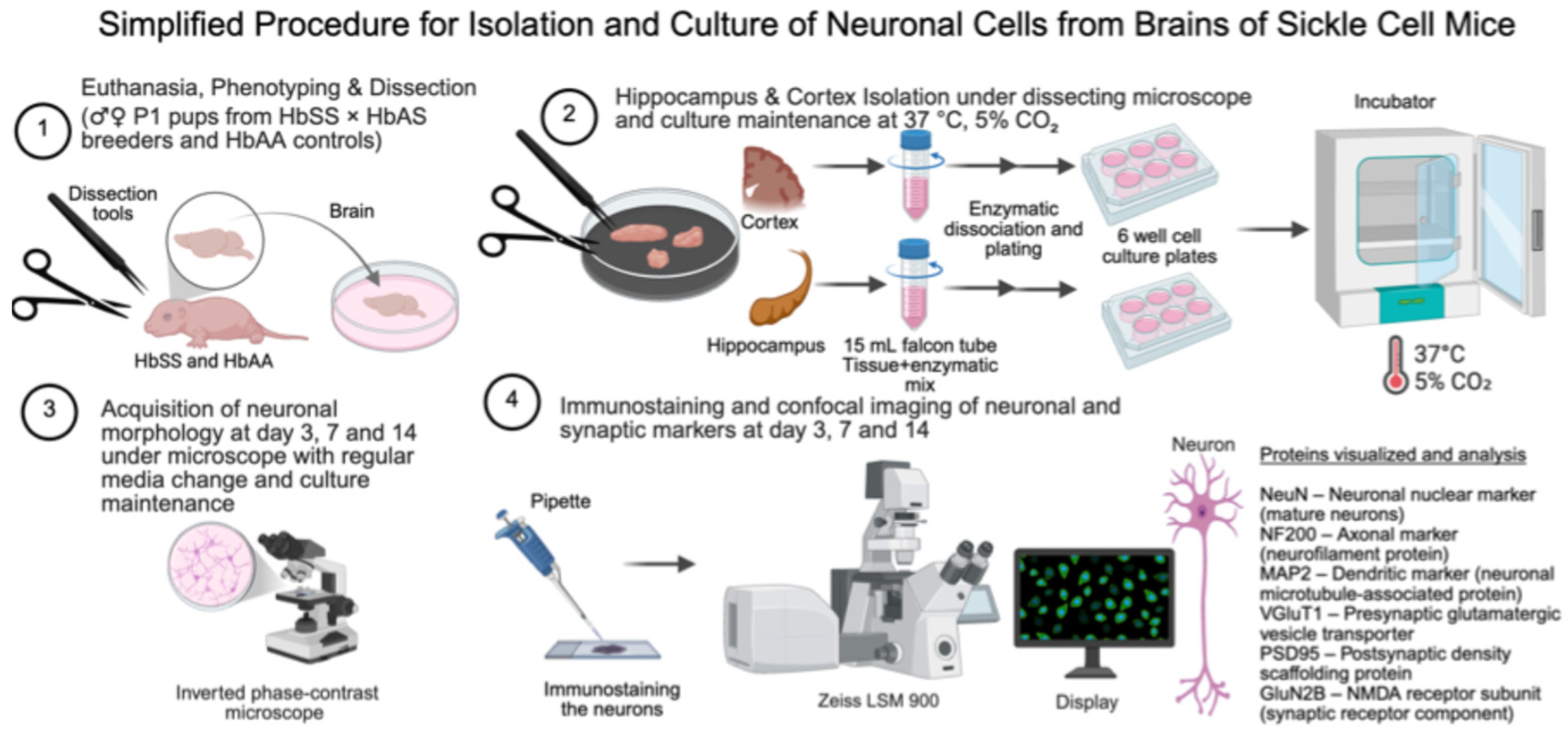
Simplified Procedure for Isolation and Culture of Neuronal Cells from Brains of Sickle Cell Mice

 , , , , , , and
Abstract
1. Introduction
2. Materials and Methods
2.1. Mice
2.2. Reagents Used in Neuronal Isolation and Culture
2.3. Step-by-Step Protocol for Primary Culture of Hippocampal and Cortical Neurons from Newborn Mice (P1)
Reagent Preparation for Dissection and Primary Neuron Culture
- Laminin: A 200 µg/mL stock solution was prepared by dissolving laminin (1 mg) in 1× HBSS (5 mL). Aliquots of 100 μL were made and frozen at −80 °C for up to 6 months.
- Poly-D-lysine: A 2 mg/mL stock solution was prepared by dissolving poly-D-lysine (5 mg) in ddH2O (2.5 mL). Aliquots of 110 µL were made and frozen at −20 °C. A 20 µg/mL working solution was prepared by diluting 2 mg/mL poly-D-lysine (100 µL) in 1× HBSS (10 mL). Approximately 1.2 mL was aliquoted per 12 coverslips and kept on ice until use. The remainder was stored at 4 °C for up to one week.
- Dissection Solution (DS): NaCl (4.003 g), KCl (0.201 g), Na2PO4 (0.012 g), KH2PO4 (0.015 g), HEPES (1.180 g), D-glucose (3.000 g), and sucrose (7.496 g) were combined in diH2O. The pH was adjusted to 7.40 with NaOH, and the final volume was brought to 500 mL with diH2O. The solution was sterile-filtered using a 0.2 µm vacuum filter and stored at 4 °C.
- The Neurobasal Medium + B27 (NBM/B27): Neurobasal™ Medium (250 mL) and B27 Supplement (5 mL) were combined, sterile-filtered through a 0.2 µm filter, and stored at 4 °C for up to 2 months.
- Conditioned Plus Media (CM+): B27 Supplement (1 mL), GlutaMAX (125 µL), L-Glutamine (125 µL), Penicillin–Streptomycin (500 µL), and FBS (5 mL) were combined and brought to a final volume of 50 mL with Neurobasal™ Medium. The solution was sterile-filtered through a 0.2 µm filter and stored at 4 °C for up to 2 weeks.
- Conditioned Minus Media (CM−): B27 Supplement (1 mL), GlutaMAX (125 µL), and Penicillin–Streptomycin (500 µL) were combined and brought to a final volume of 50 mL with Neurobasal™ Medium. The solution was sterile-filtered through a 0.2 µm filter and stored at 4 °C for up to 2 weeks.
- APV: A 5 mM APV stock was prepared by dissolving APV (9.86 mg) in ddH2O (10 mL). The solution was sterile-filtered through a 0.2 µm filter. Aliquots of 120 μL and 500 µL were stored at −20 °C.
- Ara-C: A 1 mM Ara-C stock was prepared by dissolving Ara-C (27.97 mg) in ddH2O (100 mL). The solution was sterile-filtered through a 0.2 µm filter. Aliquots were prepared and stored at 4 °C or −20 °C for up to 6 months.
- Papain: A 1 U/µL stock was prepared by dissolving papain (5 mg) in diH2O (5 mL). The solution was sterile-filtered through a 0.2 µm filter and kept on ice. It may be stored at 4 °C for up to one week.
- Enzyme Stock Preparation: In total, 5 mM APV (500 µL), L-cysteine (6 mg), and EDTA (9.3 mg) were combined and brought to a final volume of 50 mL with the dissection solution. The mixture was vortexed thoroughly until dissolved, then sterile-filtered through a 0.2 µm filter. Aliquots of 4 mL for hippocampal neurons and 12 mL for cortical neurons were stored at −20 °C.
- Complete Enzyme Solution (ES): Immediately before use, one aliquot of enzyme stock solution was thawed and supplemented with 50 units of papain to prepare the complete enzyme solution. The solution was then activated in a 37 °C water bath for 30 min prior to tissue digestion.
- Bovine Serum Albumin/Papain Inhibitor Stock (BSA/TI): In total, 10 mL of dissection solution was added to a large beaker, and BSA (1 g) was sprinkled evenly on top. The solution was allowed to dissolve completely without agitation or vortexing. Once dissolved, the Trypsin Inhibitor (1 g) was added in the same manner and allowed to dissolve fully. The final solution was sterile-filtered through a 0.2 µm filter, and aliquots of 400 µL were stored at −20 °C.
- High Inhibitor (HI) Solution: The HI solution was freshly prepared by combining dissection solution (DS, 3.0 mL), BSA/TI stock (300 µL; final concentration 10 mg/mL), and 5 mM of APV stock (30 µL; final concentration 50 µM). The mixture was gently mixed, and 1.5 mL was aliquoted into two 15 mL conical tubes and kept on ice until use.
- Low Inhibitor (LI) Solution: The LI solution was freshly prepared by combining DS (8.0 mL), BSA/TI stock (80 µL; final concentration 1 mg/mL), and 5 mM APV stock (80 µL; final concentration 50 µM). The mixture was gently mixed, and 2.5 mL was aliquoted into three 15 mL conical tubes and kept on ice until use.
2.4. Neonatal Pup Phenotyping
- Sex: The sex of neonatal pups was determined on postnatal day 1 by visual inspection of the anogenital region. Males were identified based on a greater anogenital distance and the presence of a distinct pigmentation spot above the urethral opening, indicative of developing scrotal skin. This non-invasive method is reliable for early sex differentiation in mouse pups.
2.5. Surface Preparation of Coverslips for Neuronal Seeding
- Sterilization: Round glass coverslips (15 mm or 12 mm) were sterilized by autoclaving. Under sterile conditions in a biosafety cabinet, each coverslip was dipped in 70% ethanol and set upright in a sterile Petri plate to dry. Once dried, coverslips were centered flat in the 6/24 cell culture plate wells to ensure an even coating and prevent solution runoff. Care was taken to avoid contact between coverslips and the walls of the well.
- Coating Solution: Prior to use, a laminin aliquot was thawed at 4 °C and kept on ice. A working solution of poly-D-lysine (20 µg/mL) was prepared by diluting the 2 mg/mL stock (100 µL) into 10 mL of 1× HBSS. To prepare the final coating solution, 100 µL of laminin stock was diluted into a 1.1 mL poly-D-lysine solution.
2.6. Dissection of Neonatal Brain for Hippocampal and Cortical Collection
- Inside the biosafety cabinet, one 60 mm Petri dish containing 3 mL of ice-cold dissection solution was prepared for brain sample collection, along with two sterile tubes containing 3 mL of ice-cold dissection solution for hippocampus sample collection and 9 mL for cortical region sample collection. Two enzyme solution (ES) aliquots were thawed, and 50 units of papain were added to each to prepare the complete ES, which was then incubated in a 37 °C water bath for 30 min to ensure optimal papain activity.
- Pups were euthanized by placing them inside a CO2 chamber with a fill rate of 70% displacement of the chamber volume per minute. The flow rate was adjusted based on chamber size, and pups were monitored until respiration ceased.
- Once the pups were unconscious, their bodies were disinfected with 70% ethanol using a spray bottle. Euthanasia was confirmed, followed by decapitation using large straight scissors.
- A midline cranial incision was made through the skin with small, straight scissors under aseptic conditions. Another midline incision was made through the skull, followed by two small incisions, one near each eye. Using forceps, the skull was carefully peeled back to expose the brain. The brain was removed from the skull by gently detaching the occipital lobe and underlying nerves with small forceps. Once extracted, the brain was placed in a Petri dish containing ice-chilled dissection solution.
- Using a dissecting microscope, the hippocampus was isolated from the rest of the brain by making a midline incision between the two hemispheres with fine forceps and small straight scissors. While the cerebellum was held with forceps, another pair of forceps was used to carefully ‘unroll’ the cortex from the hemisphere until the hippocampus became visible. The hippocampus was then removed using small straight scissors, and the procedure was repeated for the second hemisphere.
- Meninges were carefully removed and discarded. Following hippocampal removal, the frontal, parietal, and occipital cortices were carefully separated from the underlying white matter using fine forceps under a dissecting microscope.
- Each hippocampus (from both hemispheres) was cut into 3–4 pieces in a 60 mm Petri dish containing ice-cold dissection solution using fine scissors. The hippocampal tissue was transferred into a pre-labeled tube containing 3 mL of dissection solution. Separately, the dissected cortical regions (cut into pieces) were transferred into another tube containing 9 mL of dissection solution. Both tubes were kept on ice before proceeding to the culture hood for further processing.
2.7. Cell Dissociation and Plating
- After the 30 min incubation of complete ES at 37 °C, the enzyme solution with papain was filtered through a 0.2 μm filter into two sterile tubes.
- The tissues were transferred into the complete ES in designated sterile tubes for the hippocampus and cortex using a transfer pipette, allowing them to gently slide down via gravity while minimizing the carryover of the dissection solution.
- Under sterile conditions, tubes containing hippocampal and cortical tissues immersed in complete ES were incubated at 37 °C for 25 min.
- HI and LI solutions were prepared in 15 mL conical tubes and kept on ice during incubation.
- Tissues were transferred with a sterile transfer pipette before a sequence of washes was performed in individual tubes by gently swirling up and down ~3 times, using as little residual solution as possible. After every wash, the tube was placed back on ice and the tissue was allowed to settle to the bottom of the tube for ~1 min before proceeding to the next wash as follows in the sequence described below.
- ✓
- One wash with dissection solution (4 mL hippocampal; 12 mL cortical);
- ✓
- Two washes with HI Solution (2 mL hippocampal; 6 mL cortical)
- ✓
- Two washes with LI Solution (3 mL hippocampal; 9 mL cortical);
- ✓
- One wash with NBM/B27 Media (4 mL hippocampal; 12 mL cortical);
- ✓
- One wash NBM/B27 Media (1.5 mL hippocampal; 4.5 mL cortical).
- The cell suspension was visually triturated using a manual micropipette with a decreasing tip diameter:
- ✓
- This was performed ~15–20 times with a 5 mL sterile serological pipette.
Note: The pipetting should be gentle, and a consistent force should be used to initiate dissociation without shearing cells. Frothing or bubbling of the solution was avoided.- ✓
- This was performed ~20 times with a 1 mL pipette tip.
Note: The tip was slightly cut to widen the bore, reducing mechanical stress and helping prevent cell lysis.- ✓
- This was performed ~15 times with a 20–200 μL pipette tip.
Note: This step helped break up finer clumps. Pipetting was performed slowly and steadily to avoid the formation of bubbles or creation of shear stress.
- The suspension was left undisturbed for ~3 min to allow larger pieces of tissue to settle to the bottom.
- Immediately before plating, the coating solution was aspirated, and coverslips were gently rinsed three times with 1× HBSS (100 µL per wash) using just enough volume to cover only the coated central area of each coverslip, for 1 min per wash. After the final rinse, the HBSS was completely aspirated, and 100 µL of cell suspension was loaded onto each coverslip at 1 × 10⁴ live cells/coverslip.
- Coverslips were incubated at 37 °C and 5% CO2 for at least 2–3 h to allow cells to adhere.
- Warm CM+ was added to each well (500 µL for 24-well plates and 2 mL for 6-well plates).
- At day 2, cultures were treated for 24 h with Ara-C to inhibit glial growth by diluting Ara-C stock in CM− to achieve a final concentration of 2 µM.
- After incubation, 80% of the media was removed and replaced with fresh CM− (250 µL for 24-well plates and 1 mL for 6-well plates).
- The medium was refreshed twice weekly by replacing half of the volume with fresh, warmed CM−.
2.8. Treatments
2.9. Laser Scanning Confocal Microscopy (LSCM) for Neuron-Specific Markers
2.10. Evaluation of PSD 95 and GluN2B
2.11. Statistics
3. Results
3.1. Developmental Progression of Cortical and Hippocampal Neurons in Control and Sickle Cell Models Across 3, 7, and 14 DIVs
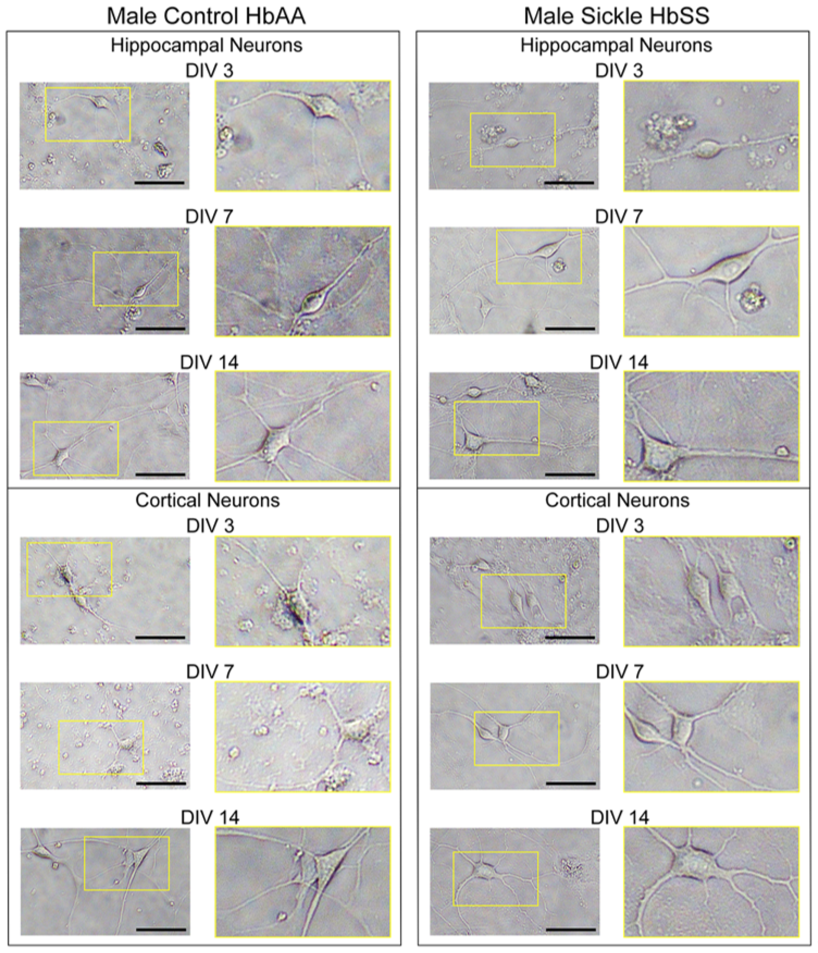
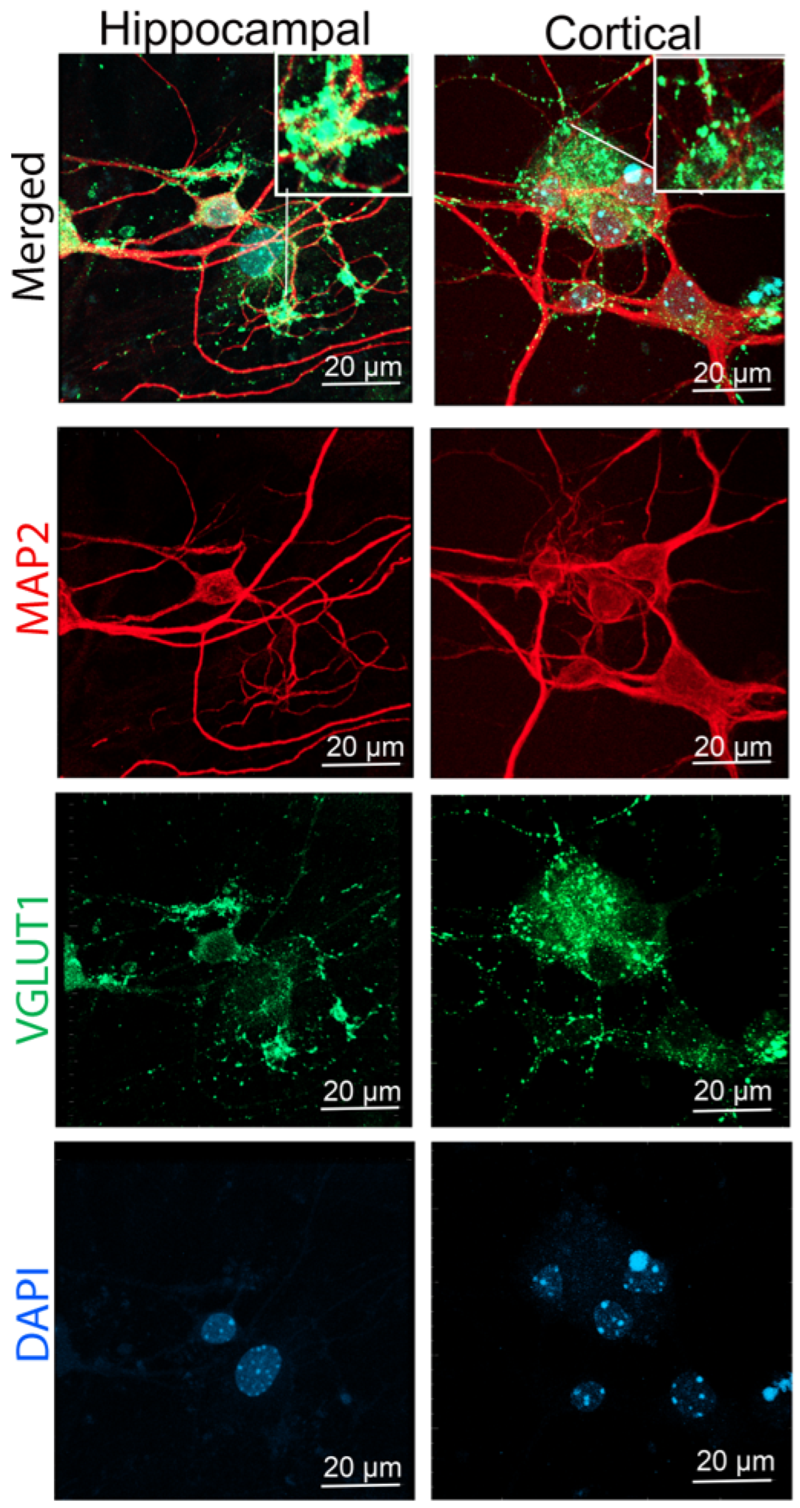
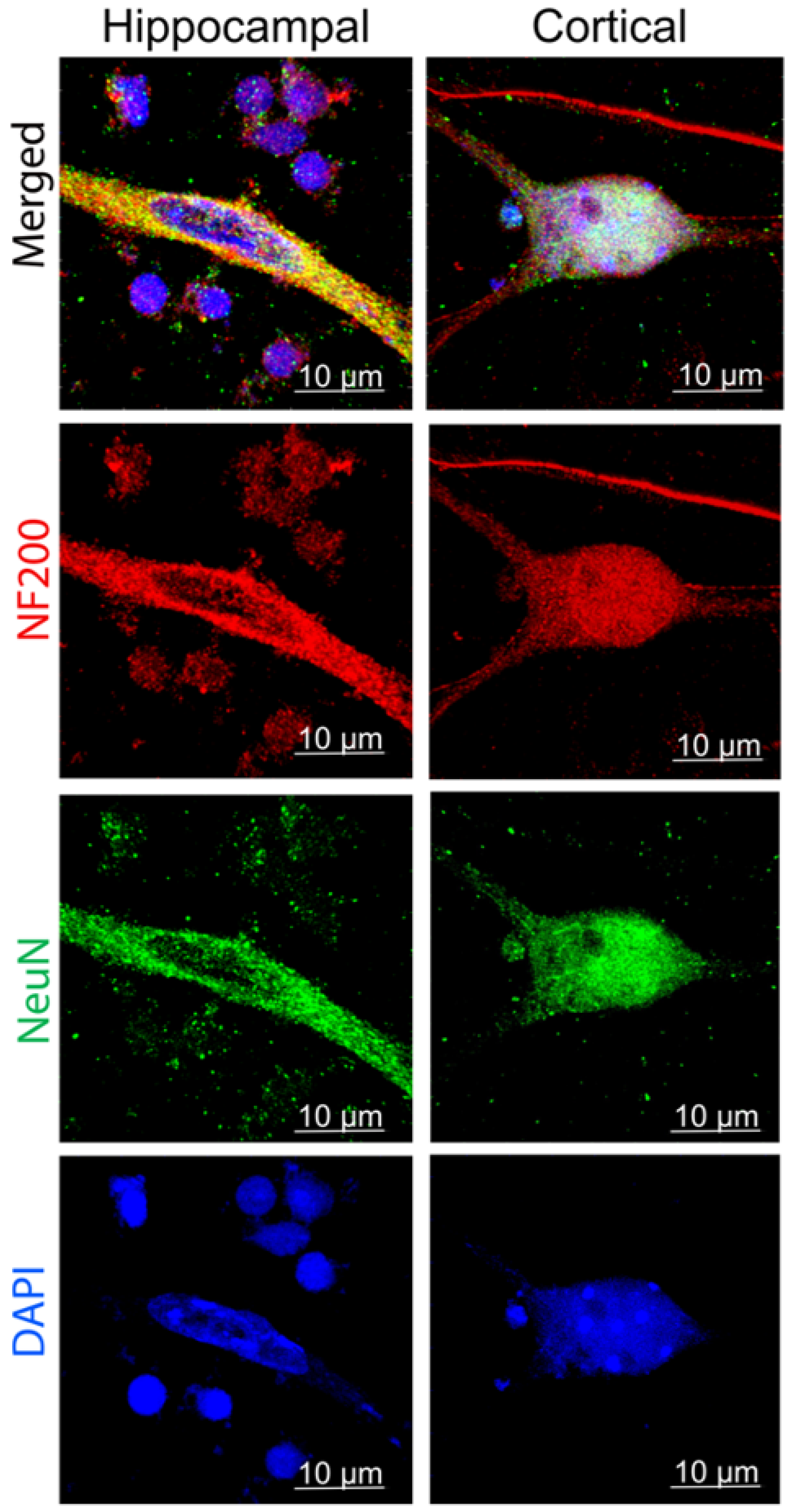
- DIV 3—Initial Attachment and Onset of Neurite Outgrowth (Figure 1): By DIV 3, cultured neurons showed successful attachment and early neurite outgrowth across all groups, including male and female, control and sickle-cell-derived neurons. While some variation in cell spreading and morphology of neurites was noted between cortical and hippocampal neurons, the cultures consistently displayed features of neuronal structure. These results suggest that the protocol is broadly applicable across sex and genotypes, although further studies will be needed to systematically characterize morphological differences. These morphological trends in hippocampal and cortical neurons were further supported by the presence of neuronal markers (Figure 2). Control and sickle cell neurons from male hippocampal and cortical cultures showed robust NeuN+ staining and emerging NF200+ neurites, indicative of healthy neuronal cells.
- DIV 7—Neurite Expansion and Early Network Formation (Figure 1): By DIV 7, neuronal maturation advanced substantially in control and sickle-cell-derived cultures. Qualitatively, cortical neurons demonstrated extensive branching with the emergence of neurite interconnections, while hippocampal neurons extended processes. Sickle cell neurons began to show more apparent variability in soma size and neurite density, especially in hippocampal cultures. Further, control and sickle cell neurons from male hippocampal and cortical cultures showed robust NeuN+ expression and NF200+ expression in neurites, indicating the preservation and specificity of neuronal integrity (Figure 2).
- DIV 14—Network Maturation and Structural Differentiation (Figure 1): At DIV 14, neuronal networks in control cultures showed dense and highly branched neurite webs observed in both cortical and hippocampal regions. Cortical neurons showed rich arborization and complex interconnectivity, while hippocampal neurons displayed organized, long projections that formed bundled tracts. Control and sickle cell hippocampal and cortical neurons maintained constant NeuN+ expression and NF200+ neurite networks at DIV 14 (Figure 2).
3.2. MAP2 and VGLUT1 in Sickle Cell vs. Control Hippocampal Neurons Across Developmental Timepoints
3.3. Genotype, Development, and Sickle Cell Microenvironment Modulate PSD 95 and GluN2B Colocalization in Cultured Hippocampal Neurons
3.4. PSD 95 Distribution in Hippocampal Neurons in Culture
4. Discussion
5. Conclusions
Supplementary Materials
Author Contributions
Funding
Institutional Review Board Statement
Data Availability Statement
Acknowledgments
Conflicts of Interest
Abbreviations
| APV | DL-2-Amino-5-Phosphonopentanoic Acid |
| Ara-C | Cytosine β-D-Arabinofuranoside Hydrochloride |
| BSA/TI | Bovine Serum Albumin/Trypsin Inhibitor |
| CM+ | Conditioned Medium Plus (with FBS and supplements) |
| CM- | Conditioned Medium Minus (without FBS) |
| DAPI | 4′,6-Diamidino-2-Phenylindole |
| DIVs | Days In Vitro |
| DS | Dissection Solution |
| EDTA | Ethylenediaminetetraacetic Acid |
| FBS | Fetal Bovine Serum |
| GluN2B | Glutamate [NMDA] Receptor Subunit Epsilon-2 |
| HBSS | Hank’s Balanced Salt Solution |
| HbAA | Homozygous Humanized Mice Expressing Hemoglobin A |
| HbSS | Homozygous Humanized Sickle Cell Mice Expressing Hemoglobin S |
| HEPES | N-(2-Hydroxyethyl) Piperazine-N′-(2-Ethanesulfonic Acid) |
| HI | High Inhibitor Solution |
| LI | Low Inhibitor Solution |
| MAP2 | Microtubule-Associated Protein 2 |
| NBM/B27 | Neurobasal Medium with B27 Supplement |
| NeuN | Neuronal Nuclei Marker (RBFOX3) |
| NF200 | Neurofilament 200 kDa |
| NMDAR | N-Methyl-D-Aspartate Receptor |
| P1 | Postnatal Day 1 |
| PBS | Phosphate-Buffered Saline |
| PSD 95 | Postsynaptic Density Protein 95 |
| RT | Room Temperature |
| SCD | Sickle Cell Disease |
| TNF-α | Tumor Necrosis Factor-alpha |
| VGLUT1 | Vesicular Glutamate Transporter 1 |
References
- Molokie, R.E.; Wang, Z.J.; Yao, Y.; Powell-Roach, K.L.; Schlaeger, J.M.; Suarez, M.L.; Shuey, D.A.; Angulo, V.; Carrasco, J.; Ezenwa, M.O.; et al. Sensitivities to Thermal and Mechanical Stimuli: Adults With Sickle Cell Disease Compared to Healthy, Pain-Free African American Controls. J. Pain 2020, 21, 957–967. [Google Scholar] [CrossRef]
- McClish, D.K.; Smith, W.R.; Dahman, B.A.; Levenson, J.L.; Roberts, J.D.; Penberthy, L.T.; Aisiku, I.P.; Roseff, S.D.; Bovbjerg, V.E. Pain Site Frequency and Location in Sickle Cell Disease: The PiSCES Project. Pain 2009, 145, 246–251. [Google Scholar] [CrossRef]
- Vichinsky, E.P.; Neumayr, L.D.; Gold, J.I.; Weiner, M.W.; Rule, R.R.; Truran, D.; Kasten, J.; Eggleston, B.; Kesler, K.; McMahon, L.; et al. Neuropsychological Dysfunction and Neuroimaging Abnormalities in Neurologically Intact Adults with Sickle Cell Anemia. JAMA 2010, 303, 1823–1831. [Google Scholar] [CrossRef]
- Kohli, D.R.; Li, Y.; Khasabov, S.G.; Gupta, P.; Kehl, L.J.; Ericson, M.E.; Nguyen, J.; Gupta, V.; Hebbel, R.P.; Simone, D.A.; et al. Pain-Related Behaviors and Neurochemical Alterations in Mice Expressing Sickle Hemoglobin: Modulation by Cannabinoids. Blood 2010, 116, 456–465. [Google Scholar] [CrossRef]
- Hillery, C.A.; Kerstein, P.C.; Vilceanu, D.; Barabas, M.E.; Retherford, D.; Brandow, A.M.; Wandersee, N.J.; Stucky, C.L. Transient Receptor Potential Vanilloid 1 Mediates Pain in Mice with Severe Sickle Cell Disease. Blood 2011, 118, 3376–3383. [Google Scholar] [CrossRef]
- Kenyon, N.; Wang, L.; Spornick, N.; Khaibullina, A.; Almeida, L.E.; Cheng, Y.; Wang, J.; Guptill, V.; Finkel, J.C.; Quezado, Z.M.N. Sickle Cell Disease in Mice Is Associated with Sensitization of Sensory Nerve Fibers. Exp. Biol. Med. 2015, 240, 87–98. [Google Scholar] [CrossRef] [PubMed]
- Wang, L.; Almeida, L.E.F.; de Souza Batista, C.M.; Khaibullina, A.; Xu, N.; Albani, S.; Guth, K.A.; Seo, J.S.; Quezado, M.; Quezado, Z.M.N. Cognitive and Behavior Deficits in Sickle Cell Mice Are Associated with Profound Neuropathologic Changes in Hippocampus and Cerebellum. Neurobiol. Dis. 2016, 85, 60–72. [Google Scholar] [CrossRef] [PubMed]
- Hazra, R.; Pu, H.; Foley, L.M.; Little-Ihrig, L.; Hitchens, T.K.; Ghosh, S.; Ofori-Acquah, S.F.; Hu, X.; Novelli, E.M. White-Matter Abnormalities and Cognitive Dysfunction Are Linked to Astrocyte Activation in Sickle Mice. PNAS Nexus 2023, 2, pgad149. [Google Scholar] [CrossRef] [PubMed]
- He, Y.; Chen, Y.; Tian, X.; Yang, C.; Lu, J.; Xiao, C.; DeSimone, J.; Wilkie, D.J.; Molokie, R.E.; Wang, Z.J. CaMKIIα Underlies Spontaneous and Evoked Pain Behaviors in Berkeley Sickle Cell Transgenic Mice. Pain 2016, 157, 2798–2806. [Google Scholar] [CrossRef]
- Khaibullina, A.; Almeida, L.E.F.; Kamimura, S.; Zerfas, P.M.; Smith, M.L.; Vogel, S.; Wakim, P.; Vasconcelos, O.M.; Quezado, M.M.; Horkayne-Szakaly, I.; et al. Sickle Cell Disease Mice Have Cerebral Oxidative Stress and Vascular and White Matter Abnormalities. Blood Cells Mol. Dis. 2021, 86, 102493. [Google Scholar] [CrossRef]
- Tajerian, M.; Hung, V.; Nguyen, H.; Lee, G.; Joubert, L.-M.; Malkovskiy, A.V.; Zou, B.; Xie, S.; Huang, T.-T.; Clark, J.D. The Hippocampal Extracellular Matrix Regulates Pain and Memory after Injury. Mol. Psychiatry 2018, 23, 2302–2313. [Google Scholar] [CrossRef]
- Xia, S.-H.; Hu, S.-W.; Ge, D.-G.; Liu, D.; Wang, D.; Zhang, S.; Zhang, Q.; Yuan, L.; Li, Y.-Q.; Yang, J.-X.; et al. Chronic Pain Impairs Memory Formation via Disruption of Neurogenesis Mediated by Mesohippocampal Brain-Derived Neurotrophic Factor Signaling. Biol. Psychiatry 2020, 88, 597–610. [Google Scholar] [CrossRef] [PubMed]
- Zhao, W.; Zhao, L.; Chang, X.; Lu, X.; Tu, Y. Elevated Dementia Risk, Cognitive Decline, and Hippocampal Atrophy in Multisite Chronic Pain. Proc. Natl. Acad. Sci. USA 2023, 120, e2215192120. [Google Scholar] [CrossRef] [PubMed]
- Duric, V.; McCarson, K.E. Persistent Pain Produces Stress-like Alterations in Hippocampal Neurogenesis and Gene Expression. J. Pain 2006, 7, 544–555. [Google Scholar] [CrossRef] [PubMed]
- Tyrtyshnaia, A.A.; Manzhulo, I.V.; Konovalova, S.P.; Zagliadkina, A.A. Neuropathic Pain Causes a Decrease in the Dendritic Tree Complexity of Hippocampal CA3 Pyramidal Neurons. Cells Tissues Organs 2020, 208, 89–100. [Google Scholar] [CrossRef]
- Stratton, H.J.; Khanna, R. Sculpting Dendritic Spines during Initiation and Maintenance of Neuropathic Pain. J. Neurosci. 2020, 40, 7578–7589. [Google Scholar] [CrossRef]
- Zhou, J.-Z.; Deng, J.; Luo, D.-X.; Mai, J.-W.; Wu, J.-Y.; Duan, Y.-J.; Dong, B.; Xin, W.-J.; Xu, T.; Wei, J.-Y. Sex Differences in Functional and Structural Alterations of Hippocampus Region in Chronic Pain: A DTI and Resting-State fMRI Study. Front. Neurosci. 2024, 18, 1428666. [Google Scholar] [CrossRef]
- Apkarian, A.V.; Bushnell, M.C.; Treede, R.-D.; Zubieta, J.-K. Human Brain Mechanisms of Pain Perception and Regulation in Health and Disease. Eur. J. Pain 2005, 9, 463–484. [Google Scholar] [CrossRef]
- Bushnell, M.C.; Ceko, M.; Low, L.A. Cognitive and Emotional Control of Pain and Its Disruption in Chronic Pain. Nat. Rev. Neurosci. 2013, 14, 502–511. [Google Scholar] [CrossRef]
- Huang, J.-Y.; Jin, Y.-X.; Dong, W.-Y.; Zhao, W.; Cheng, P.-K.; Miao, J.-H.; Liu, A.; Wang, D.; Li, J.; Zhang, Z.; et al. Intra-Somatosensory Cortical Circuits Mediating Pain-Induced Analgesia. Nat. Commun. 2025, 16, 1859. [Google Scholar] [CrossRef]
- Campbell, C.M.; Moscou-Jackson, G.; Carroll, C.P.; Kiley, K.; Haywood, C.; Lanzkron, S.; Hand, M.; Edwards, R.R.; Haythornthwaite, J.A. An Evaluation of Central Sensitization in Patients with Sickle Cell Disease. J. Pain 2016, 17, 617–627. [Google Scholar] [CrossRef]
- Darbari, D.S.; Vaughan, K.J.; Roskom, K.; Seamon, C.; Diaw, L.; Quinn, M.; Conrey, A.; Schechter, A.N.; Haythornthwaite, J.A.; Waclawiw, M.A.; et al. Central Sensitization Associated with Fetal Hemoglobin Levels in Adults with Sickle Cell Anemia. Scand. J. Pain 2017, 17, 279–286. [Google Scholar] [CrossRef]
- Cataldo, G.; Rajput, S.; Gupta, K.; Simone, D.A. Sensitization of Nociceptive Spinal Neurons Contributes to Pain in a Transgenic Model of Sickle Cell Disease. Pain 2015, 156, 722–730. [Google Scholar] [CrossRef]
- Cramer, T.M.L.; Tyagarajan, S.K. Protocol for the Culturing of Primary Hippocampal Mouse Neurons for Functional in Vitro Studies. STAR Protoc. 2024, 5, 102991. [Google Scholar] [CrossRef]
- Moutin, E.; Hemonnot, A.-L.; Seube, V.; Linck, N.; Rassendren, F.; Perroy, J.; Compan, V. Procedures for Culturing and Genetically Manipulating Murine Hippocampal Postnatal Neurons. Front. Synaptic Neurosci. 2020, 12, 19. [Google Scholar] [CrossRef] [PubMed]
- Sagi, V.; Song-Naba, W.L.; Benson, B.A.; Joshi, S.S.; Gupta, K. Mouse Models of Pain in Sickle Cell Disease. Curr. Protoc. Neurosci. 2018, 85, e54. [Google Scholar] [CrossRef] [PubMed]
- Argueta, D.A.; Avalos, B.; Goel, Y.; Tran, H.; Fotio, Y.; Piomelli, D.; DiPatrizio, N.V.; Gupta, K. Neuroprotective, Anti-Inflammatory, and Analgesic Activity of Palmitoylethanolamide in Sickle Cell Mice. Blood Adv. 2025, 9, 3056–3068. [Google Scholar] [CrossRef] [PubMed]
- Lei, J.; Paul, J.; Wang, Y.; Gupta, M.; Vang, D.; Thompson, S.; Jha, R.; Nguyen, J.; Valverde, Y.; Lamarre, Y.; et al. Heme Causes Pain in Sickle Mice via Toll-Like Receptor 4-Mediated Reactive Oxygen Species- and Endoplasmic Reticulum Stress-Induced Glial Activation. Antioxid. Redox Signal. 2021, 34, 279–293. [Google Scholar] [CrossRef]
- Lee, H.; Park, J.-H.; Trotter, J.H.; Maher, J.N.; Keenoy, K.E.; Jang, Y.M.; Lee, Y.; Kim, J.-I.; Weeber, E.J.; Hoe, H.-S. Reelin and APP Cooperatively Modulate Dendritic Spine Formation In Vitro and In Vivo. Exp. Neurobiol. 2023, 32, 42–55. [Google Scholar] [CrossRef]
- Lomeli, N.; Di, K.; Pearre, D.C.; Chung, T.-F.; Bota, D.A. Mitochondrial-Associated Impairments of Temozolomide on Neural Stem/Progenitor Cells and Hippocampal Neurons. Mitochondrion 2020, 52, 56–66. [Google Scholar] [CrossRef]
- Dunn, K.W.; Kamocka, M.M.; McDonald, J.H. A Practical Guide to Evaluating Colocalization in Biological Microscopy. Am. J. Physiol. Cell Physiol. 2011, 300, C723–C742. [Google Scholar] [CrossRef] [PubMed]
- Pászty, C.; Brion, C.M.; Manci, E.; Witkowska, H.E.; Stevens, M.E.; Mohandas, N.; Rubin, E.M. Transgenic Knockout Mice with Exclusively Human Sickle Hemoglobin and Sickle Cell Disease. Science 1997, 278, 876–878. [Google Scholar] [CrossRef]
- Kaech, S.; Banker, G. Culturing Hippocampal Neurons. Nat. Protoc. 2006, 1, 2406–2415. [Google Scholar] [CrossRef] [PubMed]
- Beaudoin, G.M.J.; Lee, S.-H.; Singh, D.; Yuan, Y.; Ng, Y.-G.; Reichardt, L.F.; Arikkath, J. Culturing Pyramidal Neurons from the Early Postnatal Mouse Hippocampus and Cortex. Nat. Protoc. 2012, 7, 1741–1754. [Google Scholar] [CrossRef] [PubMed]
- Chambliss, C.; Manci, E.; Fields, E.; Bueno, J.; Michael, A.; Eldeiry, E.; Hall, C.; Gee, B.; Chonat, S.; Archer, D.R. Characterizing Pregnancy Outcomes in a Humanized Mouse Model of Sickle Cell Disease. Br. J. Haematol. 2025, 207, 813–823. [Google Scholar] [CrossRef]
- Manci, E.A.; Hillery, C.A.; Bodian, C.A.; Zhang, Z.G.; Lutty, G.A.; Coller, B.S. Pathology of Berkeley Sickle Cell Mice: Similarities and Differences with Human Sickle Cell Disease. Blood 2006, 107, 1651–1658. [Google Scholar] [CrossRef]
- Valverde, Y.; Benson, B.; Gupta, M.; Gupta, K. Spinal Glial Activation and Oxidative Stress Are Alleviated by Treatment with Curcumin or Coenzyme Q in Sickle Mice. Haematologica 2016, 101, e44–e47. [Google Scholar] [CrossRef]
- Hardy, R.A.; Rached, N.A.; Jones, J.A.; Archer, D.R.; Hyacinth, H.I. Role of Age and Neuroinflammation in the Mechanism of Cognitive Deficits in Sickle Cell Disease. Exp. Biol. Med. 2021, 246, 106–120. [Google Scholar] [CrossRef]
- Seibenhener, M.L.; Wooten, M.W. Isolation and Culture of Hippocampal Neurons from Prenatal Mice. J. Vis. Exp. 2012, 65, e3634. [Google Scholar] [CrossRef]
- Fabry, M.E.; Bouhassira, E.E.; Suzuka, S.M.; Nagel, R.L. Transgenic Mice and Hemoglobinopathies. In Hemoglobin Disorders; Humana Press: Totowa, NJ, USA, 2003; Volume 82, pp. 213–241. [Google Scholar] [CrossRef]
- Dotti, C.G.; Sullivan, C.A.; Banker, G.A. The Establishment of Polarity by Hippocampal Neurons in Culture. J. Neurosci. 1988, 8, 1454–1468. [Google Scholar] [CrossRef]
- El-Husseini, A.E.D.; Schnell, E.; Chetkovich, D.M.; Nicoll, R.A.; Bredt, D.S. PSD-95 Involvement in Maturation of Excitatory Synapses. Science 2000, 290, 1364–1368. [Google Scholar] [CrossRef]
- Grabrucker, A.M.; Schmeisser, M.J.; Schoen, M.; Boeckers, T.M. Postsynaptic ProSAP/Shank Scaffolds in the Cross-Hair of Synaptopathies. Trends Cell Biol. 2011, 21, 594–603. [Google Scholar] [CrossRef]
- Yoshii, A.; Constantine-Paton, M. Postsynaptic BDNF-TrkB Signaling in Synapse Maturation, Plasticity, and Disease. Dev. Neurobiol. 2010, 70, 304–322. [Google Scholar] [CrossRef]
- De Luca, P.; Mele, M.; Tanqueiro, S.; Napoli, F.; Butkevičiūtė, U.; Souto, A.C.; Costa, R.O.; Schwarz, A.; Drexel, M.; Sebastião, A.M.; et al. Synaptic Accumulation of GluN2B-Containing NMDA Receptors Mediates the Effects of BDNF-TrkB Signalling on Synaptic Plasticity and in Hyperexcitability during Status Epilepticus. J. Biomed. Sci. 2025, 32, 82. [Google Scholar] [CrossRef]




Disclaimer/Publisher’s Note: The statements, opinions and data contained in all publications are solely those of the individual author(s) and contributor(s) and not of MDPI and/or the editor(s). MDPI and/or the editor(s) disclaim responsibility for any injury to people or property resulting from any ideas, methods, instructions or products referred to in the content. |
© 2026 by the authors. Licensee MDPI, Basel, Switzerland. This article is an open access article distributed under the terms and conditions of the Creative Commons Attribution (CC BY) license.
Share and Cite
Goel, Y.; Arellano, M.A.; O’Daniel, K.; Argueta, D.A.; Lomeli, R.; Lomeli, N.; Ordaz, D.A.; Bota, D.A.; Kumaresan, V.; Gupta, K. Simplified Procedure for Isolation and Culture of Neuronal Cells from Brains of Sickle Cell Mice. Cells 2026, 15, 976. https://doi.org/10.3390/cells15110976
Goel Y, Arellano MA, O’Daniel K, Argueta DA, Lomeli R, Lomeli N, Ordaz DA, Bota DA, Kumaresan V, Gupta K. Simplified Procedure for Isolation and Culture of Neuronal Cells from Brains of Sickle Cell Mice. Cells. 2026; 15(11):976. https://doi.org/10.3390/cells15110976
Chicago/Turabian StyleGoel, Yugal, Mya A. Arellano, Kendall O’Daniel, Donovan A. Argueta, Reina Lomeli, Naomi Lomeli, Dahlia A. Ordaz, Daniela A. Bota, Vidhya Kumaresan, and Kalpna Gupta. 2026. "Simplified Procedure for Isolation and Culture of Neuronal Cells from Brains of Sickle Cell Mice" Cells 15, no. 11: 976. https://doi.org/10.3390/cells15110976
APA StyleGoel, Y., Arellano, M. A., O’Daniel, K., Argueta, D. A., Lomeli, R., Lomeli, N., Ordaz, D. A., Bota, D. A., Kumaresan, V., & Gupta, K. (2026). Simplified Procedure for Isolation and Culture of Neuronal Cells from Brains of Sickle Cell Mice. Cells, 15(11), 976. https://doi.org/10.3390/cells15110976