Article

Drosophila Models Reveal NAT Complex Roles in Heart Development and Enable Functional Validation of Congenital Heart Disease Variants

1
Center for Precision Disease Modeling, Department of Medicine, University of Maryland School of Medicine, Baltimore, MD 21201, USA
2
Division of Endocrinology, Diabetes, and Nutrition, Department of Medicine, University of Maryland School of Medicine, Baltimore, MD 21201, USA
*
Authors to whom correspondence should be addressed.
Cells 2025, 14(20), 1596; https://doi.org/10.3390/cells14201596 (registering DOI)
Submission received: 12 September 2025 / Revised: 7 October 2025 / Accepted: 13 October 2025 / Published: 14 October 2025
(This article belongs to the Special Issue Drosophila as a Model for Understanding Human Disease)

Abstract

N-terminal acetylation, catalyzed by N-terminal acetyltransferase (NAT) complexes, is one of the most prevalent protein modifications in eukaryotic cells, yet its role in heart development remains poorly understood. Here, we use Drosophila as an in vivo platform to investigate the functions of NAT complex components in cardiac development and congenital heart disease (CHD). Focusing on the NatA complex, we showed that cardiac-specific knockdown of each of its three subunits (Naa15-16, vnc, and san) led to developmental lethality, structural disorganization, fibrosis, and impaired cardiac function in Drosophila. Remarkably, human NAA16 completely rescued the cardiac defects in Naa15-16 silenced Drosophila, whereas a CHD-associated variant (NAA16-R70C) failed to do so, providing direct functional evidence of its pathogenicity. Together, these findings suggest the NatA complex as a critical regulator of heart development and provide functional validation linking variants in NatA complex genes to CHD. Further studies in mammalian models will be required to provide additional supporting evidence.

1. Introduction

Protein N-terminal acetylation is one of the most prevalent modifications in eukaryotic cells, occurring in nearly 80–90% of human proteins [1,2]. This modification is catalyzed by a group of enzymes known as N-terminal acetyltransferases (NATs), which transfer an acetyl group from acetyl-CoA to the N-terminal amino acid of a nascent protein [3,4]. NATs play essential roles in modulating protein stability, localization, interaction, and function [5,6,7]. In mammals, eight major NAT complexes (NatA–H) have been identified, each with distinct catalytic and auxiliary subunits and substrate specificities (Figure 1A) [8]. Among these, the NatA complex, which is composed primarily of the catalytic subunit NAA10 or NAA11 and the auxiliary subunit NAA15 or NAA16, is the most extensively studied and is responsible for acetylating a broad range of proteins following initiator methionine removal [9,10].
Although N-terminal acetylation has been widely recognized as a critical regulator of proteome function, its roles in organ-specific development and disease have only recently gained attention. In particular, emerging evidence points to the involvement of NAT complexes in cardiac development and disease. Genetic studies have implicated mutations in NAT components, especially those of the NatA complex, in congenital heart diseases (CHDs) [11,12,13]. For instance, loss-of-function mutations in NAA15 and NAA16 have been linked to syndromic forms of CHD, suggesting that protein acetylation may play an essential role in heart development [11,12,13]. Multiple studies suggested that dysregulation of NAT could cause various cardiac diseases, including cardiac arrhythmia and cardiomyopathy [14,15]. Reduction in NAA15 levels leads to altered expression of 562 proteins and changes in the N-terminal acetylation of 9 proteins, which are implicated in NAA15-mediated CHD [15]. Several case reports have linked mutations in NAT10 to Ogden syndrome, a condition in which cardiac defects are among the major clinical manifestations [16,17,18]. Mutations in NAA30 have also been reported to be potentially associated with heart disease [19]. However, no direct in vivo evidence has demonstrated that NAT complexes are required for heart development, and mechanistic studies linking NAT dysfunction to cardiac phenotypes remain limited. Furthermore, a comprehensive investigation to determine which specific NAT complexes are involved in heart development has not yet been conducted.
Recent genomic efforts, including those by the NIH Pediatric Cardiac Genomics Consortium (PCGC), have expanded the landscape of CHD-associated genes and revealed multiple de novo variants in NAT complex components, with NAA15 and NAA16 among the most frequently affected [11,12,13]. Although bioinformatic tools predict many of these candidate genes to be disease-associated, the lack of in vivo evidence presents a significant challenge in confirming their relevance to CHD. Additionally, the chance of the genetic variant identified in a given CHD gene causing the disease is relatively low [20,21]. A mutation identified in CHD patients may not affect the protein structure and function, which will not cause CHD. A large number of genetic variants identified in CHD patients remain unconfirmed regarding their association with the disease. This gap is largely attributed to the high cost and long timelines associated with mammalian models commonly used for such studies. Consequently, there is an urgent need for a low-cost, high-throughput animal model to validate the disease association of these candidate genes and their genetic variants.
Drosophila melanogaster has emerged as a powerful model for studying heart development due to its conserved cardiac structure, rapid genetic manipulation, and suitability for high-throughput in vivo functional assays [22,23,24]. The Drosophila heart, though structurally simpler, shares significant molecular and functional similarities with the vertebrate heart, particularly during early developmental stages [25,26]. Additionally, Drosophila provides a tractable platform for validating disease-associated variants through tissue-specific genetic manipulations and phenotype-rescue strategies [27].
In this study, we investigate the roles of NAT complexes, particularly the NatA complex, in heart development using Drosophila models. The NAT complex components are highly conserved between human and Drosophila, indicating their conserved roles in heart development during different species (Figure 1B). We first characterize the expression and function of several NAT complex components in the developing Drosophila heart. We then apply in vivo genetic approaches to dissect the functional consequences of cardiac-specific knockdown of NAT components, with a focus on Naa15-16, the Drosophila homolog of human NAA15 and NAA16. Furthermore, we employ gene replacement strategies that we developed previously [27,28,29] to validate a missense variant of NAA16 (R70C) identified in CHD patients, providing critical insights into its pathogenicity. Our findings reveal essential roles for NatA complex components in heart development and demonstrate the utility of Drosophila as an efficient platform for functional validation of CHD-associated genetic variants.

2. Results

2.1. Silencing NatA Complex Components Naa15-16, Vnc, or San Impaired Drosophila Survival

First, we examined the expression patterns of genes encoding the NAT complexes in the Drosophila embryonic heart at different developmental stages. We previously published a single-cell RNA-seq dataset that includes the gene expression levels of all genes encoding NAT complex components [30]. We reanalyzed single-cell RNA-seq data from cardiac cells collected across key embryonic stages, spanning the migration of bilateral cardiac progenitors (stage 13 onward) to the formation of the linear heart tube [30]. Among these genes, NatA complex component Naa15-16 displayed consistently high and stable expression in cardiac progenitor cells across all developmental stages (Supplementary Figure S1). Other NAT complex components, including san, Naa20A, and Naa30A, were also expressed in cardiac progenitor cells, but at comparatively lower levels (Supplementary Figure S1). Notably, vnc expression was undetectable in cardiac progenitor cells at any of the examined stages, suggesting it was not among the highly expressed genes captured in this single-cell RNA-seq dataset.
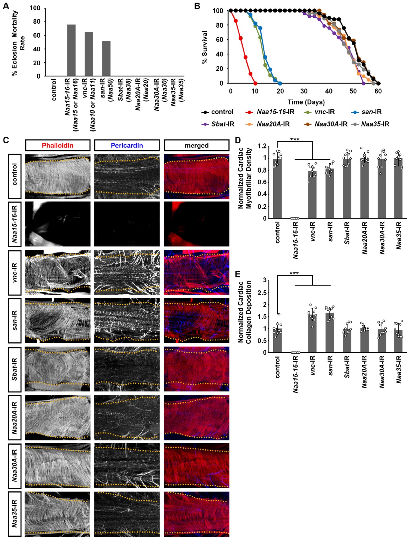
Next, we used the Drosophila UAS-Gal4 system [31] to specifically knock out individual components of the NAT complexes in the heart. This was achieved by crossing the heart-specific Gal4 driver, 4XHand-Gal4 [27], with available UAS RNAi lines NAT complexes component obtained from the Bloomington Drosophila Stock Center. The UAS RNAi lines obtained from the Bloomington Drosophila Stock Center can achieve 60–70% reduction using 4XHand-Gal4 compared to controls (Supplementary Figure S2). Cardiac-specific silencing of NatA complex components Naa15-16, vnc, and san significantly reduced eclosion rates, as indicated by the decreased number of adult flies successfully emerging from pupal cases (Figure 2A). Furthermore, the few adult flies that did eclose exhibited markedly shortened lifespans compared with controls (Figure 2B). These results the essential roles of the NatA complex components, Naa15-16, vnc, and san, in maintaining normal heart development and function in Drosophila. We did not observe any significant changes when silencing other NAT complex components in the Drosophila heart (Figure 2A,B).

2.2. Silencing NatA Complex Components Naa15-16, Vnc, or San Induced Cardiac Structural and Functional Defects in Adult Drosophila

Cardiac-specific knockdown of the NatA complex components Naa15-16, vnc, or san caused early lethality and markedly reduced lifespan (Figure 2A,B). To investigate the underlying cardiac abnormalities, we examined heart morphology in these flies. Notably, silencing Naa15-16 resulted in a complete loss of the heart structure in adult Drosophila (Figure 2C). We did not detect any cardiac myofibrillar organization and cardiac fibrillar density (Figure 2D). Further work is needed to determine how heart structures are lost in Naa15-16–silenced Drosophila. Knockdown of vnc or san caused severe disorganization of cardiac actin filaments, as revealed by phalloidin staining (Figure 2C), and significantly reduced cardiac muscle fiber density (Figure 2C,D). In addition, we observed marked accumulation of Pericardin in hearts with vnc or san knockdown (Figure 2C,E). Notably, this collagen signals detected by the pericardin antibody were completely absent in the hearts of NAA15-16-IR Drosophila (Figure 2E). This excessive Pericardin deposition observed upon vnc or san knockdown indicates fibrotic remodeling, a hallmark of cardiac injury [27,32,33].
To access cardiac functional defects resulting from vnc or san silencing, we employed optical coherence tomography (OCT), which serves as the Drosophila equivalent of echocardiography in humans [34,35]. The orthogonal view provided by OCT enables real-time measurements of heart tube diameter and heart period (Figure 3A). Because heart-specific silence Naa15-16 led to a complete loss of the heart structure in adult Drosophila, no cardiac signals could be detected using OCT. We observed that silencing vnc or san significantly increased the heart period compared to control flies (Figure 3B), but did not alter the systolic or diastolic diameters (Figure 3C–E). In contrast, silencing other NAT complex components did not produce any notable morphological or functional heart defects (Figure 2 and Figure 3).
Altogether, these findings demonstrated that components of the NatA complex are essential for maintaining normal cardiac structure and function. Among them, Naa15-16 stands out due to its consistently high expression in the developing Drosophila heart and the severe phenotypes observed upon its knockdown, highlighting its particularly critical role within the NAT complexes. However, further work is needed to perform detailed analyses of heart changes due to NatA complex components silencing during embryogenesis and larval developmental stages.

2.3. Expression of Wild-Type Human NAA16 and Its CHD-Associated Variant in the Drosophila Heart Did Not Cause Any Detectable Structural and Functional Cardiac Defects

Recent studies from the Pediatric Cardiac Genomic Consortium (PCGC) have identified more than 1000 candidate genes potentially associated with CHD [11,12,13]. Among them, several de novo mutations have been found in genes encoding components of the NAT complexes (Table 1). Notably, NAA15 and NAA16, which share the same Drosophila homolog, Naa15-16, are recurrently affected. Most variants in these genes are frameshift mutations, predicted to cause loss of function and considered pathogenic (Table 1). However, one missense variant R70C in NAA16 remains of uncertain significance regarding its disease association (Table 1). The detailed information of this R70C variant was previously reported (RadialSVM score: 0.314, RadialSVM pred: D, ExAC Overall: 2.49E-05, HHE Rank: 50.7, pLI score: 0.00) [11]. We compared the amino acid sequences between Drosophila and humans and confirmed that the mutant residue R70 is conserved in both species. This residue is located within the tetratricopeptide repeat (TPR) domain, which is also conserved between Drosophila and humans. TPR domain can act as modular docking sites, enabling proteins to form multi-protein complexes [36].
Since the missense variant R70C in NAA16 was identified in a heterozygous state in a CHD patient, we first sought to determine whether it exerted a dominant-negative or gain-of-function effect. To this end, we performed cardiac-specific overexpression of either wild-type human NAA16 or the R70C variant in Drosophila hearts. We found cardiac-specific overexpression of the R70C variant did not produce additional phenotype compared with wild-type NAA16 (Figure 4). Further, neither construct induced morphological or functional cardiac abnormalities (Figure 4), indicating that the R70C variant may not function in a gain-of-function or dominant-negative mutation.

2.4. Cardiac Structural and Functional Defects Caused by Naa15-16 Silencing in Drosophila Can Be Rescued by Expression of Wild-Type but Not Mutant Human NAA16

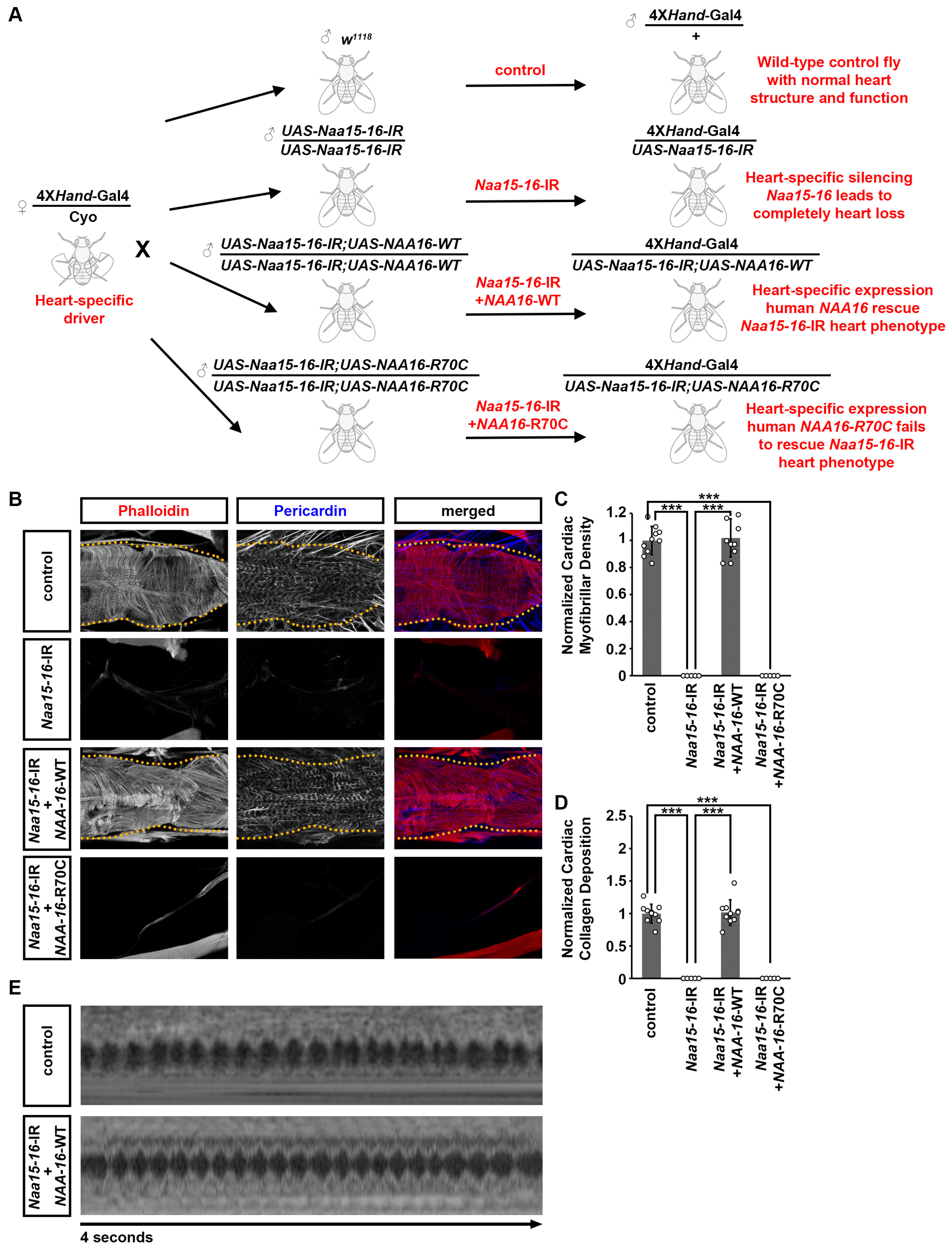
To further validate the function of the NAA16 variant identified in the CHD patients, we performed rescue experiments in Drosophila using the gene replacement strategy we developed previously [27,28,29,38,39] (Figure 5A). First, we assessed the functional conservation between Drosophila Naa15-16 and human NAA16 to establish a platform for variant validation. Cardiac-specific expression of wild-type human NAA16 in the Naa15-16 silenced Drosophila completely rescued the morphological and functional cardiac defects, confirming their conserved function (Figure 5). In contrast, the NAA16 R70C variant failed to restore the normal cardiac morphology (Figure 5B). Because heart-specific silencing of NAA15-16 resulted in a complete loss of cardiac structure in adult Drosophila, no cardiac signals could be detected by OCT in Drosophila with NAA15-16 silencing or those together with expressing the NAA16 R70C variant. These results suggest that the R70C variant functions as a hypomorphic allele, exhibiting partial loss of activity that is insufficient to sustain normal cardiac development in the absence of endogenous Naa15-16.

3. Discussion

3.1. Drosophila as a High-Throughput Platform for Identifying Genes Required for Heart Development

Drosophila offers a powerful, high-throughput genetic system for dissecting conserved molecular mechanisms of heart development [22,23,24]. Though late cardiogenesis anomalies in humans involve chambers or structures that do not have direct morphological counterparts in Drosophila, Drosophila is considered a valuable model for studying early cardiogenesis defects because many signaling pathways, transcriptional regulators, and structural proteins involved in vertebrate heart development are highly conserved in flies [40,41,42]. Perturbations in these conserved components often produce analogous phenotypes, including defects in myocardial organization, contractility, fibrosis, and arrhythmia [23,32,43].
Importantly, Drosophila allows rapid in vivo testing of gene function and variant pathogenicity in the context of a beating heart [44,45,46]. Although the Drosophila heart does not recapitulate chamber-specific features, it provides a genetically tractable and high-throughput system to uncover conserved pathways and validate CHD-associated variants before moving into more complex mammalian models [47,48,49]. Its simple tubular heart shares evolutionary and functional conservation with the vertebrate heart, and genetic tools such as tissue-specific RNAi and CRISPR enable rapid gene functional testing. Our previous large-scale screens have successfully uncovered essential cardiac genes conserved in humans, demonstrating the utility of Drosophila as a platform for functional annotation of CHD candidate genes [27]. Here, we build upon this framework by leveraging the Drosophila heart to interrogate the role of NAT complex components in cardiac development, which has never been accessed in mammals.

3.2. The NatA Complex Is Required for Heart Development and Function

N-terminal acetylation, mediated by NAT complexes, is one of the most prevalent protein modifications in eukaryotes, regulating protein stability, localization, and interactions [5,6,7]. Among these, the NatA complex, which is composed of NAA10 or NAA11 (catalytic) and NAA15 or NAA16 and NAA50 (auxiliary subunit) (Figure 1A), is responsible for modifying nearly 40–50% of the proteome. In our study, we focused on the NatA complex components Naa15-16, Drosophila homolog of NAA15 and NAA16, vnc, Drosophila homolog of NAA10 and NAA11, and san, Drosophila homolog of NAA50. They were all highly conserved from human to Drosophila and essential for Drosophila heart development. Cardiac-specific knockdown of these genes resulted in structural heart defects, early lethality, and reduced lifespan.
The observed loss of myofibrillar organization and abnormal extracellular matrix deposition following Naa15-16, vnc, or san silencing point toward roles in cytoskeletal integrity and ECM remodeling. Given that N-terminal acetylation regulates protein stability and interactions [2,7,50,51], it is possible that NatA substrates include sarcomeric proteins, actin-binding factors, or ECM regulators essential for cardiac morphogenesis. Future studies using proteomic profiling and biochemical assays will help define these substrates and clarify how loss of NatA activity disrupts cardiac structure and function.
Notably, data from the PCGC revealed that approximately 20% of CHD-associated variants are linked to both CHD and neurodevelopmental disabilities (NDD), while only about 2% are associated with isolated CHD [12]. The patients harboring the NAA15 variants presented with CHD but exhibited no signs of NDD [12], suggesting that NAA15 plays a heart-specific role in development, rather than contributing to broader neurodevelopmental phenotypes.
These results provide strong in vivo evidence supporting a critical and previously underappreciated role for NatA complex components in heart development and maintenance. No studies have investigated the roles of Naa15-16, vnc and san in Drosophila heart development. Their exact functions at different stages of cardiac development remain unknown. Further studies were necessary to examine these NatA complex components to define their role in heart development across developmental stages, as well as their downstream targets and molecular pathways.

3.3. Drosophila Enables Variant-Level Validation

One of the major challenges in CHD research is assessing the functional impact of the growing number of candidate variants identified through large-scale exome and genome sequencing efforts. While bioinformatic tools can provide preliminary predictions, experimental validation is essential to distinguish pathogenic variants from benign polymorphisms. However, traditional mammalian models such as mice are time-consuming, costly, and low throughput, making them suboptimal for systematic variant testing. In contrast, Drosophila offers a genetically tractable, scalable, and cost-effective in vivo system that enables efficient evaluation of both gene function and variant pathogenicity in the context of a functioning heart.
In this study, we used the Drosophila heart model to perform variant-level validation of a missense mutation, R70C, in the human NAA16 gene, which was identified in CHD patients by the PCGC. Using a “gene replacement” strategy, we first established that wild-type human NAA16 could fully rescue the severe cardiac morphological and functional defects caused by Naa15-16 knockdown in the fly heart, thereby demonstrating functional conservation between human and Drosophila orthologs. We then introduced the disease-associated NAA16-R70C variant in the same rescue model. This mutant form failed to restore heart structure or function, indicating an LOF effect and confirming the variant pathogenicity in vivo. Additionally, the R70C variant did not alter heart development when overexpressed, but failed to rescue the phenotype in the Naa15-16-IR model. This suggests that the variant protein is not gain-of-function or dominant-negative but rather represents a loss-of-function allele, retaining insufficient activity to support normal cardiac development in the absence of endogenous Naa15-16.
This approach provides compelling evidence for the utility of Drosophila in validating disease-associated alleles at the variant level. It demonstrates that specific human missense mutations can be functionally tested in a whole-organism context that recapitulates key aspects of cardiac development and physiology. Moreover, this strategy enables rapid screening of multiple variants to prioritize those most likely to contribute to disease phenotypes. As such, Drosophila represents a powerful complementary system to mammalian models for bridging the gap between genomic discovery and functional interpretation in the era of precision medicine.

3.4. Potentials of Drosophila as a Preclinical Model for CHD-Linked Ogden Syndrome and Therapeutic Testing

Ogden syndrome is a rare, X-linked developmental disorder caused by mutations in NAA10, which encodes the catalytic subunit of the NatA complex. Patients with Ogden syndrome exhibit a broad spectrum of clinical manifestations, including severe developmental delay, craniofacial dysmorphisms, hypotonia, and notably, CHDs [16,52,53]. Despite its clinical severity, the molecular mechanisms linking NAA10 dysfunction to cardiac phenotypes remain poorly understood, and no targeted therapies are currently available.
In this study, we showed that Ogden syndrome can be modeled in Drosophila by silencing vnc, the Drosophila homolog of NAA10, specifically in the heart. Loss of vnc in the Drosophila heart led to marked cardiac morphological abnormalities, including disorganization of myocardial actin filaments, reduced muscle fiber density, and excessive Pericardin accumulation, a hallmark of cardiac fibrosis. These phenotypes phenocopy aspects of human cardiac pathology and underscore the importance of NatA-mediated acetylation in maintaining cardiac structure and function.
Our findings provide in vivo support for the pathogenicity of NAA10 dysfunction in cardiac tissue and highlight Drosophila as a powerful model system for studying Ogden syndrome. The mechanistic role of NAA10 in heart development and its involvement in associated diseases requires further investigation. Importantly, Drosophila has previously been recognized as a high-throughput, genetically accessible model for validating candidate heart disease genes [27], as well as for exploring the molecular mechanisms and drug discovery pathways relevant to cardiovascular and other systemic diseases [54,55,56,57]. Future studies can leverage the Drosophila model to conduct small-molecule screens aimed at rescuing vnc-associated cardiac defects and identify the potential treatment for Ogden syndrome. Once promising drugs are identified in the Drosophila model with vnc silencing, further validation in mammalian models will be required.
Taken together, our findings demonstrate that the NatA complex plays a conserved and essential role in heart development. Using Drosophila, we developed a high-throughput platform for validating CHD-associated genes and variants and identified a path forward for modeling and potentially treating heart defects associated with Ogden syndrome. This work not only advances our understanding of NatA complex biology but also positions the Drosophila heart as a potentially beneficial preclinical tool for CHD research.

4. Materials and Methods

4.1. Drosophila Lines

Drosophila stocks were obtained from the Bloomington Drosophila Stock Center (BDSC; Indiana University Bloomington, Bloomington, IN, USA). The following lines were used in this study: UAS-Naa15-16-RNAi (BDSC ID 34990), UAS-vnc-RNAi (BDSC ID 56861), UAS-san-RNAi (BDSC ID 36632), UAS-Sbat-RNAi (BDSC ID 55161), UAS-Naa20A-RNAi (BDSC ID 36899), UAS-Naa30A-RNAi (BDSC ID 63983), UAS-Naa35-RNAi (BDSC ID 61901), Control w1118 (BDSCID 3605) flies were used in the crosses. The 4XHand-Gal4/Cyo driver was generated in our lab and used to direct cardiac-specific expression of RNAi-based silencing (-IR) constructs. The wild-type and R70C NAA16 cDNA were synthesized by GenScript (Piscataway, NJ, USA) and individually cloned into the pUAST-attB vector. Transgenic lines were generated via germline transformation into a fixed chromosomal docking site to ensure equivalent expression levels of the wild-type and variant NAA16 alleles in the Drosophila hearts.

4.2. Fly Quantitative RT-PCR Analysis

RNA was isolated from a single 4-day-old adult female fly using TRIzol reagent (Invitrogen, Waltham, MA, USA). RNA purity and concentration were assessed with a NanoDrop-1000 spectrophotometer (Thermo Scientific, Waltham, MA, USA). Total RNA (1 μg) was reverse transcribed using SuperScript IV (Invitrogen, Waltham, MA, USA). SYBR Green–based quantitative real-time PCR (Power SYBR Master Mix; Applied Biosystems, Waltham, MA, USA) was performed on a StepOnePlus system (Applied Biosystems) with gene-specific primer pairs (Integrated DNA Technologies, Coralville, IA, USA). Primer sequences are listed in Supplemental Table S1. Relative gene expression was calculated using the 2-DDCT method and normalized to Gapdh as an endogenous control. Four biological samples were analyzed per genotype.

4.3. Lethality at Eclosion

Eclosion lethality was assessed as the percentage of flies expressing an RNAi-based silencing construct (straight wings) that fail to emerge as adults, relative to their non-expressing siblings (curly wings) as previously described [58,59]. The eclosion lethal rate (percentage) was calculated as [((curly—straight)/curly) × 100]. At least 400 flies (female and male) were analyzed per genotype.

4.4. Adult Drosophila Survival Assay

Drosophila larvae were maintained at 25 °C to induce UAS-transgene expression. After eclosion, adult male flies were collected and kept at 25 °C, with 20 flies per vial. Mortality was recorded every 48 h as previously described [58,59]. One hundred flies were analyzed per genotype.

4.5. Immunochemistry

Adult Drosophila females (4 days old) were dissected and fixed for 10 min in 4% paraformaldehyde prepared in phosphate-buffered saline (1X PBS) as previously described [58,59]. Samples were incubated overnight at 4 °C with primary antibodies diluted in 2% bovine serum albumin (BSA; Sigma, St. Louis, MO, USA) with 0.1% Triton-X (Sigma, St. Louis, MO, USA) in 1X PBS. Following washes, secondary antibodies were applied for 2 h at room temperature in 2% BSA (Sigma, St. Louis, MO, USA) with 0.1% Triton-X (Sigma, St. Louis, MO, USA) in 1X PBS. Alexa Fluor 488 phalloidin (Thermo Fisher, Waltham, MA, USA; A12379) was used at a 1:1000 dilution. Mouse anti-Pericardin antibody (EC11; Developmental Studies Hybridoma Bank, Iowa City, IA, USA) was used at a 1:500 dilution, followed by Alexa Fluor 568 secondary antibody (Thermo Fisher, Waltham, MA, USA; A11011) at 1:1000 dilution. Confocal images were performed using a ZEISS LSM900 microscope with a 63X Plan-Apochromat 1.4 N.A. oil objective (ZEISS, Jena, Germany) and ZEN blue edition (version 3.0) acquisition software. Segment A3 of the heart was imaged by collecting Z-stacks. Control groups were imaged first to establish the laser intensity and exposure settings, which were then kept constant for all genotypes. The exposure time was adjusted to achieve approximately 70% of maximum signal saturation to allow consistent fluorescence intensity comparisons. Image processing was conducted in ImageJ (version 1.52a. 10 adult flies were imaged per genotype, and representative images are shown in the figures.

4.6. Heart Structural Analysis and Quantitation

Drosophila heart cardiac myofibril density and Pericardin deposition were quantified as previously described [58,59]. For each genotype, 10 adult flies (4-day-old females) were analyzed. Image processing and quantification were performed using ImageJ (version 1.52a). The Z-stack projections were examined, and image planes containing cardiac myofibrils were selected for analysis, avoiding the underlying ventral muscle layer. The cardiac myofibril number was quantified by using the MyofibrilJ plugin for Fiji (version 1.53q) [60]. The entire heart region within segment A3 was outlined using the freehand selection function in Fiji, and the number of cardiac myofibrils was measured. Cardiac myofibrillar density was calculated as the number of cardiac myofibril divided by the area of the selected heart region. Pericardin deposition was quantified in the same heart segment A3 based on the mean fluorescence intensity. Both cardiacmyofibril density and Pericardin deposition were normalized to those obtained from the control flies.

4.7. Optical Coherence Tomography (OCT)

Cardiac function in adult Drosophila was assessed using OCT [58,59]. The OCT system (Bioptigen) was constructed as described by the Biophotonics Group, Duke University, NC, USA. Further, 4-day-old adult female flies were anesthetized with carbon dioxide (CO2) for 3–5 min, then gently positioned on a glass plate with petroleum jelly (Vaseline) to immobilize them with the dorsal side facing the OCT imaging source. Flies were allowed to recover for at least 10 min prior to imaging to ensure full mobility and normal heart activity. For each genotype, ten control and ten RNAi-expressing flies were examined. OCT recordings were acquired from the same anatomical location, the cardiac chamber within abdominal segment A3 for all samples. Each measurement was obtained from three different positions within the abdominal segment A3, and the values were averaged to determine the cardiac diameter. M-mode imaging was used to record the heart wall movement during the cardiac cycle. ImageJ software (version 1.52a) was used to process the images. The diastolic dimension and systolic diameter were processed, measured, and determined based on three consecutive heartbeats. The heart period was determined by counting the total number of beats that occurred during a 15 s recording and then dividing 15 by the number of beats.

4.8. Statistical Analysis

Statistical tests were performed using PAST.exe software (latest v.4.13) [Natural History Museum, University of Oslo (UiO), Oslo, Norway]. Data normality was assessed using the Shapiro–Wilk test (α = 0.05). Normally distributed data were analyzed by a one-way analysis of variance (ANOVA) followed by a Tukey–Kramer post-test for comparing multiple groups. Non-normal distributed data were analyzed by a Kruskal–Wallis H-test followed by a Dunn’s test for comparisons between multiple groups. Values are presented as mean ± standard deviation (s.d.) Statistical significance was defined as * p < 0.05, ** p < 0.01, *** p < 0.001. Details for sample sizes used for quantitation have been provided in the figure legends.

Supplementary Materials

The following supporting information can be downloaded at https://www.mdpi.com/article/10.3390/cells14201596/s1, Figure S1: Relative gene expression level for NAT complex components in Drosophila heart. The relative gene expression level for genes encoding NAT complex components in Drosophila cardiogenic progenitors at embryonic stages 13, 14-E (early), 14-L (late), 15, and 16. vnc expression was undetectable in cardiac progenitor cells at any of the examined stages. None of the changes reached statistical significance; Figure S2: Relative gene expression level after NAT complex components silenced in Drosophila heart. The relative gene expression level in adult heart (4-day-old females) following the expression of UAS-RNAi transgenes targeting genes encoding NAT complex components (4XHand-Gal4). [mean ± s.d.; n = 4 biological replicates per genotype, 30 flies per each sample (4-day-old females); Student’s t-test; statistical significance: *** p < 0.001]; Table S1: Primer sequences used for quantitative RT-PCR analysis.

Author Contributions

Methodology, J.-Y.Z. and Z.H.; validation, J.-Y.Z., H.L. (Hangnoh Lee), and Z.H.; formal analysis, J.-Y.Z. and H.S.; investigation, J.-Y.Z., H.S., H.L. (Hangnoh Lee) and H.L. (Hanhan Liu); resources, J.-Y.Z. and H.L. (Hanhan Liu); data curation, J.-Y.Z. and Z.H.; writing—original draft preparation, J.-Y.Z. and Z.H.; writing—review and editing, Z.H.; supervision, J.-Y.Z. and Z.H.; project administration, Z.H.; funding acquisition, Z.H. All authors have read and agreed to the published version of the manuscript.

Funding

Z.H. is supported by U.S. National Institutes of Health (NIH) grants R01-HL134940, R01-HD111480 and R01-HL180768.

Institutional Review Board Statement

Not applicable.

Informed Consent Statement

Not applicable.

Data Availability Statement

The original contributions presented in this study are included in the article/Supplementary Materials. Further inquiries can be directed to the corresponding authors.

Acknowledgments

We thank the Bloomington Drosophila Stock Center at Indiana University Bloomington (IN) for the Drosophila lines and the Developmental Studies Hybridoma Bank at the University of Iowa (IA) for the antibody.

Conflicts of Interest

The authors declare that the research was conducted in the absence of any commercial or financial relationships that could be construed as a potential conflict of interest.

References

  1. Varland, S.; Osberg, C.; Arnesen, T. N-Terminal Modifications of Cellular Proteins: The Enzymes Involved, Their Substrate Specificities and Biological Effects. Proteomics 2015, 15, 2385–2401. [Google Scholar] [CrossRef]
  2. Varland, S.; Silva, R.D.; Kjosås, I.; Faustino, A.; Bogaert, A.; Billmann, M.; Boukhatmi, H.; Kellen, B.; Costanzo, M.; Drazic, A.; et al. N-Terminal Acetylation Shields Proteins from Degradation and Promotes Age-Dependent Motility and Longevity. Nat. Commun. 2023, 14, 6774. [Google Scholar] [CrossRef] [PubMed]
  3. Deng, S.; Marmorstein, R. Protein N-Terminal Acetylation: Structural Basis, Mechanism, Versatility, and Regulation. Trends Biochem. Sci. 2021, 46, 15–27. [Google Scholar] [CrossRef] [PubMed]
  4. Øye, H.; Lundekvam, M.; Caiella, A.; Hellesvik, M.; Arnesen, T. Protein N-Terminal Modifications: Molecular Machineries and Biological Implications. Trends Biochem. Sci. 2025, 50, 290–310. [Google Scholar] [CrossRef]
  5. Nguyen, K.T.; Mun, S.-H.; Lee, C.-S.; Hwang, C.-S. Control of Protein Degradation by N-Terminal Acetylation and the N-End Rule Pathway. Exp. Mol. Med. 2018, 50, 1–8. [Google Scholar] [CrossRef]
  6. McTiernan, N.; Kjosås, I.; Arnesen, T. Illuminating the Impact of N-Terminal Acetylation: From Protein to Physiology. Nat. Commun. 2025, 16, 703. [Google Scholar] [CrossRef] [PubMed]
  7. Ree, R.; Varland, S.; Arnesen, T. Spotlight on Protein N-Terminal Acetylation. Exp. Mol. Med. 2018, 50, 1–13. [Google Scholar] [CrossRef]
  8. Koufaris, C.; Demetriadou, C.; Nicolaidou, V.; Kirmizis, A. Bioinformatic Analysis Reveals the Association of Human N-Terminal Acetyltransferase Complexes with Distinct Transcriptional and Post-Transcriptional Processes. Biochem. Genet. 2024, 63, 2987–3008. [Google Scholar] [CrossRef]
  9. Deng, S.; Magin, R.S.; Wei, X.; Pan, B.; Petersson, E.J.; Marmorstein, R. Structure and Mechanism of Acetylation by the N-Terminal Dual Enzyme NatA/Naa50 Complex. Structure 2019, 27, 1057–1070.e4. [Google Scholar] [CrossRef]
  10. Eiyama, A.; Okamoto, K. Protein N-Terminal Acetylation by the NatA Complex Is Critical for Selective Mitochondrial Degradation. J. Biol. Chem. 2015, 290, 25034–25044. [Google Scholar] [CrossRef]
  11. Jin, S.C.; Homsy, J.; Zaidi, S.; Lu, Q.; Morton, S.; DePalma, S.R.; Zeng, X.; Qi, H.; Chang, W.; Sierant, M.C.; et al. Contribution of Rare Inherited and de Novo Variants in 2,871 Congenital Heart Disease Probands. Nat. Genet. 2017, 49, 1593–1601. [Google Scholar] [CrossRef]
  12. Homsy, J.; Zaidi, S.; Shen, Y.; Ware, J.S.; Samocha, K.E.; Karczewski, K.J.; DePalma, S.R.; McKean, D.; Wakimoto, H.; Gorham, J.; et al. De Novo Mutations in Congenital Heart Disease with Neurodevelopmental and Other Congenital Anomalies. Science 2015, 350, 1262–1266. [Google Scholar] [CrossRef] [PubMed]
  13. Zaidi, S.; Choi, M.; Wakimoto, H.; Ma, L.; Jiang, J.; Overton, J.D.; Romano-Adesman, A.; Bjornson, R.D.; Breitbart, R.E.; Brown, K.K.; et al. De Novo Mutations in Histone-Modifying Genes in Congenital Heart Disease. Nature 2013, 498, 220–223. [Google Scholar] [CrossRef]
  14. Yoshinaga, D.; Craven, I.; Feng, R.; Prondzynski, M.; Shani, K.; Tharani, Y.; Mayourian, J.; Joseph, M.; Walker, D.; Bortolin, R.H.; et al. Dysregulation of N-Terminal Acetylation Causes Cardiac Arrhythmia and Cardiomyopathy. Nat. Commun. 2025, 16, 3604. [Google Scholar] [CrossRef]
  15. Ward, T.; Tai, W.; Morton, S.; Impens, F.; Van Damme, P.; Van Haver, D.; Timmerman, E.; Venturini, G.; Zhang, K.; Jang, M.Y.; et al. Mechanisms of Congenital Heart Disease Caused by NAA15 Haploinsufficiency. Circ. Res. 2021, 128, 1156–1169. [Google Scholar] [CrossRef]
  16. Li, F.; Wang, W.; Li, Y.; Liu, X.; Zhu, Z.; Tang, J.; Hu, Y. NAA10 Gene Related Ogden Syndrome with Obstructive Hypertrophic Cardiomyopathy: A Rare Case Report. Medicine 2024, 103, e36034. [Google Scholar] [CrossRef]
  17. Wu, Y.; Lyon, G.J. NAA10-Related Syndrome. Exp. Mol. Med. 2018, 50, 1–10. [Google Scholar] [CrossRef] [PubMed]
  18. Makwana, R.; Patel, R.; O’Neill, R.; Marchi, E.; Lyon, G.J. The Cardiovascular Manifestations and Management Recommendations for Ogden Syndrome. Pediatr. Cardiol. 2025. [Google Scholar] [CrossRef]
  19. Varland, S.; Brønstad, K.M.; Skinner, S.J.; Arnesen, T. A Nonsense Variant in the N-Terminal Acetyltransferase NAA30 May Be Associated with Global Developmental Delay and Tracheal Cleft. Am. J. Med. Genet. A 2023, 191, 2402–2410. [Google Scholar] [CrossRef]
  20. Forrest, I.S.; Chaudhary, K.; Vy, H.M.T.; Petrazzini, B.O.; Bafna, S.; Jordan, D.M.; Rocheleau, G.; Loos, R.J.F.; Nadkarni, G.N.; Cho, J.H.; et al. Population-Based Penetrance of Deleterious Clinical Variants. JAMA 2022, 327, 350–359. [Google Scholar] [CrossRef] [PubMed]
  21. Sun, B.B.; Kurki, M.I.; Foley, C.N.; Mechakra, A.; Chen, C.-Y.; Marshall, E.; Wilk, J.B.; Biogen Biobank Team; Chahine, M.; Chevalier, P.; et al. Genetic Associations of Protein-Coding Variants in Human Disease. Nature 2022, 603, 95–102. [Google Scholar] [CrossRef]
  22. Souidi, A.; Jagla, K. Drosophila Heart as a Model for Cardiac Development and Diseases. Cells 2021, 10, 3078. [Google Scholar] [CrossRef] [PubMed]
  23. Taghli-Lamallem, O.; Plantié, E.; Jagla, K. Drosophila in the Heart of Understanding Cardiac Diseases: Modeling Channelopathies and Cardiomyopathies in the Fruitfly. J. Cardiovasc. Dev. Dis. 2016, 3, 7. [Google Scholar] [CrossRef] [PubMed]
  24. Ahmad, S.M. Conserved Signaling Mechanisms in Drosophila Heart Development. Dev. Dyn. 2017, 246, 641–656. [Google Scholar] [CrossRef]
  25. Curtis, N.J.; Ringo, J.M.; Dowse, H.B. Morphology of the Pupal Heart, Adult Heart, and Associated Tissues in the Fruit Fly, Drosophila Melanogaster. J. Morphol. 1999, 240, 225–235. [Google Scholar] [CrossRef]
  26. Rotstein, B.; Paululat, A. On the Morphology of the Drosophila Heart. J. Cardiovasc. Dev. Dis. 2016, 3, 15. [Google Scholar] [CrossRef] [PubMed]
  27. Zhu, J.-Y.; Fu, Y.; Nettleton, M.; Richman, A.; Han, Z. High Throughput in Vivo Functional Validation of Candidate Congenital Heart Disease Genes in Drosophila. eLife 2017, 6, e22617. [Google Scholar] [CrossRef]
  28. Alabdi, L.; Altuwaijri, N.; Zhu, J.-Y.; Efthymiou, S.; Lee, H.; Duan, J.; Salem, I.; Yu, P.; Abdullah, N.L.; Alzahrani, F.; et al. SLK Is Mutated in Individuals with a Neurodevelopmental Disorder. eBioMedicine 2025, 116, 105725. [Google Scholar] [CrossRef]
  29. Zhu, J.-Y.; Hannan, S.B.; Dräger, N.M.; Vereshchagina, N.; Krahl, A.-C.; Fu, Y.; Elliott, C.J.H.; Han, Z.; Jahn, T.R.; Rasse, T.M. Autophagy Inhibition Rescues Structural and Functional Defects Caused by the Loss of Mitochondrial Chaperone Hsc70-5 in Drosophila. Autophagy 2021, 17, 3160–3174. [Google Scholar] [CrossRef]
  30. Huang, X.; Fu, Y.; Lee, H.; Zhao, Y.; Yang, W.; van de Leemput, J.; Han, Z. Single-Cell Profiling of the Developing Embryonic Heart in Drosophila. Development 2023, 150, dev201936. [Google Scholar] [CrossRef]
  31. Duffy, J.B. GAL4 System in Drosophila: A Fly Geneticist’s Swiss Army Knife. Genesis 2002, 34, 1–15. [Google Scholar] [CrossRef]
  32. Vaughan, L.; Marley, R.; Miellet, S.; Hartley, P.S. The Impact of SPARC on Age-Related Cardiac Dysfunction and Fibrosis in Drosophila. Exp. Gerontol. 2018, 109, 59–66. [Google Scholar] [CrossRef]
  33. Zheng, W.; Ocorr, K.; Tatar, M. Extra-Cellular Matrix Induced by Steroids and Aging through a G-Protein Coupled Receptor in a Drosophila Model of Renal Fibrosis. Dis. Model. Mech. 2020, 13, dmm041301. [Google Scholar] [CrossRef]
  34. Choma, M.A.; Izatt, S.D.; Wessells, R.J.; Bodmer, R.; Izatt, J.A. Images in Cardiovascular Medicine: In Vivo Imaging of the Adult Drosophila Melanogaster Heart with Real-Time Optical Coherence Tomography. Circulation 2006, 114, e35–e36. [Google Scholar] [CrossRef] [PubMed]
  35. Yelbuz, T.M.; Choma, M.A.; Thrane, L.; Kirby, M.L.; Izatt, J.A. Optical Coherence Tomography: A New High-Resolution Imaging Technology to Study Cardiac Development in Chick Embryos. Circulation 2002, 106, 2771–2774. [Google Scholar] [CrossRef] [PubMed]
  36. Zeytuni, N.; Zarivach, R. Structural and Functional Discussion of the Tetra-Trico-Peptide Repeat, a Protein Interaction Module. Structure 2012, 20, 397–405. [Google Scholar] [CrossRef] [PubMed]
  37. Jacobs, J.P.; Franklin, R.C.G.; Béland, M.J.; Spicer, D.E.; Colan, S.D.; Walters, H.L., 3rd; Bailliard, F.; Houyel, L.; St Louis, J.D.; Lopez, L.; et al. Nomenclature for Pediatric and Congenital Cardiac Care: Unification of Clinical and Administrative Nomenclature-the 2021 International Paediatric and Congenital Cardiac Code (IPCCC) and the Eleventh Revision of the International Classification of Diseases (ICD-11). World J. Pediatr. Congenit. Heart Surg. 2021, 12, E1–E18. [Google Scholar]
  38. Zhu, J.-Y.; Fu, Y.; Richman, A.; Zhao, Z.; Ray, P.E.; Han, Z. A Personalized Model of COQ2 Nephropathy Rescued by the Wild-Type COQ2 Allele or Dietary Coenzyme Q10 Supplementation. J. Am. Soc. Nephrol. 2017, 28, 2607–2617. [Google Scholar] [CrossRef]
  39. Zhao, F.; Zhu, J.-Y.; Richman, A.; Fu, Y.; Huang, W.; Chen, N.; Pan, X.; Yi, C.; Ding, X.; Wang, S.; et al. Mutations in NUP160 Are Implicated in Steroid-Resistant Nephrotic Syndrome. J. Am. Soc. Nephrol. 2019, 30, 840–853. [Google Scholar] [CrossRef]
  40. Olson, E.N. Gene Regulatory Networks in the Evolution and Development of the Heart. Science 2006, 313, 1922–1927. [Google Scholar] [CrossRef]
  41. Cui, M.; Wang, Z.; Bassel-Duby, R.; Olson, E.N. Genetic and Epigenetic Regulation of Cardiomyocytes in Development, Regeneration and Disease. Development 2018, 145, dev171983. [Google Scholar] [CrossRef]
  42. Cannon, L.; Zambon, A.C.; Cammarato, A.; Zhang, Z.; Vogler, G.; Munoz, M.; Taylor, E.; Cartry, J.; Bernstein, S.I.; Melov, S.; et al. Expression Patterns of Cardiac Aging in Drosophila. Aging Cell 2017, 16, 82–92. [Google Scholar] [CrossRef] [PubMed]
  43. Bodmer, R. Drosophila as a Model for Congenital Heart Disease and Arrhythmia. J. Card. Fail. 2006, 12, S153. [Google Scholar] [CrossRef]
  44. Dorn, G.W., 2nd; Clark, C.F.; Eschenbacher, W.H.; Kang, M.-Y.; Engelhard, J.T.; Warner, S.J.; Matkovich, S.J.; Jowdy, C.C. MARF and Opa1 Control Mitochondrial and Cardiac Function in Drosophila. Circ. Res. 2011, 108, 12–17. [Google Scholar] [CrossRef]
  45. Smeir, E.; Leberer, S.; Blumrich, A.; Vogler, G.; Vasiliades, A.; Dresen, S.; Jaeger, C.; Gloaguen, Y.; Klose, C.; Beule, D.; et al. Depletion of Cardiac Cardiolipin Synthase Alters Systolic and Diastolic Function. iScience 2021, 24, 103314. [Google Scholar] [CrossRef]
  46. Birker, K.; Ge, S.; Kirkland, N.J.; Theis, J.L.; Marchant, J.; Fogarty, Z.C.; Missinato, M.A.; Kalvakuri, S.; Grossfeld, P.; Engler, A.J.; et al. Mitochondrial MICOS Complex Genes, Implicated in Hypoplastic Left Heart Syndrome, Maintain Cardiac Contractility and Actomyosin Integrity. eLife 2023, 12, e83385. [Google Scholar] [CrossRef]
  47. Wessells, R.J.; Bodmer, R. Screening Assays for Heart Function Mutants in Drosophila. Biotechniques 2004, 37, 58–60, 62, 64 passim. [Google Scholar] [CrossRef] [PubMed]
  48. Petersen, C.E.; Tripoli, B.A.; Schoborg, T.A.; Smyth, J.T. Analysis of Drosophila Cardiac Hypertrophy by Microcomputerized Tomography for Genetic Dissection of Heart Growth Mechanisms. Am. J. Physiol. Heart Circ. Physiol. 2022, 322, H296–H309. [Google Scholar] [CrossRef]
  49. Gomez, A.; Gonzalez, S.; Oke, A.; Luo, J.; Duong, J.B.; Esquerra, R.M.; Zimmerman, T.; Capponi, S.; Fung, J.C.; Nystul, T.G. A High-Throughput Method for Quantifying Drosophila Fecundity. Toxics 2024, 12, 658. [Google Scholar] [CrossRef]
  50. van de Kooij, B.; de Vries, E.; Rooswinkel, R.W.; Janssen, G.M.C.; Kok, F.K.; van Veelen, P.A.; Borst, J. N-Terminal Acetylation Can Stabilize Proteins Independent of Their Ubiquitination. Sci. Rep. 2023, 13, 5333. [Google Scholar] [CrossRef]
  51. François, C.M.; Pihl, T.; Dunoyer de Segonzac, M.; Hérault, C.; Hudry, B. Metabolic Regulation of Proteome Stability via N-Terminal Acetylation Controls Male Germline Stem Cell Differentiation and Reproduction. Nat. Commun. 2023, 14, 6737. [Google Scholar] [CrossRef]
  52. Shishido, A.; Morisada, N.; Tominaga, K.; Uemura, H.; Haruna, A.; Hanafusa, H.; Nozu, K.; Iijima, K. A Japanese Boy with NAA10-Related Syndrome and Hypertrophic Cardiomyopathy. Hum. Genome Var. 2020, 7, 23. [Google Scholar] [CrossRef] [PubMed]
  53. Sidhu, M.; Brady, L.; Tarnopolsky, M.; Ronen, G.M. Clinical Manifestations Associated with the N-Terminal-Acetyltransferase NAA10 Gene Mutation in a Girl: Ogden Syndrome. Pediatr. Neurol. 2017, 76, 82–85. [Google Scholar] [CrossRef]
  54. Zhu, J.-Y.; van de Leemput, J.; Han, Z. Promoting Mitochondrial Dynamics by Inhibiting the PINK1/PRKN Pathway to Relieve Diabetic Nephropathy. Dis. Model. Mech. 2024, 17, dmm050471. [Google Scholar] [CrossRef] [PubMed]
  55. Kaun, K.R.; Devineni, A.V.; Heberlein, U. Drosophila Melanogaster as a Model to Study Drug Addiction. Hum. Genet. 2012, 131, 959–975. [Google Scholar] [CrossRef]
  56. Kruger, L.; Denton, T.T. A Standardized Method for Incorporation of Drugs into Food for Use with Drosophila Melanogaster. Anal. Biochem. 2020, 599, 113740. [Google Scholar] [CrossRef]
  57. DeLoriea, J.; Millet-Boureima, C.; Gamberi, C. Protocol to Build a Drug-Testing Pipeline Using Large Populations of Drosophila Melanogaster. STAR Protoc. 2023, 4, 102747. [Google Scholar] [CrossRef]
  58. Zhu, J.-Y.; van de Leemput, J.; Han, Z. Distinct Roles of COMPASS Subunits to Drosophila Heart Development. Biol. Open 2024, 13, bio061736. [Google Scholar] [CrossRef]
  59. Zhu, J.-Y.; Lee, H.; Huang, X.; van de Leemput, J.; Han, Z. Distinct Roles for COMPASS Core Subunits Set1, Trx, and Trr in the Epigenetic Regulation of Drosophila Heart Development. Int. J. Mol. Sci. 2023, 24, 17314. [Google Scholar] [CrossRef]
  60. Spletter, M.L.; Barz, C.; Yeroslaviz, A.; Zhang, X.; Lemke, S.B.; Bonnard, A.; Brunner, E.; Cardone, G.; Basler, K.; Habermann, B.H.; et al. A Transcriptomics Resource Reveals a Transcriptional Transition during Ordered Sarcomere Morphogenesis in Flight Muscle. eLife 2018, 7, e34058. [Google Scholar] [CrossRef] [PubMed]
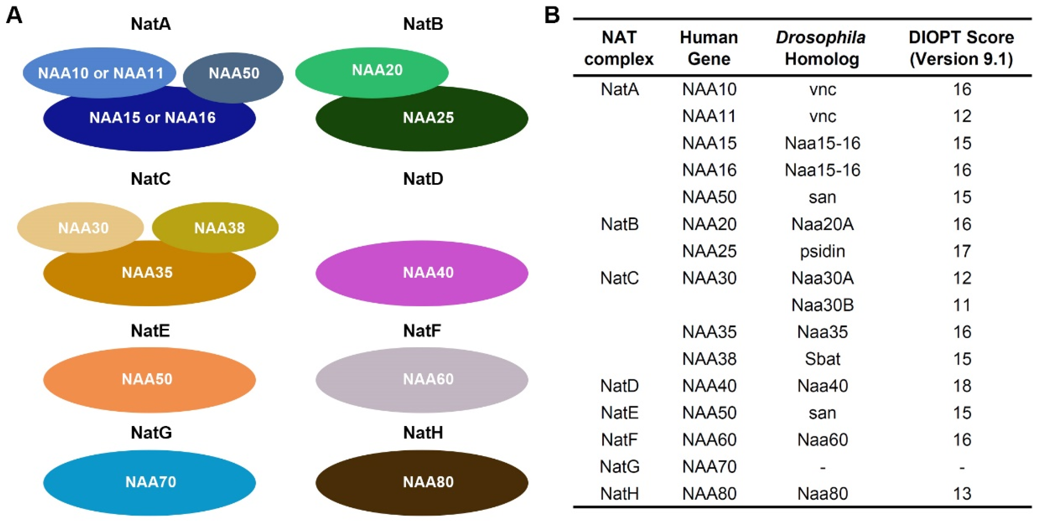
Figure 1. NAT complexes and their components. (A) Schematic representation of eight different NAT complexes. NAA10, NAA11, NAA20, NAA30, NAA40, NAA50, NAA60, NAA70, and NAA80 are the catalytic enzymes in each complex. (B) Human and Drosophila NAT complexes and their subunits. DIOPT, DRSC integrative ortholog prediction tool (version 9.1; max score Drosophila-human is 19).
Cells 14 01596 g001
Figure 2. Drosophila survival and cardiac structure following heart-specific silencing of NAT complex components. (A) Eclosion lethality induced by 4XHand-Gal4-driven expression of UAS-RNAi transgenes targeting individual NAT complex components. Crossed with a CyO (curly wing) balancer allowed identification of progeny: adults with curly wings (CyO) lacked transgene expression, whereas straight-winged flies expressed 4XHand-Gal4 > RNAi. The eclosion lethal rate (percent) was calculated as [((curly–straight)/curly) × 100]. All lines also carried 4XHand-Gal4. n = 400+ flies (female and male) per genotype. (B) Survival curves of adult flies expressing RNAi (-IR) transgenes targeting individual NAT complex components in heart cells. Control, 4XHand-Gal4+/−. n = 100 male flies (20 flies/vial) per genotype. (C) Representative confocal images of adult (4-day-old females) cardiac structure following the expression of UAS-RNAi transgenes targeting each NAT complex component (4XHand-Gal4). Cardiac actin myofibers were visualized by phalloidin staining (red). Pericardin was detected by immunofluorescence (blue). Dotted lines indicate heart tube boundaries. Control, 4XHand-Gal4+/−. (D) Quantitation of adult heart cardiac myofibrillar density relative to control (see image in (C)) [mean ± s.d.; n = 10 flies (4-day-old females) per genotype; Kruskal–Wallis H-test followed by a Dunn’s test; statistical significance: *** p < 0.001]. (E) Quantitation of adult heart cardiac collagen (Pericardin) deposition relative to control (see images in (C)) [mean ± s.d.; n = 10 flies (4-day-old females) per genotype; Kruskal–Wallis H-test followed by a Dunn’s test; statistical significance: *** p < 0.001].
Cells 14 01596 g002
Figure 3. Cardiac function in flies following heart-specific silencing of NAT complex components. (A) Representative OCT images from heartbeat recording of Drosophila (4-day-old females) expressing UAS-RNAi transgenes targeting individual NAT complex components. Control, 4XHand-Gal4+/−. (B) Quantitation of heart period (see images in (A)). (C) Representative images for diastolic and systolic heart diameter are shown for flies that express UAS-RNAi transgenes targeting san and vnc. Control, 4XHand-Gal4+/−. (D) Quantitation of adult heart diastolic diameter (see images in (C)). (E) Quantitation of adult heart systolic diameter (see images in (C)). [mean ± s.d.; n = 10 flies (4-day-old females) per genotype; Kruskal–Wallis H-test followed by a Dunn’s test; statistical significance: *** p < 0.001].
Cells 14 01596 g003
Figure 4. Drosophila cardiac phenotypes induced by expression wild-type human NAA16 and its CHD-associated variant. (A) Representative confocal images of adult (4-day-old females) Drosophila hearts expressing either wild-type human NAA16 or its CHD-associated variant R70C. Cardiac actin myofibers were visualized by phalloidin staining (red). Pericardin was detected by immunofluorescence (blue). Dotted lines indicated the boundaries of the heart tube. Control, 4XHand-Gal4+/−. (B) Quantitation of adult heart cardiac myofibrillar density relative to control (see image in (A)) [mean ± s.d.; n = 10 flies (4-day-old females) per genotype; Kruskal–Wallis H-test followed by a Dunn’s test. (C) Quantitation of adult heart cardiac collagen (Pericardin) deposition relative to control (see images in (A)) [mean ± s.d.; n = 10 flies (4-day-old females) per genotype; Kruskal–Wallis H-test followed by a Dunn’s tes. (D) Representative OCT images from heartbeat recording of Drosophila (4-day-old females) expressing either wild-type human NAA16 or its CHD-associated variant R70C. Control, 4XHand-Gal4+/−.
Cells 14 01596 g004
Figure 5. Defects caused by Drosophila NAA15-16 silencing can be rescued by wild-type human NAA16, but not the CHD-associated variant. (A) Schematic representation of the “Gene Replacement” strategy used to rescue the heart phenotype caused by heart-specific silencing of Drosophila NAA15-16, using human wild-type NAA16 and the disease-associated variant R70C. (B) Representative confocal images of adult (4-day-old females) Drosophila hearts UAS-RNAi transgenes targeting NAA15-16 (4XHand-Gal4), and together with human wildtype NAA16 or its CHD-associated variant. Cardiac actin myofibers were visualized by phalloidin staining (red). Pericardin was detected by immunofluorescence (blue). Dotted lines indicated the boundaries of the heart tube. Control, 4XHand-Gal4+/−. (C) Quantitation of adult heart cardiac myofibrillar density relative to control (see image in (A)) [mean ± s.d.; n = 10 flies (4-day-old females) per genotype; Kruskal–Wallis H-test followed by a Dunn’s test; statistical significance: *** p < 0.001]. (D) Quantitation of adult heart cardiac collagen (Pericardin) deposition relative to control (see images in (A)) [mean ± s.d.; n = 10 flies (4-day-old females) per genotype; Kruskal–Wallis H-test followed by a Dunn’s test; statistical significance: *** p < 0.001]. (E) Representative OCT images from heartbeat recording of Drosophila (4-day-old females) expressing UAS-RNAi transgenes targeting NAA15-16 (4XHand-Gal4), and together with human wildtype NAA16 or its CHD-associated variant. Control, 4XHand-Gal4+/−.
Cells 14 01596 g005
Table 1. NAT complex-related genetic variants identified by PCGC in congenital heart disease. CTD, conotruncal defects; TOF, Tetralogy of Fallot; HTX, Heterotaxy [37].
Patient IDCardiac CategoryCardiac DiagnosesHuman GeneAA ChangesEffect
1-01119CTDAtrial septal defect, secundum; Transposition D-loop with ventricular septal defect; Usual coronary arteries in D-loop TGANAA16R70CMissense
1-01943TOFTetralogy of FallotNAA16L765fsFrameshift
1-06626TOFLSVC to coronary sinus; Tetralogy of Fallot with pulmonary atresiaNAA16E630fsFrameshift
1-00455HTXAortic valve position relative to the pulmonary valve, anterior; Atrial inversion; Congenital tricuspid valve abnormality; D-looped ventricles; Dextrocardia; DORV, ventricular defect uncommitted; Heterotaxy; Hypoplastic right ventricle; Hypoplastic tricuspid valve; IDD; Left superior vena cava to right atrium; Pulmonary stenosis, bilateral branch pulmonary artery; Pulmonary stenosis, valvar; Right aortic arch; Right superior vena cava absent; Totally anomalous pulmonary venous return, mixedNAA15K336fsFrameshift
1-00141TOFPulmonary stenosis, valvar; Single left coronary; Tetralogy of FallotNAA15S761XNonsense
Disclaimer/Publisher’s Note: The statements, opinions and data contained in all publications are solely those of the individual author(s) and contributor(s) and not of MDPI and/or the editor(s). MDPI and/or the editor(s) disclaim responsibility for any injury to people or property resulting from any ideas, methods, instructions or products referred to in the content.