Article

Impact of Tobacco Smoke Exposure on Male Fertility: An In Vivo Study Using Drosophila melanogaster

by
Natasha Gomes de Miranda
1,2,
Ana Gajeiro
1,2,
Ana Martins-Bessa
1,3,* and
Isabel Gaivão
1,2,*
1
Animal and Veterinary Research Center (CECAV), Associate Laboratory for Animal and Veterinary Sciences (AL4AnimalS), University of Trás-os-Montes and Alto Douro (UTAD), 5000-801 Vila Real, Portugal
2
Department of Genetics and Biotechnology (DGB–ECVA), University of Trás-os-Montes and Alto Douro (UTAD), Quinta de Prados, 5001-801 Vila Real, Portugal
3
Department of Veterinary Sciences (DCV-ECAV), University of Trás-os-Montes and Alto Douro (UTAD), Quinta de Prados, 5001-801 Vila Real, Portugal
*
Authors to whom correspondence should be addressed.
Cells 2025, 14(21), 1689; https://doi.org/10.3390/cells14211689
Submission received: 31 August 2025 / Revised: 2 October 2025 / Accepted: 10 October 2025 / Published: 28 October 2025
(This article belongs to the Special Issue Drosophila as a Model for Understanding Human Disease)

Highlights

What are the main findings?
  • Paternal tobacco smoke exposure significantly impairs the fertility, prolificacy, and longevity of offspring of two strains of Drosophila melanogaster: Oregon K (a wild-type strain proficient in all major DNA repair pathways) and mus308 (a DNA repair-deficient strain).
  • The DNA repair-deficient mus308 strain showed higher vulnerability to spermatozoa cytotoxicity and a greater magnitude of reduction in spermatozoa count.
What is the implication of the main finding?
  • The reproductive toxicity of tobacco smoke is genotype-dependent, suggesting that compromised DNA repair pathways exacerbate the transgenerational impact of environmental toxicants.
  • D. melanogaster is a robust and sensitive in vivo model to mechanistically evaluate the paternal reproductive risks and transgenerational effects of complex smoke-related compounds.

Abstract

Tobacco smoke has been consistently associated with impaired spermatozoa quality in men, including decreased concentration, motility, and increased morphological abnormalities. Key tobacco-related toxins such as nicotine and cadmium induce oxidative stress, leading to DNA damage in germ cells. This study aimed to evaluate the effects of tobacco smoke exposure on male fertility using Drosophila melanogaster as an in vivo model. Fertility, fecundity, parental toxicity, lifespan, and spermatozoa morphology were assessed in two strains: the wild-type Oregon K and the DNA repair-deficient mus308. Males were exposed to whole-tobacco smoke in a controlled environment for periods of 0, 1 and 7 min. Tobacco smoke exposure reduced fertility, fecundity, and offspring longevity in both strains. Additionally, spermatozoa from mus308 males exhibited a higher frequency of morphological abnormalities. These findings demonstrate the detrimental impact of tobacco smoke on male reproductive function and suggest increased vulnerability in organisms with impaired DNA repair capacity.

1. Introduction

Infertility affects approximately 10–15% of human couples worldwide, with male factors accounting for nearly 20% of cases. Considering environmental factors, tobacco smoke exposure is robustly associated with detrimental impacts on sperm quality parameters, specifically manifesting as diminished concentration and motility, alongside elevated morphological abnormalities [1,2]. Tobacco (Nicotiana tabacum) contains numerous toxic components—such as nicotine and cadmium—that are known mutagens and carcinogens. These toxic components drive oxidative stress and subsequent DNA damage within germ cells, a pathway that ultimately compromises fertilization potential and elevates the risk of adverse reproductive outcomes [3,4,5].
The accumulation of tobacco smoke-derived toxic compounds is observed at concentrations in seminal fluid exceeding those found in the bloodstream, emphasizing a direct toxicological impact on spermatogenesis. Several studies have demonstrated a dose-dependent relationship between tobacco exposure and the severity of sperm impairment, because men who have smoked for more than five years have significantly worse seminal parameters than non-smokers or those who have smoked for less time [6,7,8]. Notably, cadmium has been implicated in increased oxidative stress, depletion of antioxidant defences, and dysfunction of sperm plasma membrane enzymes such as Ca2+-ATPase. Cadmium exposure also disrupts essential trace elements like zinc and magnesium, which are critical for DNA replication, chromatin stability, and overall sperm functionality [9,10,11,12].
The detrimental effects of tobacco smoke on male reproductive health have been extensively studied in mammalian models. However, research using dipteran species, particularly Drosophila melanogaster, remains limited despite their increasing use in toxicological and genotoxicity assays. Adopting Drosophila melanogaster as an in vivo model offers significant logistical benefits, including its accelerated life cycle, elevated reproductive output, and economical laboratory upkeep. The fruit fly Drosophila melanogaster is extensively employed in genotoxicity research because many of its genetic repair mechanisms and cellular pathways are evolutionarily conserved with those found in mammals. This similarity enables scientists to perform detailed mechanistic studies on toxic effects in a simpler and high-throughput model organism. Although Drosophila is useful for preliminary reproductive toxicity screening, its distinct reproductive physiology and absence of key mammalian pathways limit result extrapolation. Thus, complementary mammalian studies are essential for accurate assessment of EDCs and teratogens [5,13,14,15,16,17,18,19].
This study aimed to evaluate the effects of tobacco smoke exposure on male fertility using D. melanogaster in vivo. Additionally, the study sought to characterize changes in spermatozoa morphology, thereby contributing to the limited body of literature on Drosophila reproductive toxicology.

2. Materials and Methods

2.1. Strains

In D. melanogaster, the repair of DNA double-strand breaks (DSBs) predominantly relies on three established mechanisms: homologous recombination (HR), the classical non-homologous end joining (c-NHEJ) pathway, and the microhomology-mediated end joining (MMEJ) pathway. The present investigation utilized two distinct fly strains:
-
Oregon K (OK): This strain serves as the wild-type control and is fully competent in processing DNA lesions across all primary repair mechanisms, specifically HR, c-NHEJ, and MMEJ [20,21,22].
-
mus308: This line is homozygous for the mus308 mutation, which results in a non-functional dmPolQ protein (the functional equivalent of human DNA polymerase θ, or PolQ). The mus308 mutation imparts severe deficits in the repair of DNA interstrand cross-links and complex, persistent DNA damage. Its principal role is recognized in alternative end-joining pathways, particularly MMEJ, in addition to its involvement in translesion DNA synthesis and damage bypass [20,21].

2.2. Medium Conditions

All fly populations were maintained under carefully regulated laboratory conditions, including a controlled temperature and a consistent 12:12 h light–dark cycle. Under these standardized conditions, the species completes its life cycle in approximately 10–12 days, with adult flies living on average between 40 and 50 days. For colony maintenance, newly eclosed adults were transferred to clean glass containers supplied with the standard nutritional medium (Table 1). The culture vials were then securely closed using cotton plugs, a technique employed to facilitate necessary gas exchange while safeguarding against microbial contamination [22].

2.3. Fertility, Prolificacy, and Toxicity Assessment

Flies were divided into four experimental groups based on genotype and treatment: Oregon K control, Oregon K exposed to tobacco smoke, mus308 control, and mus308 exposed to tobacco smoke. Males and females aged between 0 and 1 days were selected. A total of 180 males were used, equally distributed between the Oregon K and mus308 strains (90 males per strain). For the toxicity and longevity tests, 75 males were used per strain. For the fertility and prolificity tests, a total of 45 replicates were used per group, with one male per replicate.
Males in the treatment groups were exposed to tobacco smoke by being placed in a 600 mL glass beaker that was covered with a net. A lit Marlboro® Gold cigarette butt was placed inside to initiate the exposure, creating a smoke concentration of approximately 1 mg/L. This specific brand was chosen because it is a low-yield cigarette (0.6 mg of nicotine, 6 mg of tar, and 7 mg of carbon monoxide under the ISO regime. These values increase dramatically under the Health Canada intense regime, as ventilation is blocked, allowing for more smoke delivery. The exposure consisted of three sequential intervals of 0, 1, and 7 min. This duration was determined through preliminary tests to be the maximum survival time for the flies under these conditions.
Following eclosion of the offspring, the number of adult males, females, and pupal deaths was recorded for each vial. Reproductive fitness (fertility) was quantified by the percentage of exposed males capable of successfully generating viable progeny. Reproductive output (prolificacy) was calculated as the aggregate count of offspring yielded from all tested males within a given cohort. Toxicity assessment was based on measuring the acute mortality rate observed in the exposed male population following the treatment phase. All trials were conducted using 3 independent biological replications, with 15 mating tubes constituting each experimental group.

2.4. Longevity Assay

Following the completion of progeny counts, the F1 offspring of each experimental condition (Oregon K control, 31 total offspring; Oregon K 1 min tobacco exposure, 32 total; Oregon K, 7 min tobacco exposure, 41 total; mus308 control, 27 total; mus308 1 min tobacco exposure, 32 total; and mus308 7 min tobacco exposure, 41 total) were moved to 3 replicate vials corresponding to their respective strain and exposure group. All groups were subsequently housed in 200 mL glass vials, each containing approximately 25 mL of culture medium. To ensure a steady nutritional quality and prevent the build-up of pathogens or contamination from differing developmental stages, the flies were moved to fresh culture media every 7 to 9 days. The total duration in days, measured from adult eclosion until the time of death for every individual, defined longevity, with the final dataset incorporating individuals from both sexes [22].

2.5. Morphological Examination of Spermatozoa

One virgin male with Oregon K and mus308 strains, aged 8–9 days, underwent tobacco smoke exposure for one minute before anesthesia via ethyl ether. Dissection of the seminal vesicles occurred in 10 μL of Ringer’s solution (130 mM NaCl, 35 mM KCl, 2 mM CaCl2; pH 6.5, adjusted with NaOH). The vesicles were subsequently moved to a second slide containing another 10 μL aliquot of fresh Ringer’s solution. Using a fine pin, the vesicles were carefully punctured to liberate the spermatozoa. Tweezers assisted in gently swirling the vesicle within the drop to enhance spermatozoa release [23]. Following this, 5 μL of the resultant spermatozoa suspension was combined with 20 μL of 1% low-melting-point (LMP) agarose (Pronadisa Micro & Molecular Biology, Madrid, Spain) pre-warmed to 37 °C. This final mixture was uniformly dispersed onto a microscope slide previously treated with 150 μL of 0.5% normal melting point (NMP) agarose (Sigma-Aldrich, St. Louis, MO, USA). An 18 × 18 mm coverslip was carefully seated over the preparation and pressed gently for gel flattening. To allow for solidification, the slides were placed at 4 °C for 5 min. After the coverslip was gingerly taken off, dehydration was achieved by sequentially immersing the slides in 70%, 90%, and 96% ethanol, followed by air-drying at room temperature. The process concluded with staining using 20 μL (1 μg/mL) of DAPI (4′,6-diamidino-2-phenylindole) to enable morphological assessment and counting of spermatozoa (1 male fly per slide from the post-treatment groups was evaluated), utilizing an Olympus BX61 (Olympus/Evident Scientific, Tokyo, Japan) fluorescence microscope at 200× magnification.

2.6. Statistical Analysis

For the longevity analysis, data were processed utilizing the OASIS 2 (Online Application for Survival Analysis 2) web-based platform. Survival curves were constructed employing the Kaplan–Meier approach, reporting 95% confidence intervals. Assessment of overall differences across the survival curves was carried out via the log-rank test (chi-square test), with corresponding p-values being documented. To establish the statistical variance separating the experimental treatment cohorts from the control group, an F-test was employed, which was executed immediately following the required analysis of variance (ANOVA) applied to the prolificacy data in its entirety. Statistical significance was designated at a threshold of p < 0.05.

3. Results

3.1. Male Fertility

Fertility was evaluated as the percentage of males producing at least one offspring per vial after exposure to tobacco smoke for 1 and 7 min in the Oregon K (OK) and mus308 strains. The results are summarized in Table 2.
Exposure to tobacco smoke for 1 min significantly increased male fertility in the OK strain compared to the control group (87% vs. 60%, p < 0.05). In contrast, in the mus308 strain, 1 min exposure resulted in a decrease in male fertility in the treated group relative to the control group (93% vs. 100%, p = 0.475); however, this difference was not statistically significant. After 7 min of exposure, male fertility in the OK-treated group increased to 100%, while in the mus308-treated group it decreased to 60%, with no significant differences.

3.1.1. Prolificacy

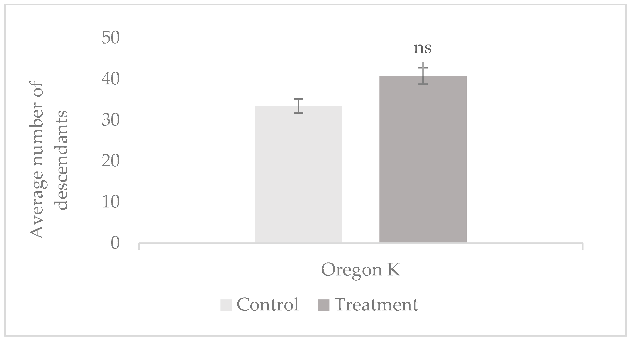
Prolificacy was evaluated by measuring the average number of offspring per test tube in the control and treatment groups of both Oregon K (OK) and mus308 strains. The 1 min tobacco smoke exposure experiments were based on 37 to 45 replicates per group using 37 to 45 males in total, while the 7 min exposures were based on 9 to 15 replicates per group using 15 males in total. The results are presented in Figure 1 and Figure 2.
Results indicate an increase in prolificacy in both strains following exposure to tobacco smoke. Specifically, in the mus308 line (Figure 1), the number of descendants per tube was 32 (treatment) compared to 27 (control). For the OK line following 7 min exposure (Figure 2), the number of descendants was 41 (treatment) and 33 (control). These findings suggest that short-term exposure to tobacco smoke may stimulate an increase in prolificacy, particularly in the Oregon K strain, where the number of offspring per tube increased. A post hoc comparison between the OK control and OK-treated group showed a statistically significant difference (p = 0.034, d = 0.65). However, no significant differences were observed in the mus308 strain (p = 0.473, d = 0.28, CI 95% = [−0.60, 1.16]) or in either strain after 7 min exposures (p > 0.05), indicating that the effect may be strain- and time-dependent.

3.1.2. Male Toxicity

Toxicity was evaluated based on the mortality rate of males exposed to tobacco smoke for 1 min, as shown in the data, which are summarized in Table 3, based on a total of 75 males per strain exposed to tobacco smoke.
Exposure to tobacco smoke resulted in notable male mortality in both strains. The mus308 strain showed a higher mortality rate (51%) compared to the Oregon K strain (40%), suggesting increased sensitivity to tobacco-induced toxicity. These results indicate that tobacco smoke exerts toxic effects on adult males with genotoxically compromised strains, like mus308, being more susceptible. No relevant mortality values were obtained in these studies in the control groups. No studies with the same methodology were found in the literature.

3.2. Offspring Longevity

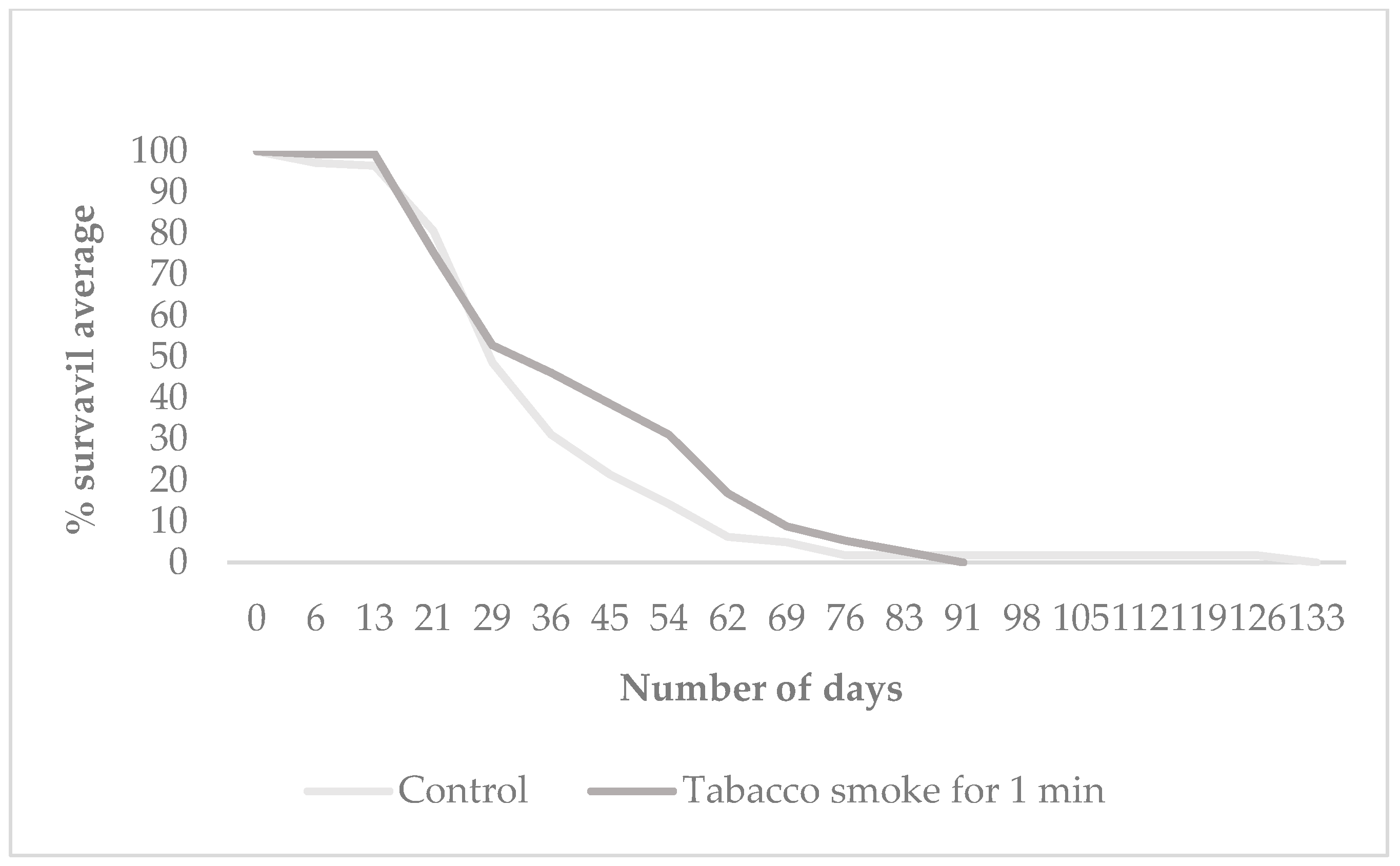
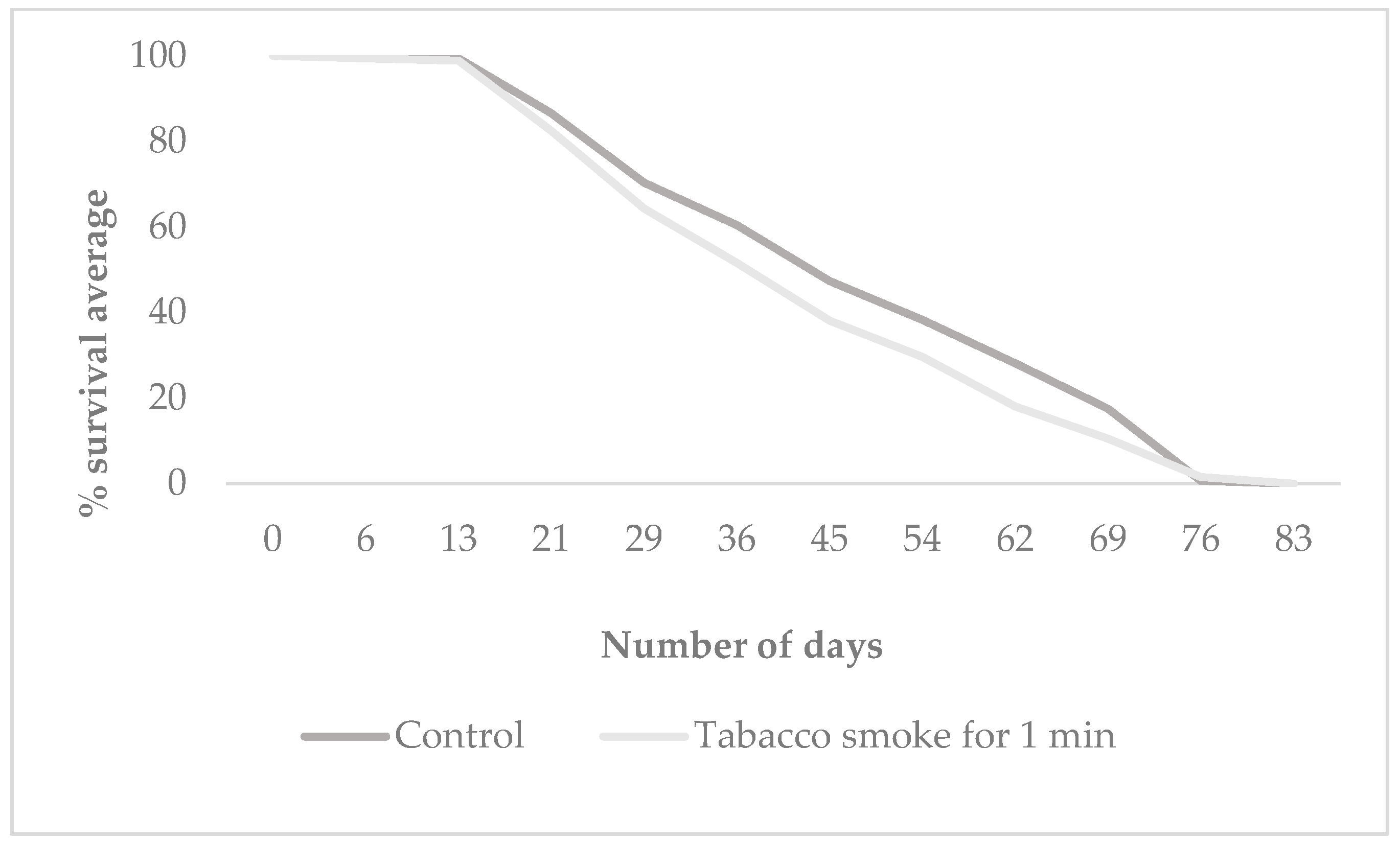
Longevity was assessed by measuring the life expectancy of the F1 offspring exposed to tobacco smoke for 1 min, derived from the control and treatment groups mus 308 and O.K. Figure 3 presents the mean survival time (in days) of F1 individuals from both Oregon K and mus308 strains under control and tobacco smoke exposure conditions. Figure 4 and Figure 5 display Kaplan–Meier survival curves indicating the percentage of surviving F1 flies over time for each group. Each dataset represents the average of three independent replicates, totalling nine tests per condition, with 57 individuals on average per group.
Exposure to tobacco smoke resulted in a reduction in the mean lifespan of F1 offspring in both the Oregon K (OK) and mus308 strains compared to their respective control groups. In the OK strain, the mean lifespan decreased from 50 days (control) to 43 days (treatment). A significant difference in survival was found between the groups (χ2 = 7.23, p = 0.007). Similarly, in the mus308 strain, the mean lifespan was reduced from 54 days in the control group to 44 days following exposure. However, statistical analysis using the log-rank test for the mus308 strain revealed a χ2 = 4.48 and a p-value = 0.346 indicating no significant difference in survival between the control and treated groups (HR = 1.25, CI 95% = [0.98, 1.60]). This suggests that the impact of paternal tobacco smoke exposure on offspring longevity is strain-dependent, with a significant effect on the overall survival curve observed only in the Oregon K strain. The difference in results between the mean lifespan (Figure 3) and the overall survival curve (Figure 4) reflects the power of the Log-rank test to detect time-dependent survival differences, even when the mean lifespans are not significantly different by ANOVA.

3.3. Spermatozoa Morphology

Spermatozoa morphology was analyzed using a modified version of the Single-Cell Gel Electrophoresis (SIM) technique as described by [23]. Samples were observed under a fluorescence microscope Olympus BX61 (Olympus/Evident Scientific, Tokyo, Japan, and images were captured using an integrated digital camera connected to the cellSens Standard software (Version 4.2). The number of spermatozoa was quantified on each slide, and morphological differences between the control and treatment groups were assessed qualitatively. An average total of 330 spermatozoa were counted on the slides analyzed for the OK strain, while 238 were counted on the slides analyzed for the mus 308 strain, all from the control group.
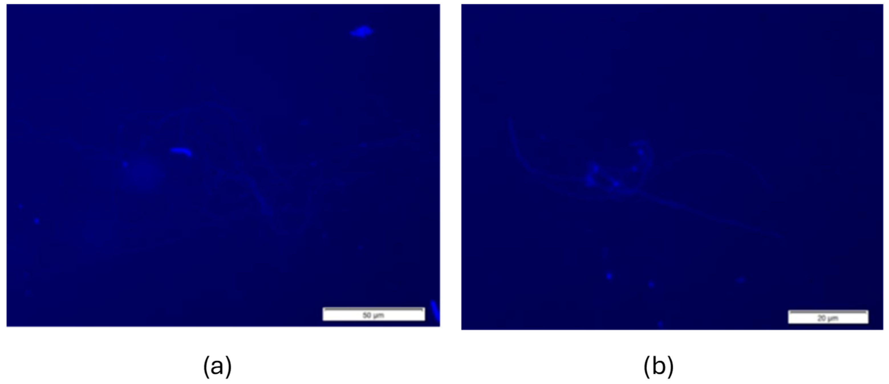
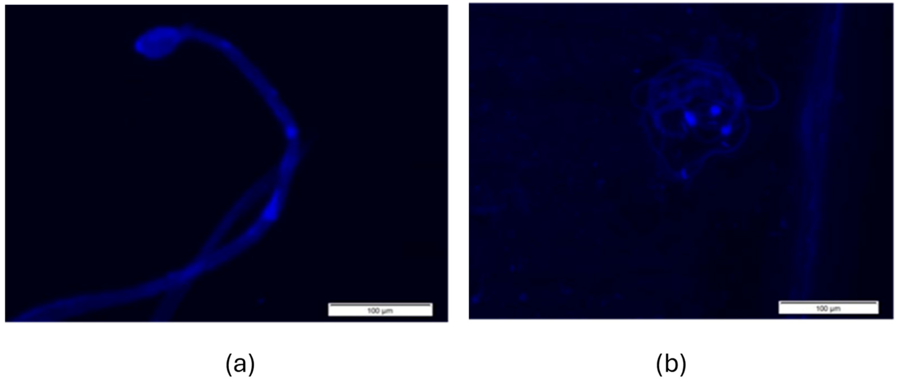
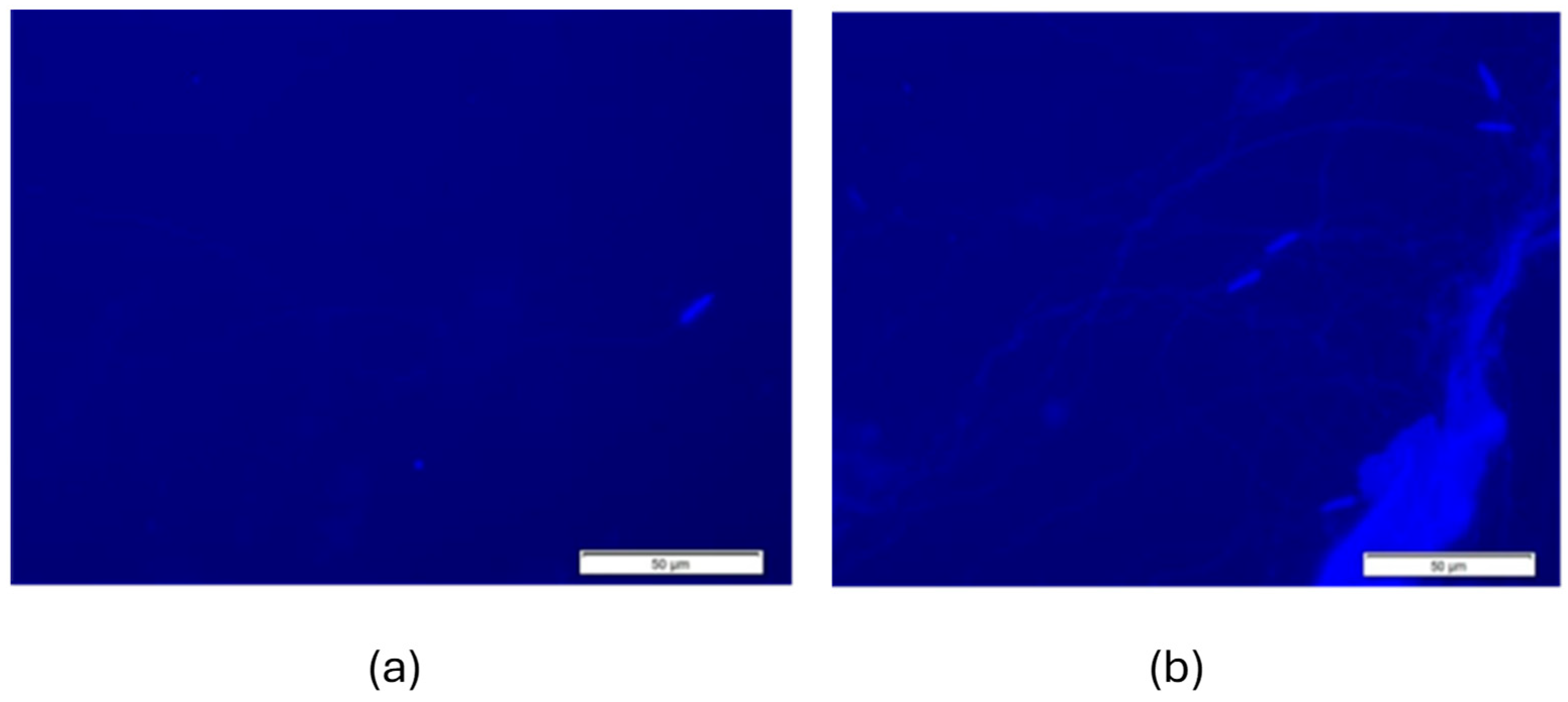
Figure 6 and Figure 7 display representative fluorescence micrographs of D. melanogaster spermatozoa from control groups, illustrating the typical morphology in the absence of exposure to tobacco smoke. Comparisons with treated groups were made to identify abnormalities potentially induced by tobacco smoke exposure.
Fluorescence microscopy revealed differences in the shape and organization of the spermatozoa head between the two strains. In the control group (Figure 6), spermatozoa from the Drosophila OK strain show no clusters and a flat head, which is normal for Drosophila. Figure 6a is representative of normal morphology, while Figure 6b shows a more oval-shaped spermatozoa head.
Figure 7a,b show that the spermatozoa of the Drosophila mus308 control strain generally have a more oval-shaped spermatozoa head than the OK strain, even though it is a control group.
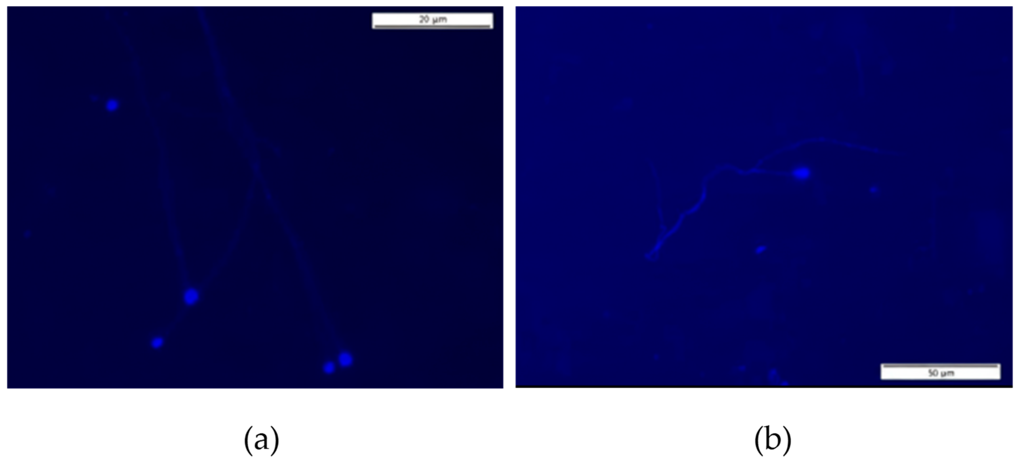
Figure 8 and Figure 9 display representative fluorescence micrographs of D. melanogaster spermatozoa from the Oregon K and mus308 strain, illustrating typical morphology in the exposure to tobacco smoke for 1 min.
Morphological changes were evident after 1 min of exposure to tobacco smoke. In Figure 8a,b of the O.K strain, the morphology is relatively preserved and similar to the control. In Figure 9 of the mus308 strain, almost all heads (a) have a round shape. In Figure 9b, the spermatozoa still preserves an oval shape. Both treated strains exhibited a significant reduction in total spermatozoa count when compared to controls (163 in OK and 152 in mus308 for the treated groups, compared to 330 and 238 in the control groups; p < 0.05). Specifically, the reduction in OK was statistically significant (p = 0.014, d = 0.81, CI 95% = [0.15, 1.47]), and in mus308, the reduction was highly significant (p = 0.002, d = 1.25, CI 95% = [0.49, 2.01])—suggesting a cytotoxic effect of smoke exposure on spermatogenesis or viability.
The observed spermatozoa abnormalities and reduction in cell numbers are in line with evidence from other species, including mammals, where exposure to tobacco has been associated with genomic instability, such as DNA strand breaks, altered chromatin organization, and structural defects in spermatozoa cells [3,24,25,26,27,28], mus308 is particularly relevant, as it is involved in DNA repair via translesion synthesis, and its deficiency may exacerbate damage caused by genotoxic agents like tobacco smoke [28]. Similar effects have been reported in other genotoxicity assays in Drosophila, highlighting the model’s relevance in toxicological studies [27,28,29,30,31].
Following smoke exposure, mus308 spermatozoa heads predominantly shifted to a rounded morphology, as seen in the fluorescence images. This transition from an oval to a rounded form may indicate compromised chromatin condensation or nuclear architecture, possibly due to oxidative stress induced by tobacco smoke. This observation is in line with previous findings demonstrating a relationship between oxidative damage and altered spermatozoa head shape in Drosophila and other model organisms [31].
The data suggest that tobacco smoke induces morphological and quantitative alterations in Drosophila spermatozoa, with the mus308 strain displaying heightened sensitivity, likely due to impaired DNA repair capacity.

4. Discussion

The observed dose-dependent effect of tobacco smoke on male Drosophila fertility, where low-level (1 min) exposure increased fertility in OK flies while higher doses were inhibitory, is a phenomenon that may be interpreted as a hormetic response. This concept, historically linked to the Arndt–Schulz principle, suggests that low doses of a stressor can stimulate a biological process that is inhibited by higher doses. Given the limited dose–response resolution of the present study, our results should be considered as a possible hypothesis rather than a definitive conclusion, providing a basis for future research to further investigate if a hormetic effect is present [25,26,27,28].
The toxicological analysis confirmed tobacco smoke’s harmful effects, particularly in the mus308 strain, which showed a higher male mortality rate (51%) compared to the OK strain (40%) following exposure. These findings are consistent with previous studies by Moratore et al. (2009), which demonstrated that aqueous tobacco extracts induce significant mortality in D. melanogaster (around 50% mortality at 10% tobacco concentration after 24 h) [32]. Other insect models, such as flour beetles and cabbage aphids, have similarly shown reduced viability and reproductive performance after tobacco exposure, reinforcing the compound’s broad-spectrum toxicity [5,28].
Regarding longevity, the observed decline in the mean lifespan of F1 offspring across both strains is consistent with the published findings. While Linford et al. (2013) and Sun et al. (2013) reported that D. melanogaster can live between 50 and 91 days under standard conditions, our control groups exhibited values near the lower end of this range [33,34]. In contrast, the treatment groups displayed significantly reduced lifespans, suggesting that even brief parental exposure to tobacco smoke adversely impacts progeny survival. In survival analyses, only the mus308 offspring exhibited a statistically significant decrease in survival probability, suggesting that intact DNA repair confers resilience to progeny. This result is in strong agreement with the observations of Piper and Partridge (2016), who highlighted the high sensitivity of D. melanogaster lifespan to environmental stressors, such as xenobiotic exposure and diet [35,36,37,38].
Spermatozoa morphology analysis, conducted using a DAPI-based fluorescence microscopy protocol adapted from the standard Sperm Integrity Morphology (SIM) technique, revealed typical flagellar features previously described by Kanippayoor et al. (2013) and López-Fernández et al. (2007) [15,39]. Despite the limited number of studies employing this specific technique in Drosophila, our results corroborate existing knowledge regarding normal spermatozoa architecture and provide visual evidence of altered spermatozoa counts in the mus308 strain following tobacco exposure. This strain, which is deficient in microhomology-mediated end joining (MMEJ) repair due to a mutation in the mus308 gene (encoding DNA polymerase θ), demonstrated increased sensitivity, suggesting that defective DNA repair pathways may exacerbate the cytotoxic and reproductive impacts of tobacco [14,16,24,31].
To further investigate tobacco-induced genotoxicity, our previous work on human lymphocytes in the comet assay revealed a dose-dependent increase in DNA damage, particularly basal (non-oxidative) damage, with the highest levels observed at 100% tobacco concentration. Interestingly, oxidative DNA damage remained low across all treatment groups, indicating that tobacco’s genotoxicity may be driven primarily through mechanisms other than oxidative stress. These observations are in line with findings by Piperakis et al. (1998), who also reported basal and oxidative damage following tobacco exposure, although our results suggest a predominance of non-oxidative lesions [40]. This aligns with Kleinsasser et al. (2005), who demonstrated nicotine-induced genotoxicity in lymphocytes, further supporting the relevance of tobacco constituents as direct DNA-damaging agents [41].
Therefore, the present study corroborates the findings of recent studies which demonstrate that toxic compounds in tobacco smoke, including nicotine and polycyclic hydrocarbons, induce oxidative stress, DNA damage and apoptosis in various human cell lines, including reproductive and epithelial cells. In animal models such as rats and mice, exposure to tobacco has been associated with hormonal changes, mitochondrial dysfunction, systemic inflammation, and impaired reproductive function. These findings reinforce the harmful effects of smoking on fertility and cellular integrity, as well as its role in the development of chronic diseases [42,43,44,45,46,47,48,49,50,51].
The results discussed in the following study, such as spermatozoa quantification, have some limitations that must be considered, as it is a new method with a few promising results in spermatozoa from flies. Therefore, future investigations into improving morphological studies of spermatozoa in D. melanogaster as a practical model for investigating the reproductive effects of tobacco are necessary. Furthermore, adapting the comet assay to specifically evaluate genotoxic damage in Drosophila spermatozoa is a promising way to advance our understanding of tobacco-induced reproductive toxicity and quantify exposure to tobacco smoke. The results of the present study, particularly those concerning spermatozoa quantification and morphological analysis, have some limitations that must be considered. This is a new method with few promising results regarding spermatozoa from flies. Therefore, future investigations are necessary to improve quantitative and morphological studies of spermatozoa in D. melanogaster as a practical model for studying the reproductive effects of tobacco. Additionally, adapting the comet assay to evaluate genotoxic damage specifically in Drosophila spermatozoa is a promising approach to advancing our understanding of tobacco-induced reproductive toxicity and quantifying exposure to tobacco smoke.

5. Conclusions

The present study concludes that tobacco smoke exposure profoundly impacts male fertility by significantly altering key biological parameters, including fertility, fecundity, and offspring longevity in the mus308 and OK strains of D. melanogaster. The two mus 308 strains exhibited more pronounced morphological alterations in spermatozoa, indicating a higher susceptibility to the toxic effects of tobacco smoke compared to the OK strain. These data establish that tobacco smoke, even at sub-lethal concentrations, modulates essential biological processes like fertility and lifespan, a response contingent upon the organism’s genetic background and intrinsic DNA repair capacity. The mus308 strain emerged as a sensitive genetic model for studying genotoxic stress. These insights highlight the utility of D. melanogaster as a valuable in vivo model for assessing environmental genotoxins and their transgenerational effects.

Author Contributions

Conceptualization, methodology, investigation, data curation, and visualization were carried out by I.G., A.M.-B., A.G. and N.G.d.M.; writing (original draft preparation, review, and editing) were carried out by I.G., A.M.-B. and N.G.d.M.; validation, formal analysis, supervision, funding acquisition, and project administration were carried out by A.M.-B. and I.G. All authors have read and agreed to the published version of the manuscript.

Funding

This work was supported by the projects UI/00772/2020 (Doi:10.54499/UI/00772/2020) and AL4AnimalS (LA/P/0059/2020) funded by the Portuguese Foundation for Science and Technology (FCT).

Institutional Review Board Statement

Not applicable.

Informed Consent Statement

Not applicable.

Data Availability Statement

The original contributions presented in this study are included in the article. Further inquiries can be directed to the corresponding authors.

Conflicts of Interest

The authors declare no conflicts of interest. The funders had no role in the design of the study; in the collection, analyses, or interpretation of data; in the writing of the manuscript; or in the decision to publish the results.

Abbreviations

The following abbreviations are used in this manuscript:
OKOregon K
DSBsDNA double-strand breaks
HRHomologous recombination
c-NHEJClassical non-homologous end joining
MMEJMicrohomology-mediated end joining
NMPNormal melting point
LMPLow melting point
SIMInclusion sperm in microgels
DAPI4′,6-diamidino-2-phenylindole

References

  1. Zenzes, M.T. Smoking and reproduction: Gene damage to human gametes and embryos. Hum. Reprod. Update 2000, 6, 122–131. [Google Scholar] [CrossRef]
  2. Leslie, S.W.; Soon-Sutton, T.L.; Khan, M.A.B. Male Infertility. In StatPearls; StatPearls: Treasure Island, FL, USA, 2024; Available online: https://www.ncbi.nlm.nih.gov/books/NBK562258/ (accessed on 10 October 2024).
  3. Agarwal, A.; Baskaran, S.; Parekh, N.; Cho, C.L.; Henkel, R.; Vij, S.; Arafa, M.; Panner, S.M.K.; Shah, R. Male infertility. Nat. Rev. Dis. Prim. 2021, 7, 319–333. [Google Scholar] [CrossRef] [PubMed]
  4. Belcheva, A.; Ivanova-Kicheva, M.; Tzvetkova, P.; Marinov, M. Effects of cigarette smoking on sperm plasma membrane integrity and DNA fragmentation. Int. J. Androl. 2004, 27, 296–300. [Google Scholar] [CrossRef] [PubMed]
  5. Collodel, G.; Capitani, S.; Pammolli, A.; Giannerini, V.; Geminiani, M.; Moretti, E. Semen quality of male idiopathic infertile smokers and nonsmokers: An ultrastructural study. J. Androl. 2010, 31, 108–113. [Google Scholar] [CrossRef] [PubMed]
  6. Künzle, R.; Mueller, M.D.; Hänggi, W.; Birkhäuser, M.H.; Drescher, H.; Bersinger, N.A. Semen quality of male smokers and nonsmokers in infertile couples. Fertil. Steril. 2003, 79, 287–291. [Google Scholar] [CrossRef]
  7. Colagar, A.H.; Jorsaraee, G.A.; Marzony, E.T. Cigarette smoking and the risk if male infertility. Pak. J. Biol. Sci. 2007, 10, 3870–3874. [Google Scholar] [CrossRef]
  8. Rau-Hansen, C.H.; Thulstrup, A.M.; Aggerholm, A.S.; Jensen, M.S.; Toft, G.; Bonde, J.P. Is smoking a risk factor for decreased semen quality? A cross—Sectional analysis. Hum. Reprod. 2007, 22, 188–196. Available online: https://pubmed.ncbi.nlm.nih.gov/16966350/ (accessed on 10 October 2024). [CrossRef]
  9. Gaur, D.S.; Talekar, M.; Pathak, V.P. Effect of cigarette smoking on semen quality of infertile men. Med. J. Armed Forces India 2007, 23, 119–123. [Google Scholar] [PubMed]
  10. Chia, S.E.; Ong, C.N.; Tsakok, F.M. Effects of cigarette smoking on human semen quality. Arch. Androl. 1994, 33, 163–168. [Google Scholar] [CrossRef]
  11. Kumosani, T.A.; Elshal, M.F.; Al-Jonaid, A.A.; Abduljabar, H.S. The influence of smoking on semen quality, seminal microelements and Ca2+-ATPase activity among infertile and fertile men. Clin. Biochem. 2008, 41, 14–15. [Google Scholar] [CrossRef]
  12. Pantaleão, S.M.; Spano, M.A.; Morelli, S.; Nepomoceno, J.C.; Takahashi, C.S. Impacto Genotóxico de Poluentes Químicos Presentes na Água e Sedimento do Rio Japaratuba (Sergipe). Ph.D. Thesis, Universidade Federal de Uberlândia, Uberlândia, Brazil, 2006. [Google Scholar]
  13. Ouchi, R.Y.; Manzato, A.J.; Ceron, C.R.; Bonilla-Rodriguez, G.O. Evaluation of the effects of a single exposure to ethidium bromide in Drosophila melanogaster (Diptera-Drosophilidae). Bull. Environ. Contam. Toxicol. 2007, 78, 489–493. [Google Scholar] [CrossRef] [PubMed]
  14. Tiwari, A.K.; Pragya, P.; Ravi Ram, K.; Chowdhuri, D.K. Environmental chemical mediated male reproductive toxicity: Drosophila melanogaster as an alternate animal model. Theriogenology 2011, 76, 197–216. [Google Scholar] [CrossRef]
  15. Kanippayoor, R.L.; Alpern, J.H.M.; Moehring, A.J. Protamines and spermatogenesis in Drosophila and Homo sapiens: A comparative analysis. Spermatogenesis 2013, 3, e24376. [Google Scholar] [CrossRef] [PubMed]
  16. Rand, M.D. Drosophotoxicology: The growing potential for Drosophila in neurotoxicology. Neurotoxicology Teratol. 2010, 32, 74–83. [Google Scholar] [CrossRef]
  17. Papanikolopoulou, K.; Skoulakis, E.M. The power and richness of modelling tauopathies in Drosophila. Mol. Neurobiol. 2011, 44, 122–133. [Google Scholar] [CrossRef]
  18. Rand, M.D.; Tennessen, J.M.; Mackay, T.F.C.; Anholt, R.R.H. Perspectives on the Drosophila melanogaster Model for Advances in Toxicological Science. Curr. Protoc. 2023, 3, e870. [Google Scholar] [CrossRef]
  19. Trizzino, M.; Kapusta, A.; Brown, C.D. Transposable elements generate regulatory novelty in a tissue-specific fashion. BMC Genomics 2018, 19, 468. [Google Scholar] [CrossRef]
  20. Rodríguez, R.; Gaivão, I.; Aguado, L.; Espina, M.; Garcia, J.; Martínez-Camblor, P.; Sierra, L.M. The Comet Assay in Drosophila: A Tool to Study Interactions between DNA Repair Systems in DNA Damage Responses In Vivo and Ex Vivo. Cells 2023, 12, 1979. [Google Scholar] [CrossRef]
  21. Alexander, J.; Beagan, K.; Kelly, J.; Orr-Weaver, T.L.; McVey, M. Multiple mechanisms contribute to double-strand break repair at rereplication forks in Drosophila follicle cells. Proc. Natl. Acad. Sci. USA 2016, 113, 13809–13814. [Google Scholar] [CrossRef]
  22. Miranda, N.; Tkach, V.V.; Barros, A.N.; Martins-Bessa, A.; Gaivão, I. Biological and Behavioral Responses of Drosophila melanogaster to Dietary Sugar and Sucralose. Int. J. Mol. Sci. 2025, 26, 8951. [Google Scholar] [CrossRef] [PubMed]
  23. Guo, R.; Henke, A.L.; Reinhardt, K. Sperm viability varies with buffer and genotype in Drosophila melanogaster. Fly 2021, 15, 1–7. [Google Scholar] [CrossRef]
  24. Dalton, J.E.; Kacheria, T.S.; Knott, S.R.; Lebo, M.S.; Nishitani, A.; Sanders, L.E.; Stirling, E.J.; Winbush, A.; Arbeitman, M.N. Dynamic, mating-induced gene expression changes in female head and brain tissues of Drosophila melanogaster. BMC Genom. 2010, 11, 541. [Google Scholar] [CrossRef]
  25. Sepaniak, S.; Forges, T.; Gerard, H.; Foliguet, B.; Bene, M.C.; Monnier-Barbarino, P. The influence of cigarette smoking on human sperm quality and DNA fragmentation. Toxicology 2006, 223, 54–60. [Google Scholar] [CrossRef]
  26. Evenson, D.P.; Jost, L.K.; Corzett, M.; Balhorn, R. Characteristics of human sperm chromatin structure following an episode of influenza and high fever: A case study. J. Androl. 2000, 21, 739–746. [Google Scholar] [CrossRef]
  27. Aitken, R.J.; De Iuliis, G.N.; Thomson, L.K.; Mitchell, L.A.; Finnie, J.M.; Koppers, A.J.; Hedges, A.; Nixon, B. DNA Damage in Human Spermatozoa Is Highly Correlated with the Efficiency of Chromatin Remodeling and the Formation of 8-Hydroxy-2′-Deoxyguanosine, a Marker of Oxidative Stress. Biol. Reprod. 2009, 81, 517–524. [Google Scholar] [CrossRef]
  28. Boyd, J.B.; Golino, M.D.; Nguyen, T.D.; Green, M.M. Isolation and characterization of X-linked mutants of Drosophila melanogaster which are sensitive to mutagens. Genetics 1976, 84, 485–506. [Google Scholar] [CrossRef]
  29. Graf, U.; Würgler, F.E.; Katz, A.J.; Frei, H.; Juon, H.; Hall, C.B.; Kale, P.G. Somatic mutation and recombination test in Drosophila melanogaster. Environ. Mutagen. 1984, 6, 153–188. [Google Scholar] [CrossRef]
  30. Valko, M.; Rhodes, C.J.; Moncol, J.; Izakovic, M.; Mazur, M. Free radicals, metals and antioxidants in oxidative stress-induced cancer. Chem. Biol. Interact. 2006, 160, 1–40. [Google Scholar] [CrossRef] [PubMed]
  31. Gualtieri, R.; Kalthur, G.; Barbato, V.; Longobardi, S.; Di Rella, F.; Adiga, S.K.; Talevi, R. Sperm Oxidative Stress during In Vitro Manipulation and Its Effects on Sperm Function and Embryo Development. Antioxidants 2021, 10, 1025. [Google Scholar] [CrossRef] [PubMed]
  32. Moratore, C. Use of Drosophila melanogaster as bioindicators in the evaluation of the lethality of extracts of nicotiana tabacum. Arq. Inst. Biol. 2009, 76, 471–474. [Google Scholar] [CrossRef]
  33. Linford, N.J.; Bilgir, C.; Ro, J.; Pletcher, S.D. Measurement of Lifespan in Drosophila melanogaster. J. Vis. Exp. 2013, 71, e50068. [Google Scholar] [CrossRef] [PubMed]
  34. Sun, Y.; Yolitz, J.; Wang, C.; Spangler, E.; Zhan, M.; Zou, S. Aging Studies in Drosophila melanogaster. Methods Mol. Biol. 2013, 1048, 77–93. [Google Scholar] [CrossRef] [PubMed]
  35. Piper, M.D.W.; Partridge, L. Protocols to Study Aging in Drosophila. In Methods in Molecular Biology; Humana Press: New York, NY, USA, 2016; Volume 1478, pp. 291–302. [Google Scholar] [CrossRef]
  36. Calabrese, E.J. Hormesis is central to toxicology, pharmacology and risk assessment. Hum. Exp. Toxicol. 2010, 29, 249–261. [Google Scholar] [CrossRef]
  37. Calabrese, E.J.; Baldwin, L.A. Hormesis: The dose-response revolution. Annu. Rev. Pharmacol. Toxicol. 2003, 43, 175–197. [Google Scholar] [CrossRef] [PubMed]
  38. Calabrese, E.J. Hormesis: A Revolution in Toxicology, Risk Assessment and Medicine. EMBO Rep. 2013, 14, 497–498. [Google Scholar] [CrossRef]
  39. López-Fernández, C.; Baltanás, A.; De La Torre, J.; Gosálvez, J. Drosophila melanogaster and Eucypris virens giant spermatozoa as visualized by cell inclusion in microgels. J. Exp. Zool. Part A Ecol. Genet. Physiol. 2007, 307, 140–144. [Google Scholar] [CrossRef]
  40. Piperakis, S.; Visvardis, E.E.; Sagnou, M.; Tassiou, A. Effects of smoking and aging on oxidative DNA damage of human lymphocytes. Carcinogenesis 1998, 19, 695–698. [Google Scholar] [CrossRef]
  41. Kleinsasser, N.H.; Sassen, A.W.; Semmler, M.P.; Harréus, U.A.; Licht, A.K.; Richter, E. The Tobacco Alkaloid Nicotine Demonstrates Genotoxicity in Human Tonsillar Tissue and Lymphocytes. Toxicol. Sci. 2005, 86, 342–353. [Google Scholar] [CrossRef]
  42. La Maestra, S.; Bonomo, P.; Martone, F.; Calandra, A.; De Lisa, M.; Ferraro, M.; De Flora, S.; Micale, R.T. Effect of cigarette smoke on DNA damage, oxidative stress, and morphological alterations in mouse testis and spermatozoa. Int. J. Hyg. Environ. Health 2015, 218, 117–122. [Google Scholar] [CrossRef]
  43. Moktar, A.; Singh, R.; Vadhanam, M.V.; Ravoori, S.; Lillard, J.W., Jr.; Gairola, C.G.; Gupta, R.C. Cigarette smoke condensate-induced oxidative DNA damage and its removal in human cervical cancer cells. Int. J. Oncol. 2011, 39, 941–947. [Google Scholar] [CrossRef]
  44. Omolaoye, T.S.; El Shahawy, O.; Skosana, B.T.; Boillat, T.; Loney, T.; du Plessis, S.S. The mutagenic effect of tobacco smoke on male fertility. Environ. Sci. Pollut. Res. Int. 2022, 29, 62055–62066. [Google Scholar] [CrossRef]
  45. Russo, M.; Cocco, S.; Secondo, A.; Adornetto, A.; Bassi, A.; Nunziata, A.; Polichetti, G.; De Felice, B.; Damiano, S.; Serù, R.; et al. Cigarette smoke condensate causes a decrease of the gene expression of Cu-Zn superoxide dismutase, Mn superoxide dismutase, glutathione peroxidase, catalase, and free radical-induced cell injury in SH-SY5Y human neuroblastoma cells. Neurotox Res. 2011, 19, 49–54. [Google Scholar] [CrossRef] [PubMed]
  46. Rao, P.; Ande, A.; Sinha, N.; Kumar, A.; Kumar, S. Effects of Cigarette Smoke Condensate on Oxidative Stress, Apoptotic Cell Death, and HIV Replication in Human Monocytic Cells. PLoS ONE 2016, 11, e0155791. [Google Scholar] [CrossRef]
  47. Xu, W.-J.; Fang, P.; Zhu, Z.-J.; Xu, D.-X.; Wang, T.-T.; Dai, J.-B. The alteration of protein profile induced by cigarette smoking via oxidative stress in mice epididymis. Int. J. Biochem. Cell Biol. 2013, 45, 571–582. [Google Scholar] [CrossRef]
  48. Dai, J.-B.; Wang, Z.-X.; Qiao, Z.-D. The hazardous effects of tobacco smoking on male fertility. Asian J. Androl. 2015, 17, 954–960. [Google Scholar] [CrossRef]
  49. Saleh, R.A.; Agarwal, A.; Sharma, R.K.; Nelson, D.R.; Thomas, A.J., Jr. Effect of cigarette smoking on levels of seminal oxidative stress in infertile men: A prospective study. Fertil. Steril. 2002, 78, 491–499. [Google Scholar] [CrossRef]
  50. Pasqualotto, F.F.; Grempel, R.G.; Pasqualotto, E.B. Effect of cryopreservation on sperm DNA integrity: A review. Int. Braz. J. Urol. 2011, 37, 592–598. [Google Scholar] [CrossRef]
  51. Fraga, C.G.; Motchnik, P.A.; Wyrobek, A.J.; Rempel, D.M.; Ames, B.N. Smoking and low antioxidant levels increase oxidative damage to sperm DNA. Mutat. Res. 1996, 351, 199–203. [Google Scholar] [CrossRef] [PubMed]
Figure 1. Mean number of offspring per tube in control and treatment groups of Oregon K and mus308 strains after 1 min exposure to tobacco smoke. Error bars represent the standard deviation (SD). The 95% Confidence Interval (CI) is OK control [26.16, 35.68]; OK treatment [25.71, 38.65]; mus308 control [23.15, 31.57]; and mus308 treatment [27.95, 36.47]. The p-value is 0.473. The symbol (ns) indicates no statistically significant difference.
Cells 14 01689 g001
Figure 2. Mean number of offspring per tube in Oregon K and mus308 strains after 7 min exposure to tobacco smoke. Error bars represent the standard deviation (SD). The 95% Confidence Interval (CI) for the mean number of offspring is control OK [24.04, 42.84]; treatment OK [27.35, 54.12]; control mus308 [14.00, 29.87]; and treatment mus308 [10.37, 28.52]. The symbol (ns) indicates no statistically significant difference.
Cells 14 01689 g002
Figure 3. The average survival time (in days) of F1 male offspring from Oregon K (OK) and mus308 strains after 1 min exposure to tobacco smoke. Error bars represent the standard deviation (SD). The 95% Confidence Interval (CI) is OK control [44.12, 55.88]; OK treatment [37.39, 48.61]; mus308 control [48.45, 59.55]; and mus308 treatment [39.12, 48.88]. The ANOVA p-value is p = 0.218 in the OK line and p = 0.072 in the mus308 line. The symbol (ns) indicates no statistically significant difference.
Cells 14 01689 g003
Figure 4. Kaplan–Meier survival curve of F1 offspring from the Oregon K strain (wild type) following 1 min exposure to tobacco smoke. A significant difference in the overall survival curve was found between the groups (Log-rank test: χ2 = 7.23, p = 0.007; Hazard Ratio (HR) = 1.34, CI 95% = [1.08, 1.66]).
Cells 14 01689 g004
Figure 5. Kaplan–Meier survival curve of F1 offspring from the mus308 strain following 1 min exposure of paternal males to tobacco smoke. A marked decrease in survival was observed in the treatment group, indicating increased vulnerability in DNA repair-deficient progeny. Statistical analysis was performed using the log-rank test. The value χ2 = 4.48 and p-value = 0.346.
Cells 14 01689 g005
Figure 6. Fluorescence micrographs of spermatozoa cells from the Oregon K (OK) strain control group, stained with DAPI. (a): The spermatozoon presents a flat head and a long tail. (b) Spermatozoa present oval heads and long tails.
Cells 14 01689 g006
Figure 7. Fluorescence micrographs of spermatozoa from the mus308 strain control group, stained with DAPI. (a): The spermatozoa are oval-headed and (b): the spermatozoa are oval-headed and are grouped together.
Cells 14 01689 g007
Figure 8. Fluorescence micrographs of spermatozoa from the OK lineage stained with DAPI in the tobacco smoke treatment group (1 min). (a) Isolated spermatozoa and (b): grouped spermatozoa.
Cells 14 01689 g008
Figure 9. Fluorescence micrographs of spermatozoa of the mus 308 strain stained with DAPI in the tobacco smoke treatment group (1 min). (a): The spermatozoa have rounded heads, and (b): the spermatozoa have oval heads.
Cells 14 01689 g009
Table 1. Constituents of the D. melanogaster standard medium.
IngredientsAmount (per Litre Distilled Water)
Sugar100 g
Agar-agar 12 g
Inactive yeast100 g
Propionic acid5 mL
Table 2. Percentage of fertile males after tobacco smoke exposure (1 min and 7 min) in Oregon K (OK) and mus308 strains. The 95% Confidence Interval (CI) is reported for each percentage.
GroupsExposure Time% Fertile MalesIC 95%
Control O.K0 min60[45.7, 74.3]
Treatment O.K1 min87[77.2, 96.8]
Treatment O.K7 min100[79.0, 100.0]
Control mus3080 min100[92.0, 100.0]
Treatment mus3081 min93[85.5, 100.0]
Treatment mus3087 min60[35.2, 84.8]
Table 3. Percentage of male mortality following 1 min tobacco smoke exposure in Oregon K and mus308 strains (treatment group). The 95% Confidence Interval (CI) is reported for each percentage. The table is based on a total of 75 males per strain exposed to tobacco smoke.
Strain% of Male MortalityIC 95%
Oregon K40[28.9%, 51.1%]
mus 30851[39.7%, 62.3%]
Disclaimer/Publisher’s Note: The statements, opinions and data contained in all publications are solely those of the individual author(s) and contributor(s) and not of MDPI and/or the editor(s). MDPI and/or the editor(s) disclaim responsibility for any injury to people or property resulting from any ideas, methods, instructions or products referred to in the content.