Article

mRNA Multipeptide-HLA Class II Immunotherapy for Melanoma

by
Apostolos P. Georgopoulos
1,2,3,*,†,
Lisa M. James
1,2,4,† and
Matthew Sanders
1,2,3
1
The HLA Cancer Research Group, Brain Sciences Center, Department of Veterans Affairs Health Care System, Minneapolis VAMC, One Veterans Drive, Minneapolis, MN 55417, USA
2
Department of Neuroscience, University of Minnesota Medical School, Minneapolis, MN 55455, USA
3
Institute for Health Informatics, University of Minnesota Medical School, Minneapolis, MN 55455, USA
4
Department of Psychiatry, University of Minnesota Medical School, Minneapolis, MN 55455, USA
*
Author to whom correspondence should be addressed.
These authors contributed equally to this work.
Cells 2025, 14(18), 1430; https://doi.org/10.3390/cells14181430
Submission received: 7 August 2025 / Revised: 4 September 2025 / Accepted: 8 September 2025 / Published: 12 September 2025

Abstract

Human Leukocyte Antigen (HLA) Class II (HLA-II) molecules bind peptides of phagocytosed non-self proteins and present them on the cell surface to circulating CD4+ T lymphocytes. A successful binding of the presented peptide with the T cell receptor (TCR) activates the CD4+ T cell, leading to the production of antibodies against the peptide (and the protein of its origin) by the B cell and augmentation of the cytotoxic and memory functions of CD8+ T cells. The first and essential step in this process is the successful formation of a stable peptide-HLA-II complex (pHLA-II), which is achieved when the peptide binds with high affinity to the HLA-II molecule. Such highly antigenic non-self peptides occur in melanoma-associated proteins and could be used as antitumor agents when bound to a matching HLA-II molecule. The objective of this study was to identify such peptides from 15 melanoma-associated proteins. We determined in silico the predicted binding affinity (IC50) of all pHLA-II pairs between 192 common HLA-II molecules and all possible linear 15-amino acid (15-mer) peptides (epitopes) of 15 known melanoma-associated antigens (N = 3466 epitopes) for a total of 192 × 3466 = 665,472 determinations. From this set, we identified epitopes with strong antigenicity (predicted best binding affinity [PBBA] IC50 < 50 nM). Of a total of 665,472 pHLA-II tested, 5941 (0.89%) showed strong PBBA, stemming from 117 HLA-II alleles and 679 distinct epitopes. This set of 5941 pHLA-II pairs with predicted high antigenicity possesses the requisite information for devising multipeptide vaccines with those epitopes alone or in combination with the corresponding HLA-II molecules. The results obtained have a major implication for cancer therapy, namely that the administration of subsets of the 679 high antigenicity epitopes above, alone or in combination with their associated HLA-II molecules, would be successful in engaging CD4+ T helper lymphocytes to augment the cytotoxic action and memory of CD8+ T lymphocytes and induce the production of antitumor antibodies by B cells. This therapy would be effective in other solid tumors (in addition to melanoma) and would be enhanced by concomitant immunotherapy with immune checkpoint inhibitors.

1. Introduction

1.1. Melanoma

Malignant melanoma originates from melanocytes, specialized cells that produce the pigment melanin. Environmental influences (e.g., exposure to ultraviolet radiation), coupled with genetic events such as mutations in regulatory genes, transform typical-functioning melanocytes into melanoma cancer cells [1]. Cutaneous melanoma is the most serious form of skin cancer, accounting for the vast majority of skin cancer deaths [2], but it can occur in other tissues (e.g., mucosal, uveal) [3]. The high mortality rate of melanoma is attributable to conversion from in situ to invasive form and metastases [1]. While there has been a significant increase in global melanoma incidence over the last several decades, improvements in detection and advances in treatment have contributed to a decline in overall mortality [4,5,6]. Still, many invasive melanoma cases result in death, highlighting the need for novel interventions.

1.2. Melanoma Cancer Antigens

As melanoma progresses, the impacted cells express specific antigens that can be used for diagnostic purposes and have become targets of immunotherapy [7]. Melanoma antigens include melanocyte differentiation antigens (MDAs), which are exclusively expressed in melanocytes and overexpressed in melanoma, cancer testis antigens (CTAs), which are typically expressed in normal germline cells but can be expressed in many types of cancer, and other proteins, such as S100, which is expressed in melanomas as well as other cancers and disorders. MDAs include Tyrosinase, TRP-1 and TRP-2, gp100 (also called premelanosomal protein; PMEL17), and Melan-A (also called melanoma antigen recognized by T cells; MART-1). CTAs include those of the melanoma-associated antigen (MAGE), B-M antigen-1 (BAGE), G antigen (GAGE), and NY-ESO-1 families. The role of each of these antigens, as well as their potential for diagnostic purposes or therapeutic intervention, has been reviewed elsewhere [7]. Melanoma is prone to a high rate of mutations [8], producing what are now commonly referred to as neoantigens [9,10]. Neoantigens are patient-specific, vary with regard to immunogenicity, and may be heterogeneous both within a tumor and relative to metastatic sites [11,12].

1.3. Melanoma Treatment

Treatment for early stages of melanoma typically involves surgical removal of the tumor and surrounding tissue, resulting in high survival rates [13]; however, more advanced stages of melanoma require additional interventions, including targeted therapies and/or immunotherapy. More specifically, with respect to the latter, the development of immune checkpoint inhibitors (ICIs), T cell therapies, and other melanoma immunotherapies has significantly improved outcomes for some patients, albeit with several limitations, including immune-related adverse effects, immunotherapy resistance, re-occurrence of the tumor, and time-consuming and costly manufacturing of treatment components [14,15,16]. Combination immunotherapies hold promise. Indeed, greater than 50% five-year survival has been documented in patients with metastatic melanoma treated with combined ICIs [17]; however, the lack of sustained benefit in a large portion of cases highlights the need for further development of melanoma immunotherapy. Recent technological advances in vaccine development have ushered in a new era of cancer treatment that holds promise for melanoma and other malignancies.

Melanoma Vaccines

Vaccination, generally speaking, is aimed at generating a durable immune response against specific antigens through human leukocyte antigen (HLA)-mediated T cell and B cell activation. The same principles apply to melanoma vaccines, which target either tumor-specific antigens (TSAs) or tumor-associated antigens (TAAs), including MDAs and CTAs [11]. Numerous melanoma vaccine formulations and platforms have been developed over the last decades, including the use of peptides, nucleic acids, viral vectors, dendritic and B cells, with recent substantial progress and development centered around synthetic peptides or nucleic-acid encapsulated liquid nanoparticles [12]. Historically, cancer vaccines for malignant melanoma have demonstrated limited efficacy [18]. However, numerous promising synthetic peptide or nucleic acid-based melanoma vaccines are under investigation in clinical trials [19,20,21]. A critical issue with regard to the effectiveness of nucleic-acid-based vaccines is the selection of antigens that are immunogenic [22], a feature that depends on the patient’s leukocyte antigen (HLA) genetic makeup.

1.4. Human Leukocyte Antigen (HLA)

HLA, also commonly referred to as Major Histocompatibility Complex (MHC), comprises cell-surface glycoproteins that play a critical role in human immune surveillance and response. Their primary function is presentation of antigens (peptides) to lymphocytes in order to identify and facilitate immune system activation against foreign antigens and cancers [23]. There are two main classes of HLA (Class I and Class II) that differ in terms of their structure and antigen presentation pathways but share a similar goal of host protection [24]. Briefly, HLA-I molecules of the three classical HLA-I genes (A, B, C) are expressed on the surface of all nucleated cells, including tumors, and are instrumental in the presentation of short (8–10 amino acid residues, mostly 9-mer [25]) endogenous antigen peptides to cytotoxic CD8+ T cells. HLA-II molecules of the three classical HLA-II genes (DPB1, DQB1, DRB1) are expressed on professional antigen presenting cells (APC), including macrophages, B cells, and dendritic cells (DC), and are critical for presentation of larger (13–22 amino acid residues, mostly 15-mer [26] exogenous antigens to CD4+ helper T cells which stimulate production of antibodies and augment the functions of CD8+ T cells. Subsets of CD8+ and CD4+ T cells acquire immunological memory following activation for faster and effective activation in future encounters with the antigen to which they were exposed. The HLA region is the most highly polymorphic in the human genome [27], contributing to population-level protection against diverse pathogens; however, each individual carries only 12 classical HLA alleles, including 2 from each Class I gene (HLA-A, HLA-B, and HLA-C) and 2 from each Class II gene (HLA-DPB1, HLA-DQB1, HLA-DRB1), that code for the HLA molecules each individual possesses. Variability across HLA polymorphisms is primarily located in the binding groove [28], the amino acid sequence and structure of which determine which antigens can bind with sufficiently high affinity to confer stability to the peptide-HLA (pHLA) complex for an effective presentation to, and activation of, CD8+ and CD4+ T cells [29]. Single amino acid substitutions alter the binding groove [30,31,32], thereby influencing the repertoire of antigen peptides that can bind with HLA for presentation to T cells [33].

1.4.1. Peptide-HLA Binding

HLA molecules of both classes possess very high specificity and degeneracy in peptide binding [30,34]. With respect to the former, a specific HLA molecule can bind with high affinity to only a few non-self antigens among a large number available, and can distinguish peptide antigens by even single amino acid substitutions [30,31,32]; and with respect to the latter, a particular HLA molecule can bind a large number of peptides.

1.4.2. Binding of pHLA to T Cell Receptor (TCR)

The pHLA complex on the surface of the cell presents its peptide to circulating T cells. Although the term TCR is used for both CD8+ and CD4+ T cells, it actually differs in its biophysics between the two cases. Nevertheless, in both cases, peptide-TCR binding is characterized by very high specificity, sensitivity, and degeneracy. With respect to specificity, even a single amino acid residue substitution in the peptide can make a big difference in the probability of its binding to TCR, for both pHLA-I and pHLA-II molecules. With respect to sensitivity, a CD8+ T cell can be activated by a single pHLA-I complex [35], and a CD4+ T cell can be activated by a single pHLA-II complex [36]. And with respect to degeneracy, a single TCR of a CD8+ or CD4+ cell can be engaged by many pHLA complexes.

1.5. HLA Expression in Melanoma Cells

1.5.1. General

Both HLA Class I and Class II can be expressed on melanoma cells [37,38]; HLA-I expression occurs in >90% of melanoma cells, whereas HLA-II expression occurs in 50–60% of the cells [37]. Higher expression of HLA (e.g., Class II HLA-DR) has been linked with better prognosis [38,39,40,41], particularly in association with ICI immunotherapy [41]. Secretion of interferon-γ by cytotoxic CD8+ T lymphocytes increases HLA expression in melanoma, thereby increasing antitumor immunogenicity [11]; however, as with other types of cancer, alterations in HLA expression are common in melanoma [42]. Downregulation or loss of HLA is generally associated with worse outcomes [43].

1.5.2. Influence of HLA Melanoma Expression on Outcomes of Immunotherapy

Mounting evidence has documented the influence of HLA on immunotherapy outcomes. In particular, Class II HLA-DR expression has been associated with better outcomes of immune checkpoint blockade immunotherapy, including longer overall survival and progression-free survival for patients with melanoma [41]. However, the influence of HLA on treatment outcomes is not limited to overall HLA expression but also rests on the specific HLA that an individual carries. For example, alleles of the Class I HLA-B44 supertype have been associated with better immune checkpoint blockade immunotherapy outcomes, whereas alleles of the HLA-B62 supertype (particularly HLA-B*15:01) are associated with poor outcomes [44]. Notably, compared to the HLA-B62 supertype, immunogenicity of the HLA-B44 supertype with melanoma antigens is significantly better yet varies across melanoma antigens [45], highlighting the relevance of both the tumor antigens and HLA composition for immunotherapy outcomes. Indeed, personalized treatment approaches based on high-affinity antigen peptide-HLA (pHLA) complexes that are highly immunogenic have become a focus of cancer immunotherapy [46,47,48,49].

1.5.3. HLA and Melanoma Vaccines: The Present Study

Melanoma vaccines represent a rapidly developing approach to melanoma immunotherapy aimed at augmenting the ability of a patient’s immune system to attack cancer cells by enhancing antigen-specific immune responses. With this approach, bioinformatics tools can be leveraged to predict pHLA binding affinity and immunogenicity in order to identify candidate epitopes that can be provided to a patient via various vaccine platforms, including peptide-based vaccines and nucleic acid-based vaccines [50,51]. We have previously used an in silico approach to evaluate the immunogenicity of all possible 9-mers of 11 melanoma-associated antigens to 2462 HLA-I molecules [45] and identified nine HLA-I molecules that would be highly immunogenic to peptide epitopes from all 11 antigens. Here, we extend that to HLA-II molecules by evaluating the predicted binding affinity of 192 common HLA-II molecules to all possible linear 15-mer epitopes of 15 known melanoma antigens to identify (a) peptide epitopes with strong antigenicity (i.e., high binding affinity to HLA-II molecules, and (b) HLA-II molecules with strong binding to many melanoma antigens. The former would be useful in including in multipeptide melanoma vaccines, whereas the latter would form the basis of a new melanoma immunotherapy by inducing their synthesis in tumor, DC, or B cells by delivering their mRNA blueprints, as discussed below.

2. Materials and Methods

2.1. Melanoma/Cancer Antigens

Fifteen cancer antigens (Table 1) were used based on their known occurrence in melanoma tumors and used in various treatments [7]; of those, six are specific to melanoma and nine are expressed in melanoma as well as in other tumors (labeled [CTA] in Table 1). The amino acid (AA) sequences of the 15 melanoma antigens used were retrieved from the Uniprot Database (https://www.uniprot.org/uniprotkb; accessed 1 April 2025) and are given in Table S1 (Supplementary Material).

2.2. In Silico Determination of Predicted Best Binding Affinities (PBBA) to Melanoma Antigens

We estimated in silico the predicted best binding affinity of the HLA alleles above to the 15 melanoma/cancer antigens above (Table 1). Predicted binding affinities were obtained for antigen epitopes using the Immune Epitope Database (IEDB; http://tools.iedb.org/mhci/, accessed on 4 September 2025) NetMHCpan (ver. 4.1 BA) tool [52]. We tested all 232 alleles of DPB1, DQB1, and DRB1HLA-II genes published in the Supplementary Tables of Hurley et al. [53]. Of those, 192 alleles could be modeled by the software tool above, and the results were analyzed as follows. We used the sliding window approach [54,55] to test exhaustively all possible linear 15-mer epitopes of the 15 antigens analyzed (Table 1); the epitope length of 15 amino acids is optimal for HLA-II molecule binding [26,56,57]. The method is illustrated in Figure 1 for PMEL17. For each pair of peptide-HLA molecules (pHLA-II) tested, this tool gave, as an output, the IC50 of the predicted binding affinity; the smaller the IC50, the stronger the binding affinity. The predicted best binding affinity (PBBA) for each pHLA-II pair was the minimum IC50 value of all epitopes tested for the pair. An IC50 value of IC50 < 50 nm is regarded as strong [58]; we called PBBAs < 50 nM “hits”. Given a protein of N amino acid length and an epitope length of 15 AA, there were N-15+1 PBBAs. The number of epitopes tested for each antigen (across the 192 HLA-II alleles) is given in Table 1.

2.3. Statistical Analysis

The IBM-SPSS statistical package (version 30) was used for standard analyses. All p-values reported are two-sided.

3. Results

3.1. Predicted Antigenicity of Melanoma-Associated Epitopes

3.1.1. Peptide Epitopes

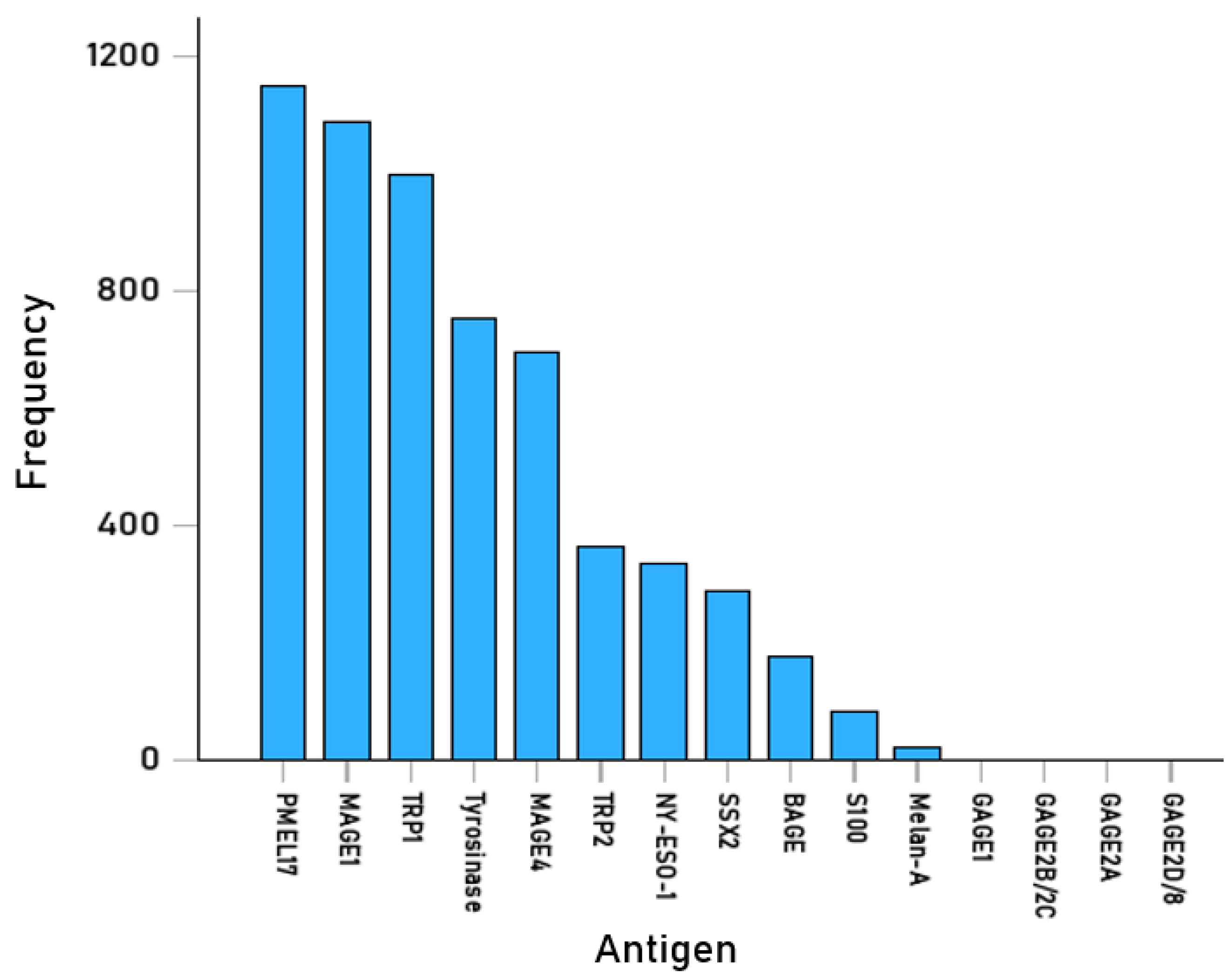
We analyzed 15-mer epitopes from 15 melanoma-associated antigens (Table 1) and 192 common HLA-II allele molecules (Table S2; Supplementary Material). The total number of pHLA-II PBBA determinations was 3466 epitopes × 192 alleles = 665,472 (Table 1). Of those, 5941 (0.89%) were hits (PBBA IC50 < 50 nM; Table S3), indicating strong antigenicity (“antigenic” group), and were retained for further analyses. The frequency of occurrence of these epitopes across the 15 antigens is given in Table 1 and illustrated in Figure 2. It could be that antigens PMEL17, MAGE1, and TRP1 had high numbers of antigenic epitopes, most likely due to their long amino acid sequences (Table 1). In contrast, none of the GAGE group had any strong binders (Table 1).

3.1.2. HLA-II Alleles

The 5941 immunogenic pHLA-II pairs came from combinations of 117 distinct HLA-II alleles and 679 distinct epitopes (the remaining HLA and melanoma antigen epitopes were not strong binding “hits”). All 5941 epitope/allele combinations with strong binding are shown in Table S3, Table S4 (sorted by allele), and Table S5 (sorted by peptide/epitope). These alleles and the number of antigens of the associated epitopes are shown in Table 2. It can be seen that five alleles (DRB1*01:01, DRB1*01:18, DRB1*01:24, DRB1*01:29, DRB1*10:01) had hits from all 11 antigens that showed strong bindings. The frequency of occurrence of the 679 distinct epitopes with strong binding is shown in Table S6 and illustrated in Figure 3. Finally, Table S7 shows all pHLA-II pairs arranged by allele, and Table S8 shows the number and percentage of hits across the 117 alleles, ranked from high to low percentage. Finally, the frequencies of those alleles among five ethnic populations are given in Table S9 (Supplementary Material).
With respect to HLA-II genes, we found the following. (a) There were no DQB1 alleles with strong binding; (b) the proportion of DPB1 alleles did not differ significantly between the antigenic group (23/117 = 0.197) and the original group (41/192 = 0.213) (p = 0.72, Z = 0.36, test of two proportions); in contrast, (c) the proportion of DRB1 alleles was highly significantly higher in the antigenic group (94/117 = 0.803) than the original group (116/192 = 0.604) (p = 0.0001, Z = 3.81, test of two proportions).

4. Discussion

4.1. Methodological Considerations

Two aspects of our methodology are noteworthy with respect to peptides and alleles tested. With respect to the former, a key point was the testing of all possible linear 15-mer peptides of all 15 melanoma-associated antigens. This procedure lends credibility to the set of peptides identified as strong binders (IC50 < 50 nM), eliminating guessing or peptide selection for vaccines based on a general description of antigens as “immunogenic”. And with respect to the latter, testing 192 common alleles [53] provided a wide spectrum of HLA-II representation. The combination of these approaches broadly and robustly covers the interactions between melanoma antigens and HLA-II molecules.

4.2. Antigenicity and Immunogenicity

The terms “antigenicity” and “immunogenicity” are being used in the literature frequently interchangeably. Strictly speaking, in the context of antigen processing and presentation by HLA-I/HLA-II molecules, antigenicity refers to the probability and strength of attachment of a specific peptide to a specific HLA molecule to form a stable pHLA complex; the immunogenicity of that pHLA complex refers to the probability and strength of eliciting an immune response by the T cells that the pHLA complex engages. The following points are noteworthy. (a) The key factor for both antigenicity and immunogenicity is the pHLA complex, not the peptides by themselves. (b) High antigenicity (i.e., strong binding) is a prerequisite for high immunogenicity. (c) The next step in an immune response is the binding of the peptide presented by the pHLA complex to the T cell receptor (TCR). Although the same generic notation (TCR) is used for both CD8+ and CD4+ molecules, the biophysics are different in the two cases, since two different, T cell-specific, coreceptors are involved, namely CD8 for the pHLA-I-specific CD8+ T cells and CD4 for the pHLA-II-specific CD4+ T cells. Both coreceptors increase the stability of the pHLA complex and facilitate the ultimate activation of the corresponding T cells for an effective discharge of their effector actions (cytotoxicity for CD8+ T cells; initiation of antibody production and enhancement in CD8+ T cells’ actions by CD4+ T cells). (d) Importantly, as mentioned above, the peptide-TCR binding is characterized by very high specificity, sensitivity, and degeneracy. With respect to specificity, even a single amino acid residue substitution in the peptide can make a big difference in the probability of its binding to TCR, for both pHLA-I and pHLA-II molecules. With respect to sensitivity, a CD8+ T cell can be activated by a single pHLA-I complex [35], and a CD4+ T cell can be activated by a single pHLA-II complex [36]. And with respect to degeneracy, a single TCR of a CD8+ or CD4+ cell can be engaged by many pHLA complexes. (e) As mentioned above, the strong, high-affinity binding of a peptide to the HLA molecule renders the resulting pHLA complex very stable. This pHLA stability increases the time for which the pHLA complex is presented to T cells and thus has a direct impact on immunogenicity by increasing the chance of pHLA-TCR binding.
In summary, the primary and most important factor for both antigenicity and immunogenicity is the strength of binding affinity of a peptide to an HLA molecule. The determination of this binding affinity in vitro is tedious, time-consuming, and practically unrealistic for testing large combinations of pHLA complexes. For example, to test in vitro 665,472 pHLA-II complexes evaluated in this study would be infeasible. Fortunately, due to major improvements in bioinformatics, the pHLA binding affinity can be estimated in silico with fair certainty [59]. For that purpose, in this study, we used the freely available NetMHCpan tool [52], a standard in the field.

4.3. Antigenic pHLA-II Complexes for Melanoma

Here we identified 5941 (out of 665,472 tested, 0.89%) pHLA-II pairs with strong binding affinities, comprising 679 (out of 3466 tested, 19.6%) distinct 15-mer peptides and 117 (out of 192 tested, 60.9%) HLA-II molecules. The peptides came from all but the GAGE antigens (Table 1), and the HLA-II alleles came from the DPB1 and DRB1 genes. Interestingly, the percentage of peptides from 15 melanoma-associated antigens found here to bind with high affinity (IC50 < 50 nM) to HLA-II molecules is comparable to ~0.5% of peptides from the whole human proteome found to bind, with the same high affinity, to HLA-I allele super types [58]. Finally, all but the GAGE antigens comprised peptides that bound strongly to HLA-II (Figure 2), and five HLA-II alleles bound with high affinity to peptides of all of them (DRB1*01:01, DRB1*01:18, DRB1*01:24, DRB1*01:29, DRB1*10:01).

4.4. Implications for Multipeptide HLA-II Restricted Melanoma Vaccines

Most melanoma vaccines use HLA-I-restricted peptides, but HLA-II-restricted peptides [60] have also been used [47]. Moreover, some mutated neoantigen peptides used in a melanoma vaccine engaged CD4+ T cells [46], indicating binding to HLA-II. Table S7 gives all 5941 pHLA pairs arranged by allele. The first step in an application is to determine HLA-II alleles carried by the patient. Of those, only the two alleles of the DPB1 gene and the two alleles of the DRB1 gene are useful, since no high-affinity binding peptide was found for alleles of the DQB1 gene. Next, amino acid sequences of peptides with high-affinity binding to the DPB1 and DRB1 alleles of the patient are selected from Table S7, synthesized (and may be embedded in longer peptide sequences), pulsed ex vivo into dendritic cells (DC) of the patients, and reintroduced to the patient, following the protocol of DC cancer treatment [61]. This treatment will guarantee that the peptides provided will bind with high affinity to the HLA-II molecules that the patient carries for the formation of a stable pHLA-II complex, an essential prerequisite for peptide presentation to circulating CD4+ T cells and initiation of antibody production by B cells. In parallel, mRNA blueprints of the same peptides are introduced into the tumor, leading to the synthesis of the peptides that will be the targets for cytotoxic antibodies. Since this is a peptide-HLA Class II complex, it will engage and activate CD4+ T cells. In addition, activated CD4+ T cells will augment the protective functions of the CD8+ T cells, with respect to both their cytotoxic action and the establishment of memory T-cells [62]. Although the details of this effect are still being investigated, with new insights being uncovered, there is general agreement about this beneficial effect (against tumor growth) of CD4+ → CD8+ T cell interaction.
Whereas the enhancement in CD8+ T cell function by activated CD4+ T cells is relatively well understood [62], the role of antitumor antibodies is unclear, in contrast to their known role in infection. For example, in a viral infection or vaccination of an immunocompetent host, production of antibodies by B cells typically takes 1–3 weeks. Antibodies act by various mechanisms to protect the host by eliminating current infection and preventing reinfection [63,64,65]. The therapeutic and prophylactic effects of such antibodies against pathogens have been well established. In contrast, with respect to cancer, the therapeutic effect of antitumor antibodies is less clear. Circulating antibodies to TAAs have been found in patients with cancer, indicating a humoral immune response to TAAs. This observation is complemented by the development of antibodies to TAA in response to peptide/epitope vaccines for melanoma [66]. However, the effect of such antibodies on tumor reduction/elimination is unclear. Indirect evidence for their positive effect comes from the observation that melanoma tumors expressing HLA-II proteins, HLA-II(+), have better prognosis than HLA-II(─) tumors [38,41], but this differential effect could also be due to the facilitation of CD8+ T cells by activated CD4+ T cells.

4.5. Melanoma Immunotherapy Based on the Synthesis of New HLA Molecules

4.5.1. HLA-II Molecules

Cancer treatment with vaccines containing peptides with predicted high-affinity binding to the HLA-II molecules of the patient is possible only if such peptide-HLA combinations exist. For example, of the 192 HLA-II alleles tested in this study, only 117 were found to bind with high affinity to any of the 3466 peptide epitopes tested exhaustively from 15 melanoma antigens. A definite way to ensure the availability of a high-affinity bound pHLA-II pair would be to induce the synthesis of specific HLA-II molecules that would satisfy that condition [67,68]. More specifically, ideal candidates would be the HLA-II molecules found to bind with high affinity to peptides from all 11 TAA, namely DRB1*01:01, DRB1*01:18, DRB1*01:24, DRB1*01:29, and DRB1*10:01 (Table 2). Nucleic acid (mRNA or viral vector-based) blueprints of these molecules and associated peptides would be administered intratumorally to patients with melanoma tumors expressing HLA-II molecules (~60% in primary and ~50% in metastatic tumors [37]). In HLA-II(─) tumors, they would be pulsed ex vivo to the patient’s DC or B cells, according to the respective protocols of cancer treatment [69]. In both cases, the co-administered peptides would form a stable pHLA-II complex, engaging and activating circulating CD4+ T cells for augmentation of CD8+ T cell cytotoxic and other functions (e.g., promoting memory CD8+ T cells), and the production of antitumor antibodies by B cells.

4.5.2. HLA-I Molecules

Essentially the same approach would be followed using HLA-I molecules and associated high-affinity binding peptides, as proposed recently [68]. In fact, we have already identified nine HLA-I molecules (from a set of 2462 HLA-I alleles) that bound with high affinity to 9-mer peptides from 11 melanoma-associated antigens (A*02:14, B*07:10, B*35:10, B*40:10, B*40:12, B*44:10, C*07:11, C*07:13, C*07:14) [70]. Nucleic acid (mRNA or viral vector-based) blueprints of these molecules and associated peptides would be injected intratumorally. Since only the α chain is polymorphic in HLA-I molecules, its synthesis would be simpler than that of the more complex HLA-II molecule. Since practically all melanoma tumors express HLA-I molecules, this treatment would be effective.

4.5.3. Tumor Microenvironment

It should be mentioned that the outcome of the aforementioned interventions would depend on the status of the immune (dys)function at the tumor’s microenvironment [71]. For example, if the expression of the HLA-I/HLA-II molecules is suppressed or impaired, epitopes of the peptides/HLA-II molecules introduced to the tumor may not be adequately presented to CD8+ and CD4+ T cells, the function of which might be impaired as well. Therefore, the expected outcome could be quite variable. These considerations lead to an approach that would involve a direct antigen–antibody reaction without the need to involve the patient’s immune system. This approach involves the blood group AB antigens and the antibodies against them, as discussed elsewhere [72].

4.5.4. Challenges

The main concern in this approach of inducing the synthesis of new HLA molecules is the possibility of autoimmunity. Since these are non-self molecules to the patient, they are highly immunogenic, and it is expected that they would be attacked by humoral and cell-mediated mechanisms, in a situation closely resembling the rejection of an HLA-incompatible organ transplant [73]. In fact, this reaction would be beneficial, as it would lead to the rejection of the tumor, similarly to the case of a HLA-incompatible transplant [72]. Now, one would expect autoimmunity to occur if epitopes of the new HLA molecules are shared with proteins in other tissues, in which case, autoimmunity would occur due to shared epitope/molecular mimicry. Interestingly, such reactions have not been reported in HLA-incompatible organ transplantation, but autoimmune reactions have been documented, actually due to autoimmunity to cryptic self-antigens that appear during chronic damage of the transplanted organ. Apparently, the appearance of such new, antigenic self-antigens would depend on the size of the transplanted organ and the duration of chronic rejection (typically months). However, in the case of melanoma (and other solid tumors), these factors would be minimal since the size of the tumor is much smaller than common transplants (e.g., kidney, lung, liver) and its rejection would be faster. In any case, such autoimmune reactions would be treated using standard protocols, as applied in post-transplantation management of autoimmune reactions.

5. Limitations

The general limitation of this study is that it is an in silico investigation of predicted peptide-HLA-II binding affinities. This bioinformatics approach allows for screening a large number of pHLA pairs at the expense of the certainty provided by in vitro assessments of binding affinities. However, the methods and bioinformatics tools used to derive those predictions have improved tremendously during the past several years, aided by the concomitant increase in computer power available for large-scale computations, and, as mentioned above, the pHLA binding affinity can be estimated in silico with fair certainty [59]. For example, the results of this study provide new information on predicted binding affinities based on 665,472 in silico estimations, a feat that would have taken a very long time to accomplish by in vitro methods. Similarly, in a comparable study of binding affinity of all 9-mer epitopes from 11 melanoma-associated antigens (N = 3123) to 2462 HLA-I molecules [70], we estimated in silico predicted binding affinities of 3123 × 2462 = 7,688,826 pHLA-I pairs, a practically infeasible task to complete in vitro. However, if the focus of the scientific question is on the binding affinity of a particular peptide to a particular HLA molecule, an in vitro assessment should accompany the in silico prediction [74,75,76].

6. Conclusions

The first and single most important step in adaptive immunity is the formation of a stable pHLA complex. This involves both a peptide and an HLA molecule. Here, we carried out an exhaustive testing of all 15-mer peptides of 15 melanoma-associated antigens to 192 common HLA-II allele proteins, for a total of 665,472 predicted binding affinity estimations. The 5941 pHLA-II pairs found to bind with strong affinity included 679 distinct 15-mer peptides from 11/15 antigens and 117/192 HLA-II molecules of the DRB1 and DQB1 genes. Specific peptide-HLA-II molecule combinations can be used for the development of effective vaccines against melanoma, where the synthesis of both the peptide and the HLA-II can be accomplished by injecting intratumorally their mRNA blueprints in HLA-II(+) melanoma tumors, or, in HLA-II(−) tumors, pulsing those blueprints to dendritic cells or B cells ex vivo, with further procedures dictated by the standard DC/B cell treatment protocols [61,69]. This treatment, combined with intratumorally injection of mRNA blueprints of high-binding-affinity HLA-I peptides and molecules [68], would be a useful addition to mRNA-based vaccines against melanoma. Obviously, this and other treatments would be more effective when applied to melanoma tumors at an early stage. In this regard, blood biomarkers such as the fatty acid and protein composition of circulating CD81-positive exosomes [77] hold great promise.
Finally, it should be mentioned that neuroinformatics has been a continuously evolving strong force in vaccine development and design. As explicitly expressed in a recent review of this topic, “Immunoinformatics represents a transformative approach to vaccine research, improving clinical trial efficiency and enabling the development of more reliable, flexible, and personalized vaccines. This approach has the potential to significantly enhance global healthcare outcomes by accelerating the vaccine development process and optimizing vaccination strategies.” ([78], Abstract).

Supplementary Materials

The following supporting information can be downloaded at: https://www.mdpi.com/article/10.3390/cells14181430/s1, Table S1: Amino acid sequences of the 15 melanoma antigens analyzed (Table 1). Table S2. The 192 HLA-II alleles used. Table S3. All estimated strong binding affinities (IC50 < 50 nM) for 15-mer sequences for the 117 HLA-II alleles with strong binding and the 11/15 antigens that had any strong affinity. Table S4. High binding affinity peptide-HLA-II pairs arranged by HLA-II allele. Table S5. High binding affinity peptide-HLA-II pairs arranged by peptide. Table S6. Frequency of occurrence of the 679 unique 15-AA peptides with strong PBBAs. Table S7. Number of strong binders (PBBA IC50 < 50 nM) per antigen and allele. Table S8. Counts (N) and percentages (%) of hits across all 11 antigens tested. Number of alleles = 117 alleles. Table S9. Relative frequencies of the alleles binding strongly to peptides, across the 7 ethnic population groups.

Author Contributions

A.P.G. conceived the study and analyzed the data. M.S. obtained the in silico predictions using the Immune Epitope Database (IEDB) NetMHCpan (ver. 4.1 BA) tool [52]. A.P.G. and L.M.J. wrote the paper. A.P.G., L.M.J. and M.S. reviewed and approved the paper. All authors have read and agreed to the published version of the manuscript.

Funding

Partial funding for this study was provided by the University of Minnesota (American Legion Brain Sciences Chair) and the U.S. Department of Veterans Affairs. The sponsors had no role in the current study design, analysis, or interpretation, or in the writing of this paper. The contents do not represent the views of the U.S. Department of Veterans Affairs or the United States Government.

Institutional Review Board Statement

This article simply and solely analyzed publicly available data and does not contain any studies with human participants or animal experiments performed by any of the authors. Hence, ethical approval is not applicable to this study.

Informed Consent Statement

Not applicable.

Data Availability Statement

All data used for analysis are freely available from public databases, as stated in the Materials and Methods section of the manuscript.

Conflicts of Interest

The authors declare no conflicts of interest.

References

  1. Centeno, P.P.; Pavet, V.; Marais, R. The journey from melanocytes to melanoma. Nat. Rev. Cancer 2023, 23, 372–390. [Google Scholar] [CrossRef]
  2. American Cancer Society. Cancer Facts and Figures 2025; American Cancer Society: Atlanta, GA, USA, 2025. [Google Scholar]
  3. Elder, D.E.; Bastian, B.C.; Cree, I.A.; Massi, D.; Scolyer, R.A. The 2018 World Health Organization classification of cutaneous, mucosal, and uveal melanoma: Detailed analysis of 9 distinct subtypes defined by their evolutionary pathway. Arch. Pathol. Lab. Med. 2020, 144, 500–522. [Google Scholar] [CrossRef]
  4. Patel, V.R.; Roberson, M.L.; Pignone, M.P.; Adamson, A.S. Risk of mortality after a diagnosis of melanoma in situ. JAMA Dermatol. 2023, 159, 703–710. [Google Scholar] [CrossRef] [PubMed]
  5. Sun, Y.; Shen, Y.; Liu, Q.; Zhang, H.; Jia, L.; Chai, Y.; Jiang, H.; Wu, M.; Li, Y. Global trends in melanoma burden: A comprehensive analysis from the Global Burden of Disease Study, 1990–2021. J. Am. Acad. Dermatol. 2025, 92, 100–107. [Google Scholar] [CrossRef] [PubMed]
  6. Waseh, S.; Lee, J.B. Advances in melanoma: Epidemiology, diagnosis, and prognosis. Front. Med. 2023, 10, 1268479. [Google Scholar] [CrossRef] [PubMed]
  7. Pitcovski, J.; Shahar, E.; Aizenshtein, E.; Gorodetsky, R. Melanoma antigens and related immunological markers. Crit. Rev. Oncol. Hematol. 2017, 115, 36–49. [Google Scholar] [CrossRef]
  8. Lawrence, M.S.; Stojanov, P.; Polak, P.; Kryukov, G.V.; Cibulskis, K.; Sivachenko, A.; Carter, S.L.; Stewart, C.; Mermel, C.H.; Roberts, S.A.; et al. Mutational heterogeneity in cancer and the search for new cancer-associated genes. Nature 2013, 499, 214–218. [Google Scholar] [CrossRef]
  9. Jiang, T.; Shi, T.; Zhang, H.; Song, Y.; Wei, J.; Ren, S.; Zhou, C. Tumor neoantigens: From basic research to clinical applications. J. Hematol. Oncol. 2019, 12, 93. [Google Scholar] [CrossRef]
  10. Xie, N.; Shen, G.; Gao, W.; Huang, Z.; Huang, C.; Fu, L. Neoantigens: Promising targets for cancer therapy. Sig. Transduct. Target Ther. 2023, 8, 9. [Google Scholar] [CrossRef]
  11. Kalaora, S.; Nagler, A.; Wargo, J.A.; Samuels, Y. Mechanisms of immune activation and regulation: Lessons from melanoma. Nat. Rev. Cancer 2022, 22, 195–207. [Google Scholar] [CrossRef]
  12. Cui, C.; Ott, P.A.; Wu, C.J. Advances in vaccines for melanoma. Hematol. Oncol. Clin. North Am. 2024, 38, 1045–1060. [Google Scholar] [CrossRef]
  13. Svedman, F.C.; Pillas, D.; Taylor, A.; Kaur, M.; Linder, R.; Hansson, J. Stage-specific survival and recurrence in patients with cutaneous malignant melanoma in Europe—A systematic review of the literature. Clin. Epidemiol. 2016, 8, 109–122. [Google Scholar] [CrossRef]
  14. Huang, A.C.; Zappasodi, R. A decade of checkpoint blockade immunotherapy in melanoma: Understanding the molecular basis for immune sensitivity and resistance. Nat. Immunol. 2022, 23, 660–670. [Google Scholar] [CrossRef]
  15. Knight, A.; Karapetyan, L.; Kirkwood, J.M. Immunotherapy in melanoma: Recent advances and future directions. Cancers 2023, 15, 1106. [Google Scholar] [CrossRef] [PubMed]
  16. Wang, X.; Ma, S.; Zhu, S.; Zhu, L.; Guo, W. Advances in immunotherapy and targeted therapy of malignant melanoma. Biomedicines 2025, 13, 225. [Google Scholar] [CrossRef] [PubMed]
  17. Larkin, J.; Chiarion-Sileni, V.; Gonzalez, R.; Grob, J.-J.; Rutkowski, P.; Lao, C.D.; Cowey, L.; Schadendorf, D.; Wagstaff, J.; Drummer, R.; et al. Five-year survival with combined nivolumab and ipilimumab in advanced melanoma. N. Engl. J. Med. 2019, 381, 1535–1546. [Google Scholar] [CrossRef] [PubMed]
  18. Seta, T.; Nakamura, S.; Oura, M.; Yokoyoma, K.; Nishikawa, Y.; Hoshino, N.; Ninomiya, K.; Shimoi, T.; Hotta, K.; Nakayama, T. Efficacy and safety of cancer vaccine therapy in malignant melanoma: A systematic review. Int. J. Clin. Oncol. 2025, 30, 1080–1097. [Google Scholar] [CrossRef]
  19. Khaddour, K.; Buchbinder, E.I. Individualized neoantigen-directed melanoma therapy. Am. J. Clin. Dermatol. 2025, 26, 225–235. [Google Scholar] [CrossRef]
  20. Liao, H.C.; Liu, S.J. Advances in nucleic acid-based cancer vaccines. J. Biomed. Sci. 2025, 32, 10. [Google Scholar] [CrossRef]
  21. Yaremenko, A.V.; Khan, M.M.; Zhen, X.; Tang, Y.; Tao, W. Clinical advances of mRNA vaccines for cancer immunotherapy. Med 2025, 6, 100562. [Google Scholar] [CrossRef]
  22. Chekaoui, A.; Garofalo, M.; Gad, B.; Staniszewska, M.; Chiaro, J.; Pancer, K.; Cryciuk, A.; Cerullo, V.; Salmaso, S.; Caliceti, P.; et al. Cancer vaccines: An update on recent achievements and prospects for cancer therapy. Clin. Exp. Med. 2024, 25, 24. [Google Scholar] [CrossRef]
  23. Medhasi, S.; Chantratita, N. Human leukocyte antigen (HLA) system: Genetics and association with bacterial and viral infections. J. Immunol. Res. 2022, 2022, 9710376. [Google Scholar] [CrossRef]
  24. Neefjes, J.; Jongsma, M.; Paul, P.; Bakke, O. Towards a systems understanding of MHC class I and MHC class II antigen presentation. Nat. Rev. Immunol. 2011, 11, 823–836. [Google Scholar] [CrossRef] [PubMed]
  25. Cerundolo, V.; Elliott, T.; Elvin, J.; Bastin, J.; Rammensee, H.G.; Townsend, A. The binding affinity and dissociation rates of peptides for class I major histocompatibility complex molecules. Eur. J. Immunol. 1991, 21, 2069–2075. [Google Scholar] [CrossRef] [PubMed]
  26. Chicz, R.M.; Urban, R.G.; Lane, W.S.; Gorga, J.C.; Stern, L.J.; Vignali, D.A.; Strominger, J.L. Predominant naturally processed peptides bound to HLA-DR1 are derived from MHC-related molecules and are heterogeneous in size. Nature 1992, 358, 764–768. [Google Scholar] [CrossRef] [PubMed]
  27. Trowsdale, J.; Knight, J.C. Major histocompatibility complex genomics and human disease. Annu. Rev. Genomics Hum. Genet. 2013, 14, 301–323. [Google Scholar] [CrossRef]
  28. Dendrou, C.A.; Petersen, J.; Rossjohn, J.; Fugger, L. HLA variation and disease. Nat. Rev. Immunol. 2018, 18, 325–339. [Google Scholar] [CrossRef]
  29. Sette, A.; Vitiello, A.; Reherman, B.; Fowler, P.; Nayersina, R.; Kast, W.M.; Melief, C.J.; Oseroff, C.; Yuan, L.; Ruppert, J.; et al. The relationship between class I binding affinity and immunogenicity of potential cytotoxic T cell epitopes. J. Immunol. 1994, 153, 5586–5592. [Google Scholar] [CrossRef]
  30. Hov, J.R.; Kosmoliaptsis, V.; Traherne, J.A.; Olsson, M.; Bobreg, K.M.; Bergquist, A.; Schrumpf, E.; Bradley, J.A.; Taylor, C.J.; Lie, B.A.; et al. Electrostatic modifications of the human leukocyte antigen-DR P9 peptide-binding pocket and susceptibility to primary sclerosing cholangitis. Hepatology 2011, 53, 1967–1976. [Google Scholar] [CrossRef]
  31. Davenport, M.P.; Quinn, C.L.; Chicz, R.M.; Green, B.N.; Willis, A.C.; Lane, W.S.; Bell, J.I.; Hill, A.V. Naturally processed peptides from two disease-resistance-associated HLA-DR13 alleles show related sequence motifs and the effects of the dimorphism at position 86 of the HLA-DR beta chain. Proc. Natl. Acad. Sci. USA 1995, 92, 6567–6571. [Google Scholar] [CrossRef]
  32. van Deutekom, H.W.; Keşmir, C. Zooming into the binding groove of HLA molecules: Which positions and which substitutions change peptide binding most? Immunogenetics 2015, 67, 425–436. [Google Scholar] [CrossRef]
  33. Paul, S.; Weiskopf, D.; Angelo, M.A.; Sidney, J.; Peters, B.; Sette, A. HLA class I alleles are associated with peptide-binding repertoires of different size, affinity, and immunogenicity. J. Immunol. 2013, 191, 5831–5839. [Google Scholar] [CrossRef]
  34. Margulies, D.H.; Corr, M.; Boyd, L.F.; Khilko, S.N. MHC class I/peptide interactions: Binding specificity and kinetics. J. Mol. Recognit. 1993, 6, 59–69. [Google Scholar] [CrossRef] [PubMed]
  35. Sykulev, Y.; Joo, M.; Vturina, I.; Tsomides, T.J.; Eisen, H.N. Evidence that a single peptide-MHC complex on a target cell can elicit a cytolytic T cell response. Immunity 1996, 4, 565–571. [Google Scholar] [CrossRef] [PubMed]
  36. Irvine, D.J.; Purbhoo, M.A.; Krogsgaard, M.; Davis, M.M. Direct observation of ligand recognition by T cells. Nature 2002, 419, 845–849. [Google Scholar] [CrossRef] [PubMed]
  37. Taramelli, D.; Fossati, G.; Mazzocchi, A.; Delia, D.; Ferrone, S.; Parmiani, G. Classes I and II HLA and melanoma-associated antigen expression and modulation on melanoma cells isolated from primary and metastatic lesions. Cancer Res. 1986, 46, 433–939. [Google Scholar]
  38. Johnson, D.B.; Estrada, M.V.; Salgado, R.; Sanchez, V.; Doxie, D.B.; Opalenik, S.R.; Vilgelm, A.E.; Feld, E.; Johnson, A.S.; Greenplate, A.R.; et al. Melanoma-specific MHC-II expression represents a tumour-autonomous phenotype and predicts response to anti-PD-1/PD-L1 therapy. Nat. Commun. 2016, 7, 10582. [Google Scholar] [CrossRef]
  39. Chen, Y.-Y.; Chang, W.-A.; Lin, E.-S.; Chen, Y.-J.; Kuo, P.-L. Expressions of HLA Class II genes in cutaneous melanoma were associated with clinical outcome: Bioinformatics approaches and systematic analysis of public microarray and RNA-Seq datasets. Diagnostics 2019, 9, 59. [Google Scholar] [CrossRef]
  40. Costantini, F.; Barbieri, G. The HLA-DR mediated signalling increases the migration and invasion of melanoma cells, the expression and lipid raft recruitment of adhesion receptors, PD-L1 and signal transduction proteins. Cell. Signal. 2017, 36, 189–203. [Google Scholar] [CrossRef]
  41. Amrane, K.; Le Meur, C.; Besse, B.; Hemon, P.; Le Noac’h, P.; Pradier, O.; Berthou, C.; Abgral, R.; Uguen, A. HLA-DR expression in melanoma: From misleading therapeutic target to potential immunotherapy biomarker. Front. Immunol. 2024, 14, 1285895. [Google Scholar] [CrossRef]
  42. Mendez, R.; Aptsiauri, N.; Del Campo, A.; Maleno, I.; Cabrera, T.; Ruiz-Cabello, F.; Garrido, F.; Garcia-Lora, A. HLA and melanoma: Multiple alterations in HLA class I and II expression in human melanoma cell lines from ESTDAB cell bank. Cancer Immunol. Immunother. 2009, 58, 1507–1515. [Google Scholar] [CrossRef] [PubMed]
  43. Jiang, N.; Yu, Y.; Wu, D.; Wang, S.; Fang, Y.; Miao, H.; Ma, P.; Huang, H.; Zhang, M.; Zhang, Y.; et al. HLA and tumour immunology: Immune escape, immunotherapy and immune-related adverse events. J. Cancer Res. Clin. Oncol. 2023, 149, 737–747. [Google Scholar] [CrossRef] [PubMed]
  44. Chowell, D.; Morris, L.G.T.; Grigg, C.M.; Weber, J.K.; Samstein, R.M.; Makarov, V.; Kuo, F.; Kendall, S.M.; Requena, D.; Riaz, N.; et al. Patient HLA class I genotype influences cancer response to checkpoint blockade immunotherapy. Science 2018, 359, 582–587. [Google Scholar] [CrossRef] [PubMed]
  45. Georgopoulos, A.P.; James, L.M.; Charonis, S.A.; Sanders, M. Melanoma and Human Leukocyte Antigen (HLA): Immunogenicity of 69 HLA Class I Alleles With 11 Antigens Expressed in Melanoma Tumors. Cancer Inform. 2023, 22, 11769351231172604. [Google Scholar] [CrossRef]
  46. Ott, P.A.; Hu, Z.; Keskin, D.B.; Shukla, S.A.; Sun, J.; Bozym, D.J.; Zhang, W.; Luoma, A.; Giobbie-Hurder, A.; Peter, L.; et al. An immunogenic personal neoantigen vaccine for patients with melanoma. Nature 2017, 547, 217–221. [Google Scholar] [CrossRef]
  47. Sahin, U.; Derhovanessian, E.; Miller, M.; Kloke, B.P.; Simon, P.; Löwer, M.; Bukur, V.; Tadmor, A.D.; Luxemburger, U.; Schrörs, B.; et al. Personalized RNA mutanome vaccines mobilize poly-specific therapeutic immunity against cancer. Nature 2017, 547, 222–226. [Google Scholar] [CrossRef]
  48. James, L.M. HLA-Based Immunotherapy for Cancer. In Cancer Immunology—Cellular Mechanisms, Therapeutic Advances and Emerging Frontiers; Scheffel, T.B., Ed.; IntechOpen: London, UK, 2025. [Google Scholar] [CrossRef]
  49. Georgopoulos, A.P. Human leukocyte antigen (HLA) and cancer immunotherapy. Explor. Immunol. 2025, submitted.
  50. Fan, T.; Zhang, M.; Yang, J.; Zhu, Z.; Cao, W.; Dong, C. Therapeutic cancer vaccines: Advancements, challenges and prospects. Signal Transduct. Target. Ther. 2023, 8, 450. [Google Scholar] [CrossRef]
  51. Sahin, U.; Türeci, Ö. Personalized vaccines for cancer immunotherapy. Science 2018, 359, 1355–1360. [Google Scholar] [CrossRef]
  52. Reynisson, B.; Alvarez, B.; Paul, S.; Peters, B.; Nielsen, M. NetMHCpan-4.1 and NetMHCIIpan-4.0: Improved predictions of MHC antigen presentation by concurrent motif deconvolution and integration of MS MHC eluted ligand data. Nucleic Acids Res. 2020, 48, W449–W454. [Google Scholar] [CrossRef]
  53. Hurley, C.K.; Kempenich, J.; Wadsworth, K.; Sauter, J.; Hofmann, J.A.; Schefzyk, D.; Schmidt, A.H.; Galarza, P.; Cardozo, M.B.R.; Dudkiewicz, M.; et al. Common, intermediate and well-documented HLA alleles in world populations: CIWD version 3.0.0. HLA 2020, 95, 516–531. [Google Scholar] [CrossRef]
  54. Charonis, S.; Tsilibary, E.P.; Georgopoulos, A. SARS-CoV-2 virus and Human Leukocyte Antigen (HLA) Class II: Investigation in silico of binding affinities for COVID-19 protection and vaccine development. J. Immunol. Sci. 2020, 4, 12–23. [Google Scholar] [CrossRef]
  55. Charonis, S.A.; Tsilibary, E.P.; Georgopoulos, A.P. In silico investigation of binding affinities between human leukocyte antigen class I molecules and SARS-CoV-2 virus spike and ORF1ab proteins. Explor. Immunol. 2021, 1, 16–26. [Google Scholar] [CrossRef] [PubMed]
  56. Engelhard, V.H. Structure of peptides associated with class I and class II MHC molecules. Annu. Rev. Immunol. 1994, 12, 181–207. [Google Scholar] [CrossRef] [PubMed]
  57. Omran, A.; Amberg, A.; Ecker, G.F. Exploring diverse approaches for predicting interferon-gamma release: Utilizing MHC class II and peptide sequences. Brief Bioinform. 2025, 26, bbaf101. [Google Scholar] [CrossRef] [PubMed]
  58. Istrail, S.; Florea, L.; Halldórsson, B.V.; Kohlbacher, O.; Schwartz, R.S.; Yap, V.B.; Yewdell, J.W.; Hoffman, S.L. Comparative immunopeptidomics of humans and their pathogens. Proc. Natl. Acad. Sci. USA 2004, 101, 13268–13272. [Google Scholar] [CrossRef]
  59. Zeng, X.; Bai, G.; Sun, C.; Ma, B. Recent progress in antibody epitope prediction. Antibodies 2023, 12, 52. [Google Scholar] [CrossRef]
  60. Wang, Y.; Kim, M.; Su, S.; Halwatura, L.; You, S.; Kim, H.L. Using major histocompatibility complex (MHC) II expression to predict antitumor response to CD4 + lymphocyte depletion. Sci. Rep. 2025, 15, 5469. [Google Scholar] [CrossRef]
  61. Alvarez-Dominguez, C.; Calderón-Gonzalez, R.; Terán-Navarro, H.; Salcines-Cuevas, D.; Garcia-Castaño, A.; Freire, J.; Gomez-Roman, J.; Rivera, F. Dendritic cell therapy in melanoma. Ann. Transl. Med. 2017, 5, 386. [Google Scholar] [CrossRef]
  62. Castellino, F.; Germain, R.N. Cooperation between CD4+ and CD8+ T cells: When, where, and how. Annu. Rev. Immunol. 2006, 24, 519–540. [Google Scholar] [CrossRef]
  63. Forthal, D.N. Functions of antibodies. Microbiol. Spectr. 2014, 2, AID-0019-2014. [Google Scholar] [CrossRef]
  64. Borrow, P. Mechanisms of viral clearance and persistence. J. Viral Hepat. 1997, 4 (Suppl. S2), 16–24. [Google Scholar] [CrossRef]
  65. Burton, D.R. Antiviral neutralizing antibodies: From in vitro to in vivo activity. Nat. Rev. Immunol. 2023, 23, 720–734. [Google Scholar] [CrossRef] [PubMed]
  66. Reed, C.M.; Cresce, N.D.; Mauldin, I.S.; Slingluff, C.L., Jr.; Olson, W.C. Vaccination with melanoma helper peptides induces antibody responses associated with improved overall survival. Clin. Cancer Res. 2015, 21, 3879–3887. [Google Scholar] [CrossRef] [PubMed]
  67. Ostrand-Rosenberg, S.; Thakur, A.; Clements, V. Rejection of mouse sarcoma cells after transfection of MHC class II genes. J. Immunol. 1990, 144, 4068–4071. [Google Scholar] [CrossRef] [PubMed]
  68. Georgopoulos, A.P.; James, L.M. Direct administration of human leucocyte antigen (dHLA) molecules into tumour sites: Proposal for a new immunotherapy for cancer. BJC Rep. 2023, 1, 17. [Google Scholar] [CrossRef]
  69. Wennhold, K.; Shimabukuro-Vornhagen, A.; von Bergwelt-Baildon, M. B cell-based cancer immunotherapy. Transfus. Med. Hemother. 2019, 46, 36–46. [Google Scholar] [CrossRef]
  70. Georgopoulos, A.P.; James, L.M.; Sanders, M. Nine human leukocyte antigen (HLA) class I alleles are omnipotent against 11 antigens expressed in melanoma tumors. Cancer Inform. 2024, 23, 11769351241274160. [Google Scholar] [CrossRef]
  71. Anderson, N.M.; Simon, M.C. The tumor microenvironment. Curr. Biol. 2020, 30, R921–R925. [Google Scholar] [CrossRef]
  72. Georgopoulos, A.P.; James, L.M. Solid tumor rejection by personalized incompatible Human Leukocyte Antigen (HLA) and ABH antigens. Explor. Immunol. 2025. submitted. [Google Scholar]
  73. Cozzi, E.; Colpo, A.; De Silvestro, G. The mechanisms of rejection in solid organ transplantation. Transfus. Apher. Sci. 2017, 56, 498–505. [Google Scholar] [CrossRef]
  74. Gfeller, D.; Bassani-Sternberg, M. Predicting Antigen Presentation-What Could We Learn from a Million Peptides? Front. Immunol. 2018, 9, 1716. [Google Scholar] [CrossRef] [PubMed]
  75. Andersen, R.S.; Thrue, C.A.; Junker, N.; Lyngaa, R.; Donia, M.; Ellebæk, E.; Svane, I.M.; Schumacher, T.N.; Thor Straten, P.; Hadrup, S.R. Dissection of T-cell antigen specificity in human melanoma. Cancer Res. 2012, 72, 1642–1650. [Google Scholar] [CrossRef]
  76. Linnemann, C.; van Buuren, M.M.; Bies, L.; Verdegaal, E.M.; Schotte, R.; Calis, J.J.; Behjati, S.; Velds, A.; Hilkmann, H.; Atmioui, D.E.; et al. High-throughput epitope discovery reveals frequent recognition of neo-antigens by CD4+ T cells in human melanoma. Nat. Med. 2015, 21, 81–85. [Google Scholar] [CrossRef]
  77. Paolino, G.; Huber, V.; Camerini, S.; Casella, M.; Macone, A.; Bertuccini, L.; Iosi, F.; Moliterni, E.; Cecchetti, S.; Ruspantini, I.; et al. The fatty acid and protein profiles of circulating CD81-positive small extracellular vesicles are associated with disease stage in melanoma patients. Cancers 2021, 13, 4157. [Google Scholar] [CrossRef]
  78. Gomase, V.S.; Sharma, R.; Dhamane, S.P. Immunoinformatics approach for optimization of targeted vaccine design: New paradigm in clinical trials and healthcare management. Rev. Recent Clin. Trials 2025. [Google Scholar] [CrossRef]
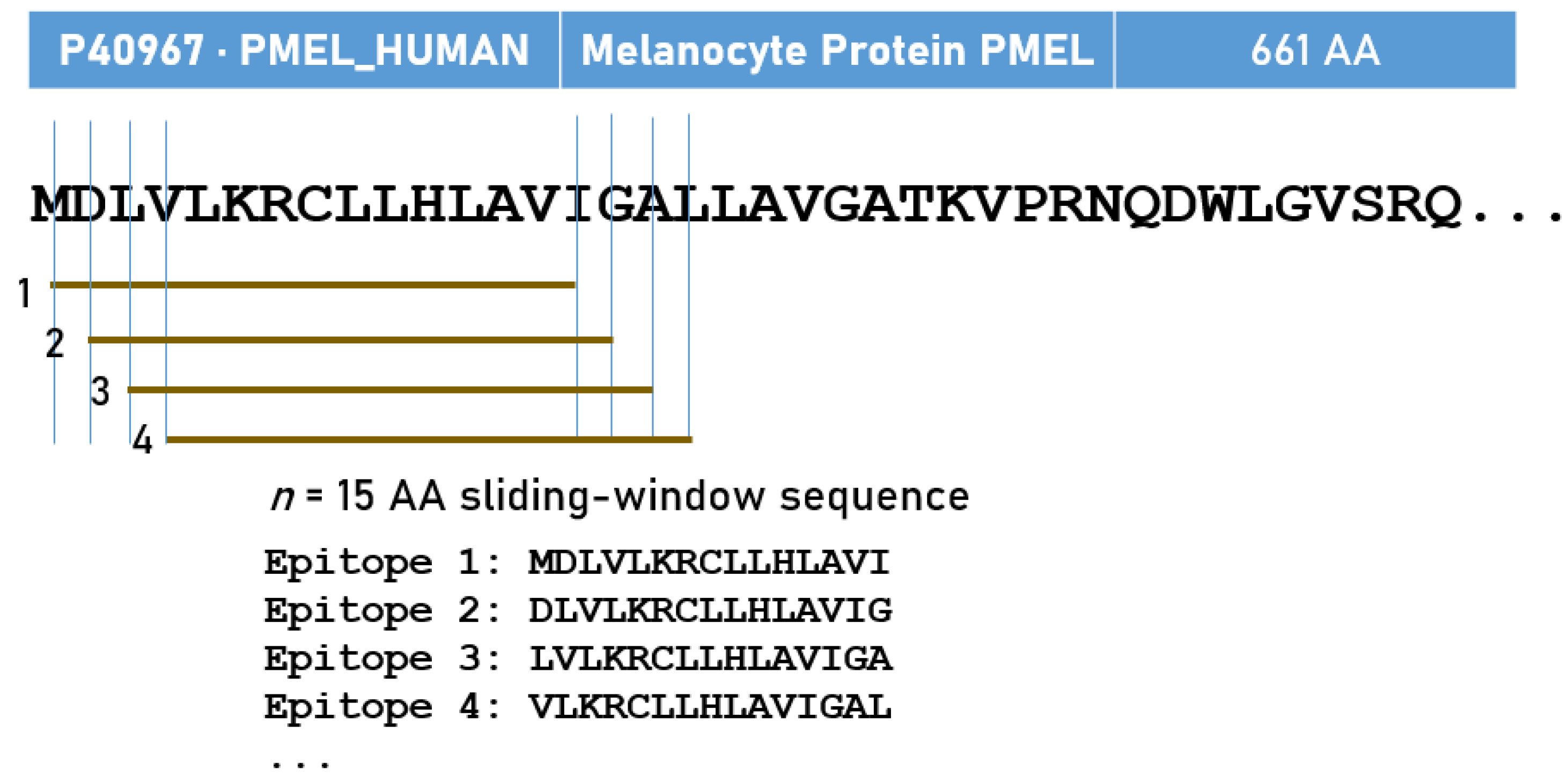
Figure 1. Schematic diagram illustrating the sliding-window method used to evaluate the immunogenicity of all 15-AA linear epitopes of melanoma antigens.
Cells 14 01430 g001
Figure 2. Frequency distribution (per antigen) of 5941 pHLA-II pairs with strong binding affinities.
Cells 14 01430 g002
Figure 3. Frequency distribution of 679 distinct epitopes of HLA-II pairs with strong binding affinity. (Table S6, Supplementary Material).
Cells 14 01430 g003
Table 1. The 15 cancer/melanoma-related antigens, the number of epitopes tested, and hits (PBBA < 50 nm). N tested is the number of 15-AA epitopes × 192 HLA-II alleles. The list of antigens is from [7]. [CTA] denotes that the antigen is a member of the Cancer-Testis Antigens family of antigens, also expressed in other tumors.
UniprotCancer AntigenN (AA)N (15-AA)N TestedHits% Hits
1O75767 TRP223722342,8163640.85
2P04271 S100927814,976830.55
3P0DTW1GAGE1 [CTA]11710319,77600.00
4P14679Tyrosinase 52951598,8807510.76
5P17643TRP1 537523100,4169980.99
6P40967 PMEL17/gp100661647124,22411510.93
7P43355MAGE1 [CTA]30929556,64010871.92
8P43358 MAGE4 [CTA]31730358,1766951.19
9P78358 NY-ESO-1 [CTA]18016631,8723331.04
10Q13066GAGE2B/2C11610219,58400.00
11Q13072 BAGE [CTA]432955681743.13
12Q16385 SSX2 [CTA]18817433,4082860.86
13Q16655Melan-A/MART-111810419,968190.10
14Q6NT46GAGE2A [CTA]11610219,58400.00
15Q9UEU5GAGE2D/GAGE8 [CTA]11610219,58400.00
Total3466665,4725941
Table 2. Counts (N) of melanoma antigens for which an allele was a strong binder. The highest number of antigens per allele is 11, since four antigens did not show any strong PBBA (Table 1). Alleles with strong binding to all 11 antigens are in bold. Alleles within the same N are arranged alphabetically.
AlleleNAlleleNAlleleNAlleleN
DRB1*01:0111DRB1*11:628DPB1*46:015DRB1*04:102
DRB1*01:1811DRB1*11:658DPB1*47:015DRB1*08:022
DRB1*01:2411DRB1*11:748DPB1*72:015DRB1*08:302
DRB1*01:2911DRB1*13:018DPB1*81:015DRB1*11:272
DRB1*10:0111DRB1*13:058DRB1*04:015DRB1*11:542
DRB1*01:1110DRB1*13:118DRB1*11:375DRB1*12:032
DRB1*01:2010DRB1*13:148DRB1*13:075DRB1*14:042
DPB1*33:019DRB1*13:508DRB1*15:035DRB1*14:382
DPB1*71:019DRB1*14:328DRB1*15:075DPB1*04:021
DRB1*07:019DRB1*15:018DRB1*16:055DPB1*105:01
DRB1*11:139DRB1*15:068DRB1*14:124DPB1*16:011
DRB1*11:149DRB1*16:098DRB1*15:374DPB1*19:011
DRB1*11:429DRB1*01:027DPB1*01:013DPB1*34:011
DRB1*13:029DRB1*08:047DRB1*04:043DPB1*41:011
DRB1*13:239DRB1*11:037DRB1*04:053DPB1*49:011
DRB1*13:979DRB1*11:847DRB1*04:723DPB1*55:011
DRB1*16:029DRB1*13:967DRB1*08:013DRB1*01:031
DRB1*03:118DRB1*15:027DRB1*08:243DRB1*03:151
DRB1*09:018DRB1*15:157DRB1*11:113DRB1*04:441
DRB1*11:018DRB1*16:017DRB1*11:193DRB1*11:061
DRB1*11:028DRB1*04:086DRB1*12:163DRB1*11:071
DRB1*11:048DRB1*13:216DRB1*13:613DRB1*12:021
DRB1*11:088DRB1*14:066DRB1*13:663DRB1*13:331
DRB1*11:108DPB1*02:015DRB1*14:013DRB1*14:021
DRB1*11:128DPB1*02:025DRB1*14:543DRB1*14:051
DRB1*11:288DPB1*04:015DRB1*16:043DRB1*14:071
DRB1*11:298DPB1*126:05DPB1*40:012DRB1*14:231
DRB1*11:468DPB1*15:015DRB1*03:012
DRB1*11:498DPB1*23:015DRB1*03:042
DRB1*11:588DPB1*39:015DRB1*03:132
Disclaimer/Publisher’s Note: The statements, opinions and data contained in all publications are solely those of the individual author(s) and contributor(s) and not of MDPI and/or the editor(s). MDPI and/or the editor(s) disclaim responsibility for any injury to people or property resulting from any ideas, methods, instructions or products referred to in the content.