Brief Report

The TIP60-CD44 Axis Modulates Colorectal Cancer Stemness

Department of Physiological Sciences, College of Veterinary Medicine, Oklahoma State University, Stillwater, OK 74078, USA
*
Author to whom correspondence should be addressed.
Cells 2025, 14(10), 686; https://doi.org/10.3390/cells14100686
Submission received: 27 February 2025 / Revised: 1 May 2025 / Accepted: 5 May 2025 / Published: 9 May 2025
(This article belongs to the Collection Cancer Stem Cells and Drug Resistance)

Abstract

:
HIV-1 Tat-interactive protein of 60 kDa (TIP60) is a lysine acetyltransferase protein that can acetylate histone and non-histone proteins. This study highlights TIP60’s role in regulating colorectal cancer (CRC) stemness. The depletion of TIP60 resulted in a marked decrease in cellular proliferation, highlighting TIP60’s involvement in the progression of CRC. Additionally, the loss of TIP60 impacted colony formation, transitioning from densely packed structures to dispersed spindle networks along with the loss of E-cadherin, indicating its role in the epithelial–mesenchymal transition (EMT). Three-dimensional culture models suggest that TIP60 is vital for spheroid formation, highlighting its importance in maintaining cancer stem cell properties in CRC. TIP60-depleted cells showed increased invasion in a 3D basement membrane extract (BME) invasion matrix, demonstrating its essential role in cellular invasiveness. Mechanistically, the reduction of TIP60 resulted in a decrease in CD44 expression, a critical marker for cancer stem cells (CSCs). Notably, CD44 overexpression restored the efficiency of spheroid formation and cell proliferation while reversing the EMT phenotype. Developing the TIP60-CD44 axis as a therapeutic target to treat CRC stemness and metastasis will help decrease the burden due to the deadly disease.

1. Introduction

Colorectal cancer (CRC) is the second leading cause of cancer-related mortality worldwide [1]. Advancements in therapeutic strategies have significantly improved the survival rates of patients diagnosed with CRC [2]. Despite the development of innovative targeted medications and several therapeutic combinations, tumor recurrence and drug resistance remain significant obstacles to conventional, targeted, and immunotherapeutic therapies [3]. Colorectal cancer stem cells (CRCSCs) are the primary determinant of cancer development, recurrence, and dissemination [4]. Cancer stem cells (CSCs) are a distinct group of cells in the tumor bulk that can renew themselves and differentiate into other types of cells [5]. CSCs have a role in tumor recurrence, metastasis, heterogeneity, and multidrug resistance [6,7].
HIV-1 Tat-interactive protein (TIP60), also known as lysine acetyltransferase 5 (KAT5), was first discovered as a cellular acetyltransferase protein interacting with HIV-1 Tat [8]. TIP60 belongs to the MYST (Moz, Ybf2/Sas3, Sas2, and TIP60) family of acetyltransferases and has been shown to acetylate histones [9] and non-histone proteins [10,11]. The MYST family members regulate chromatin remodeling, gene-specific transcription, and DNA damage repair [12]. TIP60 downregulation has been associated with several cancer types, such as colorectal, gastric, and breast cancer [13]. TIP60 has been shown to regulate cellular migration and invasion [14] and inhibit epithelial–mesenchymal transition (EMT) [15]. Apart from cancer, TIP60 has also been shown to be involved in regulating stem cell functions. In hematopoietic stem cells (HSCs), TIP60 is essential for its maintenance [16]. The TIP60 histone acetyltransferase in embryonic stem cells (ESCs) triggers the activation of genes necessary for cell division and suppresses genes that induce differentiation [17]. CD44 belongs to the cell adhesion molecule (CAM) class and is a single-chain transmembrane glycoprotein. CD44 is implicated in various types of cancers, such as breast cancer, prostate cancer, colorectal cancer, and pancreatic cancer. CD44 is involved in tumor growth, EMT, cellular invasion, metastasis, and resistance to chemotherapy [18]. It is an established marker for CSCs [19].
There remains a lack of knowledge regarding the role that TIP60 plays in the control of cancer stemness. Our research sheds light on the significance of TIP60 in the stemness of CRC, demonstrating that TIP60 affects the cancer stemness characteristics in CRC. Mechanistically, we identified that TIP60 controls cancer stemness via CD44.

2. Materials and Methods

2.1. Cell Culture

Colorectal cancer cell lines HCT116 (Cat. No. CCL-247) and 293T (Cat. No. CRL-3216) were purchased from ATCC (Manassas, VA, USA)and were grown in DMEM High glucose (Gibco, Thermo Fisher Scientific, Waltham, MA, USA; Cat. No. 11-995-081) supplemented with 10% Fetal Bovine Serum (Corning, NY, USA; Cat. No. 35-010-CV).

2.2. Generation of Stable Cell Line

An shRNA designed against the TIP60 (TOP 5′-CTGATCGAGTTCAGCTATGAACTCGAGTTCATAGCTGAACTCGATCAG-3′, Bottom 5′-CAAAAACTGATCGAGTTCAGCTATGAACTCGAGTTCATAGCTGAACTCGATCAG-3′) was cloned into a pLKO vector using AgeI and EcoRI restriction sites. For the amplification of the plasmids, the construct (TIP60-targeting, ShTIP60, and control vector, ShLuc) plasmids were transformed into Stbl2 cells. The lentivirus was generated by transfecting 5 × 10⁶ 293T cells with TIP60-targeting (ShTIP60) and control vector (ShLuc) plasmids using polyethylenimine (Fisher Scientific, Waltham, MA, USA; Cat. No. 24314-2) with a 1:3 ratio and incubated for 24 h. After 24 h, the media were changed, and fresh medium was added and incubated for 48 h. After 48 h, the virus was harvested and used to infect 2 × 10⁶ HCT116 cells with polybrene (Millipore Sigma, Burlington, MA, USA; Cat. No. TR1003) reagent (1 mg/mL). After 24 h, the media containing the virus were replaced with fresh growth media, and puromycin (Fisher Scientific, Waltham, MA, USA; Cat. No. AAJ672368EQ) (5 μg/mL) was added to the growth media for selection. The cells were selected until the mock-transfected cells died.
TIP60 wild-type (TIP60_WT) and TIP60 catalytic dead (TIP60_KD) were generated by retrovirus transfection to 5 × 10⁶ 293T cells with the plasmids (TIP60 wild-type (TIP60_WT) and TIP60 catalytic dead (TIP60_KD)) utilizing Lipofectamine 2000 (Invitrogen, Thermo Fisher Scientific, Waltham, MA, USA; Cat. No. 11668019) following the manufacturer’s instructions. The virus was collected 72 h post-transfection and used to infect 2 × 10⁶ HCT116 cells with polybrene at a 0.4 mg/mL concentration. After 6 h, the virus-containing media were substituted with growth media. Puromycin (5 μg/mL) was added to the growth medium for selection after 24 h. The cells were selected until the mock-transfected cells died, and the puromycin-selected cells were maintained for two weeks to generate stocks.

2.3. CD44 Overexpression

To overexpress CD44 in HCT116 cell lines, a plasmid containing the full-length CD44s (Cat. No.137812) gene was obtained from Addgene (Watertown, MA, USA). HCT116 control (ShLuc) and TIP60-depleted (ShTIP60) cells were placed in 6-well plates. When the cell culture reached 60% confluency, the plasmid (1:3) was introduced into the cells using polyethylenimine. After 72 h post-transfection, the media were substituted with a fresh medium. The cells were selected utilizing neomycin (1 mg/mL) (Research Product International, Mount Prospect, IL, USA; Cat. No. N20040-25).

2.4. RNA Isolation and Quantitative PCR (qPCR)

HCT116 control (ShLuc) and TIP60-depleted (ShTIP60) cells were on a 10 cm dish. After reaching 70% confluency, the cells were trypsinized (Corning, NY, USA; Cat. No. 25-053 CI) and counted utilizing a hemacytometer (Fisher Scientific, Waltham, MA, USA; Cat. No. 22-600-100). One hundred thousand HCT116 control cells (ShLuc) and TIP60-depleted (ShTIP60) cells were seeded into 6-well ultra-low attachment plates (Corning, NY, USA; Cat. No. 07-200-601) and incubated for 7 days. The cells were collected and washed with ice-cold PBS, and the whole RNA was extracted using an RNeasy Kit (Qiagen, Hilden, Germany; Cat. No. 74104), following the manufacturer’s instructions, and reverse-transcribed to cDNA using the iSCRIPT cDNA synthesis kit (Bio-Rad Laboratories, Hercules, CA, USA; Cat. No. 1708891). Quantitative real-time PCR (RT-qPCR) was performed using iTaq Universal SYBR Green Supermix (Bio-Rad Laboratories, Hercules, CA, USA; Cat. No. 1725124) on Quant Studio 6 Pro (Applied Biosystem, Thermo Fisher Scientific, Waltham, MA, USA). The amplification conditions were as follows: 50 °C for 2 min, 95 °C for 5 min; 40 cycles of amplification (95 °C for 15 s, 60 °C for 1 min); and the melt curve step (95 °C for 15 s, 60 °C for 1 min, 60 °C → 95 °C (0.1 °C/s)). The mRNA expression of the gene was normalized to the average of the two housekeeping genes ACTB and GAPDH. Gene expression analysis was performed using qPCR and computed using ΔΔCt. The ΔCt was calculated by subtracting the housekeeping gene’s Ct value from the target gene’s Ct value (ΔCt = Ct_target − Avg Ct_housekeeping, ACTB and GAPDH). To compute ΔΔCt, the values were subtracted from the control sample’s ΔCt from each experimental sample’s ΔCt (ΔΔCt = ΔCt_experimental − ΔCt_control). Finally, gene expression in fold change was calculated using the method 2−ΔΔCt [20]. The primers utilized for detecting mRNA expression levels were purchased from Integrated DNA Technologies (IDT), and the sequences of each forward primer (F) and reverse primer (R) are as Table 1.

2.5. Immunofluorescence

Cells were grown in 6-well plates (Corning Costar, NY, USA; Cat. No. 07-200-83) on 15 mm coverslips. At 70% confluency, the cells were fixed in 100% ice-cold methanol for 15 min. Cells were blocked in 3% BSA (Sigma-Aldrich, St. Louis, MO, USA; Cat. No. A2153) for 1 h and incubated with mouse anti-E-cadherin (BD Biosciences, San Jose, CA, USA; Cat. No. 610182) primary antibody at a 1:200 ratio for 1 h at room temperature. Cells were washed twice with PBS, and the secondary antibody, goat anti-mouse (Invitrogen, Thermo Fisher Scientific, Waltham, MA, USA; Cat. No. A11029), was added at a 1:1000 ratio and incubated for 1 h. After incubation, the cells were washed twice with 1× PBS. The stained cells were then mounted onto the glass slides using DAPI (Vector Laboratories, Newark, CA, USA; Cat. No. NC1848443). Images were captured using LSM 980 with Airyscan 2 confocal laser scanning microscope (Carl Zeiss Microscopy GmbH, Jena, Germany).

2.6. Cell Invasion Assay

Two hundred HCT116 ShTIP60 and ShLuc cells were seeded into a 96-well ultra-low attachment round-bottom plate (Corning, Cat. No. 07-201-680). Immediately after seeding, the cells were centrifuged at 1200 rpm for 5 min and incubated for 72 h for spheroid formation. After 72 h, 100 µL of BME invasion matrix (R&D Systems, Minneapolis, MN, USA; Cat. No. 3500096K) was added to each well. The cells were further incubated for 72-96 h. Images were taken at 10× magnification on a BioTek Cytation 5 imager (Agilent Technologies, Santa Clara, CA, USA).

2.7. Spheroid Formation Assay (3D Culture)

HCT116 control (ShLuc) and TIP60-depleted (ShTIP60) cells were cultured in a 10 cm dish. After reaching 70% confluency, the cells were trypsinized (Corning, Cat. No. 25-053 CI) and counted utilizing a hemacytometer (Fisher Scientific, Waltham, MA, USA; Cat. No. 22-600-100). One hundred cells were seeded in each well of 96-well flat ultra-low attachment plates (Corning, NY, USA; Cat. No. 07-200-603). The cells were incubated for 7 days, and images were taken using a BioTek Cytation 5 imager (Agilent Technologies, Santa Clara, CA, USA). For live and dead cell staining of spheroid culture, Calcein AM (Thermo Fisher Scientific, Waltham, MA, USA; Cat. No. C1430) and Ethidium Homodimer-1 (EthD1) (Thermo Fisher Scientific, Waltham, MA, USA; Cat. No. E1169) were added at a concentration of 1 μg/mL. The cells were incubated for 30 min at 37 °C in a cell culture incubator. After the incubation, images were taken using a BioTek Cytation 5 imager (Agilent Technologies, Santa Clara, CA, USA).

2.8. Colony Formation Assay and Absorbance Assay

HCT116 control (ShLuc) and TIP60-depleted (ShTIP60) cells were grown in a 10 cm dish. The cells were trypsinized and counted using a hemacytometer after 70% confluency. One thousand HCT116 control (ShLuc) and TIP60-depleted (ShTIP60) cells were seeded in 6-well plates and incubated for 11 days. On day 11, cells were fixed with a 0.5% Crystal Violet (Fisher Scientific, Waltham, MA, USA; Cat. No. NC0827019) staining solution. The cells were washed twice with 1× PBS, and 250 µL of Crystal Violet solution was added to each well for 5 min. After incubation, the cells were washed three times with PBS. The plates were air-dried, and images were taken using a BioTek Cytation 5 imager (Agilent Technologies, Santa Clara, CA, USA). For the absorbance assay, 1% SDS (Bio-Rad Laboratories, Hercules, CA, USA; Cat. No. 1610302) was used to destain Crystal Violet. Five hundred microliters of 1% SDS were added to each well of 6-well plates and incubated for 6 h on a shaker at room temperature. After the incubation, 100 μL of the destained solution was added to 96-well plates for HCT116 ShLuc and ShTIP60 cells. Absorbance readings were taken at 590 nm on a BioTek Cytation 5 imager (Agilent Technologies, Santa Clara, CA, USA).

2.9. Western Blot Analysis

Cell lysates were extracted using cold RIPA buffer (Cell Biolabs Inc., San Diego, CA, USA; Cat. No. AKR-190) with protease inhibitor and quantified using Pierce BCA Protein Assay Kit (Thermo Fisher Scientific, Waltham, MA, USA; Cat. No. PI23225). Denatured lysates were subjected to SDS-PAGE, transferred to PVDF membranes, and blocked with 3% BSA (Sigma-Aldrich, St. Louis, MO, USA; Cat. No. A8022) for 1 h. The membranes were then incubated with CD44 primary antibody (Thermo Fisher Scientific, Waltham, MA, USA; Cat. No. MA5-13890) at a dilution of 1:1000 and incubated with the membrane overnight at 4 °C in 1% BSA in TBST. Beta-tubulin primary antibody (Santa Cruz Biotechnology, Dallas, TX, USA; Cat No. sc9104) was incubated overnight at 4 °C with the diluted 1% BSA in TBST buffer. HRP conjugated secondary anti-mouse antibody (Santa Cruz Biotechnology, Dallas, TX, USA; Cat. No. sc516102) against CD44 and anti-rabbit antibody (Santa Cruz Biotechnology, Dallas, TX, USA; Cat. No. sc2357) against β-tubulin were used at a concentration of 1:5000 and incubated at room temperature for 1 h. Pierce ECL (Thermo Fisher Scientific, Waltham, MA, USA; Cat. No. 32106) was used for chemiluminescence. Images were acquired using an Amersham Imager 600 (GE Healthcare Life Sciences, Chicago, IL, USA).

2.10. Statistical Analysis

All the experiments were conducted with three independent biological repetitions. The evaluation of data, calculation of mean values, and standard deviation were performed utilizing Microsoft Excel. Statistical significance (p ≤ 0.05) was assessed through an unpaired, two-tailed Student’s t-test. Error bars indicate the standard deviation derived from three biological replicates.

3. Results and Discussion

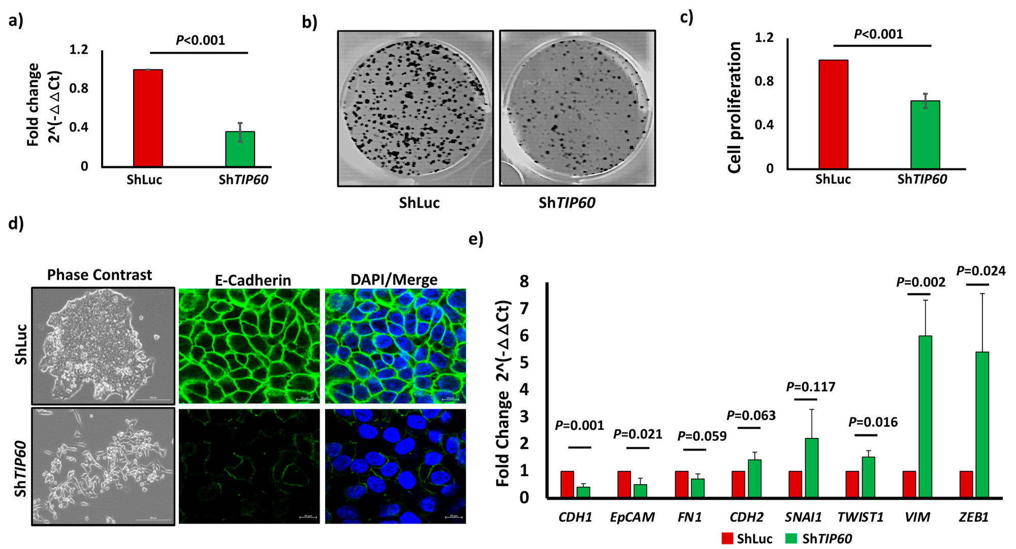
3.1. TIP60 Drives Cell Proliferation and Negatively Regulates EMT in CRC

To investigate the functional role of TIP60 in CRC, we utilized shTIP60 to deplete TIP60 expression in CRC cell lines. We achieved a knockdown efficiency of 64% (p = 0.0002) for HCT116 cell lines, as analyzed by qPCR (Figure 1a). To identify the role of TIP60 in CRC proliferation, we performed a colony formation assay (CFA) by seeding 1000 cells in 6-well plates and incubating them for 11 days. The CFA shows a significant reduction in cell proliferation (Figure 1b), which was further confirmed by the absorbance assay showing reduced cell proliferation of 37% (p = 0.0005) in TIP60-depleted (ShTIP60) cells (Figure 1c). This finding is in agreement with previous studies in squamous cell carcinoma [21] and prostate cancer [22], which suggests that TIP60 is necessary for cellular proliferation in CRC. Since TIP60 regulates cellular invasion and metastasis, we looked at the cell morphology to find out if there are any changes between control (ShLuc) and TIP60-depleted (ShTIP60) cells. The phase-contrast image shows that depletion of TIP60 results in a phenotypic change from compact cell colonies to random spindle networks in cultures, suggesting that TIP60-depleted cells undergo EMT. We also tested additional conditions to understand the role of TIP60 in CRC by overexpressing the TIP60 wild-type (TIP60_WT) and mutating its catalytic domain (TIP60_KD), which is essential for its lysine acetyltransferase activity. The phase-contrast image shows a compact colony in HCT116 TIP60_WT cells, whereas in TIP60_KD, it resembles a mesenchymal phenotype, suggesting a dominant negative effect due to mutation in the catalytic domain (Figure S1a). To further confirm TIP60-depleted (ShTIP60) cells undergoing EMT, we performed immunofluorescence staining of E-cadherin and qPCR analysis for EMT genes in HCT116 ShLuc and ShTIP60 cells. E-cadherin staining in ShLuc showed linear junctional staining, whereas in ShTIP60 cells, there was a loss of junctional staining with a punctate pattern, confirming that TIP60-depleted cells undergo EMT (Figure 1d, right panel). qPCR analysis for EMT genes revealed that epithelial markers such as E-cadherin (CDH1) (p = 0.0013) and EpCAM (p = 0.0218) were significantly downregulated. In contrast, mesenchymal markers such as TWIST1 (p = 0.0163), Vimentin (VIM) (p = 0.0028), and ZEB1 (p = 0.0243) were significantly upregulated (Figure 1e). In breast cancer, it has been demonstrated that TIP60 is essential for the epithelial phenotype [15]. The findings of our study indicate that the loss of epithelial phenotype is a consequence of the depletion of TIP60, which suggests that it plays a role as an epithelial gatekeeper in CRC. Together, these data suggest that TIP60 is essential for cell proliferation and epithelial phenotypes in CRC.

3.2. TIP60 Modulates Cancer Stem Cell Characteristics in CRC

To determine TIP60’s functional significance in regulating CRCSCs, we performed a spheroid formation assay by seeding 100 cells per well of ShLuc and ShTIP60 cells in 96-well ultra-low attachment plates. Spheroid formation assay shows that TIP60-depleted cells completely abrogate sphere-forming ability in 3D culture (Figure 2a,b). It has been shown that TIP60 is involved in stem cell function [16,17]. In an in vitro condition, sphere-forming ability is one of the assays for determining the self-renewal and multipotency of CSC subpopulations inside tumors or cancer cell lines, suggesting the importance of TIP60 in regulating CRC stemness. To understand the role of TIP60 in CRC stemness, we also performed a spheroid culture of TIP60_WT and TIP60_KD. Spheroid formation assay in TIP60_WT shows no significant differences compared to ShLuc cells. However, quantification shows that TIP60_WT forms bigger spheroids than ShLuc. In contrast, TIP60_KD shows a reduced number of spheroid (p = 0.0009) formations, smaller in size, indicating the partial loss of TIP60 functions (Figure 2c,d). This suggests that TIP60 plays a crucial role in CRC stemness. The 2D culture in Figure 1d,e shows that TIP60-depleted cells undergo EMT. To find EMT regulation in the 3D culture condition, we seeded 200 cells per well in 96-well round-bottom ultra-low attachment plates, resuspended in a spheroid-formation extracellular matrix (ECM). Since ShTIP60 fails to form spheroids in 3D culture, after seeding 200 cells per well, we spun down the cells at 1500 rpm for 5 min and incubated them for 3 days at 37 °C in sphere-forming conditioned media so the cells could assemble to form aggregates. Our results show that control (ShLuc) cells fail to invade in a 3D invasion matrix, whereas TIP60-depleted (ShTIP60) cells infiltrate in 3D culture (Figure 2e), suggesting TIP60 is involved in maintaining the epithelial phenotype. Moreover, immunofluorescence labeling of E-cadherin in 3D culture (Figure 2f) exhibits a linear junctional staining similar to that shown in 2D culture, confirming the role of TIP60 in cellular migration and invasion. These data suggest that TIP60 regulates CRC stemness and inhibits cellular invasion in CRC.

3.3. The TIP60-CD44 Axis Regulates Cancer Stem Cell Properties

To have a mechanistic insight on how TIP60 regulates CRC stemness and to identify downstream targets for TIP60, we selected genes that are involved in the regulation of pluripotency, self-renewal, and proliferation in embryonic stem cells/adult stem cells and cancer stem cells such as LGR5 [23], MYC [24], OCT4 [25], NANOG [26], and CD44 [19] (Figure 3a). qPCR results shows that CD44 (p = 0.0011) (Figure 3b) and MYC (p = 0.0038) (Figure S1b) were significantly downregulated in TIP60-depleted cells (ShTIP60) in 3D culture, whereas the expression of LGR5, OCT4, and NANOG was undetected. Since CD44 is a marker for CSCs across various solid tumors [19], we were interested in how TIP60 downregulation changes CD44 expression level and whether CD44 acts as a downstream target for TIP60. The role of MYC and TIP60 has already been established, where MYC recruits TIP60 to chromatin [27]. Meanwhile, the TIP60-CD44 axis has not yet been explored. To identify the role of CD44 in CRC, we overexpressed CD44 by transfecting CD44 full-length in HCT116 control (ShLuc) and TIP60-depleted (ShTIP60) cells. Western blot results show that overexpression of CD44 upregulates TIP60 expression levels (Figure 3c). To identify the role of the TIP60-CD44 axis in CRC cell proliferation, we performed a colony formation assay that shows CD44 overexpression restores cell proliferation in HCT116 cells (Figure 3d and S1c). To further confirm the role of CD44 in cancer stemness, we performed a spheroid formation assay. Overexpression of CD44 in TIP60-depleted (ShTIP60) cells restores spheroid formation efficiency (Figure S1d,e), suggesting that the TIP60-CD44 axis maintains the CSC property. We also performed CalceinAM (stains live cells) and EthD1 (stains dead cells) staining. CalceinAM and EthD1 staining show that TIP60-depleted cells (ShTIP60) fail to survive in 3D culture (Figure 3e), suggesting that TIP60-depleted cells undergo anoikis in 3D culture. Furthermore, immunofluorescence staining (Figure 3f) and the phase-contrast image (Figure S1f) also show that TIP60-depleted cells overexpressing CD44 (ShTIP60_CD44) undergo colony compaction, suggesting an EMT reversal phenotype. Taken together, these data suggest that the TIP60-CD44 axis regulates CRC stemness.
Our findings reveal that TIP60 regulates CRC stemness, cell proliferation, epithelial phenotype, and cellular invasion. Our results are consistent with the knockdown of TIP60 in non-small-cell lung cancer, showing reduced cell proliferation, migration, and invasion [28]. It has also been shown that in CRC, TIP60 expression is downregulated (13%) in tumor samples with peritoneal dissemination, distant metastasis, and a higher stage of TNM classification [29]. Similarly, in gastric cancer, where TIP60 is downregulated in 61% of specimens, it correlates with tumor invasion and lymph node metastasis [30]. These data suggest that TIP60 plays a critical role during tumor initiation and progression, but its downregulation may occur during the transition to a metastatic and advanced stage of CRC. In breast cancer, TIP60 acts as a tumor suppressor gene, and its loss induces genomic instability, leading to the development of cancer [31]. These studies highlight that TIP60 is a dynamic epigenetic regulator that may induce tumor-promoting and tumor-suppressing actions. In summary, this study reveals that TIP60 regulates cancer stem cells’ properties, cellular proliferation, and EMT in CRC. Targeting TIP60 will offer a novel therapeutic approach against CRCSCs.

Supplementary Materials

The following supporting information can be downloaded at https://www.mdpi.com/article/10.3390/cells14100686/s1, Figure S1. (a) Phase-contrast image of HCT116 TIP60_WT and TIP60_KD cells. (b) Bar chart showing MYC expression in HCT116_ShLuc and HCT116_ShTIP60 cells. (c) Bar chart showing cell proliferation in HCT116_ShLuc, HCT116_ShTIP60, HCT116_ShLuc_CD44, and HCT116_ShTIP60_CD44 cells. (d) Bar chart showing the number of spheroids formed per 100 cells in HCT116_ShLuc, HCT116_ShTIP60, HCT116_ShLuc_CD44, and HCT116_ShTIP60_CD44 cells. (e) Phase-contrast image of the spheroid in HCT116_ShLuc, HCT116_ShTIP60, HCT116_ShLuc_CD44, and ShTIP60_CD44 cells, Scale bars: 200 μm. (f) Phase-contrast image of HCT116_ShLuc, HCT116_ShTIP60, HCT116_ShLuc_CD44, and ShTIP60_CD44 cells, Scale bars: 200 μm. Error bars indicate the standard deviation for three biological repetitions, and the p-value is determined by unpaired two-tailed Student’s t-test (NS, not significant; ND, not detected).

Author Contributions

Conceptualization, S.J. and A.M.; writing—original draft preparation, S.J. and A.M.; writing—review and editing, S.J.; supervision, S.J.; funding acquisition, S.J. All authors have read and agreed to the published version of the manuscript.

Funding

Jha lab is funded by a start-up and RAC grants from the Oklahoma State University College of Veterinary Medicine, Department of Physiological Sciences to S.J.

Institutional Review Board Statement

Not applicable.

Informed Consent Statement

Not applicable.

Data Availability Statement

All the data are available from the corresponding authors upon request.

Acknowledgments

The authors thank the members of the Jha laboratory for their helpful discussions and comments.

Conflicts of Interest

The authors declare no conflicts of interest.

Abbreviations

The following abbreviations are used in this manuscript:
BMEBasement membrane extract
CalceinAMCalcein Acetoxymethyl Ester
CAMsCell adhesion molecules
CRC Colorectal cancer
CRCSCsColorectal cancer stem cells
CSCs Cancer stem cells
CtThreshold cycle
DAPI4′,6-diamidino-2-phenylindole
DMEMDulbecco’s Modified Eagle Medium
ECLEnhanced chemiluminescence
ECMExtracellular matrix
EMTEpithelial–mesenchymal transition
ESCsEmbryonic stem cells
EthD1Ethidium Homodimer-1
FBSFetal Bovine Serum
HRPHorseradish peroxidase
HSCsHematopoietic stem cells
KAT5Lysine acetyltransferase 5
MYSTMoz, Ybf2/Sas3, Sas2, and TIP60
PVDFPolyvinylidene difluoride
RT-qPCRQuantitative real-time PCR
SDSSodium dodecyl sulfate
SDS-PAGESodium dodecyl sulfate-polyacrylamide gel electrophoresis
TIP60HIV-1 Tat-interactive protein

References

  1. Bray, F.; Laversanne, M.; Sung, H.; Ferlay, J.; Siegel, R.L.; Soerjomataram, I.; Jemal, A. Global cancer statistics 2022: GLOBOCAN estimates of incidence and mortality worldwide for 36 cancers in 185 countries. CA Cancer J. Clin. 2024, 74, 229–263. [Google Scholar] [CrossRef] [PubMed]
  2. Gmeiner, W.H. Recent Advances in Therapeutic Strategies to Improve Colorectal Cancer Treatment. Cancers 2024, 16, 1029. [Google Scholar] [CrossRef] [PubMed] [PubMed Central]
  3. Hammond, W.A.; Swaika, A.; Mody, K. Pharmacologic resistance in colorectal cancer: A review. Ther. Adv. Med. Oncol. 2016, 8, 57–84. [Google Scholar] [CrossRef] [PubMed] [PubMed Central]
  4. Lin, C.C.; Liao, T.T.; Yang, M.H. Immune Adaptation of Colorectal Cancer Stem Cells and Their Interaction with the Tumor Microenvironment. Front. Oncol. 2020, 10, 588542. [Google Scholar] [CrossRef] [PubMed] [PubMed Central]
  5. Yu, Z.; Pestell, T.G.; Lisanti, M.P.; Pestell, R.G. Cancer stem cells. Int. J. Biochem. Cell Biol. 2012, 44, 2144–2151. [Google Scholar] [CrossRef] [PubMed] [PubMed Central]
  6. Phi, L.T.H.; Sari, I.N.; Yang, Y.G.; Lee, S.H.; Jun, N.; Kim, K.S.; Lee, Y.K.; Kwon, H.Y. Cancer Stem Cells (CSCs) in Drug Resistance and their Therapeutic Implications in Cancer Treatment. Stem Cells Int. 2018, 2018, 5416923. [Google Scholar] [CrossRef] [PubMed] [PubMed Central]
  7. Marzagalli, M.; Fontana, F.; Raimondi, M.; Limonta, P. Cancer Stem Cells-Key Players in Tumor Relapse. Cancers 2021, 13, 376. [Google Scholar] [CrossRef] [PubMed] [PubMed Central]
  8. Yamamoto, T.; Horikoshi, M. Novel substrate specificity of the histone acetyltransferase activity of HIV-1-Tat interactive protein Tip60. J. Biol. Chem. 1997, 272, 30595–30598. [Google Scholar] [CrossRef] [PubMed]
  9. Ikura, T.; Ogryzko, V.V.; Grigoriev, M.; Groisman, R.; Wang, J.; Horikoshi, M.; Scully, R.; Qin, J.; Nakatani, Y. Involvement of the TIP60 histone acetylase complex in DNA repair and apoptosis. Cell 2000, 102, 463–473. [Google Scholar] [CrossRef] [PubMed]
  10. Tang, Y.; Luo, J.; Zhang, W.; Gu, W. Tip60-dependent acetylation of p53 modulates the decision between cell-cycle arrest and apoptosis. Mol. Cell 2006, 24, 827–839. [Google Scholar] [CrossRef] [PubMed]
  11. Sykes, S.M.; Mellert, H.S.; Holbert, M.A.; Li, K.; Marmorstein, R.; Lane, W.S.; McMahon, S.B. Acetylation of the p53 DNA-binding domain regulates apoptosis induction. Mol. Cell 2006, 24, 841–851. [Google Scholar] [CrossRef] [PubMed] [PubMed Central]
  12. Sun, Y.; Jiang, X.; Chen, S.; Fernandes, N.; Price, B.D. A role for the Tip60 histone acetyltransferase in the acetylation and activation of ATM. Proc. Natl. Acad. Sci. USA 2005, 102, 13182–13187. [Google Scholar] [CrossRef] [PubMed] [PubMed Central]
  13. Ghobashi, A.H.; Kamel, M.A. Tip60: Updates. J. Appl. Genet. 2018, 59, 161–168. [Google Scholar] [CrossRef] [PubMed]
  14. Chen, G.; Cheng, Y.; Tang, Y.; Martinka, M.; Li, G. Role of Tip60 in human melanoma cell migration, metastasis, and patient survival. J. Investig. Dermatol. 2012, 132, 2632–2641. [Google Scholar] [CrossRef] [PubMed]
  15. Zhang, Y.; Subbaiah, V.K.; Rajagopalan, D.; Tham, C.Y.; Abdullah, L.N.; Toh, T.B.; Gong, M.; Tan, T.Z.; Jadhav, S.P.; Pandey, A.K.; et al. TIP60 inhibits metastasis by ablating DNMT1-SNAIL2-driven epithelial-mesenchymal transition program. J. Mol. Cell Biol. 2016, 8, 384–399. [Google Scholar] [CrossRef] [PubMed]
  16. Numata, A.; Kwok, H.S.; Zhou, Q.L.; Li, J.; Tirado-Magallanes, R.; Angarica, V.E.; Hannah, R.; Park, J.; Wang, C.Q.; Krishnan, V.; et al. Lysine acetyltransferase Tip60 is required for hematopoietic stem cell maintenance. Blood 2020, 136, 1735–1747. [Google Scholar] [CrossRef] [PubMed] [PubMed Central]
  17. Chen, P.B.; Hung, J.H.; Hickman, T.L.; Coles, A.H.; Carey, J.F.; Weng, Z.; Chu, F.; Fazzio, T.G. Hdac6 regulates Tip60-p400 function in stem cells. Elife 2013, 2, e01557. [Google Scholar] [CrossRef] [PubMed] [PubMed Central]
  18. Hassn Mesrati, M.; Syafruddin, S.E.; Mohtar, M.A.; Syahir, A. CD44: A Multifunctional Mediator of Cancer Progression. Biomolecules 2021, 11, 1850. [Google Scholar] [CrossRef] [PubMed] [PubMed Central]
  19. Wang, L.; Zuo, X.; Xie, K.; Wei, D. The Role of CD44 and Cancer Stem Cells. Methods Mol. Biol. 2018, 1692, 31–42. [Google Scholar] [CrossRef] [PubMed]
  20. Livak, K.J.; Schmittgen, T.D. Analysis of relative gene expression data using real-time quantitative PCR and the 2(-Delta Delta C(T)) Method. Methods 2001, 25, 402–408. [Google Scholar] [CrossRef] [PubMed]
  21. Stacy, A.J.; Zhang, J.; Craig, M.P.; Hira, A.; Dole, N.; Kadakia, M.P. TIP60 upregulates ΔNp63α to promote cellular proliferation. J. Biol. Chem. 2019, 294, 17007–17016. [Google Scholar] [CrossRef] [PubMed] [PubMed Central]
  22. Shiota, M.; Yokomizo, A.; Masubuchi, D.; Tada, Y.; Inokuchi, J.; Eto, M.; Uchiumi, T.; Fujimoto, N.; Naito, S. Tip60 promotes prostate cancer cell proliferation by translocation of androgen receptor into the nucleus. Prostate 2010, 70, 540–554. [Google Scholar] [CrossRef] [PubMed]
  23. Hirsch, D.; Barker, N.; McNeil, N.; Hu, Y.; Camps, J.; McKinnon, K.; Clevers, H.; Ried, T.; Gaiser, T. LGR5 positivity defines stem-like cells in colorectal cancer. Carcinogenesis 2014, 35, 849–858. [Google Scholar] [CrossRef] [PubMed] [PubMed Central]
  24. Elbadawy, M.; Usui, T.; Yamawaki, H.; Sasaki, K. Emerging Roles of C-Myc in Cancer Stem Cell-Related Signaling and Resistance to Cancer Chemotherapy: A Potential Therapeutic Target Against Colorectal Cancer. Int. J. Mol. Sci. 2019, 20, 2340. [Google Scholar] [CrossRef] [PubMed] [PubMed Central]
  25. Zhou, H.; Hu, Y.U.; Wang, W.; Mao, Y.; Zhu, J.; Zhou, B.; Sun, J.; Zhang, X. Expression of Oct-4 is significantly associated with the development and prognosis of colorectal cancer. Oncol Lett. 2015, 10, 691–696. [Google Scholar] [CrossRef] [PubMed] [PubMed Central]
  26. Zhang, J.; Espinoza, L.A.; Kinders, R.J.; Lawrence, S.M.; Pfister, T.D.; Zhou, M.; Veenstra, T.D.; Thorgeirsson, S.S.; Jessup, J.M. NANOG modulates stemness in human colorectal cancer. Oncogene 2013, 32, 4397–4405. [Google Scholar] [CrossRef] [PubMed] [PubMed Central]
  27. Frank, S.R.; Parisi, T.; Taubert, S.; Fernandez, P.; Fuchs, M.; Chan, H.M.; Livingston, D.M.; Amati, B. MYC recruits the TIP60 histone acetyltransferase complex to chromatin. EMBO Rep. 2003, 4, 575–580. [Google Scholar] [CrossRef] [PubMed] [PubMed Central]
  28. Shibahara, D.; Akanuma, N.; Kobayashi, I.S.; Heo, E.; Ando, M.; Fujii, M.; Jiang, F.; Prin, P.N.; Pan, G.; Wong, K.K.; et al. TIP60 is required for tumorigenesis in non-small cell lung cancer. Cancer Sci. 2023, 114, 2400–2413. [Google Scholar] [CrossRef] [PubMed] [PubMed Central]
  29. Sakuraba, K.; Yasuda, T.; Sakata, M.; Kitamura, Y.H.; Shirahata, A.; Goto, T.; Mizukami, H.; Saito, M.; Ishibashi, K.; Kigawa, G.; et al. Down-regulation of Tip60 gene as a potential marker for the malignancy of colorectal cancer. Anticancer Res. 2009, 29, 3953–3955. [Google Scholar] [PubMed]
  30. Sakuraba, K.; Yokomizo, K.; Shirahata, A.; Goto, T.; Saito, M.; Ishibashi, K.; Kigawa, G.; Nemoto, H.; Hibi, K. TIP60 as a potential marker for the malignancy of gastric cancer. Anticancer Res. 2011, 31, 77–79. [Google Scholar] [PubMed]
  31. Bassi, C.; Li, Y.T.; Khu, K.; Mateo, F.; Baniasadi, P.S.; Elia, A.; Mason, J.; Stambolic, V.; Pujana, M.A.; Mak, T.W.; et al. The acetyltransferase Tip60 contributes to mammary tumorigenesis by modulating DNA repair. Cell Death Differ. 2016, 23, 1198–1208. [Google Scholar] [CrossRef] [PubMed] [PubMed Central]
Figure 1. TIP60 is essential for cellular proliferation and epithelial phenotypes in CRC. (a) Bar chart showing qPCR result for TIP60 expression in HCT116 control (ShLuc) and TIP60-depleted (ShTIP60) cells. (b) Colony formation assay in ShLuc and ShTIP60 cells. (c) Bar chart showing cell proliferation after TIP60 depletion. (d) Phase-contrast image of HCT116 ShLuc, ShTIP60 cells (left panel), Scale bars: 200 μm and immunofluorescence staining of E-cadherin (right panel), Scale bars: 10 μm. (e) Bar chart showing qPCR results in fold change for EMT genes in HCT116 ShLuc, ShTIP60 cells. Error bars indicate standard deviation for three biological repetitions and p-value by unpaired two-tailed Student’s t-test.
Cells 14 00686 g001
Figure 2. TIP60 regulates colorectal cancer stemness. (a) Phase-contrast image of HCT116 ShLuc and ShTIP60 cells in 3D culture, Scale bars: 200 μm. (b) Bar chart showing number of spheroids formed per 100 cells. (c) Phase-contrast image of HCT116 TIP60_WT and TIP60_KD cells in 3D culture, Scale bars: 200 μm. (d) Bar chart showing number of spheroids formed per 100 cells. (e) Phase-contrast image of control (ShLuc) and TIP60-depleted (ShTIP60) cells cultured in 3D BME invasion matrix, Scale bars: 1000 μm. (f) Immunofluorescence staining of E-Cadherin in 3D culture of HCT116 control (ShLuc) and TIP60-depleted (ShTIP60) cells, Scale bars: 20 μm. ND, not detected. Error bars indicate standard deviation for three biological repetitions, with p-value by unpaired two-tailed Student’s t-test.
Cells 14 00686 g002
Figure 3. The TIP60-CD44 axis regulates cancer stemness. (a) Bar chart showing the expression of CD44, MYC, LGR5, OCT4, and NANOG normalized to the housekeeping gene (ACTB and GAPDH) in HCT116 cells grown in 3D. (b) Bar chart showing CD44 expression level in control (ShLuc) and TIP60-depleted (ShTIP60) cells. (c) Western blot data showing the TIP60 expression in ShLuc, ShTIP60, ShLuc_CD44, and ShTIP60_CD44, β-tubulin used as an internal control. (d) Colony formation assay in HCT116 ShLuc, ShTIP60, ShLuc_CD44, and ShTIP60_CD44 cells. (e) Phase-contrast image of HCT116 ShLuc, ShTIP60, ShLuc_CD44, and ShTIP60_CD44 spheroid culture (left panel) and CalceinAM and EthD1 staining of HCT116 ShLuc, ShTIP60, ShLuc_CD44, and ShTIP60_CD44 spheroids (right panel), Scale bars: 200 μm. (f) Immunofluorescence staining of E-cadherin in HCT116 ShLuc, ShTIP60, ShLuc_CD44, and ShTIP60_CD44 cells. UD, undetected, Scale bars: 20 μm. Error bars indicate the standard deviation for three biological repetitions and the p-value by unpaired two-tailed Student’s t-test.
Cells 14 00686 g003
Table 1. PCR primer sequences.
GenesForward 5′ to 3′Reverse 5′ to 3′
TIP60AATGTGGCCTGCATCCTAAC TGTTTTCCCTTCCACTTTGG
CD44CACATATTGCTTCAATGCCTCAGCCATCACGGTTGACAATAGTTATG
MYCTCAAGAGGTGCCACGTCTCCTCTTGGCAGCAGGATAGTCCTT
OCT4GGAGTCTGGAGACCATGTTTCTGGAACCATACTCGAACCACATCCTT
LGR5TTCAATCCCTGCGCCTAGATTGCAGGCCGCTGAAACA
NANOGTGGAAGCCACTAGGGAAAGCTGGAGTCACACACTAGTTCACGAATAA
CDH1TTACTGCCCCCAGAGGATGATGCAACGTCGTTACGAGTCA
EpCAMGCTGGCCGTAAACTGCTTTGACATTTGGCAGCCAGCTTTG
FN1AACCCTTCCACACCCCAATCACTGGGTTGCTGACCAGAAG
CDH2CCGGTTTCATTTGAGGGCACTCCCTCAGGAACTGTCCCAT
SNAI1TCTTTCCTCGTCAGGAAGCCGATCTCCGGAGGTGGGATGG
SNAI2CTCCTCATCTTTGGGGCGAGCTTCAATGGCATGGGGGTCT
TWIST1TCGGACAAGCTGAGCAAGATTGCAGCTTGCCATCTTGGAGT
VIMCTGCCAACCGGAACAATGACCATTTCACGCATCTGGCGTT
ZEB1AGGATGACCTGCCAACAGACCTTCAGGCCCCAGGATTTCTT
ACTBCCAGATCATGTTTGAGACCTTCAACCCAGAGGCGTACAGGGATAGC
GAPDHCAGCCTCAAGATCATCAGCATGTGGTCATGAGTCCTTCCA
Disclaimer/Publisher’s Note: The statements, opinions and data contained in all publications are solely those of the individual author(s) and contributor(s) and not of MDPI and/or the editor(s). MDPI and/or the editor(s) disclaim responsibility for any injury to people or property resulting from any ideas, methods, instructions or products referred to in the content.

