Article

Gene Fusion Detection in NSCLC Routine Clinical Practice: Targeted-NGS or FISH?

1
Pathology Unit, IRCCS San Raffaele Scientific Institute, 20132 Milan, Italy
2
Department of Oncology, IRCCS San Raffaele Scientific Institute, 20132 Milan, Italy
3
Candiolo Cancer Institute, FPO-IRCCS, 10060 Turin, Italy
*
Author to whom correspondence should be addressed.
Cells 2023, 12(8), 1135; https://doi.org/10.3390/cells12081135
Submission received: 9 February 2023 / Revised: 5 April 2023 / Accepted: 10 April 2023 / Published: 11 April 2023

Abstract

:
The ability to identify the broadest range of targetable gene fusions is crucial to facilitate personalized therapy selection for advanced lung adenocarcinoma (LuADs) patients harboring targetable receptor tyrosine kinase (RTK) genomic alterations. In order to evaluate the most effective testing approach for LuAD targetable gene fusion detection, we analyzed 210 NSCLC selected clinical samples, comparing in situ (Fluorescence In Situ Hybridization, FISH, and ImmunoHistoChemistry, IHC) and molecular (targeted RNA Next-Generation Sequencing, NGS, and RealTime-PCR, RT-PCR) approaches. The overall concordance among these methods was high (>90%), and targeted RNA NGS was confirmed to be the most efficient technique for gene fusion identification in clinical practice, allowing the simultaneous analysis of a large set of genomic rearrangements at the RNA level. However, we observed that FISH was useful to detect targetable fusions in those samples with inadequate tissue material for molecular testing as well as in those few cases whose fusions were not identified by the RNA NGS panel. We conclude that the targeted RNA NGS analysis of LuADs allows accurate RTK fusion detection; nevertheless, standard methods such as FISH should not be dismissed, as they can crucially contribute to the completion of the molecular characterization of LuADs and, most importantly, the identification of patients as candidates for targeted therapies.

1. Introduction

Gene fusion detection is crucial in the routine clinical practice of non-small cell lung cancer (NSCLC). While for early and locally advanced NSCLC, only EGFR mutational status is mandatory and ALK fusion analysis is optional, in metastatic NSCLC, ALK, ROS1, RET and NTRK rearrangements must be investigated together with EGFR, KRAS, BRAF, HER2 and MET mutations in order to identify those oncogene-addicted tumors that can benefit from targeted therapies. Indeed, as recommended by international guidelines, specific tyrosine kinase inhibitors (TKIs) are routinely used for stage IV tumors that carry a rearrangement in the ALK, ROS1, NTRK or RET (RTKs) gene [1,2].
Therefore, a main goal for molecular diagnostics in pathology is the ability to identify the broadest range of targetable gene fusions in a short turnaround time in order to facilitate personalized therapy selection for NSCLC patients harboring one of these genomic alterations [3].
It has been reported that 2–7% of lung adenocarcinomas (LuADs) have oncogenic ALK rearrangements, which cause the constitutive ligand-independent activation of ALK and the overexpression of the gene by fusing the intact kinase domain of ALK to the N-terminal regions of several different gene partners. EML4 has been found to be the most prevalent gene fusion partner, but more than 20 other ones have also been identified [4].
ROS1 and RET alterations occur in 1–2% and 1–3% of LuADs, respectively. ROS1 and RET are tyrosine kinase receptors, and similarly to ALK, they undergo genomic rearrangements resulting in fusion proteins that preserve and constitutively activate the tyrosine kinase. The kinase domains of ROS1 and RET are paired with a wide range of partners, the most common being CD74 for ROS1 and KIF5B for RET [4].
In all solid tumor types, including LuADs, NTRK-gene (NTRK1, NTRK2 and NTRK3) alterations have a very low prevalence (0.2–0.4%). NTRK1/2/3 gene alterations are mainly gene fusions, and a wide variety of gene partners has been reported. All of the described fusions result in the constitutive activation of their encoded tropomyosin receptor kinases (TRKA, B and C) and, as a consequence, in the dysregulated activation of the downstream pathways [5].
Because RTK gene fusions are mostly caused by chromosomal translocations and intra-chromosomal rearrangements, FISH using break-apart probes has been considered the gold standard method of detection for a long time [2,6]. Since RTK gene fusions result in increased mRNA and protein levels, RT-PCR and IHC have also been successfully used in consideration of their high sensitivity and cost-effectiveness [7]. In particular, positive ALK IHC using an appropriately validated assay may be used to prescribe ALK inhibitors [2].
It is important to underline that standard methods such as IHC, FISH and RT-PCR, traditionally used to investigate the presence of gene rearrangements, have the limitation of examining only one alteration in a single test; therefore, they require more material for all the analyses, resulting in a longer time for complete molecular characterization. Moreover, FISH cannot detect small intrachromosomal rearrangements as the interphase FISH analytical resolution is 100–200 kb, which limits its sensitivity [8].
Given the significant survival benefit obtained from targeted therapies in NSCLC patients with ALK, ROS1, RET, NTRK1, NTRK2 and NTRK3 gene rearrangements, the identification of these patients should be more and more comprehensive, cost-effective and time-efficient. In the past five years, there has been a growing awareness of the need to move from highly selective testing methods, such as the gold standard FISH analysis, toward a multiplex testing approach that avoids sequential analysis, requires a smaller amount of sample material and saves time.
Next-generation sequencing (NGS) has been demonstrated to be an efficient strategy to address these issues [9,10], allowing the simultaneous analysis of a large set of genomic alterations, including additional clinically relevant RNA alterations such as the exon 14skipped isoform of MET and giving a more accurate molecular characterization of each analyzed tumor. Moreover, unlike FISH, NGS data can be used to confirm that the chimeric transcript is in frame and therefore constitutively expressed.
In order to evaluate the most efficient and cost-effective testing approach for the simultaneous detection of NSCLC gene fusions at the RNA level, in the period 2015–2021, we analyzed 210 NSCLC selected clinical samples using two commercially available RNA NGS-targeted panels and compared the results to those obtained by standard methods (FISH, IHC and RT-PCR). We also collected follow-up clinical data for selected patients who tested positive for an ALK, ROS1 or RET fusion and who were treated with targeted therapies at our institution.

2. Materials and Methods

2.1. Study Cohort

From our molecular diagnostics database, we selected a total of 210 NSCLC cases of lung adenocarcinoma (LuAD) stage IV that were diagnosed at our institution in the period 2015–2021 and for which there was enough tissue material to conduct additional molecular studies. The cohort included 126 males (60%) and 84 females (40%). The patients’ median age at diagnosis was 68 years, ranging from 34 to 89 years. The samples included both surgical samples and tissue biopsies and were reviewed by two expert pathologists (G.A. and C.D.). The histologic diagnosis of lung adenocarcinoma was confirmed for all of them, and for all the specimens, the percentage of the tumor component was >50%.
All the cases included in this study had been analyzed for EGFR, KRAS and BRAF mutations using either a mass spectrometry multiplexed genotyping platform as previously described [11], or a targeted NGS approach [12], and they all resulted in wild type for the mentioned genes. All information regarding human material was managed using anonymous numerical codes, and all samples were handled in compliance with the Declaration of Helsinki (https://www.wma.net/policies-post/wma-declaration-of-helsinki-ethical-principles-for-medical-research-involving-human-subjects/, last accessed on 16 January 2023).

2.2. FISH Analysis

All 210 cases were successfully analyzed by FISH for ALK, ROS1, RET, NTRK1, NTRK2 and NTRK3. ALK, ROS1, RET, NTRK1, NTRK2 and NTRK3 rearrangements were tested by a break-apart FISH probe approach: the ZytoLight SPEC ALK/EML4 TriCheck Probe, ZytoLight SPEC ROS1 Dual Color Break Apart Probe, ZytoLight SPEC RET Dual Break Apart Probe, ZytoLight SPEC NTRK1 Dual Break Apart Probe, ZytoLight SPEC NTRK2 Dual Break Apart Probe and ZytoLight SPEC NTRK3 Dual Break Apart Probe (Zytovision GmbH, Bremerhaven, Germany) were used, following the manufacturer’s suggested protocol. FISH testing was performed on formalin-fixed, paraffin-embedded (FFPE) tissue, utilizing 4μm tumor tissue sections of LuAD specimens as previously described [13]. For each case, a minimum of 50 tumor nuclei were observed independently by two expert cytogenetists using a Zeiss Axioscope system (Zeiss, Milan, Italy), and the analysis was performed using Metafer v4.1.1 software, which is able to count probe signals and identify the number of fused or split signals (MetaSystems s.r.l., Milan, Italy). A sample was called true positive if  ≥15% of tumor nuclei showed convincing split signals (green and red signal distance ≥ 2 signal diameters) or a single 3′ signal, according to international guidelines [2,6]. The presence of an EML4::ALK fusion was indicated by green and red split signals, each of which fused to a blue signal in ≥15% of observed tumor nuclei.

2.3. ALK IHC

All 210 cases included in the study were analyzed for ALK protein expression by immunohistochemistry (IHC). IHC was performed on FFPE sections using VENTANA ALK (Clone D5F3) CDx Kit and Benchmark Ultra Immunostainer (Ventana Medical Systems, Tucson, AZ, USA) with OptiView detection kit (Ventana Medical Systems, Tucson, AZ, USA), according to the manufacturer’s instructions. The stained sections were analyzed by an expert pathologist (G.A. or C.D.). Presence of strong granular cytoplasmic staining in tumor cells (any percentage of positive tumor cells) was considered positive for ALK, while absence of strong granular cytoplasmic staining in tumor cells was considered negative for ALK.

2.4. RNA Extraction

In all the cases, tumor-rich areas (>50%) were selected by the pathologist in order to perform manual macrodissection prior to total RNA extraction. Total RNA was extracted from all 210 FFPE samples by using the Maxwell RSC RNA FFPE Kit and the Maxwell RSC Instrument (Promega, Milan, Italy), according to the manufacturer’s instructions. Elution was performed in 50 μL and RNA was quantified using the Qubit RNA HS Assay Kit on Qubit 3.0 Fluorometer (ThermoFisher Scientific, Waltham, MA, USA).

2.5. Targeted RNA-Based NGS

A total of 94 cases were analyzed by the Oncomine Focus Assay (OFA) RNA Panel (ThermoFisher Scientific, Waltham, MA, USA), and 116 cases were analyzed by the Oncomine Comprehensive Assay v3 (OCAv3) RNA Panel (ThermoFisher Scientific, Waltham, MA, USA). OFA Panel is designed to detect gene fusions involving 23 fusion drivers (ABL1, ALK, AKT3, AXL, BRAF, EGFR, ERBB2, ERG, ETV1, ETV4, ETV5, FGFR1, FGFR2, FGFR3, MET, NTRK1, NTRK2, NTRK3, PDGFRA, PPARG, RAF1, RET and ROS1). OCAv3 includes the analysis of 51 genes for fusion detection (AKT2, ALK, AR, AXL, BRCA1, BRCA2, BRAF, CDKN2A, EGFR, ERBB2, ERBB4, ERG, ESR1, ETV1, ETV4, ETV5, FGFR1, FGFR2, FGFR3, FGR, FLT3, JAK2, KRAS, MDM4, MET, MYB, MYBL1, NF1, NOTCH1, NOTCH4, NRG1, NTRK1, NTRK2, NTRK3, NUTM1, PDGFRA, PDGFRB, PIK3CA, PRKACA, PRKACB, PTEN, PPARG, RAD51B, RAF1, RB1, RELA, RET, ROS1, RSPO2, RSPO3 and TERT). Targeted libraries were prepared either manually or by using the Ion Chef Instrument (ThermoFisher Scientific, Waltham, MA, USA), following the user guide instructions, and then they were sequenced on the 530™ Chip utilizing the S5 Prime sequencer (ThermoFisher Scientific, Waltham, MA, USA). Raw data analysis was performed using Torrent Suite v5.12 (ThermoFisher Scientific, Waltham, MA, USA), and a fusion with a known partner was called present by the Ion Reporter if there were more than 20 supporting reads.

2.6. Real-Time PCR

ALK, ROS1, RET, NTRK1, NTRK2 and NTRK3 gene fusions and a MET exon 14 skipping alteration were evaluated in 198 cases using the AmoyDx Lung Cancer PCR Panel (Amoy Diagnostics, Xiamen, China), according to the manufacturer’s instructions. The assays were carried out using the SLAN Real-time PCR System (YanengBIO, Shenzhen, China). This panel enables the analysis of 167 hotspot alterations in 11 genes (EGFR, ALK, ROS1, KRAS, BRAF, HER2, RET, MET, NTRK1, NTRK2 and NTRK3) involved in NSCLC at both DNA level for driver mutations and RNA level for gene fusions. RT-PCR results were interpreted in accordance with the kit’s manual.

3. Results

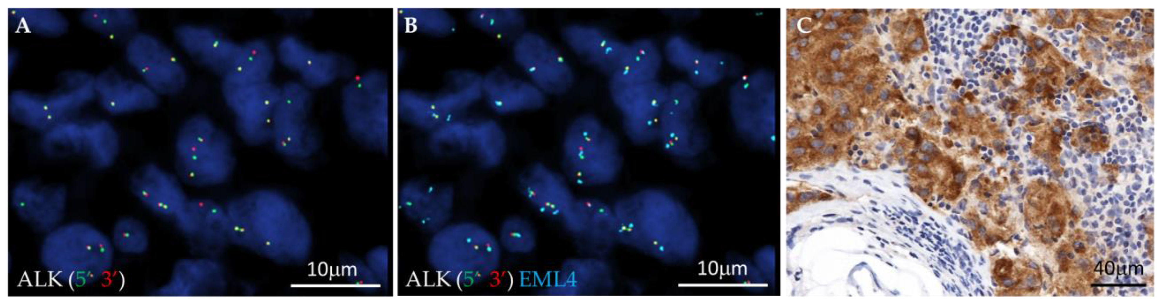
3.1. Fusion Variant Detection by FISH and IHC: In Situ Approach

A total of 77 cases of the 210 successfully investigated by FISH showed a positive result for ALK, ROS1, RET, NTRK1, NTRK2 or NTRK3 rearrangements. ALK rearrangements were the most common, with 48 (23%) cases defined as positive: 43 cases showed an ALK split signal in ≥50% of the analyzed neoplastic cells, and in the remaining 5 cases, ALK rearrangement was observed in percentages varying from 15% to 40%. The most common observed ALK fusion was EML4::ALK (38 cases, Figure 1), identified by the three-color EML4-ALK FISH probe (green 5′ and red 3′ ALK split signals, each of them fused with the split aqua EML4 signal) (Figure 1A,B); the hybridization pattern observed in the remaining 10 cases suggested an ALK rearrangement not involving EML4 (green 5′ and red 3′ ALK split signals, without aqua EML4 signal involvement). Reflex ALK IHC was performed in all the 48 ALK FISH-positive cases: all the cases showed strong cytoplasmic immunoreactivity and were scored as ALK-positive (Figure 1C).
In one case (Pt ID 186), we observed only rare cells (8%) with a green 5′ and red 3′ ALK split signal and EML4 involvement. This observation was associated with weak ALK cytoplasmatic immunoreactivity; therefore, the case was defined as ALK-negative.
ALK protein expression was not detected in the remaining 162 cases of the study cohort, which were all scored as ALK IHC-negative .
In total, 19 cases (9%) showed a red 5′ and green 3′ ROS1 split signal by FISH: 17 cases in ≥50% of the analyzed neoplastic cells and, in the remaining 2 cases, in 25% and 26%, respectively.
In 8 cases (4.1%), a red 5′ and green 3′ RET FISH split signal was observed in ≥50% of the tumor cells, while 1 case showed a single green 3′ RET signal, which was interpreted as positive according to the guidelines [14].
One case showed a green 5′ and red 3′ NTRK3 split signal in ≥50% of the tumor cells; therefore, it was defined as NTRK-positive, and it has been reported elsewhere [15].

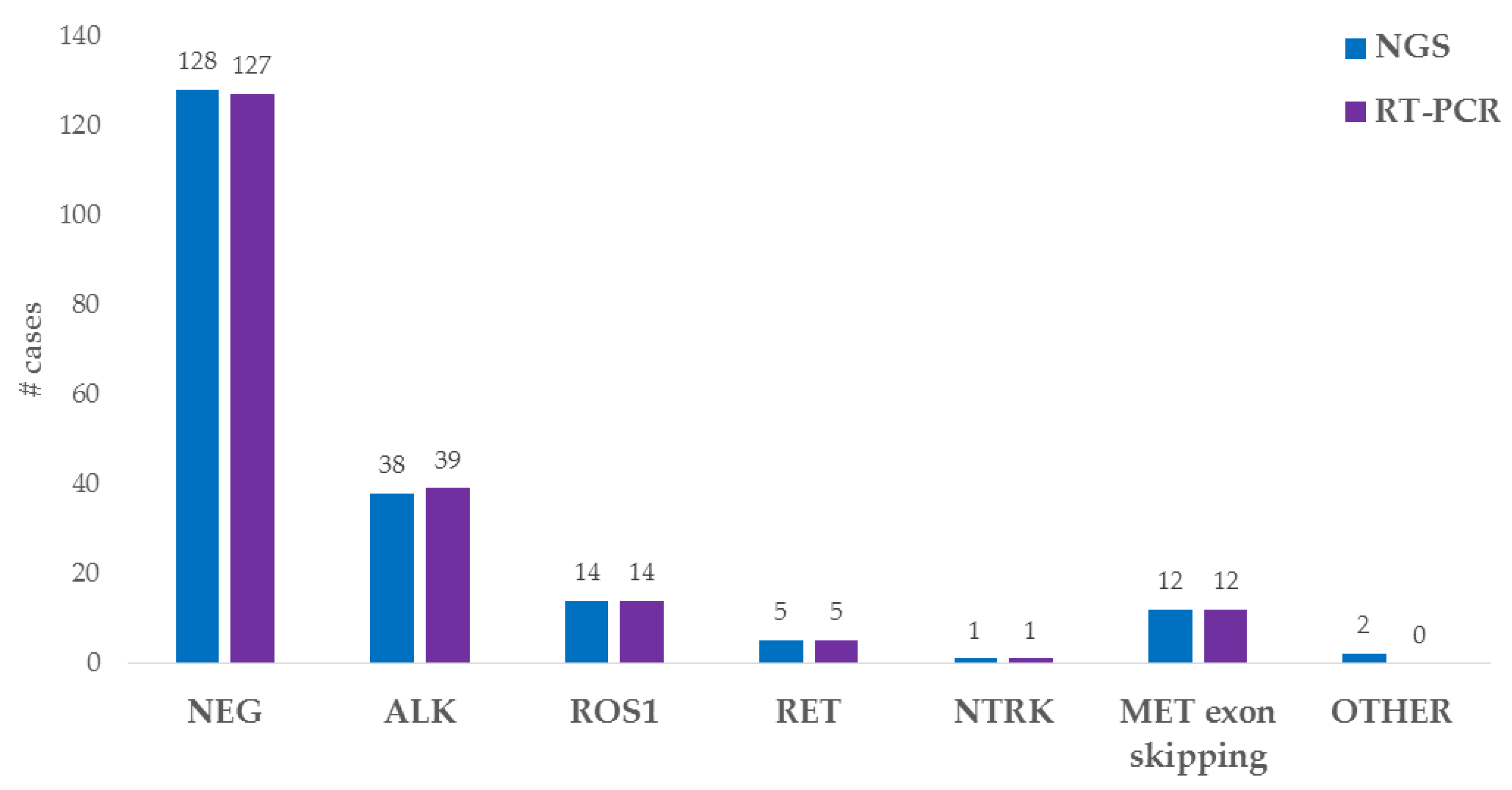
3.2. Fusion Variant Detection by NGS and Real-Time PCR: Molecular Approach

The two RNA-based NGS fusion panels used in this study can detect the majority of NSCLC-targetable fusion genes in an accurate and efficient manner. We succeeded in analyzing a total of 200 NSCLCs out of the 210 cohort cases, with a high success rate (95%). Ten cases did not meet the used panel quality control and were thus considered failed for NGS analysis. The NGS Fusion Sample Quality Control check failed (the total mapped fusion panel reads ≤ 20,000) because of the poor quality and quantity of the total RNA sample. In fact, for 8 samples, only very small tissue biopsies (<500 neoplastic cells) were available; the other 2 samples had been exposed to prolonged formalin fixation (>72 h). These data emphasize the crucial importance of proper tumor sampling and fixation processing, which can have a negative impact on molecular analysis if not correctly carried out.
Among the 10 cases inadequate for NGS analysis, 3 were ALK FISH-positive without EML4 involvement, and for those, ALK IHC was scored as positive; the other 7 cases did not show any rearrangement in the ALK, ROS1, RET, NTRK1, NTRK2 or NTRK3 genes.
The targeted RNA NGS analysis identified the presence of a fusion in 72 out of the 200 (36%) successfully analyzed cases. In particular, 38 ALK fusions (35 cases with EML4 as the partner gene and 3 cases with KIF5B), 14 ROS1 fusions (5 cases involving CD74, 4 cases involving SDC4, 4 cases involving SLC34A2 and 1 case involving EZR), 5 RET fusions (all of them having KIF5B as the partner gene) and 1 NTRK fusion (EML4(2)::NTRK3(14)) were identified.
In addition to the ALK, ROS1, RET, NTRK1, NTRK2 and NTRK3 fusions, the used NGS panels can detect a wide range of gene alterations. This extended analysis allowed us to identify 12 cases carrying MET exon 14 skipping, as well as 2 other gene fusions (EIF3E(1)::RSPO2(2) and FGFR3(17)::TACC3(10)).
We analyzed 198 NGS-tested samples also by RT-PCR (the 2 cases carrying gene fusions not involving ALK, ROS1, RET, NTRK1, NTRK2 or NTRK3 were excluded because those genes are not detected by the RT-PCR kit panel). We identified the presence of an alteration in 71 cases (36%): 39 ALK fusions, 14 ROS1 fusions, 5 RET fusions, 1 NTRK fusion and 12 MET exon 14 skipping. Three discordant cases were observed: two NGS-negative cases tested positive for ALK fusion by RT-PCR, and one case with an ALK fusion detected by NGS tested negative by RT-PCR. This last ALK fusion was not included in the RT-PCR kit panel.
The NGS and RT-PCR data are summarized in Figure 2.

3.3. Comparison of the In Situ and Molecular Approaches

Concordant results were observed in 182 cases (91%) out of the 200 analyzed by both FISH and NGS. In particular, 57 cases resulted positive using both approaches, showing ALK (37), ROS1 (14), RET (5) or NTRK (1) rearrangements (Table 1). The other 125 concordant cases included 111 ALK/ROS1/RET/NTRK FISH-negative and NGS-negative cases for all the targeted fusions and 14 cases that were NGS-positive for a fusion involving genes other than ALK, ROS1, RET, NTRK1, NTRK2 or NTRK3 (i.e., 12 MET exon 14 skipping, EIF3E::RSPO2 and FGFR3::TACC3).
A total of 198 cases were analyzed by both FISH and RT-PCR for ALK/ROS1/RET/NTRK, and we observed 181 (91.4%) concordant cases, with 58 and 123 cases being positive and negative, respectively, by the two methods (Table 1).
Discordant results between FISH and NGS were found in 18 cases (9%) (Table 2): 17 (94%) targeted RNA NGS-negative cases had a positive result by FISH, and 1 (6 %) case positive for a fusion by NGS had a FISH-negative result. In particular, 13 NGS-negative cases presented gene rearrangement in ≥50% of the analyzed cells by FISH (6 ALK fusions, 3 ROS1 fusions and 4 RET fusions), and 4 cases showed 20–25% of rearranged cells by FISH (2 ALK and 2 ROS1 rearrangements).
According to the previously published studies [2], all the FISH ALK-positive cases showed ALK positivity by reflex IHC.
The RT-PCR data were in line with the NGS results, but we observed 17 (8.6%) cases that were discordant with the FISH results. In fact, 16 FISH-positive samples did not show any gene fusions by RT-PCR analysis.
One case (Pt ID186) showed ALK split signals in rare cells (8%) by FISH and had been therefore scored negative because this value was below the cut-off of positivity (i.e., 15%); nevertheless, both NGS and RT-PCR analyses could identify an ALK fusion.

3.4. Selected Patients’ Clinical Follow-Up

A total of five cases with discordant in situ/NGS results and sufficient clinical data after initiation of TKI therapy were identified (cases 182, 183, 184, 190 and 194). Two FISH-ALK-rearranged patients (Pt ID 182 and Pt ID 183) treated with Alectinib did not develop progression after 24 and 12 months, respectively. One FISH-ALK-rearranged (Pt ID184) and one FISH-ROS1-rearranged (Pt ID194) patients received Crizotinib as first-line therapy, and they progressed after 6 and 18 months, respectively. The FISH-ALK-rearranged patient was then treated with Ceritinib, experienced progression after 22 months, received Brigatinib as third-line therapy and died of the disease after 12 months. The FISH-ROS1-rearranged patient showed response to Lorlatinib, given as second-line therapy, at 24-month follow-up. One FISH-RET-rearranged patient had progression on Selpercatinib at 6 months and died of the disease after another 6 months.

4. Discussion

Nowadays, testing for gene fusions is essential to identify NSCLC patients who can benefit from personalized, targeted treatment. Though in the first instance international and national guidelines for advanced LuAds [16] recommend only ALK and ROS1 testing, an updated and efficient molecular diagnostics of LuAds should not exclude the simultaneous analysis of all the targetable gene fusions, as there are already approved targeted therapies that can be employed for selected patients. Importantly, a wide-ranging molecular analysis should be performed as soon as possible at the diagnosis of stage IV LuADs in order to guarantee the patient the most appropriate available treatment in a rapid and cost-efficient manner.
While the low percentage of RTK fusions observed in LuADS [4] may discourage an initial comprehensive analysis of all of them, the identification of these low-prevalence alterations, especially in younger, non-smoking patients, can play a key role in their cure [15]. In this study, we demonstrated how, in a clinical setting, a comprehensive multiplexing approach for gene fusion detection that simultaneously queries multiple potentially actionable targets (e.g., ALK, ROS1, RET, NTRK1, NTRK2, NTRK3 and MET ex14skipping) is feasible by targeted NGS using total RNA extracted from FFPE NSCLC clinical samples. We analyzed a total of 210 NSCLC samples by FISH, and we succeeded in characterizing 200 of these samples (95%) by targeted RNA NGS and 198 samples (94%) by RT-PCR. The results of our analyses, together with already published data [17,18,19,20], showed an excellent concordance rate (91%) of the targeted RNA NGS results with the so-far considered gold standard FISH technique, underlining the essential role of the NGS testing approach in the molecular pathology diagnostics laboratory.
Nevertheless, the minimal discordance (9%) found in the comparison of the FISH results with the NGS and RT-PCR data prompts thoughts and suggests the need for careful planning of diagnostic strategies.
Any targeted molecular assay (both NGS and RT-PCR) is intrinsically limited by the fact that it cannot detect gene fusions that are not present in the panel design used, which is commonly based on the literature’s most recent evidence. Although the most frequent targetable gene fusions are usually included in the commercially available kits, rare and unknown fusions could be missed by these approaches, especially for the less frequent and therefore poorly studied RET and NTRK1, NTRK2 and NTRK3 fusions.
In recent years, Anchored Multiplex PCR (AMP) RNA NGS assays, based on the RACE (Rapid Amplification of cDNA Ends) PCR principle, have been developed [21] and successfully employed in the identification of gene rearrangements without prior knowledge of the target gene fusion partners [22,23]. This approach can be definitely advantageous when analyzing promiscuous gene fusions, such as those involving RTK genes in LuADs, though it requires higher RNA quantities (50–200 ng) and, therefore, cannot be always applicable.
We successfully demonstrated that, in those targeted RNA NGS/RT-PCR-negative cases, ALK IHC and FISH analyses using break-apart ALK, ROS1, RET, NTRK1, NTRK2 and NTRK3 probes did help to unveil the presence of gene fusions. While a FISH split signal for these genes cannot indicate whether the rearrangement results in an in-frame functional gene fusion, it may still identify a targetable alteration, especially in those non-smoking LuAD patients who do not carry any other targetable molecular alteration. In the case of FISH ALK split signals, ALK IHC successfully demonstrated the protein overexpression and its localization, therefore confirming the significance of a FISH ALK-positive result when NGS data are negative for the presence of a gene fusion. In fact, ALK IHC has been approved as a stand-alone companion diagnostic test [2].
Other less common rearrangements unveiled by a positive FISH finding could be further studied with other methods, such as AMP-PCR RNA NGS and RNAseq, in order to establish their role as a target for a specific drug.
For the 17 FISH-positive and NGS-negative discordant cases, we can hypothesize different explanations. The discordance may be due to the presence of a gene fusion different from those covered by the NGS and RT-PCR panels used. In fact, targeted approaches investigate only specific genome regions of interest with selected primers of amplification, which do not cover all the possible gene rearrangements. Further studies are needed in order to identify the gene breakpoints and compare those gene fusions to the ones included in the NGS and RT-PCR panel designs. For the cases that showed a FISH split signal with a percentage of tumor cells lower than 50%, we may also think of a too low representation of the altered cells and, therefore, a limited sensitivity of the NGS technique. On the contrary, the discordant FISH-negative NGS-positive case, which showed a low percentage of ALK split signals by FISH (8%) and an EML4::ALK NGS/RT-PCR-identified fusion, would indicate a high sensitivity of both methods. Moreover, this discrepancy would also suggest the importance of a more careful application of the FISH interpretation guidelines cut-off for ALK and other RTKs, which is ≥15%.
In our study cohort, the significance of the FISH-positive and NGS/RT-PCR-negative discordant findings was ultimately corroborated by the patients’ responses to targeted therapy.All three ALK FISH-positive and ALK IHC-positive cases clinically responded to the ALK-targeted treatment, and two of them are still alive after 24 months and 12 months, respectively, while the third one died of the disease after 40 months following three lines of targeted treatment. Though the presented cases are very few and anecdotal, they still indicate the strength of the FISH data and suggest the key utility of the method in those LuAD cases that tested fusion-negative by targeted RNA NGS/RT-PCR.
The availability of different testing methods as alternatives to targeted RNA NGS assays is also decisive for those samples in which the NGS assays failed due to technical problems such as poor RNA quality and/or quantity extracted from the FFPE samples. In fact, formalin fixation can negatively affect nucleic acid quality by inducing both fragmentation and sequence artifacts that may impact the downstream assay performance, especially when RNA analysis for gene fusion detection is requested, therefore negatively affecting the possibility to obtain useful molecular data [11]. Moreover, tumor molecular characterization may also be challenging because of the lack of adequate tumor cellularity in the tissue sample: very small biopsies are often obtained in LuAD patients, limiting the amount of tissue available for tumor molecular characterization. For those low-quality and very limited samples, in situ approaches such as ALK IHC and ALK/ROS1/RET/NTRK FISH, which can also evaluate a small number of tumor cells, can be useful options.

5. Conclusions

Our experience demonstrated that the targeted RNA NGS characterization of LuADs allows accurate RTK fusion detection; therefore, it should be routinely performed in efficient molecular pathology diagnostic laboratories. Nevertheless, standard methods such as IHC and FISH should not be dismissed and completely replaced by NGS techniques. Currently, they are definitely cost-effective compared to the costs of molecular assays, and, though they are not used upfront, they should still be available as they can crucially contribute to completing the molecular characterization of LuADs, either when NGS cannot be performed because of limited tissue availability or when no molecular targetable alterations are found, especially in younger and non-smoking patients.

Author Contributions

Conceptualization and supervision, L.P. and M.G.C.; methodology and validation, E.B., G.G., V.D.P., A.T., G.M. (Giovanna Marra), G.M. (Gilda Magliacane) and M.R.; software and investigation, L.P., M.G.C., E.B. and G.G.; formal analysis, L.P., M.G.C., E.B., G.A., G.G. and C.D.; resources, M.G.C., C.D., F.R.O., C.L., V.G. and A.B.; data curation, L.P., M.G.C., E.B., F.R.O., G.A., C.L., V.G. and A.B.; writing—original draft preparation and visualization, L.P., M.G.C. and E.B.; writing—review and editing, L.P., M.G.C., E.B., G.G., M.R., F.R.O., G.A., C.L., V.G., C.D. and A.B.; project administration, M.G.C. and L.P.; funding acquisition, M.G.C., C.D., V.G. and A.B. All authors have read and agreed to the published version of the manuscript.

Funding

This research received no external funding.

Institutional Review Board Statement

The study was conducted according to the guidelines of the Declaration of Helsinki.

Informed Consent Statement

Informed consent was obtained from all subjects involved in the study.

Data Availability Statement

The data presented in this study are available upon request from the corresponding author.

Acknowledgments

The authors would like to thank Elena Dal Cin and Mara Bertolazzi for their precious technical support in this study.

Conflicts of Interest

The authors declare no conflict of interest.

References

  1. Mosele, F.; Remon, J.; Mateo, J.; Westphalen, C.B.; Barlesi, F.; Lolkema, M.P.; Normanno, N.; Scarpa, A.; Robson, M.; Meric-Bernstam, F.; et al. Recommendations for the use of next-generation sequencing (NGS) for patients with metastatic cancers: A report from the ESMO Precision Medicine Working Group. Ann. Oncol. 2020, 31, 1491–1505. [Google Scholar] [CrossRef] [PubMed]
  2. Lindeman, N.I.; Cagle, P.T.; Aisner, D.L.; Arcila, M.E.; Beasley, B.; Bernicker, E.H.; Colasacco, C.; Dacic, S.; Hirsch, F.R.; Kerr, K.; et al. Updated Molecular Testing Guideline for the Selection of Lung Cancer Patients for Treatment With Targeted Tyrosine Kinase Inhibitors Guideline From the College of American Pathologists, the International Association for the Study of Lung Cancer, and the Association for Molecular Pathology. Arch. Pathol. Lab. Med. 2018, 13, 323–358. [Google Scholar]
  3. Kazdal, D.; Hofman, V.; Christopoulos, P.; Ilié, M.; Stenzinger, A.; Hofman, P. Fusion-positive non-small cell lung carcinoma: Biological principles, clinical practice, and diagnostic implications. Genes Chromosom. Cancer 2022, 61, 244–260. [Google Scholar] [CrossRef]
  4. Saigí, M.; Carcereny, E.; Morán, T.; Cucurull, M.; Domènech, M.; Hernandez, A.; Martinez-Cardús, A.; Pros, E.; Sanchez-Cespedes, M. Biological and clinical perspectives of the actionable gene fusions and amplifications involving tyrosine kinase receptors in lung cancer. Cancer Treat. Rev. 2022, 109, 102430. [Google Scholar] [CrossRef] [PubMed]
  5. Westphalen, C.B.; Krebs, M.G.; Le Tourneau, C.; Sokol, E.S.; Maund, S.L.; Wilson, T.R.; Jin, D.X.; Newberg, J.Y.; Fabrizio, D.; Veronese, L.; et al. Genomic context of NTRK1/2/3 fusion-positive tumours from a large real-world population. NPJ Precis. Oncol. 2021, 5, 69. [Google Scholar] [CrossRef] [PubMed]
  6. Lindeman, N.I.; Cagle, P.T.; Beasley, M.B.; Chitale, D.A.; Dacic, S.; Giaccone, G.; Jenkins, R.B.; Kwiatkowski, D.J.; Saldivar, J.S.; Squire, J.; et al. Molecular testing guidelines for selection of lung cancer patients for ALK tyrosine kinase inhibitors. Guideline from the College of American Pathologists, International Association for the Study of Lung Cancer, and Association for Molecular Pathology. J. Mol. Diagn. 2013, 15, 415–453. [Google Scholar] [CrossRef] [Green Version]
  7. Tuononen, K.; Sarhadi, V.K.; Wirtanen, A.; Rönty, M.; Salmenkivi, K.; Knuuttila, A.; Remes, S.; Telaranta-Keerie, A.I.; Bloor, S.; Ellonen, P.; et al. Targeted resequencing reveals ALK fusions in non-small cell lung carcinomas detected by FISH, immunohistochemistry, and real-time RT-PCR: A comparison of four methods. Biomed. Res. Int. 2013, 2013, 757490. [Google Scholar] [CrossRef] [PubMed]
  8. Cui, C.; Shu, W.; Li, P. Fluorescence in situ hybridization: Cell-based genetic diagnostic and research applications. Front. Cell Dev. Biol. 2016, 4, 89. [Google Scholar] [CrossRef] [Green Version]
  9. Lazzari, C.; Bulotta, A.; Cangi, M.G.; Bucci, G.; Pecciarini, L.; Bonfiglio, S.; Lorusso, V.; Ippati, S.; Arrigoni, G.; Grassini, G.; et al. Next generation sequencing in non-small cell lung cancer: Pitfalls and opportunities. Diagnostics 2020, 10, 1092. [Google Scholar] [CrossRef]
  10. Gregorc, V.; Mazzarella, L.; Lazzari, C.; Graziano, P.; Vigneri, P.; Genova, C.; Toschi, L.; Ciliberto, G.; Bonanno, L.; Delmonte, A.; et al. Prospective Validation of the Italian Alliance against Cancer Lung Panel in Patients with Advanced Non–Small-Cell Lung Cancer. Clin. Lung Cancer 2021, 22, e637–e641. [Google Scholar] [CrossRef]
  11. Magliacane, G.; Grassini, G.; Bartocci, P.; Francaviglia, I.; Cin, E.D.; Barbieri, G.; Arrigoni, G.; Pecciarini, L.; Doglioni, C.; Cangi, M.G. Rapid targeted somatic mutation analysis of solid tumors in routine clinical diagnostics. Oncotarget 2015, 6, 30592–30603. [Google Scholar] [CrossRef] [Green Version]
  12. Redegalli, M.; Grassini, G.; Magliacane, G.; Pecciarini, L.; Lena, M.S.; Smart, C.E.; Johnston, R.L.; Waddell, N.; Maestro, R.; Macchini, M.; et al. Routine molecular profiling in both resectable and unresectable pancreatic adenocarcinoma: Relevance of cytological samples. Clin. Gastroenterol. Hepatol. 2022. online ahead of print. [Google Scholar] [CrossRef] [PubMed]
  13. Francaviglia, I.; Magliacane, G.; Lazzari, C.; Grassini, G.; Brunetto, E.; Cin, E.D.; Girlando, S.; Medicina, D.; Smart, C.E.; Bulotta, A.; et al. Identification and monitoring of somatic mutations in circulating cell-free tumor DNA in lung cancer patients. Lung Cancer 2019, 134, 225–232. [Google Scholar] [CrossRef] [PubMed]
  14. Belli, C.; Penault-Llorca, F.; Ladanyi, M.; Normanno, N.; Scoazec, J.-Y.; Lacroix, L.; Reis-Filho, J.; Subbiah, V.; Gainor, J.; Endris, V.; et al. ESMO recommendations on the standard methods to detect RET fusions and mutations in daily practice and clinical research. Ann. Oncol. 2021, 32, 337–350. [Google Scholar] [CrossRef] [PubMed]
  15. Lazzari, C.; Pecciarini, L.; Doglioni, C.; Pedica, F.; Gajate, A.M.S.; Bulotta, A.; Gregorc, V.; Cangi, M.G. Case report: EML4::NTRK3 gene fusion in a patient with metastatic lung adenocarcinoma successfully treated with entrectinib. Front. Oncol. 2022, 12, 1038774. [Google Scholar] [CrossRef] [PubMed]
  16. Postmus, P.E.; Kerr, K.M.; Oudkerk, M.; Senan, S.; Waller, D.A.; Vansteenkiste, J.; Escriu, C.; Peters, S.; ESMO Guidelines Committee. Early and locally advanced non-small-cell lung cancer (NSCLC): ESMO Clinical Practice Guidelines for diagnosis, treatment and follow-up. Ann. Oncol. 2017, 28 (Suppl. S4), iv1–iv21. [Google Scholar] [CrossRef] [PubMed]
  17. Kirchner, M.; Neumann, O.; Volckmar, A.-L.; Stögbauer, F.; Allgäuer, M.; Kazdal, D.; Budczies, J.; Rempel, E.; Brandt, R.; Talla, S.B.; et al. RNA-Based Detection of Gene Fusions in. Cancers 2019, 11, 1309. [Google Scholar] [CrossRef] [PubMed] [Green Version]
  18. Sun, L.; McNulty, S.N.; Evenson, M.J.; Zhu, X.; Robinson, J.A.; Mann, P.R.; Duncavage, E.J.; Pfeifer, J.D. Clinical Implications of a Targeted RNA-Sequencing Panel in the Detection of Gene Fusions in Solid Tumors. J. Mol. Diagn. 2021, 23, 1749–1760. [Google Scholar] [CrossRef]
  19. Tabbò, F.; Muscarella, L.A.; Gobbini, E.; Trombetta, D.; Castellana, S.; Rigutto, A.; Galetta, D.; Maiello, E.; Martelli, O.; Tiseo, M.; et al. Detection of ALK fusion variants by RNA-based NGS and clinical outcome correlation in NSCLC patients treated with ALK-TKI sequences. Eur. J. Cancer 2022, 174, 200–211. [Google Scholar] [CrossRef]
  20. Luca, C.; Pepe, F.; Pisapia, P.; Iaccarino, A.; Righi, L.; Listì, A.; Russo, G.; Campione, S.; Pagni, F.; Nacchio, M.; et al. RNA-based next-generation sequencing in non-small-cell lung cancer in a routine setting: An experience from an Italian referral center. Per. Med. 2022, 19, 395–401. [Google Scholar] [CrossRef] [PubMed]
  21. Zheng, Z.; Liebers, M.; Zhelyazkova, B.; Cao, Y.; Panditi, D.; Lynch, K.D.; Chen, J.; Robinson, H.E.; Shim, H.S.; Chmielecki, J.; et al. Technical Reports Anchored multiplex PCR for targeted next-generation sequencing. Nat. Med. 2014, 20, 1479–1484. [Google Scholar] [CrossRef] [PubMed]
  22. Pfarr, N.; Kirchner, M.; Lehmann, U.; Leichsenring, J.; Merkelbach-Bruse, S.; Glade, J.; Hummel, M.; Stögbauer, F.; Lehmann, A.; Trautmann, M.; et al. Testing NTRK testing: Wet-lab and in silico comparison of RNA-based targeted sequencing assays. Genes Chromosom. Cancer 2020, 59, 178–188. [Google Scholar] [CrossRef] [PubMed]
  23. Racanelli, D.; Brenca, M.; Baldazzi, D.; Goeman, F.; Casini, B.; De Angelis, B.; Guercio, M.; Milano, G.M.; Tamborini, E.; Busico, A.; et al. Next-Generation Sequencing Approaches for the Identification of Pathognomonic Fusion Transcripts in Sarcomas: The Experience of the Italian ACC Sarcoma Working Group. Front. Oncol. 2020, 10, 489. [Google Scholar] [CrossRef] [PubMed] [Green Version]
Figure 1. Images showing an example of an EML4::ALK rearranged case. FISH analysis shows ALK red-green split signals (A) fused with EML4 aqua signals (B), and IHC staining shows strong cytoplasmic immunoreactivity (C).
Cells 12 01135 g001
Figure 2. Histogram representing NGS and RT-PCR results.
Cells 12 01135 g002
Table 1. FISH-Positive/NGS-Positive Concordant Cases.
NoPt_IDFISHNGSRT-PCRALK IHCFISH_DataNGS_DataRT-PCR_Data
12++++ALK split >50%, no EML4KIF5B(24)::ALK(20)ALK POS
23++++ALK split >50%, no EML4KIF5B(17)::ALK(20)ALK POS
34++++ALK split >50%, no EML4KIF5B(17)::ALK(20)ALK POS
45++++EML4::ALK > 50%EML4(20)::ALK(20)ALK POS
56++++EML4::ALK > 50%EML4(6)::ALK(20)ALK POS
67++++EML4::ALK > 50%EML4(6)::ALK(20)ALK POS
78++++EML4::ALK > 50%EML4(6)::ALK(20)ALK POS
89++++EML4::ALK > 50%EML4(6)::ALK(20)ALK POS
910++++EML4::ALK > 50%EML4(2)::ALK(20)ALK POS
1011++++EML4::ALK > 50%EML4(13)::ALK(20)ALK POS
1112++++EML4::ALK > 50%EML4(13)::ALK(20)ALK POS
1213++++EML4::ALK > 50%EML4(13)::ALK(20)ALK POS
1314++-+EML4::ALK > 50%EML4(6)::ALK(18)NEG
1415++++EML4::ALK > 50%EML4(13)::ALK(20)ALK POS
1516++++EML4::ALK > 50%EML4(13)::ALK(20)ALK POS
1617++++EML4::ALK > 50%EML4(20)::ALK(20)ALK POS
1718++++EML4::ALK > 50%EML4(13)::ALK(20)ALK POS
1819++++EML4::ALK > 50%EML4(20)::ALK(20)ALK POS
1920++++EML4::ALK > 50%EML4(6)::ALK(20)ALK POS
2021++++EML4::ALK > 50%EML4(13)::ALK(20)ALK POS
2122++++EML4::ALK > 50%EML4(6)::ALK(20)ALK POS
2223++++EML4::ALK >50%EML4(6)::ALK(20)ALK POS
2324++++EML4::ALK > 50%EML4(13)::ALK(20)ALK POS
2425++++EML4::ALK > 50%EML4(13)::ALK(20)ALK POS
2526++++EML4::ALK > 50%EML4(6)::ALK(20)ALK POS
2627++++EML4::ALK > 50%EML4(13)::ALK(20)ALK POS
2728++++EML4::ALK > 50%EML4(13)::ALK(20)ALK POS
2829++++EML4::ALK > 50%EML4(13)::ALK(20)ALK POS
2930++++EML4::ALK > 50%EML4(13)::ALK(20)ALK POS
3031++++EML4::ALK > 50%EML4(2)::ALK(20)ALK POS
3132++++EML4::ALK > 50%EML4(20)::ALK(20)ALK POS
3233++++EML4::ALK > 50%EML4(6)::ALK(20)ALK POS
3334++++EML4::ALK > 50%EML4(6)::ALK(20)ALK POS
3435++++EML4::ALK > 50%EML4(13)::ALK(20)ALK POS
35212++++EML4::ALK > 50%EML4(6)::ALK(20)ALK POS
3636++++EML4::ALK = 12%EML4(6)::ALK(20)ALK POS
3737++++EML4::ALK = 40% EML4(13)::ALK(20)ALK POS
38162+++-EML4::NTRK3 > 50%EML4(2)::NTRK3(14)NTRK POS
39166+++-ROS1 split > 50%SDC4(2)::ROS1(32)ROS1 POS
40167+++-ROS1 split > 50%SLC34A2(13)::ROS1(34)ROS1 POS
41168+++-ROS1 split > 50%EZR(10)::ROS1(34)ROS1 POS
42169+++-ROS1 split > 50%CD74(6)::ROS1(34)ROS1 POS
43170+++-ROS1 split > 50%SLC34A2(13)::ROS1(32)ROS1 POS
44171+++-ROS1 split > 50%SDC4(2)::ROS1(32)ROS1 POS
45172+++-ROS1 split > 50%CD74(6)::ROS1(34)ROS1 POS
46173+++-ROS1 split > 50%CD74(6)::ROS1(34)ROS1 POS
47174+++-ROS1 split > 50%SLC34A2(13)::ROS1(32)ROS1 POS
48175+++-ROS1 split > 50%SLC34A2(13)::ROS1(32)ROS1 POS
49176+++-ROS1 split > 50%SDC4(2)::ROS1(32)ROS1 POS
50177+++-ROS1 split > 50%SDC4(2)::ROS1(32)ROS1 POS
51209+++-ROS1 split > 50%CD74(6)::ROS1(34)ROS1 POS
52210+++-ROS1 split > 50%CD74(6)::ROS1(34)ROS1 POS
53163+++-RET split > 50%KIF5B(15)::RET(12)RET POS
54164+++-RET split > 50%KIF5B(15)::RET(12)RET POS
55165+++-RET split > 50%KIF5B(15)::RET(12)RET POS
56207+++-RET split > 50%KIF5B(23)::RET(12)RET POS
57208+++-RET split > 50%KIF5B(15)::RET(12)RET POS
Abbreviations: No, Number; +, positive for presence of rearrangement/expression; -, negative for presence of rearrangement/expression.
Table 2. FISH and NGS/RT-PCR Discordant Cases.
NoPt_IDFISHNGSRT-PCRALK IHCFISH_DataNGS_DataRT-PCR_Data
1178+--+ALK split = 25%, no EML4No fusionsNEG
2179+--+ALK split = 25%, no EML4No fusionsNEG
3180+--+ALK split = 50%, no EML4No fusionsNEG
4181+--+ALK split = 50%, no EML4No fusionsNEG
5182+-++EML4::ALK > 50%No fusionsALK POS
6183+--+EML4::ALK > 50%No fusionsNEG
7184+--+EML4::ALK > 50%No fusionsNEG
8185+-++EML4::ALK > 50%No fusionsALK POS
9192+---ROS1 split > 50%No fusionsNEG
10193+---ROS1 split > 50%No fusionsNEG
11194+---ROS1 split > 50%No fusionsNEG
12195+---ROS1 split = 25%No fusionsNEG
13196+---ROS1 split = 25%No fusionsNEG
14189+---RET split > 50%No fusionsNEG
15190+---RET split > 50%No fusionsNEG
16191+---RET split > 50%No fusionsNEG
17188+---RET single 3′ > 50%No fusionsNEG
18186-++-ALK split in rare cellEML4(6)::ALK(20)ALK POS
Abbreviations: No, Number; +, positive for presence of rearrangement/expression; -, negative for presence of rearrangement/expression.
Disclaimer/Publisher’s Note: The statements, opinions and data contained in all publications are solely those of the individual author(s) and contributor(s) and not of MDPI and/or the editor(s). MDPI and/or the editor(s) disclaim responsibility for any injury to people or property resulting from any ideas, methods, instructions or products referred to in the content.

