
Alzheimer’s Disease and Its Possible Evolutionary Origin: Hypothesis

Abstract
1. Introduction
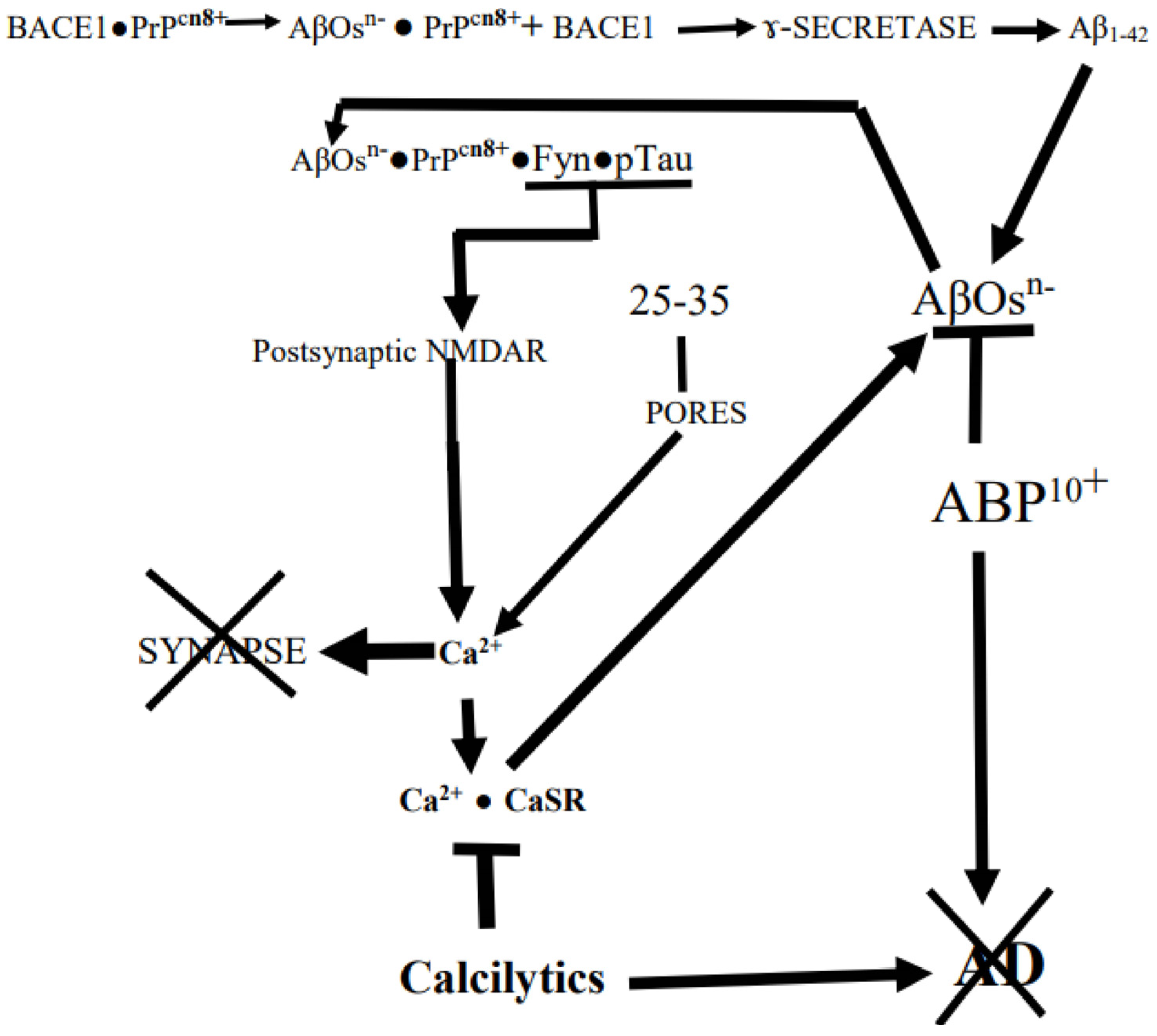
2. Aβ Oligomerslikely AD-Starters, but Not the Terminators
3. The Deadly AD Family
4. Neuron–Astrocyte collaboration
5. Where Does AD Start?
6. Origins
7. How Might AD Start?
8. The Spreading of LOAD/SAD Connectopathy
9. CaSRs Participation in Driving the Connectopathy
10. Why Is LOAD/SAD a Disease of Aging?
11. The Clinical Emergence of LOAD/SAD after Its Long Stealthy Prelude
12. The Lethal Tau-Driven Finale
13. The Synapses Pruning AβOs-PrPC-Tau Combination Also Induces Neurons to Suicidally Try to Enter Their Cell Cycle
14. Do AβOs Really Co-Drive AD Pathology?
15. How Might AD Be Treated?
Author Contributions
Funding
Conflicts of Interest
Abbreviations
| AβPP | Amyloid-β pecursor protein |
| Aβ | Amyloid-β |
| AβOs | Amyloid-β oligomers |
| AD | Alzheimer’s disease |
| α-7nAChRs | Nicotonic acetylcholine receptors |
| AMPA | α-amino-3-hydroxy-5-methyl-4-isoxazolepropionic acid |
| ANTs | Astrocyte•Neuron Teams |
| Apo E | Apolipoprotein-E |
| BACE1 | Beta-site amyloid precursor protein cleaving enzyme-1 (β-secretase 1) |
| BBB | Blood–Brain Barrier |
| BDNF | Brain-derived neurotrophic factor |
| BOLD fMRI | Blood Oxygen Level Dependent functional Magnetic Resonance Imaging |
| CaSRs | Ca2+-sensing receptors |
| Cdk | Cyclin-dependent kinase |
| CSF | Cerebrospinal fluid |
| DG | Dentate gyrus |
| DMN | default-mode network |
| EAATs | Excitatory Amino Acid Transporters |
| EOAD/FAD | Early-Onset or Familial AD |
| EC | Entorhinal cortical |
| ECM | Extracellular Matrix |
| EVs | Extracellular Vesicles |
| GSK3β | Glycogen synthase kinase-3 β |
| IDPs | Intrinsically disordered proteins |
| ISF | Interstitial fluid |
| LEC | Lateral entorhinal cortex |
| LRP1 | LDL receptor-related protein 1 |
| MAPK (ERK) | Mitogen-activated protein kinase (extracellular signal-regulated kinase) |
| MARCKS | Myristoylated alanine-rich C kinase substrate |
| MCI | Mild Cognitive Impairment |
| NAHAs | Normal Adult Human cerebral Astrocytes |
| NFTs | Neurofibrillary tangles |
| NMDARs | N-methyl-D-aspartate receptors |
| PCM1 | pericentriolar material-1 |
| PET | Positron Emission Tomography |
| PKC | Protein Kinase C |
| PrPC | Cellular prion protein |
| PQCs | protein quality and quantity control systems |
| ROS | Reactive Oxygen Species |
| LOAD/SAD | Late onset or sporadic AD |
| HPTOs | Hyper-phosphorylated tau oligomers |
| PQCs | Protein quality and quantity control systems |
| PSD | Postsynaptic density |
| SDC | somatodendritic compartment |
References
- Kraepelin, E. Allgemeine Psychiatrie. In Psychiatrie, 8th ed.; Barth: Leipzig, Germany, 1909; Volume I. [Google Scholar]
- Kraepelin, E. Klinische Psychiatrie. In Psychiatrie, 8th ed.; Barth: Leipzig, Germany, 1910; Volume II. [Google Scholar]
- Broxmeyer, L. Dr Oskar Fischer’s curious little Alzheimer’s germ. Curr. Opin. Neurosci. 2017, 1, 160–178. [Google Scholar]
- Delacourte, A. The natural and molecular history of Alzheimer’s disease. J. Alzheimer’s Dis. 2006, 9, 187–194. [Google Scholar] [CrossRef] [PubMed]
- Hanseeuw, B.J.; Betensky, R.A.; Jacobs, H.I.; Schultz, A.P.; Sepulcre, J.; Becker, J.A.; Orozco Cosio, D.M.; Farrell, M.; Quiroz, Y.T.; Mormino, E.C.; et al. Association of Amyloid and Tau with Cognition in Preclinical Alzheimer Disease A Longitudinal Study. JAMA Neurol. 2019, 76, 915–924. [Google Scholar] [CrossRef] [PubMed]
- Jucker, M.; Walker, L.C. Propagation and spread of pathogenic protein assemblies in neurodegenerative diseases. Nat. Neurosci. 2018, 21, 1341–1349. [Google Scholar] [CrossRef] [PubMed]
- Ye, L.; Hamaguchi, T.; Fritschi, S.K.; Eisele, Y.S.; Obermüller, U.; Jucker, M.; Walker, L.C. Progression of Seed-Induced Aβ Deposition within the Limbic Connectome. Brain Pathol. 2015, 25, 743–752. [Google Scholar] [CrossRef] [PubMed]
- Gimbel, D.A.; Nygaard, H.B.; Coffey, E.E.; Gunther, E.C.; Laurén, J.; Gimbel, Z.A.; Strittmatter, S.M. Memory impairment in transgenic Alzheimer mice requires cellular prion protein. J. Neurosci. 2010, 30, 6367–6374. [Google Scholar] [CrossRef] [PubMed]
- Jonsson, T.; Atwal, J.K.; Steinberg, S.; Snaedal, J.; Jonsson, P.V.; Bjornsson, S.; Stefansson, H.; Sulem, P.; Gudbjartsson, D.; Maloney, J.; et al. A mutation in APP protects against Alzheimer’s disease and age-related cognitive decline. Nature 2012, 488, 96–99. [Google Scholar] [CrossRef]
- Coskuner-Weber, O.; Uversky, V.N. Insights into the Molecular Mechanisms of Alzheimer’s and Parkinson’s Diseases with Molecular Simulations: Understanding the Roles of Artificial and Pathological Missense Mutations in Intrinsically Disordered Proteins Related to Pathology. Int. J. Mol. Sci. 2018, 19, 336. [Google Scholar] [CrossRef]
- Uversky, V.N. Dancing Protein Clouds: Intrinsically Disordered Proteins in Health and Disease. Part A; Academic Press-Elsevier: Cambridge, MA, USA, 2019. [Google Scholar]
- Davies, P. The Demon in the Machine: How Hidden Webs of Information Are Finally Solving the Mystery of Life; Allen Lane-Penguin Random House: London, UK, 2019. [Google Scholar]
- Jones, C.L.; Tepe, J.J. Proteasome Activation to Combat Proteotoxicity. Molecules 2019, 24, 2841. [Google Scholar] [CrossRef]
- Gomes, C.M.; Faísca, P.F.N. Protein Folding: An Introduction; Springer: Cham, Switzerland, 2019. [Google Scholar]
- Valastyan, J.S.; Lindquist, S. Mechanisms of protein-folding diseases at a glance. Dis. Model. Mech. 2014, 7, 9–14. [Google Scholar] [CrossRef]
- Pilla, E.; Schneider, K.; Bertolotti, A. Coping with Protein Quality Control Failure. Annu. Rev. Cell Dev. Biol. 2017, 33, 439–465. [Google Scholar] [CrossRef] [PubMed]
- Trumbore, C.N. Shear-induced amyloid formation of IDPs in the brain. Prog. Mol. Biol. Transl. Sci. 2019, 166, 225–309. [Google Scholar] [PubMed]
- Ovsepian, S.V.; O’Leary, V.B. Neuronal Activity and Amyloid Plaque Pathology: An Update. J. Alzheimer’s Dis. 2016, 49, 13–19. [Google Scholar] [CrossRef]
- Cirrito, J.R.; Yamada, K.A.; Finn, M.B.; Sloviter, R.S.; Bales, K.R.; May, P.C.; Schoepp, D.D.; Paul, S.M.; Mennerick, S.; Holtzman, D.M. Synaptic Activity Regulates Interstitial Fluid Amyloid-β Levels In Vivo. Neuron 2005, 48, 913–922. [Google Scholar] [CrossRef] [PubMed]
- Garcia-Osta, A.; Alberini, C.M. Amyloid beta mediates memory formation. Learn. Mem. 2009, 16, 267–272. [Google Scholar] [CrossRef]
- Ricciarelli, R.; Puzzo, D.; Bruno, O.; Canepa, E.; Gardella, E.; Rivera, D.; Privitera, L.; Domenicotti, C.; Marengo, B.; Marinari, U.M.; et al. A novel mechanism for cyclic adenosine monophosphate-mediated memory formation: Role of amyloid beta. Ann. Neurol. 2014, 75, 602–607. [Google Scholar] [CrossRef]
- Welikovitch, L.A.; Carmo, S.D.; Maglóczky, Z.; Szocsics, P.; Lőke, J.; Freund, T.; Cuello, A.C. Evidence of intraneuronal Aβ accumulation preceding tau pathology in the entorhinal cortex. Acta Neuropathol. 2018, 136, 901–917. [Google Scholar] [CrossRef]
- Khan, U.A.; Liu, L.; Provenzano, F.A.; Berman, D.E.; Profaci, C.P.; Sloan, R.; Mayeux, R.; Duff, K.E.; Small, S.A. Molecular drivers and cortical spread of lateral entorhinal cortex dysfunction in preclinical Alzheimer’s disease. Nat. Neurosci. 2014, 17, 304–311. [Google Scholar] [CrossRef]
- Morrison, J.M.; Hof, P.R. Life and death of neurons in the aging brain. Science 1997, 278, 412–419. [Google Scholar] [CrossRef]
- Feng, C.; Bao, X.; Shan, L.; Ling, Y.; Ding, Y.; Wang, J.; Cao, Y.; Wang, Q.; Cui, W.; Xu, S. Calcium-Sensing Receptor Mediates β-Amyloid-Induced Synaptic Formation Impairment and Cognitive Deficits via Regulation of Cytosolic Phospholipase A2/Prostaglandin E2 Metabolic Pathway. Front. Aging Neurosci. 2020, 12, 144. [Google Scholar] [CrossRef]
- Kurochkin, I.V. Amyloidogenic determinant as a substrate recognition motif of insulin-degrading enzyme. FEBS Lett. 1998, 427, 153–156. [Google Scholar] [CrossRef] [PubMed]
- Perneczky, R. (Ed.) Biomarkers for Preclinical Alzheimer’s Disease; Human Press: New York, NY, USA; Springer: Berlin/Heidelberg, Germany, 2018. [Google Scholar]
- Xie, L.; Helmerhorst, E.; Taddei, K.; Plewright, B.; van Bronswijk, W.; Martins, R. Alzheimer’s Amyloid Peptides Compete for Insulin Binding to the Insulin Receptor. J. Neurosci. 2002, 22, RC221. [Google Scholar] [CrossRef]
- Beckerman, M. Fundamentals of Neurodegeneration and Protein Misfolding Disorders. In Biological and Medical Physics, Biomedical Engineering, 1st ed.; Springer: Cham, Switzerland, 2015. [Google Scholar]
- Cline, E.N.; Bicca, M.A.; Viola, K.L.; Klein, W.L. The Amyloid-β Oligomer Hypothesis: Beginning of the Third Decade. J. Alzheimer’s Dis. 2018, 64, S567–S610. [Google Scholar] [CrossRef] [PubMed]
- Matsumura, S.; Shinoda, K.; Yamada, M.; Yokojima, S.; Inoue, M.; Ohnishi, T.; Shimada, T.; Kikuchi, K.; Masui, D.; Hashimoto, S.; et al. Two Distinct Amyloid -Protein (A) Assembly Pathways Leading to Oligomers and Fibrils Identified by Combined Fluorescence Correlation Spectroscopy, Morphology, and Toxicity Analyses. J. Biol. Chem. 2011, 286, 11555–11562. [Google Scholar] [CrossRef] [PubMed]
- Viola, K.L.; Klein, W.L. Amyloid β oligomers in Alzheimer’s disease pathogenesis, treatment, and diagnosis. Acta Neuropathol. 2015, 129, 183–206. [Google Scholar] [CrossRef] [PubMed]
- Bayer, T.A.; Wirths, O. Intracellular accumulation of amyloid-Beta—A predictor for synaptic dysfunction and neuron loss in Alzheimer’s disease. Front. Aging Neurosci. 2010, 2, 1–10. [Google Scholar] [CrossRef] [PubMed]
- Trumbore, C.N. Shear-Induced Amyloid Formation in the Brain: III. The Roles of Shear Energy and Seeding in a Proposed Shear Model. J. Alzheimer’s Dis. 2018, 65, 47–70. [Google Scholar] [CrossRef]
- Jin, M.; Shepardson, N.; Yang, T.; Chen, G.; Walsh, D.; Selkoe, D.J. Soluble amyloid β-protein dimers isolated from Alzheimer cortex directly induce Tau hyperphosphorylation and neuritic degeneration. Proc. Natl. Acad. Sci. USA 2011, 108, 5819–5824. [Google Scholar] [CrossRef]
- Selkoe, D.; Mandelkow, E.; Holtzman, D. Deciphering Alzheimer Disease. Cold Spring Harb. Perspect. Med. 2012, 2, a011460. [Google Scholar] [CrossRef]
- Gunn, A.P.; Masters, C.L.; Cherny, R.A. Pyroglutamate-Aβ: Role in the natural history of Alzheimer’s disease. Int. J. Biochem. Cell Biol. 2010, 42, 1915–1918. [Google Scholar] [CrossRef]
- Harigaya, Y.; Saido, T.C.; Eckman, C.B.; Prada, C.M.; Shoji, M.; Younkin, S.G. Amyloid beta protein starting pyroglutamate at position 3 is a major component of the amyloid deposits in the Alzheimer’s disease brain. Biochim. Biophys. Res. Comm. 2000, 276, 422–427. [Google Scholar] [CrossRef]
- Jawhar, S.; Wirths, O.; Bayer, T.A. Pyroglutamate amyloid-β (Aβ): A hatchet man in Alzheimer disease. J. Biol. Chem. 2011, 286, 38825–38832. [Google Scholar] [CrossRef]
- Lee, J.; Gillman, A.L.; Jang, H.; Ramachandran, S.; Kagan, B.L.; Nussinov, R.; Arce, F.T. Role of the fast kinetics of pyroglutamate-modified amyloid-β oligomers in membrane binding and membrane permeability. Biochemistry 2014, 53, 4704–4714. [Google Scholar] [CrossRef]
- Moro, M.L.; Phillips, A.S.; Gaimster, K.; Paul, C.; Mudher, A.; Nicoll, J.A.R.; Boche, D. Pyroglutamate and Isoaspartate modified Amyloid-Beta in ageing and Alzheimer’s disease. Acta Neuropathol. Commun. 2018, 6, 3. [Google Scholar] [CrossRef] [PubMed]
- Pagano, K.; Galante, D.; D’Arrigo, C.; Corsaro, A.; Nizzari, M.; Florio, T.; Molinari, H.; Tomaselli, S.; Ragona, L. Effects of Prion Protein on Aβ42 and Pyroglutamate-Modified AβpΕ3-42 Oligomerization and Toxicity. Mol. Neurobiol. 2019, 56, 1957–1971. [Google Scholar] [CrossRef]
- Nussbaum, J.M.; Schilling, S.; Cynis, H.; Silva, A.; Swanson, E.; Wangsanut, T.; Tayler, K.; Wiltgen, B.; Hatami, A.; Rönicke, R.; et al. Prion-like behaviour and tau-dependent cytotoxicity of pyroglutamylated amyloid-β. Nature 2012, 485, 651–655. [Google Scholar] [CrossRef] [PubMed]
- Wu, G.; Miller, R.A.; Connolly, B.; Marcus, J.; Renger, J.; Savage, M.J. Pyroglutamate- Modified Amyloid-β Protein Demonstrates Similar Properties in an Alzheimer’s Disease Familial Mutant Knock-In Mouse and Alzheimer’s Disease Brain. Neurodegener. Dis. 2014, 14, 53–66. [Google Scholar] [CrossRef] [PubMed]
- Ahmed, M.; Davis, J.; Aucoin, D.; Sato, T.; Ahuja, S.; Aimoto, S.; Elliott, J.I.; Van Nostrand, W.E.; Smith, S.O. Structural conversion of neurotoxic amyloid-beta(1–42) oligomers to fibrils. Nat. Struct. Mol. Biol. 2010, 17, 561–567. [Google Scholar] [CrossRef]
- Krone, M.G.; Baumketner, A.; Bernstein, S.L.; Wyttenbach, T.; Lazo, N.D.; Teplow, D.B.; Bowers, M.T.; Shea, J.-E. Effects of familial Alzheimer’s disease mutations on the folding nucleation of the amyloid beta-protein. J. Mol. Biol. 2008, 381, 221–228. [Google Scholar] [CrossRef]
- Lṻhrs, T.; Ritter, C.; Adrian, M.; Riek-Loher, D.; Bohrmann, B.; Döbeli, H.; Schubert, D.; Riek, R. 3D structure of Alzheimer’s amyloid-beta(1–42) fibrils. Proc. Natl. Acad. Sci. USA 2005, 102, 17342–17347. [Google Scholar] [CrossRef]
- Jang, H.; Arce, F.T.; Ramachandran, S.; Kagan, B.L.; Lal, R.; Nussinov, R. Familial Alzheimer’s Disease Osaka Mutant (ΔE22) β-Barrels Suggest an Explanation for the Different Aβ1–40/42 Preferred Conformational States Observed by Experiment. J. Phys. Chem. B 2013, 117, 11518–11529. [Google Scholar] [CrossRef] [PubMed]
- Knight, J.E.; Piccinin, A.M. Olfaction as a Predictor of Alzheimer’s disease Pathology in Old Age: A Growth Curve Analysis. In Proceedings of the Poster Presented at Gerontological Society of America (GSA) Scientific Meeting, New Orleans, LA, USA, 22 November 2016. [Google Scholar]
- Gessel, M.M.; Bernstein, S.; Kemper, M.; Teplow, D.B.; Bowers, M.T. Familial Alzheimer’s Disease Mutations Differentially Alter Amyloid β-Protein Oligomerization. ACS Chem. Neurosci. 2012, 3, 909–918. [Google Scholar] [CrossRef] [PubMed]
- Kalimo, H.; Lalowski, M.; Bogdanovic, N.; Philipson, O.; Bird, T.D.; Nochlin, D.; Gerard, D.; Schellenberg, G.D.; Brundin, R.M.; Olofsson, T.; et al. The Arctic AβPP mutation leads to Alzheimer’s disease pathology with highly variable topographic deposition of differentially truncated Aβ. Acta Neuropathol. Commun. 2013, 1, 60. [Google Scholar] [CrossRef] [PubMed]
- Melchor, J.P.; McVoy, L.; Van Nostrand, W.E. Charge Alterations of E22 Enhance the Pathogenic Properties of the Amyloid b-Protein. J. Neurochem. 2000, 74, 2209–2212. [Google Scholar] [CrossRef]
- Päiviö, A.; Jarvet, J.; Gräslund, A.; Lannfelt, L.; Westlind-Danielsson, A. Unique physicochemical profile of beta-amyloid peptide variant Abeta1-40E22G protofibrils: Conceivable neuropathogen in arctic mutant carriers. J. Mol. Biol. 2004, 339, 145–159. [Google Scholar] [CrossRef] [PubMed]
- Ovchinnikova, O.Y.; Ovchinnikova, O.Y.; Finder, V.H.; Vodopivec, I.; Nitsch, R.M.; Glockshuber, R. The Osaka FAD mutation E22Δ leads to the formation of a previously unknown type of amyloid β fibrils and modulates Aβ neurotoxicity. J. Mol. Biol. 2011, 408, 780–791. [Google Scholar] [CrossRef]
- Schṻtz, A.K.; Vagt, T.; Huber, M.; Ovchinnikova, O.Y.; Cadalbert, R.; Wall, J.; Güntert, P.; Böckmann, A.; Glockshuber, R.; Meier, B.H. Atomic-resolution three-dimensional structure of amyloid β fibrils bearing the Osaka mutation. Angew. Chem. Int. Ed. 2015, 54, 331–335. [Google Scholar] [CrossRef]
- Kong, C.; Xie, H.; Gao, Z.; Shao, M.; Li, H.; Shi, R.; Cai, L.; Gao, S.; Sun, T.; Li, C. Binding between Prion Protein and Aβ Oligomers Contributes to the Pathogenesis of Alzheimer’s Disease. Virol. Sin. 2019, 34, 475–488. [Google Scholar] [CrossRef]
- Kudo, W.; Lee, H.-P.; Zou, W.-Q.; Wang, X.; Perry, G.; Zhu, X.; Smith, M.A.; Petersen, R.B.; Lee, H.-G. Cellular prion protein is essential for oligomeric amyloid-β-induced neuronal cell death. Human Mol. Gen. 2012, 21, 1138–1144. [Google Scholar] [CrossRef]
- Kostylev, M.A.; Kaufman, A.C.; Nygaard, H.B.; Patel, P.; Haas, L.T.; Gunther, E.C.; Vortmeyer, A.; Strittmatter, S.M. Prion-Protein-interacting Amyloid-β Oligomers of High Molecular Weight Are Tightly Correlated with Memory Impairment in Multiple Alzheimer Mouse Models. J. Biol. Chem. 2015, 290, 17415–17438. [Google Scholar] [CrossRef]
- Jarosz-Griiiths, H.H.; Noble, E.; Rushworth, J.V.; Hooper, N.M. Amyloid β receptors: The good, the bad, and the prion protein. J. Biol. Chem. 2016, 291, 3174–3183. [Google Scholar] [CrossRef]
- Actor-Engel, H.S.; Schwartz, S.L.; Crosby, K.C.; Sinnen, B.L.; Prikhodko, O.; Ramsay, H.J.; Bourne, J.N.; Winborn, C.S.; Lucas, A.; Smith, K.R.; et al. Precision Mapping of amyloid-β binding reveals perisynaptic localization and spatially restricted plasticity deficits. eNeuro 2021, 8, ENEURO.0416-21.2021. [Google Scholar] [CrossRef] [PubMed]
- Dohler, F.; Sepulveda-Falla, D.; Krasemann, S.; Altmeppen, H.; Schlüter, H.; Hildebrand, D.; Zerr, I.; Matschke, J.; Glatzel, M. High molecular mass assemblies of amyloid-β oligomers bind prion protein in patients with Alzheimer’s disease. Brain 2014, 137, 873–886. [Google Scholar] [CrossRef]
- Brody, A.H.; Strittmatter, S.M. Synaptotoxic Signaling by Amyloid Beta Oligomers in Alzheimer’s Disease through Prion Protein and mGluR5. Adv. Pharmacol. 2018, 82, 293–323. [Google Scholar] [PubMed]
- Smith, L.M.; Strittmater, S.M. Binding Sites for Amyloid-β Oligomers and Synaptic Toxicity. Cold Spring Harb. Perspect. Med. 2017, 7, a024075. [Google Scholar] [CrossRef]
- Griffiths, H.H.; Whitehouse, I.J.; Hooper, N.M. Regulation of amyloid-β production by the prion protein. Prion 2012, 6, 217–222. [Google Scholar] [CrossRef] [PubMed]
- Zhou, J.; Liu, B. Alzheimer’s Disease and Prion Protein. Intractable Rare Dis. Res. 2013, 2, 35–44. [Google Scholar] [CrossRef]
- Chen, S.; Yadav, S.P.; Surewicz, W.K. Interaction between human prion protein and amyloid-beta (Abeta) oligomers: Role of N-terminal residues. J. Biol. Chem. 2010, 285, 26377–26383. [Google Scholar] [CrossRef]
- Markham, K.A. Molecular Features of the Zn2+ Binding Site in the Prion Protein Probed by 113Cd NMR. Biophys. J. 2019, 116, 610–620. [Google Scholar] [CrossRef]
- Zhang, Y.; Zhao, Y.; Zhang, L.; Yu, W.; Wang, Y.; Chang, W. Cellular Prion Protein as a Receptor of Toxic Amyloid-β42 Oligomers Is Important for Alzheimer’s Disease. Front. Cell Neurosci. 2019, 13, 339. [Google Scholar] [CrossRef]
- Lauren, J. Cellular prion protein as a therapeutic target in Alzheimer’s disease. J. Alzheimer’s Dis. 2014, 38, 227–244. [Google Scholar] [CrossRef] [PubMed]
- Iida, M.; Mashima, T.; Yamaoki, Y.; So, M.; Nagata, T.; Katahira, M. The anti-prion RNA aptamer R12 disrupts the Alzheimer’s disease-related complex between prion and amyloid β. FEBS J. 2019, 286, 2355–2365. [Google Scholar] [CrossRef] [PubMed]
- Li, S.; Jin, M.; Liu, L.; Dang, Y.; Ostaszewski, B.L.; Selkoe, D.J. Decoding the synaptic dysfunction of bioactive human AD brain soluble Aβ to inspire novel therapeutic avenues for Alzheimer’s. Acta Neuropathol. Commun. 2018, 6, 121. [Google Scholar] [CrossRef]
- Chakravarthy, B.; Ménard, M.; Brown, L.; Hewitt, M.; Atkinson, T.; Whitfield, J. A synthetic peptide corresponding to a region of the human pericentriolar material 1 (PCM-1) protein binds β-amyloid (Aβ1–42) oligomers. J. Neurochem. 2013, 126, 415–424. [Google Scholar] [CrossRef]
- Chakravarthy, B.; Ito, S.; Atkinson, T.; Gaudet, C.; Ménard, M.; Brown, L.; Whitfield, J. Evidence that a synthetic amyloid-ß oligomer-binding peptide (ABP) targets amyloid-ß deposits in transgenic mouse brain and human Alzheimer’s disease brain. Biochem. Biophys. Res. Commun. 2014, 445, 656–660. [Google Scholar] [CrossRef] [PubMed]
- Graff, J.M.; Stumpo, D.J.; Blackshear, P.J. Characterization of the Phosphorylation Sites in the Chicken and Bovine Myristoylated Alanine-rich C Kinase Substrate Protein, a Prominent Cellular Substrate for Protein Kinase C. J. Biol. Chem. 1989, 264, 11912–11919. [Google Scholar] [CrossRef] [PubMed]
- Chakravarthy, B.R.; Wong, J.; Durkin, J.P. Evidence that the modulation of membrane-associated protein kinase C (PKC) by an endogenous inhibitor plays a role in N1E-115 murine neuroblastoma cell differentiation. J. Neurochem. 1995, 65, 1569–1579. [Google Scholar] [CrossRef] [PubMed]
- Chakravarthy, B.; Ménard, M.; Brown, L.; Atkinson, T.; Whitfield, J. Identification of protein kinase C inhibitory activity associated with a polypeptide isolated from a phage display system with homology to PCM-1, the pericentriolar material-1 protein. Biochem. Biophys. Res. Commun. 2012, 424, 147–151. [Google Scholar] [CrossRef]
- Chiarini, A.; Armato, U.; Whitfield, J.F.; Dal Pra, I. Targeting human astrocytes’ calcium- sensing receptors for treatment of Alzheimer’s disease. Curr. Pharm. Des. 2017, 23, 4990–5000. [Google Scholar] [CrossRef]
- Dal Pra, I.; Armato, U.; Chiarini, A.F. Family C G-Protein-Coupled Receptors in Alzheimer’s Disease and Therapeutic Implications. Front. Pharmacol. 2019, 10, 1282. [Google Scholar] [CrossRef]
- Gorvin, C.M.; Morten Frost, M.; Malinauskas, T.; Cranston, T.; Boon, H.; Siebold, C.; Jones, E.Y.; Hannan, F.M.; Thakker, R.V. Calcium-sensing receptor residues with loss- and gain- of-function mutations are located in regions of conformational change and cause signalling bias. Hum. Mol. Genet. 2018, 27, 3720–3733. [Google Scholar] [CrossRef] [PubMed]
- Millucci, L.; Millucci, L.; Ghezzi, L.; Bernardini, G.; Santucci, A. Conformations and biological activities of amyloid beta peptide 25–35. Curr. Prot. Pept. Sci. 2010, 11, 54–67. [Google Scholar] [CrossRef] [PubMed]
- Wang, Y.; Liu, L.; Hu, W.; Li, G. Mechanism of soluble beta-amyloid in primary cultures of rat cortical neurons. Neurosci. Lett. 2016, 618, 72–76. [Google Scholar] [CrossRef]
- Kandel, N.; Matos, J.O.; Tatulian, S.A. Structure of amyloid β25–35 in lipid environment and cholesterol—Dependent membrane pore formation. Sci. Rep. 2019, 9, 2689. [Google Scholar] [CrossRef] [PubMed]
- Ferreira, D.; Ferreira, D.; Verhagen, C.; Hernández-Cabrera, J.A.; Cavallin, L.; Guo, C.-J.; Ekman, U.; Muehlboeck, J.-S.; Simmons, A.; Barroso, J.; et al. Distinct subtypes of Alzheimer’s disease based on patterns of brain atrophy: Longitudinal trajectories and clinical applications. Sci. Rep. 2017, 7, 46263. [Google Scholar] [CrossRef] [PubMed]
- Cordone, S.; Annarumma, L.; Rossini, P.M.; Gennaro, L.D. Sleep and β-Amyloid Deposition in Alzheimer Disease: Insights on Mechanisms and Possible Innovative Treatments. Front. Pharmacol. 2019, 10, 695. [Google Scholar] [CrossRef]
- Hobson, J.A. The Dreaming Brain; Psycnet.apa.org/record/1988-97471-000; American Psychological Association: Washington, DC, USA, 1988. [Google Scholar]
- Zott, B.; Busche, M.A.; Sperling, R.A.; Konnerth, A. What Happens with the Circuit in Alzheimer’s Disease in Mice and Humans? Annu. Rev. Neurosci. 2018, 41, 277–297. [Google Scholar] [CrossRef]
- Fletcher, E.; Raman, M.; Huebner, P.; Liu, A.; Mungas, D.; Carmichael, O.; DeCarli, C. Loss of Fornix White Matter Volume as a Predictor of Cognitive Impairment in Cognitively Normal Elderly Individuals. JAMA Neurol. 2013, 70, 1389–1395. [Google Scholar] [CrossRef]
- Karikari, T.K.; Pascoal, T.A.; Ashton, N.J.; Janelidze, S.; Benedet, A.L.; Rodriguez, J.L.; Chamoun, M.; Savard, M.; Kang, M.S.; Therriault, J.; et al. Blood phosphorylated tau 181 as a biomarker for Alzheimer’s disease: A diagnostic performance and prediction modelling study using data from four prospective cohorts. Lancet Neurol. 2020, 19, 422–433. [Google Scholar] [CrossRef]
- Gonzalez-Ortiz, F.; Turton, M.; Kac, P.R.; Smirnov, D.; Premi, E.; Ghidoni, R.; Benussi, L.; Cantoni, V.; Saraceno, C.; Rivolta, J.; et al. Brain-derived tau: A novel blood-based biomarker for Alzheimer’s disease-type neurodegeneration. Brain 2022, 146, 1152–1165. [Google Scholar] [CrossRef]
- Mayeux, R.; Stern, Y. Epidemiology of Alzheimer Disease. Spring Harbor. Perspect. Med. 2012, 2, a006239. [Google Scholar] [CrossRef] [PubMed]
- Vina, J.; Lloret, A. Why Women Have More Alzheimer’s Disease than Men: Gender and Mitochondrial Toxicity of Amyloid-β Peptide. J. Alzheimer’s Dis. 2010, 20, S527–S533. [Google Scholar]
- Huang, Y.; Mahley, R.W. Apolipoprotein E: Structure and Function in Lipid Metabolism, Neurobiology, and Alzheimer’s Diseases. Neurobiol. Dis. 2014, 72, 3–12. [Google Scholar] [CrossRef] [PubMed]
- Liu, C.-C.; Kanekiyo, T.; Xu, H.; Bu, G. Apolipoprotein E and Alzheimer disease: Risk, mechanisms and therapy. Nat. Rev. Neurol. 2013, 9, 106–118. [Google Scholar]
- Wang, C.; Najm, R.; Xu, Q.; Jeong, D.-E.; Walker, D.; Balestra, M.E.; Yoon, S.Y.; Yuan, H.; Li, G.; Zachary, A.; et al. Gain of toxic apolipoprotein E4 effects in human iPSC-derived neurons is ameliorated by a small- molecule structure corrector. Nat. Med. 2018, 24, 647–657. [Google Scholar] [CrossRef]
- Zlokovic, B.V. Neurovascular pathways to neurodegeneration in Alzheimer’s disease and other disorders. Nat. Rev. Neurosci. 2011, 12, 723–738. [Google Scholar]
- Konttinen, H.; Cabral-da-Silva, M.E.C.; Ohtonen, S.; Wojciechowski, S.; Shakirzyanova, A.; Caligola, S.; Giugno, R.; Ishchenko, Y.; Hernández, D.; Fazaludeen, M.F.; et al. PSEN1ΔE9, APPswe, and APOE4 Confer Disparate Phenotypes in Human iPSC-Derived Microglia. Stem Cell Rep. 2019, 13, 669–683. [Google Scholar] [CrossRef]
- Mṻller, K.; Pia, W.; Graeber, M.B. A presenilin 1 mutation in the first case of Alzheimer’s disease. Lancet Neurol. 2013, 12, 129–130. [Google Scholar]
- Andreeva, T.Y.; Lukiw, W.J.; Rogaev, E.I. Biological basis for amyloidogenesis in Alzheimer’s disease. Biochemistry 2017, 82, 122–139. [Google Scholar] [CrossRef]
- Andrew, R.J.; Kellett, K.A.B.; Thinakaran, G.; Hooper, N.M. A Greek Tragedy: The Growing Complexity of Alzheimer Amyloid Precursor Protein Proteolysis. J. Biol. Chem. 2016, 291, 19235–19244. [Google Scholar]
- Bateman, R.; Xiong, C.; Benzinger, T.L.S.; Fagan, A.M.; Goate, A.M.; Fox, N.C.; Marcus, D.; Cairns, N.J.; Xie, X.; Blazey, T.; et al. Clinical and Biomarker Changes in Dominantly Inherited Alzheimer’s Disease. N. Eng. J. Med. 2012, 367, 795–804. [Google Scholar] [CrossRef] [PubMed]
- Fleisher, A.S.; Chen, K.; Quiroz, Y.T.; Jakimovich, L.J.; Gomez, M.G.; Langois, C.M.; Langbaum, J.B.S.; Ayutyanont, N.; Roontiva, A.; Thiyyagura, P.; et al. Florbetapir PET analysis of amyloid-β deposition in the presenilin-1 E280A autosomaldominant Alzheimer’s disease kindred: A cross-sectional study. Lancet Neurol. 2012, 11, 1057–1065. [Google Scholar] [CrossRef] [PubMed]
- Graham, W.V.; Bonito-Oliva, A.; Sakmar, T.P. Update on Alzheimer’s Disease Therapy and Prevention Strategies. Annu. Rev. Med. 2017, 68, 413–430. [Google Scholar] [CrossRef] [PubMed]
- Hardy, J.; Selkoe, D.J. The amyloid hypothesis of Alzheimer’s disease: Progress and problems on the road to therapeutics. Science 2002, 297, 353–356. [Google Scholar] [CrossRef]
- Lv, Z.-Y.; Tan, C.-C.; Yu, J.-T.; Tan, L. Spreading of Pathology in Alzheimer’s Disease. Neurotox. Res. 2017, 32, 707. [Google Scholar] [CrossRef]
- Barry, D.N.; Maguire, E.A. Remote Memory and the Hippocampus: A Constructive Critique. Trends Cogn. Sci. 2019, 23, 128–142. [Google Scholar] [CrossRef]
- McCormick, C.; Rosenthal, C.R.; Miller, T.D.; Maguire, E.A. Mind-Wandering in People with Hippocampal Damage. J. Neurosci. 2018, 38, 2745–2754. [Google Scholar] [CrossRef]
- Takehara-Nishiuchi, K. Entorhinal cortex and consolidated memory. Neurosci. Res. 2014, 84, 27–33. [Google Scholar] [CrossRef]
- Herculano-Houzel, S. The Human Advantage: How Our Brains Became Remarkable; MIT Press: Cambridge, MA, USA, 2016. [Google Scholar]
- Buzsáki, G. The Brain from Inside Out; Oxford University Press: Oxford, UK, 2019. [Google Scholar]
- van den Heuvel, M.P.; Sporns, O. Rich-club organization of the human connectome. J. Neurosci. 2011, 31, 15775–15786. [Google Scholar] [CrossRef]
- Gollo, L.L.; Zalesky, A.; Hutchison, R.M.; van den Heuvel, M.; Breakspear, M. Dwelling quietly in the rich club: Brain network determinants of slow cortical fluctuations. Philos. Trans. R. Soc. B 2015, 370, 20140165. [Google Scholar] [CrossRef]
- Dal Prà, I.; Chiarini, A.; Gui, L.; Chakravarthy, B.; Pacchiana, R.; Gardenal, E.; Whitfield, J.F.; Armato, U. Do Astrocytes Collaborate with Neurons in Spreading the “Infectious” Aβ and Tau Drivers of Alzheimer’s Disease? Neuroscientist 2015, 21, 9–29. [Google Scholar] [CrossRef]
- Armato, U.; Chiarini, A.; Chakravarthy, B.; Chioffi, F.; Pacchiana, R.; Colarusso, E.; Whitfield, J.F.; Dal Prà, I. Calcium-sensing receptor antagonist (calcilytic) NPS 2143 specifically blocks the increased secretion of endogenous Aβ42 prompted by exogenous fibrillary or soluble Aβ25–35 in human cortical astrocytes and neurons-therapeutic relevance to Alzheimer’s disease. Biochim. Biophys. Acta 2013, 1832, 1634–1652. [Google Scholar] [PubMed]
- Verkhratsky, A.; Nedergaard, M. Astroglial cradle in the life of the synapse. Phil. Trans. R. Soc. B 2014, 369, 20130595. [Google Scholar] [CrossRef] [PubMed]
- Krenick, R.; van Asperen, J.V.; Ullian, E.M. Human astrocytes are distinct contributors to the complexity of synaptic function. Brain Res. Bull. 2017, 129, 66–73. [Google Scholar] [CrossRef]
- Bushong, E.A.; Martone, M.E.; Jones, Y.Z.; Ellisman, M.H. Protoplasmic Astrocytes in CA1 Stratum Radiatum Occupy Separate Anatomical Domains. J. Neurosci. 2002, 22, 183–192. [Google Scholar] [CrossRef] [PubMed]
- Verkhratsky, A.; Rodríguez, J.J.; Parpura, V. Astroglia in neurological diseases? Future Neurol. 2013, 8, 149–158. [Google Scholar] [CrossRef]
- Lieff, J. The Secret Language of Cells; BenBella Books: Dallas, TX, USA, 2020. [Google Scholar]
- Richard, A.; Lu, X.-H. “Teaching old dogs new tricks”: Targeting neural extracellular matrix for normal and pathological aging-related cognitive decline. Neural Regen. Res. 2019, 14, 578–581. [Google Scholar]
- Tsien, R.Y. Very long-term memories may be stored in the pattern of holes in the perineuronal net. Proc. Natl. Acad. Sci. USA 2013, 110, 12456–12461. [Google Scholar] [CrossRef]
- Dehaene, S. Face à Face Avec Son Cerveau; Odile Jacob: Paris, France, 2021; p. 123. [Google Scholar]
- Pastuzyn, E.D.; Day, C.E.; Kearns, R.B.; Kyrke-Smith, M.; Taibi, A.V.; McCormick, J.; Yoder, N.; Belnap, D.M.; Erlendsson, S.; Morado, D.R.; et al. The Neuronal Gene Arc Encodes a Repurposed Retrotransposon Gag Protein that Mediates Intercellular RNA Transfer. Cell 2018, 172, 275–288. [Google Scholar] [CrossRef]
- Ferreira, S.T.; Klein, W.L. The Aβ oligomer hypothesis for synapse failure and memory loss in Alzheimer’s disease. Neurobiol. Learn. Mem. 2011, 96, 529–543. [Google Scholar] [CrossRef]
- Pickett, E.K.; Koffie, R.M.; Wegmann, S.; Henstridge, C.M.; Herrmann, A.G.; Colom-Cadena, M.; Lleo, A.; Kay, K.R.; Vaught, M.; Soberman, R.; et al. Non-Fibrillar Oligomeric Amyloid-β within Synapses. J. Alzheimer’s Dis. 2016, 3, 787–800. [Google Scholar] [CrossRef]
- Arendt, T. Synaptic degeneration in Alzheimer’s disease. Acta Neuropathol. 2009, 118, 167–179. [Google Scholar] [CrossRef]
- Jack, C.R., Jr.; Knopman, D.S.; Jagust, W.J.; Petersen, R.C.; Weiner, M.W.; Aisen, P.S.; Shaw, L.M.; Vemuri, P.; Wiste, H.J.; Weigand, S.D.; et al. Tracking pathophysiological processes in Alzheimer’s disease: An updated hypothetical model of dynamic biomarkers. Lancet Neurol. 2013, 12, 207–216. [Google Scholar] [CrossRef]
- Van Hoesen, G.W.; Solodkin, A. Cellular and system neuroanatomical changes in Alzheimer’s disease. Ann. N. Y. Acad. Sci. 1994, 747, 12–35. [Google Scholar] [CrossRef]
- Brun, A.; Englund, E. Regional pattern of degeneration in Alzheimer’s disease: Neuronal loss and histopathological grading. Histopathology 1981, 5, 549–564. [Google Scholar] [CrossRef]
- Lavenex, P.; Amaral, D.G. Hippocampal-neocortical interaction: A hierarchy of associativity. Hippocampus 2000, 10, 420–430. [Google Scholar] [CrossRef] [PubMed]
- Raslau, F.D.; Mark, I.T.; Klein, A.P.; Ulmer, J.L.; Mathews, V.; Mark, L.P. Memory part 2: The role of the medial temporal lobe. Am. J. Neuroradiol. 2015, 36, 846–849. [Google Scholar] [CrossRef] [PubMed]
- Stephan, A.H.; Barres, B.A.; Stevens, B. The complement system: An unexpected role in synaptic pruning during development and disease. Ann. Rev. Neurosci. 2012, 35, 369–389. [Google Scholar] [CrossRef] [PubMed]
- Hong, S.; Dissing-Olesen, L.; Stevens, B. New insights on the role of microglia in synaptic pruning in health and disease. Curr. Opin. Neurobiol. 2016, 36, 128–134. [Google Scholar] [CrossRef]
- Hong, S.; Beja-Glasser, V.F.; Nfonoyim, B.M.; Frouin, A.; Li, S.; Ramakrishnan, S.; Merry, K.M.; Shi, Q.; Rosenthal, A.; Barres, B.A.; et al. Complement and microglia mediate early synapse loss in Alzheimer mouse models. Science 2016, 352, 712–716. [Google Scholar] [CrossRef]
- Rajendran, L.; Paolicelli, R.C. Microglia-Mediated Synapse Loss in Alzheimer’s Disease. J. Neurosci. 2018, 38, 2911–2919. [Google Scholar] [CrossRef] [PubMed]
- Hammond, T.R.; Robinton, D.; Stevens, B. Microglia and the Brain: Complementary Partners in Development and Disease. Ann. Rev. Cell Dev. Biol. 2018, 34, 523–544. [Google Scholar] [CrossRef] [PubMed]
- Schafer, D.P.; Lehrman, E.K.; Kautzman, A.; Koyama, R.; Mardinly, A.R.; Yamasaki, R.; Ransohoff, R.M.; Greenberg, M.E.; Barres, B.A.; Stevens, B. Microglia Sculpt Postnatal Neural Circuits in an Activity and Complement-Dependent Manner. Neuron 2012, 74, 691–705. [Google Scholar] [CrossRef] [PubMed]
- Talantova, M.; Sanz-Blasco, S.; Zhang, X.; Xia, P.; Akhtar, M.W.; Okamoto, S.-i.; Dziew czapolski, G.; Nakamura, T.; Cao, G.; Pratt, A.E.; et al. Aβ induces astrocytic glutamate release, extrasynaptic NMDA receptor activation, and synaptic loss. Proc. Natl. Acad. Sci. USA 2013, 110, E2518–E2527, Correction in Proc. Natl. Acad. Sci. USA 2013, 110, 13691. [Google Scholar] [CrossRef]
- Klein, W.L. Synaptotoxic amyloid-β oligomers: A molecular basis for the cause, diagnosis, and treatment of Alzheimer’s disease? J. Alzheimer’s Dis. 2013, 33 (Suppl. 1), S49–S65. [Google Scholar] [CrossRef]
- Bloom, G.S. Amyloid-β and tau: The trigger and bullet in Alzheimer disease pathogenesis. JAMA Neurol. 2014, 71, 505–508. [Google Scholar] [CrossRef]
- Ittner, L.M.; Götz, J. Amyloid-β and tau- a toxic pas de deux in Alzheimer’s disease. Nat. Rev. Neurosci. 2011, 12, 67–72. [Google Scholar] [CrossRef]
- Tardivel, M.; Tardivel, M.; Bégard, S.; Bousset, L.; Dujardin, S.; Coens, A.; Melki, R.; Buée, L.; Colin, M. Tunneling nanotube (TNT)-mediated neuron-to neuron transfer of pathological Tau protein assemblies. Acta Neuropathol. Commun. 2016, 4, 117. [Google Scholar] [CrossRef]
- Manns, J.R.; Eichenbaum, H. Evolution of declarative memory. Hippocampus 2006, 16, 795–808. [Google Scholar] [CrossRef]
- Sugar, J.; Moser, M.-B. Episodic memory: Neuronal codes for what, where, and when. Hippocampus 2019, 29, 1190–1205. [Google Scholar] [CrossRef]
- Small, S.A. Forgetting: The Benefits of “Not Remembering”; Penguin Random House LLC: New York, NY, USA, 2021. [Google Scholar]
- Bergmann, E.; Zur, G.; Bershadsky, G.; Khan, I. The organization of mouse and human Cortico-hippocampal networks estimated by intrinsic functional connectivity. Cerebra Cortex. 2016, 26, 4497–4512. [Google Scholar] [CrossRef] [PubMed]
- Reep, R.L.; Finlay, B.L.; Darlington, R.B. The limbic system in Mammalian brain evolution. Brain Behav. Evol. 2007, 70, 57–70. [Google Scholar] [CrossRef] [PubMed]
- DeSilva, J.M.; Traniello, J.F.A.; Claxton, A.G.; Fannin, L.D. When and Why Did Human Brains Decrease in Size? A New Change-Point Analysis and Insights From Brain Evolution in Ants. Front. Ecol. Evol. 2021, 9, 742639. [Google Scholar] [CrossRef]
- Michon, M.; López, V.; Aboitiz, F. Origin and evolution of human speech: Emergence from a trimodal auditory, visual and vocal network. Prog. Brain Res. 2019, 250, 345–371. [Google Scholar]
- Humphries, M. The Spike: An Epic Journey through the Brain in 2.1 Seconds; Princeton University Press: Princeton, NJ, USA, 2021. [Google Scholar]
- Ranganath, C.; Richey, M. Two cortical systems for memory-guided behaviour. Nat. Rev. Neurosci. 2012, 13, 713–726. [Google Scholar] [CrossRef]
- Burke, S.N.; Gaynor, L.S.; Barnes, C.A.; Bauer, R.M.; Bizon, J.L.; Roberson, E.D.; Ryan, L. Shared Functions of Perirhinal and Parahippocampal Cortices: Implications for Cognitive Aging. Trends Neurosci. 2018, 41, 349–359. [Google Scholar] [CrossRef]
- Rudy, J.W. The Neurobiology of Learning and Memori, 3rd ed.; Oxford University Press: New York, NY, USA, 2021. [Google Scholar]
- Rugg, M.D.; Vilberg, K.L. Brain networks underlying episodic memory retrieval. Curr. Opin. Neurobiol. 2013, 23, 255–260. [Google Scholar] [CrossRef]
- Reagh, Z.M.; Ranganath, C. What does the functional organization of cortico- hippocampal networks tell us about the functional organization of memory? Neurosci. Lett. 2018, 680, 69–76. [Google Scholar] [CrossRef]
- Diana, R.A.; Yonelinas, A.P.; Ranganath, C. Parahippocampal cortex activation during context reinstatement predicts item recollection. J. Exp. Psychol. 2013, 142, 1287–1297. [Google Scholar] [CrossRef]
- Graham, D. An Internet in Your Head: A New Paradigm for How the Brain Works; Columbia University Press: New York, NY, USA, 2021. [Google Scholar]
- Eichenbaum, H.; Lipton, P.A. Towards a functional organization of the medial temporal lobe memory system: Role of the parahippocampal and medial entorhinal cortical areas. Hippocampus 2008, 18, 1314–1324. [Google Scholar] [CrossRef]
- Josselin, S.A.; Tonegawa, S. Memory Engrams; Recalling the past and imaging the future. Science 2020, 367, eaaw4325. [Google Scholar] [CrossRef] [PubMed]
- Rolls, E.T. Cerebral Cortex; Oxford University Press: Oxford, UK, 2016. [Google Scholar]
- Sekeres, M.J.; Winicur, G.; Moscovitch, M. The hippocampus and related neocortical structures in memory transformation. Neurosci. Lett. 2018, 680, 39–53. [Google Scholar] [CrossRef] [PubMed]
- Lisman, J.; Buzsáki, G.; Eichenbaum, H.; Nadel, L.; Ranganath, C.; Redish, A.D. Viewpoints: How the hippocampus contributes to memory, navigation and cognition. Nat. Neurosci. 2017, 20, 1434–1447. [Google Scholar] [CrossRef]
- Nilssen, E.S.; Doan, T.P.; Nigro, M.J.; Ohara, S.; Witter, M.P. Neurons and networks in the entorhinal cortex: A reappraisal of the lateral and medial entorhinal subdivisions mediating parallel cortical pathways. Hippocampus 2019, 29, 1238–1254. [Google Scholar] [CrossRef] [PubMed]
- Schultz, H.; Eommer, T.; Peters, J. The Role of the Human Entorhinal Cortex in a Representational Account of Memory. Front. Hum. Neurosci. 2015, 9, 628. [Google Scholar] [CrossRef]
- Herculano-Houzel, S. Life history changes accompany increased numbers of cortical neurons: A new framework for understanding human brain evolution. Prog. Brain Res. 2019, 250, 179–216. [Google Scholar]
- Nicolellis, M. The True Creator of Everything; Yale University Press: New Haven, CT, USA, 2020; p. 11. [Google Scholar]
- Small, S.A.; Swanson, L.W. A Network Explanation of Alzheimer’s Regional Vulnerability. Cold Spring Harb. Symp. Quant Biol. 2018, 83, 193–200. [Google Scholar] [CrossRef]
- Takehara-Nishiuchi, K. Prefrontal-hippocampal interaction during the encoding of new memories. Brain Neurosci. Adv. 2020, 4, 2398212820925580. [Google Scholar] [CrossRef]
- Butler, A.B.; Hodos, W. Comparative Vertebrate Neuroanatomy; Wiley-Liss: New York, NY, USA, 1996. [Google Scholar]
- Rolls, E.T. The storage and recall of memories in the hippocampo-cprtical system. Cell Tissue Res. 2018, 373, 577–604. [Google Scholar] [CrossRef]
- Hebb, D.O. The Organization of Behavior: A Neuropsychological Theory; John Wiley and Sons: New York, NY, USA, 1949. [Google Scholar]
- Van Essen, D.G.; Donahue, C.J.; Glasser, M.F. Development and Evolution of Cerebral and Cerebellar Cortex. Brain Behav. Evol. 2018, 91, 158–169. [Google Scholar] [CrossRef]
- Schroder, T.N.; Haak, K.V.; Jimenez, N.I.Z.; Beckmann, C.F.; Christian, F.; Doeller, C.F. Functional topography of the human entorhinal cortex. eLife 2015, 4, e60738. [Google Scholar]
- Nolte, J.; Angevine, J.B. The Human Brain in Photographs and Diagrams, 4th ed.; Elsevier: Saunders Park, PA, USA, 2014; pp. 178–181. [Google Scholar]
- Sossin, W.S. Memory synapses are defined by distinct molecular complexes: A proposal. Front. Synaptic Neurosci. 2018, 10, 5. [Google Scholar] [CrossRef] [PubMed]
- Wang, J.H. Associative Memory Cells: Basic Units of Memory Trace; Springer: Singapore, 2019. [Google Scholar]
- Aboitiz, F. A view from evolutionary neuroanatomy Palgrave Macmillan. In A Brain for Speech; Springer: London, UK, 2017. [Google Scholar] [CrossRef]
- Aboitz, F.; Montiel, J. Origin and Evolution of the Vertebrate Telencephalon, with Special Reference to the Mammalian Neocortex; Springer: New York, NY, USA, 2007. [Google Scholar]
- Basma, J.; Guley, N.; Michael, L.M., II; Arnautovic, K.; Boop, F.; Sorenson, J. The Evolutionary Development of the Brain as It Pertains to Neurosurgery. Cureus 2020, 12, e6748. [Google Scholar] [CrossRef] [PubMed]
- Striedter, G.F.; Northcutt, G. Brains through Time: A Natural History of Vertebrates; Oxford University Press: New York, NY, USA, 2020. [Google Scholar]
- Kass, J.H. Evolution of Brains from early mammals to humans. WIREs 2013, 4, 33–45. [Google Scholar] [CrossRef]
- Gloor, P. The Temporal Lobe and Limbic System; Oxford University Press: Oxford, UK, 1997; pp. 1–865. [Google Scholar]
- Schneider, G.E. Brain Structure and Its Origins; MIT Press: Cambridge, MA, USA, 2014. [Google Scholar]
- Krauzlis, R.J.; Bogadhi, A.R.; Herman, J.P.; Bollimunta, A. Selective attention without a neocortex. Cortex 2018, 102, 161–175. [Google Scholar] [CrossRef]
- Herculano-Houzel, S.; Kaas, J.H.; de Oliveira-Souza, R. Corticalization of motor control in humans is a consequence of brain scaling in primate evolution. J. Comp. Neurol. 2015, 524, 448–455. [Google Scholar] [CrossRef]
- Inhof, M.C.; Ranganath, C. The Hippocampus from Cells to Systems; Hannula, D.E., Duff, M.C., Eds.; Springer: Cham, Switzerland, 2017; pp. 559–589. [Google Scholar]
- Bruner, E.; Jacobs, H. Alzheimer’s Disease: The Downside of a Highly Evolved Parietal Lobe? J. Alzheimer’s Dis. 2013, 35, 227–240. [Google Scholar] [CrossRef]
- Buckner, R.L.; Sepulcre, J.; Talukdar, T.; Krienen, F.M.; Liu, H.; Hedden, T.; Andrews-Hanna, J.R.; Sperling, R.A.; Johnson, K.A. Cortical hubs revealed by intrinsic functional connectivity: Mapping, assessment of stability, and relation to Alzheimer’s disease. J. Neurosci. 2009, 29, 1860–1875. [Google Scholar] [CrossRef]
- del Etoile, J.; Adeli, M. Graph theory in Alzheimer’s disease. Neuroscience 2017, 23, 616–626. [Google Scholar]
- Chételat, G. Multimodal neuroimaging in Alzheimer’s disease: Early diagnosis, physiopathological mechanisms, and impact of lifestyle. J. Alzheimer’s Dis. 2018, 64, S199–S211. [Google Scholar] [CrossRef]
- Kodis, E.J.; Choi, S.; Swanson, E.; Ferreira, G.; Bloom, G.S. N-methyl-D-aspartate receptor–mediated calcium influx connects amyloid-β oligomers to ectopic neuronal cell cycle reentry in Alzheimer’s disease. Alzheimer’s Dement. 2018, 14, 1302–1312. [Google Scholar] [CrossRef] [PubMed]
- Acosta, D.; Powell, F.; Zhao, Y.; Raj, A. Regional vulnerability in Alzheimer’s disease: The role of cell-autonomous and transneuronal processes. Alzheimer’s Dement. 2018, 14, 797–810. [Google Scholar] [CrossRef] [PubMed]
- Lauren, J.; Gimbel, D.A.; Nygaard, H.B.; Gilbert, J.W.; Strittmatter, S.M. Cellular prion protein mediates impairment of synaptic plasticity by amyloid-beta oligomers. Nature 2009, 45, 1128–1132. [Google Scholar] [CrossRef] [PubMed]
- Vecchi, T.; Gatti, D. Memory as Prediction; MIT Press: Cambridge, MA, USA, 2020. [Google Scholar]
- Aboitiz, F.; Montiel, J.F. Olfaction, navigation, and the origin of isocortex. Front. Neurosci. 2015, 9, 402. [Google Scholar] [CrossRef] [PubMed]
- Krienen, F.M.; Buckner, R.L. Human Association Cortex: Expanded, Untethered, Neotenous, and Plastic. In Evolutionary Neuroscience; Academic Press: London UK, 2020; pp. 845–860. [Google Scholar]
- Todorova, R.; Zugaro, M. Isolated cortical computations during delta waves support memory consolidation. Science 2019, 366, 377–381. [Google Scholar] [CrossRef]
- Kaas, J.H. The Origin and Evolution of Neocortex: From Early Mammals to Modern Humans. Prog. Brain Res. 2019, 250, 61–81. [Google Scholar]
- Luzzati, F. A hypothesis for the evolution of the upper layers of the neocortex through co-option of the olfactory cortex developmental program. Front. Neurosci. 2015, 9, 162. [Google Scholar] [CrossRef]
- Ward, P.D.; Kirschvink, J. A New History of Life; Bloomsbury Press: New York, NY, USA, 2015. [Google Scholar]
- Panciroli, E. Beasts before Us; Bloomsbury Sigma: Oxford, UK, 2021. [Google Scholar]
- Rowe, T.B. The Emergence of Mammals in Evolutionary Neuroscience; Kaas, J.H., Ed.; Academic Press Elsevier: London, UK, 2020; pp. 263–313. [Google Scholar]
- Rowe, T.B.; Shepherd, G.M. The role of ortho-retronasal olfaction in mammalian cortical evolution. J. Comp. Neurol. 2016, 524, 471–495. [Google Scholar] [CrossRef]
- Paredes, M.F.; Sorrells, S.F.; Garcia-Verdugo, J.M.; Alvarez-Buylla, A. Brain size and limits to adult neurogenesis. J. Comp. Neurol. 2016, 524, 646–664. [Google Scholar] [CrossRef]
- Hofman, M.A. On the nature and evolution of the human mind. Prog. Brain Res. 2019, 250, 251–283. [Google Scholar]
- Roe, A.W. Columnar connectome: Toward a mathematics of brain function. Netw. Neurosci. 2019, 3, 779–791. [Google Scholar] [CrossRef] [PubMed]
- Sanides, F. Comparative neurology of the temporal lobe in primates including man with reference to speech. Brain Lang. 1975, 2, 396–419. [Google Scholar] [CrossRef] [PubMed]
- Hawkins, J.A. A Thousand Brains; Basic Books, Hachette Book Group: New York, NY, USA, 2021. [Google Scholar]
- Molnar, Z.; Kaas, J.H.; de Carlos, J.A.; Hevner, R.F.; Lein, E.; Němec, P. Evolution and Development of the Mammalian Cerebral Cortex. Brain Behav. Evol. 2014, 83, 126–139. [Google Scholar] [CrossRef] [PubMed]
- Finlay, B.L.; Darlington, R.B.; Nicastro, N. Developmental structure in brain evolution. Behav. Brain Sci. 2001, 24, 263–308. [Google Scholar] [CrossRef]
- Manns, J.R.; Eichenbaum, H. A cognitive map for object memory in the hippocampus. Learn. Mem. 2009, 16, 616–624. [Google Scholar] [CrossRef]
- Sokolowski, K.; Corbin, J.G. Wired for behaviors: From development to function of innate limbic system circuitry. Front. Mol. Neurosci. 2012, 5, 55. [Google Scholar] [CrossRef]
- Wyss, J.M.; Sripanidkulchai, K. The indusium griseum and anterior hippocampal continuation in the rat. J. Compar. Neurol. 1983, 219, 251–272. [Google Scholar] [CrossRef]
- Kṻnzle, H. The hippocampal continuation (indusium griseum): Its connectivity in the hedgehog tenrec and its status within the hippocampal formation of higher vertebrates. Anat. Embryol. 2004, 208, 183–213. [Google Scholar] [CrossRef]
- Pattison, K. Fossil Men; William Morrow-HarperCollins Publishers: New York, NY, USA, 2020. [Google Scholar]
- Boaz, N.T.; Ciochon, R.I. Dragon Bone Hill; Oxford University Press: New York, NY, USA, 2004. [Google Scholar]
- Herrmansen, R.D. Down from the Trees; Apple Academic Press: Oakville, ON, Canada, 2018. [Google Scholar]
- Hopkins, W.D.; Li, X.; Crow, T.; Roberts, N. Vertex- and atlas-based comparisons in measures of cortical thickness, gyrification and white matter volume between humans and chimpanzees. Brain Struct. Funct. 2017, 222, 229–245. [Google Scholar] [CrossRef]
- Buckner, R.L.; Krienen, F.M. The evolution of distributed association networks in the human brain. Trends Cogn. Sci. 2013, 17, 648–665. [Google Scholar] [CrossRef]
- Coolidge, F.L. Evolutionary Neuropsychology: An Introduction to the Evolution and the Structures and Functions of the Human Brain; Oxford University Press: New York, NY, USA, 2020. [Google Scholar]
- Holloway, R.L. The Human Brain Evolving: A Personal Retrospective. Annu. Rev. Anthropol. 2008, 37, 1–19. [Google Scholar] [CrossRef]
- Sykes, R.W. Kindred: Neanderthal Life, Love, Death and Art; Bloomsbury Sigma Publishers: London, UK, 2020. [Google Scholar]
- Fiddes, I.T.; Lodewijk, G.A.; Mooring, M.; Bosworth, C.M.; Ewing, A.D.; Mantalas, G.L.; Novak, A.M.; van den Bout, A.; Bishara, A.; Rosenkrantz, J.L.; et al. Human-Specific NOTCH2NL Genes Affect Notch Signaling and Cortical Neurogenesis. Cell 2019, 173, 1356–1369. [Google Scholar] [CrossRef] [PubMed]
- Lui, J.H.; Hansen, D.V.; Kriegstein, A.R. Development and evolution of the human neocortex. Cell 2011, 146, 18–38. [Google Scholar] [CrossRef] [PubMed]
- Kyrousi, C.; Cappello, S.C. Using brain organoids to study human neurodevelopment, evolution and disease. WIREs Dev. Biol. 2020, 9, e347. [Google Scholar] [CrossRef] [PubMed]
- Mayer, S.; Kriegstein, A.R. Evolutionary Neuroscience, 2nd ed.; Kaas, J.H., Ed.; Academic Press-Elsevier: San Diego, CA, USA, 2020; pp. 519–532. [Google Scholar]
- Shimojo, H.; Ohtsuka, T.; Kageyama, R. Dynamic expression of notch signaling genes in neural stem/progenitor cells. Front. Neurosci. 2011, 5, 78. [Google Scholar] [CrossRef]
- Henrique, D.; Schweisguth, F. Mechanisms of Notch signaling: A simple logic deployed in time and space. Development 2019, 146, dev172148. [Google Scholar] [CrossRef]
- Suzuki, I.K. Molecular drivers of human cerebral cortical evolution. Neurosci. Res. 2020, 151, 1–14. [Google Scholar] [CrossRef]
- Hansen, D.V.; Jan, H.; Lui, J.H.; Parker, P.R.L.; Kriegstein, A.R. Neurogenic radial glia in the outer subventricular zone of human neocortex. Nature 2010, 464, 554–561. [Google Scholar] [CrossRef]
- Xing, L.; Kubik-Zahorodna, A.; Namba, T.; Pinson, A.; Florio, M.; Prochazka, J.; Sarov, M.; Sedlacek, R.; Huttner, W.B. Expression of human-specific ARHGAP 11B in mice leads to neocortex expansion and increased memory flexibility. EMBO J. 2021, 40, e107093. [Google Scholar] [CrossRef]
- Namba, T.; Nardelli, J.; Gressens, P.; Huttner, W.B. Metabolic regulstion of neocortical expansion in development and evolution. Neuron 2021, 109, 408–419. [Google Scholar] [CrossRef]
- Badre, D. On Task: How Our Brain Gets Things Done; Princeton University Press: Princeton, NJ, USA, 2020. [Google Scholar]
- Ardesch, D.A.; Scholtens, L.H.; Li, L.; Preuss, T.M.; Rilling, J.K.; van den Heuvel, M.P. Evolutionary expansion of connectivity between multimodal association areas in the human brain compared with chimpanzee. Proc. Natl. Acad. Sci. USA 2019, 116, 7101–7106. [Google Scholar] [CrossRef] [PubMed]
- Bota, M.; Sporns, O.; Swanson, L.W. Architecture of the cerebral cortical association connectome underlying cognition. Proc. Natl. Acad. Sci. USA 2015, 112, E2093–E2101. [Google Scholar] [CrossRef] [PubMed]
- Hofman, M.A. Evolution of the human brain: When bigger is better. Front. Neuroanat. 2014, 8, 15. [Google Scholar] [CrossRef] [PubMed]
- Reardon, P.K.; Seidlitz, J.; Vandekar, S.; Liu, S.; Patel, R.; Park, M.T.M.; Alexander-Bloch, A.; Clasen, L.S.; Blumenthal, J.D.; Lalonde, F.M.; et al. Normative brain size variation and brain shape diversity in humans. Science 2018, 360, 1222–1227. [Google Scholar] [CrossRef] [PubMed]
- Alpers, M.P. A history of kuru. Papua New Guin. Med. J. 2007, 50, 10–19. [Google Scholar]
- Beckman, D.; Ott, S.; Donis-Cox, K.; Janssen, W.G.; Bliss-Moreau, E.; Rudebeck, P.H.; Baxter, M.G.; Morrison, J.H. Oligomeric Aβ in the monkey brain impacts synaptic integrity and induces accelerated cortical aging. Proc. Natl. Acad. Sci. USA 2019, 116, 26239–26246. [Google Scholar] [CrossRef]
- Augustinack, J.C.; Huber, K.E.; Postelnicu, G.M.; Kakunoori, S.; Wang, R.; van der Kouwe, A.J.W.; Wald, L.L.; Stein, T.D.; Frosch, M.P.; Fischl, B. Entorhinal verrucae geometry is coincident and correlates with Alzheimer’s lesions: A combined neuropathology and high-resolution ex vivo MRI analysis. Acta Neuropathol. 2012, 123, 85–96. [Google Scholar] [CrossRef]
- Hevner, R.F.; Wong-Riley, M.T. Entorhinal cortex of the human, monkey, and rat: Metabolic map as revealed by cytochrome oxidase. J. Comp. Neurol. 1992, 326, 451–469. [Google Scholar] [CrossRef]
- Van Hoesen, G.W.; Hyman, B.T.; Damasio, A.R. Entorhinal cortex pathology in Alzheimer’s disease. Hippocampus 1991, 1, 1–8. [Google Scholar] [CrossRef]
- Solodkin, A.; Van Hoesen, G.W. Entorhinal cortex modules of the human brain. J. Comp. Neurol. 1996, 365, 610–627. [Google Scholar] [CrossRef]
- Kageyama, G.H.; Wong-Riley, M.T. Histochemical localization of cytochrome oxidase in the hippocampus: Correlation with specific neuronal types and afferent pathways. Neuroscience 1982, 7, 2337–2361. [Google Scholar] [CrossRef] [PubMed]
- Murphy, M.P. How mitochondria produce reactive oxygen species. Biochem. J. 2009, 417, 1–13. [Google Scholar] [CrossRef]
- Roberts, G.W.; Nash, M.; Ince, P.G.; Royston, M.C.; Gentleman, S.M. On the origin of Alzheimer’s disease: A hypothesis. Neuroreport 1993, 4, 7–9. [Google Scholar] [CrossRef] [PubMed]
- Burggren, A.; Brown, J. Imaging markers of structural and functional brain changes that precede cognitive symptoms in risk for Alzheimer’s disease. Brain Imaging Behav. 2014, 8, 251–261. [Google Scholar] [CrossRef] [PubMed]
- Kobro-Flatmoen, A.; Nagelhus, A.; Witter, M.P. Reelin-immunoreactive neurons in entorhinal cortex layer II selectively express intracellular amyloid in early Alzheimer’s disease. Neurobiol. Dis. 2016, 93, 172–183. [Google Scholar] [CrossRef]
- Staranhan, A.M.; Haberman, R.P.; Gallagher, M. Cognitive decline is associated with reduced reelin expression in the entorhinal cortex of aged rats. Cerebral Cortex. 2011, 21, 392–400. [Google Scholar] [CrossRef]
- Cuchillo-Ibáñez, I.; Balmaceda, V.; Botella-López, A.; Rabano, A.; Avila, J.; Sáez-Valero, J. Beta-amyloid impairs reelin signaling. PLoS ONE 2013, 8, e72297. [Google Scholar]
- Cui, H.; Kong, Y.; Zhang, H. Oxidative Stress, Mitochondrial Dysfunction, and Aging. J. Signal Trans. 2012, 2012, 646354. [Google Scholar] [CrossRef]
- Khan, O. The interneuron energy hypothesis: Implications for brain disease. Neurobiol. Dis. 2016, 90, 75–85. [Google Scholar] [CrossRef]
- Lane, R.K.; Hilsbek, T.; Rea, S.L. The role of mitochondrial dysfunction in age-related diseases. Biochim. Biophys. Acta 2015, 1847, 1387–1400. [Google Scholar] [CrossRef]
- Sun, N.; Youle, R.J.; Finkel, T. The Mitochondrial Basis of Aging. Mol. Cell 2016, 61, 654–666. [Google Scholar] [CrossRef]
- Azzam, E.I.; Jay-Gerin, J.-P.; Pain, D. Ionizing radiation-induced metabolic oxidative stress and prolonged cell injury. Cancer Lett. 2012, 327, 48–60. [Google Scholar] [CrossRef] [PubMed]
- Schroeder, E.; Koo, E.H. To Think or Not to Think: Synaptic Activity and Aβ Release. Neuron 2005, 48, 873–875. [Google Scholar] [CrossRef] [PubMed]
- Kamenetz, F.; Tomitaet, T.; Hsieh, H.; Seabrook, G.; Borchelt, D.; Iwatsubo, T.; Sisodia, S.; Malinow, R. APP Processing and Synaptic Function. Neuron 2003, 37, 925–937. [Google Scholar] [CrossRef] [PubMed]
- Ovsepian, S.V.; O’Leary, V.B.; Zaborszky, L.; Ntziachristos, V.; Dolly, J.O. Synaptic vesicle cycle and amyloid β: Biting the hand that feeds. Alzheimer’s Dement. 2018, 14, 502–513. [Google Scholar] [CrossRef] [PubMed]
- Liao, D.; Millar, E.C.; Teravskis, P.J. Tau acts as a mediator for Alzheimer’s disease-related synaptic deficits. Eur. J. Neurosci. 2014, 39, 1201–1213. [Google Scholar] [CrossRef]
- Leterrier, C. The Axon Initial Segment: An Updated Viewpoint. J. Neurosci. 2018, 38, 2135–2145. [Google Scholar] [CrossRef]
- Li, C.; Götz, J. Somatodendritic accumulation of Tau in Alzheimer’s disease is promoted by Fyn-mediated local protein translation. EMBO J. 2017, 36, 3120–3138. [Google Scholar] [CrossRef]
- Cabré, R.; Naudí, A.; Dominguez-Gonzalez, M.; Ayala, V.; Jové, M.; Mota-Martorell, N.; Piñol-Ripoll, G.; PilarGil-Villar, M.; Rué, M.; Portero-Otín, M.; et al. Sixty years old is the breakpoint of human frontal cortex aging. Free Radic. Biol. Med. 2017, 103, 14–22. [Google Scholar] [CrossRef]
- Stancu, I.-C.; Vasconcelos, B.; Terwel, D.; Dewachter, I. Models of β-amyloid induced Tau-pathology: The long and “folded” road to understand the mechanism. Mol. Neurodegener. 2014, 9, 51–55. [Google Scholar] [CrossRef]
- Pagani, L.; Eckert, A. Amyloid-Beta interaction with mitochondria. Int. J. Alzheimer’s Dis. 2011, 2011, 925050. [Google Scholar] [CrossRef] [PubMed]
- Hamilton, A.; Zamponi, G.W.; Ferguson, S.S. Glutamate receptors function as scaffolds for the regulation of β-amyloid and cellular prion protein signaling complexes. Mol. Brain 2015, 8, 18. [Google Scholar] [CrossRef] [PubMed]
- Nygaard, H.B.; van Dyck, C.H.; Strittmatter, S.M. Fyn kinase inhibition as a novel therapy for Alzheimer’s disease. Alzheimer’s Res. Ther. 2014, 6, 8. [Google Scholar] [CrossRef] [PubMed]
- Nygaard, H.B. Targeting Fyn Kinase in Alzheimer’s Disease. Biol. Psychiatry 2018, 83, 369–376. [Google Scholar] [CrossRef]
- Salazar, S.; Strittmatter, S.M. Cellular prion protein as a receptor for amyloid-β oligomers in Alzheimer’s disease. Biochem. Biophys. Res. Commun. 2017, 483, 1143–1147. [Google Scholar] [CrossRef]
- Wang, Y.; Mandelkow, E. Tau in physiology and pathology. Nat. Rev. Neurosci. 2016, 17, 5–21. [Google Scholar] [CrossRef]
- DiChiara, T.; DiNunno, N.; Clark, J.; Bu, R.L.; Cline, E.N.; Rollins, M.G.; Gong, Y.; Brody, D.L.; Sligar, S.G.; Velasco, P.T.; et al. Alzheimer’s Toxic Amyloid Beta Oligomers: Unwelcome Visitors to the Na/K ATPase alpha3 Docking Station. Yale J. Biol. Med. 2017, 90, 45–61. [Google Scholar]
- Swerdlow, R.H.; Burns, J.M.; Khan, S.M. The Alzheimer’s disease mitochondrial cascade hypothesis. J. Alzheimer’s Dis. 2010, 20 (Suppl. 2), 265–279. [Google Scholar] [CrossRef]
- Kim, J.; Yang, Y.; Song, S.S.; Na, J.-H.; Oh, K.J.; Jeong, C.; Yu, Y.G.; Shin, Y.-K. Beta-Am yloid Oligomers Activate Apoptotic BAK Pore for Cytochrome c Release. Biophys. J. 2014, 107, 1601–1608. [Google Scholar] [CrossRef]
- Han, X.-J.; Hu, Y.-Y.; Yang, Z.-J.; Jiang, L.-P.; Shi, S.-L.; Li, Y.-R.; Guo, M.-Y.; Wu, H.-L.; Wan, Y.-Y. Amyloid β-42 induces neuronal apoptosis by targeting mitochondria. Mol. Med. Rep. 2016, 16, 4521–4528. [Google Scholar] [CrossRef]
- Bobba, A.; Amadoro, G.; Valenti, D.; Corsetti, V.; Lassandro, R.; Atlante, A. Mitochondrial respiratory chain Complexes I and IV are impaired by β-amyloidviadirect interaction and through Complex I-dependent ROS production, respectively. Mitochondrion 2013, 13, 298–311. [Google Scholar] [CrossRef] [PubMed]
- Petersen, C.A.H.; Alikhani, N.; Behbahani, H.; Wiehager, B.; Pavlov, P.F.; Alafuzoff, I.; Leinonen, V.; Ito, A.; Winblad, B.; Glaser, E.; et al. The amyloid beta-peptide is imported into mitochondria via the TOM import machinery and localized to mitochondrial cristae. Proc. Natl. Acad. Sci. USA 2008, 105, 3145–3150. [Google Scholar]
- Beckert, B.; Acker-Palmer, A.; Volknandt, W. Aβ42 oligomers impair the bioenergetics activity in hippocampal synaptosomes derived from APP-KO mice. J. Biol. Chem. 2018, 399, 453–465. [Google Scholar] [CrossRef]
- Reddy, P.H. Amyloid beta, mitochondrial structural and functional dynamics in Alzheimer’s disease. Exp. Neurol. 2009, 218, 286–292. [Google Scholar] [CrossRef] [PubMed]
- Green, D.R. Cell Death; Cold Spring Harbor Press CSH: New York, NY, YSA, 2018. [Google Scholar]
- Engel, P. Does metabolic failure at the synapse cause Alzheimer’s disease. Med. Hypotheses 2014, 83, 802–808. [Google Scholar] [CrossRef] [PubMed]
- Kumamoto, N.; Gu, Y.; Wang, J.; Janoschka, S.; Takemaru, K.-I.; Levine, J.; Ge, S. A role for primary cilia in glutamatergic synaptic integration of adult-born neurons. Nat. Neurosci. 2012, 15, 399–405. [Google Scholar] [CrossRef]
- Whitfield, J.F.; Chiarini, A.; Dal Prà, I.; Armato, U.; Chakravarthy, B. The Possible Roles of the Dentate Granule Cell’s Leptin and Other Ciliary Receptors in Alzheimer’s Neuropathology. Cells 2015, 4, 253–274. [Google Scholar] [CrossRef]
- Pampliega, O.; Soria, F.N.; Pineda-Ramirez, N.; Bezard, E. Amylod beta oligomers modulate neuronal autophagy through the primary cilium. bioRXiv 2021. [Google Scholar] [CrossRef]
- Chakravarthy, B.; Gaudet, C.; Ménard, M.; Brown, L.; Atkinson, T.; LaFerla, F.M.; Ito, S.; Armato, U.; Dal Prà, I.; Whitfield, J. Reduction of the immunostainable length of the hippocampal dentate granule cells’ primary cilia in 3xAD-transgenic mice producing human Aβ1–42 and tau. Biochem. Biophys. Res. Commun. 2012, 427, 218–222. [Google Scholar] [CrossRef]
- Vorobyeva, A.A.; Saunders, A.J. Amyloid-β interrupts canonical Sonic hedgehog signaling by distorting primary cilia structure. Cilia. 2018, 7, 5. [Google Scholar] [CrossRef]
- Dammermann, A.; Merdes, A. Assembly of centrosomal proteins and microtubule organization depends on PCM-1. J. Cell Biol. 2002, 159, 255–266. [Google Scholar] [CrossRef]
- Fokin, A.I.; Olga, N.Z.; Anton, V.B.; Elena, S.N. Centrosome-derived microtubule radial array, PCM-1 protein, and primary cilia formation. Protoplasma 2019, 256, 1361–1373. [Google Scholar] [CrossRef]
- Carter, A.P.; Diamant, A.G.; Urnavicius, L. How dynein and dynactin transport cargos: A structural perspective. Curr. Opin. Struct. Biol. 2016, 37, 62–70. [Google Scholar] [CrossRef] [PubMed]
- Zappulli, V.; Friis, K.P.; Fitzpatrick, Z.; Maguire, C.A.; Breakefield, X.O. Extracellular vesicles and intercellular communication within the nervous system. J. Clin. Investig. 2016, 126, 1198–1207. [Google Scholar] [CrossRef] [PubMed]
- Izapandah, M.; Seddigh, A.; Barough, S.E.; Fazeli, S.A.S.; Ai, J. Potential of Extracellular Vesicles in Neurodegenerative Diseases: Diagnostic and Therapeutic Indications. J. Mol. Neurosci. 2018, 66, 172–179. [Google Scholar]
- Lee, S.; Mankhong, S.; Kang, J.H. Extracellular Vesicle as a Source of Alzheimer’s Biomarkers: Opportunities and Challenges. Int. J. Mol. Sci. 2019, 20, 1728. [Google Scholar] [CrossRef] [PubMed]
- Sinha, M.S.; Ansell-Schultz, A.; Civitelli, L.; Hildesjö, C.; Larsson, M.; Lannfelt, L.; Ingelsson, M.; Hallbeck, M. Alzheimer’s disease pathology propagation by exosomes con taining toxic amyloid-beta oligomers. Acta Neuropathol. 2018, 136, 41–56. [Google Scholar] [CrossRef]
- Attems, J.; Thal, D.R.; Jellinger, K.A. The relationship between subcortical tau pathology and Alzheimer’s disease. Biochem. Soc. Trans. 2012, 40, 711–715. [Google Scholar] [CrossRef]
- Goedert, M.; David, S.; Eisenberg, D.S.; Crowther, R.A. Propagation of Tau Aggregates and Neurodegeneration. Annu. Rev. Neurosci. 2017, 40, 189–210. [Google Scholar] [CrossRef]
- Guerro-Mŭnoz, M.J.; Gerson, J.; Castillo-Carranza, D.L. Tau Oligomers: The Toxic Player at Synapses in Alzheimer’s Disease. Front. Cell Neurosci. 2015, 9, 464. [Google Scholar]
- Harris, J.A.; Devidze, N.; Verret, L.; Ho, K.; Halabisky, B.; Thwin, M.T.; Kim, D.; Hamto, P.; Lo, I.; Yu, G.-Q.; et al. Transsynaptic progression of amyloid-β-induced neuronal dysfunction within the entorhinal-hippocampal network. Neuron 2010, 68, 428–441. [Google Scholar] [CrossRef]
- Gardenal, E.; Chiarini, A.; Armato, U.; Dal Prà, I.; Verkhratsky, A.; Rodríguez, J.J. Increased Calcium-Sensing Receptor Immunoreactivity in the Hippocampus of a Tripple Transgenic Mouse Model of Alzheimer’s Disease. Front. Neurosci. 2017, 11, 81. [Google Scholar] [CrossRef]
- Kanninen, K.M.; Bister, N.; Koistinaho, J.; Malm, T. Exosomes as new diagnostic tools in CNS diseases. Biochim. Biophys. Acta Mol. Basis Dis. 2016, 1862, 403–410. [Google Scholar] [CrossRef] [PubMed]
- Green, D.R.; Levine, B. To be or not to be? How selective autophagy and cell death govern cell fate? Cell 2014, 157, 65–75. [Google Scholar] [CrossRef] [PubMed]
- Cai, Z.-Y.; Xiao, M.; Quazi, S.H.; Ke, Z.-Y. Exosomes: A novel therapeutic target for Alzheimer’s disease? Neural Regen. Res. 2018, 13, 930–935. [Google Scholar] [CrossRef] [PubMed]
- Eitan, E.; Hutchison, E.R.; Marosi, K.; Comotto, J.; Mustapic, M.; Nigam, S.M.; Suire, C.; Maharana, C.; Jicha, G.A.; Liu, D.; et al. Extracellular Vesicle-Associated Aβ Mediates Trans-Neuronal Bioenergetic and Ca 2+-Handling Deficits in Alzheimer’s Disease Models. NPJ Aging Mech. Dis. 2016, 2, 16019. [Google Scholar] [CrossRef] [PubMed]
- Malm, T.; Loppi, S.; Kanninen, K.M. Exosomes in Alzheimer’s disease. Neurochem. Int. 2016, 97, 193–199. [Google Scholar] [CrossRef]
- Domert, J.; Rao, S.B.; Agholme, L.; Brorsson, A.-C.; Marcusson, J.; Hallbeck, M.; Nath, S. Spreading of amyloid-β peptides via neuritic cell-to-cell transfer is dependent on insufficient cellular clearance. Neurobiol. Dis. 2014, 65, 82–92. [Google Scholar] [CrossRef]
- Bailey, J.A.; Maloney, B.; Ge, Y.-W.; Lahiri, D.K. Functional activity of the novel Alzheimer’s amyloid β-peptide interacting domain (AβID) in the APP and BACE1 promoter sequences and implications in activating apoptotic genes and in amyloidogenesis. Gene 2011, 488, 13–22. [Google Scholar] [CrossRef]
- Maloney, B.; Lahiri, D.K. The Alzheimer’s amyloid β-peptide (Aβ) binds a specific DNA Aβ-interacting domain (AβID) in the APP, BACE1, and APOE promoters in a sequence- specific manner: Characterizing a new regulatory motif. Gene 2011, 488, 1–12. [Google Scholar] [CrossRef]
- Chiarini, A.; Armato, U.; Gardenal, E.; Gui, L.; Dal Prà, I. Amyloid β exposed human astrocytes overproduce phospho-tau and overrelease it within exosomes, effects suppressed by calcilytic NPS 2143-further implications for Alzheimer’s therapy. Front. Neurosci. 2017, 11, 217. [Google Scholar] [CrossRef]
- Kolarova, M.; Sengupta, U.; Bartos, A.; Ricny, J.; Kayed, R. Tau Oligomers in Sera of Patients with Alzheimer’s Disease and Aged Controls. J. Alzheimer’s Dis. 2017, 58, 471–478. [Google Scholar] [CrossRef] [PubMed]
- Fagiani, F.; Lanni, C.; Racchi, M.; Pascale, A.; Govoni, S. Amyloid-β and Synaptic Vesicle Dynamics: A Cacophonic Orchestra. J. Alzheimer’s Dis. 2019, 72, 1–4. [Google Scholar] [CrossRef] [PubMed]
- Endres, K.; Deller, T. Regulation of Alpha-Secretase ADAM10 In vitro and In vivo: Genetic, Epigenetic, and Protein-Based Mechanisms. Front. Mol. Neurosci. 2017, 10, 56. [Google Scholar] [CrossRef] [PubMed]
- Das, U.; Scott, D.A.; Ganguly, A.; Koo, E.H.; Tang, Y.; Roy, S. Activity-induced convergence of APP and BACE-1 in acidic microdomains via an endocytosis-dependent pathway. Neuron 2013, 79, 447–460. [Google Scholar] [CrossRef] [PubMed]
- Sannerud, R.; Declerck, I.; Peric, A.; Raemaekers, T.; Menendez, G.; Zhou, L.; Veerle, B.; Coen, K.; Munck, S.; Strooper, B.D.; et al. ADP ribosylation factor 6 (ARF6) controls amyloid precursor protein (APP) processing by mediating the endosomal sorting of BACE. Proc. Natl. Acad. Sci. USA 2011, 108, E559–E568. [Google Scholar] [CrossRef]
- Su, R.; Han, Z.-Y.; Fan, J.-P.; Zhang, Y.-L. A possible role of myristoylated alanine-rich C kinase substrate in endocytic pathway of Alzheimer’s disease. Neurosci. Bull. 2010, 26, 338–344. [Google Scholar] [CrossRef]
- Callender, J.A.; Yanga, Y.; Lordéna, G.; Stephensonc, N.L.; Jonesb, A.C.; Brognard, J.; Newton, A.C. Protein kinase Cα gain-of-function variant in Alzheimer’s disease displays enhanced catalysis by a mechanism that evades down-regulation. Proc. Natl. Acad. Sci. USA 2018, 115, E5497–E5505. [Google Scholar] [CrossRef]
- Müller, U.C.; Deller, T.; Korte, M. Not just amyloid: Physiological functions of the amyloid precursor protein family. Nat. Rev. Neurosci. 2017, 18, 281–298. [Google Scholar] [CrossRef]
- Bode, D.C.; Baker, M.D.; Viles, J.H. Ion channel formation by Amyloid β-42 oligomers but not Amyloid-β40 in cellular membranes. J. Biol. Chem. 2017, 292, 1404–1413. [Google Scholar] [CrossRef]
- Snyder, J.S. Recalibrating the relevance of adult neurogenesis. Trends Neurosci. 2019, 42, 164–178. [Google Scholar] [CrossRef] [PubMed]
- Sorrells, S.F.; Paredes, M.F.; Cebrian-Silla, A.; Sandoval, K.; Qi, D.; Kevin, W.; Kelley, K.W.; James, D.; Mayer, S.; Chang, J.; et al. Human hippocampal neurogenesis drops sharply in children to undetectable levels in adults. Nature 2018, 555, 377–381. [Google Scholar] [CrossRef]
- Raichelen, D.A.; Alexander, G.E. Why Your Brain Needs Exercise. Sci. Am. 2020, 322, 27–31. [Google Scholar]
- Kepp, K.P. Alzheimer’s disease due to loss of function: A new synthesis of the available data. Prog. Neurobiol. 2016, 143, 36–60. [Google Scholar] [CrossRef] [PubMed]
- Bayer, T.A.; Wirths, O. Intraneuronal Aβ as a trigger for neuron loss: Can this be translated into human pathology? Biochem. Soc. Trans. 2011, 39, 857–861. [Google Scholar] [CrossRef]
- Puzzo, D.; Arancio, O. Amyloid-β peptide: Dr. Jekyll or Mr. Hyde? J. Alzheimer’s Dis. 2013, 33, S111–S120. [Google Scholar] [CrossRef]
- Kanekiyo, T.; Cirrito, J.R.; Liu, C.-C.; Shinohara, M.; Li, J.; Schuler, D.R.; Shinohara, M.; Holtzman, D.M.; Bu, G. Neuronal clearance of amyloid-β by endocytic receptor LRP1. J. Neurosci. 2013, 33, 19276–19283. [Google Scholar] [CrossRef]
- Roberts, K.F.; Elbert, D.L.; Kasten, T.P.; Patterson, B.W.; Sigurdson, W.C.; Connors, R.E.; Ovod, V.; Munsell, L.Y.; Mawuenyega, K.G.; Miller-Thomas, M.M.; et al. Amyloid-β efflux from the central nervous system into the plasma. Ann. Neurol. 2014, 76, 837–844. [Google Scholar] [CrossRef]
- Marr, R.A.; Hafez, D.M. Amyloid-beta and Alzheimer’s disease: The role of neprilysin-2 in amyloid-beta clearance. Front. Aging Neurosci. 2014, 6, 187. [Google Scholar] [CrossRef]
- Jessen, N.A.; Munk, A.S.F.; Lundgaard, I.; Nedergaard, M. The glymphatic system: A beginner’s guide. Neurochem. Res. 2015, 40, 2583–2599. [Google Scholar] [CrossRef]
- Mestre, H.; Kostrikov, S.; Mehta, R.I.; Nedergaard, M. Perivascular spaces, glymphatic dysfunction, and small vessel disease. Clin. Sci. 2017, 131, 2257–2274. [Google Scholar] [CrossRef]
- Nedergaard, M. Neuroscience. Garbage truck of the brain. Science 2013, 340, 1529–1530. [Google Scholar] [CrossRef]
- Simon, M.; Iliff, J.J. Regulation of cerebrospinal fluid (CSF) flow in neurodegenerative, neurovascular and neuroinflammaory disease. Biochim. Biophys. Acta 2016, 1862, 442–451. [Google Scholar] [CrossRef]
- Plog, B.A.; Nedergaard, M. The Glymphatic System in Central Nervous System Health and Disease: Past, Present, and Future. Annu. Rev. Pathol-Mech. Dis. 2018, 13, 379–394. [Google Scholar] [CrossRef]
- Damkier, H.H.; Brown, P.D.; Praetorius, J. Cerebrospinal fluid secretion by the choroid plexus. Physiol. Rev. 2013, 93, 1847–1892. [Google Scholar] [CrossRef]
- Asgari, M.; Zécourt, D.; Kurtcuoglu, V. How astrocyte networks may contribute to cerebral metabolite clearance. Sci. Rep. 2015, 5, 15024. [Google Scholar] [CrossRef]
- Benveniste, H.; Lee, H.; Volkow, N.D. The Glymphatic Pathway: Waste Removal from the CNS via Cerebrospinal Fluid Transport. Neuroscientist 2017, 23, 454–465. [Google Scholar] [CrossRef]
- Iliff, J.J.; Nedergaard, M. Is there a cerebral lymphatic system? Stroke 2013, 44, S93–S95. [Google Scholar] [CrossRef]
- Spampinato, S.F.; Merlo, S.; Sano, Y.; Kanda, T.; Sortino, M.A. Astrocytes contribute to Aβ-induced blood–brain barrier damage through activation of endothelial MMP9. J. Neurochem. 2017, 142, 464–477. [Google Scholar] [CrossRef]
- Takeda, S.; Hashimoto, T.D.; Roe, A.D.; Hori, Y.; Spires-Jones, T.L.; Hyman, B.T. Brain interstitial oligomeric amyloid β increases with age and is resistant to clearance from brain in a mouse model of Alzheimer’s disease. FASEB J. 2013, 27, 3239–3248. [Google Scholar] [CrossRef]
- Patterson, B.W.; Elbert, D.L.; Mawuenyega, K.G.; Kasten, T.; Ovod, V.; Ma, S.; Xiong, C.; Chott, R.; Yarasheski, K.; Sigurdson, W.; et al. Age and amyloid effects on human central nervous system amyloid-beta kinetics. Ann. Neurol. 2015, 78, 439–453. [Google Scholar] [CrossRef] [PubMed]
- Cohen, S.I.A.; Linse, S.; Luheshi, L.M.; Hellstrand, E.; White, D.A.; Rajah, L.; Otzen, D.E.; Vendruscolo, M.; Dobson, C.M.; Knowles, T.P.J. Proliferation of amyloid-β42 aggregates occurs through a secondary nucleation mechanism. Proc. Natl. Acad. Sci. USA 2013, 110, 9758–9763. [Google Scholar] [CrossRef] [PubMed]
- Mawuenyega, K.G.; Sigurdson, W.; Ovod, V.; Munsell, L.; Kasten, T.; Morris, J.C.; Yarasheski, K.E.; Bateman, R.J. Decreased clearance of CNS beta-amyloid in Alzheimer’s disease. Science 2010, 330, 1774. [Google Scholar] [CrossRef] [PubMed]
- Walker, L.C.; Schelle, J.; Jucker, M. The Prion-Like Properties of Amyloid-β Assemblies: Implications for Alzheimer’s Disease. Cold Spring Harbor Perspect. Med. 2016, 6, a024398. [Google Scholar] [CrossRef]
- Jucker, M.; Walker, L.C. Self-propagation of pathogenic protein aggregates in neurodegenerative diseases. Nature 2013, 501, 45–51. [Google Scholar] [CrossRef] [PubMed]
- Iturria-Medina, Y.; Sotero, R.C.; Toussaint, P.J.; Evans, A.C. Epidemic spreading model to characterize misfolded proteins propagation in aging and associated neurodegenerative disorders. PLoS Comput. Biol. 2014, 10, e1003956. [Google Scholar] [CrossRef] [PubMed]
- Potter, R.; Patterson, B.W.; Elbert, D.L.; Ovod, V.; Kasten, T.; Sigurdson, W.; Mawuenyega, K.; Blazey, T.; Goate, A.; Chott, R.; et al. Increased in Vivo Amyloid-β42 Production, Exchange, and Loss in Presenilin Mutation Carriers. Sci. Transl. Med. 2013, 5, 189ra77. [Google Scholar] [CrossRef]
- Mawuenyega, K.G.; Kasten, T.; Sigurdson, W.; Bateman, R.J. Amyloid-beta isoform metabolism quantitation by stable isotope-labeled kinetics. Anal. Biochem. 2013, 440, 56–62. [Google Scholar] [CrossRef]
- Reimana, E.M.; Chena, K.; Alexander, G.E.; Casellid, R.J.; Bandya, D.; Osborned, D.; Saundersk, A.M.; Hardym, J. Functional brain abnormalities in young adults at genetic risk for late-onset Alzheimer’s dementia. Proc. Natl. Acad. Sci. USA 2004, 101, 284–289. [Google Scholar] [CrossRef]
- Snyder, H.M.; Carrillo, M.C.; Grodstein, F.; Henriksen, K.; Jeromin, A.; Lovestone, S.; Michelle, M.; Mielke, M.M.; O’Bryant, S.; Sarasa, M.; et al. Developing novel blood-based biomarkers for Alzheimer’s disease. Alzheimer’s Dement 2014, 10, 109–114. [Google Scholar] [CrossRef]
- Buschea, M.A.; Chena, X.; Henninga, H.A.; Reichwaldd, J.; Staufenbield, M.; Sakmanna, B.; Konnertha, A. Critical role of soluble amyloid-β for early hippocampal hyperactivity in a mouse model of Alzheimer’s disease. Proc. Natl. Acad. Sci. USA 2012, 109, 8740–8745. [Google Scholar] [CrossRef] [PubMed]
- Busche, M.A.; Konnerth, A. Impairments of neural circuit function in Alzheimer’s disease. Phil. Trans. R. Soc. B 2016, 371, 20150429. [Google Scholar] [CrossRef]
- Kuhn, L.; Lyle, C.; Gomez, A.N.; Rothfuss, J.; Gal, Y. Robustness to Pruning Predicts Generalization in Deep Neural Networks; Cornell University: Ithaca, NY, USA, 2014. [Google Scholar]
- Karnan, E.; De Strooper, B. The amyloid cascade hypothesis: Are we poised for success or failure? J. Neurochem. 2016, 139 (Suppl. 2), 237–252. [Google Scholar] [CrossRef] [PubMed]
- Rémy, F.; Vayssière, N.; Saint-Aubert, L.; Barbeau, E.; Pariente, J. White matter disruption at the prodromal stage of Alzheimer’s disease: Relationships with hippocampal atrophy and episodic memory performance. Neuroimage Clin. 2015, 7, 482–492. [Google Scholar] [CrossRef]
- Kelleher, R.J.; Soiza, R.L. Evidence of endothelial dysfunction in the development of Alzheimer’s disease: Is Alzheimer’s a vascular disorder? Am. J. Cardiovasc. Dis. 2013, 3, 197–226. [Google Scholar] [PubMed]
- deToledo-Morrell, L.; Stoub, T.R.; Bulgakova, M.; Wilson, R.S.; Bennett, D.A.; Leurgans, S.; Wuu, J.; Turner, D.A. MRI-derived entorhinal volume is a good predictor of conversion from MCI to AD. Neurobiol. Aging 2004, 25, 1197–1203. [Google Scholar] [CrossRef]
- Petrache, A.L.; Rajulawalla, A.; Shi, A.; Wetzel, A.; Saito, T.; Saido, T.C.; Harvey, K.; Ali, A.B. Aberrant Excitatory-Inhibitory Synaptic Mechanisms in Entorhinal Cortex Microcircuits During the Pathogenesis of Alzheimer’s Disease. Cerebral Cortex. 2019, 29, 1834–1850. [Google Scholar] [CrossRef]
- Yassa, M.A.; Stark, A.M.; Bakker, A.; Albert, M.S.; Gallagher, M.; Stark, C.E.L. High-resolution structural and functional MRI of hippocampal CA3 and dentate gyrusin patients with amnestic Mild Cognitive Impairment. Neuroimage 2010, 51, 1242–1252. [Google Scholar] [CrossRef]
- Khan, T.K. Biomarkers in Alzheimer’s Disease, 1st ed.; Elsevier: Amsterdam, The Netherlands, 2016. [Google Scholar]
- Chiarini, A.; Armato, U.; Liu, D.; Dal Prà, I. Calcium-Sensing Receptors of Human Neural Cells Play Crucial Roles in Alzheimer’s Disease. Front. Physiol. 2016, 7, 134. [Google Scholar] [CrossRef]
- Chang, W.; Tu, C.; Cheng, Z.; Rodriguez, L.; Chen, T.-H.; Gassmann, M.; Bettler, B.; Margeta, M.; Jan, L.Y.; Shoback, D. Complex formation with the Type B gamma-aminobu tyric acid receptor affects the expression and signal transduction of the extracellular calcium- sensing receptor. Studies with HEK-293 cells and neurons. J. Biol. Chem. 2007, 282, 25030–25040. [Google Scholar] [CrossRef]
- Kim, J.Y.; Ho, H.; Kim, N.; Liu, J.; Tu, C.-L.; Yenari, M.A.; Chang, W. Calcium-sensing receptor (CaSR) as a novel target for ischemic neuroprotection. Ann. Clin. Transl. Neurol. 2014, 1, 851–866. [Google Scholar] [CrossRef] [PubMed]
- Baig, S.; Wilcock, G.K.; Love, S. Loss of perineuronal net N-acetylgalactosamine in Alzheimer’s disease. Acta Neuropathol. 2005, 110, 393–401. [Google Scholar] [CrossRef] [PubMed]
- Wen, T.H.; Binder, D.K.; Ethell, I.M.; Razak, K.A. The Perineuronal ‘Safety’ Net? Perineuronal Net Abnormalities in Neurological Disorders. Front. Mol. Neurosci. 2018, 11, 270. [Google Scholar] [CrossRef] [PubMed]
- Bandettini, P.A. Fmri; MIT Press: Cambridge, MA, USA, 2020. [Google Scholar]
- Chiarini, A.; Whitfield, J.; Bonafini, C.; Chakravarthy, B.; Armato, U.; Dal Prà, I. Amyloid-β25–35, an Amyloid-β1–42 Surrogate, and Proinflammatory Cytokines Stimulate VEGF-A Secretion by Cultured, Early Passage, Normoxic Adult Human Cerebral Astrocytes. J. Alzheimer’s Dis. 2010, 21, 915. [Google Scholar] [CrossRef]
- 386 Yang, S.-P.; Bae, D.-G.; Kang, H.J.; Gwag, B.J.; Gho, Y.S.; Chae, C.-B. Co-accumulation of vascular endothelial growth factor with beta-amyloid in the brain of patients with Alzheimer’s disease. Neurobiol. Aging. 2004, 25, 283–290. [Google Scholar] [CrossRef]
- Biron, K.E.; Dickstein, D.L.; Gopaul, R.; Jefferies, W.A. Amyloid triggers extensive cerebral angiogenesis causing blood brain barrier permeability and hypervascularity in Alzheimer’s disease. PLoS ONE 2011, 6, e23789. [Google Scholar] [CrossRef]
- Fioravanzo, L.; Venturini, M.; Di Liddo, R.; Marchi, F.; Grandi, C.; Parnigotto, P.P.; Folin, M. Involvement of rat hippocampal astrocytes in β-amyloid-induced angiogenesis and neuroinflammation. Curr. Alzheimer Res. 2010, 7, 591–601. [Google Scholar] [CrossRef]
- Hillman, E.M. Coupling mechanism and significance of the BOLD signal: A status report. Annu. Rev. Neurosci. 2014, 37, 161–181. [Google Scholar] [CrossRef]
- Jefferies, W.A.; Price, K.A.; Biron, K.E.; Fenninger, F.; Pfeifer, C.G.; Dickstein, D.L. Adjusting the compass: New insights into the role of angiogenesis in Alzheimer’s disease. Alzheimer’s Res. Ther. 2013, 5, 64. [Google Scholar] [CrossRef]
- Meyer, E.P.; Ulmann-Schuler, A.; Staufenbiel, M.; Krucker, T. Altered morphology and 3D architecture of brain vasculature in a mouse model for Alzheimer’s disease. Proc. Natl. Acad. Sci. USA 2008, 105, 3587–3592. [Google Scholar] [CrossRef]
- Desai, B.S.; Schneider, J.A.; Li, J.-L.; Carvey, P.M.; Hendey, B. Evidence of angiogenic vessels in Alzheimer’s disease. J. Neural. Transm. 2009, 116, 587–597. [Google Scholar] [CrossRef] [PubMed]
- Vigneau-Roy, N.; Bernier, M.; Descoteaux, M.; Whittingstall, K. Regional variations in vascular density correlate with resting-state and task-evoked blood oxygen level-dependent signal amplitude. Hum. Brain Mapp. 2014, 35, 1906–1920. [Google Scholar] [CrossRef] [PubMed]
- Dickerson, B.C.; Salat, D.H.; Greve, D.N.; Chua, E.F.; Rand-Giovannetti, E.; Rentz, D.M.; Bertram, L.; Mullin, K.; Tanzi, R.E.; Blacker, D.; et al. Increased hippocampal activation in mild cognitive impairment compared to normal aging and AD. Neurology 2005, 65, 404–411. [Google Scholar] [CrossRef]
- Ewers, M.; Sperling, R.A.; Klunk, W.E.; Weiner, M.W.; Hampel, H. Neuroimaging markers for the prediction and early diagnosis of Alzheimer’s disease dementia. Trends Neurosci. 2011, 34, 430–442. [Google Scholar] [CrossRef]
- Putcha, D.; Brickhouse, M.; O’Keefe, K.; Sullivan, C.; Rentz, D.; Marshall, G.; Dickerson, B.; Sperling, R. Hippocampal hyperactivation associated with cortical thinning in Alzheimer’s disease signature regions in non-demented elderly adults. J. Neurosci. 2011, 31, 17680–17688. [Google Scholar] [CrossRef]
- Bero, A.W.; Yan, P.; Roh, J.H.; Cirrito, J.R.; Stewart, F.R.; Raichle, M.E.; Lee, J.-M.; Holtzman, D.M. Neuronal activity regulates the regional vulnerability to amyloid-β deposition. Nat. Neurosci. 2011, 14, 750–756. [Google Scholar] [CrossRef]
- Bakker, A.; Krauss, G.L.; Albert, M.S.; Speck, C.L.; Jones, L.R.; Stark, C.E.; Yassa, M.A.; Bassett, S.S.; Shelton, A.L.; Gallagher, M. Reduction of hippocampal hyperactivity improves cognition in amnestic mild cognitive impairment. Neuron 2012, 74, 467–474. [Google Scholar] [CrossRef] [PubMed]
- Nippert, A.R.; Biesecker, K.P.; Newman, E.R. Mechanisms Mediating Functional Hyperemia in the Brain. Neurosci. 2018, 24, 73–83. [Google Scholar] [CrossRef]
- Vanitallie, T.B. Alzheimer’s disease: Innate immunity gone awry? Metabolism 2017, 69, S41–S49. [Google Scholar] [CrossRef]
- Salminen, A.; Jouhten, P.; Sarajärvi, T.; Haapasalo, A.; Hiltunen, M. Hypoxia and GABA shunt activation in the pathogenesis of Alzheimer’s disease. Neurochem. Int. 2016, 92, 13–24. [Google Scholar] [CrossRef]
- Sun, X.; He, G.; Qing, H.; Zhou, W.; Dobie, F.; Cai, F.; Staufenbiel, M.; Huang, L.E.; Song, W. Hypoxia facilitates Alzheimer’s disease pathogenesis by up-regulating BACE1 gene expression. Proc. Natl. Acad. Sci. USA 2006, 103, 18727–18732. [Google Scholar] [CrossRef] [PubMed]
- Li, L.; Zhang, X.; Yang, D.; Luo, G.; Chen, S.; Le, W. Hypoxia increases Abeta generation by altering beta- and gamma-cleavage of APP. Neurobiol. Aging 2009, 30, 1091–1098. [Google Scholar] [CrossRef] [PubMed]
- Nik, S.H.M.; Wilson, L.; Newman, M.; Croft, K.; Mori, T.A.; Musgrave, I.; Lardelli, M. The BACE1-PSEN-AβPP regulatory axis has an ancient role in response to low oxygen/oxidative stress. J. Alzheimer’s Dis. 2012, 28, 515–530. [Google Scholar]
- Bai, S.; Mao, M.; Tian, L.; Yu, Y.; Zeng, J.; Ouyang, K.; Yu, L.; Li, L.; Wang, D.; Deng, X.; et al. Calcium sensing receptor mediated the excessive generation of β-amyloid peptide induced by hypoxia in vivo and in vitro. Biochem. Biophys. Res. Commun. 2015, 459, 568–573. [Google Scholar] [CrossRef] [PubMed]
- Buée, L.; Hof, P.R.; Bouras, C.; Delacourte, A.; Perl, D.P.; Morrison, J.H.; Fillit, H.M. Pathological alterations of the cerebral microvasculature in Alzheimer’s disease and related dementing disorders. Acta Neuropathol. 1994, 87, 469–480. [Google Scholar] [CrossRef]
- De Strooper, B.; Karnan, E. The Cellular Phase of Alzheimer’s Disease. Cell 2016, 164, 603–615. [Google Scholar] [CrossRef]
- Marchesi, V.T. Alzheimer’s dementia begins as a disease of small blood vessels, damaged by oxidative-induced inflammation and dysregulated amyloid metabolism: Implications for early detection and therapy. FASEB J. 2011, 25, 5–13. [Google Scholar] [CrossRef]
- Kaufer, D.; Frieman, A. Damage to a protective shield around the brain may lead to Alzheimer’s disease. Sci. Am. 2012, 324, 43–47. [Google Scholar]
- Kimbrough, I.F.; Robel, S.; Roberson, E.D.; Sontheimer, H. Vascular amyloidosis impairs the gliovascular unit in a mouse model of Alzheimer’s disease. Brain 2015, 138, 3716–3733. [Google Scholar] [CrossRef]
- Lesné, S.E. Toxic oligomer species of amyloid-β in Alzheimer’s disease, a timing issue. Swiss Med. Wkly. 2014, 144, w14021. [Google Scholar] [CrossRef]
- Larso, M.E.; Lesné, S.E. Soluble Aβ oligomer production and toxicity. J. Neurochem. 2012, 120 (Suppl. 1), 125–139. [Google Scholar] [CrossRef] [PubMed]
- Lesné, S.; Koh, M.T.; Kotilinek, L.; Kayed, R.; Glabe, C.G.; Yang, A.; Gallagher, M.; Ashe, K.H. A specific amyloid-beta protein assembly in the brain impairs memory. Nature 2006, 440, 352–357. [Google Scholar] [CrossRef] [PubMed]
- Llorens-Martín, M.; Blazquez-Llorca, L.; Benavides-Piccione, R.; Rabano, A.; Hernandez, F.; Avila, J.; DeFelipe, J. Selective alterations of neurons and circuits related to memory loss in Alzheimer’s disease. Front. Neuroanat. 2014, 8, 38. [Google Scholar] [CrossRef] [PubMed]
- Götz, T.K.; Halliday, G.; Nisbet, R.M. Molecular Pathogenesis of the Tauopathies. Annu. Rev. Pathol. Mech. Dis. 2019, 14, 239–261. [Google Scholar] [CrossRef] [PubMed]
- Ittner, A.M.; Ittner, L.M. Dendritic Tau in Alzheimer’s Disease. Neuron 2018, 99, 13–27. [Google Scholar] [CrossRef] [PubMed]
- Rudenko, L.K.; Wallrabe, H.; Periasamy, A.; Siller, K.H.; Svindrych, Z.; Seward, M.E.; Best, M.N.; Bloom, G.S. Intraneuronal Tau Misfolding Induced by Extracellular Amyloid-β Oligomers. J. Alzheimer’s Dis. 2019, 71, 1125–1138. [Google Scholar] [CrossRef]
- Walker, L.A. Prion-like mechanisms in Alzheimer disease. Handb. Clin. Neurol. 2018, 153, 303–319. [Google Scholar]
- Morsch, R.; Simon, W.; Coleman, P.D. Neurons may live for decades with neurofibrillary tangles. J. Neuropathol. Exp. Neurol. 1999, 58, 188–197. [Google Scholar] [CrossRef]
- D’Avanzo, C.; Aronson, J.; Kim, Y.H.; Choi, S.H.; Tanzi, R.E.; Kim, D.Y. Alzheimer’s in 3D culture: Challenges and perspectives. Bioessays 2015, 37, 1139–1148. [Google Scholar] [CrossRef]
- Kahlson, M.A.; Colodner, K.J. Glial Tau Pathology in Tauopathies: Functional Consequences. J. Exp. Neurosci. 2015, 9 (Suppl. 2), 43–50. [Google Scholar] [CrossRef]
- Nisbet, R.M.; Polanco, J.-C.; Ittner, L.M.; Götz, J. Tau aggregation and its interplay with amyloid-β. Acta Neuropathol. 2015, 129, 207–220. [Google Scholar] [CrossRef] [PubMed]
- Rosenblum, W.I. Why Alzheimer trials fail: Removing soluble oligomeric beta amyloid is essential, inconsistent, and difficult. Neurobiol. Aging 2014, 35, 969–974. [Google Scholar] [CrossRef] [PubMed]
- Rapoport, M.; Dawson, H.N.; Binder, L.I.; Vitek, M.P.; Ferreira, A. Tau is essential for β-amyloid-induced neurotoxicity. Proc. Natl. Acad. Sci. USA 2002, 99, 6364–6369. [Google Scholar] [CrossRef] [PubMed]
- Roberson, E.D.; Scearce-Levie, K.; Palop, J.J.; Yan, F.; Cheng, I.H.; Wu, T.; Gerstein, H.; Yu, G.-Q.; Mucke, L. Reducing Endogenous Tau Ameliorates Amyloid ß-Induced Deficits in an Alzheimer’s Disease Mouse Model. Science 2007, 316, 750–754. [Google Scholar] [CrossRef] [PubMed]
- Tackenberg, C.; Brandt, R. Divergent Pathways Mediate Spine Alterations and Cell Death Induced by Amyloid-β, Wild-Type Tau, and R406W Tau. J. Neurosci. Neurobiol. Dis. 2009, 29, 14439–14450. [Google Scholar] [CrossRef] [PubMed]
- Jorfi, M.; D’Avanzo, C.; Kim, D.Y.; Irimia, D. Three-Dimensional Models of the Human Brain Development and Diseases. Adv. Healthc. Mater. 2018, 7, 1700723. [Google Scholar] [CrossRef]
- Takeda, S.; Wegmann, S.; Cho, H.; DeVos, S.L.; Commins, C.; Roe, A.D.; Nicholls, S.B.; Carlson, G.A.; Pitstick, R.; Nobuhara, C.K.; et al. Neuronal uptake and propagation of a rare phosphorylated high-molecular-weight tau derived from Alzheimer’s disease brain. Nat. Commun. 2015, 6, 8490. [Google Scholar] [CrossRef]
- Arboleda-Velasquez, J.F.; Lopera, F.; O’Hare, M.; Delgado-Tirado, S.; Marino, C.; Chmielewska, N.; Saez-Torres, K.L.; Amarnani, D.; Schultz, A.P.; Sperling, R.A.; et al. Resistance to autosomal dominant Alzheimer’s disease in an APOE3 Christchurch ho mozygote: A case report. Nat. Med. 2019, 25, 1680–1683. [Google Scholar] [CrossRef]
- Arendt, T.; Brückner, M.K.; Mosch, B.; Lösche, A. Selective cell death of hyperploid neurons in Alzheimer’s disease. Am. J. Pathol. 2010, 177, 15–20. [Google Scholar] [CrossRef]
- Herrup, K.; Yang, Y. Cell cycle regulation in the postmitotic neuron: Oxymoron or new biology? Nat. Rev. Neurosci. 2007, 8, 368–378. [Google Scholar] [CrossRef]
- Busser, J.; Geldmacher, D.S.; Herrup, K. Ectopic cell cycle proteins predict the sites of neuronal cell death in Alzheimer’s disease brain. J. Neurosci. 1998, 18, 2801–2807. [Google Scholar] [CrossRef] [PubMed]
- Frade, J.M.; Ovejero-Benito, M.C. Neuronal cell cycle: The neuron itself and its circumstances. Cell Cycle 2015, 14, 712–720. [Google Scholar] [CrossRef] [PubMed]
- Koseoglu, M.M.; Norambuena, A.; Sharlow, E.R.; Lazo, J.S.; Bloom, G.S. Aberrant Neuronal Cell Cycle Re-Entry: The Pathological Confluence of Alzheimer’s Disease and Brain Insulin Resistance, and Its Relation to Cancer. J. Alzheimer’s Dis. 2019, 67, 1–11. [Google Scholar] [CrossRef] [PubMed]
- Varvel, N.H.; Bhaskar, K.; Patil, A.R.; Pimplikar, S.W.; Herrup, K.; Lamb, B.T. Abeta oligomers induce neuronal cell cycle events in Alzheimer’s disease. J. Neurosci. 2008, 28, 10786–10793. [Google Scholar] [CrossRef]
- Vincent, I.; Bu, B.; Hudson, K.; Husseman, J.; Nochlin, D.; Jin, L. Constitutive Cdc25B tyrosine phosphatase activity in adult brain neurons with M phase-type alterations in Alzheimer’s disease. Neuroscience 2001, 105, 639–650. [Google Scholar] [CrossRef]
- Yang, Y.; Mufson, E.J.; Herrup, K. Neuronal cell death is preceded by cell cycle events at all stages of Alzheimer’s disease. J. Neurosci. 2003, 23, 2557–2563. [Google Scholar] [CrossRef]
- Bhaskar, K.; Maphis, N.; Xu, G.; Varvel, N.H.; Kokiko-Cochran, O.N.; Weick, J.P.; Staugaitis, S.M.; Cardona, A.; Ransohoff, R.M.; Herrup, K.; et al. Microglial derived tumor necrosis factor-α drives Alzheimer’s disease-related neuronal cell cycle events. Neurobiol. Dis. 2014, 62, 273–285. [Google Scholar] [CrossRef]
- Raina, A.K.; Zhu, X.; Smith, M.A. Alzheimer’s disease and the cell cycle. Acta Neurobiol. Exp. 2004, 64, 107–112. [Google Scholar]
- Modi, P.K.; Jaiswal, S.; Sharma, P. Regulation of Neuronal Cell Cycle and Apoptosis by MicroRNA 34a. Mol. Cell Biol. 2016, 36, 84–94. [Google Scholar] [CrossRef]
- Whitfield, J.; Chakravarthy, B. CALCIUM: The Grand-Master Cell Signaler; NRC Research Press: Ottawa, ON, Canada, 2001. [Google Scholar]
- Grant, G.D.; Cook, J.G. The Temporal Regulation of S Phase Proteins During G1. Adv. Exp. Med. Biol. 2017, 1042, 335–369. [Google Scholar]
- Absalon, S.; Kochanek, D.M.; Raghavan, V.; Krichevsky, A.M. MiR-26b, upregulated in Alzheimer’s disease, activates cell cycle entry, tau-phosphorylation, and apoptosis in postmitotic neurons. J. Neurosci. 2013, 33, 14645–14659. [Google Scholar] [CrossRef]
- Arendt, T. Cell cycle activation and aneuploid neurons in Alzheimer’s disease. Mol. Neurobiol. 2012, 46, 125–135. [Google Scholar] [CrossRef]
- Vincent, I.; Jicha, G.; Rosado, M.; Dickson, D.W. Aberrant expression of mitotic cdc2/cyclin B1 kinase in degenerating neurons of Alzheimer’s disease brain. J. Neurosci. 1997, 17, 3588–3598. [Google Scholar] [CrossRef] [PubMed]
- Vignarelli, P. Mitotic chromosome condensation in vertebrates. Exp. Cell Res. 2012, 318, 1435–1441. [Google Scholar] [CrossRef] [PubMed]
- Herrup, K. The case for rejecting the amyloid cascade hypothesis. Nat. Neurosci. 2015, 18, 794–799. [Google Scholar] [CrossRef]
- Goure, W.F.; Krafft, G.A.; Jerecic, J.; Hefti, F. Targeting the proper amyloid-beta neuronal toxins: A path forward for Alzheimer’s disease immunotherapeutics. Alzheimer’s Res. Therap. 2014, 6, 42. [Google Scholar] [CrossRef] [PubMed]
- van Dyck, C.H. Anti-Amyloid-β Monoclonal Antibodies for Alzheimer’s Disease: Pitfalls and Promise. Biol. Psychiatry 2018, 83, 311–319. [Google Scholar] [CrossRef]
- Biogen Plans Regulatory Filing for Aducanumab in Alzheimer’s Disesae. Available online: https://investors.biogen.com/news-releases/news-release-details/biogen-plans-regulatory-filing-aducanumab-alzheimers-disease (accessed on 22 October 2019).
- Larkin, H.D. Lecanemab Gains FDA Approval for Early Alzheimer Disease. JAMA 2023, 329, 363. [Google Scholar] [CrossRef]
- Dodart, J.-C.; Bales, K.R.; Gannon, K.S.; Greene, S.J.; DeMattos, R.B.; Mathis, C.; DeLong, C.A.; Wu, S.; Xin, W.; Holtzman, D.M.; et al. Immunization reverses memory deficits without reducing brain Abeta burden in Alzheimer’s disease model. Nat. Neurosci. 2002, 5, 452–457. [Google Scholar] [CrossRef]
- Chui, D.H.; Tanahashi, H.; Ozawa, K.; Ikeda, S.; Checler, F.; Ueda, O.; Suzuki, H.; Araki, W.; Inoue, H.; Shirotani, K.; et al. Transgenic mice with Alzheimer presenilin 1 mutation show accelerated neurodegeneration without amyloid plaque formation. Nat. Med. 1999, 5, 560–564. [Google Scholar] [CrossRef]
- Knight, E.M.; Kim, S.H.; Kottwitz, J.C.; Hatami, A.; Albay, R.; Suzuki, A.; Lublin, A.; Alberini, C.M.; Klein, W.L.; Szabo, P.; et al. Effective anti-Alzheimer Aβ therapy involves depletion of specific Aβ oligomer subtypes. Neuroimmunol. Neuroinflammat. 2016, 3, e237. [Google Scholar] [CrossRef] [PubMed]
- Mintun, M.A.; Lo, A.C.; Evans, C.D.; Wessels, A.M.; Ardayfio, P.A.; Andersen, S.W.; Shcherbinin, S.; Sparks, J.D.; Sims, J.R.; Brys, M.; et al. Donanemab in Early Alzheimer’s Disease. N. Engl. J. Med. 2021, 384, 1691–1704. [Google Scholar] [CrossRef] [PubMed]
- Liu, V.; Leong, A.T.L.; Zhao, Y.; Zhao, Y.; Xian, L.; Mak, H.K.F.; Tsang, A.C.O.; Lao, G.K.K.; Leung, G.K.K.; Wu, E.X. A low-cost and shielding-free ultra-low-field brain MRI scanner. Nat. Commun. 2021, 12, 7238. [Google Scholar] [CrossRef] [PubMed]
- Kang, M.S.; Shin, M.; Ottoy, J.; Aliaga, A.A.; Mathotaarachchi, S.; Quispialaya, K.; Pascoal, T.A.; Collins, D.L.; Chakravarty, M.M.; Mathieu, A.; et al. Pre clinical in vivo longitudinal assessment of KG207-M as a disease-modifying Alzheimer’s disease therapeutic. Journal of Cerebral Blood Flow and Metabolism. J. Cereb. Blood Flow Metab. 2022, 42, 788–801. [Google Scholar] [CrossRef] [PubMed]
- Nemeth, E.F. The search for calcium receptor antagonists (calcilytics). J. Mol. Endocrinol. 2002, 29, 15–21. [Google Scholar] [CrossRef]
- John, M.R.; Harfst, E.; Loeffler, J.; Belleli, R.; Mason, J.; Bruin, G.J.M.; Seuwen, K.; Klickstein, L.B.; Mindeholm, L.; Widler, L.; et al. AXT914 a novel, orally-active parathyroid hormone-releasing drug in two early studies of healthy volunteers and post menopausal women. Bone 2014, 64, 204–210. [Google Scholar] [CrossRef] [PubMed]

Disclaimer/Publisher’s Note: The statements, opinions and data contained in all publications are solely those of the individual author(s) and contributor(s) and not of MDPI and/or the editor(s). MDPI and/or the editor(s) disclaim responsibility for any injury to people or property resulting from any ideas, methods, instructions or products referred to in the content. |
© 2023 by the authors. Licensee MDPI, Basel, Switzerland. This article is an open access article distributed under the terms and conditions of the Creative Commons Attribution (CC BY) license (https://creativecommons.org/licenses/by/4.0/).
Share and Cite
Whitfield, J.F.; Rennie, K.; Chakravarthy, B. Alzheimer’s Disease and Its Possible Evolutionary Origin: Hypothesis. Cells 2023, 12, 1618. https://doi.org/10.3390/cells12121618
Whitfield JF, Rennie K, Chakravarthy B. Alzheimer’s Disease and Its Possible Evolutionary Origin: Hypothesis. Cells. 2023; 12(12):1618. https://doi.org/10.3390/cells12121618
Chicago/Turabian StyleWhitfield, James F., Kerry Rennie, and Balu Chakravarthy. 2023. "Alzheimer’s Disease and Its Possible Evolutionary Origin: Hypothesis" Cells 12, no. 12: 1618. https://doi.org/10.3390/cells12121618
APA StyleWhitfield, J. F., Rennie, K., & Chakravarthy, B. (2023). Alzheimer’s Disease and Its Possible Evolutionary Origin: Hypothesis. Cells, 12(12), 1618. https://doi.org/10.3390/cells12121618