Article

Short-Term Exposure to Bisphenol A Does Not Impact Gonadal Cell Steroidogenesis In Vitro

by
Neena Roy
1,
Clara Lazzaretti
1,
Elia Paradiso
1,
Chiara Capponi
2,
Tommaso Ferrari
1,
Francesca Reggianini
1,
Samantha Sperduti
1,3,
Lara Baschieri
1,4,
Elisa Mascolo
1,
Carmela Perri
1,
Manuela Varani
5,
Giulia Canu
5,
Tommaso Trenti
5,
Alessia Nicoli
6,
Daria Morini
6,
Francesca Iannotti
6,
Maria Teresa Villani
6,
Elena Vicini
2,
Manuela Simoni
1,3,7 and
Livio Casarini
1,3,*
1
Unit of Endocrinology, Department of Biomedical, Metabolic and Neural Sciences, University of Modena and Reggio Emilia, 41126 Modena, Italy
2
Department of Anatomical, Histological, Forensic and Orthopedic Sciences, Sapienza University of Rome, 00161 Rome, Italy
3
Center for Genomic Research, University of Modena and Reggio Emilia, 42121 Modena, Italy
4
International PhD School in Clinical and Experimental Medicine (CEM), University of Modena and Reggio Emilia, 42121 Modena, Italy
5
Department of Laboratory Medicine and Pathological Anatomy, Azienda USL/Azienda Ospedaliero-Universitaria of Modena, 41126 Modena, Italy
6
Department of Obstetrics and Gynaecology, Fertility Center, ASMN, Azienda Unità Sanitaria Locale-IRCCS di Reggio Emilia, 42123 Reggio Emilia, Italy
7
Unit of Endocrinology, Department of Medical Specialties, Azienda Ospedaliero-Universitaria of Modena, 41125 Modena, Italy
*
Author to whom correspondence should be addressed.
Cells 2023, 12(11), 1537; https://doi.org/10.3390/cells12111537
Submission received: 27 April 2023 / Revised: 30 May 2023 / Accepted: 31 May 2023 / Published: 2 June 2023

Abstract

:
Bisphenol A (BPA) is a ubiquitous, synthetic chemical proven to induce reproductive disorders in both men and women. The available studies investigated the effects of BPA on male and female steroidogenesis following long-term exposure to the compound at relatively high environmental concentrations. However, the impact of short-term exposure to BPA on reproduction is poorly studied. We evaluated if 8 and 24 h exposure to 1 nM and 1 µM BPA perturbs luteinizing hormone/choriogonadotropin (LH/hCG)-mediated signalling in two steroidogenic cell models, i.e., the mouse tumour Leydig cell line mLTC1, and human primary granulosa lutein cells (hGLC). Cell signalling studies were performed using a homogeneous time-resolved fluorescence (HTRF) assay and Western blotting, while gene expression analysis was carried out using real-time PCR. Immunostainings and an immunoassay were used for intracellular protein expression and steroidogenesis analyses, respectively. The presence of BPA leads to no significant changes in gonadotropin-induced cAMP accumulation, alongside phosphorylation of downstream molecules, such as ERK1/2, CREB and p38 MAPK, in both the cell models. BPA did not impact STARD1, CYP11A1 and CYP19A1 gene expression in hGLC, nor Stard1 and Cyp17a1 expression in mLTC1 treated with LH/hCG. Additionally, the StAR protein expression was unchanged upon exposure to BPA. Progesterone and oestradiol levels in the culture medium, measured by hGLC, as well as the testosterone and progesterone levels in the culture medium, measured by mLTC1, did not change in the presence of BPA combined with LH/hCG. These data suggest that short-term exposure to environmental concentrations of BPA does not compromise the LH/hCG-induced steroidogenic potential of either human granulosa or mouse Leydig cells.

1. Introduction

Bisphenol A (BPA) is an endocrine disruptor affecting various physiological functions, including reproduction, in both men and women [1]. It is extensively used in the manufacture of polycarbonate plastics, epoxy resins, and in polyester-styrene resins, and used in the food-packaging industry in interior liners for food cans, milk containers, and baby bottles, and in dentistry as a sealant [2,3]. BPA can leach into food and water from its packaging, under various conditions such as pH changes, sterilisation, or increased temperature [4,5,6]. This endocrine disruptor has a half-life of less than 6 h after oral administration [7]; it may be accumulated in human tissues and has been detected in blood, urine, fat, mammary tissue, and in the placenta [8,9,10,11,12]. About 4.4–8.8 µM BPA has also been found in the aspirated antral fluid of women undergoing in vitro fertilisation treatment [11].
In females, BPA interferes with ovarian follicle development and ovarian steroidogenesis [1,13,14]. BPA is known to mimic, enhance or inhibit estrogenic signals by binding through oestrogen receptors, ERα and ERβ [15]. However, it inhibited follicle growth and induced atresia in cultured mouse antral follicles through a genomic estrogenic independent pathway [16]. Granulosa cells play a crucial role in ovarian follicle growth, steroidogenesis, and oocyte survival and nourishment. Previous studies have reported that steroid hormone synthesis by granulosa cells has been impaired following BPA exposure [16,17,18,19], possibly via the targeting of Cyp11a1 [14]. Evidence suggests that BPA exerts profound effects even in males, damaging spermatogenesis and steroidogenesis, and Leydig cells were extensively used to study the effects of BPA. Previous reports showed that the effect of BPA on Leydig cells varies according to the dose selected. In pubertal Wistar/ST rats, high-dose exposure to BPA decreased cell Leydig number and expression of steroidogenic enzymes [20], and these results were also independently confirmed in Leydig cells isolated from adult males and exposed to a low dose of BPA [21]. Other in vitro studies in human, mouse, and rat foetal testes have reported the association of BPA with decreased testosterone levels [22]. These issues are due, at least in part, to the deleterious effect exerted by BPA on luteinizing hormone (LH)/choriogonadotropin (hCG) receptor (LHCGR) mRNA levels [23], and second messenger activation [24]. LH and hCG bind to their receptor, activating Gαs protein which in turn triggers the cyclic adenosine monophosphate (cAMP)/protein kinase A (PKA) pathway, leading to the phosphorylation of extracellular-regulated kinase 1/2 (pERK1/2) and cAMP response element-binding protein (CREB), thereby mediating progesterone and testosterone production and gonadal cell proliferation [25]. Thus, both in vitro and in vivo studies have suggested that BPA exposure alters male steroidogenesis through the perturbation of gonadotropin receptor-mediated intracellular signalling.
Currently available studies are mostly based on the effect of BPA on male and female steroidogenesis following long-term exposure (e.g., for 48 h [26]), to the compound at micromolar concentrations that are higher than the concentrations found in the environment. Very few data are available on the short-term effect of BPA on male and female reproduction in the presence of gonadotropins [24]. Hence, in the current study, we evaluated the impact of short-term exposure to BPA on LH/hCG-mediated signalling in two steroidogenic cell models, i.e., the mouse tumour Leydig cell line mLTC1, and the human primary granulosa lutein cells (hGLC).

2. Materials and Methods

2.1. Human Samples and Patients’ Selection

Human primary granulosa lutein cells (hGLC) were isolated from the follicular fluid aspirate of about forty women undergoing oocyte retrieval for assisted reproduction at the Santa Maria Nuova Hospital (Reggio Emilia, Italy). Patients matching the following criteria were included in this study: absence of any endocrine abnormalities, severe viral or bacterial infections, and age between 25 and 45 years. The study was approved by the local ethics committee (documents ‘Protocollo n. 2017/0015890 del 26/06/2017′, ‘Protocollo n2018/0080377 del 16/07/2018′, ‘Protocollo n. 2018/0080389 del 16/07/2018′, ‘Protocollo n. 2019/0009846 del 24/01/2019′ and ‘Protocollo n. 2019/0048906 del 23/04/2019′), and written consent was obtained from each patient.

2.2. Isolation and Culture of Human Granulosa Lutein Cells

hGLC was isolated from the follicular fluid, using methods previously described [27,28,29]. Briefly, hGLCs were purified using a 50% Percoll gradient (GE Healthcare, Little Chalfont, UK) to separate them from other cellular components following centrifugation. Haemolysis buffer was added to remove any red blood cell contamination, and this reaction was blocked by adding medium containing DMEM/F12 (Gibco, Thermo Fisher Scientific, Waltham, MA, USA), 10% fetal bovine serum (FBS), 2 mM L-glutamine, 100 IU/mL penicillin, 0.1 mg/mL streptomycin (all from Thermo Fisher Scientific, Waltham, MA, USA) and 250 ng/mL Fungizone (Merck KGaA, Darmstadt, Germany). Blood red cell debris was excluded by centrifugation, and hGLC was washed in Dulbecco’s phosphate-buffered saline (DPBS; Merck KGaA). Finally, the cells were resuspended and cultured in the medium for 6 days to allow them to recover the expression of gonadotropin receptors [30]. After 6 days of culturing, cells were serum-starved overnight and used for analyses.

2.3. mLTC1 Cell Line

Mouse Leydig tumour cells (mLTC1) are a commonly used and validated model for studying Leydig cell steroidogenesis [16,31,32,33,34]. mLTC-1 cells (ATCC CRL-2065, LCG Standards, Molsheim, France) were available in-house [34,35] and grown in RPMI 1640 medium supplemented with 10% FBS, 100 IU/mL penicillin, 0.1 mg/mL streptomycin, 2 mM glutamine, and 1 mM HEPES, at 37 °C and 5% CO2.

2.4. Dose-Finding Experiments for BPA Concentration

We performed a cell viability assay using 3-(4,5-dimethylthiazol-2-yl)-2,5-diphenyltetrazolium bromide (MTT) to select the best BPA concentrations to be used for the in vitro study. The concentrations used ranged between 1 fM and 1 mM, and we found that 1 mM BPA is toxic to cells (Figure S1). Hence, we chose non-toxic 1 nM and 1 µM BPA concentrations, which are biologically relevant, having been reported present in the environment [36] and even in follicular fluid [11].

2.5. cAMP Production

hGLC and mLTC1 cells were seeded in 96-well plates (2 × 10⁴ cells/well). After 24 h, cells were treated for 20 min with the phosphodiesterase inhibitor 3-isobutyl-1-methylxanthine (IBMX) (Sigma-Aldrich, St. Louis, MO, USA). This was followed by 3 h exposure of hGLC to BPA, and 1 h exposure of mLTC1 to BPA, in the presence or absence of the three times (3×) the 50% effective concentration (EC50) of LH (1500 pM) and hCG (300 pM) [27,33,37]. The reactions were then stopped by removing the media and rapidly freezing the cells at −80 °C. The next day, samples were collected in 30 µL of the phosphate-buffered saline (PBS), and the total cAMP produced was measured using a cAMP-Gs Dynamic kit for homogeneous time-resolved fluorescence (HTRF), following the manufacturer’s instruction (Cisbio, Codolet, France).

2.6. Western Blotting

hGLC and mLTC1 cells were seeded in 24-well plates (1 × 10⁵ cells/well) and serum-starved 12 h before treatments. The cells were treated with 1 nM and 1 µM BPA, in the presence or absence of 3xEC50 LH and hCG. Cells treated for 15 min with phorbol 12-myristate 13-acetate (PMA) served as a positive control. Untreated cells were added as negative controls. The reactions were stopped by removing the media and immediately lysing cells in RIPA Laemmli buffer containing protease and phosphatase inhibitors. Phospho-p38 MAPK, phospho-ERK1/2, phospho-CREB and total ERK1/2 were evaluated via Western blotting using validated specific antibodies and protocols, as previously described [27,37,38]. The signal was developed using a chemiluminescent detection solution (Cyanagen, Bologna, Italy) and acquired using an image analysis system (VersaDoc Imaging System and QuantityOne software 4.6; Bio-Rad Laboratories, Inc., Hercules, CA, USA). Signals were semi-quantified using ImageJ software (National Institutes of Health, Bethesda, MD, USA).

2.7. Gene Expression

5 × 10⁴ cells/well of hGLC and mLTC1 cells were seeded in a 24-well plate and serum-starved overnight. Cells were treated for 8 and 24 h with BPA at 1 nM and 1 µM concentrations, in the presence or in the absence of 3xEC50 LH and hCG. Treatments were stopped by removing the media and immediately freezing the cells at –80 °C. The total RNA was extracted using the phenol–chloroform method using RNA Extracol (EURx Sp. z o.o., Gdańsk, Poland). The extracted RNA was reverse-transcribed using Multiscribe reverse transcriptase (Applied Biosystems, Thermo Fisher Scientific, Waltham, MA, USA), and the gene expression was evaluated via real-time PCR. The following genes were evaluated in hGLC: STARD1 (fw 5′-AAGAGGGCTGGAAGAAGGAG-3′; rev 5′-TCTCCTTGACATTGGGGTTC-3′), CYP17A1 (fw 5′-AGCCGCACACCAACTATCAG-3′; rev 5′-GCAAACTCACCGATGCTGGA-3′) and CYP19A1 (fw 5′-TACATTATAACATCACCAGCATCG-3′; rev 5′-TCATAATTCCACACCAAGAGAA-3′). The genes evaluated in mLTC1 were Stard1 (fw 5′-ACAGACTCTATGAAGAACTT-3′; rev 5′-GACCTTGATCTCCTTGAC-3′) and Cyp17a1 (fw 5′-CGAACACCGTCTTTCAATGACC-3′; rev 5′-TGGCAAACTCTCCAATGCTG-3′). The real time data were normalized to the endogenous control: RPS7 (fw 5′-AATCTTTGTTCCCGTTCCTCA-3′; rev 5′-CGAGTTGGCTTAGGCAGAA-3′) for hGLC [39], and Hprt (fw 5′-GCGTCGTGATTAGCGATGATG-3′; rev 5′-TCTCGAGCAAGTCTTTCAGTCC-3′) for mLTC1 cells [37], using the 2−ΔΔCt method [40].

2.8. Immunofluorescence

Some 3.5 × 10⁴ cells/well were seeded onto 3-chamber slides, and serum-starved for 12 h before treatment. The cells were treated with 1 µM BPA, in the presence or absence of LH/hCG. For hGLCs, the treatment was carried out in the presence of 1 µM androstendione in all the conditions. After 24 h of treatment, the media were removed and cells were washed in PBS, fixed for 10 min with 4% paraformaldehyde (PFA) (Electron Microscopy Sciences, Hatfield, PA, USA) at 4 °C, washed twice with PBS, and incubated for 10 min with 0.5% TritonX-100 (Sigma-Aldrich, Merck KGaA, Darmstadt, Germany) at room temperature (RT). The reduction of nonspecific background signal was achieved by incubating cells with 1 M glycine (Sigma-Aldrich), and subsequently, with 5% normal donkey serum (Jackson Laboratories Immuno Research, Ely, UK) at room temperature for 30 min. Cells were incubated for 2 h with mouse anti-StAR antibody (1:100, sc-166821, Santa Cruz Biotechnology, Santa Cruz, CA, USA) at room temperature, and then for 1 h with the secondary antibody Alexa Fluor® 488 AffiniPure Donkey Anti-Mouse IgG (H + L) (1:200, #715-545-150 Jackson Immuno Research) at room temperature. Following extensive washes in PBS, cells were counterstained with DAPI (Invitrogen, Thermo Fischer Scientific, Waltham, MA, USA) and mounted with Vectashield mounting medium (Vector Laboratories, Newark, CA, USA). Digital pictures were acquired using a Zeiss Airyscan 2 confocal microscope.

2.9. Analysis of Steroidogenesis

hGLC and mLTC1 were seeded in 48-well plates at a concentration of 3 × 10⁴ cells/well, and serum-starved 12 h before treatment. Cells were treated with BPA in the presence or absence of LH and hCG, for 8 and 24 h. For hGLCs, the treatment was carried out in the presence of 1 µM androstenedione as a substrate for oestrogen production [41]. The media were collected and analyzed for progesterone and oestradiol concentrations in hGLCs, and for progesterone and testosterone in mLTC1. The analysis was carried out using an Architect Immunoassay Analyzer (Abbott Laboratories, Abbott Park, IL, USA).

2.10. Statistical Analysis

Statistic analyses were performed using GraphPad Prism 9 (GraPhPad Software Inc., San Diego, CA, USA). The results were analysed using the Kruskal–Wallis test, after testing for normality with D’Agostino and Pearson tests. Values of p < 0.05 were considered significant.

3. Results

3.1. Cell Signalling Analysis

In hGLC and mLTC1 cells, we evaluated the effect of BPA on cAMP production mediated by LH and hCG. While the treatment with gonadotropins induced an intracellular cAMP increase (Figure 1; Kruskal–Wallis test, p < 0.05), no different effects between gonadotropin administered alone or in combination with 1 nM and 1 µM BPA were found (p ≥ 0.05). Similar results were obtained in a control experiment (p ≥ 0.05), where hGLC and mLTC1 cells were treated with the adenylyl cyclase activator forskolin, or with the Gαs protein activator cholera toxin, in the presence of BPA (Figure S2). These data suggest that early LH/hCG-induced effectors could not be hindered by BPA in granulosa and Leydig cells.
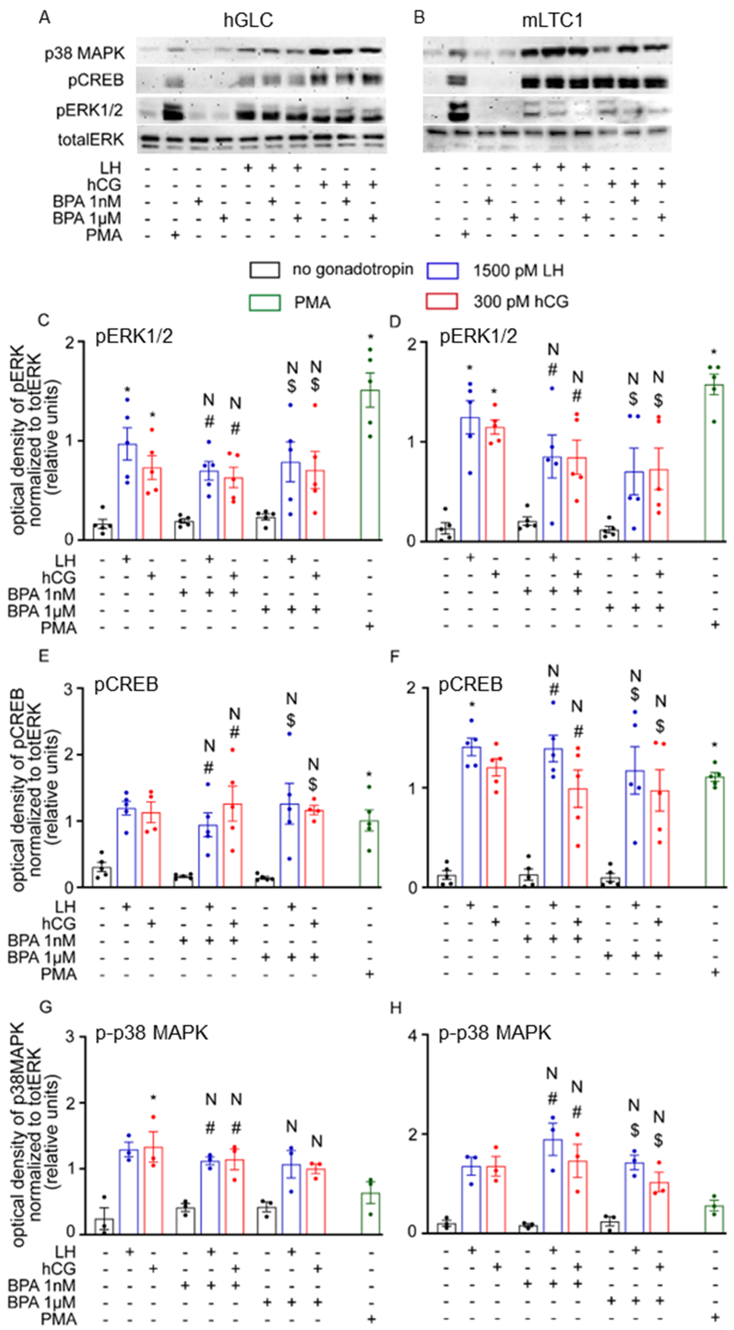
The effect of BPA on the gonadotropin-induced phosphorylation of molecules downstream of cAMP, such as ERK1/2, CREB and p38MAPK, was evaluated in both hGLC and mLTC1 cells. PMA-treated cells served as a positive control. Western blot analysis revealed that LH/hCG alone and together with BPA induced similar phosphorylation patterns in ERK1/2, CREB and p38 MAPK in both the models in vitro (Figure 2; Kruskal–Wallis test, p ≥ 0.05). Indeed, both the hormones, as well as PMA, induced phospho-protein activation. However, the presence of 1 nM and 1 µM BPA did not impact the results both in hGLC and mLTC1 cells.

3.2. Gene Expression Analysis

The effects of BPA on the 8 and 24 h expression of genes regulating LH/hCG-induced steroid synthesis were evaluated in hGLC, using real-time PCR. Gonadotropins upregulated the 8 h expression of STARD1 and CYP19A1 target genes (Figure 3; Kruskal–Wallis test, p < 0.05), while CYP17A1 gene expression increased after 24 h (p < 0.05). However, gene expression patterns were not altered in hGLC treated with LH/hCG alone, and in combination with 1 nM and 1 µM BPA (p ≥ 0.05).
Gene expression analysis was performed in mLTC1, where the effects of BPA addition to cell treatment with gonadotropins were investigated. Stard1 and Cyp17a1 gene expression was evaluated using real-time PCR; this was not the case for the Cyp19a1 gene, which is silent in the Leydig cell [42]. In mLTC1 cells, LH/hCG significantly increased both 8 and 24 h Stard1 and Cyp17a1 gene expression (Figure 4; Kruskal–Wallis test, p < 0.05). However, gonadotropin–BPA cotreatment is not linked to any change in gene expression patterns at both timepoints evaluated (p ≥ 0.05).
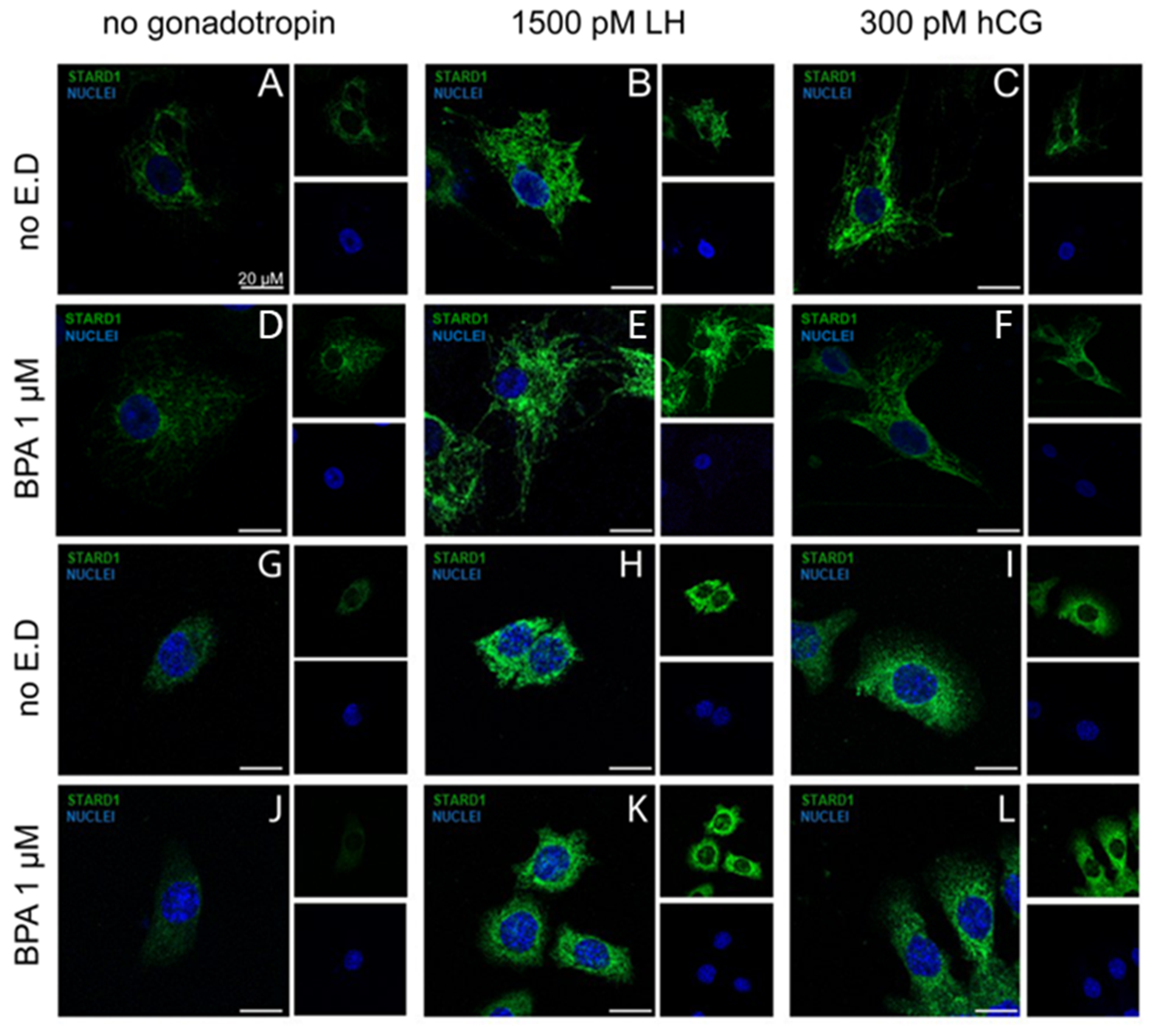
We also investigated the effect of BPA on StAR protein expression, as a key regulator of steroidogenesis. hGLC and mLTC1 cells were treated for 24 h by LH/hCG, in the presence or in the absence of BPA, and the StAR protein was evaluated using immunostaining. As expected, gonadotropins treatments increased the cytoplasmic levels of StAR immunoreactivity (Figure 5A–C). However, in line with our gene expression data, no changes were induced by BPA co-treatments (Figure 5D–F,J,L).

3.3. BPA Did Not Alter Steroidogenesis

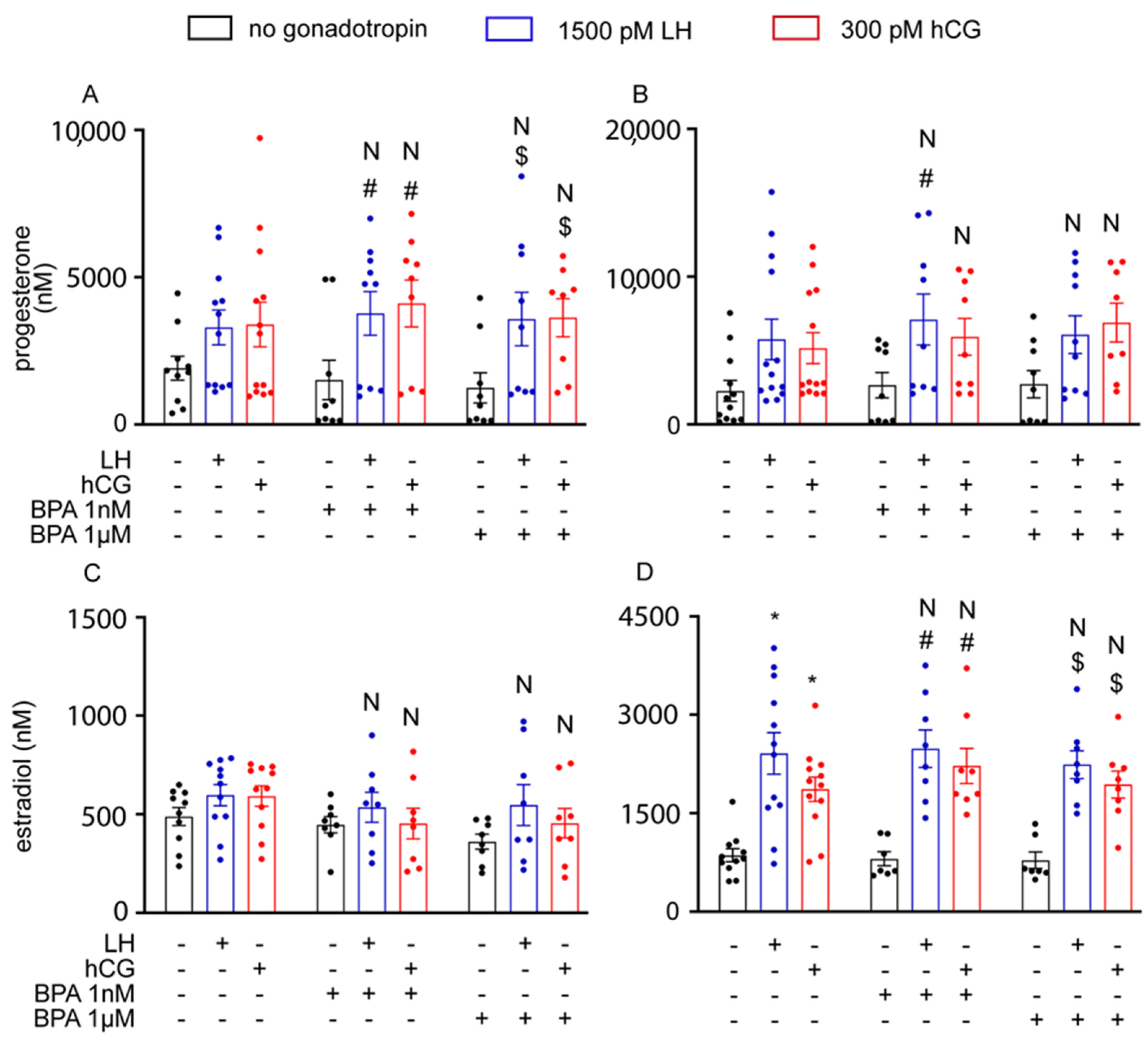
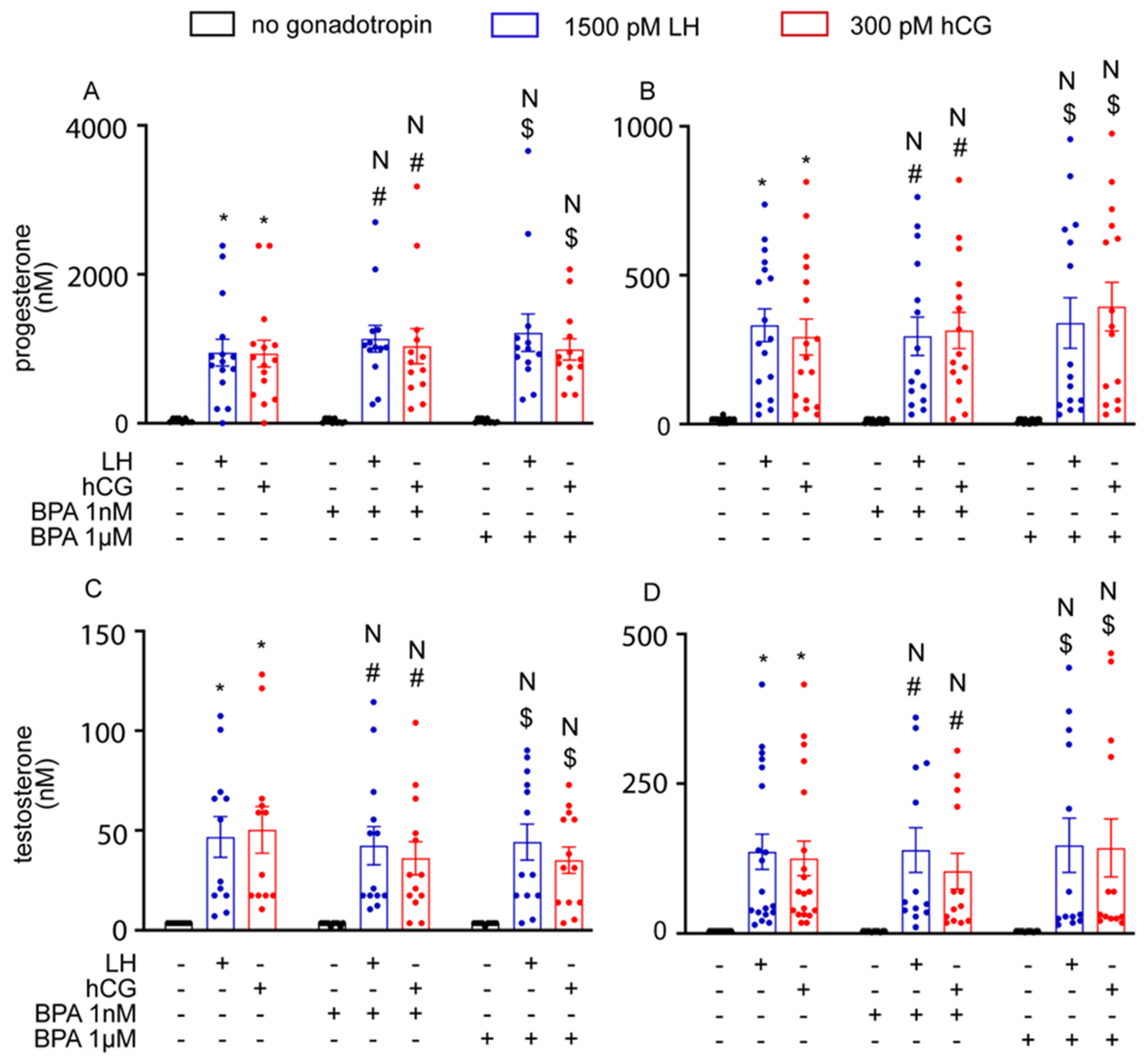
To further assess the effect of BPA on steroidogenesis, we also measured the production of progesterone and oestradiol in the culture media using hGLC, and progesterone and testosterone in the culture media using mLTC1. To this end, cells were treated for 8 and 24 h with gonadotropins, in the presence or in the absence of BPA. LH and hCG enhanced progesterone production via hGLC at both the timepoints evaluated, while oestradiol synthesis by hGLC required 24 h (Figure 6; Kruskal–Wallis test, p < 0.05, n = 5). However, BPA did not significantly impact steroid production (p ≥ 0.05). These results match those obtained from the culture media of mLTC1 cells, wherein the production of progesterone and testosterone by mLTC1 was increased upon cell treatment with LH and hCG (Figure 7; Kruskal–Wallis test, p < 0.05), and the addition of BPA did not change steroid levels (p ≥ 0.05).

4. Discussion

We found that short-term exposure to BPA at concentrations comparable to those found in human biological fluids [11] did not produce any effect on the LH/hCG-mediated steroidogenic pathway, both in hGLC and mLTC1 cells. In this study, we tested two environmental BPA concentrations, i.e., 1 nM and 1 µM, and performed cell signalling, gene expression, and steroidogenesis analyses. All the endpoints evaluated herein are known targets of gonadotropin action, and were not affected by the presence of BPA. These results suggest that short-term exposure to BPA does not impact the steroidogenic activity of gonadal cells, in vitro.
BPA is a ubiquitous environmental contaminant in Europe, America, Asia, and Australia, with concentrations ranging from 10 to >100,000 μg/kg dry weight [36]. The compound may induce adverse reproductive disorders in both men and women, although the data available do not converge in a single direction [26]. It is a common view that in females, BPA interferes with ovarian follicle development and steroidogenesis [13,43]. In cultured human luteinized granulosa cells, relatively high BPA concentrations (2 and 20 µg/mL, corresponding to about 8.7 and 87.6 µM) decreased oestradiol and progesterone production within 48 h [44]. The molecular mechanism by which BPA can impair steroidogenesis should involve both the mRNA and protein expression of steroidogenic enzymes, such as cholesterol monooxygenase (CYP11A1), 3β-hydroxysteroid dehydrogenase (3β-HSD), and aromatase, without affecting StAR expression [44]. However, the same study found opposite results under lower concentrations. Acute exposure to a similar concentration range of the compound (about 0.9–90.0 µM) did not alter the 48 h steroidogenesis in granulosa cells in vitro, suggesting that the main impact of BPA on ovarian cells could be exerted under conditions of chronic exposure [44]. We observed a similar effect in LH/hCG-induced progesterone and oestradiol levels when hGLC cells were treated with 1 nM and 1 µM BPA, which did not produce any effect on steroidogenesis and confirmed previous observations [44]. Our study indicates that the endocrine disruptor did not change cAMP levels, phospho-protein activation, and the expression of gonadotropin target genes and steroid synthesis. Overall, our results disagree with previous in vitro studies demonstrating the disruptive effects of BPA in human granulosa cell steroidogenesis [45,46]. For instance, higher BPA concentrations (40–100 µM) than those used in the present study induced the upregulation of aromatase expression and oestrogen synthesis [45]. Another study testing the 48 h exposure of cumulus granulosa cells to BPA revealed that the compound increases progesterone and decreases oestradiol synthesis [45], while experiments in a human ovarian granulosa cell-derived cell line, KGN, demonstrated that fM–pM doses of BPA increase oestradiol production. Taken together, data provided by in vitro studies are controversial, and do not allow us to achieve conclusions about the short-term effect of BPA on steroidogenesis. We may hypothesize that several perturbing factors, such as genetic background [47], polyphenol-containing plastic materials used for cell culture [48], or the additive effect of reagents for in vitro studies [49], could modulate or even mask the eventual perturbation of the endocrine signal exerted by BPA.
In males, BPA has been shown to disrupt steroidogenesis [50]. In vitro studies using mouse, rat and human cultured foetal testes have demonstrated decreased testosterone production induced by treatment with 1 fM to 10 µM BPA [22]. Interestingly, BPA affected only the functions of humans, but not rodent Leydig cells, suggesting that the endocrine disruption exerted by this compound is not necessarily similar among mammals. Moreover, the action of BPA is differently exerted according to the given experimental conditions. In cultured mLTC1 cells, exposure to pM–µM BPA concentrations inhibited hCG-induced cAMP and progesterone production [24]. These results do not align with those described in the present study, wherein we did not find any significant effect on LH/hCG-stimulated cAMP and progesterone production to be linked to the presence of 1 nM and 1µM BPA. The reason for this difference may be related to the exposure time and the concentrations that we used. The authors exposed cells to higher concentrations of BPA for 48 h [24], while we used BPA at lower concentrations, with exposure for 1 h for cAMP and for both 8 and 24 h exposure for progesterone production. Rather, our results match those of previous studies reporting that exposure to 1 nM–1 µM BPA for 4 or 24 h did not impact testosterone levels, regardless of the presence of hCG [51], and that even 10 µM BPA failed to inhibit hCG-induced upregulaton of the Stard1 gene [52]. Again, these findings point out the relevance of careful experimental settings for investigating the effects of endocrine disruptors in vitro.

5. Conclusions

Steroidogenic signalling plays a critical role in fertility. BPA, an endocrine-disrupting compound, is known to affect steroidogenic pathways affecting male and female fertility. In this study, we observed that short-term exposure to environmental concentrations of BPA does not compromise the steroidogenic potential of either human granulosa or mouse Leydig cells. Our findings add to those that report weak or no acute action of BPA in gonadal cells. However, several papers reporting opposite results suggest that the issue must be further investigated, using an experimental setting aiming to exclude potential confounding factors that may affect results.

Supplementary Materials

The following supporting information can be downloaded at: https://www.mdpi.com/article/10.3390/cells12111537/s1, Figure S1: Cell viability assay using MTT in hGLC (A) and mLTC1 (B); Figure S2: The effect of cAMP accumulation in hGLC (A) and in mLTC1 (B) through the direct activation of adenylate cyclase activity by Forskolin (50 µM) in the presence of BPA at 1 nM and 1 µM concentrations.

Author Contributions

Conceptualization, N.R., C.L., E.V., M.S. and L.C.; methodology, N.R., C.L. and C.C.; validation, E.V. and L.C.; formal analysis, N.R., C.L., E.P., C.C., T.F., F.R., S.S., L.B., E.M., C.P., M.V., G.C., T.T., A.N., D.M., F.I. and M.T.V.; investigation, N.R., C.L., E.P., C.C., T.F., F.R., S.S., L.B., E.M., C.P., M.V., G.C., T.T., A.N., D.M., F.I. and M.T.V.; resources, T.T., M.T.V., E.V., M.S. and L.C.; data curation, N.R., E.V. and L.C.; writing—original draft preparation, N.R.; writing—review and editing, N.R., E.V., M.S. and L.C.; visualization, N.R., C.L., C.C., E.V. and L.C.; supervision, E.V., M.S. and L.C.; project administration, E.V., M.S. and L.C.; funding acquisition, E.V., M.S. and L.C. All authors have read and agreed to the published version of the manuscript.

Funding

This research and APC were funded by the Italian Ministero dell’Università e della Ricerca, grant “Progetti di Rilevante Interesse Nazionale” (PRIN) 2017.

Institutional Review Board Statement

The study was approved by the local ethics committee (documents ‘Protocollo n. 2017/0015890 del 26/06/2017′, ‘Protocollo n2018/0080377 del 16/07/2018′, ‘Protocollo n. 2018/0080389 del 16/07/2018′, ‘Protocollo n. 2019/0009846 del 24/01/2019′ and ‘Protocollo n. 2019/0048906 del 23/04/2019′).

Informed Consent Statement

Informed consent was obtained from all subjects involved in the study.

Data Availability Statement

All other data are available from the corresponding author upon reasonable request.

Acknowledgments

L.C. was supported by Le Studium Loire Valley Institute for Advanced Studies, Orléans and Tours, France, through the ARD CVL Biopharmaceuticals programme, funded by the Centre-Val de Loire region.

Conflicts of Interest

The authors declare no conflict of interest.

References

  1. Peretz, J.; Vrooman, L.; Ricke, W.A.; Hunt, P.A.; Ehrlich, S.; Hauser, R.; Padmanabhan, V.; Taylor, H.S.; Swan, S.H.; Vandevoort, C.A.; et al. Bisphenol A and reproductive health: Update of experimental and human evidence, 2007–2013. Environ. Health Perspect. 2014, 122, 775–786. [Google Scholar] [CrossRef] [PubMed] [Green Version]
  2. Rubin, B.S. Bisphenol A: An endocrine disruptor with widespread exposure and multiple effects. J. Steroid Biochem. Mol. Biol. 2011, 127, 27–34. [Google Scholar] [CrossRef] [PubMed]
  3. Staples, C.A.; Dorn, P.B.; Klecka, G.M.; O’Block, S.T.; Harris, L.R. A review of the environmental fate, effects, and exposures of bisphenol A. Chemosphere 1998, 36, 2149–2173. [Google Scholar] [CrossRef]
  4. Nam, S.H.; Seo, Y.M.; Kim, M.G. Bisphenol A migration from polycarbonate baby bottle with repeated use. Chemosphere 2010, 79, 949–952. [Google Scholar] [CrossRef]
  5. Lim, D.S.; Kwack, S.J.; Kim, K.B.; Kim, H.S.; Lee, B.M. Potential risk of bisphenol a migration from polycarbonate containers after heating, boiling, and microwaving. J. Toxicol. Environ. Health-Part A Curr. Issues 2009, 72, 1285–1291. [Google Scholar] [CrossRef]
  6. Krishnan, A.V.; Stathis, P.; Permuth, S.F.; Tokes, L.; Feldman, D. Bisphenol-a: An estrogenic substance is released from polycarbonate flasks during autoclaving. Endocrinology 1993, 132, 2279–2286. [Google Scholar] [CrossRef]
  7. Ougier, E.; Zeman, F.; Antignac, J.P.; Rousselle, C.; Lange, R.; Kolossa-Gehring, M.; Apel, P. Human biomonitoring initiative (HBM4EU): Human biomonitoring guidance values (HBM-GVs) derived for bisphenol A. Environ. Int. 2021, 154, 106563. [Google Scholar] [CrossRef]
  8. Meeker, J.D.; Calafat, A.M.; Hauser, R. Urinary bisphenol A concentrations in relation to serum thyroid and reproductive hormone levels in men from an infertility clinic. Environ. Sci. Technol. 2010, 44, 1458–1463. [Google Scholar] [CrossRef] [Green Version]
  9. Vandenberg, L.N.; Hauser, R.; Marcus, M.; Olea, N.; Welshons, W.V. Human exposure to bisphenol A (BPA). Reprod. Toxicol. 2007, 24, 139–177. [Google Scholar] [CrossRef] [PubMed]
  10. Ouchi, K.; Watanabe, S. Measurement of bisphenol A in human urine using liquid chromatography with multi-channel coulometric electrochemical detection. J. Chromatogr. B Anal. Technol. Biomed. Life Sci. 2002, 780, 365–370. [Google Scholar] [CrossRef] [PubMed]
  11. Ikezuki, Y.; Tsutsumi, O.; Takai, Y.; Kamei, Y.; Taketani, Y. Determination of bisphenol A concentrations in human biological fluids reveals significant early prenatal exposure. Hum. Reprod. 2002, 17, 2839–2841. [Google Scholar] [CrossRef] [PubMed] [Green Version]
  12. Calafat, A.M.; Kuklenyik, Z.; Reidy, J.A.; Caudill, S.P.; Ekong, J.; Needham, L.L. Urinary concentrations of bisphenol A and 4-Nonylphenol in a human reference population. Environ. Health Perspect. 2005, 113, 391–395. [Google Scholar] [CrossRef] [PubMed] [Green Version]
  13. Bloom, M.S.; Mok-Lin, E.; Fujimoto, V.Y. Bisphenol A and ovarian steroidogenesis. Fertil. Steril. 2016, 106, 857–863. [Google Scholar] [CrossRef] [Green Version]
  14. Peretz, J.; Flaws, J.A. Bisphenol A down-regulates rate-limiting Cyp11a1 to acutely inhibit steroidogenesis in cultured mouse antral follicles. Toxicol. Appl. Pharmacol. 2013, 271, 249–256. [Google Scholar] [CrossRef] [PubMed] [Green Version]
  15. Wetherill, Y.B.; Akingbemi, B.T.; Kanno, J.; McLachlan, J.A.; Nadal, A.; Sonnenschein, C.; Watson, C.S.; Zoeller, R.T.; Belcher, S.M. In vitro molecular mechanisms of bisphenol A action. Reprod. Toxicol. 2007, 24, 178–198. [Google Scholar] [CrossRef]
  16. Peretz, J.; Gupta, R.K.; Singh, J.; Hernández-Ochoa, I.; Flaws, J.A. Bisphenol A impairs follicle growth, inhibits steroidogenesis, and downregulates rate-limiting enzymes in the oestradiol biosynthesis pathway. Toxicol. Sci. 2011, 119, 209–217. [Google Scholar] [CrossRef]
  17. Grasselli, F.; Baratta, L.; Baioni, L.; Bussolati, S.; Ramoni, R.; Grolli, S.; Basini, G. Bisphenol A disrupts granulosa cell function. Domest. Anim. Endocrinol. 2010, 39, 34–39. [Google Scholar] [CrossRef]
  18. Lee, S.G.; Kim, J.Y.; Chung, J.Y.; Kim, Y.J.; Park, J.E.; Oh, S.; Yoon, Y.D.; Yoo, K.S.; Yoo, Y.H.; Kim, J.M. Bisphenol a exposure during adulthood causes augmentation of follicular atresia and luteal regression by Decreasing 17β-estradiol synthesis via downregulation of aromatase in rat ovary. Environ. Health Perspect. 2013, 121, 663–669. [Google Scholar] [CrossRef] [PubMed] [Green Version]
  19. Zhou, W.; Liu, J.; Liao, L.; Han, S.; Liu, J. Effect of bisphenol A on steroid hormone production in rat ovarian theca-interstitial and granulosa cells. Mol. Cell. Endocrinol. 2008, 283, 12–18. [Google Scholar] [CrossRef]
  20. Nakamura, D.; Yanagiba, Y.; Duan, Z.; Ito, Y.; Okamura, A.; Asaeda, N.; Tagawa, Y.; Li, C.M.; Taya, K.; Zhang, S.Y.; et al. Bisphenol A may cause testosterone reduction by adversely affecting both testis and pituitary systems similar to oestradiol. Toxicol. Lett. 2010, 194, 16–25. [Google Scholar] [CrossRef]
  21. Nanjappa, M.K.; Simon, L.; Akingbemi, B.T. The industrial chemical bisphenol A (BPA) interferes with proliferative activity and development of steroidogenic capacity in rat Leydig cells. Biol. Reprod. 2012, 86, 1–12. [Google Scholar] [CrossRef]
  22. N’Tumba-Byn, T.; Moison, D.; Lacroix, M.; Lecureuil, C.; Lesage, L.; Prud’homme, S.M.; Pozzi-Gaudin, S.; Frydman, R.; Benachi, A.; Livera, G.; et al. Differential Effects of Bisphenol A and Diethylstilbestrol on Human, Rat and Mouse Fetal Leydig Cell Function. PLoS ONE 2012, 7, e51579. [Google Scholar] [CrossRef] [Green Version]
  23. Mannelli, C.; Szóstek, A.Z.; Lukasik, K.; Carotenuto, C.; Ietta, F.; Romagnoli, R.; Ferretti, C.; Paulesu, L.; Wołczynski, S.; Skarzynski, D.J. Bisphenol A modulates receptivity and secretory function of human decidual cells: An in vitro study. Reproduction 2015, 150, 115–125. [Google Scholar] [CrossRef] [PubMed] [Green Version]
  24. Nikula, H.; Talonpoika, T.; Kaleva, M.; Toppari, J. Inhibition of hCG-stimulated steroidogenesis in cultured mouse Leydig tumor cells by bisphenol A and octylphenols. Toxicol. Appl. Pharmacol. 1999, 157, 166–173. [Google Scholar] [CrossRef] [PubMed]
  25. Menon, B.; Franzo-Romain, M.; Damanpour, S.; Menon, K.M.J. Luteinizing hormone receptor mRNA down-regulation is mediated through ERK-dependent induction of RNA binding protein. Mol. Endocrinol. 2011, 25, 282–290. [Google Scholar] [CrossRef] [Green Version]
  26. Palak, E.; Lebiedzinska, W.; Lupu, O.; Pulawska, K.; Anisimowicz, S.; Mieczkowska, A.N.; Sztachelska, M.; Niklinska, G.N.; Milewska, G.; Lukasiewicz, M.; et al. Molecular insights underlying the adverse effects of bisphenol A on gonadal somatic cells’ steroidogenic activity. Reprod. Biol. 2023, 23, 100766. [Google Scholar] [CrossRef]
  27. Casarini, L.; Lispi, M.; Longobardi, S.; Milosa, F.; la Marca, A.; Tagliasacchi, D.; Pignatti, E.; Simoni, M. LH and hCG Action on the Same Receptor Results in Quantitatively and Qualitatively Different Intracellular Signalling. PLoS ONE 2012, 7, e46682. [Google Scholar] [CrossRef] [Green Version]
  28. Casarini, L.; Moriondo, V.; Marino, M.; Adversi, F.; Capodanno, F.; Grisolia, C.; La Marca, A.; La Sala, G.B.; Simoni, M. FSHR polymorphism p.N680S mediates different responses to FSH in vitro. Mol. Cell. Endocrinol. 2014, 393, 83–91. [Google Scholar] [CrossRef]
  29. Sperduti, S.; Paradiso, E.; Anzivino, C.; Lazzaretti, C.; Limoncella, S.; D’Alessandro, S.; Roy, N.; Reggianini, F.; Ferrari, T.; Melli, B.; et al. LH increases the response to FSH in granulosa-lutein cells from sub/poor-responder patients in vitro. Hum. Reprod. 2023, 38, 103–112. [Google Scholar] [CrossRef]
  30. Nordhoff, V.; Sonntag, B.; Von Tils, D.; Götte, M.; Schüring, A.N.; Gromoll, J.; Redmann, K.; Casarini, L.; Simoni, M. Effects of the FSH receptor gene polymorphism p.N680S on cAMP and steroid production in cultured primary human granulosa cells. Reprod. Biomed. Online 2011, 23, 196–203. [Google Scholar] [CrossRef] [PubMed] [Green Version]
  31. Casarini, L.; Riccetti, L.; Limoncella, S.; Lazzaretti, C.; Barbagallo, F.; Pacifico, S.; Guerrini, R.; Tagliavini, S.; Trenti, T.; Simoni, M.; et al. Probing the effect of sildenafil on progesterone and testosterone production by an intracellular FRET/BRET combined approach. Biochemistry 2019, 58, 799–808. [Google Scholar] [CrossRef]
  32. Victor Rebois, R. Establishment of gonadotropin-responsive murine leydig tumor cell line. J. Cell Biol. 1982, 94, 70–76. [Google Scholar] [CrossRef] [PubMed]
  33. Riccetti, L.; Yvinec, R.; Klett, D.; Gallay, N.; Combarnous, Y.; Reiter, E.; Simoni, M.; Casarini, L.; Ayoub, M.A. Human Luteinizing Hormone and Chorionic Gonadotropin Display Biased Agonism at the LH and LH/CG Receptors. Sci. Rep. 2017, 7, 940. [Google Scholar] [CrossRef] [PubMed] [Green Version]
  34. Limoncella, S.; Lazzaretti, C.; Paradiso, E.; D’Alessandro, S.; Barbagallo, F.; Pacifico, S.; Guerrini, R.; Tagliavini, S.; Trenti, T.; Santi, D.; et al. Phosphodiesterase (PDE) 5 inhibitors sildenafil, tadalafil and vardenafil impact cAMP-specific PDE8 isoforms-linked second messengers and steroid production in a mouse Leydig tumor cell line. Mol. Cell. Endocrinol. 2022, 542, 111527. [Google Scholar] [CrossRef] [PubMed]
  35. Fanelli, F.; Magagnoli, M.; Mezzullo, M.; Lispi, M.; Limoncella, S.; Tommasini, A.; Pelusi, C.; Santi, D.; Simoni, M.; Pagotto, U.; et al. Exploring the human chorionic gonadotropin induced steroid secretion profile of mouse Leydig tumor cell line 1 by a 20 steroid LC-MS/MS panel. J. Steroid Biochem. Mol. Biol. 2023, 229, 106270. [Google Scholar] [CrossRef]
  36. Corrales, J.; Kristofco, L.A.; Baylor Steele, W.; Yates, B.S.; Breed, C.S.; Spencer Williams, E.; Brooks, B.W. Global assessment of bisphenol a in the environment: Review and analysis of its occurrence and bioaccumulation. Dose-Response 2015, 13, 1559325815598308. [Google Scholar] [CrossRef] [Green Version]
  37. Riccetti, L.; De Pascali, F.; Gilioli, L.; Potì, F.; Giva, L.B.; Marino, M.; Tagliavini, S.; Trenti, T.; Fanelli, F.; Mezzullo, M.; et al. Human LH and hCG stimulate differently the early signalling pathways but result in equal testosterone synthesis in mouse Leydig cells in vitro. Reprod. Biol. Endocrinol. 2017, 15, 1–12. [Google Scholar] [CrossRef] [Green Version]
  38. Paradiso, E.; Lazzaretti, C.; Sperduti, S.; Antoniani, F.; Fornari, G.; Brigante, G.; Di Rocco, G.; Tagliavini, S.; Trenti, T.; Morini, D.; et al. Sphingosine-1 phosphate induces cAMP/PKA-independent phosphorylation of the cAMP response element-binding protein (CREB) in granulosa cells. Mol. Cell. Endocrinol. 2021, 520, 111082. [Google Scholar] [CrossRef]
  39. Casarini, L.; Lazzaretti, C.; Paradiso, E.; Limoncella, S.; Riccetti, L.; Sperduti, S.; Melli, B.; Marcozzi, S.; Anzivino, C.; Sayers, N.S.; et al. Membrane Estrogen Receptor (GPER) and Follicle-Stimulating Hormone Receptor (FSHR) Heteromeric Complexes Promote Human Ovarian Follicle Survival. iScience 2020, 23, 101812. [Google Scholar] [CrossRef]
  40. Livak, K.J.; Schmittgen, T.D. Analysis of relative gene expression data using real-time quantitative PCR and the 2-ΔΔCT method. Methods 2001, 25, 402–408. [Google Scholar] [CrossRef]
  41. Horton, R.; Tait, J.F. Androstenedione production and interconversion rates measured in peripheral blood and studies on the possible site of its conversion to testosterone. J. Clin. Investig. 1966, 45, 301–313. [Google Scholar] [CrossRef]
  42. Miller, W.L.; Auchus, R.J. The molecular biology, biochemistry, and physiology of human steroidogenesis and its disorders. Endocr. Rev. 2011, 32, 81–151. [Google Scholar] [CrossRef] [PubMed] [Green Version]
  43. Caserta, D.; Di Segni, N.; Mallozzi, M.; Giovanale, V.; Mantovani, A.; Marci, R.; Moscarini, M. Bisphenol a and the female reproductive tract: An overview of recent laboratory evidence and epidemiological studies. Reprod. Biol. Endocrinol. 2014, 12, 37. [Google Scholar] [CrossRef] [PubMed] [Green Version]
  44. Mansur, A.; Adir, M.; Yerushalmi, G.; Hourvitz, A.; Gitman, H.; Yung, Y.; Orvieto, R.; Machtinger, R. Does BPA alter steroid hormone synthesis in human granulosa cells in vitro? Hum. Reprod. 2016, 31, 1562–1569. [Google Scholar] [CrossRef] [Green Version]
  45. Kwintkiewicz, J.; Nishi, Y.; Yanase, T.; Giudice, L.C. Peroxisome proliferator-activated receptor-γ mediates bisphenol A inhibition of FSH-stimulated IGF-1, aromatase, and oestradiol in human granulosa cells. Environ. Health Perspect. 2010, 118, 400–406. [Google Scholar] [CrossRef] [PubMed] [Green Version]
  46. Pogrmic-Majkic, K.; Samardzija Nenadov, D.; Fa, S.; Stanic, B.; Trninic Pjevic, A.; Andric, N. BPA activates EGFR and ERK1/2 through PPARΓ to increase expression of steroidogenic acute regulatory protein in human cumulus granulosa cells. Chemosphere 2019, 229, 60–67. [Google Scholar] [CrossRef] [PubMed]
  47. Zhong, R.; He, H.; Jin, M.; Lu, Z.; Deng, Y.; Liu, C.; Shen, N.; Li, J.; Wang, H.; Ying, P.; et al. Genome-wide gene-bisphenol A, F and triclosan interaction analyses on urinary oxidative stress markers. Sci. Total Environ. 2022, 807, 150753. [Google Scholar] [CrossRef]
  48. Togola, A.; Desmarchais, A.; Téteau, O.; Vignault, C.; Maillard, V.; Buron, C.; Bristeau, S.; Guérif, F.; Binet, A.; Elis, S. Bisphenol S is present in culture media used for ART and cell culture. Hum. Reprod. 2021, 36, 1032–1042. [Google Scholar] [CrossRef]
  49. Sonavane, M.; Gassman, N.R. Bisphenol A co-exposure effects: A key factor in understanding BPA’s complex mechanism and health outcomes. Crit. Rev. Toxicol. 2019, 49, 371–386. [Google Scholar] [CrossRef]
  50. Shamhari, A. ‘Afifah; Hamid, Z.A.; Budin, S.B.; Shamsudin, N.J.; Taib, I.S. Bisphenol a and its analogues deteriorate the hormones physiological function of the male reproductive system: A mini-review. Biomedicines 2021, 9, 1744. [Google Scholar] [CrossRef]
  51. Murono, E.P.; Derk, R.C.; De León, J.H. Differential effects of octylphenol, 17β-estradiol, endosulfan, or bisphenol A on the steroidogenic competence of cultured adult rat Leydig cells. Reprod. Toxicol. 2001, 15, 551–560. [Google Scholar] [CrossRef] [PubMed]
  52. Takamiya, M.; Lambard, S.; Huhtaniemi, I.T. Effect of bisphenol A on human chorionic gonadotrophin-stimulated gene expression of cultured mouse Leydig tumour cells. Reprod. Toxicol. 2007, 24, 265–275. [Google Scholar] [CrossRef] [PubMed]
Figure 1. Effect of BPA on cAMP production in vitro: (A) in human granulosa cells (hGLC); (B) in mouse Leydig cells (mLTC1). cAMP was measured using an HTRF assay. Forskolin and cholera toxin served as the positive control in hGLC and mLTC1 cells, respectively. Data are represented as mean ± SEM. CTX = cholera toxin. *, Significantly different vs. “no gonadotropin” alone; # vs. “no gonadotropin” + BPA 1 nM; $ vs. “no gonadotropin” + BPA 1 µM; N, not significantly different vs. LH/hCG treatment in the absence of BPA (Kruskal–Wallis test, p ≥ 0.05).
Cells 12 01537 g001
Figure 2. Effect of BPA on activation of pERK, pCREB and p38MAPK: Western blotting analyses in hGLC (A) and in mLTC1 (B) after exposure to BPA without or with hCG or LH. Relative semi-quantification of the activation of pERK (C,D), pCREB (E,F) and p38MAPK (G,H) in hGLC and mLTC1 cells, respectively. Data are represented as mean ± SEM. *, Significantly different vs. “no gonadotropin” alone; # vs. “no gonadotropin” + BPA 1 nM; $ vs. “no gonadotropin” + BPA 1 µM; N, not significantly different vs. LH/hCG treatment in the absence of BPA (Kruskal–Wallis test, p ≥ 0.05).
Cells 12 01537 g002
Figure 3. Effect of BPA on steroidogenic gene expression in hGLC after 8 h (A,C,E) and 24 h (B,D,F) of treatment. Genes: STARD1 (A,B), CYP17A1 (C,D) and CYP19A1 (E,F). Data are represented as mean ± SEM. *, Significantly different vs. “no gonadotropin” alone; # vs. “no gonadotropin” + BPA 1 nM; $ vs. “no gonadotropin” + BPA 1 µM; N, not significantly different vs. LH/hCG treatment in the absence of BPA (Kruskal–Wallis test, p ≥ 0.05).
Cells 12 01537 g003
Figure 4. Effect of BPA on steroidogenic gene expression in mLTC1 after 8 h (A,C) and 24 h (B,D). Genes: Stard1 (A,C) and Cyp17a1 (B,D). Data are represented as mean ± SEM. *, Significantly different vs. “no gonadotropin” alone; # vs. “no gonadotropin” + BPA 1 nM; $ vs. “no gonadotropin” + BPA 1 µM; N, not significantly different vs. LH/hCG treatment in the absence of BPA (Kruskal–Wallis test, p ≥ 0.05).
Cells 12 01537 g004
Figure 5. Immunofluorescence images of the effect of BPA on StAR protein expression in LH/hCG-treated hGLC (AF) and in mLTC1 cells (GL). Cells were treated 24 h with 1 µM of BPA, in the absence or presence of LH/hCG. Images are representative of three independent experiments (magnification 40×. Subfigure size is reduced to 50%).
Cells 12 01537 g005
Figure 6. The effect of BPA on progesterone and oestradiol levels in hGLC cells after 8 h (A,C) and 24 h (B,D). Data are represented as mean ± SEM. *, Significantly different vs. “no gonadotropin” alone; # vs. “no gonadotropin” + BPA 1 nM; $ vs. “no gonadotropin” + BPA 1 µM; N, not significantly different vs. LH/hCG treatment in the absence of BPA (Kruskal–Wallis test, p ≥ 0.05).
Cells 12 01537 g006
Figure 7. The effect of BPA on progesterone and testosterone levels in mLTC1 cells after 8 h (A,C) and 24 h (B,D). Mean ± SEM. *, Significantly different vs. “no gonadotropin” alone; # vs. “no gonadotropin” + BPA 1 nM; $ vs. “no gonadotropin” + BPA 1 µM; N, not significantly different vs. LH/hCG treatment in the absence of BPA (Kruskal–Wallis test, p ≥ 0.05).
Cells 12 01537 g007
Disclaimer/Publisher’s Note: The statements, opinions and data contained in all publications are solely those of the individual author(s) and contributor(s) and not of MDPI and/or the editor(s). MDPI and/or the editor(s) disclaim responsibility for any injury to people or property resulting from any ideas, methods, instructions or products referred to in the content.