Communication

Involvement of Substance P (SP) and Its Related NK1 Receptor in Primary Sjögren’s Syndrome (pSS) Pathogenesis

1
Institute of Biochemistry and Cell Biology, National Research Council (IBBC-CNR), Department of Sense Organs, Sapienza University of Rome, Viale del Policlinico 155, 00185 Rome, Italy
2
Department of Internal Medicine and Medical Specialties, Rheumatology Unit, Sapienza University of Rome, Viale del Policlinico 155, 00185 Rome, Italy
3
Department of Sense Organs, Sapienza University of Rome, Viale del Policlinico 155, 00185 Rome, Italy
4
Division of Rheumatology, Department of Clinical Internal, Anaesthesiologic and Cardiovascular Sciences, Sapienza University, Viale del Policlinico 155, 00185 Rome, Italy
5
Department of Medico-Surgical Sciences and Biotechnology, Sapienza University of Rome, 00185 Rome, Italy
6
Department of Radiological, Oncological and Pathological Sciences, Sapienza University of Rome, Viale del Policlinico 155, 00185 Rome, Italy
*
Author to whom correspondence should be addressed.
These authors contributed equally to this work.
These authors also contributed equally to this work.
Cells 2023, 12(10), 1347; https://doi.org/10.3390/cells12101347
Submission received: 21 March 2023 / Revised: 21 April 2023 / Accepted: 1 May 2023 / Published: 9 May 2023

Abstract

:
Primary Sjögren’s Syndrome (pSS) is a systemic autoimmune disease that primarily attacks the lacrimal and salivary glands, resulting in impaired secretory function characterized by xerostomia and xerophthalmia. Patients with pSS have been shown to have impaired salivary gland innervation and altered circulating levels of neuropeptides thought to be a cause of decreased salivation, including substance P (SP). Using Western blot analysis and immunofluorescence studies, we examined the expression levels of SP and its preferred G protein-coupled TK Receptor 1 (NK1R) and apoptosis markers in biopsies of the minor salivary gland (MSG) from pSS patients compared with patients with idiopathic sicca syndrome. We confirmed a quantitative decrease in the amount of SP in the MSG of pSS patients and demonstrated a significant increase in NK1R levels compared with sicca subjects, indicating the involvement of SP fibers and NK1R in the impaired salivary secretion observed in pSS patients. Moreover, the increase in apoptosis (PARP-1 cleavage) in pSS patients was shown to be related to JNK phosphorylation. Since there is no satisfactory therapy for the treatment of secretory hypofunction in pSS patients, the SP pathway may be a new potential diagnostic tool or therapeutic target.

1. Introduction

Primary Sjögren’s Syndrome (pSS) is a systemic autoimmune disease that primarily affects the lacrimal and salivary glands, resulting in impaired secretory function characterized by xerostomia and xerophthalmia [1].
The characteristic autoimmune feature of pSS etiopathogenesis is based on the development of autoimmune epithelitis characterized by focal lymphocytic infiltration of the exocrine glands, often in association with acinar epithelial cell atrophy and progressive fibrosis diagnosed mainly through biopsy of the minor labial salivary glands [2].
The pathology is characterized by a broad spectrum of circulating autoantibodies, of which antinuclear antibodies (ANAs) are the most frequently detected (in approximately 70% of pSS patients) and anti-Ro/SSA and La/SSB antibodies are the most specifically detected [3]. There is convincing evidence that the innate immune response in the early phase of pathology is activated mainly by the involvement of CD4+ T cells infiltrating the salivary and lacrimal glands, whereas T cell activation and B cell accumulation occur at later stages [4]. Furthermore, activated T cells contribute to pathogenesis by releasing pro-inflammatory cytokines (e.g., TNFα, IFNs, IL-1, IL-2, and IL-6), chemokines, and increased expression of adhesion molecules, apoptosis-related factors, co-stimulatory molecules, autoantigens, and functional innate immune receptors, leading to chronic inflammatory damage to exocrine glands and progressive loss of their physiological function [5].
The prevalence of pSS is 0.15–3.3% depending on the diagnostic criteria used. Ninety-five percent of pSS patients are women, typically peri- and post-menopausal, but young post-menarche patients may also be affected [6]. Diagnostic criteria of the disease include the presence of a detectable focal lymphocytic sialadenitis (aggregates of at least 50 cells per 4 mm2) with a focus score ≥1, the presence of autoantibodies (i.e., anti-Ro/SSA and anti-La/SSA), and impaired salivary and ocular secretions, as defined by the American–European Consensus Group (AECG) criteria [7] and the American College of Rheumatology (ACR)/European League Against Rheumatism (EULAR) criteria [8].
Patients with pSS have been shown to have impaired innervation of the salivary glands along with changes in circulating levels of neuropeptides, which are thought to be a cause of the decreased salivation [9,10]. One possible explanation is that this autoimmune disease and/or local inflammation may cause vasoneural dysregulation and peripheral nerve injury, resulting in decreased fluid flow and acinar cell atrophy, followed by destruction of the salivary and lacrimal glands [11]. Moreover, apoptosis has emerged as a possible mechanism for damage to the salivary and lacrimal glands in pSS, leading to impairment of their secretory function [12].
Salivary secretion is controlled by the autonomic nervous system and influenced by the sensory nervous system. Activation of the parasympathetic nervous system leads to a sharp increase in salivary flow with a low protein concentration, and its function is influenced by neurons containing neuropeptides with strong sialagogue action, including substance P (SP), which is released by the nerve fibers innervating the salivary glands [13].
SP, a neuropeptide of 11 amino acids, was the first to be identified and is the best characterized member of the tachykinin peptide family, and its biological activity is mediated by its preferred G-protein-coupled TK receptor 1 (neurokinin receptor 1; NK1R) through the activation of phospholipase Cβ (PLCβ). Since SP and NK1R are widely distributed in the brain and sensory/autonomic nervous system, their involvement in many physiological functions can be explained [14].
In the central nervous system (CNS), SP is mainly found in neuronal cells, where it frequently co-localizes with classical transmitters and other neuropeptides and is endowed with neurotrophic and neuroprotective properties, as has been extensively demonstrated by our group and others [15,16,17,18,19].
In the peripheral nervous system (PNS), SP is found in high concentration in primary small-diameter sensory neurons (C-fibers) [20,21] and in the dorsal horns of the spinal cord, justifying its role as a sensory neurotransmitter important for pain perception [22]. In addition, SP influences smooth muscle contraction, epithelial permeability, and mediates inflammatory processes by increasing neutrophil and macrophage traffic and activating mast cells, monocytes, or lymphocytes, which release their mediators such as histamine, interleukins (IL-1, IL-2), and immunoglobulins, respectively [23].
The presence of SP nerve fibers has been demonstrated around the excretory ducts of human salivary glands, which contributes to changes in fluid and electrolyte secretion followed by a gradual increase in saliva production [24]. In salivary glands, immunoreactive SP nerve fibers have been shown to be found mainly around blood vessels and in direct contact with the acini, and their number is significantly reduced in pSS tissues [10]; however, no data are available on the presence and distribution of the associated receptor NKR1. The aim of this study was to characterize the expression of both SP and NKR1 in biopsies of the minor salivary gland (MSG) and to analyze their levels in pSS patients compared to patients with idiopathic sicca syndrome. Exploring their contribution to the pathogenesis of the disease could be useful to identify new potential diagnostic tools or therapeutic targets.

2. Materials and Methods

2.1. pSS and Sicca Patients’ Selection and Enrollment

Minor salivary gland (MSG) biopsies were obtained from patients with suspected pSS and followed up at our dedicated Sjögren Clinic at Sapienza University (Department of Clinical Internal, Anaesthesiologic, and Cardiovascular Sciences—Rheumatology Unit). The total number of patients enrolled was 30. Following the biopsy procedure, the patient was classified as pSS if they met the AECG criteria [7], while those who did not fulfill these criteria were classified as an idiopathic sicca syndrome patient. Inclusion criteria included the presence of a detectable focal lymphocytic sialadenitis (aggregates of at least 50 cells per 4 mm2) with a focus score ≥1, and presence of autoantibodies (i.e., anti-Ro/SSA and anti-La/SSA). Exclusion criteria included ongoing treatment with corticosteroids, hydroxychloroquine, immunosuppressants, or pilocarpine (within 3 months). Informed and written consent was obtained from patients enrolled in the study. Permission for the use of MSG samples for research purposes was obtained by our ethical committee of Sapienza University of Rome, under protocol number 4688.

2.2. MSG Biopsy Collection

Surgical procedures and tissue sample collection of MSG biopsies were performed by otolaryngology specialists of the Department of Sense Organs, Division of Otolaryngology, Sapienza University of Rome. A local anesthetic was injected into the lower lip, followed by a small incision in the lip mucosa. Two MSG biopsies per patient were obtained. One of the two biopsy samples was embedded in paraffin for immunohistochemistry/immunofluorescence (IHC/IF) analysis and the remaining one was immediately frozen after the surgical procedure and stored at −80 °C until it was used for Western Blot (WB) analysis.

2.3. Protein Extraction and WB Analysis

For protein characterization, extracts from the MSG biopsies were obtained using a lysis buffer (RIPA buffer), and the total protein concentration was measured using the BioRad protein assay (DC Protein Assay, Biorad Laboratories, CA, USA, #500-0116). WB analysis was performed using selective antibodies against substance P (SP, Santa Cruz Biotechnology, Dallas, TX, USA, sc-517213, 1:1000) and its cognate receptor Neurokinin 1 (NK1R, Santa Cruz Biotechnology, Dallas, TX, USA, sc-365091, 1:1000), cleaved PARP-1 (Santa Cruz Biotechnology, Dallas, TX, USA, sc-56196, 1:1000), and phosphorylated JNK (pJNK, cell signaling technology, Danvers, MA, USA, #9251, 1:1000). The images obtained from the WBs were analyzed using ImageJ software for Windows. All samples were normalized for protein loading using β-actin (Santa Cruz Biotechnology, Dallas, TX, USA, sc-47778, 1:10,000) as the protein loading control. The values were determined from the ratio between the arbitrary units (a.u.) derived from the protein band and the respective β-actin band and expressed as mean ± Standard Deviation (SD).

2.4. Immunohistochemistry and Immunofluorescence

Immunohistochemistry (IHC) was used to characterize the infiltrates and to detect the presence of Germinal Centers (GCs). GC+ samples were defined by the presence of at least one GC (nodular aggregate segregated in T and B cells areas with positive staining for CD21L and Bcl6 on sequential sections [25]); the methods for the staining procedure were reported in our previous work [26].
After collection, MSGs were immediately fixed in 10% formalin and embedded in paraffin. Paraffin-embedded sections were dewaxed using 2 changes of xylol, 15 min each. After hydration in graded ethanol solutions (100%, 95%), the slides were hydrated in distilled water. The slides were then immersed in antigen retrieval solution (10 mM sodium citrate, 0.05% Tween 20, pH 6.0), using a microwave oven operated at 640 W for 15 min. After cooling, the slides were transferred to PBS 1X and incubated for 24 h at 4 °C with primary antibodies: substance P (Santa Cruz Biotechnology, sc-517213, 1:100) and e-cadherin (ECAD, ThermoFisher Scientific, Waltham, MA, USA, #53-3249-82, 1:100) in PBS 1X to perform immunofluorescence (IF). The slides were then washed in PBS 1X and incubated for 1 h at R.T. with the appropriate secondary antibody: Donkey anti-Mouse Alexa Fluor 546 (ThermoFisher Scientific, Waltham, MA, USA, A-Catalog #A10036, 1:300). The nuclei were stained with 4′,6-diamidino-2- phenylindole (DAPI, 1:10,000, SeraCare, Milford, CT, USA, KPL Dapi 1 mg 71-03-01). The slides were mounted using aqueous mounting medium. Negative controls were performed by omitting the primary antibodies (ctl neg).
All immune-reacted sections were analyzed in detail and representative images were captured using the Zeiss LSM 780 laser-scanning confocal head with a Zeiss Axio Imager Z1 microscope; LSM510 Image Examiner Software was used to process the images, which were then composed into figures using Adobe illustrator 27.4 and Photoshop CS6.
To quantify SP immunoreactivity, 5 (40× magnification) images were captured for each group. During the image acquisitions, the exposure parameters, such as gain and time, were kept constant, to avoid observing differences among experimental groups due to artifacts. The analysis of SP IF staining (%Area SP+) was performed using the ImageJ software (version 1.53, National Institutes of Health, Bethesda, MD, USA, https://imagej.nih.gov/ij/download.html) and the statistical significance was calculated using the t-Student test, p < 0.001 (***).

2.5. Data Analysis

Statistical analyses were performed using GraphPad InStat3 for Windows. The data from WBs were analyzed using one-way analysis of variance (ANOVA) followed by a Tukey’s post hoc test. All results are expressed as mean ± SD, with n being the number of independent experiments. The significance level was set at p < 0.05 (*), p < 0.01 (**), and p < 0.001 (***).

3. Results

3.1. Lower Expression of SP and Higher Expression of NK1R in MSGs of pSS Patients Compared with Sicca Patients

As previously reported, immunohistochemical experiments indicated a reduction in SP in pSS [10], but no data are available on quantitative protein expression of SP and its associated receptor (NK1R).
Here, we demonstrated by Western blot analysis of MSG extracts that the levels of SP and NK1R significantly differed between the two groups. The level of SP was significantly decreased in the pSS patients compared with the sicca group (** p < 0.01) as shown in Figure 1A. In contrast, NK1R showed the opposite trend (Figure 1B) with a significant increase in pSS patients compared to sicca patients (*** p < 0.001).

3.2. SP and ECAD Localization in MSG Sections from pSS and Sicca Patients’ Immunofluorescence

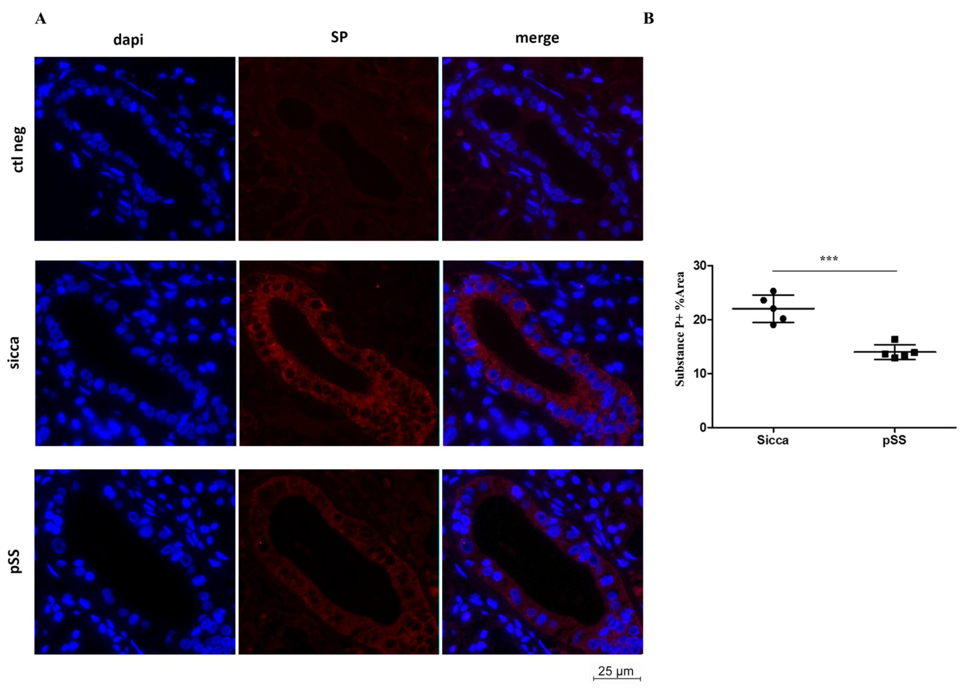
Using Immunofluorescence (IF) technique, we investigated the expression of substance P in MSG sections from pSS and sicca patients. We detected that SP expression (red) in pSS MSGs was reduced when compared to sicca MSGs, particularly in ductal epithelial cells (Figure 2A). When quantitatively analyzed, SP immunoreactivity levels appeared significantly lower in pSS patients compared to sicca individuals (*** p < 0.001, t = 6.302; Figure 2B). To assess the primary antibody specificity, a negative control (ctl neg) was also performed (first row, Figure 2A).
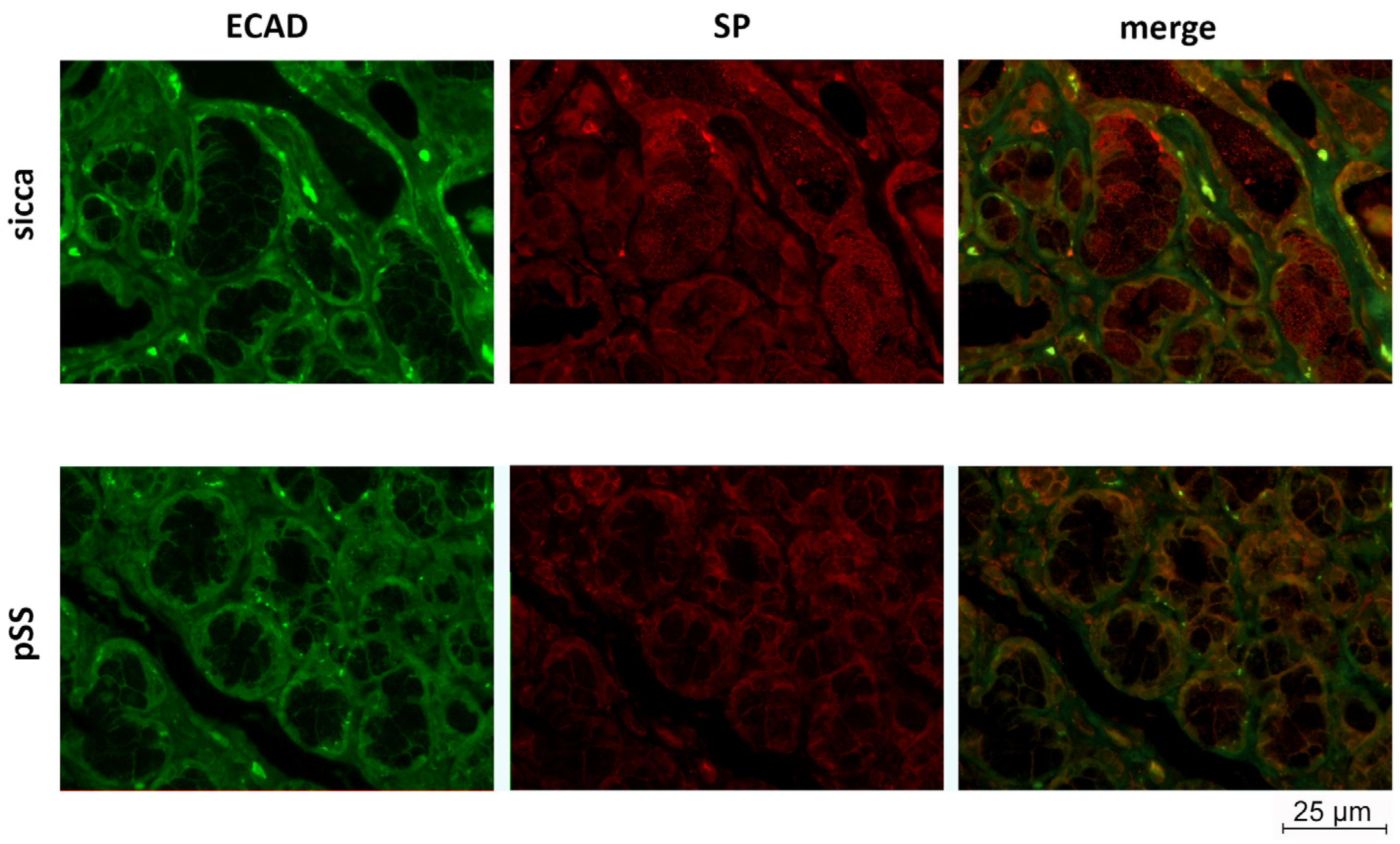
To further investigate the localization of SP in the MSGs of sicca and pSS patients, a double IF analysis for SP and E-Cadherin (ECAD), a membranous glycoprotein expressed in epithelial cells, was performed. SP localization was detected in both sicca and pSS patients, in ductal and acinar cells, and mainly in the epithelial compartment as shown by the merge images of both groups (Figure 3).

3.3. Western Blot Analysis Reveals Increase in PARP-1 Cleavage and JNK Phosphorylation in MSGs of pSS Patients

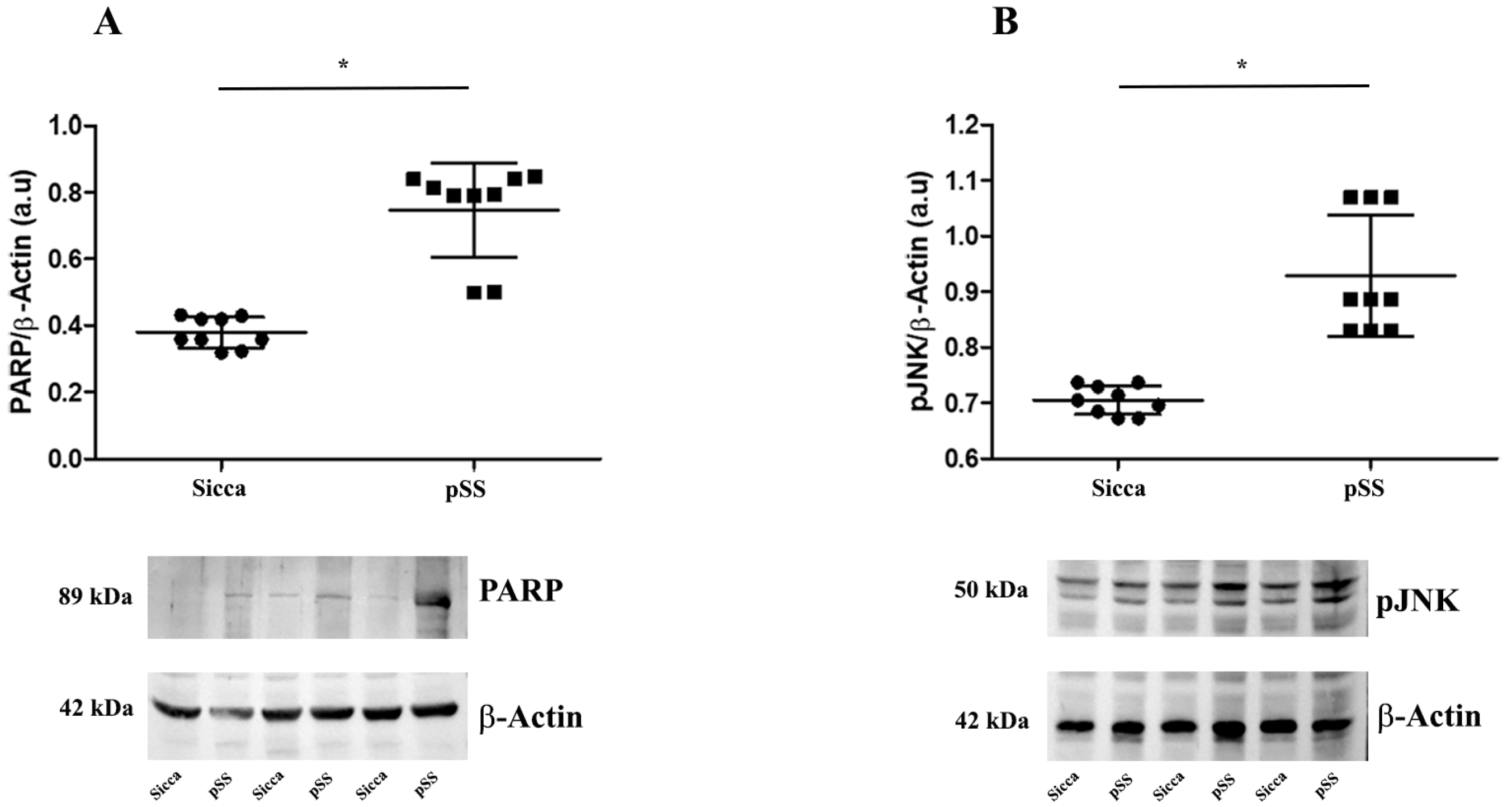
Since apoptosis is involved in the pathogenesis of pSS [27], we investigated proteolysis of poly (ADP-ribose) polymerase-1 (PARP-1), an enzyme involved in DNA repair [28], and the activation of c-Jun N-terminal kinases (JNK) known to trigger the caspase pathway. To this end, we performed Western blotting analysis with an antibody that recognizes the 89 kDa fragment released by caspase-3-mediated cleavage of PARP-1 and an antibody against the phosphorylated form of JNK (pJNK).
The representative bands of cleaved PARP-1 (c-PARP-1) and pJNK WBs are shown in Figure 4A,B, respectively. They show a significant increase in both proteins in the MSGs of pSS compared with those of sicca patients (* p < 0.05).

4. Discussion

Over the past few years, the role of SP has been intensely investigated in different systems such as inflammatory bowel disease (IBD) [23], in motor and non-motor Parkinson’s disease (PD) [29], and amyloid precursor protein (APP) metabolism in an Alzheimer’s disease (AD) model [17].
SP could be also involved in the impairment of salivary secretion observed in pSS patients. Treatments for pSS-related xerostomia have been classified as symptomatic, topical, or systemic stimulants, and regenerative treatments, but there is no satisfactory therapy to treat salivary hypofunction. The result of various clinical trials is that parasympathomimetics, including pilocarpine, able to stimulate exocrine gland secretion, are the most beneficial treatments for the management of salivary dysfunction in pSS [30].
Although a subjective, transient improvement in salivary function has been consistently observed with pilocarpine therapy, the exact mechanism is unclear. It has been hypothesized that pilocarpine could improve salivary secretion through local stimulation of neuropeptidergic fibers. The results of Sato et al. [31] in healthy humans showed that oral treatment with pilocarpine triggered an increased release of neuropeptides, including SP, in both saliva and blood, suggesting that this sialagogue neuropeptide should be an important factor contributing to the stimulatory pilocarpine treatment.
To confirm this hypothesis, preclinical experiments were performed in rats that showed a comparable effect on salivary secretion when treated with SP or with parasympathomimetics. Interestingly, the use of a selective SP antagonist has been shown to reduce the increased salivary secretion elicited by pilocarpine treatment [32]. These data confirm that the stimulatory effect of pilocarpine on rat submandibular glands is not only due to activation of muscarinic receptors, but probably also to the direct stimulation of SP nerve fibers.
The contribution of the SP pathway to the pathogenesis of the disease could be useful to identify new therapeutic targets, as already suggested in studies on pSS and other forms of keratoconjunctivitis sicca [14].
Indeed, clinical trials with the administration of SP analogs (eledoisin and physalaemin) in the form of eye drops have shown significant improvement in tear secretion in these patients [33,34,35].
Additional evidence is provided by the NOD (non-obese diabetic) mouse model of SS, which is genetically predisposed to the development of autoimmune exocrinopathy and mimics the sialoadenitis of Sjögren’s syndrome [36].
According to these previous studies, our results also point toward SP’s involvement in pSS, showing that SP levels strongly decreased in pSS MSGs compared to sicca MSGs, when analyzed by WB. This result was confirmed by the low level of SP detected using IF in pSS ductal epithelial cells, as reported in Figure 2, as well as in both acinar and ductal cells, detected in combination with ECAD IF, in Figure 3. The role of SP in the epithelium has been also studied in other tissues like the eye, where it has been found to be protective towards diabetes-related wounds in the corneal epithelium, when interacting with its related receptor NK1R [37].
Consistent with our results, the salivary glands of these mice, together with the presence of autoantibodies, were characterized by decreased levels of β-adrenergic, muscarinic, and neuropeptide signal transduction responses and by increased levels of apoptosis. The authors proposed that apoptosis is a key factor in the development of the disease and triggers autoimmunity [38].
In agreement with this finding and other clinical evidence [27], we demonstrated an increase in cleaved PARP-1 (c-PARP-1) in the MSGs of pSS patients, confirming that excessive apoptosis may contribute to pSS salivary gland dysfunction.
As previously reported, c-Jun N-terminal kinase (JNK) signaling was shown to mediate apoptosis in human salivary gland cells in vitro [39]. We confirmed the key role of this signaling pathway in pSS patients’ MSGs, showing a significant increase in JNK activation (pJNK) associated with enhanced apoptotic processes.
The therapeutic possibility of intervening and attempting to restore or stimulate salivary gland functions associated with pSS is essential from a clinical perspective, especially to prevent the complications of decreased or absent salivation [40].
Since pilocarpine treatment is the most effective treatment for improving salivary secretion by locally stimulating neuropeptidergic fibers and, in particular, by increasing the production and release of SP, this underscores the importance of our experimental results with regard to possible drug therapy, as previously suggested [33,34,35].
Furthermore, because there is no satisfactory therapy for the treatment of secretory hypofunction in pSS patients, the new data on the upregulation of the NK1 receptor represent a nodal point in the possibility of using low doses of SP or synthetic selective NK1 agonists to treat pSS symptoms.

Author Contributions

C.S. and M.F. conceived and designed the study; P.T. coordinated the experimental plan; M.F. and M.G.B. performed the sample collection; S.C. and R.P. performed the characterization of the human samples; P.R. and E.F. carried out the sample preparation, and performed the Western blot and immunofluorescence; P.R. and E.F. analyzed the data; B.C., M.L. and C.G. performed the microscopy image collection; C.S., P.R. and E.F. wrote the manuscript; C.S., M.F., S.C., B.C., P.T. and A.G. reviewed and edited the manuscript; all the authors critically discussed the results and approved its final version and qualify for authorship. All authors have read and agreed to the published version of the manuscript.

Funding

This work was supported by Sapienza University Intramural Grant 2022 to F.M. (RM12218161859E20).

Institutional Review Board Statement

The study was conducted in accordance with the Declaration of Helsinki, and approved by the Institutional Review Board (or Ethics Committee) of Policlinico Umberto I, Sapienza University of Rome (protocol code 4688).

Informed Consent Statement

Informed consent was obtained from all subjects involved in the study.

Data Availability Statement

Data presented in this study are available on request from the corresponding author.

Conflicts of Interest

The authors declare no conflict of interest.

References

  1. Katsifis, G.E.; Rekka, S.; Moutsopoulos, N.M.; Pillemer, S.; Wahl, S.M. Systemic and Local Interleukin-17 and Linked Cytokines Associated with Sjögren’s Syndrome Immunopathogenesis. Am. J. Pathol. 2009, 175, 1167. [Google Scholar] [CrossRef]
  2. Fox, R.I. Sjögren’s syndrome. Lancet 2005, 366, 321–331. [Google Scholar] [CrossRef]
  3. Negrini, S.; Emmi, G.; Greco, M.; Borro, M.; Sardanelli, F.; Murdaca, G.; Indiveri, F.; Puppo, F. Sjögren’s syndrome: A systemic autoimmune disease. Clin. Exp. Med. 2022, 22, 9. [Google Scholar] [CrossRef]
  4. Verstappen, G.M.; Kroese, F.G.M.; Bootsma, H. T cells in primary Sjögren’s syndrome: Targets for early intervention. Rheumatology 2021, 60, 3088–3098. [Google Scholar] [CrossRef]
  5. Retamozo, S.; Flores-Chavez, A.; Consuegra-Fernández, M.; Lozano, F.; Ramos-Casals, M.; Brito-Zerón, P. Cytokines as therapeutic targets in primary Sjögren syndrome. Pharmacol. Ther. 2018, 184, 81–97. [Google Scholar] [CrossRef] [PubMed]
  6. de Paiva, C.S.; Hwang, C.S.; Pitcher, J.D.; Pangelinan, S.B.; Rahimy, E.; Chen, W.; Yoon, K.C.; Farley, W.J.; Niederkorn, J.Y.; Stern, M.E.; et al. Age-related T-cell cytokine profile parallels corneal disease severity in Sjogren’s syndrome-like keratoconjunctivitis sicca in CD25KO mice. Rheumatology 2010, 49, 246–258. [Google Scholar] [CrossRef] [PubMed]
  7. Vitali, C.; Bombardieri, S.; Jonsson, R.; Moutsopoulos, H.M.; Alexander, E.L.; Carsons, S.E.; Daniels, T.E.; Fox, P.C.; Fox, R.I.; Kassan, S.S.; et al. Classification criteria for Sjögren’s syndrome: A revised version of the European criteria proposed by the American-European Consensus Group. Ann. Rheum. Dis. 2002, 61, 554–558. [Google Scholar] [CrossRef] [PubMed]
  8. Shiboski, C.H.; Shiboski, S.C.; Seror, R.; Criswell, L.A.; Labetoulle, M.; Lietman, T.M.; Rasmussen, A.; Scofield, H.; Vitali, C.; Bowman, S.J.; et al. 2016 American College of Rheumatology/European League Against Rheumatism Classification Criteria for Primary Sjögren’s Syndrome: A Consensus and Data-Driven Methodology Involving Three International Patient Cohorts. Arthritis Rheumatol. 2017, 69, 35–45. [Google Scholar] [CrossRef] [PubMed]
  9. Ekström, J.; Ekman, R.; Håkanson, R.; Sjögren, S.; Sundler, F. Calcitonin gene-related peptide in rat salivary glands: Neuronal localization, depletion upon nerve stimulation, and effects on salivation in relation to substance P. Neuroscience 1988, 26, 933–949. [Google Scholar] [CrossRef]
  10. Batbayar, B.; Nagy, G.; Kövesi, G.; Zelles, T.; Fehér, E. Morphological basis of sensory neuropathy and neuroimmunomodulation in minor salivary glands of patients with Sjögren’s syndrome. Arch. Oral Biol. 2004, 49, 529–538. [Google Scholar] [CrossRef]
  11. Konttinen, Y.T.; Hukkanen, M.; Kemppinen, P.; Segerberg, M.; Sorsa, T.; MalmstrÖM, M.; Rose, S.; Itescu, S.; Polak, J.M. Peptide-containing nerves in labial salivary glands in Sjögren’s syndrome. Arthritis Rheum. 1992, 35, 815–820. [Google Scholar] [CrossRef]
  12. Manganelli, P.; Fietta, P. Apoptosis and Sjögren syndrome. Semin. Arthritis Rheum. 2003, 33, 49–65. [Google Scholar] [CrossRef]
  13. Ferreira, J.N.; Hoffman, M.P. Interactions between developing nerves and salivary glands. Organogenesis 2013, 9, 199. [Google Scholar] [CrossRef]
  14. Severini, C.; Improta, G.; Falconieri-Erspamer, G.; Salvadori, S.; Erspamer, V. The tachykinin peptide family. Pharmacol. Rev. 2002, 54, 285–322. [Google Scholar] [CrossRef] [PubMed]
  15. Amadoro, G.; Pieri, M.; Ciotti, M.T.; Carunchio, I.; Canu, N.; Calissano, P.; Zona, C.; Severini, C. Substance P provides neuroprotection in cerebellar granule cells through Akt and MAPK/Erk activation: Evidence for the involvement of the delayed rectifier potassium current. Neuropharmacology 2007, 52, 1366–1377. [Google Scholar] [CrossRef]
  16. Lallemend, F.; Lefebvre, P.P.; Hans, G.; Rigo, J.M.; Van De Water, T.R.; Moonen, G.; Malgrange, B. Substance P protects spiral ganglion neurons from apoptosis via PKC-Ca2+-MAPK/ERK pathways. J. Neurochem. 2003, 87, 508–521. [Google Scholar] [CrossRef]
  17. Marolda, R.; Ciotti, M.T.; Matrone, C.; Possenti, R.; Calissano, P.; Cavallaro, S.; Severini, C. Substance P activates ADAM9 mRNA expression and induces α-secretase-mediated amyloid precursor protein cleavage. Neuropharmacology 2012, 62, 1954–1963. [Google Scholar] [CrossRef] [PubMed]
  18. Salthun-Lassalle, B.; Traver, S.; Hirsch, E.C.; Michel, P.P. Substance P, neurokinins A and B, and synthetic tachykinin peptides protect mesencephalic dopaminergic neurons in culture via an activity-dependent mechanism. Mol. Pharmacol. 2005, 68, 1214–1224. [Google Scholar] [CrossRef]
  19. Severini, C.; Petrella, C.; Calissano, P. Substance P and Alzheimer’s Disease: Emerging Novel Roles. Curr. Alzheimer Res. 2016, 13, 964–972. [Google Scholar] [CrossRef]
  20. Cuello, A.C.; Ribeiro-da-Silva, A.; Ma, W.; De Koninck, Y.; Henry, J.L. Organization of substance P primary sensory neurons: Ultrastructural and physiological correlates. Regul. Pept. 1993, 46, 155–164. [Google Scholar] [CrossRef] [PubMed]
  21. Schirinzi, T.; Maftei, D.; Grillo, P.; Bovenzi, R.; Bissacco, J.; Simonetta, C.; Mascioli, D.; Zenuni, H.; Cerroni, R.; Vincenzi, M.; et al. Olfactory neuron Substance P is overexpressed in Parkinson’s disease reflecting gut dysfunction. Mov. Disord. 2023. [Google Scholar] [CrossRef]
  22. Snijdelaar, D.G.; Dirksen, R.; Slappendel, R.; Crul, B.J.P. Substance P. Eur. J. Pain 2000, 4, 121–135. [Google Scholar] [CrossRef]
  23. O’Connor, T.M.; O’Connell, J.; O’Brien, D.I.; Goode, T.; Bredin, C.P.; Shanahan, F. The role of substance P in inflammatory disease. J. Cell. Physiol. 2004, 201, 167–180. [Google Scholar] [CrossRef] [PubMed]
  24. Hauser-Kronberger, C.; Albegger, K.; Saria, A.; Hacker, G.W. Neuropeptides in human salivary (submandibular and parotid) glands. Acta Otolaryngol. 1992, 112, 343–348. [Google Scholar] [CrossRef]
  25. Fisher, B.A.; Jonsson, R.; Daniels, T.; Bombardieri, M.; Brown, R.M.; Morgan, P.; Bombardieri, S.; Ng, W.-F.; Tzioufas, A.G.; Vitali, C.; et al. Standardisation of labial salivary gland histopathology in clinical trials in primary Sjögren’s syndrome. Ann. Rheum. Dis. 2017, 76, 1161–1168. [Google Scholar] [CrossRef] [PubMed]
  26. Colafrancesco, S.; Priori, R.; Smith, C.G.; Minniti, A.; Iannizzotto, V.; Pipi, E.; Lucchesi, D.; Pontarini, E.; Nayar, S.; Campos, J.; et al. CXCL13 as biomarker for histological involvement in Sjögren’s syndrome. Rheumatology 2020, 59, 165–170. [Google Scholar] [CrossRef]
  27. Jimenez, F.; Aiba-Masago, S.; Al Hashimi, I.; Vela-Roch, N.; Fernandes, G.; Yeh, C.K.; Talal, N.; Dang, H. Activated caspase 3 and cleaved poly(ADP-ribose)polymerase in salivary epithelium suggest a pathogenetic mechanism for Sjögren’s syndrome. Rheumatology 2002, 41, 338–342. [Google Scholar] [CrossRef]
  28. Lazebnik, Y.A.; Kaufmann, S.H.; Desnoyers, S.; Poirier, G.G.; Earnshaw, W.C. Cleavage of poly(ADP-ribose) polymerase by a proteinase with properties like ICE. Nature 1994, 371, 346–347. [Google Scholar] [CrossRef]
  29. Tirassa, P.; Schirinzi, T.; Raspa, M.; Ralli, M.; Greco, A.; Polimeni, A.; Possenti, R.; Mercuri, N.B.; Severini, C. What substance P might tell us about the prognosis and mechanism of Parkinson’s disease? Neurosci. Biobehav. Rev. 2021, 131, 899–911. [Google Scholar] [CrossRef]
  30. Salum, F.G.; Medella-Junior, F.D.A.C.; Figueiredo, M.A.Z.; Cherubini, K. Salivary hypofunction: An update on therapeutic strategies. Gerodontology 2018, 35, 305–316. [Google Scholar] [CrossRef]
  31. Sato, Y.; Itoh, H.; Suzuki, Y.; Tatsuta, R.; Takeyama, M. Effect of pilocarpine on substance P and calcitonin gene-related peptide releases correlate with salivary secretion in human saliva and plasma. J. Clin. Pharm. Ther. 2013, 38, 19–23. [Google Scholar] [CrossRef] [PubMed]
  32. Ueha, T.; Nemoto, A.; Kurihara, K. Studies on the salivary secretion induced by substance P in perfused submandibular gland of rat. Proc. Finn. Dent. Soc. 1989, 85, 345–353. [Google Scholar] [PubMed]
  33. De Caro, G.; Cordella, M.; Miani, P. The treatment of sjoegren’s syndrome with physalaemin. Ophthalmologica 1969, 158, 284–287. [Google Scholar] [CrossRef] [PubMed]
  34. Jaeger, W. Treatment of severe courses of keratoconjunctivitis sicca with Eledoisin. Klin. Monbl. Augenheilkd. 1988, 192, 163–166. [Google Scholar] [CrossRef]
  35. Spinelli, D.; Trabucchi, P.; Formenti, F. [Eledoisin eyedrops in the therapy of keratoconjunctivitis sicca]. Ophtalmologie 1988, 2, 145–147. [Google Scholar] [PubMed]
  36. Robinson, C.P.; Yamachika, S.; Bounous, D.I.; Brayer, J.; Jonsson, R.; Holmdahl, R.; Peck, A.B.; Humphreys-Beher, M.G. A novel NOD-derived murine model of primary Sjögren’s syndrome. Arthritis Rheum. 1998, 41, 150–156. [Google Scholar] [CrossRef]
  37. Yang, L.; Di, G.; Qi, X.; Qu, M.; Wang, Y.; Duan, H.; Danielson, P.; Xie, L.; Zhou, Q. Substance P promotes diabetic corneal epithelial wound healing through molecular mechanisms mediated via the neurokinin-1 receptor. Diabetes 2014, 63, 4262–4274. [Google Scholar] [CrossRef]
  38. Humphreys-Beher, M.G.; Yamachika, S.; Yamamoto, H.; Maeda, N.; Nakagawa, Y.; Peck, A.B.; Robinson, C.P. Salivary gland changes in the NOD mouse model for Sjögren’s syndrome: Is there a non-immune genetic trigger? Eur. J. Morphol. 1998, 36, 247–251. [Google Scholar]
  39. Fu, J.; Shi, H.; Cao, N.; Wu, S.; Zhan, T.; Xie, L.; Wang, Z.; Ye, L.; Li, C.; Shen, Y.; et al. Toll-like receptor 9 signaling promotes autophagy and apoptosis via divergent functions of the p38/JNK pathway in human salivary gland cells. Exp. Cell Res. 2019, 375, 51–59. [Google Scholar] [CrossRef]
  40. Jensen, S.B.; Vissink, A. Salivary gland dysfunction and xerostomia in Sjögren’s syndrome. Oral Maxillofac. Surg. Clin. N. Am. 2014, 26, 35–53. [Google Scholar] [CrossRef]
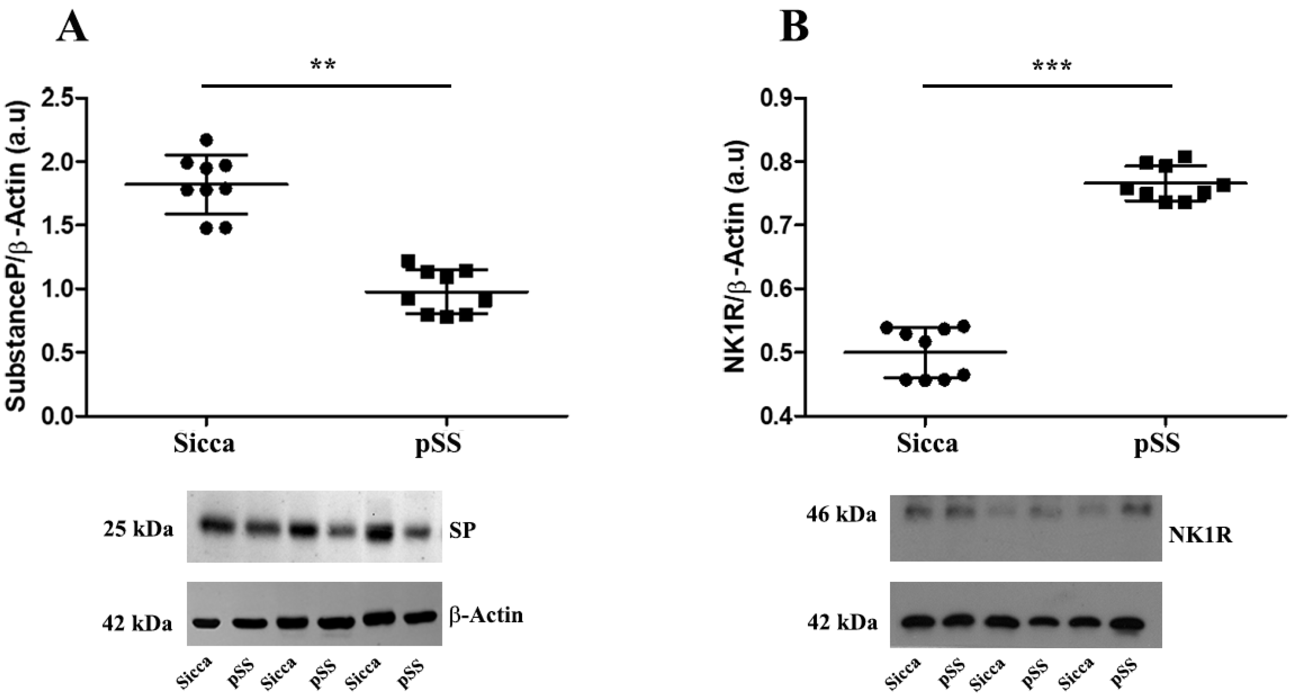
Figure 1. (A,B) Representative Western blot of SP (A) and NK1R (B) levels in MSGs from sicca and pSS patients. Data are expressed as mean optical density in arbitrary units (a.u.) and are given as mean ± SD of n = 9 independent experiments. Statistical significance calculated using one-way analysis of variance (ANOVA) for repeated measures followed by Tukey’s post hoc test, indicated with ** p < 0.01; *** p < 0.001. β-actin expression was used to normalize sample variability. Molecular weight markers (kDa) are shown on the left.
Cells 12 01347 g001
Figure 2. (A,B) SP expression and localization was severely affected in MSG sections from pSS and sicca patients. (A) Representative images of immunofluorescence staining (red) of SP in pSS and sicca patient MSG sections. (B) Quantitative analysis showing lower expression of SP in pSS compared to sicca patients. n = 5 independent experiments and statistical significance calculated using t-Student test. *** p < 0.001; t = 6.302. Scale bar 25 µm; 40× magnification. First row: negative control (ctl neg).
Cells 12 01347 g002
Figure 3. SP and ECAD expression and localization was severely affected in MSG sections from pSS and sicca patients. Representative images of double immunofluorescence staining of substance P (SP, red) and e-cadherin (ECAD, green) in pSS and sicca patients MSG sections. Scale bar 25 µm; 40× magnification.
Cells 12 01347 g003
Figure 4. (A,B) Representative Western blot of c-PARP-1 (A) and pJNK (B) levels in MSGs from sicca and pSS patients. Data are expressed as mean optical density in arbitrary units (a.u.) and are given as mean ± SD of n = 9 independent experiments. Statistical significance was calculated using one-way analysis of variance (ANOVA) for repeated measures followed by Tukey’s post hoc test, indicated with * p < 0.05. β-actin expression was used to normalize sample variability. Molecular weight markers (kDa) are shown on the left.
Cells 12 01347 g004
Disclaimer/Publisher’s Note: The statements, opinions and data contained in all publications are solely those of the individual author(s) and contributor(s) and not of MDPI and/or the editor(s). MDPI and/or the editor(s) disclaim responsibility for any injury to people or property resulting from any ideas, methods, instructions or products referred to in the content.