Article

Reconstitution of Caruncle Placenta through the 20α-HSD/Casp-3 Apoptotic Pathway during Early Pregnancy in Bovines


1
Institute of Applied Humanimal Science, Hankyong National University, Ansung-si 17579, Republic of Korea
2
General Graduate School of Animal Life Convergence Science, Hankyong National University, Ansung-si 17579, Republic of Korea
3
School of Animal Life Convergence Science, Hankyong National University, Ansung-si 17579, Republic of Korea
*
Author to whom correspondence should be addressed.
These authors contributed equally to this work.
Cells 2023, 12(1), 162; https://doi.org/10.3390/cells12010162
Submission received: 30 November 2022 / Revised: 21 December 2022 / Accepted: 27 December 2022 / Published: 30 December 2022
(This article belongs to the Section Cellular Aging)

Abstract

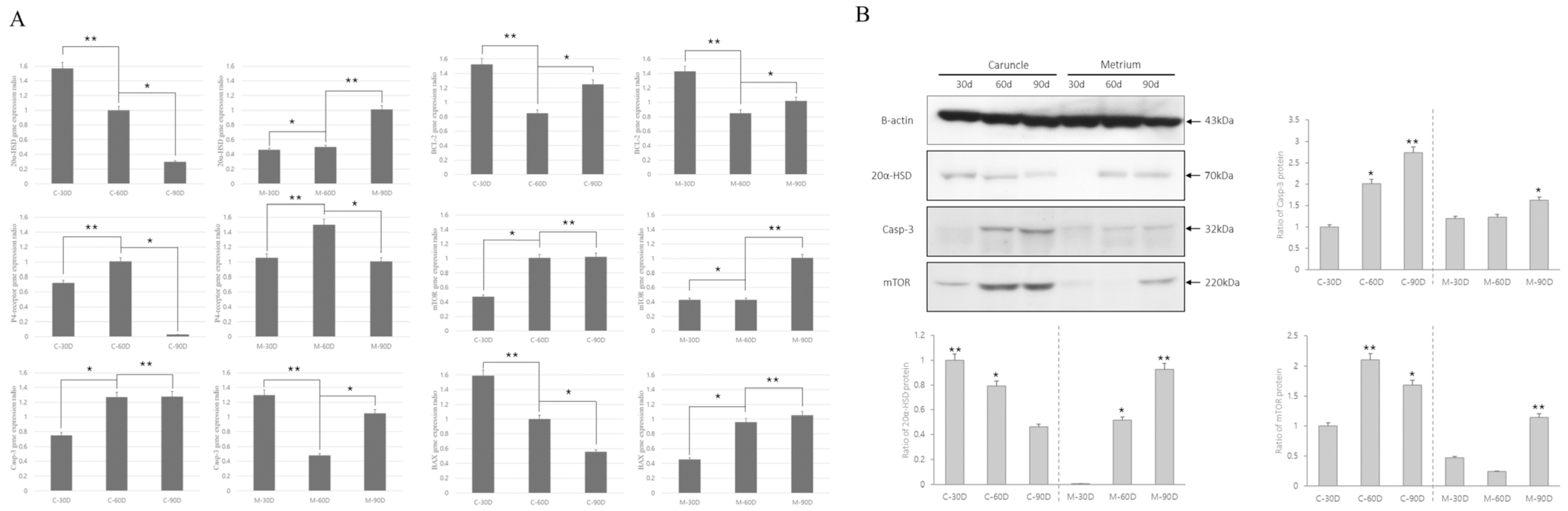
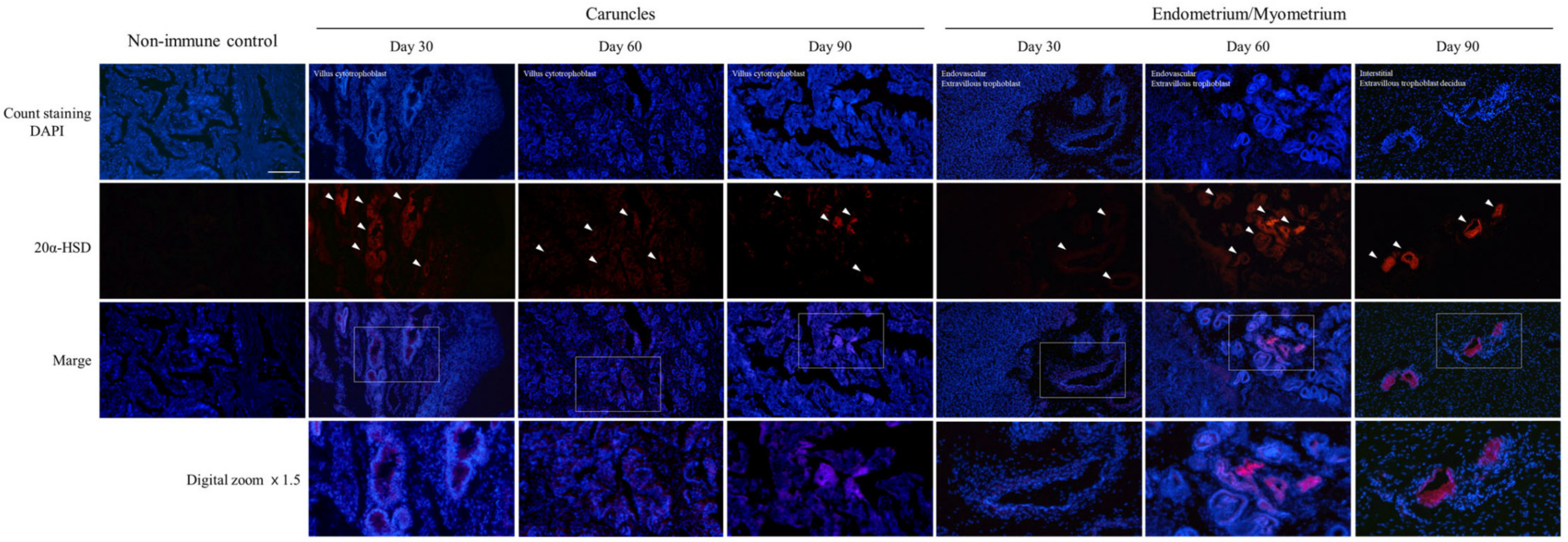
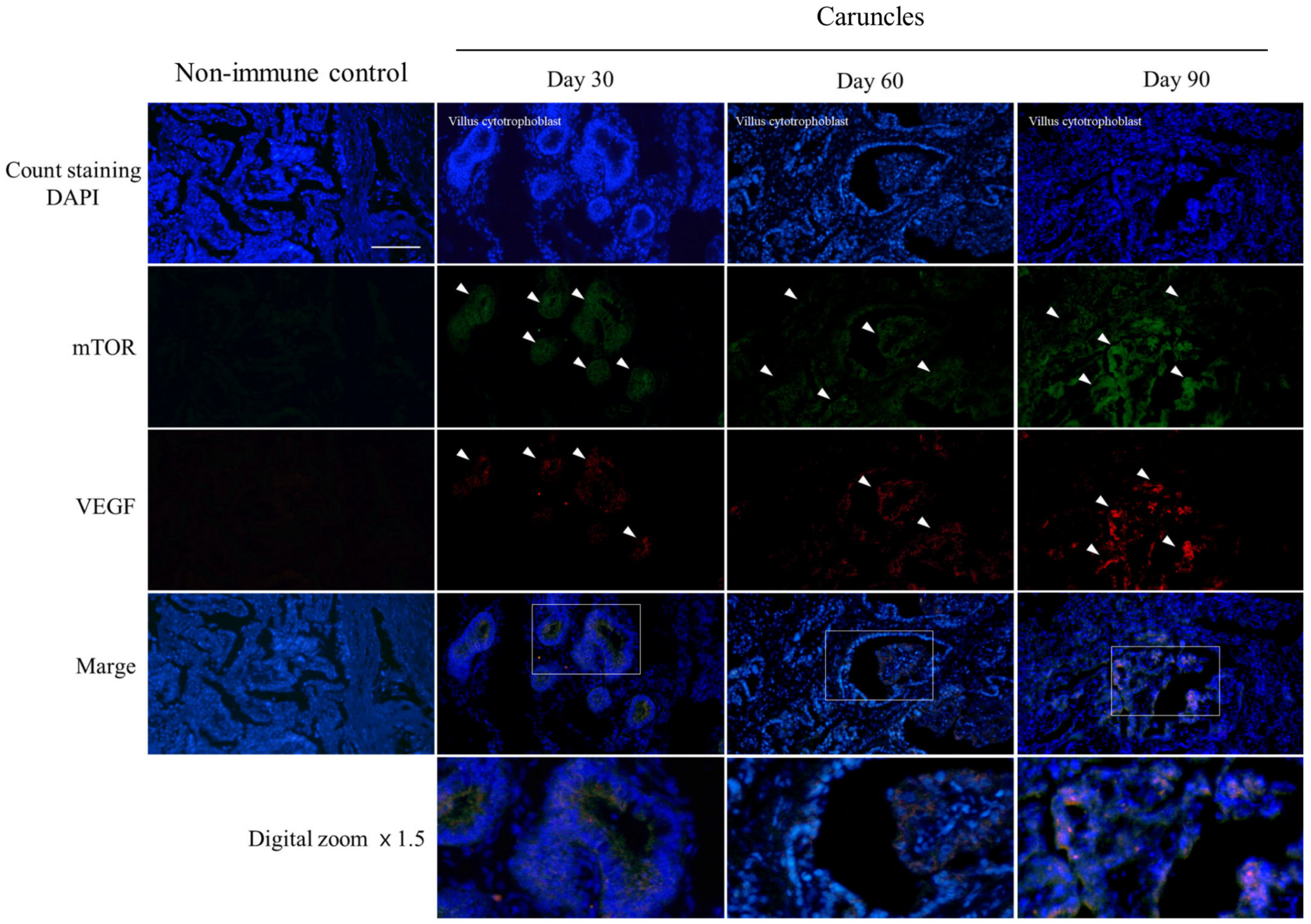
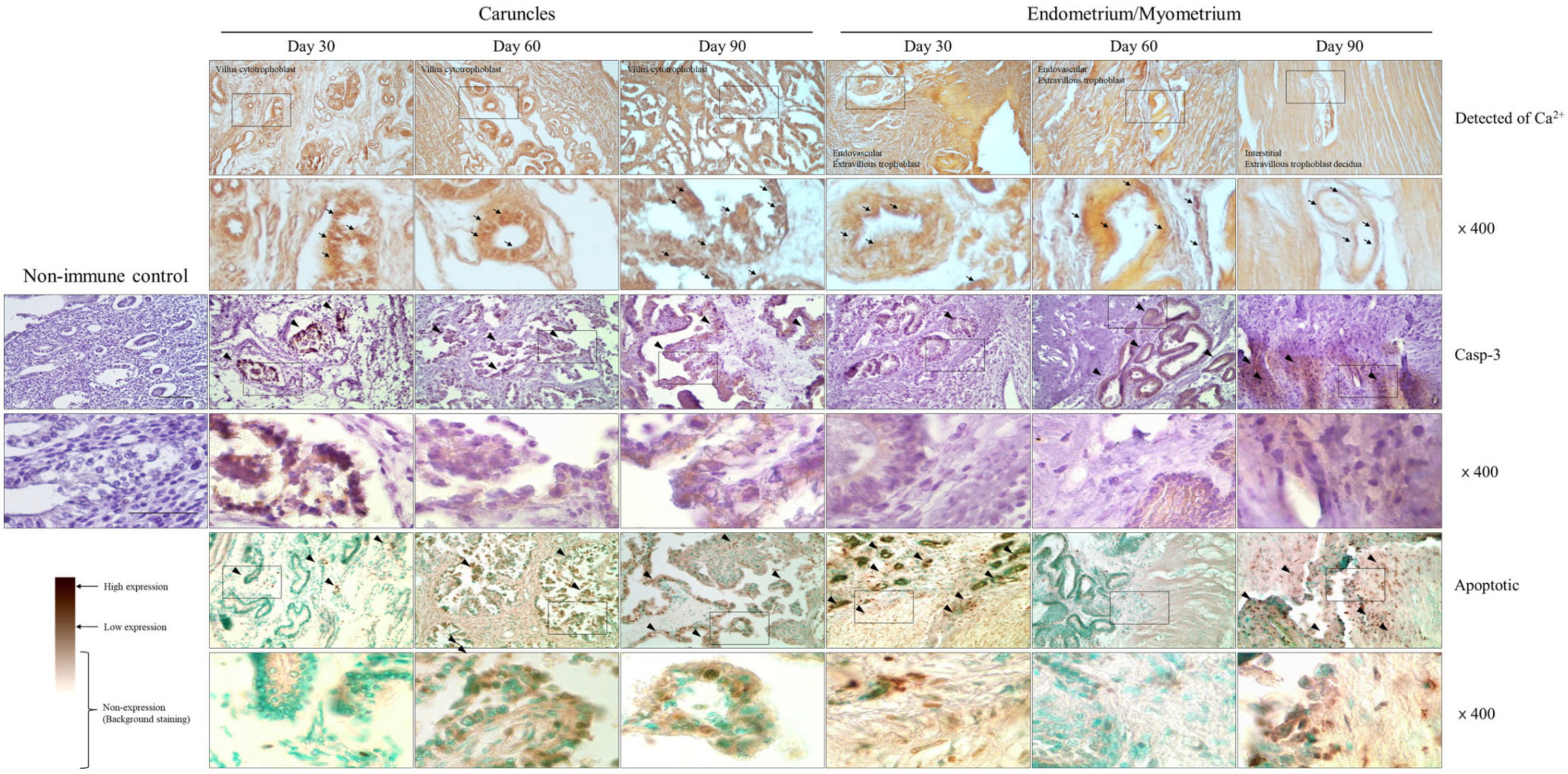
Trophoblast cells of endometrium during bovine pregnancy with different characteristics undergo dynamic changes during uterine remodeling, which can be observed as continuous changes, as P4 secreted by the mother is replaced by placental hormones. In this context, the present study analyzed tissues’ morphological changes through uterine apoptosis during early pregnancy. In addition, the expression pattern associated with apoptosis genes and 20α-HSD was determined in the endometrium and caruncle tissues. The localization of 20α-HSD, VEGF, Casp3, and mTOR protein was also determined in endometrium and caruncle during early pregnancy. From around 30 days, caruncle trophoblast cells with very high invasiveness expanded the villus section as the gestation period progressed. The surrounding cells detached and reorganized into new cells. In addition, an analysis of the effect of apoptosis on cell reorganization in the caruncle revealed that the expression of 20α-HSD/Casp-3 signals in the villus section gradually increased from 30 to 90 days. However, on the 30th day, glandular epithelial cells occurred sporadically in the trophoblast cell section. Moreover, the apoptosis of trophoblast cells increased at 90 days. Taken together, the results of the present study show that changes in the uterus during early pregnancy promote changes during later pregnancy by inducing the reorganization through the stimulation of 20α-HSD and Casp-3, promoting uterine and caruncle tissues, unlike cell development mediated by hormone signaling.
Keywords: apoptosis; progesterone; pregnancy; P4; ruminants; placenta apoptosis; progesterone; pregnancy; P4; ruminants; placenta