Article

Changes in miR-124-1, miR-212, miR-132, miR-134, and miR-155 Expression Patterns after 7,12-Dimethylbenz(a)anthracene Treatment in CBA/Ca Mice

1
Doctoral School of Health Sciences, Faculty of Health Sciences, University of Pécs, 7624 Pécs, Hungary
2
Department of Public Health Medicine, Medical School, University of Pécs, 7624 Pécs, Hungary
3
Institute of Transdisciplinary Discoveries, Medical School, University of Pécs, 7624 Pécs, Hungary
4
Institute of Physiology, Medical School, University of Pécs, 7624 Pécs, Hungary
*
Authors to whom correspondence should be addressed.
These authors contributed equally to this work.
Cells 2022, 11(6), 1020; https://doi.org/10.3390/cells11061020
Submission received: 20 February 2022 / Revised: 13 March 2022 / Accepted: 16 March 2022 / Published: 17 March 2022
(This article belongs to the Special Issue Regulatory Roles of Non-coding RNAs in Cancer)

Abstract

:
Specific gene and miRNA expression patterns are potential early biomarkers of harmful environmental carcinogen exposures. The aim of our research was to develop an assay panel by using several miRNAs for the rapid screening of potential carcinogens. The expression changes of miR-124-1, miR-212, miR-132, miR-134, and miR-155 were examined in the spleen, liver, and kidneys of CBA/Ca mice, following the 20 mg/bwkg intraperitoneal 7,12-dimethylbenz(a)anthracene (DMBA) treatment. After 24 h RNA was isolated, the miRNA expressions were analyzed by a real-time polymerase chain reaction and compared to a non-treated control. DMBA induced significant changes in the expression of miR-134, miR-132, and miR-124-1 in all examined organs in female mice. Thus, miR-134, miR-132, and miR-124-1 were found to be suitable biomarkers for the rapid screening of potential chemical carcinogens and presumably to monitor the protective effects of chemopreventive agents.

1. Introduction

Cancer has become a major public health problem worldwide. Due to the increasing trendline of lifestyle-related risk factors, as well as current trend of population ageing, cancer morbidity is predicted to reach 28.4 million cases worldwide by 2040, which is a 47% increase compared to the estimated number of cases in 2020 [1]. According to the World Health Organization (WHO), however, about half of all malignant tumors are preventable [2], and early diagnosis—in general—increases the chances of successful treatment. Consequently, the timely identification of harmful exogenic substances that may play a part in tumor development, the early detection of tumors, along with the continuous development of more accurate diagnostic and more effective therapeutic procedures are all important factors in the prevention and successful treatment of cancer.
Specific gene expression patterns, such as increased expression of HA-RAS or C-MYC oncogenes, are early biomarkers of harmful environmental exposures at the different stages of tumorigenesis and represent the underlying mechanisms of the carcinogenic process [3,4,5,6]. Moreover, previously, we found that miR-9-3 and mTORC1 can be used as biomarkers for early detection of DMBA-induced damages in female mice [7].
MicroRNAs (miRNAs) are highly conserved RNA molecules of 19 to 22 nucleotides in length that regulate approximately 30–50% of human gene expression [8,9]. They bind to the 3′ untranslated region of partially or fully complementary messenger RNAs (mRNAs) and thereby inhibit their translation and ultimately the protein synthesis [10]. Since miRNAs are stable in vivo and show a mostly altered expression pattern in malignantly transformed cells [11,12], distinct miRNA patterns can be observed following exposure to carcinogenic agents [13].
The carcinogenesis-related regulatory functions of miRNAs can be highly diverse, ranging from oncogenic to tumor suppressing effects, depending on the nature of the genes they regulate [14]. The complexity of their regulation is supported by the fact that some miRNAs can have both oncogenic and tumor suppressor properties, because a miRNA can be partially or fully complementary to several different mRNAs. For example, the miR-125 as a tumor suppressor reduces the antiapoptotic effect of the MUC1 and Bcl-2 genes [15,16] and suppresses vascular endothelial growth factor (VEGF) expression, thereby inhibiting tumor proliferation and metastasis formation [17]. However, miR-125 also acts as a tumor promoter by regulating the tumor suppressor protein TP53INP1 (Tumor protein P53 induced nuclear protein 1), and, as a result, stimulates cell proliferation and migration [15].
MiR-124-1, miR-212, miR-132, miR-134, and miR-155 are implicated in the regulation of oncogenes, tumor suppressor genes, cell growth, proliferation, invasion, migration, metastasis, and apoptosis through various signaling pathways [18,19,20]. Moreover, these miRNAs have been found to show altered expression levels in a variety of malignant cancer cells [18,19,21]. MiR-124 acts as a tumor suppressor by inhibiting the translation of the cell cycle regulator cyclin dependent kinase 6 (CDK6) and by reducing the phosphorylation of the retinoblastoma protein (Rb) [19]. Furthermore, loss of miR-124 function by hypermethylation is frequently found in tumor cells [18]. Proteins regulated by miR-212 affect cell proliferation and apoptosis as well as processes involved in tumorigenesis, such as invasion and metastasis formation [22]. For example, as a target of miR-212, the transcription factor SOX4 has been found to be involved in the invasion, migration, and metastasis of various tumors [23]. The miR-132 has both tumor-specific oncogenic and tumor suppressor properties. Its oncogenic effect has been observed in pancreatic carcinoma (PC) by inhibiting the Phosphatase and tensin homolog (PTEN) tumor suppressor gene [24]. However, in non-small-cell lung carcinoma (NSCLC) miR-132 has been shown to inhibit migration and invasion of cells [25], thus indicating a tumor suppressor characteristic. The miR-134 increases the expression of P21 and P57 CDKIs and represses the expression of cyclin D1, cyclin D2, and cyclin-dependent kinase 4 (CDK4) proteins, which are promoters of the cell cycle and are overexpressed in many tumors. Thus, miR-134, as a tumor suppressor, reduces cell proliferation [26] and enhances apoptosis [27]. MiR-155 is significantly overexpressed in cervical cancer (CEC) tissues compared to normal tissues [28].
Many chemical compounds, with the ability to cause cancer in humans, have been identified. 7,12-dimethylbenz(a)anthracene (DMBA) is one of the most potent mutagenic and carcinogenic environmental polycyclic aromatic hydrocarbons [29,30]. Polycyclic aromatic hydrocarbons are formed during imperfect burning of organic compounds; thus, for example, cigarette smoke and car exhaust gases are characteristic and significant sources of DMBA exposure [31]. DMBA is activated by cyto-chrome P450-metabolizing enzymes [32,33]. The resulting metabolites can alkylate the DNA or other cellular macromolecules, which explains its initiator and promoter effects [6]. In addition, DMBA enhances the formation of reactive oxygen species (ROS) [34], and this effect also contributes to cellular dysfunction and altered behavior [35]. DMBA is often used in animal experiments as an initiator carcinogenic agent [36], leading to the formation of tumors histologically similar to human cancers [37]. The expression of certain genes (e.g., H-RAS, C-MYC, and P53) and miRNAs (e.g., miR-21 and miR-146a) is altered 24 to 48 h after treatment with DMBA; thus, these expression changes can be considered as early biomarkers of the carcinogenic process [38,39].
Increased expression levels of certain miRNAs, namely miR-128b, miR-152, miR-125b, miR-205, miR-27a, miR-146a, miR-222, miR-23a, miR-24, miR-150, etc., were represented in lung cancer, while the expression levels of miR-29a, miR-221, miR-223, miR-25, miR-92, miR-99a, etc., were increased in colorectal cancer tissues [40]. Moreover, it has been reported that patients with papillary thyroid carcinomas showed increased levels of miR-221, miR-222, and miR-146 in the tumors [41]. Thus, the role of miRNAs as early epigenetic biomarkers in various tumors has been confirmed [42]. Therefore, we hypothesized that altered expression patterns of certain miRNAs may be biomarkers of early damage caused by the carcinogenic agent, DMBA.
The aim of our research was to develop an assay panel for the rapid screening of potential carcinogens or chemopreventive agents. Earlier, we investigated the expression of miR-330, miR-29a, miR-9-1, miR-9-3, and mTORC1 after treatment with DMBA in vivo and found that miR-9-3 and mTORC1 expression in female mice could be suitable biomarkers for rapid identification for possible carcinogenic effects [7]. Following up on our previous study, to expand the options for the biomarkers in the assay panel, we analyzed the expression of miR-124-1, miR-212, miR-132, miR-134, and miR-155 after DMBA exposure in CBA/Ca mice spleen, liver, and kidneys. The aim of the present study was to investigate whether any or some of these miRNAs could be used as potential biomarkers of early carcinogenic damage as well as to elucidate the probable role of miRNAs in the process of tumorigenesis.

2. Materials and Methods

2.1. Animal Treatment

Two groups of CBA/Ca mice were used in our study. Both control and DMBA-treated groups consisted of 12 (six males and six females) 6–8 weeks old mice. Both the treated and the control groups received 0.1 mL of corn oil (Sigma-Aldrich, St. Louis, MO, USA) intraperitoneally, but in the treated group, 20 mg/bwkg DMBA was dissolved in the corn oil. After 24 h of DMBA exposure, the mice were euthanized, cervical dislocation was performed, and the livers, kidneys, and spleens were removed. Total cellular RNA level was determined as described below.
In accordance with the guidelines concerning laboratory animals, mice received humane care. The experiment was approved by Regional Animal Ethical Committee Pécs and conducted according to the current ethical regulations (ethical permission no.: BA02/2000-79/2017).

2.2. Isolation of Total RNA

Total cellular RNA was isolated using TRIZOL reagent, according to the manufacturer’s instructions (Thermo Fisher Scientific, Waltham, MA, USA). The RNA quality was determined by NanoDrop absorption photometry. Only RNA fractions with A > 2.0 at 260/280 nm were used for the reverse transcription polymerase chain reaction process.

2.3. Reverse Transcription Polymerase Chain Reaction (RT-PCR)

Following the manufacturer’s instructions, the one-step PCR, including reverse transcription and target amplification, was performed in a 96-well plate using Kapa SYBR FAST One-step RTQCR kit (Kapa Biosystems, Wilmington, MA, USA) on a LightCycler 480 qPCR platform.
The temperature program was set as follows: 5 min incubation at 42 °C and 3 min incubation at 95 °C. Then, 45 cycles (95 °C—5 s, 56 °C—15 s, and 72 °C—5 s) were performed, and at the end of each cycle, a fluorescent reading was made. Each run was performed with melting curve analysis (95 °C—5 s, 65 °C—60 s, and 97 °C ∞) to confirm the amplification specificity. The reaction mixture used was the following: 10 μL KAPA SYBR FASTqPCR Master Mix, 0.4 μL KAPA RT Mix, 0.4 μL dUTP, 0.4 μL primers, and 5 μL miRNA template supplemented with sterile double-distilled water to a total volume of 20 μL.
Primer sequences for the examined miRNAs (miR-124-1, miR-212, miR-132, miR-134, and miR-155) and the internal control gene (mouse U6) are summarized in Table 1.
Primers were synthetized by Integrated DNA Technologies (Integrated DNA Technologies Inc., Coralville, IA, USA), and sequences were taken from previous publications [43,44].

2.4. Calculations and Statistical Analysis

Relative miRNA expression levels were calculated and compared using the 2−ΔΔCT method. During the statistical analysis for the testing the distribution of results, we used the Kolmogorov–Smirnov test. To compare means, we used the Levene’s type T-probe. For calculations and analysis, IBM SPSS 21 (International Business Machines Corporation, Armonk, NY, USA) statistical software was used. We determined the level of statistical significance at p < 0.05.
Differences in miRNA expressions were expressed on the figures as percentages of the respective control group.

3. Results

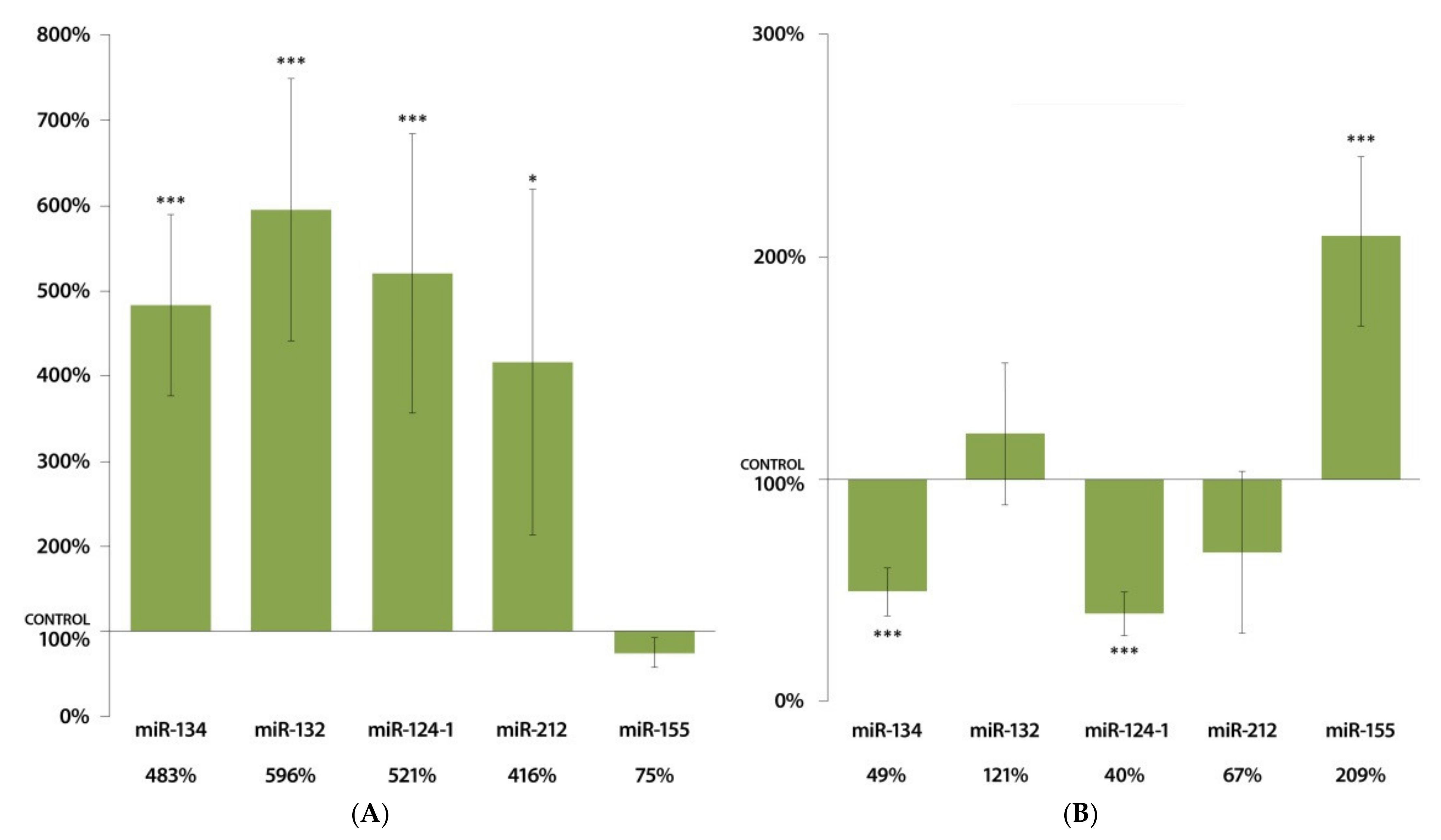
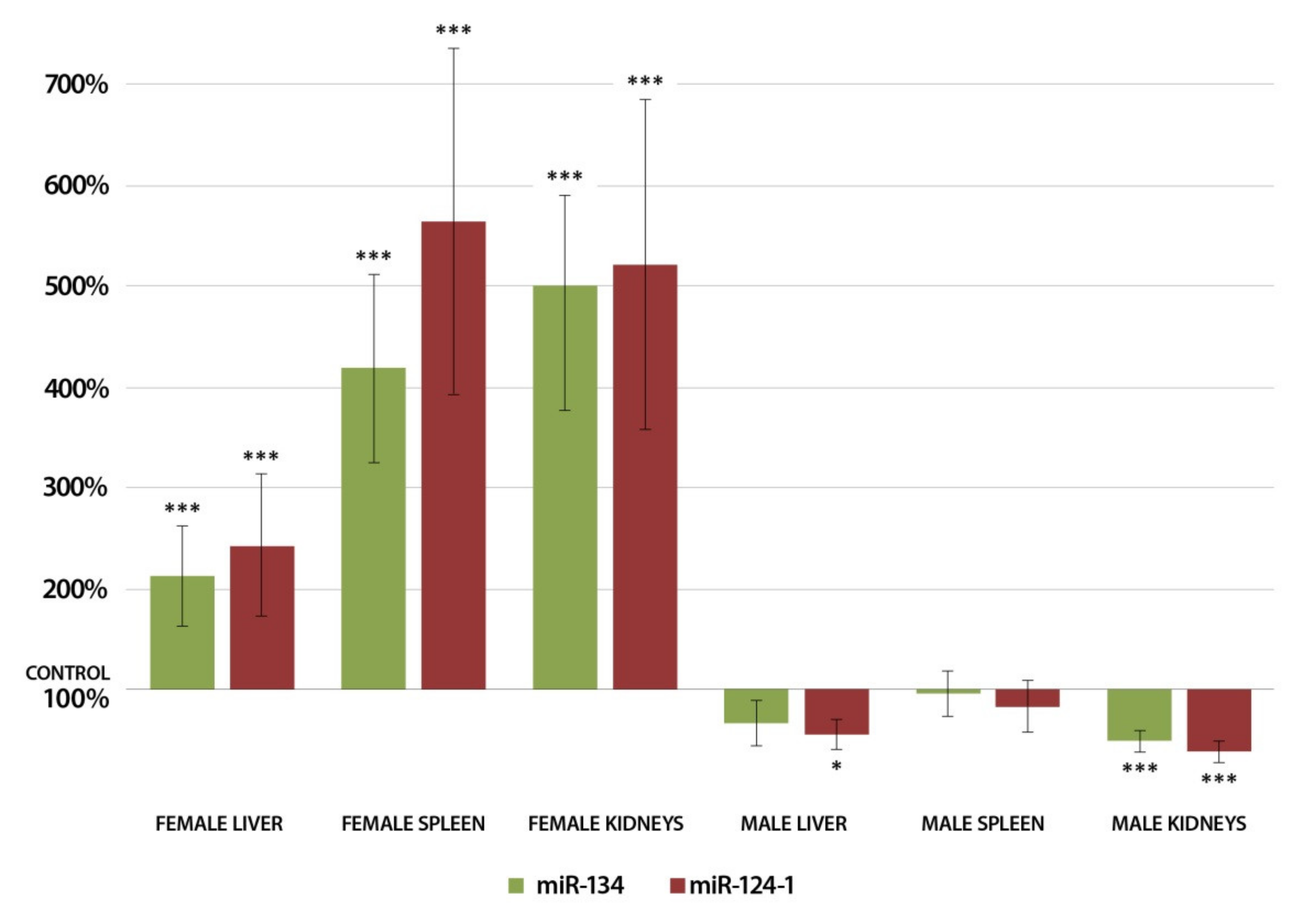
3.1. Changes in miRNA Expression in the Liver 24 h after DMBA Treatment

A total of 24 h after DMBA treatment, there was a highly significant (p < 0.001) increase in the expressions of miR-134, miR-132, miR-124-1, miR-212, and miR-155 in the liver of DMBA-treated female mice compared to the control group (Figure 1A). In contrast, only miR-124-1 decreased expression was significant (p < 0.05) among the tested miRNAs in the liver of treated males (Figure 1B).

3.2. Changes in miRNA Expression in the Spleen 24 h after DMBA Treatment

In the spleens of female mice, DMBA induced a highly significant (p < 0.001) rise in the expressions of miR-124-1, miR-132, and miR-134 compared to the control group (Figure 2A). A highly significant (p < 0.001) increase in miR-132, miR-212, and miR-155 expression could also be observed in the spleens of males after DMBA treatment, as shown in Figure 2B.

3.3. Changes in miRNA Expression in the Kidneys 24 h after DMBA Treatment

Figure 3A illustrates that DMBA treatment resulted highly significant (p < 0.001) increase in the expression of miR-134, miR-132, miR-124-1, and significant (p < 0.05) increase in the expression of miR-212 in the kidneys of female mice. On the other hand, miR-134 and miR-124-1 showed a highly significantly decreased expression, and miR-155 demonstrated a highly significantly increased expression in the kidneys of male mice in response to DMBA compared to untreated male mice (Figure 3B).

4. Discussion

We found a highly significant increase in the expressions of miR-134, miR-132, and miR-124-1 following exposure to DMBA in all the examined organs of female mice; however, less-marked and sometimes opposite changes in miRNA expression could be detected in DMBA-treated males. These results indicate that miR-134, miR-132, and miR-124-1 could be potential biomarkers of early carcinogenic damage in females and that gender-specific hormonal differences affect miRNA expression patterns caused by carcinogenic factors, significantly. DMBA, a well-known carcinogen, affects various members of the cell signaling pathways, including miRNA expressions, which, in turn, influence the activation or inhibition of other key elements of signal transduction pathways.
DMBA induces a dose-dependent increase in transforming growth factor beta 1 (TGF-β1) levels [45] and the higher expression of TGF-β1 increases miR-132 expression [46]. However, miR-132 blocks the DMBA-activated TGF β1/Smad2/3 signaling [47], suggesting that there is a mutual feedback between TGF-β1 and miR-132. In addition, miR-132 regulates liver tumor cell proliferation, apoptosis, migration, and invasion by suppressing the DMBA-induced transcription factor SRY-related HMG-box (SOX) [48,49]. It is also known that miR-132 represses the Akt/mTOR signaling pathway, which is activated by DMBA and involved in promoting tumor formation [50]. The significant increases in miR-132 expression, observed in our study, may be an early response to DMBA-induced signaling processes such as SOX, TGF-β1, or mTOR activation. Our present results suggest that miR-132 cannot be used as a biomarker in males. However, a highly significant increase in miR-132 expression was found in females, correlating with literature data [45,46]. Thus, miR-132 could be used as a gender-specific biomarker in female mice.
Both in vitro and in vivo studies confirm the complex role of miR-212 in carcinogenesis [51,52]. MiR-212 inhibits cell proliferation, migration, and invasion in renal cell carcinoma (RCC) and exerts tumor suppressor effects by downregulating T-box transcription factor TBX15 (TBX15) [51,52]. In the present study, the results regarding miR-212 expression in the liver were opposite in the two genders (decreasing in males and increasing in females), which supports the mechanism of tumor development via the Forkhead Box A1 (FOXA1) signaling pathway, while miR-212 suppresses the growth of hepatocellular carcinoma (HCC) by downregulating the expression of the FOXA1 gene [53]. This effect is gender-specific because FOXA1 is involved in both estrogen and androgen signaling, acting as a central regulator of sexual dimorphism in liver tumors, which are more common in males [54]. An increase in miR-212 expression in females reduces the protective effect of estrogen by suppressing FOXA1, whereas in males, the reduced miR-212 levels increase FOXA1 expression, which favors the hepatic tumor-promoting effect of androgen signaling [52,53,54]. These observations are supported by the report that the suppression of FOXA1 resulted in more and larger tumor cells in females and reduced tumor growth in males [55]. This highlights the interactions between sex hormones and signal transducers in liver tumors, but further studies are required to understand the exact role of miRNAs, for example miR-212, in the regulation of these processes. Interestingly, miR-212 expression in the spleen of female mice did not show any significant changes after DMBA treatment; therefore, miR-212 could only be used as a biomarker limited to the liver organ at most.
According to an earlier investigation, increased expression of miR-155 expression could be observed in vivo in the liver, spleen, lung, and kidneys 3 and 6 h after DMBA treatment [39]. In accordance with this, our results showed a significant increase in miR-155 expression in the spleen and kidneys of males and the liver of females 24 h after DMBA exposure. However, unidirectional, positive changes were found only in the liver from the studied organs in both sexes, although the changes were not significant in males, which are explained by the miR-155 upregulating effect of estrogen [56]. This effect may be due to the metabolic activation of DMBA by cytochrome P450 (CYP) enzymes, mainly cytochrome P450 1A1 (CYP1A1) and 1B1 (CYP1B1), which are typically present in the liver with high enzyme activity, and thus the effect of DMBA on miR-155 expression may be more rapid and dominant in this tissue type [57]. It is also known that miR-155 is overexpressed in liver injury, and miR-155 exerts a protective effect by suppressing nuclear factor-kappa B (NF-kB) signaling [58]. In addition, miR-155 has also been identified as an oncomiR in various types of human tumors, which inhibits the activity of tumor suppressor targets such as TP53INP1 or the von Hippel–Lindau (VHL) tumor suppressor protein [59,60]. The data in the literature as well as in our study highlight the complex function of miR-155 in carcinogenesis and indicate the need for further studies to fully understand the observed changes and the mechanisms influencing miR-155 levels. Therefore, based on the present results, as well as the mentioned multifarious factors, miR-155 cannot be used as a robust early biomarker in our target assay panel.
The CASC3 protein is a direct target of miR-124, through which miR-124 is capable of inactivating the p38-MAPK, JNK, or ERK signaling pathways, thereby inhibiting cell proliferation [61]. The tumor suppressor effect of miR-124 was also observed through the inhibition of cell cycle-related cyclin D1, cyclin-dependent kinase 2 (CDK2), cyclin-dependent kinase 4 (CDK4), and cyclin-dependent kinase 6 (CDK6) [62,63]. Despite these previous findings, the expected increase in miR-124-1 expression following DMBA exposure was only observed in the female group, with highly significant increases in all three examined organs. DMBA activates the signal transducer and activator of transcription 3 (STAT3) transcription factor [64], which has a repressive effect on miR-124 [65]. The STAT3-inducing effect was also observed after testosterone treatment, which may explain the downregulation of miR-124-1 in the male group [66]. Therefore, miR-124-1 was found to be potential biomarker only in females.
Strong expression of miR-134 leads to reduced expression of the C-MYC oncogene, and the Cyclin E and Cyclin D1 cell cycle regulatory proteins. Furthermore, the increased expression of miR-134 coincides with growing cyclin-dependent kinase inhibitor 1B (p27) levels, which also inhibits the cell cycle [67,68]. It also acts as a tumor suppressor by regulating intracellular signal transduction pathways such as the RAS/MAPK/ERK or the RAS/PI3K/AKT signaling pathways that stimulate cell proliferation and invasion and inhibit apoptosis [69,70]. According to literature data, there is a significantly reduced expression of miR-134 in renal cell carcinoma [69]. This observation correlates with our findings that there was a significant decrease in miR-134 expression in the kidney tissue of the male mice 24 h after DMBA exposure. However, a not-significant reduction was also observed in the other tested organs, so the effect of miR-134 as a tumor suppressor was weaker in male mice. In contrast, DMBA treatment caused a significant increase in miR-134 expression after 24 h in the kidneys, liver, and spleen of the females. Upstream regulators of miR-134, such as the NF-kB transcription factor, whose aberrant activation has been reported in malignant tumor cell lines, may underlie the clear and significant sex differences [71]. DMBA increases the expression of NF-kB [72], resulting in a stronger repression of miR-134 [73], which may explain the observed decreases in miR-134 expression in male mice in our study. In female mice, however, the estrogen receptor (ER) inhibits NF-kB activation [74], thus blocking its repressive effect on miR-134 and leading to an increased miR-134 expression.
Increased expression of miR-124 also has a silencing effect on NF-kB by regulating TRAF6 [75], thereby reducing the NF-kB-induced miR-134 inhibition. This is also reflected in our findings (Figure 4) since we found very similar expression patterns of miR-134 and miR-124-1. Thus, it is conceivable that miR-124-1 regulates miR-134 expression through the TRAF6/NF-kB pathway [73,75].
Considering the effects of sex-specific hormonal differences and the statistically significant expressions of miR-134 in the examined organs, miR-134 was found to be a usable biomarker only in females.
Figure 5 shows the summary of the affected relevant signaling pathways and the role of the expression changes of the investigated miRNAs (miR-134, miR-132, miR-124-1, miR-212, and miR-155) 24 h after DMBA treatment.

5. Conclusions

We investigated the expression of miR-134, miR-132, miR-124-1, miR-212, and miR-155 as well as in a previous study other miRNAs and the mTORC1 after in vivo DMBA treatment to establish a biomarker system of early carcinogenic damages [7].
In our present study, we observed statistically significant increases in expression of miR-134, miR-132, and miR-124-1 in all examined organs of female mice in contrast to the males, mainly due to different effects of sex hormones, based upon literature. Summarizing our present and previous results, miR-9-3, miR-124-1, miR-132, and miR-134 miRNAs, as well as the mTORC1 gene, were usable for detecting carcinogen exposure in female CBA/Ca mice. In addition to examining the early effects of DMBA, the model can also be used to study chemopreventive effects, by the simultaneous use of supposed chemopreventive agents and DMBA, measuring to what extent these agents are able to reduce/prevent the DMBA-induced miRNA and epigenetic changes after 24 h [76,77,78,79,80].
The complexity of the molecular processes involved in carcinogenesis makes it necessary to extend the test systems to the widest possible range of signaling pathways and their regulators. The miR-9-3, miR-124-1, miR-132, and miR-134 miRNAs and the mTORC1 gene are involved in signaling pathways such as RAS/MAPK/ERK, RAS/PI3K/AKT, p38 MAPK, or JNK, which typically show higher activity in the early phase of carcinogenesis. In addition, these four miRNAs regulate the synthesis of various growth factors, growth factor receptors, signal transduction proteins, transcription factors, cell cycle regulators, tumor suppressor genes, inflammatory mediators, and, indirectly, even additional miRNAs that play key roles in proliferative activity and/or carcinogenesis [76]. With our panel—due to the broad involvement of signaling pathways and regulatory proteins concerned in carcinogenesis—we can detect early molecular processes induced by chemical carcinogens and the protective effects of chemopreventive agents with higher sensitivity and complexity than conventional gene expression models. Compared to single-element gene or miRNA expression assays, this robust analytical strategy gives an opportunity for more efficient qualitative and quantitative detection of putative carcinogen exposure. Furthermore, in the future, the model can be further optimized by including additional miRNAs and genes and by exploring possible differences in their importance.
Applied in practice, the five biomarker-based assay panel designed to predict carcinogenesis could provide a valuable tool for further investigation of chemopreventive and/or complementary therapeutic tumor suppressor compounds, thus opening new opportunities for reducing tumor incidence and mortality by using them as a tool for primary, secondary, and tertiary prevention.

Author Contributions

Conceptualization, A.T., L.S., R.M. and I.K.; data curation, A.T., L.S., Z.O. and I.K.; investigation, A.T., L.S., R.M., A.D., R.D., N.G. and T.V.; supervision, A.T., F.B. and I.K.; visualization, A.T.; writing—original draft, A.T. and I.K.; writing—review and editing, A.T., B.L.R., B.N., Z.O., E.P., J.L.S., F.B. and I.K. All authors have read and agreed to the published version of the manuscript.

Funding

This research received no external funding.

Institutional Review Board Statement

The experiment was approved by Regional Animal Ethical Committee Pécs and conducted according to the current ethical regulations. (Ethical permission no.: BA02/2000-79/2017).

Acknowledgments

This work was supported by the European Union’s Horizon 2020 OPEN FET RIA (NEURAM, No, 712821), the Higher Education Institutional Excellence Program of the Ministry for Innovation and Technology in Hungary, within the framework of the “Innovation for the sustainable life and environment” thematic program of the University of Pecs.

Conflicts of Interest

The authors declare no conflict of interest.

References

  1. Sung, H.; Ferlay, J.; Siegel, R.L.; Laversanne, M.; Soerjomataram, I.; Jemal, A.; Bray, F. Global Cancer Statistics 2020: GLOBOCAN Estimates of Incidence and Mortality Worldwide for 36 Cancers in 185 Countries. CA Cancer J. Clin. 2021, 71, 209–249. [Google Scholar] [CrossRef] [PubMed]
  2. Danaei, G.; Vander Hoorn, S.; Lopez, A.D.; Murray, C.J.; Ezzati, M. Comparative Risk Assessment Collaborating Group (Cancers). Causes of cancer in the world: Comparative risk assessment of nine behavioural and environmental risk factors. Lancet 2005, 366, 1784–1793. [Google Scholar] [CrossRef] [Green Version]
  3. Frenk, S.; Houseley, J. Gene expression hallmarks of cellular ageing. Biogerontology 2018, 19, 547–566. [Google Scholar] [CrossRef] [Green Version]
  4. Kwa, M.; Makris, A.; Esteva, F.J. Clinical utility of gene-expression signatures in early stage breast cancer. Nat. Rev. Clin. Oncol. 2017, 14, 595–610. [Google Scholar] [CrossRef]
  5. Yang, Z.-Y.; Liu, X.-Y.; Shu, J.; Zhang, H.; Ren, Y.-Q.; Xu, Z.-B.; Liang, Y. Multi-view based integrative analysis of gene expression data for identifying biomarkers. Sci. Rep. 2019, 9, 1775–1781. [Google Scholar] [CrossRef]
  6. Budán, F.; Varjas, T.; Nowrasteh, G.; Varga, Z.; Boncz, I.; Cseh, J.; Prantner, I.; Antal, T.; Pázsit, E.; Gobel, G.; et al. Early modification of c-myc, Ha-ras and p53 expressions by N-methyl-N-nitrosourea. Vivo 2008, 22, 793–797. [Google Scholar]
  7. Tomesz, A.; Szabo, L.; Molnar, R.; Deutsch, A.; Darago, R.; Mathe, D.; Budan, F.; Ghodratollah, N.; Varjas, T.; Nemeth, B.; et al. Effect of 7,12-Dimethylbenz(α)anthracene on the Expression of miR-330, miR-29a, miR-9-1, miR-9-3 and the mTORC1 Gene in CBA/Ca Mice. Vivo 2020, 34, 2337–2343. [Google Scholar] [CrossRef] [PubMed]
  8. Bartel, D.P. MicroRNAs: Target Recognition and Regulatory Functions. Cell 2009, 136, 215–233. [Google Scholar] [CrossRef] [Green Version]
  9. Rajewsky, N. microRNA target predictions in animals. Nat. Genet. 2006, 38, S8–S13. [Google Scholar] [CrossRef]
  10. Bartel, D.P. Metazoan MicroRNAs. Cell 2018, 173, 20–51. [Google Scholar] [CrossRef] [Green Version]
  11. Calin, G.A.; Sevignani, C.; Dumitru, C.D.; Hyslop, T.; Noch, E.; Yendamuri, S.; Shimizu, M.; Rattan, S.; Bullrich, F.; Negrini, M.; et al. Human microRNA genes are frequently located at fragile sites and genomic regions involved in cancers. Proc. Natl. Acad. Sci. USA 2004, 101, 2999–3004. [Google Scholar] [CrossRef] [PubMed] [Green Version]
  12. Visone, R.; Croce, C.M. MiRNAs and Cancer. Am. J. Pathol. 2009, 174, 1131–1138. [Google Scholar] [CrossRef] [PubMed]
  13. Li, M.; Huo, X.; Davuljigari, C.B.; Dai, Q.; Xu, X. MicroRNAs and their role in environmental chemical carcinogenesis. Environ. Geochem. Health 2019, 41, 225–247. [Google Scholar] [CrossRef] [PubMed]
  14. Ji, Q.; Hao, X.; Meng, Y.; Zhang, M.; DeSano, J.; Fan, D.; Xu, L. Restoration of tumor suppressor miR-34 inhibits human p53-mutant gastric cancer tumorspheres. BMC Cancer 2008, 8, 266. [Google Scholar] [CrossRef] [Green Version]
  15. Sun, Y.-M.; Lin, K.-Y.; Chen, Y.-Q. Diverse functions of miR-125 family in different cell contexts. J. Hematol. Oncol. 2013, 6, 314. [Google Scholar] [CrossRef] [Green Version]
  16. Rajabi, H.; Jin, C.; Ahmad, R.; McClary, A.C.; Joshi, M.D.; Kufe, D. Mucin 1 Oncoprotein Expression Is Suppressed by the miR-125b Oncomir. Genes Cancer 2010, 1, 62–68. [Google Scholar] [CrossRef]
  17. Bi, Q.; Tang, S.; Xia, L.; Du, R.; Fan, R.; Gao, L.; Jin, J.; Liang, S.; Chen, Z.; Xu, G.; et al. Ectopic Expression of MiR-125a Inhibits the Proliferation and Metastasis of Hepatocellular Carcinoma by Targeting MMP11 and VEGF. PLoS ONE 2012, 7, e40169. [Google Scholar] [CrossRef]
  18. Furuta, M.; Kozaki, K.-I.; Tanaka, S.; Arii, S.; Imoto, I.; Inazawa, J. miR-124 and miR-203 are epigenetically silenced tumor-suppressive microRNAs in hepatocellular carcinoma. Carcinogenesis 2010, 31, 766–776. [Google Scholar] [CrossRef] [Green Version]
  19. Lujambio, A.; Esteller, M. CpG island hypermethylation of tumor suppressor microRNAs in human cancer. Cell Cycle 2007, 6, 1455–1459. [Google Scholar] [CrossRef]
  20. Alzahrani, A.; Hanieh, H. Differential modulation of Ahr and Arid5a: A promising therapeutic strategy for autoimmune encephalomyelitis. Saudi Pharm. J. 2020, 28, 1605–1615. [Google Scholar] [CrossRef]
  21. Chen, Y.; Zhou, Y.; Han, F.; Zhao, Y.; Tu, M.; Wang, Y.; Huang, C.; Fan, S.; Chen, P.; Yao, X.; et al. A novel miR-1291-ERRα-CPT1C axis modulates tumor cell proliferation, metabolism and tumorigenesis. Theranostics 2020, 10, 7193–7210. [Google Scholar] [CrossRef] [PubMed]
  22. Chen, W.; Song, J.; Bian, H.; Yang, X.; Xie, X.; Zhu, Q.; Qin, C.; Qi, J. The functions and targets of miR-212 as a potential biomarker of cancer diagnosis and therapy. J. Cell. Mol. Med. 2020, 24, 2392–2401. [Google Scholar] [CrossRef] [PubMed] [Green Version]
  23. Hanieh, H.; Ahmed, E.A.; Vishnubalaji, R.; Alajez, N. SOX4: Epigenetic regulation and role in tumorigenesis. Semin. Cancer Biol. 2020, 67, 91–104. [Google Scholar] [CrossRef]
  24. Zhang, H.; Liu, A.; Feng, X.; Tian, L.; Bo, W.; Wang, H.; Hu, Y. MiR-132 promotes the proliferation, invasion and migration of human pancreatic carcinoma by inhibition of the tumor suppressor gene PTEN. Prog. Biophys. Mol. Biol. 2019, 148, 65–72. [Google Scholar] [CrossRef] [PubMed]
  25. Guo, H.; Zhang, X.; Chen, Q.; Bao, Y.; Dong, C.; Wang, X. miR-132 suppresses the migration and invasion of lung cancer cells by blocking USP9X-induced epithelial-mesenchymal transition. Am. J. Transl. Res. 2018, 10, 224–234. [Google Scholar] [PubMed]
  26. Sun, C.-C.; Li, S.-J.; Li, D.-J. Hsa-miR-134 suppresses non-small cell lung cancer (NSCLC) development through down-regulation of CCND1. Oncotarget 2016, 7, 35960–35978. [Google Scholar] [CrossRef] [PubMed] [Green Version]
  27. Qin, Q.; Wei, F.; Zhang, J.; Wang, X.; Li, B. miR-134 inhibits non-small cell lung cancer growth by targeting the epidermal growth factor receptor. J. Cell. Mol. Med. 2016, 20, 1974–1983. [Google Scholar] [CrossRef]
  28. Park, S.; Eom, K.; Kim, J.; Bang, H.; Wang, H.Y.; Ahn, S.; Kim, G.; Jang, H.; Kim, S.; Lee, D.; et al. MiR 9, miR 21, and miR 155 as potential biomarkers for HPV positive and negative cervical cancer. BMC Cancer 2017, 17, 658. [Google Scholar] [CrossRef]
  29. Bosland, M.C.; Prinsen, M.K. Induction of dorsolateral prostate adenocarcinomas and other accessory sex gland lesions in male Wistar rats by a single administration of N methyl N nitrosourea, 7,12 dimethylbenz(a)anthracene, and 3,2′ dimethyl 4 aminobiphenyl after sequential treatment with cyproterone acetate and testosterone propionate. Cancer Res. 1990, 50, 691–699. [Google Scholar]
  30. El-Sohemy, A.; Archer, M.C. Inhibition of N-methyl-N-nitrosourea- and 7,12-dimethylbenz[a] anthracene-induced rat mammary tumorigenesis by dietary cholesterol is independent of Ha-ras mutations. Carcinogenesis 2000, 21, 827–831. [Google Scholar] [CrossRef] [Green Version]
  31. Lee, L.; Lee, J.; Waldman, S.; Casper, R.; Grynpas, M. Polycyclic aromatic hydrocarbons present in cigarette smoke cause bone loss in an ovariectomized rat model. Bone 2002, 30, 917–923. [Google Scholar] [CrossRef]
  32. Gao, J.; Lauer, F.T.; Dunaway, S.; Burchiel, S.W. Cytochrome P450 1B1 Is Required for 7,12-Dimethylbenz(a)-anthracene (DMBA) Induced Spleen Cell Immunotoxicity. Toxicol. Sci. 2005, 86, 68–74. [Google Scholar] [CrossRef] [PubMed] [Green Version]
  33. Lindhe, O.; Granberg, L.; Brandt, I. Target cells for cytochrome p450 catalysed irreversible binding of 7,12 dime-thylbenz[a]anthracene (DMBA) in rodent adrenal glands. Arch Toxicol. 2002, 76, 460–466. [Google Scholar] [CrossRef] [PubMed]
  34. Hosny, S.; Sahyon, H.; Youssef, M.; Negm, A. Oleanolic Acid Suppressed DMBA-Induced Liver Carcinogenesis through Induction of Mitochondrial-Mediated Apoptosis and Autophagy. Nutr. Cancer 2020, 73, 968–982. [Google Scholar] [CrossRef] [PubMed]
  35. Li, R.; Jia, Z.; Trush, M.A. Defining ROS in biology and medicine. React. Oxyg. Species 2016, 1, 9–21. [Google Scholar] [CrossRef] [PubMed] [Green Version]
  36. Currier, N.; Solomon, S.E.; Demicco, E.G.; Chang, D.L.F.; Farago, M.; Ying, H.; Dominguez, I.; Sonenshein, G.E.; Cardiff, R.D.; Xiao, Z.-X.J.; et al. Oncogenic Signaling Pathways Activated in DMBA-Induced Mouse Mammary Tumors. Toxicol. Pathol. 2005, 33, 726–737. [Google Scholar] [CrossRef]
  37. Li, N.; Chen, X.; Liao, J.; Yang, G.; Wang, S.; Josephson, Y.; Han, C.; Chen, J.; Huang, M.-T.; Yang, C.S. Inhibition of 7,12-dimethylbenz[a]anthracene (DMBA)-induced oral carcinogenesis in hamsters by tea and curcumin. Carcinogenesis 2002, 23, 1307–1313. [Google Scholar] [CrossRef]
  38. Budán, F.; Varjas, T.; Nowrasteh, G.; Prantner, I.; Varga, Z.; Ember, A.; Cseh, J.; Gombos, K.; Pázsit, E.; Gobel, G.; et al. Early modification of c-myc, Ha-ras and p53 expressions by chemical carcinogens (DMBA, MNU). Vivo 2009, 23, 591–598. [Google Scholar]
  39. Juhász, K.; Gombos, K.; Szirmai, M.; Révész, P.; Magda, I.; Gocze, K.; Ember, I. DMBA induces deregulation of miRNA expression of let-7, miR-21 and miR-146a in CBA/CA mice. Vivo 2012, 26, 113–117. [Google Scholar]
  40. Volinia, S.; Calin, G.A.; Liu, C.G.; Ambs, S.; Cimmino, A.; Petrocca, F.; Visone, R.; Iorio, M.; Roldo, C.; Ferracin, M.; et al. A microRNA expression signature of human solid tumors defines cancer gene targets. Proc. Natl. Acad. Sci. USA 2006, 103, 2257–2261. [Google Scholar] [CrossRef] [Green Version]
  41. He, H.; Jazdzewski, K.; Li, W.; Liyanarachchi, S.; Nagy, R.; Volinia, S.; Calin, G.A.; Liu, C.-G.; Franssila, K.; Suster, S.; et al. The role of microRNA genes in papillary thyroid carcinoma. Proc. Natl. Acad. Sci. USA 2005, 102, 19075–19080. [Google Scholar] [CrossRef] [PubMed] [Green Version]
  42. Chen, X.; Ba, Y.; Ma, L.; Cai, X.; Yin, Y.; Wang, K.; Guo, J.; Zhang, Y.; Chen, J.; Guo, X.; et al. Characterization of microRNAs in serum: A novel class of biomarkers for diagnosis of cancer and other diseases. Cell Res. 2008, 18, 997–1006. [Google Scholar] [CrossRef] [PubMed] [Green Version]
  43. Shor, B.; Cavender, D.; Harris, C. A kinase dead knock in mutation in mTOR leads to early embryonic lethality and is dis-pensable for the immune system in heterozygous mice. BMC Immunol. 2009, 10, 28. [Google Scholar] [CrossRef] [Green Version]
  44. Uchida, S.; Hara, K.; Kobayashi, A.; Funato, H.; Hobara, T.; Otsuki, K.; Yamagata, H.; McEwen, B.S.; Watanabe, Y. Early Life Stress Enhances Behavioral Vulnerability to Stress through the Activation of REST4-Mediated Gene Transcription in the Medial Prefrontal Cortex of Rodents. J. Neurosci. 2010, 30, 15007–15018. [Google Scholar] [CrossRef]
  45. Kong, Y.-H.; Xu, S.-P. Salidroside prevents skin carcinogenesis induced by DMBA/TPA in a mouse model through suppression of inflammation and promotion of apoptosis. Oncol. Rep. 2018, 39, 2513–2526. [Google Scholar] [CrossRef] [Green Version]
  46. Wang, Y.; Han, B.; Wang, Y.; Wang, C.; Zhang, H.; Xue, J.; Wang, X.; Niu, T.; Niu, Z.; Chen, Y. Mesenchymal stem cell–secreted extracellular vesicles carrying TGF-β1 up-regulate miR-132 and promote mouse M2 macrophage polarization. J. Cell. Mol. Med. 2020, 24, 12750–12764. [Google Scholar] [CrossRef] [PubMed]
  47. Chen, L.; Zhu, Q.; Lu, L.; Liu, Y. MiR-132 inhibits migration and invasion and increases chemosensitivity of cisplatin-resistant oral squamous cell carcinoma cells via targeting TGF-β1. Bioengineered 2020, 11, 91–102. [Google Scholar] [CrossRef] [Green Version]
  48. Huang, J.; Lu, D.; Xiang, T.; Wu, X.; Ge, S.; Wang, Y.; Wang, J.; Cheng, N. MicroRNA-132-3p regulates cell proliferation, apoptosis, migration and invasion of liver cancer by targeting Sox4. Oncol. Lett. 2020, 19, 3173–3180. [Google Scholar] [CrossRef] [Green Version]
  49. Foronda, M.; Martínez, P.; Schoeftner, S.; López, G.G.; Schneider, R.; Flores, J.M.; Pisano, D.; Blasco, M.A. Sox4 Links Tumor Suppression to Accelerated Aging in Mice by Modulating Stem Cell Activation. Cell Rep. 2014, 8, 487–500. [Google Scholar] [CrossRef] [Green Version]
  50. Sobinoff, A.P.; Mahony, M.; Nixon, B.; Roman, S.D.; McLaughlin, E.A. Understanding the Villain: DMBA-Induced Preantral Ovotoxicity Involves Selective Follicular Destruction and Primordial Follicle Activation through PI3K/Akt and mTOR Signaling. Toxicol. Sci. 2011, 123, 563–575. [Google Scholar] [CrossRef] [Green Version]
  51. Deng, J.-H.; Zheng, G.-Y.; Li, H.-Z.; Ji, Z.-G. MiR-212-5p inhibits the malignant behavior of clear cell renal cell carcinoma cells by targeting TBX15. Eur. Rev. Med. Pharmacol. Sci. 2019, 23, 10699–10707. [Google Scholar] [PubMed]
  52. Gu, C.; Wang, Z.; Jin, Z.; Li, G.; Kou, Y.; Jia, Z.; Yang, J.; Tian, F. MicroRNA-212 inhibits the proliferation, migration and invasion of renal cell carcinoma by targeting X-linked inhibitor of apoptosis protein (XIAP). Oncotarget 2017, 8, 92119–92133. [Google Scholar] [CrossRef] [PubMed] [Green Version]
  53. Dou, C.; Wang, Y.; Li, C.; Liu, Z.; Jia, Y.; Li, Q.; Yang, W.; Yao, Y.; Liu, Q.; Tu, K. MicroRNA-212 suppresses tumor growth of human hepatocellular carcinoma by targeting FOXA1. Oncotarget 2015, 6, 13216–13228. [Google Scholar] [CrossRef] [PubMed] [Green Version]
  54. Zhao, Y.; Li, Z. Interplay of estrogen receptors and FOXA factors in the liver cancer. Mol. Cell. Endocrinol. 2015, 418 Pt 3, 334–339. [Google Scholar] [CrossRef] [Green Version]
  55. Li, Z.; Tuteja, G.; Schug, J.; Kaestner, K.H. Foxa1 and Foxa2 Are Essential for Sexual Dimorphism in Liver Cancer. Cell 2012, 148, 72–83. [Google Scholar] [CrossRef] [Green Version]
  56. Zhang, C.; Zhao, J.; Deng, H. 17β-Estradiol up-regulates miR-155 expression and reduces TP53INP1 expression in MCF-7 breast cancer cells. Mol. Cell. Biochem. 2013, 379, 201–211. [Google Scholar] [CrossRef]
  57. Granberg, A.L.; Brunström, B.; Brandt, I. Cytochrome P450-dependent binding of 7,12-dimethylbenz[a]anthracene (DMBA) and benzo[a]pyrene (B[a]P) in murine heart, lung, and liver endothelial cells. Arch. Toxicol. 2000, 74, 593–601. [Google Scholar] [CrossRef]
  58. Yuan, K.; Zhang, X.; Lv, L.; Zhang, J.; Liang, W.; Wang, P. Fine-tuning the expression of microRNA-155 controls acetaminophen-induced liver inflammation. Int. Immunopharmacol. 2016, 40, 339–346. [Google Scholar] [CrossRef] [Green Version]
  59. Kong, W.; He, L.; Richards, E.J.; Challa, S.; Xu, C.-X.; Permuthwey, J.; Lancaster, J.M.; Coppola, D.M.; Sellers, T.; Djeu, J.Y.; et al. Upregulation of miRNA-155 promotes tumour angiogenesis by targeting VHL and is associated with poor prognosis and triple-negative breast cancer. Oncogene 2013, 33, 679–689. [Google Scholar] [CrossRef] [Green Version]
  60. Gironella, M.; Seux, M.; Xie, M.-J.; Cano, C.; Tomasini, R.; Gommeaux, J.; Garcia, S.; Nowak, J.; Yeung, M.L.; Jeang, K.-T.; et al. Tumor protein 53-induced nuclear protein 1 expression is repressed by miR-155, and its restoration inhibits pancreatic tumor development. Proc. Natl. Acad. Sci. USA 2007, 104, 16170–16175. [Google Scholar] [CrossRef] [Green Version]
  61. Xu, L.; Dai, W.; Li, J.; He, L.; Wang, F.; Xia, Y.; Chen, K.; Li, S.; Liu, T.; Lu, J.; et al. Methylation-regulated miR-124-1 suppresses tumorigenesis in hepatocellular carcinoma by targeting CASC3. Oncotarget 2016, 7, 26027–26041. [Google Scholar] [CrossRef] [PubMed] [Green Version]
  62. Liu, X.; Kang, J.; Sun, S.; Luo, Y.; Ji, X.; Zeng, X.; Zhao, S. iASPP, a microRNA-124 target, is aberrantly expressed in astrocytoma and regulates malignant glioma cell migration and viability. Mol. Med. Rep. 2017, 17, 1970–1978. [Google Scholar] [CrossRef] [PubMed]
  63. Kawano, S.; Nakamachi, Y. miR-124a as a key regulator of proliferation and MCP-1 secretion in synoviocytes from patients with rheumatoid arthritis. Ann. Rheum. Dis. 2011, 70 (Suppl. 1), i88–i91. [Google Scholar] [CrossRef] [PubMed]
  64. Kwon, Y.-J.; Ye, D.-J.; Baek, H.-S.; Chun, Y.-J. 7,12-Dimethylbenz[α]anthracene increases cell proliferation and invasion through induction of Wnt/β-catenin signaling and EMT process. Environ. Toxicol. 2018, 33, 729–742. [Google Scholar] [CrossRef] [PubMed]
  65. Pang, Y.; Wu, J.; Li, X.; Wang, C.; Wang, M.; Liu, J.; Yang, G. NEAT1/miR-124/STAT3 feedback loop promotes breast cancer progression. Int. J. Oncol. 2019, 55, 745–754. [Google Scholar] [CrossRef]
  66. Ho, C.-H.; Lu, Y.-C.; Fan, C.-K.; Yu, H.-J.; Liu, H.-T.; Wu, C.-C.; Chen, K.-C.; Liu, S.-P.; Cheng, P.-C. Testosterone regulates the intracellular bacterial community formation of uropathogenic Escherichia coli in prostate cells via STAT3. Int. J. Med. Microbiol. 2020, 310, 151450. [Google Scholar] [CrossRef]
  67. Razavipour, S.F.; Harikumar, K.; Slingerland, J.M. p27 as a Transcriptional Regulator: New Roles in Development and Cancer. Cancer Res. 2020, 80, 3451–3458. [Google Scholar] [CrossRef]
  68. Ahn, H.; Im, E.; Lee, D.Y.; Lee, H.-J.; Jung, J.H.; Kim, S.-H. Antitumor Effect of Pyrogallol via miR-134 Mediated S Phase Arrest and Inhibition of PI3K/AKT/Skp2/cMyc Signaling in Hepatocellular Carcinoma. Int. J. Mol. Sci. 2019, 20, 3985. [Google Scholar] [CrossRef] [Green Version]
  69. Liu, Y.; Zhang, M.; Qian, J.; Bao, M.; Meng, X.; Zhang, S.; Zhang, L.; Zhao, R.; Li, S.; Cao, Q.; et al. miR-134 Functions as a Tumor Suppressor in Cell Proliferation and Epithelial-to-Mesenchymal Transition by Targeting KRAS in Renal Cell Carcinoma Cells. DNA Cell Biol. 2015, 34, 429–436. [Google Scholar] [CrossRef] [Green Version]
  70. Vitiello, P.P.; Cardone, C.; Martini, G.; Ciardiello, D.; Belli, V.; Matrone, N.; Martinelli, E. Receptor tyrosine kinase-dependent PI3K activation is an escape mechanism to vertical suppression of the EGFR/RAS/MAPK pathway in KRAS-mutated human colorectal cancer cell lines. J. Exp. Clin. Cancer Res. 2019, 38, 41. [Google Scholar] [CrossRef]
  71. Dolcet, X.; Llobet, D.; Pallares, J.; Matias Guiu, X. NF kB in development and progression of human cancer. Virchows Arch. 2005, 446, 475–482. [Google Scholar] [CrossRef] [PubMed]
  72. Karnam, K.C.; Ellutla, M.; Bodduluru, L.N.; Kasala, E.R.; Uppulapu, S.K.; Kalyankumarraju, M.; Lahkar, M. Preventive effect of berberine against DMBA-induced breast cancer in female Sprague Dawley rats. Biomed. Pharmacother. 2017, 92, 207–214. [Google Scholar] [CrossRef] [PubMed]
  73. Shuang, T.; Wang, M.; Zhou, Y.; Shi, C.; Wang, D. NF-κB1, c-Rel, and ELK1 inhibit miR-134 expression leading to TAB1 upregulation in paclitaxel-resistant human ovarian cancer. Oncotarget 2017, 8, 24853–24868. [Google Scholar] [CrossRef] [PubMed] [Green Version]
  74. Biswas, D.K.; Singh, S.; Shi, Q.; Pardee, A.B.; Iglehart, J.D. Crossroads of Estrogen Receptor and NF-κB Signaling. Sci. STKE 2005, 2005, pe27. [Google Scholar] [CrossRef]
  75. Xie, C.; Zhang, L.-Z.; Chen, Z.-L.; Zhong, W.-J.; Fang, J.-H.; Zhu, Y.; Xiao, M.-H.; Guo, Z.-W.; Zhao, N.; He, X.; et al. A hMTR4-PDIA3P1-miR-125/124-TRAF6 Regulatory Axis and Its Function in NF kappa B Signaling and Chemoresistance. Hepatology 2020, 71, 1660–1677. [Google Scholar] [CrossRef]
  76. Molnar, R.; Szabo, L.; Tomesz, A.; Deutsch, A.; Darago, R.; Ghodratollah, N.; Varjas, T.; Nemeth, B.; Budan, F.; Kiss, I. In vivo effects of olive oil and trans-fatty acids on miR-134, miR-132, miR-124-1, miR-9-3 and mTORC1 gene expression in a DMBA-treated mouse model. PLoS ONE 2021, 16, e0246022. [Google Scholar] [CrossRef]
  77. Szabo, L.; Molnar, R.; Tomesz, A.; Deutsch, A.; Darago, R.; Nowrasteh, G.; Varjas, T.; Nemeth, B.; Budan, F.; Kiss, I. The effects of flavonoids, green tea polyphenols and coffee on DMBA induced LINE-1 DNA hypomethylation. PLoS ONE 2021, 16, e0250157. [Google Scholar] [CrossRef]
  78. Varjas, T.; Nowrasteh, G.; Budán, F.; Nadasi, E.; Horváth, G.; Makai, S.; Gracza, T.; Cseh, J.; Ember, I. Chemopreventive effect ofPanax ginseng. Phytother. Res. 2009, 23, 1399–1403. [Google Scholar] [CrossRef]
  79. Varjas, T.; Nowrasteh, G.; Budán, F.; Horváth, G.; Cseh, J.; Gyöngyi, Z.; Makai, S.; Ember, I. The effect of fenugreek on the gene expression of arachidonic acid metabolizing enzymes. Phytother. Res. 2011, 25, 221–227. [Google Scholar] [CrossRef]
  80. Szabo, L.; Molnar, R.; Tomesz, A.; Deutsch, A.; Darago, R.; Varjas, T.; Ritter, Z.; Szentpeteri, J.L.; Andreidesz, K.; Mathe, D.; et al. Olive Oil Improves While Trans Fatty Acids Further Aggravate the Hypomethylation of LINE-1 Retrotransposon DNA in an Environmental Carcinogen Model. Nutrients 2022, 14, 908. [Google Scholar] [CrossRef]
Figure 1. Expression results of miR-134, miR-132, miR-124-1, miR-212, and miR-155 in the liver of (A) female and (B) male CBA/Ca mice 24 h after DMBA treatment, compared to non-treated mice (100%) (* p < 0.05; *** p < 0.001).
Cells 11 01020 g001
Figure 2. Expression results of miR-134, miR-132, miR-124-1, miR-212, and miR-155 in the spleen of (A) female and (B) male CBA/Ca mice 24 h after DMBA treatment, compared to non-treated mice (100%) (*** p < 0.001).
Cells 11 01020 g002
Figure 3. Expression results of miR-134, miR-132, miR-124-1, miR-212, and miR-155 in the kidneys of (A) female and (B) male CBA/Ca mice 24 h after DMBA treatment, compared to non-treated mice (100%) (* p < 0.05; *** p < 0.001).
Cells 11 01020 g003
Figure 4. Expression results of miR-134 and miR-124-1 in the liver, kidneys, and spleen of CBA/Ca mice 24 h after DMBA treatment, compared to non-treated mice (100%) (* p < 0.05; *** p < 0.001).
Cells 11 01020 g004
Figure 5. Summary of the affected relevant signaling pathways and the role of the expression changes of the investigated miRNAs (miR-134, miR-132, miR-124-1, miR-212, and miR-155) 24 h after DMBA treatment.
Cells 11 01020 g005
Table 1. The used primer sequences for the examined miRNAs (miR-124-1, miR-212, miR-132, miR-134, and miR-155) and the internal control gene (mouse U6).
miRNAFORWARDREVERSE
miR-134TGTGACTGGTTGACCAGAGGGTGACTAGGTGGCCCACAG
miR-132ACCGTGGCTTTCGATTGTTACGACCATGGCTGTAGACTGTT
miR-124-1TCTCTCTCCGTGTTCACAGCACCGCGTGCCTTAATTGTAT
miR-212GGCACCTTGGCTCTAGACTGGCCGTGACTGGAGACTGTTA
miR-155GACTGTTAATGCTAATCGTGATAGGTGCAGGGTCCGAGGTATTC
mouse U6CGCTTCGGCAGCACATATACTTCACGAATTTGCGTGTCAT
Publisher’s Note: MDPI stays neutral with regard to jurisdictional claims in published maps and institutional affiliations.

