Review

Toward Establishing an Ideal Adjuvant for Non-Inflammatory Immune Enhancement

1
Nebuta Research Institute for Life Sciences, Aomori University, Aomori 030-0943, Japan
2
Department of Vaccine Immunology, Hokkaido University Graduate School of Medicine, and Hokkaido University International Institute for Zoonosis Control, Sapporo 060-8638, Japan
3
Department of Medical Biology, Akita University Graduate School of Medicine 1-1-1 Hondo, Akita 010-8543, Japan
*
Author to whom correspondence should be addressed.
Cells 2022, 11(24), 4006; https://doi.org/10.3390/cells11244006
Submission received: 10 November 2022 / Revised: 7 December 2022 / Accepted: 7 December 2022 / Published: 11 December 2022
(This article belongs to the Special Issue Innate-Acquired Linkage in Immunotherapy)

Abstract

:
The vertebrate immune system functions to eliminate invading foreign nucleic acids and foreign proteins from infectious diseases and malignant tumors. Because pathogens and cancer cells have unique amino acid sequences and motifs (e.g., microbe-associated molecular patterns, MAMPs) that are recognized as “non-self” to the host, immune enhancement is one strategy to eliminate invading cells. MAMPs contain nucleic acids specific or characteristic of the microbe and are potential candidates for immunostimulants or adjuvants. Adjuvants are included in many vaccines and are a way to boost immunity by deliberately administering them along with antigens. Although adjuvants are an important component of vaccines, it is difficult to evaluate their efficacy ex vivo and in vivo on their own (without antigens). In addition, inflammation induced by currently candidate adjuvants may cause adverse events, which is a hurdle to their approval as drugs. In addition, the lack of guidelines for evaluating the safety and efficacy of adjuvants in drug discovery research also makes regulatory approval difficult. Viral double-stranded (ds) RNA mimics have been reported as potent adjuvants, but the safety barrier remains unresolved. Here we present ARNAX, a noninflammatory nucleic acid adjuvant that selectively targets Toll-like receptor 3 (TLR3) in antigen-presenting dendritic cells (APCs) to safely induce antigen cross-presentation and subsequently induce an acquired immune response independent of inflammation. This review discusses the challenges faced in the clinical development of novel adjuvants.

1. Introduction

The genomic DNA of vertebrates (including humans) mutates over time and thereby evolves. On the one hand, DNA mutates endogenously at a certain frequency during the replication process, regardless of survival advantage or disadvantage. Mutations are often neutral. In general, genetic drift is subject to environmental selection, and favorable traits are fixed. On the other hand, genomic DNA can also mutate in response to exogenous factors. Exogenous genetic mutations include cases in which infection is mediated by foreign agents such as retroviruses or transposons. The invasion of microorganisms and foreign genes threatens the identity of an individual’s DNA, and the host immune system defends against this crisis by eliminating non-identical nucleic acids and proteins by inducing cell death and inflammation.
The immune system is the general term for the mechanisms that identify and eliminate the presence of the ‘non-self’. Mutations can lead to evolution if they occur in germ cells and to cancer if they occur in somatic cells. Innate immunity prevents microbial invasion and specifically recognizes nucleic acids and membrane components, while acquired immunity specializes in identifying mutations in proteins and peptides. Both have surveillance mechanisms and appear to be sophisticated self-preservation mechanisms. Immune enhancement is one strategy for controlling infectious diseases and cancer [1,2]. Vaccination is a method of immune enhancement that targets certain ‘non-self’ organisms, usually by means of antigens combined with adjuvants in order to eliminate them [1,2]. Substances with an immune-enhancing role are called adjuvants. Adjuvants are a very important component of vaccines, but their own immunopotentiating effect (without antigen) is difficult to assess ex vivo and in vivo. The host response to an adjuvant usually reflects its inflammatory features rather than its immunopotentiating effect. Therefore, there are few safe and effective general-purpose vaccine adjuvants and none have been established in drug discovery research.
ARNAX is a synthetic double-stranded RNA hybrid molecule capped with DNA [3]. The molecule is chemically engineered to be relatively resistant to nucleases; ARNAX was endocytosed into Toll-like receptor 3 (TLR3)-positive cells simply by adding to the cells (without transfection reagents), and the dsRNA portion in ARNAX-activated TLR3, which exclusively induced signal transduction of the Toll/IL-1R homology (TIR) domain-containing molecule 1 (TICAM-1) pathway [3]. Efficient TLR3 activation by ARNAX required a 120–140 bp double-stranded RNA portion [3,4]. No activation of cytoplasmic RNA sensors was observed in cells sprinkled with ARNAX [4]. In dendritic cells that responded to ARNAX, levels of the co-stimulators, interferon (IFN)-message and IL-12p40 were increased (Table 1) [5,6,7]. ARNAX served as an effective immunopotentiator in animal models with implant tumors or viral infections without toxic side effects upon i.p. or s.c. injection [3,4,8]. The properties of ARNAX appear to be more reasonable than the early reported double-stranded RNA adjuvants polyI:C and polyI:C(12U) in terms of cytokine toxicity (Table 2). Thus, ARNAX would be a candidate for a safe Th1-skewing adjuvant that overcomes regulatory approval. This paper reviews the properties of ARNAX compared to current Th1 adjuvants and discusses the safety and efficacy of adjuvants in vaccine development.

2. Innate and Acquired Immunity

The immune system is classified into innate and acquired immunity. Innate immunity recognizes components of microorganisms (microbe-associated molecular patterns, MAMPs, pathogen-associated molecular patterns, PAMPs) and regulates cells through receptor signaling; MAMPs/PAMPs are characteristic motifs present in microbes but not in humans and are identified by host pattern recognition receptors (PRRs) [9]. Innate immune PRRs, represented by Toll-like receptors (TLRs), are conserved in almost all multicellular organisms [10,11]. PRRs recognize “non-self” nucleic acids and signal the exclusion of foreign organisms even in plants [12], particularly protecting the host from infectious pathogens. Thus, MAMPs are potential candidates for vaccine adjuvants, some of which are being investigated in clinical trials. However, a series of innate immune signals induced by MAMP administration, accompanied by severe inflammatory toxicity and disease signs and symptoms, forced the discontinuation of clinical trials. Insofar as vaccine side effects are representative of the innate immune response, MAMP administration would mimic the symptoms of infectious disease. In other words, the genomic and nucleic acid identity of multicellular organisms is maintained by acute inflammation caused by the host’s innate immunity. This highlights the fact that the use of MAMPs as vaccine adjuvants must be devised in some way to ensure their safety prior to clinical use.
In humans, the innate immune response of the host varies widely among individuals. In addition to genetic factors such as single nucleotide polymorphisms, epigenetics plays a major role in orchestrating the biological response to pathogens, including changes in (or activation of) cells responsible for immunity and cytokine responses [13,14]. In other words, infectious diseases are systematized as syndromes caused by host responses triggered by innate and acquired immunity in addition to pathogen virulence [15]. Thus, the challenge is to design adjuvants that evoke the immune system harmlessly rather than inducing the pathogenicity of infectious diseases.
Innate immunity can be triggered by all cells in the body after exposure to microorganisms and is not restricted to immune cells. Typical type I transmembrane receptors that sense infection by foreign pathogens are TLRs, while cytoplasmic receptors that sense intracellular infectious pathogens are RIG-I-like receptors (RLRs) and Nod-like receptors (NLRs) [11]. These are collectively referred to as PRRs [9] and are distributed in cells throughout the body, resulting in a systemic response to infection; PRRs simultaneously function as adjuvant receptors. Thus, microbial adjuvants are presumed to be involved in the pathogenesis and development of infections. For this reason, many attenuated vaccines that mimicked infectious diseases must have reached a high threshold of safety.
Activation of acquired immunity is achieved by mechanisms involving antibody production or activation of T lymphocytes [1,2,15]. The current consensus on the origin of the immune system is that innate and acquired immunity are separate systems but hierarchically linked in vertebrates [16]. Acquired immunity is essentially a mechanism by which protozoa selectively produce various surface proteins by recombination or transformation of genes, as indicated by the presence of rearrangement-activated genes (Rag) and transposase activity in protozoan prototypes of surface protein change mechanisms [17]. Protozoa evade attack by host immune cells by exchanging surface proteins. Human-acquired immunity is a derivative of a gene rearrangement mechanism incorporated into lymphocytes, the activation of which has multiple triggers independent of infection. Acquired immunity is defined as an induced response triggered by interferons and accompanied by inflammation [18]. In humans, acquired immunity occurs simultaneously with or secondary to innate immunity [9,11,16,19]. This sequence of immune activation is conserved from lamprey to humans.
Activation of acquired immunity is characterized by the proliferation of specific lymphocyte clones triggered by signals from cell adhesion clusters known as antigen-presenting complexes [20]. Dendritic cells select specific T lymphocytes and subsets to be responsible for antigen presentation [5]. That is, the activation of acquired immunity can be monitored by the proliferation of T cells and subsets with receptors that match the specific antigens presented by the major histocompatibility complex (MHC) of dendritic cells [5,21]. Activation of T lymphocytes reflects the binding of dendritic cells to T cell molecular clusters. Molecules and cytokines that join clusters on dendritic cells tend to be upregulated in response to adjuvants, leading to amplified T-cell activation [9,11,20].
B lymphocytes are responsible for antibody production; B lymphocytes have antigen receptors that recognize antigens and activate specific B-cell clones [22]. Through dendritic cell-dependent or -independent pathways, B cells are potently activated (antibody production) [23]. Activation-induced cytidine deaminase (AID) governs B-lymphocyte class-switching (switch recombination), and humoral factors and CD4+ helper T cells are involved in B-lymphocyte activation [23,24]. Adjuvants are thought to be important in enhancing the production of subclass-specific immunoglobulins by promoting class switch recombination [22,25].
Natural killer (NK) cells belong to the innate lymphocytes and are activated in response to adjuvants [3,26,27]; NK cell activation involves dendritic cell-dependent and -independent pathways, and the Rae-1 molecule of stimulatory cells appears to be involved in NK receptor activation [26,27]. Adjuvants may be associated with the activation of other innate lymphocytes, NKT cells, and γδT cells, which are ongoing topics [28,29,30]. Vaccines could be designed to selectively reproduce this sequence of events, and adjuvants could help to achieve these cascades.

3. Antibodies and T Cells in Viral Infections

Dendritic cells bind T cells displaying the MHC complex (CD4/8, MHC (peptide)–T cell receptor, TCR)) and other co-stimulatory molecules [31,32]. The dissociation constant (Kd) of the MHC (peptide)–TCR complex is weak, about 10−6 (depending on the sequence), and requires other molecules for strong adhesion [15]. Furthermore, a large number of TCRs must be assembled to allow sufficient proliferative T-cell signaling [15]. CD8+ T cells activate proliferation pathways, such as Src, protein kinase C (PKC) and PI3-kinase, through dendritic cell–T-cell adhesion. Neutralizing antibodies recognize antigen structures with four planes in the variable region and thus have high affinity, with the Kd of an antibody–antigen complex usually greater than 10−9 [15]. In chimeric antigen receptor T (CAR-T) cells, antibody F (ab) have been shown to bind well to CD19 and other molecules on target cells by external cross-linking [33]. Antibodies are often effective in recognizing not only antigens on cells but also free antigens in the liquid phase [34,35]. The multifaced recognition shows the advantage of neutralizing antibodies. The advantage of cytotoxic T lymphocytes (CTLs) lies in recognition of intracellularly processed short-chain peptides. Short-chain peptides were conceived as candidates for universal vaccines against cancer and infectious diseases. Mutant antigens, testis-specific antigens, and pan-cancer antigens, such as WT1, have been selected as cancer vaccines. However, exogenous administration of short-chain antigen peptides is usually unsuccessful because neither phagocytosis nor antigen cross-presentation occurs [36]. Furthermore, the diversity of the human MHC creates additional challenges. Because humans are hybrids, it can be difficult to extrapolate results from inbred strains of mice to human clinical trials [36].
In general, the efficacy of vaccine-induced antiviral immunity depends on the type of virus, even when the vaccine-associated enhanced disease is excluded [37]. For certain viruses, such as respiratory syncytial virus (RSV) and SARS-CoV, vaccine efficacy cannot be predicted from the antibody titer of vaccines [38]. In such cases, evaluation of “antiviral effectors” other than antibody titers is necessary for prevention [39,40], and fever and inflammatory signs are not necessarily reliable markers for assessing vaccine efficacy.
What is needed for the SARS vaccine is not an increase in neutralizing antibody titers but activation of dendritic cells by Th1 adjuvants [38,39]. This includes combined effects consisting of antigen-specific CTL induction, CD4+ T cell activation, and antibody production. In addition, other inducers for dendritic cell activation may exist. Where vaccines have proven to be effective, for example, against measles, mumps, and rubella viruses, a distinction must be made because neutralizing antibodies are effective against these viral infections [15]. In addition, antibody titers alone may not be sufficient to determine vaccine efficacy [40,41,42]. For this reason, viral vaccines require the quantification of CTLs as well as antibodies.

4. Cross-Antigen Presentation of Extrinsic Antigens

Infection presents the host immune system with antigens unique to the microorganism, the antigens being released from the infected cells. Antigens are taken up by dendritic cells from foreign sources (Figure 1), except in rare cases where the pathogen directly infects dendritic cells (e.g., measles infection) [43]. Foreign antigens are usually presented on the surface of dendritic cells by class II presentation [5,15]; CD4+ T lymphocytes recognize the MHC class II-peptide complex in dendritic cells and are activated (Table 1). Notably, foreign antigens from infected cells are taken up by professional antigen-presenting dendritic cells (mouse CD8α+ DC, CD103+ DC, human CD141+ DC) and presented to CD8+ T lymphocytes by MHC class I presentation. Another mechanism known as the cross-antigen presentation can occur in response to uptake antigens [44,45], and notably, the action of PAMPs (=adjuvants) experimentally promotes cross-antigen presentation [46,47] (Table 1). Phenotypically, after infection or vaccine administration, lymphocytes proliferate (i.e., lymph node swelling), and specific antibodies and CD8+ T lymphocytes increase in response to the antigen/adjuvant complex, but not either alone [48]. Microorganisms are eliminated only as a result of the immune-boosting effects of the antigen/adjuvant combination. Lymphocyte proliferation may interfere with the proliferation and metabolism of other cell types but is not in itself a safety concern. Thus, adverse vaccine events are largely defined by the innate immune response induced by the adjuvant.
This figure shows a possible route of cross-antigen presentation hypothesized to be caused by dendritic cells that have phagocytosed foreign substances (antigens). Foreign proteins are degraded by proteases, such as cathepsins, and are trimmed by insulin-regulated aminopeptidase (IRAP) and other enzymes in (phagolysosomes or) endosomes. MHC I molecules are recruited to this endosome either from the plasma membrane or from the ER. The degraded/trimmed peptide is mounted on MHC I and further trimmed by endoplasmic reticulum aminopeptidase (ERAP). MHC I/peptide complexes are transported on the cell membrane. This pathway is called cross-antigen presentation because it differs from normal MHC I presentation. Normally, endogenous peptides that have been degraded by proteasomes in dendritic cells drop into the endoplasmic reticulum (ER) in a transporter-associated antigen-processing (TAP)-dependent manner and localized MHC I loads the peptides and move to the cell membrane. Peptides can be generated using a TAP-independent mechanism. Adjuvants facilitate the mechanism of cross-antigen presentation of phagocytosed foreign antigens. The TLR3-TICAM-1 pathway experimentally promotes the presentation of foreign antigens, but the mechanism of cross-antigen presentation upon in vivo stimulation by TLR3 agonist alone remains to be addressed (dashed lines).

5. Recognition of Subthreshold Antigens by Dendritic Cells with Adjuvant

With an adjuvant alone, dendritic cells do not mount an immune response [48,49]. However, repeated stimulation of PRR accelerates the immune response of dendritic cells even in the absence of antigens and potentiates the response to antigens encountered in the future [50,51]. This is explained by changes in dendritic cell epigenetics and metabolism [51] and a concept known as trained immunity [52]. TLRs are important triggers for initiating trained immunity [48,50,51]. Repeated stimulation of TLRs in dendritic cells enables these cells to sense barely detectable amounts of antigen. Moreover, the signal response of TLR becomes amplified compared with the first stimulation. However, the establishment of trained immunity is accompanied by a number of risks, including inflammatory side effects, exhaustion and the exacerbation of autoimmune diseases.
Innate immune activation can potentially induce acquired immunity. It has been shown that repeated innate stimulation provides a method for preventing latent cancer and subclinical infections, as immune surveillance facilitates their clearance. If a TLR-specific adjuvant successfully activates dendritic cells without involving other cells of the body, this enables the dendritic cells to respond to lesser antigen signals without side effects [53,54]. Most TLRs and PRRs are, however, broadly distributed across human tissues and organs, with the exception of TLR3, TLR7, and TLR9. TLR7 and TLR9 are exclusively expressed in plasmacytoid dendritic cells (pDCs), which mainly serve as cytokine inducers with a lesser ability for cross-antigen presentation [55,56]. It is TLR3 that confers potent cross-antigen presentation in dendritic cells [53,54]. Since human antigen-presenting dendritic cells (represented by CD141+ cells) predominantly express TLR3 [57], TLR3-specific ligands may achieve this goal of amplified antigen-sensing without adverse toxicity [53,54,58]. Activation of PRRs by such non-inflammatory agonists induces immune cell recruitment, signaling, and the secretion of appropriate effector molecules, inducing favorable immune responses to mitigate invading pathogens or cancers [58,59]. The administration of a safe adjuvant may be a prerequisite to appropriately sensitize the initial response of acquired immunity.

6. TICAM-1 Pathway in Dendritic Cells

Dendritic cells can be discriminated into multiple subsets based on their surface markers. Direct stimulation of TLRs of specific dendritic cell subsets is an efficient method of cross-antigen presentation [60,61]. However, TLRs that are predominantly expressed by dendritic cells must be selected. Both mouse CD8α+/CD103+ and human CD141+ dendritic cells (professional antigen-presenting cells, APCs) highly express TLR3 [57,62,63], which binds to TICAM-1 but not to the MyD88 adapter [11,64], suggesting that the TLR3- TICAM-1 pathway can achieve this goal [65]. TLR3 is highly expressed in APCs and low in other cell types except epithelial cells [66,67]. If so, targeting TLR3 in dendritic cells with specific adjuvants would minimize cytokine toxicity of the systemic response, minimize effects on other cells [68,69], and raise antigen-specific T lymphocytes without signs of inflammation (Figure 2). Notably, all human TLRs except TLR3 are linked to MyD88 and induce inflammatory responses [70], and a TLR3-specific adjuvant (herein referred to as ARNAX) is currently under development based on a nonclinical proof of concept (POC) [65].
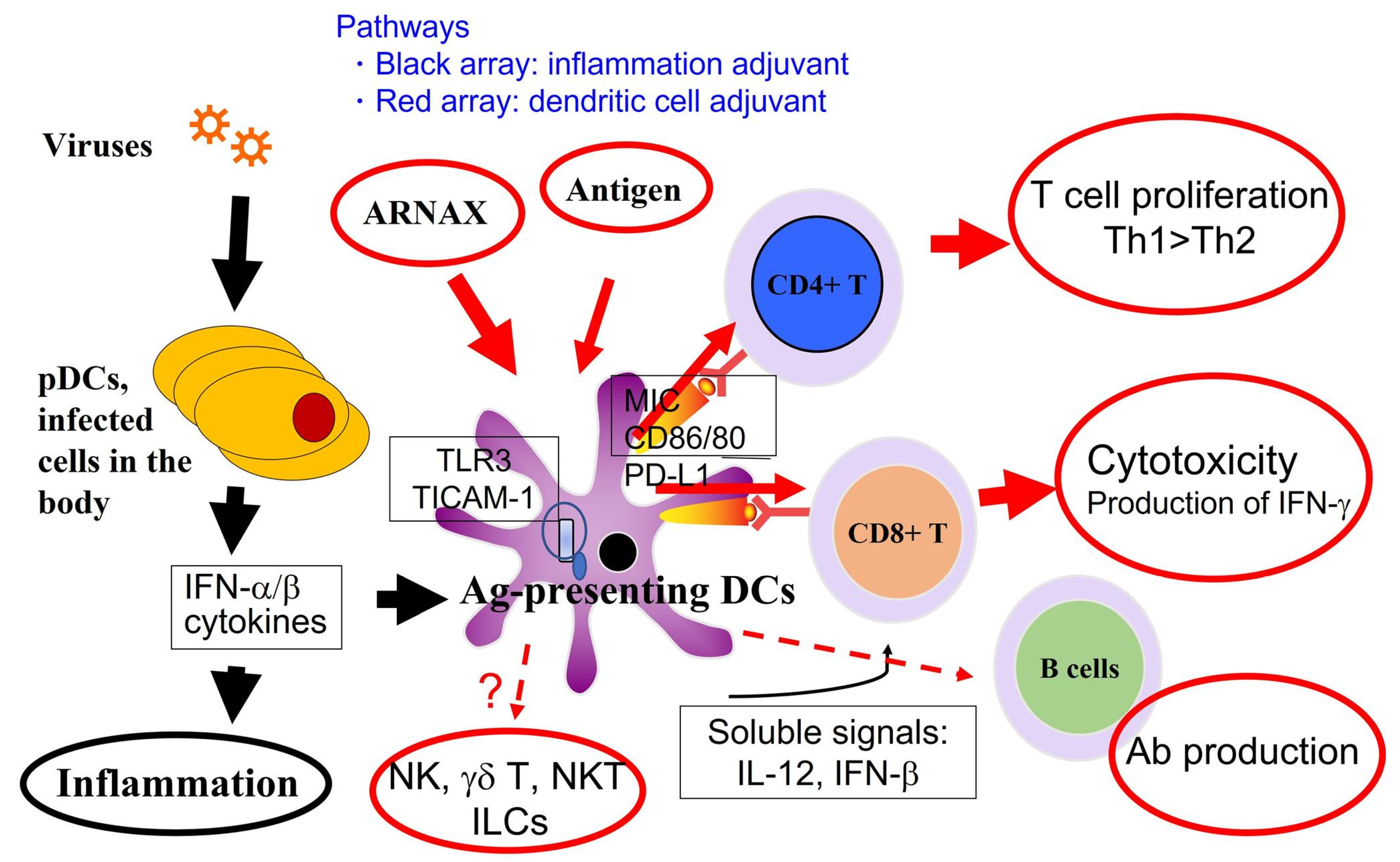
This figure depicts the outcome of ARNAX-dependent TLR3 activation in APCs (e.g., human CD141+ DCs). TLR3-specific activation by ARNAX does not increase cytokine/IFN levels in blood circulation. Cytokinemia is usually induced secondary to TLR stimulation of other infected cells in the body. Dendritic cells are activated either by adjuvant with DC-targeting or inflammation. Red arrays indicate the DC-targeting pathway, and black arrays the inflammatory pathway induced by viruses, targeting various cells in the body. In response to ARNAX, three representative immune cell activations are observed without inflammatory signs: Th1 polarization, CD8+ T cell activation, and Ab production. DC-targeting results in DC-mediated activation of NK cells, although they belong to innate lymphocytes. How ARNAX regulates other lymphocyte moieties, including γδT or NKT cells, remains elusive. The DC-targeting pathway is superior to the inflammatory pathway in that it elicits cellular immunity with fewer adverse events.
Another adjuvant called monophosphoryl lipid A (MPLA), which activates the TLR4-TICAM-1 pathway, has been approved as a low-toxicity adjuvant [71] and is currently applied in several vaccines (such as GSK AS02 and AS04) [72]. TLR4 ligands activate two adaptors (MyD88 and TICAM-1) that are differentially biased against the drug (Table 2); MPLA favors TICAM-1 over MyD88 [65,70]. TLR4 is expressed in mouse APCs but not in human APCs [57]. TLR3 and TLR 4 bind with different ligands but have in common the activation of TICAM-1. Both TLR3 and TLR4 agonists select for the TICAM-1 adaptor, making them suitable as adjuvants to minimize inflammation induction and promote antigen presentation (Figure 1 and Figure 2). TLR4 is not expressed in human APCs but is expressed in other dendritic cell subsets and macrophages [73]. TLR4, unlike the case of TLR3, requires TICAM-2 (TRAM) for activation of TICAM-1 [11,47,70].
No other examples of TLRs adopting TICAM-1 as an adaptor have been reported so far; STING may be regulated by TICAM-1 in a limited variety of cells [74,75]. TLR7 and TLR9 are expressed specifically in pDC and adopt MyD88 as an adaptor [55,76]. pDC is a potent inducer of type I interferon (IFN) and cytokines, inducing systemic inflammation, but it has a low capacity for cross-antigen presentation [56,77]. However, there are opposing views as to pDC antigen presentation capacity in several papers [77,78]. Cross-antigen presentation may occur under the activation of MyD88 in other dendritic cell subsets; TLRs that use MyD88 as an adaptor is not limited to APC expression [11], and safety issues, along with the side effect of systemic inflammation, are barriers and have not been approved under the Pharmaceutical Affairs Law, except TLR4. Thus, the TICAM-1 pathway would provide the conditions for a safe dendritic cell adjuvant.

7. Cross-Presentation Induced by the TICAM-1 Pathway in Dendritic Cells

TLR3 is highly expressed on antigen-presenting dendritic cells (CD11+ DCs) [57] and initiates the mechanism of foreign antigen presentation (called cross-antigen presentation) (Figure 1); TLR4 is also expressed on other CD1c positive dendritic cells and macrophages [75], in a process called dendritic cell maturation via the TICAM-1 pathway, conferring direct antigen-presenting capacity to dendritic cells.
TICAM-1 signaling activates downstream transcription factors NF-kB, IRF-3, and AP-1 to promote their nuclear translocation in immune cells [58,64,79]. In other cells, including tumor cells, TICAM-1 signaling activates the RIP-1/3 pathway and also promotes IRF-3 transcription and type I IFN induction [80,81,82]. TICAM-1 appears to induce less NF-κB activation than MyD88 [64,79]. Transcription factors of the AP-1 family are surmised to be strongly involved in cross-antigen presentation [83]. SEC61 is also reported to be downstream of IRF-3 and involved in cross-antigen presentation [84]. Tlr3˗/˗, Ticam1˗/˗, Irf3˗/˗, and Ifnar˗/˗ mice lose the ability to induce proliferation of CD8+ T lymphocytes in antigen presentation [4]. Positive feedback amplification of type I IFN by lymphocyte IFN-γ production and dendritic cell IFNAR is thought to promote cross-antigen presentation [85].
Compared to other RNA adjuvants, ARNAX is an adjuvant that specifically activates only TLR3 during cross-antigen presentation (Table 2). Any combination of antigens can constitute a vaccine; even if ARNAX were introduced into humans, trained immunity could be safely activated. A nontoxic trained immune response targeting CD141+ dendritic cells would trigger cross-antigen presentation in response to reduced antigen levels.

8. Adjuvants for Cancer Vaccines

It was first reported in 1990 that cancer cells express specific antigens [86]. Hundreds of tumor-associated antigens (TAAs) have since been identified [87,88]. This raised the question as to whether acquired immunity recognizes cancer-specific antigens as “nonself” and eliminates them during so-called, antitumor immunity. For more than 30 years, peptide vaccine therapy using cancer antigens has been investigated; however, clinical trials have been unsuccessful to date [89]. In 2012, it was demonstrated that cancer immunity plays a role in cancer treatment as a surveillance mechanism by inhibiting immune checkpoints, which is different from the strategy involved in vaccines [90]. Acquired immunity can successfully eliminate not only infectious agents but also cancerous cells.
One possible reason why cancer vaccines have been unsuccessful to date may be that peptide vaccine were administered without immune enhancers (adjuvants), unlike vaccines for infectious diseases. It is worth noting that 25% of cancers originate from infection, and an understanding of infectious disease and the use of adjuvants in the context of trained immunity may be crucial in the development of anticancer therapies [91].
Previously, peptide vaccine therapy for cancer has predominantly involved the subcutaneous administration of cancer antigens, e.g., 8–10 amino acids (CD8 epitope) or ~13 amino acids (CD4 epitope) [89]. A method of directly administering the antigens to patients and observing tumor regression was adopted as an efficacy marker. Two main problems were encountered: (1) dendritic cells do not present antigens simply by exposure to exogenously administered peptides [89], and (2) the adjuvant used was inadequate [92,93]. In many cases, the peptide was mixed with oil (montanide or squaran) to enhance immunity locally without stimulating cell proliferation. Initially, there were no approved adjuvants other than alum, which was contraindicated as a result of its potential to drive Th2 polarization [94,95,96], which promotes tumor growth [97]. Alum may activate NALP3 inflammasome to deteriorate tumor microenvironment (TME) [98,99,100]. In addition, the results of non-clinical tests using animal models were not directly applicable to human clinical trials because the antigens differed from those in humans. Furthermore, the administration of an antigen alone does not mimic pathogen infection, and, in the same way, a single antigen only cannot trigger anticancer immunity by T cells [53,54].
Many infections are cured following an immune response. If cancer immunity is to mimic the effects of infectious disease vaccines, the antigens (either mutant antigens or testis-specific antigens) must trigger immunity, particularly with the mutations or altered expression levels of the antigen, and epitopes must be targeted for MHC presentation despite individual differences.
Solid tumors present a tissue microenvironment that may be refractory to treatment [98,99], somewhat resembling cases of tuberculosis or DNA virus infections. Chemotherapy has been established as a superior treatment to immunotherapy in infectious diseases and is the preferred therapy being actively considered in anticancer therapy. It is difficult to define the functional profile of TME in each tumor that comprises variable rates of stromal cells and cancer cells [92,98,99]. The stroma contains environmental factors such as regulatory T cells (Treg), myeloid-derived suppressor cells, neutrophils, cancer-associated fibroblasts, and tolerogenic dendritic cells that control immune activation in a complex manner [92,98]. These immune-regulatory factors, as well as a cytokine environment induced by dead cells, may affect the properties of TME in individual tumors [98,99]. Inflammation in cancer is generally controlled by environmental factors, including TME [92,98,99]. Acquired immunity is affected by both activation and exhaustion and is also affected by the time axis for the construction of the TME.

9. Adjuvant in Prophylactic Vaccine against Infectious Diseases

Vaccines are one method of deliberately activating immunity to protect against infection. A typical vaccine consists of an antigen and an adjuvant, and immune enhancement usually results from the addition of an adjuvant. However, live vaccines, inactivated whole-particle vaccines, and mRNA vaccines are examples where endogenous adjuvants have ensured immune enhancement. In general, attenuated and inactivated viral and bacterial vaccines contain live adjuvants, the amount of which often cannot be adjusted, resulting in lot-to-lot variation in vaccines. This inevitably results in adverse reactions related to infection. Such side effects often result in candidate vaccines failing toxicity tests, and, therefore, not gaining approval. Many of the licensed routine and optional vaccines use alum as an adjuvant, purportedly from a conventional point of view. The advantages of alum in prophylactic vaccines are that it barely promotes serious fever and inflammation, and does not affect the impurity of the antigen preparation (including the PAMP components). However, despite its long-term use, the molecular response to alum has not been fully defined [95,96], making it impossible to clearly define and standardize adjuvant function [101,102]. Although alum has been used in many vaccinations without serious troubles, there are several counter-reports on vaccine applications. For example, alum in the human papillomavirus vaccine preventing uterine cervical cancer might participate in a chronic fatigue-like syndrome according to several reports [103,104].
Immune responses to vaccines vary from individual to individual and between infectious and mutant strains. Vaccines can also induce adverse events, such as an over-amplified immune response, which can cause damage to the host [105,106]. In general, vaccines require immune-boosting by the inclusion of safe adjuvants. It is essential to establish non-toxic and versatile adjuvants that can be used against a wide range of antigens in all patients. Next-generation vaccines that are superior to mRNA vaccines need to be supported by the development of adjuvants that meet safety standards.

10. Perspectives

Adjuvants include a wide range of compounds recognized by PRRs. TLR signals (with the exception of TLR3) converge onto MyD88 [11,70] and are classified as inflammatory adjuvants. Only TLR3 can induce selective activation of antigen-presenting dendritic cells through TICAM-1 alone [64,79] and is thereby considered a safe dendritic cell adjuvant. Thus, adjuvants can be categorized into inflammatory adjuvants and dendritic cell adjuvants by clear definitions, and the current categorization based on identified and unidentified receptors is merely a historical convenience [107]. Adjuvants should be defined based on their comprehensive analysis [108,109], molecular interaction and activation of the immune system, especially involving innate immune receptors (PRRs), and it is necessary to evaluate their functional identification, efficacy, and safety separately. It is molecularly understood that adjuvants command dendritic cells to activate the main response, acquired immunity, while the accompanied induction of excessive inflammation and cytokine storms is a separate side response.
PLGA-Riboxxim, a recently published dsRNA adjuvant, were created under a conceptualization similar to ARNAX. PLGA-Riboxxim carries a TLR3/RIG-I ligand, and targets DCs to potentially activate therapeutic immunity [110]. If the additional RIG-I activation works better for diseased patients, this type of immune-enhancing strategy may be advantageous to protect patients, particularly vulnerable older populations, from infection or cancer.
Our proposal is to enhance trained immunity with dendritic cell adjuvants such as the TLR3 agonist ARNAX to lower the antigen threshold for detecting latent infection and cancer and facilitate the creation of more sensitive and effective preventive vaccines. Guidelines and potential biomarkers for the development of next-generation adjuvants have not yet been provided [107]. Given that the host response to adjuvants reflects the natural response to foreign nucleic acids/proteins, pharmaceutical law needs to reasonably relax restrictions on adjuvant approval. Based on knowledge of the structural–functional relationship of MAMPs/PAMPs and modification of dendritic cell-targeted reagents, it is possible to create a new class of adjuvants based on the active artificial adjuvants with the removal of the inflammatory component—a forthcoming consideration for the development of ideal vaccines.

Author Contributions

Conceptualization, T.S., M.T. and M.M.; writing—original draft preparation, T.S.; writing—review and editing, T.S.; supervision, M.T. and M.M.; All authors have read and agreed to the published version of the manuscript.

Funding

This work was supported in part by the Japan Agency for Medical Research and Development (AMED) (Grant number: 19fk0108112h0201, 21ck0106480h0003).

Acknowledgments

Authors are grateful to the Aomori Yamada Gakuen Educational Corporation and the Institute of Advanced Immunotherapy (IAI) Co. Ltd. for supporting the Nebuta Research Institute at Aomori University. We also thank C. Yamazaki, the managing assistant at Aomori University. Helpful discussions by M. Shingai, T. Kawakita, and K. Taniguchi (Hokkaido University, Sapporo) are gratefully acknowledged.

Conflicts of Interest

The authors declare no conflict of interest.

References

  1. Del Giudice, G.; Rappuoli, R.; Didierlaurent, A.M. Correlates of adjuvanticity: A review on adjuvants in licensed vaccines. Semin Immunol. 2018, 39, 14–21. [Google Scholar] [CrossRef] [PubMed]
  2. Laupèze, B.; Hervé, C.; Di Pasquale, A.; Tavares Da Silva, F. Adjuvant Systems for vaccines: 13 years of post-licensure experience in diverse populations have progressed the way adjuvanted vaccine safety is investigated and understood. Vaccine 2019, 37, 5670–5680. [Google Scholar] [CrossRef] [PubMed]
  3. Matsumoto, M.; Tatematsu, M.; Nishikawa, F.; Azuma, M.; Ishii, N.; Morii-Sakai, A.; Shime, H.; Seya, T. Defined TLR3-specific adjuvant that induces NK and CTL activation without significant cytokine production in vivo. Nat. Commun. 2015, 18, 6280. [Google Scholar] [CrossRef] [PubMed] [Green Version]
  4. Takeda, Y.; Kataoka, K.; Yamagishi, J.; Ogawa, S.; Seya, T.; Matsumoto, M. A TLR3-Specific Adjuvant Relieves Innate Resistance to PD-L1 Blockade without Cytokine Toxicity in Tumor Vaccine Immunotherapy. Cell Rep. 2017, 19, 1874–1887. [Google Scholar] [CrossRef] [Green Version]
  5. Inaba, K.; Turley, S.; Iyoda, T.; Yamaide, F.; Shimoyama, S.; e Sousa, C.R.; Germain, R.N.; Mellman, I.; Steinman, R.M. The formation of immunogenic major histocompatibility complex class II-peptide ligands in lysosomal compartments of dendritic cells is regulated by inflammatory stimuli. J. Exp. Med. 2000, 191, 927–936. [Google Scholar] [CrossRef] [Green Version]
  6. Cho, K.J.; Roche, P.A. Regulation of MHC Class II-Peptide Complex Expression by Ubiquitination. Front. Immunol. 2013, 4, 369. [Google Scholar] [CrossRef] [Green Version]
  7. Wu, J.; Meng, Z.; Jiang, M.; Zhang, E.; Trippler, M.; Broering, R.; Bucchi, A.; Krux, F.; Dittmer, U.; Yang, D.; et al. Toll-like receptor-induced innate immune responses in non-parenchymal liver cells are cell type-specific. Immunology 2010, 129, 363–374. [Google Scholar] [CrossRef]
  8. Takaki, H.; Ichimiya, S.; Matsumoto, M.; Seya, T. Mucosal Immune Response in Nasal-Associated Lymphoid Tissue upon Intranasal Administration by Adjuvants. J. Innate Immun. 2018, 10, 515–521. [Google Scholar] [CrossRef]
  9. Schenten, D.; Medzhitov, R. The control of adaptive immune responses by the innate immune system. Adv. Immunol. 2011, 109, 87–124. [Google Scholar] [CrossRef] [PubMed]
  10. Oshiumi, H.; Matsuo, A.; Matsumoto, M.; Seya, T. Pan-vertebrate toll-like receptors during evolution. Curr. Genom. 2008, 9, 488–493. [Google Scholar] [CrossRef]
  11. Kawai, T.; Akira, S. The roles of TLRs, RLRs and NLRs in pathogen recognition. Int. Immunol. 2009, 21, 317–337. [Google Scholar] [CrossRef] [PubMed] [Green Version]
  12. Zhou, F.; Emonet, A.; Dénervaud Tendon, V.; Marhavy, P.; Wu, D.; Lahaye, T.; Geldner, N. Co-incidence of Damage and Microbial Patterns Controls Localized Immune Responses in Roots. Cell 2020, 180, 440–453.e18. [Google Scholar] [CrossRef] [PubMed] [Green Version]
  13. Godoy-Tena, G.; Ballestar, E. Epigenetics of Dendritic Cells in Tumor Immunology. Cancers 2022, 14, 1179. [Google Scholar] [CrossRef] [PubMed]
  14. Papalexi, E.; Satija, R. Single-cell RNA sequencing to explore immune cell heterogeneity. Nat. Rev. Immunol. 2018, 18, 35–45. [Google Scholar] [CrossRef] [PubMed]
  15. Harper, D.R. Viruses: Biology, Application and Control; Garland Press: New York, NY, USA, 2012; pp. 65–130. [Google Scholar]
  16. Pancer, Z.; Amemiya, C.T.; Ehrhardt, G.R.; Ceitlin, J.; Gartland, G.L.; Cooper, M.D. Somatic diversification of variable lymphocyte receptors in the agnathan sea lamprey. Nature 2004, 430, 174–180. [Google Scholar] [CrossRef] [Green Version]
  17. Teixeira, S.M.; El-Sayed, N.M.; Araújo, P.R. The genome and its implications. Adv. Parasitol. 2011, 75, 209–230. [Google Scholar] [CrossRef]
  18. Fitzgerald-Bocarsly, P.; Feng, D. The role of type I interferon production by dendritic cells in host defense. Biochimie 2007, 89, 843–855. [Google Scholar] [CrossRef] [Green Version]
  19. Seya, T.; Matsumoto, M.; Ebihara, T.; Oshiumi, H. Functional evolution of the TICAM-1 pathway for extrinsic RNA sensing. Immunol. Rev. 2009, 227, 44–53. [Google Scholar] [CrossRef]
  20. Greenwald, R.J.; Freeman, G.J.; Sharpe, A.H. The B7 family revisited. Annu. Rev. Immunol. 2005, 23, 515–548. [Google Scholar] [CrossRef]
  21. Burnet, F.M. The concept of immunological surveillance. Prog. Exp. Tumor Res. 1970, 13, 1–27. [Google Scholar] [CrossRef] [PubMed]
  22. Gupta, S.L.; Khan, N.; Basu, S.; Soni, V. B-Cell-Based Immunotherapy: A Promising New Alternative. Vaccines 2022, 10, 879. [Google Scholar] [CrossRef] [PubMed]
  23. Takaki, H.; Kure, S.; Oshiumi, H.; Sakoda, Y.; Suzuki, T.; Ainai, A.; Hasegawa, H.; Matsumoto, M.; Seya, T. Toll-like receptor 3 in nasal CD103+ dendritic cells is involved in immunoglobulin A production. Mucosal Immunol. 2018, 11, 82–96. [Google Scholar] [CrossRef] [PubMed] [Green Version]
  24. Muramatsu, M.; Kinoshita, K.; Fagarasan, S.; Yamada, S.; Shinkai, Y.; Honjo, T. Class switch recombination and hypermutation require activation-induced cytidine deaminase (AID), a potential RNA editing enzyme. Cell 2000, 102, 553–563. [Google Scholar] [CrossRef] [Green Version]
  25. Monroy-Iglesias, M.J.; Crescioli, S.; Beckmann, K.; Le, N.; Karagiannis, S.N.; Van Hemelrijck, M.; Santaolalla, A. Antibodies as biomarkers for cancer risk: A systematic review. Clin. Exp. Immunol. 2022, 209, 46–63. [Google Scholar] [CrossRef] [PubMed]
  26. Ebihara, T.; Azuma, M.; Oshiumi, H.; Kasamatsu, J.; Iwabuchi, K.; Matsumoto, K.; Saito, H.; Taniguchi, T.; Matsumoto, M.; Seya, T. Identification of a polyI:C-inducible membrane protein that participates in dendritic cell-mediated natural killer cell activation. J. Exp. Med. 2015, 212, 1337. [Google Scholar] [CrossRef] [PubMed] [Green Version]
  27. Hamerman, J.A.; Ogasawara, K.; Lanier, L.L. Cutting edge: Toll-like receptor signaling in macrophages induces ligands for the NKG2D receptor. J. Immunol. 2004, 172, 2001–2005. [Google Scholar] [CrossRef] [Green Version]
  28. Ebihara, T. Dichotomous Regulation of Acquired Immunity by Innate Lymphoid Cells. Cells 2020, 9, 1193. [Google Scholar] [CrossRef]
  29. Van Acker, H.H.; Campillo-Davo, D.; Roex, G.; Versteven, M.; Smits, E.L.; Van Tendeloo, V.F. The role of the common gamma-chain family cytokines in γδ T cell-based anti-cancer immunotherapy. Cytokine Growth Factor Rev. 2018, 41, 54–64. [Google Scholar] [CrossRef]
  30. Gustafsson, K.; Herrmann, T.; Dieli, F. Editorial: Understanding Gamma Delta T Cell Multifunctionality-Towards Immunotherapeutic Applications. Front. Immunol. 2020, 11, 921. [Google Scholar] [CrossRef]
  31. Yamazaki, S.; Inaba, K.; Tarbell, K.V.; Steinman, R.M. Dendritic cells expand antigen-specific Foxp3+ CD25+ CD4+ regulatory T cells including suppressors of alloreactivity. Immunol. Rev. 2006, 212, 314–329. [Google Scholar] [CrossRef]
  32. Dolfi, D.V.; Duttagupta, P.A.; Boesteanu, A.C.; Mueller, Y.M.; Oliai, C.H.; Borowski, A.B.; Katsikis, P.D. Dendritic cells and CD28 costimulation are required to sustain virus-specific CD8+ T cell responses during the effector phase in vivo. J. Immunol. 2011, 186, 4599–4608. [Google Scholar] [CrossRef] [PubMed] [Green Version]
  33. Singh, A.P.; Zheng, X.; Lin-Schmidt, X.; Chen, W.; Carpenter, T.J.; Zong, A.; Wang, W.; Heald, D.L. Development of a quantitative relationship between CAR-affinity, antigen abundance, tumor cell depletion and CAR-T cell expansion using a multiscale systems PK-PD model. MAbs 2020, 12, 1688616. [Google Scholar] [CrossRef] [PubMed] [Green Version]
  34. Ghiotto-Ragueneau, M.; Battifora, M.; Truneh, A.; Waterfield, M.D.; Olive, D. Comparison of CD28-B7.1 and B7.2 functional interaction in resting human T cells: Phosphatidylinositol 3-kinase association to CD28 and cytokine production. Eur. J. Immunol. 1996, 26, 34–41. [Google Scholar] [CrossRef] [PubMed]
  35. Carreno, B.M.; Collins, M. The B7 family of ligands and its receptors: New pathways for costimulation and inhibition of immune responses. Annu. Rev. Immunol. 2002, 20, 29–53. [Google Scholar] [CrossRef]
  36. Ward, J.P.; Gubin, M.M.; Schreiber, R.D. The Role of Neoantigens in Naturally Occurring and Therapeutically Induced Immune Responses to Cancer. Adv. Immunol. 2016, 130, 25–74. [Google Scholar] [CrossRef] [PubMed]
  37. Gartlan, C.; Tipton, T.; Salguero, F.J.; Sattentau, Q.; Gorringe, A.; Carroll, M.W. Vaccine-Associated Enhanced Disease and Pathogenic Human Coronaviruses. Front. Immunol. 2022, 13, 882972. [Google Scholar] [CrossRef]
  38. Iwata-Yoshikawa, N.; Nagata, N.; Takaki, H.; Matsumoto, M.; Suzuki, T.; Hasegawa, H.; Seya, T. Prophylactic Vaccine Targeting TLR3 on Dendritic Cells Ameliorates Eosinophilic Pneumonia in a Mouse SARS-CoV Infection Model. Immunohorizons 2022, 6, 275–282. [Google Scholar] [CrossRef]
  39. Iwata-Yoshikawa, N.; Shiwa, N.; Sekizuka, T.; Sano, K.; Ainai, A.; Hemmi, T.; Kataoka, M.; Kuroda, M.; Hasegawa, H.; Suzuki, T.; et al. A lethal mouse model for evaluating vaccine-associated enhanced respiratory disease during SARS-CoV-2 infection. Sci. Adv. 2022, 8, eabh3827. [Google Scholar] [CrossRef]
  40. Ita, K. Coronavirus Disease (COVID-19): Current Status and Prospects for Drug and Vaccine Development. Arch. Med. Res. 2021, 52, 15–24. [Google Scholar] [CrossRef]
  41. Ascough, S.; Paterson, S.; Chiu, C. Induction and subversionof human protective immunity: Contrasting influenza and respiratory syncytial virus. Front. Immunol. 2018, 9, 323. [Google Scholar] [CrossRef]
  42. Iwata-Yoshikawa, N.; Uda, A.; Suzuki, T.; Tsunetsugu-Yokota, Y.; Sato, Y.; Morikawa, S.; Tashiro, M.; Sata, T.; Hasegawa, H.; Nagata, N. Effects of Toll-like receptor stimulation on eosinophilic infiltration in lungs of BALB/c mice immunized with UV-inactivated severe acute respiratory syndrome-related coronavirus vaccine. J. Virol. 2014, 88, 8597–8614. [Google Scholar] [CrossRef] [PubMed] [Green Version]
  43. Shingai, M.; Inoue, N.; Okuno, T.; Okabe, M.; Akazawa, T.; Miyamoto, Y.; Ayata, M.; Honda, K.; Kurita-Taniguchi, M.; Matsumoto, M.; et al. Wild-type measles virus infection in human CD46/CD150-transgenic mice: CD11c-positive dendritic cells establish systemic viral infection. J. Immunol. 2005, 175, 3252–3261. [Google Scholar] [CrossRef] [PubMed] [Green Version]
  44. Takaki, H.; Takeda, M.; Tahara, M.; Shingai, M.; Oshiumi, H.; Matsumoto, M.; Seya, T. The MyD88 pathway in plasmacytoid and CD4+ dendritic cells primarily triggers type I IFN production against measles virus in a mouse infection model. J. Immunol. 2013, 191, 4740–4747. [Google Scholar] [CrossRef] [Green Version]
  45. Joffre, O.P.; Segura, E.; Savina, A.; Amigorena, S. Cross-presentation by dendritic cells. Nat. Rev. Immunol. 2012, 12, 557–569. [Google Scholar] [CrossRef] [PubMed]
  46. Ho, N.I.; Huis In ’t Veld, L.G.M.; Raaijmakers, T.K.; Adema, G.J. Adjuvants Enhancing Cross-Presentation by Dendritic Cells: The Key to More Effective Vaccines? Front. Immunol. 2018, 9, 2874. [Google Scholar] [CrossRef]
  47. Seya, T.; Akazawa, T.; Uehori, J.; Matsumoto, M.; Azuma, I.; Toyoshima, K. Role of toll-like receptors and their adaptors in adjuvant immunotherapy for cancer. Anticancer Res. 2003, 23, 4369–4376. [Google Scholar]
  48. Coban, C.; Koyama, S.; Takeshita, F.; Akira, S.; Ishii, K.J. Molecular and cellular mechanisms of DNA vaccines. Hum. Vaccin. 2008, 4, 453–456. [Google Scholar] [CrossRef]
  49. Seya, T.; Takeda, Y.; Takashima, K.; Yoshida, S.; Azuma, M.; Matsumoto, M. Adjuvant immunotherapy for cancer: Both dendritic cell-priming and check-point inhibitor blockade are required for immunotherapy. Proc. Jpn. Acad. Ser. B Phys. Biol. Sci. 2018, 94, 153–160. [Google Scholar] [CrossRef] [Green Version]
  50. Le Naour, J.; Galluzzi, L.; Zitvogel, L.; Kroemer, G.; Vacchelli, E. Trial watch: TLR3 agonists in cancer therapy. Oncoimmunology 2020, 9, 1771143. [Google Scholar] [CrossRef]
  51. Brubaker, S.W.; Bonham, K.S.; Zanoni, I.; Kagan, J.C. Innate immune pattern recognition: A cell biological perspective. Annu. Rev. Immunol. 2015, 33, 257–290. [Google Scholar] [CrossRef] [Green Version]
  52. Covián, C.; Fernández-Fierro, A.; Retamal-Díaz, A.; Díaz, F.E.; Vasquez, A.E.; Lay, M.K.; Riedel, C.A.; González, P.A.; Bueno, S.M.; Kalergis, A.M. BCG-Induced Cross-Protection and Development of Trained Immunity: Implication for Vaccine Design. Front. Immunol. 2019, 10, 2806. [Google Scholar] [CrossRef] [PubMed]
  53. Matsumoto, M.; Takeda, Y.; Tatematsu, M.; Seya, T. Toll-Like Receptor 3 Signal in Dendritic Cells Benefits Cancer Immunotherapy. Front. Immunol. 2017, 8, 1897. [Google Scholar] [CrossRef] [PubMed] [Green Version]
  54. Matsumoto, M.; Takeda, Y.; Seya, T. Targeting Toll-like receptor 3 in dendritic cells for cancer immunotherapy. Expert Opin. Biol. Ther. 2020, 20, 937–946. [Google Scholar] [CrossRef]
  55. Gilliet, M.; Cao, W.; Liu, Y.J. Plasmacytoid dendritic cells: Sensing nucleic acids in viral infection and autoimmune diseases. Nat. Rev. Immunol. 2008, 8, 594–606. [Google Scholar] [CrossRef] [PubMed]
  56. Grouard, G.; Rissoan, M.-C.; Filgueira, L.; Durand, I.; Banchereau, J.; Liu, Y.-J. The enigmatic plasmacytoid T cells develop into dendritic cells with Interleukin 3 and CD40-ligand. J. Exp. Med. 1997, 185, 1101–1111. [Google Scholar] [CrossRef] [PubMed] [Green Version]
  57. Jongbloed, S.L.; Kassianos, A.J.; McDonald, K.J.; Clark, G.J.; Ju, X.; Angel, C.E.; Chen, C.J.; Dunbar, P.R.; Wadley, R.B.; Jeet, V.; et al. Human CD141+ (BDCA-3)+ dendritic cells (DCs) represent a unique myeloid DC subset that cross-presents necrotic cell antigens. J. Exp. Med. 2010, 207, 1247–1260. [Google Scholar] [CrossRef] [Green Version]
  58. Matsumoto, M.; Seya, T. TLR3: Interferon induction by double-stranded RNA including poly(I:C). Adv. Drug Deliv. Rev. 2008, 60, 805–812. [Google Scholar] [CrossRef]
  59. Matsumoto, M.; Oshiumi, H.; Seya, T. Antiviral responses induced by the TLR3 pathway. Rev. Med. Virol. 2011, 21, 67–77. [Google Scholar] [CrossRef]
  60. Trinchieri, G. Interleukin-12 and the regulation of innate resistance and adaptive immunity. Nat. Rev. Immunol. 2003, 3, 133–146. [Google Scholar] [CrossRef]
  61. Segura, E. Review of Mouse and Human Dendritic Cell Subsets. Methods Mol. Biol. 2016, 1423, 3–15. [Google Scholar] [CrossRef]
  62. Poulin, L.F.; Salio, M.; Griessinger, E.; Anjos-Afonso, F.; Craciun, L.; Chen, J.L.; Keller, A.M.; Joffre, O.; Zelenay, S.; Nye, E.; et al. Characterization of human DNGR-1+ BDCA3+ leukocytes as putative equivalents of mouse CD8alpha+ dendritic cells. J. Exp. Med. 2010, 207, 1261–1271. [Google Scholar] [CrossRef] [PubMed] [Green Version]
  63. Desch, A.N.; Randolph, G.J.; Murphy, K.; Gautier, E.L.; Kedl, R.M.; Lahoud, M.H.; Caminschi, I.; Shortman, K.; Henson, P.M.; Jakubzick, C.V. CD103+ pulmonary dendritic cells preferentially acquire and present apoptotic cell-associated antigen. J. Exp. Med. 2011, 208, 1789–1797. [Google Scholar] [CrossRef] [PubMed] [Green Version]
  64. Oshiumi, H.; Matsumoto, M.; Funami, K.; Akazawa, T.; Seya, T. TICAM-1, an adaptor molecule that participates in Toll-like receptor 3-mediated interferon-beta induction. Nat. Immunol. 2003, 4, 161–167. [Google Scholar] [CrossRef] [PubMed]
  65. Seya, T.; Takeda, Y.; Matsumoto, M. A Toll-like receptor 3 (TLR3) agonist ARNAX for therapeutic immunotherapy. Adv. Drug Deliv. Rev. 2019, 147, 37–43. [Google Scholar] [CrossRef] [PubMed]
  66. Matsumoto, M.; Kikkawa, S.; Kohase, M.; Miyake, K.; Seya, T. Establishment of a monoclonal antibody against human Toll-like receptor 3 that blocks double-stranded RNA-mediated signaling. Biochem. Biophys. Res. Commun. 2002, 293, 1364–1369. [Google Scholar] [CrossRef] [PubMed]
  67. Matsumoto, M.; Funami, K.; Tanabe, M.; Oshiumi, H.; Shingai, M.; Seto, Y.; Yamamoto, A.; Seya, T. Subcellular localization of Toll-like receptor 3 in human dendritic cells. J. Immunol. 2003, 171, 3154–3162. [Google Scholar] [CrossRef] [Green Version]
  68. Heidegger, S.; Kreppel, D.; Bscheider, M.; Stritzke, F.; Nedelko, T.; Wintges, A.; Bek, S.; Fischer, J.C.; Graalmann, T.; Kalinke, U.; et al. RIG-I activating immunostimulatory RNA boosts the efficacy of anticancer vaccines and synergizes with immune checkpoint blockade. EBioMedicine 2019, 41, 146–155. [Google Scholar] [CrossRef] [Green Version]
  69. Ruzicka, M.; Koenig, L.M.; Formisano, S.; Boehmer, D.F.R.; Vick, B.; Heuer, E.M.; Meinl, H.; Kocheise, L.; Zeitlhöfler, M.; Ahlfeld, J.; et al. RIG-I-based immunotherapy enhances survival in preclinical AML models and sensitizes AML cells to checkpoint blockade. Leukemia 2020, 34, 1017–1026. [Google Scholar] [CrossRef] [Green Version]
  70. Akira, S. Toll-like receptor signaling. J. Biol. Chem. 2003, 278, 38105–38108. [Google Scholar] [CrossRef] [Green Version]
  71. Mata-Haro, V.; Cekic, C.; Martin, M.; Chilton, P.M.; Casella, C.R.; Mitchell, T.C. The vaccine adjuvant monophosphoryl lipid A as a TRIF-biased agonist of TLR4. Science 2007, 316, 1628–1632. [Google Scholar] [CrossRef]
  72. Garcon, N.; Chomez, P.; van Mechelen, M. Glaxo- SmithKline Adjuvant Systems in vaccines: Concepts, achievements and perspectives. Expert Rev. Vaccines 2007, 6, 723–739. [Google Scholar] [CrossRef] [PubMed]
  73. Kassianos, A.J.; Hardy, M.Y.; Ju, X.; Vijayan, D.; Ding, Y.; Vulink, A.J.; McDonald, K.J.; Jongbloed, S.L.; Wadley, R.B.; Wells, C.; et al. Human CD1c (BDCA-1)+ myeloid dendritic cells secrete IL-10 and display an immuno-regulatory phenotype and function in response to Escherichia coli. Eur. J. Immunol. 2012, 42, 1512–1522. [Google Scholar] [CrossRef] [PubMed]
  74. Takashima, K.; Oshiumi, H.; Matsumoto, M.; Seya, T. TICAM-1 is dispensable in STING-mediated innate immune responses in myeloid immune cells. Biochem. Biophys. Res. Commun. 2018, 499, 985–991. [Google Scholar] [CrossRef] [PubMed]
  75. Wang, X.; Majumdar, T.; Kessler, P.; Ozhegov, E.; Zhang, Y.; Chattopadhyay, S.; Barik, S.; Sen, G.C. STING Requires the Adaptor TRIF to Trigger Innate Immune Responses to Microbial Infection. Cell Host Microbe. 2016, 20, 329–341. [Google Scholar] [CrossRef] [PubMed] [Green Version]
  76. Kader, M.; Smith, A.P.; Guiducci, C.; Wonderlich, E.R.; Normolle, D.; Watkins, S.C.; Barrat, F.J.; Barratt-Boyes, S.M. Blocking TLR7- and TLR9-mediated IFN-α production by plasmacytoid dendritic cells does not diminish immune activation in early SIV infection. PLoS Pathog. 2013, 9, e1003530. [Google Scholar] [CrossRef] [Green Version]
  77. Hoeffel, G.; Ripoche, A.C.; Matheoud, D.; Nascimbeni, M.; Escriou, N.; Lebon, P.; Heshmati, F.; Guillet, J.G.; Gannagé, M.; Caillat-Zucman, S.; et al. Antigen crosspresentation by human plasmacytoid dendritic cells. Immunity 2007, 27, 481–492. [Google Scholar] [CrossRef]
  78. Lee, W.; Suresh, M. Vaccine adjuvants to engage the cross-presentation pathway. Front. Immunol. 2022, 13, 940047. [Google Scholar] [CrossRef]
  79. Yamamoto, M.; Sato, S.; Hemmi, H.; Hoshino, K.; Kaisho, T.; Sanjo, H.; Takeuchi, O.; Sugiyama, M.; Okabe, M.; Takeda, K.; et al. Role of adaptor TRIF in the MyD88-independent toll-like receptor signaling pathway. Science 2003, 301, 640–643. [Google Scholar] [CrossRef] [PubMed]
  80. Seya, T.; Shime, H.; Takaki, H.; Azuma, M.; Oshiumi, H.; Matsumoto, M. TLR3/TICAM-1 signaling in tumor cell RIP3-dependent necroptosis. Oncoimmunology 2012, 1, 917–923. [Google Scholar] [CrossRef] [Green Version]
  81. Hanson, B. Necroptosis: A new way of dying? Cancer Biol. Ther. 2016, 17, 899–910. [Google Scholar] [CrossRef] [Green Version]
  82. Yoneyama, M.; Suhara, W.; Fukuhara, Y.; Fukuda, M.; Nishida, E.; Fujita, T. Direct triggering of the type I interferon system by virus infection: Activation of a transcription factor complex containing IRF-3 and CBP/p300. EMBO J. 1998, 17, 1087–1095. [Google Scholar] [CrossRef]
  83. Murphy, T.L.; Tussiwand, R.; Murphy, K.M. Specificity through cooperation: BATF-IRF interactions control immune-regulatory networks. Nat. Rev. Immunol. 2013, 13, 499–509. [Google Scholar] [CrossRef] [PubMed]
  84. Rojas, J.J.; Sampath, P.; Bonilla, B.; Ashley, A.; Hou, W.; Byrd, D.; Thorne, S.H. Manipulating TLR Signaling Increases the Anti-tumor T Cell Response Induced by Viral Cancer Therapies. Cell Rep. 2016, 15, 264–273. [Google Scholar] [CrossRef] [PubMed] [Green Version]
  85. Honda, K.; Taniguchi, T. Toll-like receptor signaling and IRF transcription factors. IUBMB Life 2006, 58, 290–295. [Google Scholar] [CrossRef] [PubMed]
  86. van der Bruggen, P.; Traversari, C.; Chomez, P.; Lurquin, C.; De Plaen, E.; Van den Eynde, B.; Knuth, A.; Boon, T. A gene encoding an antigen recognized by cytolytic T lymphocytes on a human melanoma. Science 1991, 254, 1643–1647. [Google Scholar] [CrossRef] [PubMed]
  87. Uenaka, A.; Ono, T.; Akisawa, T.; Wada, H.; Yasuda, T.; Nakayama, E. Identification of a unique antigen peptide pRL1 on BALB/c RL male 1 leukemia recognized by cytotoxic T lymphocytes and its relation to the Akt oncogene. J. Exp. Med. 1994, 180, 1599–1607. [Google Scholar] [CrossRef] [Green Version]
  88. Boon, T. Tumor antigens recognized by cytolytic T lymphocytes: Present perspectives for specific immunotherapy. Int. J. Cancer 1993, 54, 177–180. [Google Scholar] [CrossRef]
  89. Rosenberg, S.A.; Yang, J.C.; Restifo, N.P. Cancer immunotherapy: Moving beyond current vaccines. Nat. Med. 2004, 10, 909–915. [Google Scholar] [CrossRef] [Green Version]
  90. Topalian, S.L.; Hodi, F.S.; Brahmer, J.R.; Gettinger, S.N.; Smith, D.C.; McDermot, D.F. Safety, activity, and immune correlates of anti-PD-1 antibody in cancer. N. Engl. J. Med. 2012, 366, 2443–2454. [Google Scholar] [CrossRef] [Green Version]
  91. Mestrallet, G.; Sone, K.; Bhardwaj, N. Strategies to overcome DC dysregulation in the tumor microenvironment. Front. Immunol. 2022, 13, 980709. [Google Scholar] [CrossRef]
  92. Seya, T.; Shime, H.; Matsumoto, M. Functional Alteration of Tumor-infiltrating Myeloid Cells in RNA Adjuvant Therapy. Anticancer Res. 2015, 35, 4385–4392. [Google Scholar] [PubMed]
  93. Tatematsu, M.; Funami, K.; Seya, T.; Matsumoto, M. Extracellular RNA Sensing by Pattern Recognition Receptors. J. Innate Immun. 2018, 10, 398–406. [Google Scholar] [CrossRef] [PubMed]
  94. Nishimura, T.; Iwakabe, K.; Sekimoto, M.; Ohmi, Y.; Yahata, T.; Nakui, M.; Sato, T.; Habu, S.; Tashiro, H.; Sato, M.; et al. Distinct role of antigen-specific T helper type 1 (Th1) and Th2 cells in tumor eradication in vivo. J. Exp. Med. 1999, 190, 617–627. [Google Scholar] [CrossRef]
  95. Franchi, L.; Nunez, G. The NALP2 inflammasome is critical for Alum-mediated IL-1b secretion but dispensable for adjuvant activity. Eur. J. Immunol. 2008, 38, 2085–2089. [Google Scholar] [CrossRef] [PubMed] [Green Version]
  96. Brewer, J.M.; Conacer, M.; Hunter, C.A.; Mohrs, M.; Brombacher, F.; Alexander, J. Aluminium hydroxide adjuvant initiates strong antigen-specific Th2 responses in the absence of IL-4- or IL-13-mediated signaling. J. Immunol. 1999, 163, 6448–6454. [Google Scholar] [PubMed]
  97. Nishimura, T.; Nakui, M.; Sato, M.; Iwakabe, K.; Kitamura, H.; Sekimoto, M.; Ohta, A.; Koda, T.; Nishimura, S. The critical role of Th1-dominant immunity in tumor immunology. Cancer Chemother Pharmacol. 2000, 46, S52–S61. [Google Scholar] [CrossRef] [PubMed]
  98. Denton, A.E.; Roberts, E.W.; Fearon, D.T. Stromal Cells in the Tumor Microenvironment. Adv. Exp. Med. Biol. 2018, 1060, 99–114. [Google Scholar] [CrossRef]
  99. Pitt, J.M.; Marabelle, A.; Eggermont, A.; Soria, J.C.; Kroemer, G.; Zitvogel, L. Targeting the tumor microenvironment: Removing obstruction to anticancer immune responses and immunotherapy. Ann. Oncol. 2016, 27, 1482–1492. [Google Scholar] [CrossRef]
  100. Rock, K.L.; Kono, H. The inflammatory response to cell death. Annu. Rev. Pathol. 2008, 3, 99–126. [Google Scholar] [CrossRef]
  101. Pulendran, B.; Arunachalam, P.S.; O’Hagan, D.T. Emerging concepts in the science of vaccine adjuvants. Nat. Rev. Drug Discov. 2021, 20, 454–475. [Google Scholar] [CrossRef]
  102. Brito, L.A.; Malyala, P.; O’Hagan, D.T. Vaccine adjuvant formulations: A pharmaceutical perspective. Semin. Immunol. 2013, 25, 130–145. [Google Scholar] [CrossRef] [PubMed]
  103. Souayah, N.; Michas-Martin, P.A.; Nasar, A.; Krivitskaya, N.; Yacoub, H.A.; Khan, H.; Qureshi, A.I. Guillain–Barré syndrome after Gardasil vaccination: Data from Vaccine Adverse Event Reporting System 2006–2009. Vaccine 2011, 29, 886–889. [Google Scholar] [CrossRef] [PubMed]
  104. Kinoshita, T.; Abe, R.T.; Hineno, A.; Tsunekawa, K.; Nakane, S.; Ikeda, S. Peripheral sympathetic nerve dysfunction in adolescent Japanese girls following immunization with the human papillomavirus vaccine. Intern. Med. 2014, 53, 2185–2200. [Google Scholar] [CrossRef] [Green Version]
  105. Guimarães, L.E.; Baker, B.; Perricone, C.; Shoenfeld, Y. Vaccines, adjuvants and autoimmunity. Pharmacol. Res. 2015, 100, 190–209. [Google Scholar] [CrossRef] [PubMed]
  106. Geoghegan, S.; O’Callaghan, K.P.; Offit, P.A. Vaccine Safety: Myths and Misinformation. Front. Microbiol. 2020, 11, 372. [Google Scholar] [CrossRef] [Green Version]
  107. Guidance for Cancer Immuno-Therapy in Japanese. Available online: https://www.mhlw.go.jp/web/t_doc?dataId=00tc3979&dataType=1&pageNo=1 (accessed on 10 November 2022).
  108. Jain, A.; Mittal, S.; Tripathi, L.P.; Nussinov, R.; Ahmad, S. Host-pathogen protein-nucleic acid interactions: A comprehensive review. Comp. Struc. Biotechnol. J. 2022, 20, 4415–4436. [Google Scholar] [CrossRef]
  109. Rostamizadeh, L.; Molavi, O.; Rashid, M.; Ramazani, F.; Baradaran, B.; Lavasanaifar, A.; Lai, R. Recent advances in cancer immunotherapy: Modulation of tumor microenvironment by Toll-like receptor ligands. BioImpact 2022, 12, 261–290. Available online: http://bi.tbzmed.ac.ir/ (accessed on 2 December 2022). [CrossRef]
  110. Koerner, J.; Horvath, D.; Herrmann, V.L.; MacKerracher, A.; Gander, B.; Yagita, H.; Rohayem, J.; Groettrup, M. PLGA-particle vaccine carrying TLR3/RIG-I ligand Riboxxim synergizes with immune checkpoint blockade for effective anti-cancer immunotherapy. Nat. Commun. 2021, 12, 2935. [Google Scholar] [CrossRef]
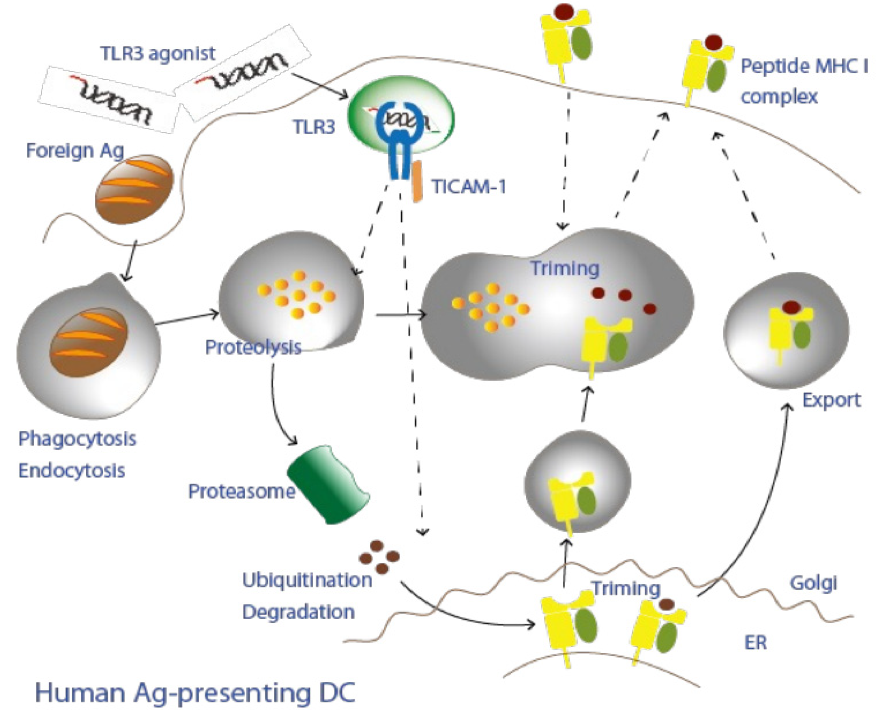
Figure 1. A putative model for antigen presentation in CD141+ dendritic cells.
Cells 11 04006 g001
Figure 2. Acquired immune response induced by TLR3/TICAM-1-mediated dendritic cell activation.
Cells 11 04006 g002
Table 1. Properties of antigen presentation.
PresentationClass IClass IICross-Present
Origin of the peptideIntracellularExtracellularVacuolar/Cytosolic
LocalizationProteasomeEndosomePhagosome
Expressing cellsAll host cellsImmune cellsDCs
Invariant chainNoYesNo
T cell binding
Response to ARNAX
CD8 in CTL
Unknown
CD4 in helper T
Unknown
CD8 in CTL
Up-regulated
Table 2. Properties of the TICAM-1 adjuvants.
LigandReceptorAdapterTarget CellsOutcome
Mouse DCHuman DCSkewingCross-PresentCytokine Storm
ARNAXTLR3TICAM-1CD8α DC
CD103 DC
CD141 DCTh1+++-
AmpligenTLR3/MDA5TICAM-1 > MAVSNot restricted Th1++(?) *+(?) *
PolyI:CMDA5/TLR3TICAM-1 < MAVSNot restricted Th1++++++
MPLATLR4TICAM-1 > MyD88CD8a DC
CD103 DC
CD1c DCTh1++++
* (?) No concrete data have been reported or available.
Publisher’s Note: MDPI stays neutral with regard to jurisdictional claims in published maps and institutional affiliations.