Review

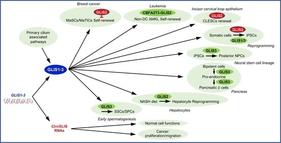
GLIS1-3: Links to Primary Cilium, Reprogramming, Stem Cell Renewal, and Disease

Cell Biology Section, Immunity, Inflammation and Disease Laboratory, National Institute of Environmental Health Sciences, National Institutes of Health, Research Triangle Park, Durham, NC 27709, USA
*
Author to whom correspondence should be addressed.
Cells 2022, 11(11), 1833; https://doi.org/10.3390/cells11111833
Submission received: 18 April 2022 / Revised: 27 May 2022 / Accepted: 2 June 2022 / Published: 3 June 2022
(This article belongs to the Special Issue Transcriptional Regulatory Mechanisms in Health and Disease)

Abstract

:
The GLI-Similar 1-3 (GLIS1-3) genes, in addition to encoding GLIS1-3 Krüppel-like zinc finger transcription factors, also generate circular GLIS (circGLIS) RNAs. GLIS1-3 regulate gene transcription by binding to GLIS binding sites in target genes, whereas circGLIS RNAs largely act as miRNA sponges. GLIS1-3 play a critical role in the regulation of many biological processes and have been implicated in various pathologies. GLIS protein activities appear to be regulated by primary cilium-dependent and -independent signaling pathways that via post-translational modifications may cause changes in the subcellular localization, proteolytic processing, and protein interactions. These modifications can affect the transcriptional activity of GLIS proteins and, consequently, the biological functions they regulate as well as their roles in disease. Recent studies have implicated GLIS1-3 proteins and circGLIS RNAs in the regulation of stemness, self-renewal, epithelial-mesenchymal transition (EMT), cell reprogramming, lineage determination, and differentiation. These biological processes are interconnected and play a critical role in embryonic development, tissue homeostasis, and cell plasticity. Dysregulation of these processes are part of many pathologies. This review provides an update on our current knowledge of the roles GLIS proteins and circGLIS RNAs in the control of these biological processes in relation to their regulation of normal physiological functions and disease.

Graphical Abstract

1. Introduction

GLI-Similar 1-3 (GLIS1-3) constitute a distinct subgroup of Krüppel-like zinc finger proteins that function as activators and repressors of gene transcription [1,2,3,4,5,6,7]. GLIS proteins contain a highly conserved DNA binding domain (DBD) consisting of five Cys2His2 zinc finger motifs that bind to GLIS binding sites, referred to as GLISBS, consisting of a G-rich consensus sequence in the regulatory regions of target genes (Figure 1). The DBD of GLIS3 exhibits a 93% and 54% homology with that of GLIS1 and GLIS2, respectively, and a 68–71% similarity with those of GLI1-3, Krüppel-like zinc finger proteins involved in hedgehog pathway-dependent transcriptional regulation. The GLIS3-DBD exhibits a 52% homology with that of members of the ZIC family of Krüppel-like zinc finger proteins. Because of the high homology in their DBDs, members of the GLI and ZIC subfamilies recognize DNA-binding sequences that are very similar to those of GLISBS. It is, therefore, likely that members of these families compete for the same G-rich binding site and thereby interfere with each other’s regulation of gene transcription and function. This is supported by data showing that GLIS2 inhibits GLISBS-dependent transcriptional activation by GLI1 [8] and the GLI1-induced transcriptional activation of Snai1 and Wingless 4 (Wnt4) by competing for the same binding site [9]. The transcriptional activation by GLIS1-3 is mediated by the recruitment of co-activators, such as EP300/p300, to the transactivation domain (TAD) at their carboxy-terminus (Figure 1). Although GLIS2 can activate gene transcription, in several cell types it appears to function largely as a repressor [3,5,8,10,11,12,13]. GLIS2 repressor function is, in part, mediated through its recruitment of the co-repressor, C-terminal binding protein 1 (CTBP1) [5,14]. Outside their DBDs, GLIS proteins exhibit little sequence homology with each other or with GLI and ZIC proteins, with two exceptions. GLIS3 and GLIS1 share a highly conserved 32 amino acid sequence (60% homology) within their TAD (referred to as TAD-HCR) that may be important for the interaction with common co-activators (Figure 1). The N-terminus of GLIS3 further contains a region of about 60 amino acids, referred to as highly conserved region (HCR), that exhibits high homology with a region present in the N-terminus of all three GLI proteins (Figure 1).
Study of loss-of-GLIS-function mutations in humans and Glis knockout mice showed that impairments in GLIS protein function are causally linked to the development of many pathologies [6,7,16]. Lack of GLIS3 is implicated in several diseases, including neonatal diabetes, congenital hypothyroidism, male infertility, polycystic kidney disease, and glaucoma [17,18,19,20,21,22,23,24,25]. Deficiency in GLIS2 causes nephronophthisis, a cystic renal disease that is the most common cause of end-stage renal disease in young adults [8,12,26,27], while GLIS1 was found to play a critical role in the regulation of intraocular pressure by maintaining normal trabecular meshwork functions [28]. GLIS1-3 have also been implicated in several malignancies, including breast and colorectal cancer, and leukemia [11,16,29,30,31,32,33,34]. A translocation involving CBFA2/RUNX1 Partner Transcriptional Co-Repressor 3 (CBFA2T3, also referred to as ETO2) and GLIS2 has been implicated in acute mega-karyoblastic leukemia, while GLIS1-Paired Box 8 (PAX8) and GLIS3-PAX8 translocations are associated with hyalinizing trabecular tumors, a rare thyroid neoplasm stemming from thyroid follicular cells [13,16,33,35,36,37,38].
Stem cell renewal, EMT, mesenchymal epithelial transition (MET), cell reprogramming, lineage determination, and differentiation are interrelated biological processes that play a key role in embryonic development, tissue homeostasis, and cell plasticity [39]. Dysregulation of these processes are causally involved in many diseases. Recent studies have demonstrated that GLIS proteins can regulate stem cell renewal, reprogramming, EMT, and cell lineage determination and that in certain cases this involves the primary cilium [11,13,40,41,42,43,44]. In this review, we focus particularly on several recent studies that identified roles for GLIS1-3 and circGLIS RNAs in the regulation of stem cell renewal, EMT, differentiation, and reprogramming, and their relevance to several diseases, including cancer. In addition, we discuss established links between these interrelated biological processes, primary cilium-associated pathways, and the regulation of GLIS transcriptional activity and function. Greater insights into the mechanisms by which GLIS1-3 regulate gene transcription and physiological functions will provide a better understanding of their roles in disease and might lead to the discovery of new therapeutic strategies in the management of these diseases.

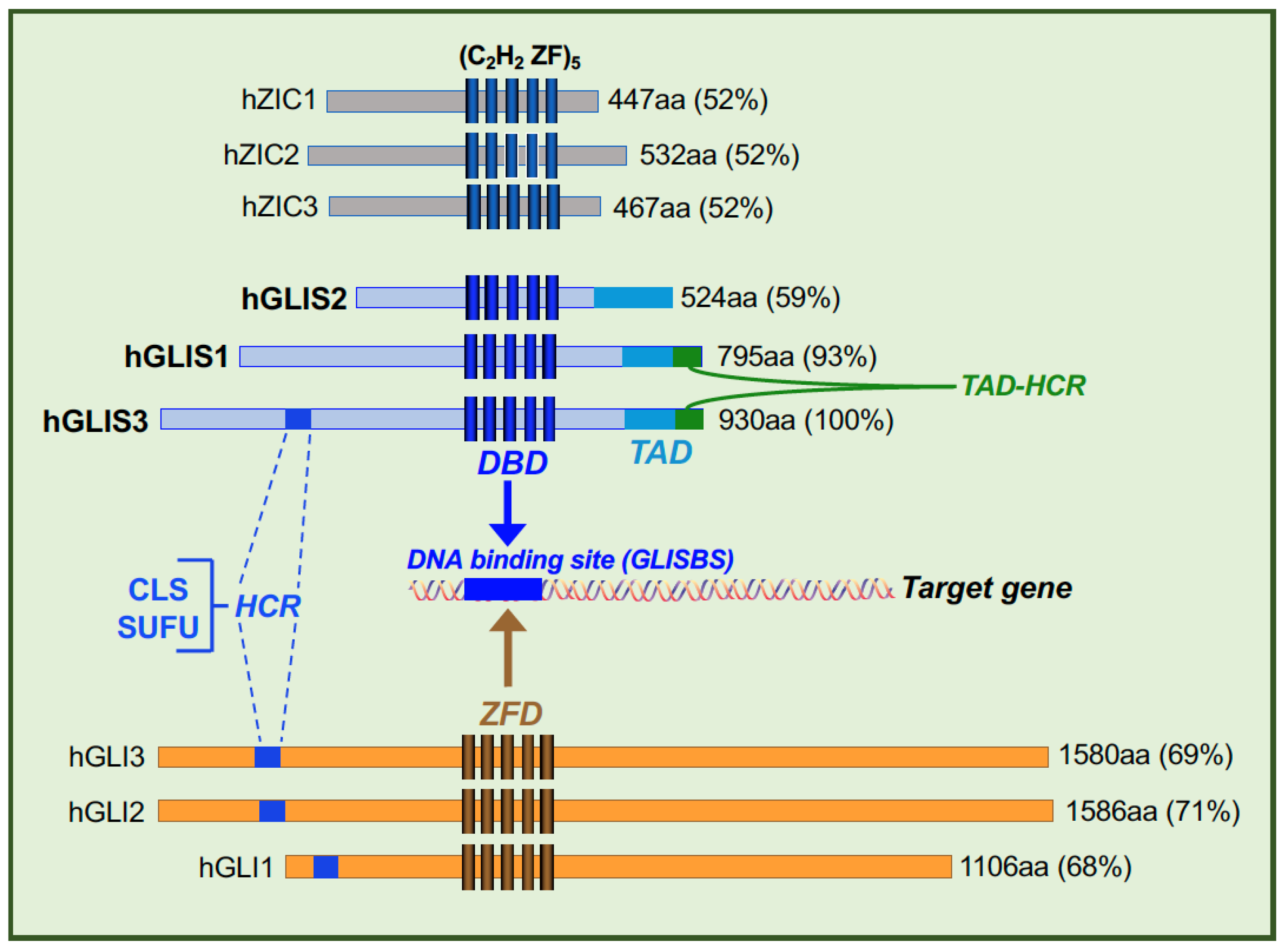
2. GLIS Proteins and Primary Cilium

Immunostaining of various tissues and cultured cells have localized GLIS3 largely to the nucleus; however, GLIS3 has also been detected in the tip of primary cilia [18,45]. GLIS3 localization to the primary cilium is supported by mass spectrometric analysis of primary cilia-associated proteins [46]. Whether there is any connection between GLIS1 and the primary cilium, has yet to be established. Phylogenetic analyses of GLIS1/3 genes have provided certain clues for potential roles of both GLIS1 and GLIS3 in primary cilium-dependent functions [47]. The primary cilium is a microtubule-based, non-motile sensory organelle protruding from the plasma membrane of most cell types [48,49,50,51]. Primary cilia play a critical role in mediating the activation of several signaling pathways by a wide range of external signals, such as Wingless (WNT) proteins, insulin growth factor 1 (IGF1), platelet-derived growth factor α (PDGFα), hedgehogs, and mechanical stress, and therefore in the regulation of many cellular functions and biological processes, including cell fate specification, proliferation, and stemness [51,52,53]. Defects in primary cilium assembly or primary cilium-associated signaling pathways are causally linked to several pathologies, collectively referred to as ciliopathies that include polydactyly, nephronophthisis, and polycystic kidney disease [49,51,54,55,56].
The interaction of external signals with their respective receptors (e.g., G protein-coupled receptors, GPCRs) in the ciliary membrane result in changes in the activity specific ciliary proteins, including several protein kinases and transcription factors [49,50]. This might involve changes in ciliary cAMP and Ca2+ levels, post-translational modifications, proteolytic processing, and protein–protein interactions as demonstrated for GLI proteins. The formation of the primary cilium is cell cycle-dependent; the primary cilium is formed in G0/G1 interphase of the cell cycle and disassembled before cell division. Transport of proteins in and out the primary cilium is mediated by the intraflagellar transport system (IFT) [50]. The transition zone (TZ) at the base of the primary cilium forms a barrier between the cytoplasm and primary cilium and controls the entry and exit of proteins, including that of GLI proteins [57,58,59]. The N-terminus of GLI1-3 contains a ciliary localization signal (CLS) with the consensus sequence SSXR-X6-R/KKR-X5-PY/L containing an Arg/Lys-rich nuclear localization-like signal [50,58,60,61]. Transportin (TNPO1, also referred to as karyopherin β2 or importin 2), interacts with the CLS and mediates the entry of GLI1-3 into the primary cilium. A consensus CLS is also present within the 60 amino acid HCR at the N-terminus of GLIS3 [6,7,62] (Figure 1). We hypothesize that TNPO1 can also mediate the entry of GLIS3 into the primary cilium and that GLIS3-mediated transcriptional regulation is mediated by a primary cilium-dependent (canonical) pathway via a still to be identified primary cilium-associated GPCR. However, GLIS3 activity might also be controlled by a primary cilium-independent (noncanonical) mechanism. In addition to the CLS, the HCR contains a consensus binding site (ΦYGHΦ) for suppressor of fused (SUFU), a protein with multiple roles in hedgehog signaling that has also been localized to the primary cilium [63] (Figure 1). SUFU has been reported to interact with GLIS3 and to protect it from proteosomal degradation [62].
GLIS2 also has been localized to primary cilia and shown to interact with SUFU [9,12]. However, GLIS2 does not contain a consensus CLS sequence suggesting that its ciliary localization might be mediated by a different mechanism. Regulation of GLIS2 transcriptional activity by primary cilium-associated mechanisms is discussed below in more detail [11,44].
GLIS proteins are post-translationally modified by phosphorylation, sumoylation, ubiquitination, and acetylation, and they can undergo proteolytic cleavage [9,11,62,64,65,66]. Although the roles of many of these post-translational modifications are still largely unknown, they can alter the subcellular localization, protein stability, proteolytic processing, and transcriptional activity of GLIS proteins and, consequently, affect the regulation of their biological functions, as shown for GLIS2 and GLIS3 [11,44,62,66].

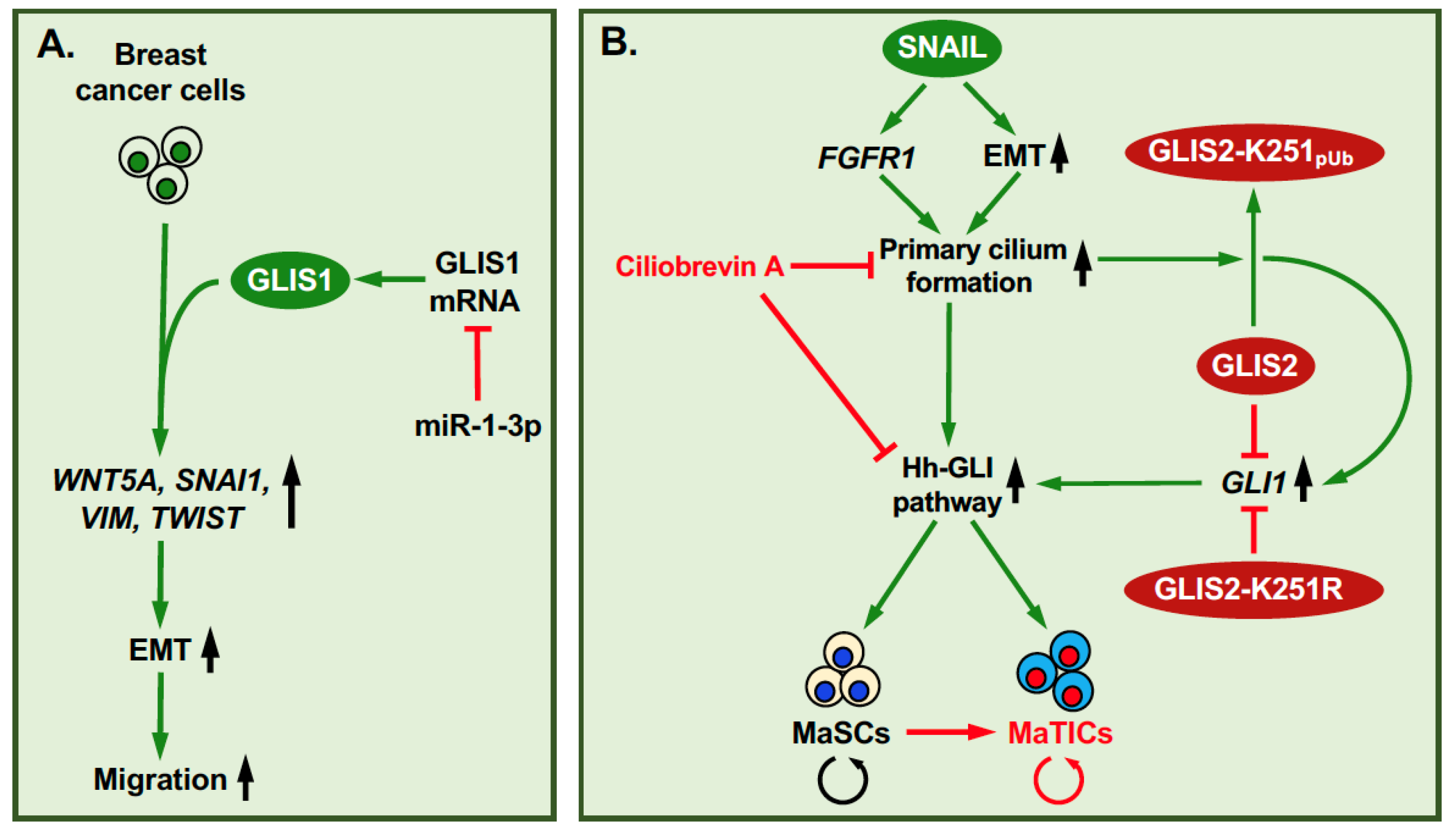
3. GLIS1-3: Regulation of Self-Renewal and EMT in Relation to Tumorigenesis

GLIS1-3 have been implicated in various malignancies, including leukemia, thyroid cancer, and breast cancer (reviewed in [16]). Dysregulation of EMT and stem cell renewal play critical roles in promoting cancer and tumor progression by inhibiting cell differentiation and increasing cell proliferation, migration, and metastasis. In several GLIS-associated malignancies, GLIS proteins have been reported to regulate stem cell renewal, EMT, and/or reprogramming in several tumor cell types [11,13,33,35,67,68].
Breast cancer is heterogeneous disease comprised of multiple tumor subtypes, including triple-negative (estrogen receptor-, progesterone receptor-, and epidermal growth factor receptor 2-negative; ERPRHER2) breast tumors. All three GLIS genes have been linked to breast cancer. GLIS3 is expressed at significantly higher levels in breast cancer samples than in normal tissue and is particularly elevated in triple-negative breast tumors [30]. In MMTV-Cut like homeobox 1 (CUX1) transgenic mice, a mouse model used to study breast cancer development, GLIS1 was found to be highly expressed in a subset of breast tumors with elevated WNT expression [69]. This correlated with increased β-catenin transcriptional activity and expression of epithelial–mesenchymal transition (EMT)-promoting genes, such as snail family transcriptional repressor 1 (SNAI1), vimentin (VIM), and twist family BHLH transcription factor 1 (TWIST), and enhanced cell migration and invasion (Figure 2A). Similarly, hypoxia-induced GLIS1 expression in MDA-MB-231 breast carcinoma cells caused an increase in WNT5A expression and cell migration [31]. A separate study reported that expression of microRNA miR-1-3p suppressed EMT and cell migration in MDA-MB-231 cells by binding to the 3′-UTR of GLIS1 mRNA resulting in reduced GLIS1 expression [70]. These studies suggest that GLIS1 plays a critical role in promoting EMT reprogramming in breast cancer cells.
A recent study identified an important role for GLIS2 in the regulation of stemness in multipotent mammary stem cells (MaSCs) and mammary tumor-initiating cells (MaTICs) [11]. MaTICs and cells from claudin-low breast tumors, which represent a subtype of triple-negative tumors thought to stem from MaTICs, exhibit many of the stem cell properties of MaSCs (Figure 2B). In addition to their self-renewal ability, MaSCs can differentiate into luminal and basal progenitors that, subsequently, give rise to ductal luminal and alveolar cells, and myoepithelial cells, respectively. As such, MaSCs play a critical role in coordinating mammary gland homeostasis, morphogenesis, and alveologenesis during embryonic development as well as postnatally [71]. Activation of the EMT program promotes and maintains the self-renewal capacity of MaSCs by inducing the formation of primary cilia that is mediated in part via increased expression of FGFR1 by EMT transcription factors, such as SNAIL2 (also known as SLUG) (Figure 2B) [52,72]. This, subsequently, leads to the primary cilium-dependent activation of the hedgehog-GLI1 signaling pathway and increased self-renewal [50,52,73]. Loss of primary cilia represses hedgehog signaling and, subsequently, reduces self-renewal in MaSCs and MaTICs, thereby reducing the tumor-producing potential of MaTICs.
The transcription factor SNAIL2 plays a critical role in the activation of EMT in both MaSCs and MaTICs [11,74]. This is accompanied by increased expression of EMT marker genes, such as N-cadherin (CDH2) and VIM, and the transcription factors, Zinc finger E-box binding homeobox 1 (ZEB1) and TWIST, and reduced expression of several epithelial markers, including E-cadherin (CDH1). The study by Wilson et al. [11] further demonstrated that GLIS2 represses MaSC self-renewal, whereas inactivation or loss of GLIS2 function promotes MaSC self-renewal indicating that GLIS2 functions as a repressor of MaSC stemness by directly suppressing the transcription of GLI1 and WNT genes (Figure 2B). Based on these findings one might predict that GLIS2 deficiency would promote expansion of MaSCs at the cost of their differentiation into different mammary gland lineages and result in defective mammary gland development. This hypothesis was supported by observations showing that Glis2-deficient mice develop only small mammary rudiments [11]. A SNAIL-dependent post-translational modification of GLIS2 was found to play a crucial role in the regulation of self-renewal of MaSCs by GLIS2. SNAIL stimulates GLIS2 polyubiquitination at K251 leading to loss of GLIS2 repressor function (Figure 2B). The latter was not due to increased proteolytic degradation or reduced GLIS2 mRNA expression, but to the loss of GLIS2 repressor activity [11]. Ciliobrevin A, an inhibitor of ciliogenesis, inhibited GLIS2 polyubiquitination and the activation of the hedgehog signaling pathway. Although the precise molecular mechanism has yet to be elucidated, these data indicate that the regulation of GLIS2 polyubiquitination by SNAIL is primary cilium-dependent (Figure 2B). The importance of GLIS2 polyubiquitination is supported by data showing that the K251R mutation converts GLIS2 into a constitutive repressor and consequently abrogates the SNAIL-induced expression of GLI1 and CDH11 in mammary cells [11].
GLIS2 has also been implicated in the regulation of self-renewal in leukemic cells [16,32,33,34,68,75,76,77,78]. Several studies demonstrated that a cryptic inversion of chromosome 16 that fuses CBFA2T3 to GLIS2, is frequently (25–30%) associated with a pediatric non-Down’s syndrome (non-DS) acute megakaryoblastic leukemia (AMKL) with poor prognosis [32,34,75,76,77,78,79,80]. Transcriptomic analysis indicated that CBFA2T3-GLIS2 regulates gene transcription partly through a CBFA2T3- or GLIS2-mediated mechanisms, but that a subset of genes are regulated via a CBFA2T3-GLIS2-specific mechanism. Among the genes regulated by CBFA2T3-GLIS2 were those with established roles in leukemia and hematopoietic stem cell self-renewal and the control of EMT and cell migration, including genes linked to the NOTCH, sonic hedgehog (SHH), WNT, hippo, and TGFβ/BMP signaling pathways [13,33,35,80,81]. Both the CBFA2T3 and GLIS2 domains are required to optimally stimulate hematopoietic stem cell self-renewal. CBFA2T3-GLIS2 was found to induce activation of super enhancers that regulate genes critical in leukemia [81]. It was further shown that CBFA2T3-GLIS2 greatly increased the expression of the ETS transcription factor ERG, a strong inducer of hematopoietic stem cell renewal, in a GLIS2-dependent manner and down-regulated the expression of GATA binding protein 1 (GATA-1), a promoter of megakaryocytic differentiation [13]. The role of CBFA2T3-GLIS2 in promoting self-renewal was supported by a recent study showing increased self-renewal by CBFA2T3-GLIS2 in a human induced pluripotent stem cells-derived model [67]. A study of murine MOZ-TIF2 acute myelogenous leukemia (AML) cells revealed that GLIS2 expression suppressed leukemic stem cell self-renewal and promoted differentiation [68].

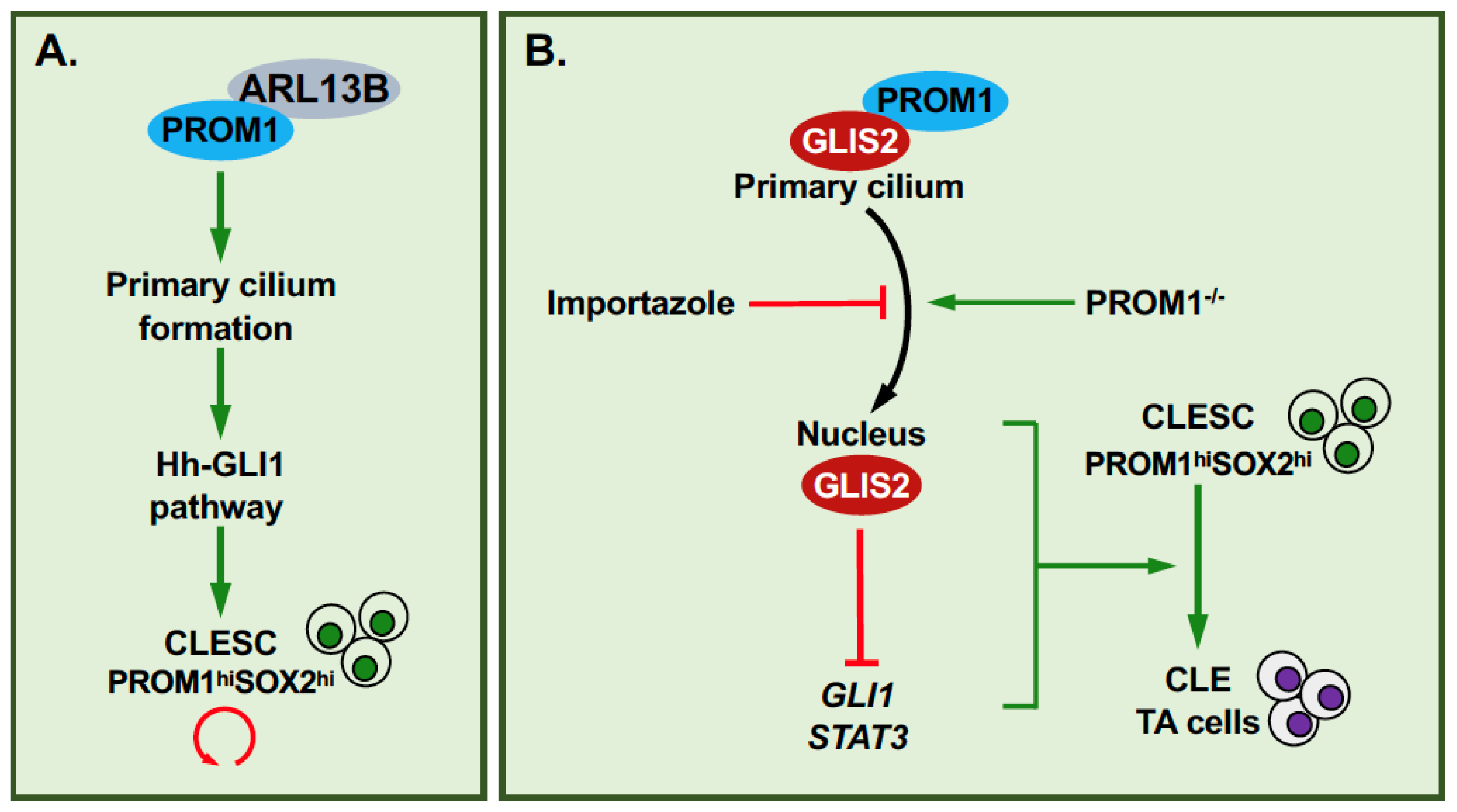
4. GLIS2, Primary Cilium, PROM1 and Cell Renewal

A recent study identified a link between GLIS2, the primary cilium, prominin-1, and cell renewal [44]. Prominin-1 (PROM1/CD133) is a cholesterol-binding, pentaspan membrane glycoprotein, associated with several plasma membrane protrusions, including primary cilia [44,82]. PROM1 has been implicated in the regulation of stem cell maintenance, differentiation, and cancer [44,82,83]. In addition, PROM1 plays a role in the regulation of ciliary dynamics and the length and function of primary cilia through its interaction with the ciliary protein, ADP-ribosylation factor-like protein 13B (Arl13b) (Figure 3A) [44,84]. Study of the incisor cervical loop epithelium (CLE) showed that PROM1hiSOX2hi CLE stem cells (CLESCs) have longer cilia than PROM1loKi67hi CLE transit amplifying cells and that loss of PROM1 function inhibits primary cilium elongation, disrupts primary cilium-associated signaling pathways, including sonic hippo (SHH) pathway activation, and significantly reduces stem cell renewal (clonogenic capacity)(Figure 3B) [44]. These observations indicated a link between PROM1, primary cilium dynamics, and the regulation of stem cell self-renewal by SHH. This study further showed that the transition of CLESCs into CLE transit amplifying cells is accompanied by a decrease in Gli1-3 mRNA expression and an increase in Glis2 mRNA expression, and a translocation of GLIS2 and PROM1 proteins from the primary cilium to the nucleus. This translocation was inhibited by importazole, an inhibitor of importin β1-mediated nuclear import. In Prom1-KO mice, GLIS2 was largely localized to the nucleus suggesting that PROM1 is required for the localization of GLIS2 to the primary cilium, while in Glis2-KO mice PROM1 expression and the number of SOX2+ stem cells were shown to be significantly reduced, indicating an interrelationship between GLIS2 and PROM1 in regulating stemness. Together, these observations led to the hypothesis that translocation of GLIS2 to the nucleus during the transition of stem cell to transit amplifying cells allows it to function as a transcription repressor, thereby reducing the expression of genes (e.g., Gli1, Stat3) that are critical for stem cell maintenance and renewal (Figure 3B) [44]. The inhibition of stem cell renewal by GLIS2 involves repression of the transcription activation of Stat3, a GLIS2 target gene, and inhibition of GLI1-mediated transcriptional activation due to competition between GLIS2 and GLI1 for binding to the same enhancer in target genes. The inhibition of CLESC renewal is consistent with the function of GLIS2 in stem cell renewal reported for MaSCs [11]. Interestingly, PROM1 has been reported to be also expressed in MaSCs and MaTICs [85] suggesting that it might play a similar role in these cells as in CLESCs.

5. Effects of GLIS1-3 on iPSC Reprogramming and Differentiation

Several studies have demonstrated that GLIS1-3 can modulate reprogramming of somatic cells into induced pluripotent stem cells (iPSCs) [43,86,87,88,89,90,91,92,93]. Over-expression of the transcription factors, octamer-binding protein 4 (OCT4 or POU5F1), SRY-box transcription factor 2 (SOX2), Krüppel-like zinc finger protein 4 (KLF4), and c-MYC (OSKM) induces reprogramming of somatic cells into iPSCs [94]. Reprogramming of fibroblasts into iPSCs requires mesenchymal–epithelial transition (MET) that is mediated in part through the repression the EMT-inducing genes SNAIL by SOX2 and OCT4, and TGFB1 and TGFBR2 by c-MYC, and induction of MET-associated genes, such as E- cadherin (CDH1) by KLF4 [95]. Over-expression of GLIS1 in human and mouse fibroblasts markedly enhanced reprogramming efficiency by either OSK or OSKM, whereas down-regulation of endogenous GLIS1 expression reduced reprogramming efficiency by OSK [43,88,96]. However, in human adipose-derived stromal cells the GLIS1-mediated increase in reprogramming was dependent on co-expression with c-MYC [87]. A recent study showed that over-expression of GLIS1 synergized with Nanog homeobox (NANOG) in improving reprogramming efficiency, but not with LIN28, and that efficiency was reduced when GLIS1 was combined with NANOG and LIN28 [86]. In human fibroblasts, GLIS1 enhanced the expression of several reprogramming-promoting genes, including estrogen receptor-related receptor B (ESRRB), LIN28A, NANOG, FOXA2, MYCL1, MYCN, and several WNT genes. The induction of ESRRB, LIN28A, NANOG, and FOXA2 was mediated through an indirect mechanism, whereas MYCL1 and MYCN were directly regulated by GLIS1 [42]. GLIS1 regulates gene transcription in coordination with other transcription factors, including OCT4, SOX2, and KLF4. GLIS1 was found to interact with KLF4 and both its ZFD and N-terminus were required for this interaction. Together these studies indicate that GLIS1 enhances reprogramming efficiency by stimulating MET and activating the canonical WNT signaling pathway. A different study showed that expression of GLIS1, Spalt like transcription factor 4 (SALL4), liver receptor homolog 1 (LRH1) in combination with Jun dimerization protein 2 (JDP2), lysine demethylase 2B (KDM2B or JHDM1B), and ID1 induce reprogramming of fibroblasts into iPSCs, but that GLIS1 was not required [43,97]. GLIS1 was reported to be highly expressed in unfertilized eggs and one-cell embryos, but at very low levels in blastocysts and pluripotent stem cells (PSCs) [42] suggesting that it does not play a significant role in maintaining pluripotency in PSCs in vivo.
In addition to GLIS1, GLIS3 has also been shown to enhance reprogramming efficiency. Co-expression of GLIS3 was shown to promote reprogramming of human adipose-derived stromal cells by OSKM as efficiently as GLIS1 [87]. In contrast to GLIS1 and GLIS3, co-expression of GLIS2 inhibited reprogramming [87]. However, a different study reported that GLIS2 knockdown in hPSCs repressed the expression of pluripotency genes, such as OCT4 and SOX2, and promoted their differentiation into endodermal and trophoblast lineages suggesting that GLIS2 expression maintains stemness of hPSCs [98]. Future studies are needed to explain these apparent contrasting effects of GLIS2 on reprogramming and hESC stemness.
In the effects of GLIS proteins on reprogramming, one needs to consider that KLF4 and GLIS proteins bind similar G-rich binding sequences suggesting that there might be some overlap between target genes they regulate. This is supported by a study showing that GLIS1 can substitute for KLF4 in reprogramming [88]. This appears to be supported by a recent report indicating that GLIS1 and KLF4 share some common functions and/or mechanisms in reprogramming [91]. In the case of GLIS2, by functioning as a repressor GLIS2 might also compete with KLF4 for binding and as a result suppress the activation of certain KLF4 target genes and reduce reprogramming efficiency.

6. GLIS2 and Reprogramming in Hepatic Fibrosis

The liver plays a critical role in the regulation of many metabolic functions, including lipid, carbohydrate and protein metabolism, glycogen storage, detoxification, bile production, and synthesis of plasma proteins. Metabolic-associated fatty liver disease (MAFLD) is a major health concern present in roughly 25% of the global population. MAFLD can further progress into non-alcoholic steatohepatitis (NASH) that is accompanied with increased hepatic fat accumulation (steatosis), inflammation, and fibrosis. A recent study [41] in mice fed a high fat diet (HFD) identified GLIS2 as a critical regulator of gene expression during the progression of MAFLD. ATAC-Seq analysis identified the Glis2 gene as one of the genomic loci that obtained an open configuration during advanced NASH, whereas loci of genes associated with hepatic identity became closed. This correlated with a higher expression of GLIS2 in NASH. This study further showed that GLIS2 was one of the transcription factors that gained motif activity during NASH progression. These observations suggested a regulatory role for GLIS2 in the regulation of gene transcription during NASH progression (Figure 4A). This was supported by data showing that specific knock down of Glis2 expression by adenovirus-associated virus (AAV) miRNA in hepatocytes from mice fed a HFD significantly reduced the expression of genes associated with extracellular matrix, inflammation, adhesion, and cell cycle, including Col1a1, Tnf, Ctgf, Ccl2, and Adgre1 (Figure 4A). This was accompanied by a decrease in hepatic fibrosis, inflammation, and apoptosis; however, hepatic steatosis remained unaffected. GLIS2 binding sites (GLIS2BS) were associated with many of these differentially expressed genes indicating that their transcription is directly regulated by GLIS2. These data suggest that in this case, GLIS2 functions as an activator of gene transcription. The GLIS2BS in many of the target genes overlap with those of several other transcription factors that are implicated in NASH-associated gene expression, such as ELF3 and members of the AP-1 family. This suggests that GLIS2 regulates gene transcription of these genes in coordination with other transcription factors. Thus, GLIS2 together with other hepatic transcription factors are part of gene regulatory network that regulates NASH-associated gene expression in hepatocytes. The study further indicates that GLIS2 plays a critical role in promoting NASH-associated reprogramming in hepatic fibrosis, inflammation, and apoptosis; however, hepatic steatosis remained unaffected. GLIS2 binding sites (GLIS2BS) were associated with many of these differentially expressed genes indicating that their transcription is directly regulated by GLIS2. These data suggest that in this case, GLIS2 functions as an activator of gene transcription. The GLIS2BS in many of the target genes overlap with those of several other transcription factors that are implicated in NASH-associated gene expression, such as ELF3 and members of the AP-1 family. This suggests that GLIS2 regulates gene transcription of these genes in coordination with other transcription factors. Thus, GLIS2 together with other hepatic transcription factors are part of gene regulatory network that regulates NASH-associated gene expression in hepatocytes. The study further indicates that GLIS2 plays a critical role in promoting NASH-associated reprogramming in hepatocytes by repressing the expression of hepatocyte identity genes (e.g., Cyp8b1, Aldh2, Idh1, and Slc2a2) and increasing that of inflammatory and fibrosis-related genes (Figure 4B) [41]. These observations contrast those in kidney, in which loss of GLIS2 function induces inflammation and fibrosis [8,12,26,27]. These differential effects of GLIS2 in kidney and liver are likely due to tissue- and context-dependent differences.

7. Additional Roles for GLIS3 in Stem and Progenitor Cells

GLIS3 has several additional roles in the regulation of progenitor cell differentiation, renewal, and survival in several tissues, including the pancreas and testis [7,19,20]. In the pancreas, GLIS3 is most highly expressed in pancreatic β and ductal cells [6,7,22,23,24,99,100]. Loss of GLIS3 function causes neonatal diabetes that is, in part, due to a reduced generation of neurogenin 3-positive (NGN3+) endocrine progenitor cells during pancreatic development. GLIS3 directly regulates the expression of NGN3, a transcription factor required for the differentiation of bipotent progenitor cells into proendocrine and productal cells [101,102]. Reduced expression of NGN3 might be partly responsible for the decreased generation of NGN3+ pro-endocrine cells and endocrine cells in GLIS3-deficient pancreas. Whether this also involves effects on self-renewal or cell survival has yet to be established. GLIS3 also has important functions in postnatal pancreas, where it is essential for the transcriptional regulation of insulin genes (Ins1 and Ins2 in mice and INS in humans) expression and several other genes [7,22,23,24]. GLIS3 was shown to regulate Ins2 transcription in coordination with other transcription factors [100]. Loss of GLIS3 function in both humans and mice also causes dilation of pancreatic ducts suggesting that GLIS3 might have a regulatory role in ductal cell homeostasis. A recent study demonstrated that Glis3 is most highly expressed in CD133+CD71 cells, a subpopulation of pancreatic ductal cells with high self-renewal capacity [103,104]. These progenitor cells are multipotent and able to differentiate along the acinar, ductal, and endocrine lineages [103]. Glis3 knockdown reduces self-renewal in these cells that appears, at least in part, due to reduced transcription of Prom1 (CD133). The stimulation of self-renewal in these cells by PROM1 appears to be mediated via the activation of PI3K/AKT and β-catenin pathways and induction of WNT signaling-related genes. These studies suggest that GLIS3 is required for the self-renewal of these progenitor cells by regulating Prom1 expression [104]. This suggests that in addition to GLIS2, GLIS3 also has a connection with PROM1 and stem cell renewal.
GLIS3 is also essential for early spermatogenesis [19,43]. It is most highly expressed in pro-spermatogonia (ProSG; also referred to as gonocytes), spermatogonial stem and progenitor cells (SSCs and SPCs, respectively), but not at later stages of spermatogenesis. Shortly after birth, ProSGs resume proliferation and give rise to SSCs, which sequentially differentiate into SPCs and differentiated spermatogonia [105,106]. In addition to its function in regulating retrotransposon silencing [107], GLIS3 is involved in the control of cell proliferation and differentiation of SSCs and SPCs [19]. ProSGs, SSCs, and SPCs are significantly reduced in Glis3-deficient mice. This, subsequently, greatly impacts the generation of spermatozoa leading to infertility. The precise molecular mechanism underlying this regulation has yet to be determined.
GLIS3 has further been shown to regulate lineage determination of human pluripotent stem cells (hPSCs) into neural progenitor cells (NPCs) [40]. Addition of activin/TGFβ/BMP pathway inhibitors directs differentiation of hPSCs along the anterior NPC lineage that is associated by decreased expression of stem cell marker genes, including OCT4, NANOG, and SOX2, and induction of anterior NPC markers, such as OTX1/2, ZIC1, and PAX6. Expression of GLIS3 in hPSCs directs differentiation along the posterior NPC lineage in lieu of the anterior lineage as indicated by the induction of posterior NPC marker genes, such as GBX2, MNX1, and HOXA2. This study demonstrated that this differentiation along the posterior lineage was mediated by increased expression of WNT genes, particularly WNT3A, which encodes a strong posteriorizing factor [40,108,109]. GLIS3 was shown to activate WNT3A transcription directly by binding to a GLISBS in the WNT3A proximal promoter region.

8. GLIS Circular RNAs

In addition to linear RNAs required for the formation of GLIS proteins, GLIS1-3 genes also generate CircGLIS RNAs. CircRNAs are lncRNAs that are generated through backsplicing of 1 or more exons that is facilitated by RNA binding proteins (RBPs). CircRNAs can localize to the nucleus and cytoplasm and can be secreted through exosomes [110,111]. They can regulate gene expression both at the transcriptional and post-transcriptional level. The most common mechanism of action is their interaction with miRNAs, thereby acting as miRNA sponges. CircRNAs can also modulate gene expression through their interaction with proteins, including RBPs. They can regulate various cellular functions, including EMT, stemness, apoptosis, senescence, and differentiation, and have been implicated in many cancers.
A recent study showed that the expression circular GLIS2 (circGLIS2) was significantly higher in colorectal cancer, the third most common cancer globally [112]. CircGLIS2 is generated from exon 2 and 3 via back-splicing. Over-expressing of circGLIS2 in human colorectal cell lines results in activation of the NF-κB signaling pathway by sponging miR-671, which results in the induction of several pro-inflammatory cytokines that, subsequently, increase cell migration and metastasis. The authors suggested that targeting circGLIS2 might provide a strategy to intervene in colorectal cancer [112].
A recent study reported that, in addition to circGLIS2, linear GLIS2 RNA also plays a role in colorectal cancer [29]. GLIS2 was identified as a moderate repressor of several p53 target genes, including BCL2 binding component 3 (BBC3, also referred to as PUMA), which encodes a pro-apoptotic protein. GLIS2 over-expression in human colon cancer HCT116 cells also repressed the expression of several focal adhesion genes. ChIP analysis indicated that GLIS2 was associated with the BBC3 proximal promoter, a region where also p53 binds. Suppression of GLIS2 expression modestly increased p53 binding to this region. Subcutaneous injection of GLIS2-over-expressing HCT116 cells enhanced tumor load in mice and reduced their overall survival.
Recently, expression of circular GLIS3 (circGLIS3) RNAs, generated by exon 2 or exons 5–8, have been implicated in several tumor types, including glioma, non-small cell lung cancer (NSCLC), and bladder cancer [113,114,115]. Expression of circGLIS3, generated from exon 2, was found to be associated high-grade gliomas that are highly invasive and resistant to therapeutic treatments. Most of these gliomas consisted of glioblastoma, the deadliest type of brain cancer. Expression of this circGLIS3 enhances migration of glioma cells. Moreover, tumors derived from intracranial injection of circGLIS3-overexpressing glioma cells in nude mice were more invasive and reduced the overall survival of the mice [113]. CircGLIS3 was found to interact with T567-phosphorylated ezrin (p-EZR), but not with unphosphorylated ezrin, causing an increase in p-EZR, a protein that promotes cell migration and correlates with high-grade gliomas. This study further showed that circGLIS3 is secreted through exosomes and enhances angiogenesis. Thus, circGLIS3 appears to promote the malignancy of gliomas by both increasing invasiveness and angiogenesis. A different report showed that high levels of GLIS3 expression correlated with high-grade gliomas and poor prognosis [116]. Knockdown of GLIS3 expression in glioma cells lines reduced cell proliferation and migration, inhibited the activation of the NF-κB signaling pathway, and suppressed the in vivo malignant behavior of cells when implanted into nude mice. This correlated with respective changes in c-MYC, MMP9, and phosphorylated p65 expression. Over-expression of GLIS3 in glioma cell lines had the inverse effect.
CircGLIS3 containing exon 2 was also identified in bladder cancer tumors and shown to increase migration in cultured bladder cancer cells by absorbing miR-1273f resulting in increased expression of S-phase kinase protein 1 (SKP1) and cyclin D1 [114]. CircGLIS3 derived from exons 5–8 was found to be associated with cancer progression in NSCLC [115]. This circGLIS3 was shown to stimulate cell proliferation and migration and inhibit apoptosis in cultured NSCLC cells that appears to be mediated in part by the down-regulation of the tumor suppressor miR-644a. CircGlis3 RNAs appear to be also involved in the regulation of normal physiological processes, including pancreatic β cell function, as was recently reported for a circGLIS3 derived from exon 4 [117].
Together, these studies demonstrate that circGLIS2 and circGLIS3 enhance self-renewal and invasiveness in various tumor cell types. Whether this involves regulation of reprogramming, stemness, and EMT needs further study. Moreover, circGLIS RNAs may have a role in the regulation normal physiological functions as well. Thus, mutations or shRNA knockdown may affect the expression of linear GLIS RNAs and/or circRNAs and yield distinct phenotypes.

9. Conclusions

Evidence is accumulating that the transcriptional activity of GLIS proteins is regulated, at least in part, by primary cilium-dependent mechanisms. In addition, several studies have linked GLIS proteins to the regulation of cell reprogramming, cell lineage determination, and self-renewal. These processes play a critical role in normal mammalian development, while defects in their regulation are causally linked to many pathologies, including cancer. A better understanding of the molecular mechanisms by which GLIS proteins regulate gene transcription are critical for elucidating their function in the control of various biological processes and their roles in disease. Moreover, future insights into the signaling pathways that regulate the subcellular localization, post-translational modification, proteolytic processing, and transcriptional activity of GLIS proteins might lead to the development of new therapeutic strategies in the management of several pathologies.

Author Contributions

A.M.J. wrote original draft and designed the Figures, D.W.S. and H.S.K. reviewed and revised the manuscript. All authors have read and agreed to the published version of the manuscript.

Funding

This work is supported by the Intramural Research Program of the NIEHS (NIH Z01-ES-100485).

Institutional Review Board Statement

Not applicable.

Informed Consent Statement

Not applicable.

Data Availability Statement

Not applicable.

Acknowledgments

The authors would like to thank all current and previous members of the Cell Biology Section for their contributions to the study of GLIS proteins.

Conflicts of Interest

The authors declare no conflict of interest.

References

  1. Zhang, F.; Jetten, A.M. Genomic structure of the gene encoding the human GLI-related, Kruppel-like zinc finger protein GLIS2. Gene 2001, 280, 49–57. [Google Scholar] [CrossRef]
  2. Kim, Y.S.; Lewandoski, M.; Perantoni, A.O.; Kurebayashi, S.; Nakanishi, G.; Jetten, A.M. Identification of Glis1, a novel Gli-related, Kruppel-like zinc finger protein containing transactivation and repressor functions. J. Biol. Chem. 2002, 277, 30901–30913. [Google Scholar] [CrossRef] [PubMed] [Green Version]
  3. Zhang, F.; Nakanishi, G.; Kurebayashi, S.; Yoshino, K.; Perantoni, A.; Kim, Y.S.; Jetten, A.M. Characterization of Glis2, a novel gene encoding a Gli-related, Kruppel-like transcription factor with transactivation and repressor functions. Roles in kidney development and neurogenesis. J. Biol. Chem. 2002, 277, 10139–10149. [Google Scholar] [CrossRef] [PubMed] [Green Version]
  4. Kim, Y.S.; Nakanishi, G.; Lewandoski, M.; Jetten, A.M. GLIS3, a novel member of the GLIS subfamily of Kruppel-like zinc finger proteins with repressor and activation functions. Nucleic Acids Res. 2003, 31, 5513–5525. [Google Scholar] [CrossRef]
  5. Kim, S.C.; Kim, Y.S.; Jetten, A.M. Kruppel-like zinc finger protein Gli-similar 2 (Glis2) represses transcription through interaction with C-terminal binding protein 1 (CtBP1). Nucleic Acids Res. 2005, 33, 6805–6815. [Google Scholar] [CrossRef] [Green Version]
  6. Jetten, A.M. GLIS1-3 transcription factors: Critical roles in the regulation of multiple physiological processes and diseases. Cell Mol. Life Sci. 2018, 75, 3473–3494. [Google Scholar] [CrossRef]
  7. Scoville, D.W.; Kang, H.S.; Jetten, A.M. Transcription factor GLIS3: Critical roles in thyroid hormone biosynthesis, hypothyroidism, pancreatic beta cells and diabetes. Pharmacol Ther 2020, 215, 107632. [Google Scholar] [CrossRef]
  8. Kim, Y.S.; Kang, H.S.; Herbert, R.; Beak, J.Y.; Collins, J.B.; Grissom, S.F.; Jetten, A.M. Kruppel-like zinc finger protein Glis2 is essential for the maintenance of normal renal functions. Mol. Cell Biol. 2008, 28, 2358–2367. [Google Scholar] [CrossRef] [Green Version]
  9. Li, B.; Rauhauser, A.A.; Dai, J.; Sakthivel, R.; Igarashi, P.; Jetten, A.M.; Attanasio, M. Increased hedgehog signaling in postnatal kidney results in aberrant activation of nephron developmental programs. Hum. Mol. Genet. 2011, 20, 4155–4166. [Google Scholar] [CrossRef] [Green Version]
  10. Kim, Y.S.; Kang, H.S.; Jetten, A.M. The Kruppel-like zinc finger protein Glis2 functions as a negative modulator of the Wnt/beta-catenin signaling pathway. FEBS Lett. 2007, 581, 858–864. [Google Scholar] [CrossRef] [Green Version]
  11. Wilson, M.M.; Callens, C.; Le Gallo, M.; Mironov, S.; Ding, Q.; Salamagnon, A.; Chavarria, T.E.; Viel, R.; Peasah, A.D.; Bhutkar, A.; et al. An EMT-primary cilium-GLIS2 signaling axis regulates mammogenesis and claudin-low breast tumorigenesis. Sci. Adv. 2021, 7, eabf6063. [Google Scholar] [CrossRef] [PubMed]
  12. Attanasio, M.; Uhlenhaut, N.H.; Sousa, V.H.; O’Toole, J.F.; Otto, E.; Anlag, K.; Klugmann, C.; Treier, A.C.; Helou, J.; Sayer, J.A.; et al. Loss of GLIS2 causes nephronophthisis in humans and mice by increased apoptosis and fibrosis. Nat. Genet. 2007, 39, 1018–1024. [Google Scholar] [CrossRef] [PubMed]
  13. Thirant, C.; Ignacimouttou, C.; Lopez, C.K.; Diop, M.; Le Mouel, L.; Thiollier, C.; Siret, A.; Dessen, P.; Aid, Z.; Riviere, J.; et al. ETO2-GLIS2 Hijacks Transcriptional Complexes to Drive Cellular Identity and Self-Renewal in Pediatric Acute Megakaryoblastic Leukemia. Cancer Cell 2017, 31, 452–465. [Google Scholar] [CrossRef] [PubMed] [Green Version]
  14. Chinnadurai, G. Transcriptional regulation by C-terminal binding proteins. Int. J. Biochem. Cell Biol. 2007, 39, 1593–1607. [Google Scholar] [CrossRef] [PubMed]
  15. Scoville, D.W.; Jetten, A.M. GLIS3: A Critical Transcription Factor in Islet beta-Cell Generation. Cells 2021, 10, 3471. [Google Scholar] [CrossRef] [PubMed]
  16. Jetten, A.M. Emerging Roles of GLI-Similar Kruppel-like Zinc Finger Transcription Factors in Leukemia and Other Cancers. Trends Cancer 2019, 5, 547–557. [Google Scholar] [CrossRef]
  17. Dimitri, P. The role of GLIS3 in thyroid disease as part of a multisystem disorder. Best Pract. Res. Clin. Endocrinol. Metab. 2017, 31, 175–182. [Google Scholar] [CrossRef]
  18. Kang, H.S.; Beak, J.Y.; Kim, Y.S.; Herbert, R.; Jetten, A.M. Glis3 is associated with primary cilia and Wwtr1/TAZ and implicated in polycystic kidney disease. Mol. Cell Biol. 2009, 29, 2556–2569. [Google Scholar] [CrossRef] [Green Version]
  19. Kang, H.S.; Chen, L.Y.; Lichti-Kaiser, K.; Liao, G.; Gerrish, K.; Bortner, C.D.; Yao, H.H.; Eddy, E.M.; Jetten, A.M. Transcription Factor GLIS3: A New and Critical Regulator of Postnatal Stages of Mouse Spermatogenesis. Stem Cells 2016, 34, 2772–2783. [Google Scholar] [CrossRef] [Green Version]
  20. Kang, H.S.; Kim, Y.S.; ZeRuth, G.; Beak, J.Y.; Kilic, G.; Jensen, J.; Sosa-Pineda, B.; Jetten, A.M. Transcription factor Glis3: A novel critical player in the regulation of pancreatic β-cell development. Mol. Cell Biol. 2009, 29, 6366–6379. [Google Scholar] [CrossRef] [Green Version]
  21. Kang, H.S.; Kumar, D.; Liao, G.; Lichti-Kaiser, K.; Gerrish, K.; Liao, X.H.; Refetoff, S.; Jothi, R.; Jetten, A.M. GLIS3 is indispensable for TSH/TSHR-dependent thyroid hormone biosynthesis and follicular cell proliferation. J. Clin. Investig. 2017, 127, 4326–4337. [Google Scholar] [CrossRef] [PubMed] [Green Version]
  22. Kang, H.S.; Takeda, Y.; Jeon, K.; Jetten, A.M. The Spatiotemporal Pattern of Glis3 Expression Indicates a Regulatory Function in Bipotent and Endocrine Progenitors during Early Pancreatic Development and in Beta, PP and Ductal Cells. PLoS ONE 2016, 11, e0157138. [Google Scholar] [CrossRef]
  23. Senee, V.; Chelala, C.; Duchatelet, S.; Feng, D.; Blanc, H.; Cossec, J.C.; Charon, C.; Nicolino, M.; Boileau, P.; Cavener, D.R.; et al. Mutations in GLIS3 are responsible for a rare syndrome with neonatal diabetes mellitus and congenital hypothyroidism. Nat. Genet. 2006, 38, 682–687. [Google Scholar] [CrossRef] [PubMed]
  24. Yang, Y.; Chang, B.H.; Samson, S.L.; Li, M.V.; Chan, L. The Kruppel-like zinc finger protein Glis3 directly and indirectly activates insulin gene transcription. Nucleic Acids Res. 2009, 37, 2529–2538. [Google Scholar] [CrossRef] [PubMed] [Green Version]
  25. Khor, C.C.; Do, T.; Jia, H.; Nakano, M.; George, R.; Abu-Amero, K.; Duvesh, R.; Chen, L.J.; Li, Z.; Nongpiur, M.E.; et al. Genome-wide association study identifies five new susceptibility loci for primary angle closure glaucoma. Nat. Genet. 2016, 48, 556–562. [Google Scholar] [CrossRef] [PubMed]
  26. Jin, H.; Zhang, Y.; Liu, D.; Wang, S.S.; Ding, Q.; Rastogi, P.; Purvis, M.; Wang, A.; Elhadi, S.; Ren, C.; et al. Innate Immune Signaling Contributes to Tubular Cell Senescence in the Glis2 Knockout Mouse Model of Nephronophthisis. Am. J. Pathol. 2020, 190, 176–189. [Google Scholar] [CrossRef]
  27. Lu, D.; Rauhauser, A.; Li, B.; Ren, C.; McEnery, K.; Zhu, J.; Chaki, M.; Vadnagara, K.; Elhadi, S.; Jetten, A.M.; et al. Loss of Glis2/NPHP7 causes kidney epithelial cell senescence and suppresses cyst growth in the Kif3a mouse model of cystic kidney disease. Kidney Int. 2016, 89, 1307–1323. [Google Scholar] [CrossRef] [Green Version]
  28. Nair, K.S.; Srivastava, C.; Brown, R.V.; Koli, S.; Choquet, H.; Kang, H.S.; Kuo, Y.M.; Grimm, S.A.; Sutherland, C.; Badea, A.; et al. GLIS1 regulates trabecular meshwork function and intraocular pressure and is associated with glaucoma in humans. Nat. Commun. 2021, 12, 4877. [Google Scholar] [CrossRef]
  29. Yao, J.; Lei, P.J.; Li, Q.L.; Chen, J.; Tang, S.B.; Xiao, Q.; Lin, X.; Wang, X.; Li, L.Y.; Wu, M. GLIS2 promotes colorectal cancer through repressing enhancer activation. Oncogenesis 2020, 9, 57. [Google Scholar] [CrossRef]
  30. Rami, F.; Baradaran, A.; Kahnamooi, M.M.; Salehi, M. Alteration of GLIS3 gene expression pattern in patients with breast cancer. Adv. Biomed. Res. 2016, 5, 44. [Google Scholar] [CrossRef]
  31. Shimamoto, K.; Tanimoto, K.; Fukazawa, T.; Nakamura, H.; Kanai, A.; Bono, H.; Ono, H.; Eguchi, H.; Hirohashi, N. GLIS1, a novel hypoxia-inducible transcription factor, promotes breast cancer cell motility via activation of WNT5A. Carcinogenesis 2020, 41, 1184–1194. [Google Scholar] [CrossRef] [PubMed]
  32. Masetti, R.; Pigazzi, M.; Togni, M.; Astolfi, A.; Indio, V.; Manara, E.; Casadio, R.; Pession, A.; Basso, G.; Locatelli, F. CBFA2T3-GLIS2 fusion transcript is a novel common feature in pediatric, cytogenetically normal AML, not restricted to FAB M7 subtype. Blood 2013, 121, 3469–3472. [Google Scholar] [CrossRef] [PubMed] [Green Version]
  33. Gruber, T.A.; Larson Gedman, A.; Zhang, J.; Koss, C.S.; Marada, S.; Ta, H.Q.; Chen, S.C.; Su, X.; Ogden, S.K.; Dang, J.; et al. An Inv(16)(p13.3q24.3)-encoded CBFA2T3-GLIS2 fusion protein defines an aggressive subtype of pediatric acute megakaryoblastic leukemia. Cancer Cell 2012, 22, 683–697. [Google Scholar] [CrossRef] [PubMed] [Green Version]
  34. Thiollier, C.; Pflumio, F.; Ballerini, P.; Crispino, J.D.; Bernard, O.; Mercher, T. Novel ETO2-GLIS2 fusion and therapeutic strategy in acute megakaryoblastic leukemia. Med. Sci. (Paris) 2012, 28, 1013–1016. [Google Scholar] [CrossRef]
  35. Thiollier, C.; Lopez, C.K.; Gerby, B.; Ignacimouttou, C.; Poglio, S.; Duffourd, Y.; Guegan, J.; Rivera-Munoz, P.; Bluteau, O.; Mabialah, V.; et al. Characterization of novel genomic alterations and therapeutic approaches using acute megakaryoblastic leukemia xenograft models. J. Exp. Med. 2012, 209, 2017–2031. [Google Scholar] [CrossRef] [Green Version]
  36. Masetti, R.; Togni, M.; Astolfi, A.; Pigazzi, M.; Manara, E.; Indio, V.; Rizzari, C.; Rutella, S.; Basso, G.; Pession, A.; et al. DHH-RHEBL1 fusion transcript: A novel recurrent feature in the new landscape of pediatric CBFA2T3-GLIS2-positive acute myeloid leukemia. Oncotarget 2013, 4, 1712–1720. [Google Scholar] [CrossRef] [Green Version]
  37. Marchio, C.; Da Cruz Paula, A.; Gularte-Merida, R.; Basili, T.; Brandes, A.; da Silva, E.M.; Silveira, C.; Ferrando, L.; Metovic, J.; Maletta, F.; et al. PAX8-GLIS3 gene fusion is a pathognomonic genetic alteration of hyalinizing trabecular tumors of the thyroid. Mod. Pathol. 2019, 32, 1734–1743. [Google Scholar] [CrossRef]
  38. Nikiforova, M.N.; Nikitski, A.V.; Panebianco, F.; Kaya, C.; Yip, L.; Williams, M.; Chiosea, S.I.; Seethala, R.R.; Roy, S.; Condello, V.; et al. GLIS Rearrangement is a Genomic Hallmark of Hyalinizing Trabecular Tumor of the Thyroid Gland. Thyroid 2019, 29, 161–173. [Google Scholar] [CrossRef]
  39. Pei, D.; Shu, X.; Gassama-Diagne, A.; Thiery, J.P. Mesenchymal-epithelial transition in development and reprogramming. Nat. Cell Biol. 2019, 21, 44–53. [Google Scholar] [CrossRef]
  40. Jeon, K.; Kumar, D.; Conway, A.E.; Park, K.; Jothi, R.; Jetten, A.M. GLIS3 Transcriptionally Activates WNT Genes to Promote Differentiation of Human Embryonic Stem Cells into Posterior Neural Progenitors. Stem Cells 2019, 37, 202–215. [Google Scholar] [CrossRef] [Green Version]
  41. Loft, A.; Alfaro, A.J.; Schmidt, S.F.; Pedersen, F.B.; Terkelsen, M.K.; Puglia, M.; Chow, K.K.; Feuchtinger, A.; Troullinaki, M.; Maida, A.; et al. Liver-fibrosis-activated transcriptional networks govern hepatocyte reprogramming and intra-hepatic communication. Cell Metab. 2021, 33, 1685–1700.e9. [Google Scholar] [CrossRef] [PubMed]
  42. Maekawa, M.; Yamanaka, S. Glis1, a unique pro-reprogramming factor, may facilitate clinical applications of iPSC technology. Cell Cycle 2011, 10, 3613–3614. [Google Scholar] [CrossRef] [PubMed] [Green Version]
  43. Scoville, D.W.; Kang, H.S.; Jetten, A.M. GLIS1-3: Emerging roles in reprogramming, stem and progenitor cell differentiation and maintenance. Stem Cell Investig. 2017, 4, 80. [Google Scholar] [CrossRef] [PubMed] [Green Version]
  44. Singer, D.; Thamm, K.; Zhuang, H.; Karbanova, J.; Gao, Y.; Walker, J.V.; Jin, H.; Wu, X.; Coveney, C.R.; Marangoni, P.; et al. Prominin-1 controls stem cell activation by orchestrating ciliary dynamics. EMBO J. 2019, 38, e99845. [Google Scholar] [CrossRef]
  45. Hashimoto, H.; Miyamoto, R.; Watanabe, N.; Shiba, D.; Ozato, K.; Inoue, C.; Kubo, Y.; Koga, A.; Jindo, T.; Narita, T.; et al. Polycystic kidney disease in the medaka (Oryzias latipes) pc mutant caused by a mutation in the Gli-Similar3 (glis3) gene. PLoS ONE 2009, 4, e6299. [Google Scholar] [CrossRef] [Green Version]
  46. Mick, D.U.; Rodrigues, R.B.; Leib, R.D.; Adams, C.M.; Chien, A.S.; Gygi, S.P.; Nachury, M.V. Proteomics of Primary Cilia by Proximity Labeling. Dev. Cell 2015, 35, 497–512. [Google Scholar] [CrossRef] [Green Version]
  47. Yasuoka, Y.; Matsumoto, M.; Yagi, K.; Okazaki, Y. Evolutionary History of GLIS Genes Illuminates their Roles in Cell Reprogramming and Ciliogenesis. Mol. Biol. Evol. 2019, 37, 100–109. [Google Scholar] [CrossRef] [Green Version]
  48. Schou, K.B.; Pedersen, L.B.; Christensen, S.T. Ins and outs of GPCR signaling in primary cilia. EMBO Rep. 2015, 16, 1099–1113. [Google Scholar] [CrossRef] [Green Version]
  49. Goetz, S.C.; Anderson, K.V. The primary cilium: A signalling centre during vertebrate development. Nat. Rev. Genet. 2010, 11, 331–344. [Google Scholar] [CrossRef]
  50. Nachury, M.V.; Mick, D.U. Establishing and regulating the composition of cilia for signal transduction. Nat. Rev. Mol. Cell Biol. 2019, 20, 389–405. [Google Scholar] [CrossRef]
  51. Anvarian, Z.; Mykytyn, K.; Mukhopadhyay, S.; Pedersen, L.B.; Christensen, S.T. Cellular signalling by primary cilia in development, organ function and disease. Nat. Rev. Nephrol. 2019, 15, 199–219. [Google Scholar] [CrossRef] [PubMed]
  52. Guen, V.J.; Chavarria, T.E.; Kroger, C.; Ye, X.; Weinberg, R.A.; Lees, J.A. EMT programs promote basal mammary stem cell and tumor-initiating cell stemness by inducing primary ciliogenesis and Hedgehog signaling. Proc. Natl. Acad. Sci. USA 2017, 114, E10532–E10539. [Google Scholar] [CrossRef] [PubMed] [Green Version]
  53. Lyu, R.; Zhou, J. The Multifaceted Roles of Primary Cilia in the Regulation of Stem Cell Properties and Functions. J. Cell. Physiol. 2017, 232, 935–938. [Google Scholar] [CrossRef] [PubMed]
  54. Braun, D.A.; Hildebrandt, F. Ciliopathies. Cold Spring Harb. Perspect. Biol. 2017, 9, a028191. [Google Scholar] [CrossRef]
  55. McConnachie, D.J.; Stow, J.L.; Mallett, A.J. Ciliopathies and the Kidney: A Review. Am. J. Kidney Dis. 2021, 77, 410–419. [Google Scholar] [CrossRef]
  56. Reiter, J.F.; Leroux, M.R. Genes and molecular pathways underpinning ciliopathies. Nat. Rev. Mol. Cell Biol. 2017, 18, 533–547. [Google Scholar] [CrossRef]
  57. Bangs, F.; Anderson, K.V. Primary Cilia and Mammalian Hedgehog Signaling. Cold Spring Harb. Perspect. Biol. 2017, 9, a028175. [Google Scholar] [CrossRef]
  58. Kong, J.H.; Siebold, C.; Rohatgi, R. Biochemical mechanisms of vertebrate hedgehog signaling. Development 2019, 146, dev166892. [Google Scholar] [CrossRef] [Green Version]
  59. Ho, E.K.; Stearns, T. Hedgehog signaling and the primary cilium: Implications for spatial and temporal constraints on signaling. Development 2021, 148, dev195552. [Google Scholar] [CrossRef]
  60. Han, Y.; Xiong, Y.; Shi, X.; Wu, J.; Zhao, Y.; Jiang, J. Regulation of Gli ciliary localization and Hedgehog signaling by the PY-NLS/karyopherin-beta2 nuclear import system. PLoS Biol. 2017, 15, e2002063. [Google Scholar] [CrossRef] [Green Version]
  61. Lu, L.; Madugula, V. Mechanisms of ciliary targeting: Entering importins and Rabs. Cell Mol. Life Sci. 2018, 75, 597–606. [Google Scholar] [CrossRef] [PubMed]
  62. ZeRuth, G.T.; Yang, X.P.; Jetten, A.M. Modulation of the transactivation function and stability of Kruppel-like zinc finger protein Gli-similar 3 (Glis3) by Suppressor of Fused. J. Biol. Chem. 2011, 286, 22077–22089. [Google Scholar] [CrossRef] [PubMed] [Green Version]
  63. Tukachinsky, H.; Lopez, L.V.; Salic, A. A mechanism for vertebrate Hedgehog signaling: Recruitment to cilia and dissociation of SuFu-Gli protein complexes. J. Cell Biol. 2010, 191, 415–428. [Google Scholar] [CrossRef] [PubMed] [Green Version]
  64. Hosking, C.R.; Ulloa, F.; Hogan, C.; Ferber, E.; Figueroa, A.; Gevaert, K.; Birchmeier, W.; Briscoe, J.; Fujita, Y. The Transcriptional Repressor Glis2 Is a Novel Binding Partner for p120 Catenin. Mol. Biol. Cell 2007, 18, 1918–1927. [Google Scholar] [CrossRef] [Green Version]
  65. ZeRuth, G.T.; Williams, J.G.; Cole, Y.C.; Jetten, A.M. HECT E3 Ubiquitin Ligase Itch Functions as a Novel Negative Regulator of Gli-Similar 3 (Glis3) Transcriptional Activity. PLoS ONE 2015, 10, e0131303. [Google Scholar] [CrossRef]
  66. Hoard, T.M.; Yang, X.P.; Jetten, A.M.; ZeRuth, G.T. PIAS-family proteins negatively regulate Glis3 transactivation function through SUMO modification in pancreatic beta cells. Heliyon 2018, 4, e00709. [Google Scholar] [CrossRef] [Green Version]
  67. Bertuccio, S.N.; Boudia, F.; Cambot, M.; Lopez, C.K.; Lordier, L.; Donada, A.; Robert, E.; Thirant, C.; Aid, Z.; Serravalle, S.; et al. The Pediatric Acute Leukemia Fusion Oncogene ETO2-GLIS2 Increases Self-Renewal and Alters Differentiation in a Human Induced Pluripotent Stem Cells-Derived Model. Hemasphere 2020, 4, e319. [Google Scholar] [CrossRef] [Green Version]
  68. Shima, H.; Takamatsu-Ichihara, E.; Shino, M.; Yamagata, K.; Katsumoto, T.; Aikawa, Y.; Fujita, S.; Koseki, H.; Kitabayashi, I. Ring1A and Ring1B inhibit expression of Glis2 to maintain murine MOZ-TIF2 AML stem cells. Blood 2018, 131, 1833–1845. [Google Scholar] [CrossRef]
  69. Vadnais, C.; Shooshtarizadeh, P.; Rajadurai, C.V.; Lesurf, R.; Hulea, L.; Davoudi, S.; Cadieux, C.; Hallett, M.; Park, M.; Nepveu, A. Autocrine Activation of the Wnt/beta-Catenin Pathway by CUX1 and GLIS1 in Breast Cancers. Biol. Open 2014, 3, 937–946. [Google Scholar] [CrossRef] [Green Version]
  70. Tao, S.; Li, H.; Ma, X.; Ma, Y.; He, J.; Gao, Y.; Li, J. Elevating microRNA-1-3p shuttled by cancer-associated fibroblasts-derived extracellular vesicles suppresses breast cancer progression and metastasis by inhibiting GLIS1. Cancer Gene Ther. 2021, 28, 634–648. [Google Scholar] [CrossRef]
  71. Fu, N.Y.; Nolan, E.; Lindeman, G.J.; Visvader, J.E. Stem Cells and the Differentiation Hierarchy in Mammary Gland Development. Physiol. Rev. 2020, 100, 489–523. [Google Scholar] [CrossRef] [PubMed]
  72. Ye, X.; Tam, W.L.; Shibue, T.; Kaygusuz, Y.; Reinhardt, F.; Ng Eaton, E.; Weinberg, R.A. Distinct EMT programs control normal mammary stem cells and tumour-initiating cells. Nature 2015, 525, 256–260. [Google Scholar] [CrossRef] [PubMed] [Green Version]
  73. Rokkam, P.; Gugalavath, S.; Gift Kumar, D.K.; Vempati, R.K.; Malla, R.R. Prognostic Role of Hedgehog-GLI1 Signaling Pathway in Aggressive and Metastatic Breast Cancers. Curr. Drug Metab. 2020, 21, 33–43. [Google Scholar] [CrossRef] [PubMed]
  74. Nassour, M.; Idoux-Gillet, Y.; Selmi, A.; Come, C.; Faraldo, M.L.; Deugnier, M.A.; Savagner, P. Slug controls stem/progenitor cell growth dynamics during mammary gland morphogenesis. PLoS ONE 2012, 7, e53498. [Google Scholar] [CrossRef] [PubMed] [Green Version]
  75. de Rooij, J.D.; Branstetter, C.; Ma, J.; Li, Y.; Walsh, M.P.; Cheng, J.; Obulkasim, A.; Dang, J.; Easton, J.; Verboon, L.J.; et al. Pediatric non-Down syndrome acute megakaryoblastic leukemia is characterized by distinct genomic subsets with varying outcomes. Nat. Genet. 2017, 49, 451–456. [Google Scholar] [CrossRef] [Green Version]
  76. Hara, Y.; Shiba, N.; Ohki, K.; Tabuchi, K.; Yamato, G.; Park, M.J.; Tomizawa, D.; Kinoshita, A.; Shimada, A.; Arakawa, H.; et al. Prognostic Impact of Specific Molecular Profiles in Pediatric Acute Megakaryoblastic Leukemia in Non-Down Syndrome. Genes Chromosomes Cancer 2017, 56, 394–404. [Google Scholar] [CrossRef]
  77. Ishibashi, M.; Yokosuka, T.; Yanagimachi, M.D.; Iwasaki, F.; Tsujimoto, S.I.; Sasaki, K.; Takeuchi, M.; Tanoshima, R.; Kato, H.; Kajiwara, R.; et al. Clinical Courses of Two Pediatric Patients with Acute Megakaryoblastic Leukemia Harboring the CBFA2T3-GLIS2 Fusion Gene. Turk. J. Haematol. 2016, 33, 331–334. [Google Scholar] [CrossRef]
  78. Thirant, C.; Lopez, C.; Malinge, S.; Mercher, T. Molecular pathways driven by ETO2-GLIS2 in aggressive pediatric leukemia. Mol. Cell Oncol. 2017, 4, e1345351. [Google Scholar] [CrossRef]
  79. Gruber, T.A.; Downing, J.R. The biology of pediatric acute megakaryoblastic leukemia. Blood 2015, 126, 943–949. [Google Scholar] [CrossRef] [Green Version]
  80. Smith, J.L.; Ries, R.E.; Hylkema, T.; Alonzo, T.A.; Gerbing, R.B.; Santaguida, M.T.; Eidenschink Brodersen, L.; Pardo, L.; Cummings, C.L.; Loeb, K.R.; et al. Comprehensive Transcriptome Profiling of Cryptic CBFA2T3-GLIS2 Fusion-Positive AML Defines Novel Therapeutic Options: A COG and TARGET Pediatric AML Study. Clin. Cancer Res. 2020, 26, 726–737. [Google Scholar] [CrossRef]
  81. Benbarche, S.; Lopez, C.K.; Salataj, E.; Aid, Z.; Thirant, C.; Laiguillon, M.C.; Lecourt, S.; Belloucif, Y.; Vaganay, C.; Antonini, M.; et al. Screening of ETO2-GLIS2-induced Super Enhancers identifies targetable cooperative dependencies in acute megakaryoblastic leukemia. Sci. Adv. 2022, 8, eabg9455. [Google Scholar] [CrossRef] [PubMed]
  82. Corbeil, D.; Marzesco, A.M.; Wilsch-Brauninger, M.; Huttner, W.B. The intriguing links between prominin-1 (CD133), cholesterol-based membrane microdomains, remodeling of apical plasma membrane protrusions, extracellular membrane particles, and (neuro)epithelial cell differentiation. FEBS Lett. 2010, 584, 1659–1664. [Google Scholar] [CrossRef] [PubMed] [Green Version]
  83. Liou, G.Y. CD133 as a regulator of cancer metastasis through the cancer stem cells. Int. J. Biochem. Cell Biol. 2019, 106, 1–7. [Google Scholar] [CrossRef] [PubMed]
  84. Jaszai, J.; Thamm, K.; Karbanova, J.; Janich, P.; Fargeas, C.A.; Huttner, W.B.; Corbeil, D. Prominins control ciliary length throughout the animal kingdom: New lessons from human prominin-1 and zebrafish prominin-3. J. Biol. Chem. 2020, 295, 6007–6022. [Google Scholar] [CrossRef] [Green Version]
  85. Liu, T.T.; Li, X.F.; Wang, L.; Yang, J.L. CD133 expressionand clinicopathologic significance in benign and malignant breast lesions. Cancer Biomark. 2020, 28, 293–299. [Google Scholar] [CrossRef]
  86. Wang, L.; Su, Y.; Huang, C.; Yin, Y.; Chu, A.; Knupp, A.; Tang, Y. NANOG and LIN28 dramatically improve human cell reprogramming by modulating LIN41 and canonical WNT activities. Biol Open 2019, 8, bio047225. [Google Scholar] [CrossRef] [Green Version]
  87. Lee, S.Y.; Noh, H.B.; Kim, H.T.; Lee, K.I.; Hwang, D.Y. Glis family proteins are differentially implicated in the cellular reprogramming of human somatic cells. Oncotarget 2017, 8, 77041–77049. [Google Scholar] [CrossRef] [Green Version]
  88. Maekawa, M.; Yamaguchi, K.; Nakamura, T.; Shibukawa, R.; Kodanaka, I.; Ichisaka, T.; Kawamura, Y.; Mochizuki, H.; Goshima, N.; Yamanaka, S. Direct reprogramming of somatic cells is promoted by maternal transcription factor Glis1. Nature 2011, 474, 225–229. [Google Scholar] [CrossRef] [Green Version]
  89. Yoshioka, N.; Dowdy, S.F. Enhanced generation of iPSCs from older adult human cells by a synthetic five-factor self-replicative RNA. PLoS ONE 2017, 12, e0182018. [Google Scholar] [CrossRef]
  90. Yoshioka, N.; Gros, E.; Li, H.R.; Kumar, S.; Deacon, D.C.; Maron, C.; Muotri, A.R.; Chi, N.C.; Fu, X.D.; Yu, B.D.; et al. Efficient generation of human iPSCs by a synthetic self-replicative RNA. Cell Stem Cell 2013, 13, 246–254. [Google Scholar] [CrossRef] [Green Version]
  91. Borisova, E.; Nishimura, K.; An, Y.; Takami, M.; Li, J.; Song, D.; Matsuo-Takasaki, M.; Luijkx, D.; Aizawa, S.; Kuno, E.; et al. Structurally-discovered KLF4 variants accelerate and stabilize reprogramming to pluripotency. iScience 2021, 25, 103525. [Google Scholar] [CrossRef]
  92. Kondrateva, E.; Demchenko, A.; Slesarenko, Y.; Pozhitnova, V.; Yasinovsky, M.; Amelina, E.; Tabakov, V.; Voronina, E.; Lavrov, A.; Smirnikhina, S. Generation of two induced pluripotent stem cell lines (RCMGi004-A and -B) from human skin fibroblasts of a cystic fibrosis patient with compound heterozygous F508del/W1282X mutations in CFTR gene. Stem Cell Res. 2021, 52, 102232. [Google Scholar] [CrossRef] [PubMed]
  93. Wang, L.; Chen, Y.; Guan, C.; Zhao, Z.; Li, Q.; Yang, J.; Mo, J.; Wang, B.; Wu, W.; Yang, X.; et al. Using low-risk factors to generate non-integrated human induced pluripotent stem cells from urine-derived cells. Stem Cell. Res. Ther. 2017, 8, 245. [Google Scholar] [CrossRef] [PubMed] [Green Version]
  94. Takahashi, K.; Yamanaka, S. Induction of pluripotent stem cells from mouse embryonic and adult fibroblast cultures by defined factors. Cell 2006, 126, 663–676. [Google Scholar] [CrossRef] [PubMed] [Green Version]
  95. Li, R.; Liang, J.; Ni, S.; Zhou, T.; Qing, X.; Li, H.; He, W.; Chen, J.; Li, F.; Zhuang, Q.; et al. A mesenchymal-to-epithelial transition initiates and is required for the nuclear reprogramming of mouse fibroblasts. Cell Stem Cell 2010, 7, 51–63. [Google Scholar] [CrossRef] [PubMed] [Green Version]
  96. Li, L.; Chen, K.; Wang, T.; Wu, Y.; Xing, G.; Chen, M.; Hao, Z.; Zhang, C.; Zhang, J.; Ma, B.; et al. Glis1 facilitates induction of pluripotency via an epigenome-metabolome-epigenome signalling cascade. Nat. Metab. 2020, 2, 882–892. [Google Scholar] [CrossRef]
  97. Liu, J.; Han, Q.; Peng, T.; Peng, M.; Wei, B.; Li, D.; Wang, X.; Yu, S.; Yang, J.; Cao, S.; et al. The oncogene c-Jun impedes somatic cell reprogramming. Nat. Cell Biol. 2015, 17, 856–867. [Google Scholar] [CrossRef]
  98. Pells, S.; Koutsouraki, E.; Morfopoulou, S.; Valencia-Cadavid, S.; Tomlinson, S.R.; Kalathur, R.; Futschik, M.E.; De Sousa, P.A. Novel Human Embryonic Stem Cell Regulators Identified by Conserved and Distinct CpG Island Methylation State. PLoS ONE 2015, 10, e0131102. [Google Scholar] [CrossRef] [Green Version]
  99. Dimitri, P.; Habeb, A.M.; Gurbuz, F.; Millward, A.; Wallis, S.; Moussa, K.; Akcay, T.; Taha, D.; Hogue, J.; Slavotinek, A.; et al. Expanding the Clinical Spectrum Associated With GLIS3 Mutations. J. Clin. Endocrinol. Metab. 2015, 100, E1362–E1369. [Google Scholar] [CrossRef] [Green Version]
  100. Scoville, D.W.; Lichti-Kaiser, K.; Grimm, S.; Jetten, A. GLIS3 binds pancreatic beta cell regulatory regions alongside other islet transcription factors. J. Endocrinol. 2019, 243, 1–14. [Google Scholar] [CrossRef]
  101. Yang, Y.; Bush, S.P.; Wen, X.; Cao, W.; Chan, L. Differential Gene Dosage Effects of Diabetes-Associated Gene GLIS3 in Pancreatic beta Cell Differentiation and Function. Endocrinology 2017, 158, 9–20. [Google Scholar] [CrossRef]
  102. Kim, Y.S.; Kang, H.S.; Takeda, Y.; Hom, L.; Song, H.Y.; Jensen, J.; Jetten, A.M. Glis3 regulates neurogenin 3 expression in pancreatic beta-cells and interacts with its activator, Hnf6. Mol. Cells 2012, 34, 193–200. [Google Scholar] [CrossRef] [PubMed]
  103. Jin, L.; Gao, D.; Feng, T.; Tremblay, J.R.; Ghazalli, N.; Luo, A.; Rawson, J.; Quijano, J.C.; Chai, J.; Wedeken, L.; et al. Cells with surface expression of CD133highCD71low are enriched for tripotent colony-forming progenitor cells in the adult murine pancreas. Stem Cell Res. 2016, 16, 40–53. [Google Scholar] [CrossRef] [PubMed] [Green Version]
  104. Tremblay, J.R.; Lopez, K.; Ku, H.T. A GLIS3-CD133-WNT-signaling axis regulates the self-renewal of adult murine pancreatic progenitor-like cells in colonies and organoids. J. Biol. Chem. 2019, 294, 16634–16649. [Google Scholar] [CrossRef]
  105. Makela, J.A.; Hobbs, R.M. Molecular regulation of spermatogonial stem cell renewal and differentiation. Reproduction 2019, 158, R169–R187. [Google Scholar] [CrossRef] [Green Version]
  106. Tan, K.; Song, H.W.; Wilkinson, M.F. RHOX10 drives mouse spermatogonial stem cell establishment through a transcription factor signaling cascade. Cell Rep. 2021, 36, 109423. [Google Scholar] [CrossRef] [PubMed]
  107. Ungewitter, E.K.; Rotgers, E.; Kang, H.S.; Lichti-Kaiser, K.; Li, L.; Grimm, S.A.; Jetten, A.M.; Yao, H.H. Loss of Glis3 causes dysregulation of retrotransposon silencing and germ cell demise in fetal mouse testis. Sci. Rep. 2018, 8, 9662. [Google Scholar] [CrossRef]
  108. Dunty, W.C., Jr.; Biris, K.K.; Chalamalasetty, R.B.; Taketo, M.M.; Lewandoski, M.; Yamaguchi, T.P. Wnt3a/beta-catenin signaling controls posterior body development by coordinating mesoderm formation and segmentation. Development 2008, 135, 85–94. [Google Scholar] [CrossRef] [Green Version]
  109. Hendrickx, M.; Van, X.H.; Leyns, L. Anterior-posterior patterning of neural differentiated embryonic stem cells by canonical Wnts, Fgfs, Bmp4 and their respective antagonists. Dev. Growth Differ. 2009, 51, 687–698. [Google Scholar] [CrossRef]
  110. Das, A.; Sinha, T.; Shyamal, S.; Panda, A.C. Emerging Role of Circular RNA-Protein Interactions. Non-Coding RNA 2021, 7, 48. [Google Scholar] [CrossRef]
  111. Fontemaggi, G.; Turco, C.; Esposito, G.; Di Agostino, S. New Molecular Mechanisms and Clinical Impact of circRNAs in Human Cancer. Cancers 2021, 13, 3154. [Google Scholar] [CrossRef] [PubMed]
  112. Chen, J.; Yang, X.; Liu, R.; Wen, C.; Wang, H.; Huang, L.; Li, W.; Zhu, Z.; Zhu, Y.; Liu, H. Circular RNA GLIS2 promotes colorectal cancer cell motility via activation of the NF-kappaB pathway. Cell Death Dis. 2020, 11, 788. [Google Scholar] [CrossRef] [PubMed]
  113. Li, Y.; Chen, J.; Chen, Z.; Xu, X.; Weng, J.; Zhang, Y.; Mo, Y.; Liu, Y.; Wang, J.; Ke, Y. CircGLIS3 Promotes High-Grade Glioma Invasion via Modulating Ezrin Phosphorylation. Front. Cell Dev. Biol. 2021, 9, 663207. [Google Scholar] [CrossRef]
  114. Wu, S.; Yang, J.; Xu, H.; Wang, X.; Zhang, R.; Lu, W.; Yang, J.; Li, X.; Chen, S.; Zou, Y.; et al. Circular RNA circGLIS3 promotes bladder cancer proliferation via the miR-1273f/SKP1/Cyclin D1 axis. Cell Biol. Toxicol. 2022, 38, 129–146. [Google Scholar] [CrossRef] [PubMed]
  115. Wu, Z.; Jiang, H.; Fu, H.; Zhang, Y. A circGLIS3/miR-644a/PTBP1 positive feedback loop promotes the malignant biological progressions of non-small cell lung cancer. Am. J. Cancer Res. 2021, 11, 108–122. [Google Scholar]
  116. Liu, Z.; Liu, L.; Qi, Y.; Li, H.; Pan, S. GLIS family zinc finger 3 promoting cell malignant behaviors and NF-kappaB signaling in glioma. Brain Res. 2021, 1770, 147623. [Google Scholar] [CrossRef] [PubMed]
  117. Xiong, L.; Chen, L.; Wu, L.; He, W.; Chen, D.; Peng, Z.; Li, J.; Zhu, X.; Su, L.; Li, Y.; et al. Lipotoxicity-induced circGlis3 impairs beta cell function and is transmitted by exosomes to promote islet endothelial cell dysfunction. Diabetologia 2022, 65, 188–205. [Google Scholar] [CrossRef]
Figure 1. GLIS1-3 proteins are most closely related to members of the GLI subfamily Krüppel-like zinc finger transcription factors. The DNA binding domain (DBD) consists of five C2H2 zinc fingers that exhibit high similarity with those of GLI1-3 and several ZIC family members. The DBD recognizes a GC-rich GLIS binding site (GLISBS) in regulatory region of target genes. Shown in parenthesis is the percent homology of each DBD relative to GLIS3-DBD. The transactivation domain (TAD) at the C-terminus of GLIS1 and GLIS3 contains a 32 amino acids sequence that is highly conserved (TAD-CR). The N-terminus of GLIS3 and GLI1-3 share a highly conserved region (HCR) of about 60 amino acids that contains a SUFU binding site and a ciliary localization signal (CLS) [6,15].
Cells 11 01833 g001
Figure 2. Roles of GLIS1 and GLIS2 in breast cancer. (A) Expression of GLIS1 in breast cancer cells increases the expression of EMT promoting genes, such as SNAI1, VIM, WNT5A, and TWIST, thereby promoting cell migration. MiR-1-3p inhibits the expression of GLIS1 and EMT. (B) Schematic of the interplay between SNAIL, primary cilium, regulation of GLIS2 repressor activity, and stem cell renewal. SNAIL induces GLIS2 polyubiquitination at K251 through a primary cilium-dependent mechanism that involves stimulation of EMT and FGFR1 expression. This polyubiquitination causes loss of GLIS2 repressor function, and subsequent activation of GLI1 and WNT genes and increased stem cell renewal [11]. These actions are inhibited by ciliobrevin A, a ciliogenesis and hedgehog pathway inhibitor. GLIS2-K251R functions as a constitutive repressor and inhibitor of stem cell renewal.
Cells 11 01833 g002
Figure 3. Link between PROM1, GLIS2, and stem cell renewal. (A) The interaction of PROM1 with the ciliary protein ARL13B promotes primary cilium formation and elongation leading to increased activation of the hedgehog (Hh)-GLI signaling pathway and stem cell renewal [84]. (B) GLIS2 interacts with PROM1 and localizes to the primary cilium. Loss of PROM1 promotes nuclear localization of GLIS2 where it represses the transcription of target genes, including GLI1 and STAT3, thereby inhibiting CLESC renewal and promoting their differentiation into CLE transient amplifying cells (TA) [44].
Cells 11 01833 g003
Figure 4. Role of GLIS2 in hepatocyte reprogramming during non-alcoholic steatohepatitis (NASH). (A) Diet-induced MAFLD can progress to the development of NASH. This is accompanied by increased expression of several transcription factors, including GLIS2, FOSL2, JUN, ATF7, NFE2L1, and ELF3, that together regulate the induction of inflammatory and fibrotic genes [41]. Down-regulation of GLIS2 by Glis2-miRNA inhibits inflammation and fibrosis but has no effect on steatosis (lipid accumulation). (B) Hepatocyte reprogramming during NASH. Hepatocyte identity genes are downregulated, whereas genes involved in fibrosis and inflammation are induced during NASH.
Cells 11 01833 g004
Publisher’s Note: MDPI stays neutral with regard to jurisdictional claims in published maps and institutional affiliations.

