Article

Angiotensinergic Neurotransmissions in the Medial Amygdala Nucleus Modulate Behavioral Changes in the Forced Swimming Test Evoked by Acute Restraint Stress in Rats

by
Camila Marchi-Coelho
1,
Willian Costa-Ferreira
1,2,
Lilian L. Reis-Silva
1,2 and
Carlos C. Crestani
1,2,*
1
School of Pharmaceutical Sciences, São Paulo State University (UNESP), Araraquara, SP 14800-903, Brazil
2
Joint UFSCar-UNESP Graduate Program in Physiological Sciences, São Carlos, SP 13565-905, Brazil
*
Author to whom correspondence should be addressed.
Cells 2021, 10(5), 1217; https://doi.org/10.3390/cells10051217
Submission received: 31 March 2021 / Revised: 7 May 2021 / Accepted: 13 May 2021 / Published: 17 May 2021
(This article belongs to the Special Issue Chronic Stress Models for Mood Disorders 2020)

Abstract

:
We investigated the role of angiotensin II type 1 (AT1 receptor) and type 2 (AT2 receptor) and MAS receptors present in the medial amygdaloid nucleus (MeA) in behavioral changes in the forced swimming test (FST) evoked by acute restraint stress in male rats. For this, rats received bilateral microinjection of either the selective AT1 receptor antagonist losartan, the selective AT2 receptor antagonist PD123319, the selective MAS receptor antagonist A-779, or vehicle 10 min before a 60 min restraint session. Then, behavior in the FST was evaluated immediately after the restraint (15 min session) and 24 h later (5 min session). The behavior in the FST of a non-stressed group was also evaluated. We observed that acute restraint stress decreased immobility during both sessions of the FST in animals treated with vehicle in the MeA. The decreased immobility during the first session was inhibited by intra-MeA administration of PD123319, whereas the effect during the second session was not identified in animals treated with A-779 into the MeA. Microinjection of PD123319 into the MeA also affected the pattern of active behaviors (i.e., swimming and climbing) during the second session of the FST. Taken together, these results indicate an involvement of angiotensinergic neurotransmissions within the MeA in behavioral changes in the FST evoked by stress.

Graphical Abstract

1. Introduction

Available pharmacological treatments for depression are only partially effective as the remission rate is only 30% for patients treated with the traditional therapy (monoamine pathways alterations), so that new pharmacological targets for depression treatment have been explored [1,2]. Other relevant aspects are evidence from clinical and preclinical studies correlating stress with the pathogenesis of several psychic disorders, including depression [3]. Indeed, studies in rodents revealed that exposition to acute stressors led to alterations in depression-like behavioral in several tasks, including the forced swimming test (FST) [4,5,6,7]. Despite these pieces of evidence, the neurobiological mechanisms involved in behavioral changes evoked by stressful stimuli are not completely understood.
The renin-angiotensin system (RAS) was first described as an endocrine system, but it has been described that biologically active angiotensins might be synthetized locally in several organs and tissues, including in the brain [8]. The RAS has been subdivided mainly into two axes: (i) the angiotensin-converting enzyme (ACE)/angiotensin II/angiotensin II type 1 (AT1) receptor axis and (ii) its counterregulatory axis the angiotensin-converting enzyme 2 (ACE2)/Angiotensin-(1–7)/MAS receptor axis [9,10,11]. The first step for angiotensin II synthesis is the hydrolysis of angiotensinogen to angiotensin I by renin [9,12]. Then, angiotensin I is cleaved to angiotensin II by ACE [9,12]. ACE-independent pathways for the formation of angiotensin II from angiotensin I and angiotensinogen have also been described [9,12]. The angiotensin II is biologically active, and its main effects, including pro-stress action, are mediated by activation AT1 receptor [10,11,12,13,14,15,16,17,18]. However, angiotensin II also activates the angiotensin II type 2 (AT2) receptor [10,11,12]. Angiotensin-(1–7) is mainly synthetized from the hydrolysis of the angiotensin II by the ACE2, but cleavage of angiotensin I and angiotensin II by other peptidases might also contribute [9,12]. The effects of angiotensin-(1–7) are mainly mediated by activation of the MAS receptor [9,10,11].
Evidence points to the involvement of the RAS in the behavioral responses evoked by aversive threats [13,14,15]. Besides, the RAS has been proposed as a potential new target for depression treatment [1,11,15]. Accordingly, activation of the AT1 receptor in the central nervous system has been demonstrated as a prominent mechanism involved in behavioral and physiological responses evoked by aversive situations [10,16,17,18]. Thus, observational studies, case reports and interventional studies revealed antidepressant effect in patients treated with AT1 receptor antagonists [1,13,14]. Studies in rodents also revealed that exposition to stress changes the angiotensin II synthesis and the AT1 receptor expression in the central nervous system [13,19], and systemic pharmacological treatment with AT1 receptor antagonists inhibits depressive-like behaviors evoked by chronic stressors [20,21]. Effect of AT1 receptor blockade in depressive-like behaviors in non-stressed animals was also demonstrated [22,23,24]. Conversely, ACE-2/Angiotensin-(1–7)/MAS receptor pathway seem to be a contraregulatory RAS mechanism to pro-stress effects of AT1 receptor [10,11,15]. Indeed, previous studies revealed that i.c.v. administration of Angiotensin-(1–7) evoked antidepressant-like effects in non-stressed animals [25,26]. The role of the AT2 receptor in physiological and behavioral responses to stress is less understood [27]. Despite these pieces of evidence, the specific brain sites whereby angiotensinergic mechanisms operate to control behavioral responses are poorly understood.
Limbic structures play a prominent role in the control of behavioral responses in the central nervous system [28,29]. In this sense, the medial amygdaloid nucleus (MeA) is part of the limbic system and has been reported as the main amygdaloid subnucleus activated by aversive stimuli [30]. The role of MeA in depressive-like behaviors is supported by evidence that intra-MeA microinjection of tricyclic antidepressants, V1b receptor antagonist, the monoamine oxidase inhibitor antidepressant drug minaprine or estradiol decreased immobility in the FST [31,32,33]. Besides, lesion of catecholaminergic terminals within the MeA inhibited the antidepressant-like effect evoked by systemic injection of tricyclic antidepressants and electroconvulsive shock [34,35]. However, the local neurochemical mechanisms involved in behavioral control by MeA is poorly understood. Besides, a role of the MeA in behavioral changes evoked by stressful experience remain to be addressed.
Angiotensinergic terminals, angiotensinogen, and all angiotensin receptors were identified within the MeA [36,37,38,39]. However, the involvement of MeA angiotensinergic neurotransmissions in behavioral responses evoked by stressful events was never investigated. Thus, in the present study we evaluated the role of AT1, AT2 and MAS receptors present in the MeA in behavioral changes in the FST evoked by an acute session of restraint stress in rats.

2. Materials and Methods

2.1. Animals

Sixty-three male Wistar rats (60-days-old, weighing 240–260 g) used in this study were obtained from São Paulo State University (UNESP) Animal Breeding Facility (Botucatu, São Paulo, Brazil). They were transferred to the Animal Facility of the School of Pharmaceutical Sciences/Laboratory of Pharmacology (Araraquara, São Paulo, Brazil) at least seven days before the beginning of the experimental procedures. During the entire experimental period animals were housed in collective plastic cages (four rats/ cage) in a temperature-controlled room at 24 °C with light-dark cycle 12:12 h (lights on between 7:00 a.m. and 7:00 p.m.) having free access to water and food. Complying the Brazilian and international guidelines for animal use and welfare, this study was approved by the Local Ethical Committee for Animal Use of the School of Pharmaceutical Sciences/UNESP (approval # 12/2018).

2.2. Surgery Procedure

Animals were subjected to inhalation anesthesia with isoflurane (2%) using a low-flow anesthesia system (Bonther, Ribeirão Preto, São Paulo, Brazil). The scalp was anesthetized with 2% lidocaine, and the skull was surgically exposed. Stainless-steel guide cannulas (26 G, 15 mm long) directed to the MeA were bilaterally implanted using a stereotaxic apparatus (Stoelting, Wood Dale, Illinois, USA). The stereotaxic coordinates for cannula implantation were: +5.6 mm from the interaural, +3.4 mm lateral from the medial suture, −8.2 mm ventral from the skull. All parameters were determined from the Paxinos and Watson [40]. The incisor bar position was set at −3.2 mm. Dental cement was applied to fix cannulas to the skull. After surgery, the animals were treated with a poly-antibiotic formulation (streptomycin and penicillin) to prevent infection (560 mg/mL/kg, i.m.) and a non-steroidal anti-inflammatory drug for post-operation analgesia (flunixin meglumine, 0.5 mg/mL/kg, s.c.). Then, the animals were kept in their collective cages for five days before the trials.

2.3. Drug Microinjection into the Brain

Microinjection needles (33 G, Small Parts, Miami Lakes, Florida, USA) used to perform intra-brain microinjections of the drugs were one mm longer than the guide cannulas fixed into the brain. The injection needles were connected to a 2 μL syringe (7002KH, Hamilton, Reno, Nevada, USA) by a polyethylene tube (PE-10). All drugs were injected in a final volume of 100 nL [41,42,43]. To avoid reflux, the needle was left in the guide cannula for 40 s after the microinjection before being removed.

2.4. Restraint Stress

For the restraint stress, animals were restrained into cylindric plastic tubes (diameter: 6.5 cm and length: 15 cm) for 60 min. The tubes had ½ inch holes that filled 20% of the tube for ventilation. Each animal was restrained only once to avoid habituation.

2.5. Forced Swimming Test (FST)

The test consisted of exposing the animals to a 40 cm high cylinder filled with 30 cm of water (25 ± 1 °C). To assess behavioral despair (depressive-like behavior), the test had two sessions (denominated pre-test and test sessions) [44,45], so as described by Porsolt et al. [46,47]. In the pre-test session, animals were individually placed into the water-filled cylinder for 15 min. Afterwards, they were gently wiped with a towel and returned to their home cages. Twenty-four hours later, the animals went to the test session, where they were re-exposed to the water-filled cylinder for 5 min. The behavior was recorded during the pre-test and test sessions using a camera coupled to a microcomputer (Microsoft LifeCam cinema HD).
Antidepressant drugs cause alterations in behavioral responses during the FST [46,47,48], such as decreased immobility (lack of movement, except necessary movements to keep the animal’s head above the water surface) and increase in latency to first bout of immobility and climbing (vertical movements of the forepaws towards the chamber) and swimming (horizontal movements towards the chamber) behaviors [44,49,50]. Therefore, all these behaviors were manually analyzed in a blind manner with the support of the software X-Plot-Rat (it can be freely downloaded at http://scotty.ffclrp.usp.br/X-Plo-Rat.html accessed on 18 October 2018) [42].

2.6. Drugs and Solutions

Losartan potassium (selective AT1 receptor antagonist) (Tocris, Westwoods Business Park, Ellisville, Missouri, USA), A-779 (selective MAS receptor antagonist) (Tocris), PD123319 ditrifluoroacetate (selective AT2 receptor antagonist) (Tocris) and urethane (Sigma-Aldrich, St Louis, Missouri, USA) were dissolved in saline (NaCl 0.9%). The poly-antibiotic formulation (Pentabiotico; Fontoura-Wyeth, Campinas, São Paulo, Brazil), isoflurane (Isoforine; Cristália, Itapira, São Paulo, Brazil), lidocaine (Harvey Química Farmacêutica Ind. e Comércio Ltd.a., Catanduva, São Paulo, Brazil), and flunixin meglumine (Banamine, Schering Plough, Cotia, São Paulo, Brazil) were used as provided.

2.7. Experimental Design

Five days after the stereotaxic surgery, the animals were brought to the experimental room in their own home-cages. All rats were allowed at least 60 min to adapt to the sound and illumination conditions of the room (25 °C and acoustically isolated) before starting the experiment. Afterwards, animals randomly received bilateral microinjections into the MeA of the selective AT1 receptor antagonist losartan (1 nmol/100 nL, n = 9), the selective MAS receptor antagonist A-779 (0.1 nmol/100 nL, n = 10), the selective AT2 receptor antagonist PD12319 (0.5 nmol/100 nL, n = 8) or vehicle (NaCl 0.9%, 100 nL, n = 21) [42,51,52,53]. Ten minutes after the drug microinjection into the MeA, the animals underwent the 60 min session of restraint stress. Immediately after the restraint, the rats were placed individually in the water-filled cylinder for a period of 15 min (pre-test session of the FST). Twenty-four hours later, the rats were submitted to the 5 min session of the FST (test session). The behavior in the FST of a control group (non-stressed, n = 15) that was not subjected to the restraint stress was recorded to determinate the behavioral changes evoked by acute restraint stress. Immobility, swimming and climbing time; as well as latency to the first bout of immobility were evaluated during both pre-test and test sessions of the FST.

2.8. Histological Determination of the Microinjection Sites

At the end of each experiment, the animals were anaesthetized with urethane (1.2 g/kg, i.p.) and 100 nL of Evan’s blue dye (1%) was bilaterally microinjected into the brain as a marker for the injection site. Afterwards, the brain was removed and postfixed in 10% formalin for at least 48 h at 4 °C. Then, serial 40 µm-thick sections of the MeA region were cut using a cryostat (CM1900, Leica, Wetzlar, Germany). The microinjection sites were identified according to Paxinos and Watson [40] in a light microscope.

2.9. Data Analysis

Data were expressed as mean ± standard error mean (SEM). Data were analyzed using the Software GraphPad Prism 7 (GraphPad Software Inc., La Jolla, California, USA). Two-way ANOVA, with treatment as main factor and time as repeated measurement, was used to analyze the behaviors during the pre-test session (first session) of the FST. Behaviors during the test session (second session) of the FST were analyzed using the one-way ANOVA. When statistical differences were identified by ANOVA, the Bonferroni post-hoc test was used to assess specific differences between experimental groups. p < 0.05 was assumed as significant.

3. Results

3.1. Determination of the Microinjection Sites

Photomicrograph of a coronal brain section representing the bilateral microinjection sites in the MeA of a representative animal is presented in Figure 1. Diagrammatic representations based on Paxinos and Watson [40] depicting the microinjection sites in the MeA of all animals used in the present study are also shown in Figure 1.

3.2. Effects of MeA Treatment with Angiotensinergic Antagonists on Behavioral Changes in the Forced Swimming Test Evoked by Acute Restraint Stress

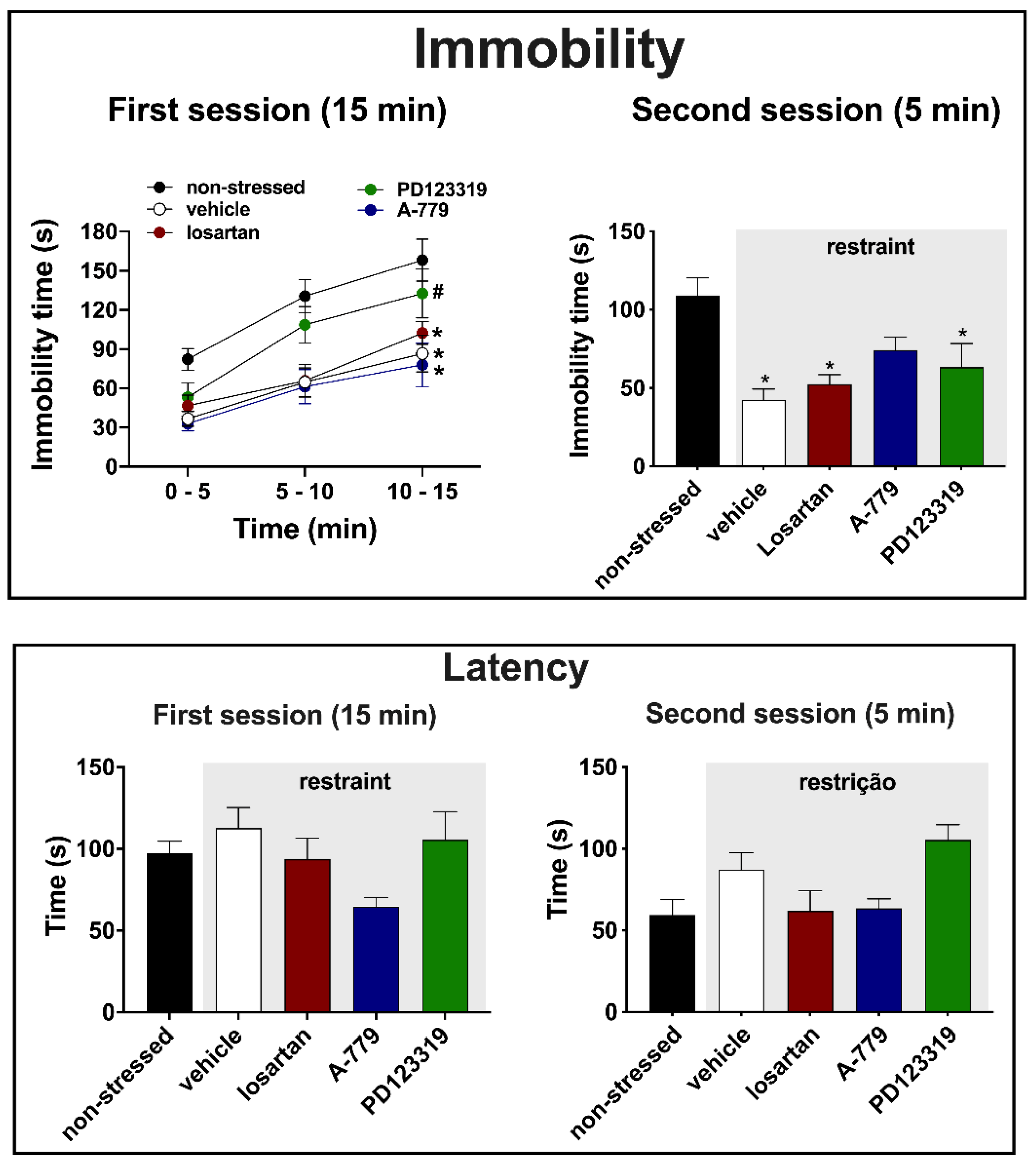
Immobility—analysis of immobility time at the pre-test session (first session, 15 min) indicated effect of time (F (1.804, 103.8) = 45.9, p < 0.0001) and treatment (F (4, 58) = 9.0, p < 0.0001), but without a time x treatment interaction (F (8, 115) = 0.9, p = 0.4635). Post-hoc analysis revealed that restraint stress decreased the immobility time in all period analyzed in animals treated with either vehicle (p < 0.0001), the selective AT1 receptor antagonist losartan (p < 0.0001) or the selective MAS receptor antagonist A-779 (p < 0.0001), when compared to the non-stressed group (Figure 2). The effect of restraint stress was not identified in animals subjected to microinjection of the selective AT2 receptor antagonist PD123319 into the MeA (p = 0.3567) (Figure 2). Besides, analysis indicated that immobility values of PD123319-treated animals were higher in relation to the vehicle group (p < 0.05) (Figure 2).
Regarding the immobility time during the test session (second session, 5 min), the analysis indicated differences between the experimental groups (F (4, 58) = 7.9, p < 0.0001) (Figure 2). Post-hoc analysis revealed that restraint stress decreased the immobility time in animals treated with either vehicle (p < 0.0001), losartan (p = 0.0034) or PD123319 (p = 0.0329) in the MeA, when compared to the non-stressed group (Figure 2). However, the effect of restraint stress was not identified in animals treated with A-779 into the MeA (p = 0.1179) (Figure 2).
Latency—analysis of latency to the first bout of immobility during the first session of the FST did not indicate differences between the experimental groups (F (4, 58) = 2.164, p = 0.0843) (Figure 2). Conversely, analysis during the second session indicated a significant effect (F (4, 58) = 2.828, p = 0.0375) (Figure 2). However, post-hoc analysis did not reveal specific difference between the experimental groups (p > 0.05) (Figure 2).
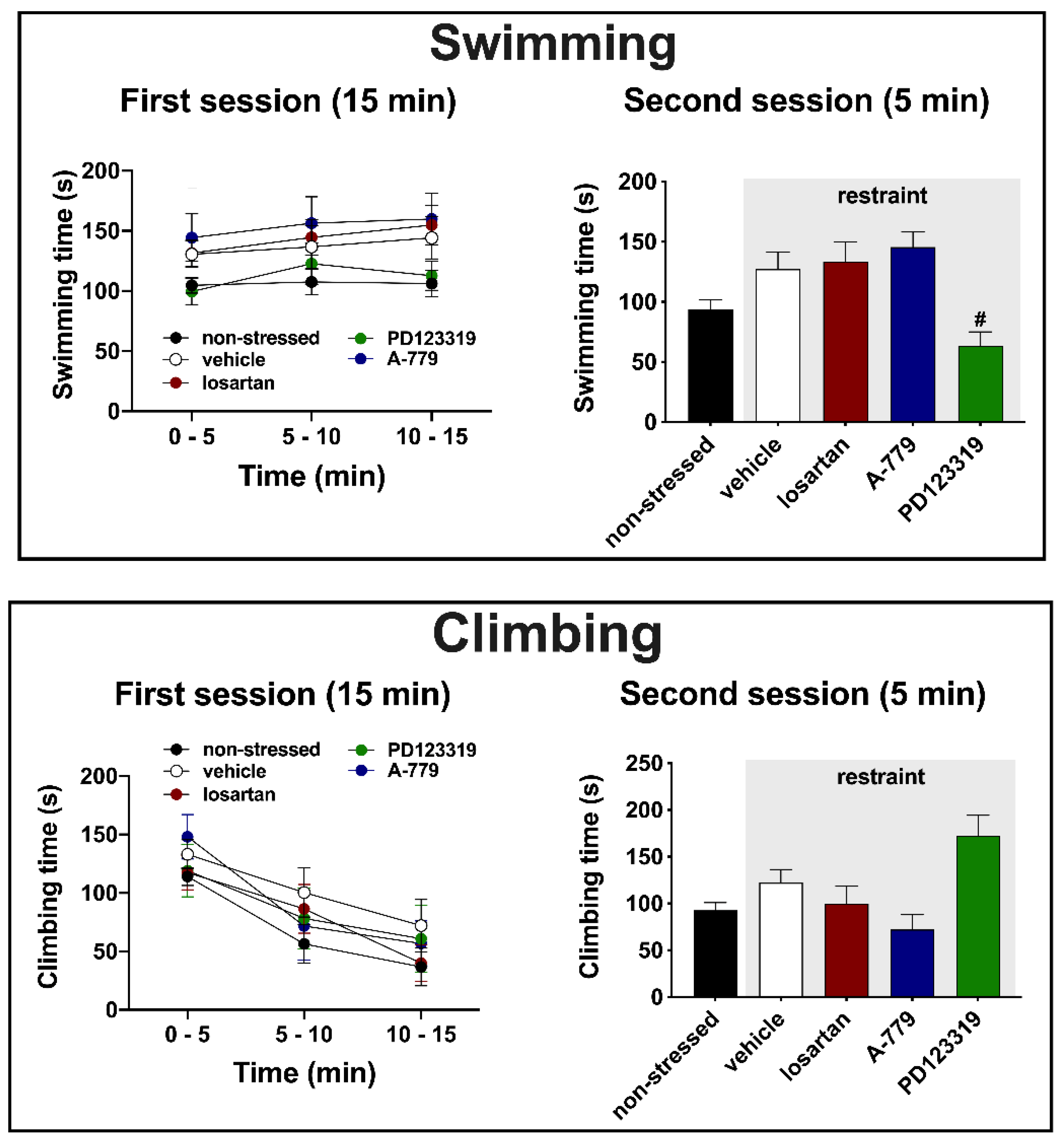
Swimming—analysis of swimming time during the first session did not indicate effect of either time (F (1.860, 103.8) = 1.510, p = 0.2263) or treatment (F (4, 58) = 2.46, p = 0.0557) or time x treatment interaction (F (8, 115) = 0.1770, p = 0.9936) (Figure 3). Conversely, analysis of swimming time during the second session indicated difference between the experimental groups (F (4, 58) = 4.074, p = 0.0075) (Figure 3). Post-hoc analysis revealed a decrease in the swimming time in animals treated with PD123319 (p = 0.0314) in relation to the vehicle group (Figure 3).
Climbing—analysis of climbing time during the first session indicated effect of time (F (1.860, 103.8) = 20.3, p < 0.0001), but without influence of treatment (F (4, 58) = 0.8928, p = 0.4742) and time x treatment interaction (F (8, 115) = 0.3281, p = 0.9537) (Figure 3). Analysis of climbing during the second session also indicated significant effect (F (4, 58) = 4.22, p = 0.0062) (Figure 3). However, post-hoc analysis did not reveal specific differences between the experimental groups (p > 0.05) (Figure 3).

4. Discussion

Results reported in the present study provide the first evidence of an involvement of MeA angiotensinergic neurotransmission in behavioral changes evoked by aversive threats. Indeed, we observed that exposure to a 60 min session of restraint session decreased the immobility during both the first (15 min session performed immediately after the restraint) and second (5 min session performed 24 h after the first session) sessions of the FST. However, we did not identify effect of restraint stress in latency to the first bout of immobility, as well as in swimming and climbing behaviors in the FST. Regarding the role of angiotensinergic receptors in the MeA, the treatment with the selective AT2 receptor antagonist PD123319 inhibited the restraint-evoked decrease in immobility during the first session, but without affecting the change during the second session. Furthermore, the effect of restraint in immobility during the second session was not identified in rats treated with the selective MAS receptor antagonist A-779 in the MeA. Finally, our results revealed a decrease in the swimming behavior in the second session of the FST in animals that received the selective AT2 antagonist into the MeA.
Previous studies provided controversial results regarding the effect of stress in behaviors in the FST. Indeed, either increase, decrease or absence of effect in immobility and others behavioral parameters in the FST were reported following exposure to an acute session of stress [4,5,6,54,55,56,57,58]. Some evidence indicated that these discrepant findings might be related to differences in the experimental protocols, such as interval between the end of stress session and the start of FST, housing conditions (collective x isolated), and intensity and characteristics (escapable or inescapable) of the aversive stimulus [4,54]. Age has also been reported as a potential factor affecting the stress effects on rodent behaviors in the FST [5].
Specifically regarding the restraint stress, it was reported decrease in immobility and increase in swimming behavior immediately after an acute 60 min session of restraint stress [4]. Nevertheless, opposite effect was observed after 2 h of acute restraint stress [6], thus suggesting that effects in the FST are affected by duration of restraint. Increase in immobility was also reported in the FST 40 min after a 7 h session of restraint [57,58], thus reinforcing the idea that longer exposition to restraint stress is related to increased immobility. Therefore, the findings obtained in the present study of decrease in immobility during both the first and second sessions of the FST corroborate results reported by Armario et al. [4]. However, to the best of our knowledge, our results are the first evaluating the behavior in the FST 24 h after an acute session of restraint in animals submitted to a previous session of forced swimming (i.e., pre-test session). Indeed, Armario et al. [4] did not identify effect of 60 min session of restraint in the FST 24 h later in animals that were not submitted to a previous session of forced swimming.
A role of MeA in behavioral responses in the FST is supported by evidence obtained in non-stressed animals that demonstrated decrease in immobility following local MeA treatment with imipramine or a selective V1b receptor antagonist [32,34]. Regarding angiotensinergic receptors in the MeA, we reported recently that MeA treatment with losartan decreased the immobility time in the FST in non-stressed animals [42]. This study also revealed that either PD123319 or A-779 microinjection into the MeA did not affect the immobility, but altered the active behavioral pattern of climbing and swimming [42]. Thus, results reported in the present study provide new evidence regarding the control of behavioral responses in the FST by angiotensinergic receptors in the MeA. Findings documented here indicate an involvement of MeA angiotensinergic neurotransmission in restraint-evoked behavioral changes in the FST. Specifically, our results revealed that the decreased immobility evoked by restraint stress in the pre-test session is mediated, at least partly, by activation of AT2 receptors in the MeA. The absence of restraint stress effect in immobility in the second session in animals treated with A-779 in the MeA provides evidence for an involvement of the MAS receptor in this behavioral response.
Interestingly, comparison of the present results with those reported previously in non-stressed animals [42] indicates that MeA angiotensinergic neurotransmissions differently control behavior in the FST in naïve (non-stressed) and stressed animals. As mentioned above, we identified that MeA treatment with losartan 10 min before the pre-test session decreased the immobility time in the test session of the FST in non-stressed animals (behavior was not evaluated in the pre-test session) [42]. Conversely, the present study indicates an involvement of AT2 and MAS receptors in the MeA in the decreased immobility evoked by restraint stress, but without a role of local AT1 receptors. Although surprising, our results are supported by previous evidence of different effects of pharmacological treatments of limbic structures in the innate anxiety and anxiogenic response induced by restraint stress in the elevated plus maze (EPM) [59,60]. For instance, Campos et al. [59] reported that the anxiogenic effect observed in naïve animals in the EPM following ventral hippocampus treatment with an endocannabinoid reuptake inhibitor shifted to an anxiolytic effect in animals submitted to restraint stress (i.e., the endocannabinoid facilitation inhibited restraint-evoked anxiogenic response). Similar effect was observed with medial prefrontal cortex treatment with the phytocannabinoid cannabidiol [60]. We have also identified that bed nucleus of stria terminalis treatment with a CB2 cannabinoid receptor antagonist led to an anxiogenic effect in the EPM in naïve animals, whereas the same treatment inhibited the anxiogenic effect evoked by restraint stress [61]. Thus, as reported previously for behaviors related to anxiety, the results reported here taken together with those of Moreno-Santos et al. [42] indicate that behavioral control in the FST by angiotensinergic mechanisms in the MeA is influenced by previous stressful experience. In this sense, to the best of our knowledge, our findings are the first indicating an influence of stressful experience in neurobiological mechanisms regulating behavior in the FST.
The decreased swimming and tendency of increase in climbing (42% increase in relation to vehicle group) during the second session of the FST in animals treated with the selective AT2 receptor antagonist in the MeA are, at least partly, in line with previous results [42]. Indeed, this previous study identified increased climbing and a tendency of decreased swimming in non-stressed animals treated with PD123319 in the MeA. Previous pharmacological studies investigating the effects of antidepressant drugs in the FST provided evidence that active behaviors, such as swimming and climbing, seem to be regulated by specific monoaminergic encephalic circuits [50]. Indeed, these studies identified that selective serotonin reuptake inhibitors increased specifically the swimming behavior, whereas selective noradrenaline uptake inhibitors acted mainly increasing climbing [49,62,63,64,65]. Therefore, the control of active behaviors during the FST by AT2 receptor in the MeA might be mediated by stimulation of facilitatory afferences to serotoninergic circuits and/or inhibition of noradrenergic pathways. Facilitation of local serotoninergic neurotransmission and/or inhibition of noradrenergic mechanisms within the MeA might also contribute. Nevertheless, further studies are needed to clarify an interaction between MeA angiotensinergic neurotransmission and monoaminergic pathways in the control of behaviors in the FST.
In summary, the findings reported here indicate that an acute 60 min session of restraint stress decreases the immobility in the first (15 min) and second (5 min) sessions of the FST in rats. The decrease in immobility in the first session seems to be mediated by AT2 receptors activation in the MeA, whereas MAS receptors is potentially involved in the behavioral change evoked by restraint stress during the second session. Our results also confirm a role of MeA AT2 receptors controlling the pattern of active behaviors (i.e., climbing and swimming) during the FST.

Author Contributions

Conceptualization, C.M.-C., W.C.-F., and C.C.C.; Methodology, C.M.-C., W.C.-F., and C.C.C.; Validation, C.M.-C., W.C.-F., and L.L.R.-S.; Formal Analysis, C.M.-C., W.C.-F., and L.L.R.-S.; Investigation, C.M.-C., W.C.-F., and L.L.R.-S.; Resources, C.C.C.; Data Curation, C.C.C.; Writing—Original Draft Preparation, C.M.-C. and W.C.-F.; Writing—Review and Editing, L.L.R.-S. and C.C.C.; Visualization, C.M.-C., W.C.-F., and C.C.C.; Supervision, C.C.C.; Project Administration, C.C.C.; Funding Acquisition, C.C.C. All authors have read and agreed to the published version of the manuscript.

Funding

This research was funded by São Paulo Research Foundation (FAPESP) (grant # 2017/19249-0 and 2019/24478-3), National Council for Scientific and Technological Development (CNPq) (grants # 431339/2018-0), and Scientific Support and Development Program of School of Pharmaceutical Sciences (UNESP). CMC was a FAPESP undergraduate fellow (FAPESP, 2018/23686-9), WCF was a FAPESP PhD fellow (2016/05218-2), and LLRS was a CNPq MSc fellow. CCC is a CNPq research fellow (process # 304108/2018-9).

Institutional Review Board Statement

This study was approved by the Ethical Committee for Animal Use of the School of Pharmaceutical Sciences/São Paulo State University (UNESP) (approval # 12/2018), which complies with Brazilian and international guidelines for animal use and welfare. The Ethical Committee for the Use of Animals of the School of Pharmaceutical Sciences (São Paulo State University - UNESP) approved the present study on October 24, 2018.

Informed Consent Statement

Not applicable.

Data Availability Statement

All relevant data are presented.

Conflicts of Interest

The authors declare no conflict of interest.

References

  1. Vian, J.; Pereira, C.; Chavarria, V.; Köhler, C.; Stubbs, B.; Quevedo, J.; Kim, S.-W.; Carvalho, A.F.; Berk, M.; Fernandes, B.S. The renin–angiotensin system: A possible new target for depression. BMC Med. 2017, 15, 144. [Google Scholar] [CrossRef]
  2. Jakubovski, E.; Bloch, M.H. Prognostic Subgroups for Citalopram Response in the STAR*D Trial. J. Clin. Psychiatry 2014, 75, 738–747. [Google Scholar] [CrossRef] [Green Version]
  3. Papaioannou, A.; Gerozissis, K.; Prokopiou, A.; Bolaris, S.; Stylianopoulou, F. Sex differences in the effects of neonatal handling on the animal’s response to stress and the vulnerability for depressive behaviour. Horm. Behav. 2002, 129, 131–139. [Google Scholar] [CrossRef]
  4. Armario, A.; Gil, M.; Marti, J.; Pol, O.; Balasch, J. Influence of various acute stressors on the activity of adult male rats in a holeboard and in the forced swim test. Pharmacol. Biochem. Behav. 1991, 39, 373–377. [Google Scholar] [CrossRef]
  5. Bernal-Morales, B.; Contreras, C.M.; Cueto-Escobedo, J. Acute restraint stress produces behavioral despair in weanling rats in the forced swim test. Behav. Process. 2009, 82, 219–222. [Google Scholar] [CrossRef] [PubMed]
  6. Sevgi, S.; Ozek, M.; Erogly, L. L-NAME prevents anxiety-like and depression-like behavior in rats exposed to restraint stress. Methods Find. Exp. Clin. Pharmacol. 2006, 28, 95–99. [Google Scholar] [CrossRef]
  7. Suvrathan, A.; Tomar, A.; Chattarji, S. Effects of chronic and acute stress on rat behaviour in the forced-swim test. Stress 2010, 13, 533–540. [Google Scholar] [CrossRef] [PubMed]
  8. Sigmund, C.D.; Diz, D.I.; Chappell, M.C. No Brain Renin-Angiotensin System Déjà vu All over Again? Hypertension 2017, 69, 1007–1010. [Google Scholar] [CrossRef]
  9. Santos, R.A.S.; Sampaio, W.O.; Alzamora, A.C.; Motta-Santos, D.; Alenina, N.; Bader, M.; Campagnole-Santo, M.J. The ACE2/Angiotensin-(1-7)/Mas axis of the renin-angiotensin system: Focus on Angiotensin-(1-7). Physiol. Rev. 2018, 98, 505–553. [Google Scholar] [CrossRef] [PubMed] [Green Version]
  10. Fontes, M.A.P.; Martins Lima, A.; Santos, R.A.S. Dos Brain angiotensin-(1–7)/Mas axis: A new target to reduce the cardiovascular risk to emotional stress. Neuropeptides 2016, 56, 9–17. [Google Scholar] [CrossRef] [PubMed]
  11. Gironacci, M.M.; Vicario, A.; Cerezo, G.; Silva, M.G. The depressor axis of the renin–angiotensin system and brain disorders: A translational approach. Clin. Sci. 2018, 132, 1021–1038. [Google Scholar] [CrossRef]
  12. Karnik, S.S.; Unal, H.; Kemp, J.R.; Tirupula, K.C.; Eguchi, S.; Vanderheyden, P.M.L.; Thomas, W.G. International Union of Basic and Clinical Pharmacology. XCIX. Angiotensin Receptors: Interpreters of Pathophysiological Angiotensinergic Stimuli [corrected]. Pharmacol. Rev. 2015, 67, 754–819. [Google Scholar] [CrossRef] [PubMed] [Green Version]
  13. Bali, A.; Jaggi, A.S. Angiotensin as stress mediator: Role of its receptor and interrelationships among other stress mediators and receptors. Pharmacol. Res. 2013, 76, 49–57. [Google Scholar] [CrossRef] [PubMed]
  14. Saavedra, J.M. Beneficial effects of Angiotensin II receptor blockers in brain disorders. Pharmacol. Res. 2017, 125, 91–103. [Google Scholar] [CrossRef]
  15. De Melo, L.A.; Almeida-Santos, A.F. Neuropsychiatric Properties of the ACE2/Ang-(1-7)/Mas Pathway: A Brief Review. Protein Pept. Lett. 2019, 27, 476–483. [Google Scholar] [CrossRef]
  16. Saavedra, J.M.; Armando, I.; Bregonzio, C.; Juorio, A.; Macova, M.; Pavel, J.; Sanchez-Lemus, E. A Centrally Acting, Anxiolytic Angiotensin II AT1 Receptor Antagonist Prevents the Isolation Stress-Induced Decrease in Cortical CRF1 Receptor and Benzodiazepine Binding. Neuropsychopharmacology 2006, 54, 271–281. [Google Scholar] [CrossRef] [PubMed] [Green Version]
  17. Saavedra, J.M.; Ando, H.; Armando, I.; Baiardi, G.; Bregonzio, C.; Jezova, M.; Zhou, J. Brain Angiotensin II, an Important Stress Hormone: Regulatory Sites and Therapeutic Opportunities. Ann. N. Y. Acad. Sci. 2004, 1018, 76–84. [Google Scholar] [CrossRef]
  18. Watanabe, T.; Fujioka, T.; Hashimoto, M.; Nakamura, S. Stress and Brain Angiotensin II Receptors. Crit. Rev. Neurobiol. 1998, 12, 305–317. [Google Scholar] [CrossRef]
  19. Wright, J.W.; Harding, J.W. Brain renin-angiotensin—A new look at an old system. Prog. Neurobiol. 2011, 95, 49–67. [Google Scholar] [CrossRef]
  20. Ayyub, M.; Najmi, A.; Akhtar, M. Protective Effect of Irbesartan an Angiotensin (AT1) Receptor Antagonist in Unpredictable Chronic Mild Stress Induced Depression in Mice. Drug Res. Stuttg. 2016, 67, 59–64. [Google Scholar] [CrossRef] [PubMed]
  21. Ping, G.; Qian, W.; Song, G.; Zhaochun, S. Valsartan reverses depressive/anxiety-like behavior and induces hippocampal neurogenesis and expression of BDNF protein in unpredictable chronic mild stress mice. Pharmacol. Biochem. Behav. 2014, 124, 5–12. [Google Scholar] [CrossRef] [PubMed]
  22. VIJAYAPANDI, P.; NAGAPPA, A.N. Biphasic Effects of Losartan Potassium on Immobility in Mice. Yakugaku Zasshi 2005, 125, 653–657. [Google Scholar] [CrossRef] [PubMed] [Green Version]
  23. Gard, P.R.; Mandy, A.; Sutcliffe, M.A. Evidence of a possible role of altered angiotensin function in the treatment, but not etiology, of depression. Biol. Psychiatry 1999, 45, 1030–1034. [Google Scholar] [CrossRef]
  24. Diniz, C.R.A.F.; Casarotto, P.C.; Fred, S.M.; Biojone, C.; Castrén, E.; Joca, S.R.L. Antidepressant-like effect of losartan involves TRKB transactivation from angiotensin receptor type 2 (AGTR2) and recruitment of FYN. Neuropharmacology 2018, 135, 163–171. [Google Scholar] [CrossRef] [PubMed]
  25. Almeida-Santos, A.F.; Kangussu, L.M.; Moreira, F.A.; Santos, R.A.S.; Aguiar, D.C.; Campagnole-Santos, M.J. Anxiolytic- and antidepressant-like effects of angiotensin-(1-7) in hypertensive transgenic (mRen2)27 rats. Clin. Sci. 2016, 130, 1247–1255. [Google Scholar] [CrossRef] [PubMed]
  26. Kangussu, L.M.; Almeida-Santos, A.F.; Bader, M.; Alenina, N.; Fontes, M.A.Ô.P.; Santos, R.A.S.; Aguiar, D.C.; Campagnole-Santos, M.J. Angiotensin-(1-7) attenuates the anxiety and depression-like behaviors in transgenic rats with low brain angiotensinogen. Behav. Brain Res. 2013. [Google Scholar] [CrossRef] [PubMed]
  27. Saavedra, J.M.; Armando, I. Angiotensin II AT2 Receptors Contribute to Regulate the Sympathoadrenal and Hormonal Reaction to Stress Stimuli. Cell. Mol. Neurobiol. 2018, 38, 85–108. [Google Scholar] [CrossRef] [PubMed]
  28. Cullinan, W.E.; Herman, J.P.; Battaglia, D.F.; Akil, H.; Watson, S.J. Pattern and time course of immediate early gene expression in rat brain following acute stress. Neuroscience 1995, 64, 477–505. [Google Scholar] [CrossRef]
  29. Furlong, T.M.; McDowall, L.M.; Horiuchi, J.; Polson, J.W.; Dampney, R.A.L. The effect of air puff stress on c-Fos expression in rat hypothalamus and brainstem: Central circuitry mediating sympathoexcitation and baroreflex resetting. Eur. J. Neurosci. 2014, 39, 1429–1438. [Google Scholar] [CrossRef]
  30. Davern, P.J.; Head, G.A. Role of the medial amygdala in mediating responses to aversive stimuli leading to hypertension. Clin. Exp. Pharmacol. Physiol. 2011, 38, 136–143. [Google Scholar] [CrossRef]
  31. Estrada, C.M.; Ghisays, V.; Nguyen, E.T.; Caldwell, J.L.; Streicher, J.; Solomon, M.B. Estrogen signaling in the medial amygdala decreases emotional stress responses and obesity in ovariectomized rats. Horm. Behav. 2018, 98, 33–44. [Google Scholar] [CrossRef]
  32. Salomé, N.; Stemmelin, J.; Cohen, C.; Griebel, G. Differential roles of amygdaloid nuclei in the anxiolytic- and antidepressant-like effects of the V1b receptor antagonist, SSR149415, in rats. Psychopharmacol. Berl. 2006, 187, 237–244. [Google Scholar] [CrossRef] [PubMed]
  33. Kazuaki, K.; Hiroaki, A.; Hironaka, A.; Susumu, O. Effects of Minaprine and Sulpiride Injected into the Amygdaloid Nucleus on the Duration of Immobility in Rats Forced to Swim. Jpn. J. Pharmacol. 1990, 53, 411–413. [Google Scholar] [CrossRef] [Green Version]
  34. Araki, H.; Kawashima, K.; Uchiyma, Y.; Aihara, H. Involvement of amygdaloid catecholaminergic mechanism in suppressive effects of desipramine and imipramine on duration of immobility in rats forced to swim. Eur. J. Pharmacol. 1985, 113, 313–318. [Google Scholar] [CrossRef]
  35. Kazuaki Kawashima; Hiroaki Araki; Yoshimi Uchiyama; Hironaka Aihara Amygdaloid catecholaminergic mechanisms involved in suppressive effects of electroconvulsive shock on duration of immobility in rat forced to swim. Eur. J. Pharmacol. 1987, 141, 1–6. [CrossRef]
  36. Lenkei, Z.; Palkovits, M.; Corvol, P.; Llorens-Cortès, C. Expression of Angiotensin Type-1 (AT1) and Type-2 (AT2) Receptor mRNAs in the Adult Rat Brain: A Functional Neuroanatomical Review. Front. Neuroendocrinol. 1997, 18, 383–439. [Google Scholar] [CrossRef] [PubMed]
  37. Lynch, K.R.; Hawelu-Johnson, C.L.; Guyenet, P.G. Localization of brain angiotensinogen mRNA by hybridization histochemistry. Brain Res. 1987, 388, 149–158. [Google Scholar] [CrossRef]
  38. Von Bohlen und Halbach, O. The renin-angiotensin system in the mammalian central nervous system. Curr. Protein Pept. Sci. 2005, 6, 355–371. [Google Scholar] [CrossRef] [PubMed]
  39. De Kloet, A.D.; Wang, L.; Ludin, J.A.; Smith, J.A.; Pioquinto, D.J.; Hiller, H.; Steckelings, U.M.; Scheuer, D.A.; Sumners, C.; Krause, E.G. Reporter mouse strain provides a novel look at angiotensin type-2 receptor distribution in the central nervous system. Brain Struct. Funct. 2016, 221, 891–912. [Google Scholar] [CrossRef] [Green Version]
  40. Paxinos, G.; Watson, C. The Rat Brain in Stereotaxic Coordinates, 3rd ed.; Acdemic Press: San Diego, CA, USA, 1997. [Google Scholar]
  41. Fortaleza, E.A.T.; Tavares, R.F.; Corrêa, F.M.A. The medial amygdaloid nucleus modulates cardiovascular responses to acute restraint in rats. Neuroscience 2009, 159, 717–726. [Google Scholar] [CrossRef]
  42. Moreno-Santos, B.; Marchi-Coelho, C.; Costa-Ferreira, W.; Crestani, C.C. Angiotensinergic receptors in the medial amygdaloid nucleus differently modulate behavioral responses in the elevated plus-maze and forced swimming test in rats. Behav. Brain Res. 2021, 397, 112947. [Google Scholar] [CrossRef] [PubMed]
  43. Fortaleza, E.A.T.; Ferreira-Junior, N.C.; Lagatta, D.C.; Resstel, L.B.M.; Corrêa, F.M.A. The medial amygdaloid nucleus modulates the baroreflex activity in conscious rats. Auton. Neurosci. 2015, 193, 44–50. [Google Scholar] [CrossRef] [PubMed] [Green Version]
  44. Petit-Demouliere, B.; Chenu, F.; Bourin, M. Forced swimming test in mice: A review of antidepressant activity. Psychopharmacology Berl. 2005, 177, 245–255. [Google Scholar] [CrossRef] [PubMed]
  45. Almeida, J.; Duarte, J.O.; Oliveira, L.A.; Crestani, C.C. Effects of nitric oxide synthesis inhibitor or fluoxetine treatment on depression-like state and cardiovascular changes induced by chronic variable stress in rats. Stress 2015, 18, 462–474. [Google Scholar] [CrossRef] [PubMed]
  46. PORSOLT, R.D.; LE PICHON, M.; JALFRE, M. Depression: A new animal model sensitive to antidepressant treatments. Nature 1977, 266, 730–732. [Google Scholar] [CrossRef]
  47. Porsolt, R.D.; Anton, G.; Blavet, N.; Jalfre, M. Behavioural despair in rats: A new model sensitive to antidepressant treatments. Eur. J. Pharmacol. 1978, 47, 379–391. [Google Scholar] [CrossRef]
  48. Abelaira, H.M.; Reus, G.Z.; Quevedo, J. Animal models as tools to study the pathophysiology of depression. Rev. Bras. Psiquiatr. 2013, 35, S112–S120. [Google Scholar] [CrossRef] [PubMed] [Green Version]
  49. Detke, M.J.; Rickels, M.; Lucki, I. Active behaviors in the rat forced swimming test differentially produced by serotonergic and noradrenergic antidepressants. Psychopharmacol. Berl. 1995, 121, 66–72. [Google Scholar] [CrossRef] [PubMed]
  50. Cryan, J.F.; Valentino, R.J.; Lucki, I. Assessing substrates underlying the behavioral effects of antidepressants using the modified rat forced swimming test. Neurosci. Biobehav. Rev. 2005, 29, 547–569. [Google Scholar] [CrossRef]
  51. Oscar, C.G.; Müller-Ribeiro, F.C.D.F.; de Castro, L.G.; Martins Lima, A.; Campagnole-Santos, M.J.; Santos, R.A.S.; Xavier, C.H.; Fontes, M.A.P. Angiotensin-(1–7) in the basolateral amygdala attenuates the cardiovascular response evoked by acute emotional stress. Brain Res. 2015, 1594, 183–189. [Google Scholar] [CrossRef]
  52. Costa-Ferreira, W.; Morais-Silva, G.; Gomes-de-Souza, L.; Marin, M.T.; Crestani, C.C. The AT1 Receptor Antagonist Losartan Does Not Affect Depressive-Like State and Memory Impairment Evoked by Chronic Stressors in Rats. Front. Pharmacol. 2019, 10, 705. [Google Scholar] [CrossRef] [Green Version]
  53. Costa-Ferreira, W.; Gomes-de-Souza, L.; Crestani, C.C. Role of angiotensin receptors in the medial amygdaloid nucleus in autonomic, baroreflex and cardiovascular changes evoked by chronic stress in rats. Eur. J. Neurosci. 2021, 53, 763–777. [Google Scholar] [CrossRef]
  54. Prince, C.R.; Anisman, H. Acute and chronic stress effects on performance in a forced-swim task. Behav. Neural Biol. 1984, 42, 99–119. [Google Scholar] [CrossRef]
  55. Platt, J.E.; Stone, E. A Chronic restraint stress elicits a positive antidepressant response on the forced swim test. Eur. J. Pharmacol. 1982, 82, 179–181. [Google Scholar] [CrossRef]
  56. Weiss, J.A.Y.M.; Goodman, P.A.; Charry, J.M.; Bailey, W.H.; Losito, G.; Corrigan, S. BEHAVIORAL DEPRESSION PRODUCED BY AN UNCONTROLLABLE STRESSOR: RELATIONSHIP TO NOREPINEPHRINE, DOPAMINE, AND SEROTONIN LEVELS IN VARIOUS REGIONS OF RAT BRAIN. Brain Res. Rev. 1981, 3, 167–205. [Google Scholar] [CrossRef]
  57. Bettio, L.E.B.; Freitas, A.E.; Neis, V.B.; Santos, D.B.; Ribeiro, C.M.; Rosa, P.B.; Farina, M.; Rodrigues, A.L.S. Guanosine prevents behavioral alterations in the forced swimming test and hippocampal oxidative damage induced by acute restraint stress. Pharmacol. Biochem. Behav. 2014, 127, 7–14. [Google Scholar] [CrossRef] [Green Version]
  58. Moretti, M.; Budni, J.; dos Santos, D.B.; Antunes, A.; Daufenbach, J.F.; Manosso, L.M.; Farina, M.; Rodrigues, A.L.S. Protective Effects of Ascorbic Acid on Behavior and Oxidative Status of Restraint-Stressed Mice. J. Mol. Neurosci. 2013, 49, 68–79. [Google Scholar] [CrossRef]
  59. Campos, A.C.; Ferreira, F.R.; Guimarães, F.S.; Lemos, J.I. Facilitation of endocannabinoid effects in the ventral hippocampus modulates anxiety-like behaviors depending on previous stress experience. Neuroscience 2010, 167, 238–246. [Google Scholar] [CrossRef]
  60. Fogaça, M.V.; Reis, F.M.C.V.; Campos, A.C.; Guimarães, F.S. Effects of intra-prelimbic prefrontal cortex injection of cannabidiol on anxiety-like behavior: Involvement of 5HT1A receptors and previous stressful experience. Eur. Neuropsychopharmacol. 2014, 24, 410–419. [Google Scholar] [CrossRef]
  61. Gomes-de-Souza, L.; Bianchi, P.C.; Costa-Ferreira, W.; Tomeo, R.A.; Cruz, F.C.; Crestani, C.C. CB1 and CB2 receptors in the bed nucleus of the stria terminalis differently modulate anxiety-like behaviors in rats. Prog. Neuro-Psychopharmacol. Biol. Psychiatry 2021, 110, 110284. [Google Scholar] [CrossRef]
  62. Carlezon, W.A.; Mague, S.D.; Andersen, S.L. Enduring behavioral effects of early exposure to methylphenidate in rats. Biol. Psychiatry 2003, 54, 1330–1337. [Google Scholar] [CrossRef] [PubMed]
  63. Estrada-Camarena, E.; Fernández-Guasti, A.; López-Rubalcava, C. Antidepressant-Like Effect of Different Estrogenic Compounds in the Forced Swimming Test. Neuropsychopharmacology 2003, 28, 830–838. [Google Scholar] [CrossRef] [PubMed] [Green Version]
  64. Mague, S.D.; Pliakas, A.M.; Todtenkopf, M.S.; Tomasiewicz, H.C.; Zhang, Y.; Stevens, W.C.; Jones, R.M.; Portoghese, P.S.; Carlezon, W.A. Antidepressant-Like Effects of κ-Opioid Receptor Antagonists in the Forced Swim Test in Rats. J. Pharmacol. Exp. Ther. 2003, 305, 323–330. [Google Scholar] [CrossRef] [Green Version]
  65. Slattery, D.A.; Desrayaud, S.; Cryan, J.F. GABA B Receptor Antagonist-Mediated Antidepressant-Like Behavior Is Serotonin-Dependent. J. Pharmacol. Exp. Ther. 2005, 312, 290–296. [Google Scholar] [CrossRef] [Green Version]
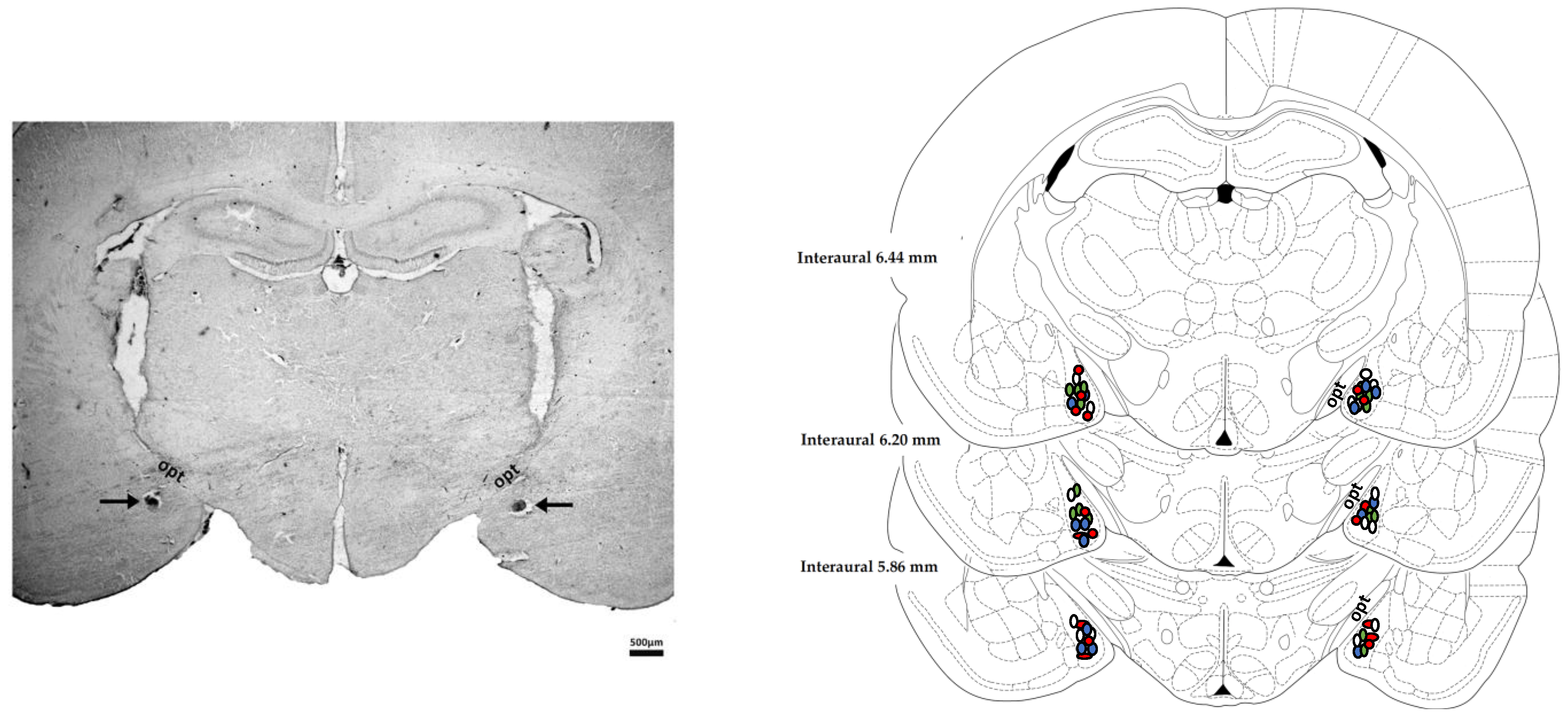
Figure 1. Microinjection sites into the MeA. (Left) Photomicrograph of a coronal brain section illustrating bilateral injection sites in the medial amygdaloid nucleus (MeA) of a representative animal. (Right) Diagrammatic representation based on Paxinos & Watson [40] showing the microinjection sites in the medial amygdaloid nucleus (MeA) of the selective AT1 receptor antagonist losartan (red circles), the selective MAS receptor antagonist A-779 (blue circles), the selective AT2 receptor antagonist PD123319 (green circles) and vehicle (white circles). The number shown in the figure can be smaller than the total number of animals used in this experiment due to the overlap of some injection sites. Opt—optical tract.
Cells 10 01217 g001
Figure 2. Immobility time and latency to the first bout of immobility in the forced swimming test (FST) of animals submitted to an acute session of restraint stress and that received bilateral microinjection into the medial amygdaloid nucleus (MeA) of the selective AT1 receptor antagonist losartan (1 nmol/100 nL, n = 9), the selective MAS receptor antagonist A-779 (0.1 nmol/100 nL, n = 10), the selective AT2 receptor antagonist PD123319 (5 nmol/100 nL, n = 8) or the vehicle (saline, 100 nL, n = 21); as well as animals who were not submitted to restraint stress (non-stressed, n = 15). (Top) Immobility time each 5 min during the pre-test session (first session, left), which was performed immediately after the end of the acute restraint session, and during the entire test session (second session, right) that occurred 24 h after the pre-test session of the FST. The bars and circles represent the mean ± SEM. Results of the first session were analyzed using two-way ANOVA followed by Bonferroni post-hoc test, whereas one-way ANOVA was used in the results of the second session. * p < 0.05 vs. non-stressed group (during the entire period for the results of the first session); # p < 0.05 vs. vehicle group (subjected to restraint stress) during the entire period. (Bottom) Latency to the first bout of immobility during the first and second sessions of the FST. The bars represent the mean ± SEM. One-way ANOVA followed by Bonferroni post-hoc test.
Cells 10 01217 g002
Figure 3. Swimming and climbing time in the forced swimming test (FST) of animals submitted to an acute session of restraint stress and that received bilateral microinjection in the medial amygdaloid nucleus (MeA) of the selective AT1 receptor antagonist losartan (1 nmol/100 nL, n = 9), the selective MAS receptor antagonist A-779 (0.1 nmol/100 nL, n = 10), the selective AT2 receptor antagonist PD123319 (5 nmol/100 nL, n = 8) or the vehicle (saline, 100 nL, n = 21); as well as animals who were not submitted to restraint stress (non-stressed, n = 15). (Top) Swimming time each 5 min during the pre-test session (first session, left), which was performed immediately after the end of the acute restraint session, and during the entire test session (second session, right) that occurred 24 h after the pre-test session of the FST. (Bottom) Climbing time each 5 min during the pre-test session (first session, left), and during the entire test session (second session, right). The bars represent the mean ± SEM. Results of the first session were analyzed using two-way ANOVA, whereas one-way ANOVA followed by Bonferroni post-hoc test was used in the results of the second session. # p < 0.05 vs. vehicle group (subjected to restraint stress).
Cells 10 01217 g003
Publisher’s Note: MDPI stays neutral with regard to jurisdictional claims in published maps and institutional affiliations.