Review

Calcium Signaling in Plant Programmed Cell Death

1
State Key Laboratory of Subtropical Silviculture, School of Forestry and Biotechnology, Zhejiang A & F University, Hangzhou 311300, China
2
Department of Biotechnology, COMSATS University Islamabad, Abbottabad Campus, University Road, Abbottabad 22060, Pakistan
*
Authors to whom correspondence should be addressed.
These authors contribute equally to this work.
Cells 2021, 10(5), 1089; https://doi.org/10.3390/cells10051089
Submission received: 21 March 2021 / Revised: 24 April 2021 / Accepted: 28 April 2021 / Published: 2 May 2021
(This article belongs to the Special Issue Programmed Cell Death Regulation in Plants)

Abstract

:
Programmed cell death (PCD) is a process intended for the maintenance of cellular homeostasis by eliminating old, damaged, or unwanted cells. In plants, PCD takes place during developmental processes and in response to biotic and abiotic stresses. In contrast to the field of animal studies, PCD is not well understood in plants. Calcium (Ca2+) is a universal cell signaling entity and regulates numerous physiological activities across all the kingdoms of life. The cytosolic increase in Ca2+ is a prerequisite for the induction of PCD in plants. Although over the past years, we have witnessed significant progress in understanding the role of Ca2+ in the regulation of PCD, it is still unclear how the upstream stress perception leads to the Ca2+ elevation and how the signal is further propagated to result in the onset of PCD. In this review article, we discuss recent advancements in the field, and compare the role of Ca2+ signaling in PCD in biotic and abiotic stresses. Moreover, we discuss the upstream and downstream components of Ca2+ signaling and its crosstalk with other signaling pathways in PCD. The review is expected to provide new insights into the role of Ca2+ signaling in PCD and to identify gaps for future research efforts.

1. Introduction

Programmed cell death (PCD) is a process that plays a fundamental role in plant development and responses to biotic and abiotic stresses [1,2]. According to the differences in the expression of the conserved PCD-inducing genes, two main types of plant PCD are distinguishable; developmental PCD (dPCD) regulated by internal factors, and environmental PCD (ePCD) induced by external stimuli [3]. The basic features of PCD include protoplast and nucleus shrinkage, chromatin condensation, cleavage of DNA and vacuolization [4]. The occurrence of PCD is meant to eliminate infected cells, thus limiting the proliferation of pathogenic bacteria [5].
It is reported that calcium (Ca2+), a universal second messenger, is critical for PCD in plants [6]. Transient changes in cytosolic Ca2+ level are rapidly induced by diverse stimuli in plants [7,8]. Substantial evidence indicates that Ca2+ plays an important role in cell death regulation [9]. The emptying of intracellular Ca2+ stores and/or alteration in intracellular Ca2+ levels has been shown to modulate cell death in almost all cell types. Ca2+ permeable channels and Ca2+ sensor CaM, CBL-CIPK and CDPK are involved in Ca2+ signal transduction and PCD.

2. The role of Ca2+ in PCD

2.1. Biotic Stresses

Plants are constantly challenged by various pathogens like viruses, bacteria, and fungi. To inhibit the spread and restrict the growth of pathogens, rapid PCD takes place at the initial infection site. Two innate immune systems play a fundamental role in PCD; PTI (pathogen-associated molecular pattern (PAMP)-triggered immunity) and ETI (effector-triggered immunity) [10,11], with the former getting more focus and hence has been better explored. The classic example of plant PCD is the hypersensitive response (HR) [12,13,14]. It is now well established that the Ca2+ signal is indispensable for the induction of HR. In soybean and tobacco, HR was prevented by Ca2+ channel blocker La3+ or EGTA, showing that Ca2+ was necessary for the induction of HR. Similarly, in Arabidopsis, Pseudomonas syringae-induced HR was preceded by an increases in cytosolic Ca2+, and was blocked by LaCl3 [15]. During the reciprocal evolution of gene-for-gene interactions, the plant’s resistance (R) gene product function as a signalling adaptor for the pathogen’s avirulence (avr) gene product, leading to refinement of HR. A study focusing on the early events in HR observed a sustained Ca2+ elevation downstream of the avrRpm1/RPM1 gene-for-gene interaction in Arabidopsis challenged by Pseudomonas syringae pv. tomato [16,17,18]. Overall, these studies illustrate that the Ca2+ signal is one of the prerequisites for the induction of HR in plants.
After the perception of different biotic and abiotic stimuli, spatial and temporal changes in cytosolic free Ca2+ concentrations ([Ca2+]cyt) are frequently observed as an immediate response [19,20]. The stress-induced increases in cytosolic Ca2+ is mediated by Ca2+ transporters, such as cyclic nucleotide gated channels (CNGCs), two-pore Ca2+ channels (TPCs), Ca2+-ATPases and glutamate receptors (GLRs) [21].
CNGCs mediate Ca2+ influx and generate the Ca2+ signal, which play a fundamental role in HR induced by pathogens. It was found that CNGC2 (also called DND1), is required for the induction of HR in Arabidopsis. cAMP-and cGMP-dependent Ca2+ elevation and induction of HR were impaired in cngc2 loss-of-function mutant (also known as dnd1) [22,23]. CNGC4 is also implicated in pathogen defense; loss-of-function mutant of AtCNGC4 (dnd2/hlm1) showed remarkably similar autoimmune phenotypes to dnd1, including defects in HR [24,25,26]. Moreover, heteropolymerization of CNGC2 and CNGC4 is necessary for the pathogen-induced intracellular Ca2+ influx. Loss of function of both CNGC2 and CNGC4 disrupts the downstream Ca2+-dependent pathogen signaling leading to HR [27]. Two other CNGC channels AtCNGC11 and AtCNGC12 also play a significant role in plant PCD by mediating Ca2+ fluxes [28,29]. Using electrophysiology, Zhang (2019) showed that CNGC12, but not CNGC11, is an active Ca2+-permeable channel in Xenopus oocytes. CNGC11 and CNGC12 knockout mutant plants exhibited partially decreased resistance to an avirulent oomycete pathogen Hyaloperonospora parasitica as well as the bacterial pathogen Pseudomonas syringae [30,31,32]. Interestingly, a 3 kb deletion across AtCNGC11 and AtCNGC12 resulted in a novel, but functional chimeric AtCNGC11/12. The mutant, named constitutive expresser of PR genes 22 (cpr22), exhibited increased resistance to pathogen infection in the hemizygous state and conditional lethality in the homozygous state [32,33]. Furthermore, HR-like spontaneous lesion formation in cpr22 was shown to be Ca2+-dependent [34]. Moreover, Ca2+ channel blockers Gd3+ and La3+ suppressed AtCNGC11/12-induced PCD. Overall, these results shed light on the critical role of CNGC11 and CNGC12 in PCD. Furthermore CNGC20, a hyperpolarization-activated Ca2+ permeable channel, regulates bak1/serk4 cell death. Notably, CNGC19, the closest homolog of CNGC20, makes a quantitative genetic contribution to bak1/serk4 cell death only in the absence of CNGC20 in Arabidopsis [35]. As 20 CNGC members have been reported in Arabidopsis, other CNGCs might also be possibly involved in the regulation of PCD in plants. In addition, the heterologous combination of CNGCs increases and enriches the regulation of PCD in plants.
Besides CNGCs, other Ca2+ transporters also play key roles in controlling intracellular Ca2+ during HR triggered by pathogens. It has been demonstrated that tonoplast-localized Ca2+ pumps ACA4/ACA11 are main players in regulating Ca2+ spike induced by bacterial elicitor peptide flg22. The double-knockout aca4/11 mutants exhibited higher basal Ca2+ levels as well as amplitude of Ca2+ signal than wild-type. These data demonstrate the important role of tonoplast-localized Ca2+ pumps in maintaining Ca2+ at homeostatic levels and for the initiation of proper PTI responses [36]. Similarly, Boursiac et al. (2010) discovered that silencing the expression of two vacuolar-localized Ca2+-ATPases resulted in spontaneous HR-like lesions and a faster pathogen response in Arabidopsis thaliana [37]. The overexpression of a rice putative voltage-gated Ca2+ permeable channel, OsTPC1, resulted in hypersensitivity to the Trichoderma viride xylanase (TvX) elicitor, with downstream events including oxidative burst, activation of OsMPK2, and hypersensitive cell death. On the other hand, these events were severely impaired in the insertional mutant, suggesting that OsTPC1 determines sensitivity to the elicitor and is a key regulator of hypersensitive cell death [38]. Glutamate receptors (GLRs) are also important transporters involved in mediating HR-induced intracellular Ca2+ influx. The increase of intracellular Ca2+, induced by HR, was impaired in the glr2.7/2.8/2.9 triple mutant, which exhibited sensitivity to pathogens. These data indicate that GLR2.7/2.8/2.9 play an important role in PTI [39].
The endoplasmic reticulum (ER) stress-induced PCD is an important response pathway in plant HR. Ca2+ pumps on the ER membrane play an important role in this process. During the bacterial blight of rice, XA10, a kind of endogenous inducer of PCD, inhibits the ER-Ca2+, leading to the production of ROS in the chloroplast, and eventually leading to cell death. In addition, CPA, a specific blocker of plant ER-type IIA Ca2+ pumps (SERCA), can induce ER stress, and via an increase in cytosolic Ca2+ concentrations, triggers PCD in soybean cells. At the same time, mitochondria release cytochrome c and caspase-like activities and thereby promote PCD together [40]. Silencing ER-localized type IIB Ca2+-ATPase (NbCA1) can induce a similar extent of PCD to that induced by pathogens [41]. The evidence shows that cell death suppressor Bax inhibitor-1 (BI-1) interacts with CaM and then coordinates with Ca2+-ATPase to influence the ion homeostasis in plant cell death regulation [42].
In recent years some progress has been made in understanding the mechanism for regulation of these calcium transporters in HR. Cyclic nucleotides, cAMP/cGMP, can bind on and activate PM channels which mediate the flux of extracellular Ca2+ and increase cytosolic Ca2+ [43,44]. The cAMP-and cGMP-dependent Ca2+ elevation and induction of HR were impaired in cngc2, indicating that CNGC2 is a typical cAMP/cGMP dependent Ca2+ channel. In addition, CNGC2 is also activated by endogenous plant elicitor peptides (PEPs), leading to cytosolic Ca2+ elevation. Physical damage to the cells results in Ca2+ elevation leading to the activation of METACASPASE4 (MC4) which in turn releases Pep1 from its protein precursor, precursor of peptide 1 (PROPEP1). The released Pep1 then binds to Pep receptors (PEPRs), which activate a cyclic GMP (cGMP)-dependent CNGC2, leading to pathogen-associated cytosolic Ca2+ elevation to regulate HR under DAMPs in PTI. cAMP and cGMP induced Ca2+ signal also regulates the Pep-dependent gene expression in Arabidopsis thaliana [45,46,47]. CNGC11 and CNGC12 are reported to be involved in PCD. Using electrophysiology, it was shown that CNGC12, but not CNGC11, functions as an active calcium channel. Furthermore, in Xenopus oocytes the cyclic nucleotide monophosphates did not modulate the activities of both CNGCs. However, the activity of CNGC12 (but not CNGC11) was significantly enhanced when CaM1 was co-expressed in oocytes [30].
LRR receptor kinase BAK1 is located on the plasma membrane, and together with FLS2/EFR forms a complex to perceive flg22, which may involve in the initial PTI-induced cytosolic Ca2+ through phosphorylation, consequently negatively regulates HR [48,49,50]. Further, BAK1 interacts with and phosphorylates CNGC20 which in turn regulates CNGC20 stability. BIK1, a key component downstream of BAK1 in plant immunity [51], activates CNGC2 and CNGC4 by phosphorylation, leading to an increase in cytosolic Ca2+ in Arabidopsis thaliana [27]. Cytosolic Ca2+ can trigger the proteolytic cleavage of BAK1 thus negatively regulating the HR. All these studies indicate that BAK1 plays a negative role in HR induced by pathogens. However, it was also discovered that overexpression of BAK1-triggered cell death was dependent on SOBIR1 in Arabidopsis thaliana [52]. Moreover, BAK1-interacting receptor kinase 1 (BIR1) was demonstrated to be involved in the negative regulation of cell death. When the function of BIR1 is compromised, BAK1 and SOBIR1 associate with each other in plants [53]. These findings suggest that maintaining the homeostasis of BAK1 through a Ca2+ dependent proteolytic process is crucial for plant HR.
The stimulus-induced Ca2+ elevation is decoded by downstream Ca2+ sensors which include CaM/CMLs, CBLs-CIPKs and CDPKs. A CaM binding protein, AtBAG6, is upregulated by stress and is involved in plant PCD. The overexpression of AtBAG6 induced the cell death phenotype in plants, which was consistent with PCD [54]. In tomatoes, the downregulation of the expression of the APR134 gene, encoding for a CaM-related protein, compromised the plant’s immune response. Similarly, increasing the expression of CML43 (an orthologue of APR134 in Arabidopsis) led to accelerated HR induced by avirulent pathogen [55,56]. These results highlight the role of the CaM-related proteins as important mediators in Ca2+-dependent signals during the plant immune responses. The extent of Ca2+ signal, ROS accumulation and PCD were significantly higher in the sensitive Brassica oleracea group than in the resistant group after inoculation with Sclerotinia sclerotiorum. Moreover, the expression of cell death-related WRKY transcription factors was also different between the sensitive and resistant B. oleracea. These findings highlight the role of WRKY transcription factors in linking the Ca2+ signal to downstream cell death in the host in response to S. sclerotiorum [57]. The calcium-dependent kinase 3 (CPK3) has been demonstrated to be a positive regulator of PCD in plants. Sphingosine or phytosphingosine (PHS) activate CPK3 which phosphorylates its binding partner, the 14-3-3 proteins. This binding leads to the disruption of the CPK3-14-3-3 protein complex and CPK3 degradation. Moreover, Arabidopsis CPK3 knockouts exhibited the FB1-resistant phenotype, revealing a novel role for CPK3 as a positive regulator of plant PCD [58]. Recently, root meristem growth factor 7 (RGF7), perceived by the RGI4/RGI5-BAK1/SERK4 receptor complexes, acts as a novel DAMP and takes an important part in Arabidopsis thaliana immunity. The expression of RGF7 precursor-encoding gene (preRGF7) is highly induced by Pseudomonas syringae, and is regulated by a signaling complex comprising of MPK3/MPK6-CPK5/CPK6-WRKY33, with MPKs and CPKs working upstream of WRKY33 [59]. It has been shown that CBL10 and CIPK6 are required for PCD triggered by kinase Pto upon recognition of Pseudomonas syringae effectors AvrPto or AvrPtoB in tomatoes. Ca2+-CBL10/CIPK6 complex promotes the accumulation of ROS by activating RbohB, and hence regulates the process of effector-triggered immunity [60]. Besides that, a study by Yang et al., (2007) has shown that BAP genes act as general negative regulators of biotic and abiotic stress-induced PCD. AtBAP1 and AtBAP2 encode small proteins containing a Ca2+-dependent phospholipid-binding C2 domain and interact with their functional partner BON1. The loss of BAP2 function results in promoting HR, while double mutant of bap1 bap2 lead to seedling lethality mediated by PAD4 and EDS1, two regulators of defense responses and cell death. On the other hand, overexpression of BAP1 or BAP2 with their partner BON1 abolishes pathogen-induced PCD [61].
Most of the previous studies in the field of plant immunity have regarded PTI and ETI as two independent parallel immunity branches, however, the latest research results show that PTI and ETI are interrelated. PTI is indispensable to ETI, plants with less efficient PTI as the first layer of the immune system also exhibit diminished plant disease resistance mediated by ETI in the second layer of the immune system. ETI can amplify PTI and induce a more lasting immunity output by enhancing the expression of core protein components in PTI, which helps plants to stimulate a strong and lasting immune response against pathogen invasion [62]. In HR-induced PCD, Ca2+ signals might serve as a link between PTI and ETI (Figure 1).

2.2. Abiotic Stress

2.2.1. Salt Stress

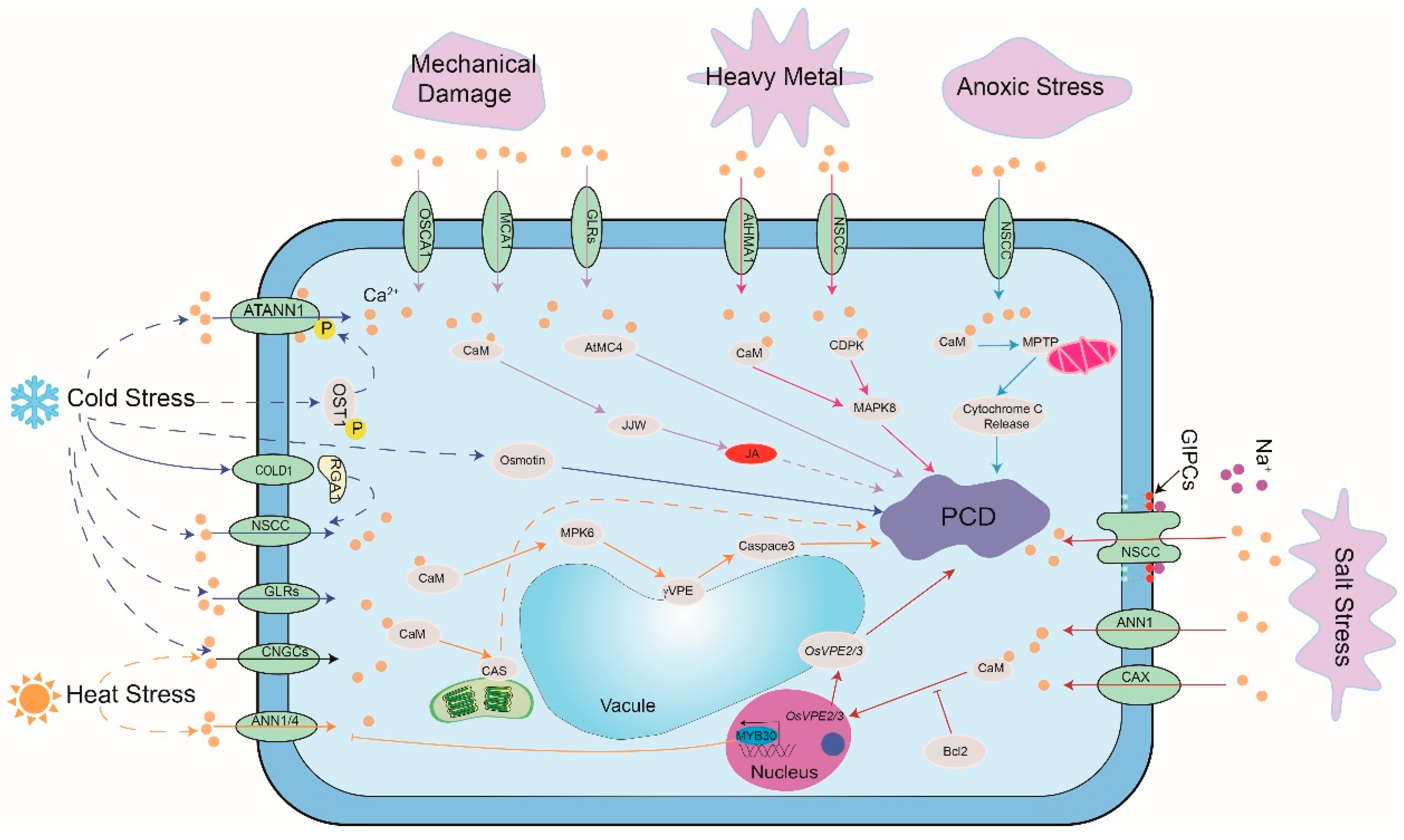
Under salt stress, the level of reactive oxygen species (ROS) in plants like grape [63], tobacco BY-2 cells [64] and barley [65] increases and results in PCD [66]. Salt stress triggers increases in cytosolic free Ca2+ concentration ([Ca2+]cyt), which, as a signaling molecule, plays an important role in regulating PCD in plant cells [67]. A low concentration (10 μmol/L) of Ca2+ channel blocker LaCl3 effectively prevented the early stages of salt stress-induced PCD in rice roots by inhibiting cytoplasmic Ca2+ elevation and ROS production [68]. Similar to the effect of La3+, the overexpression of Bcl-2, one of the most important antiapoptotic members in mammals, significantly suppressed transient cytosolic Ca2+ elevations. This led to a decrease in the expression levels of OsVPE2 and OsVPE3 (vacuolar processing enzymes), prohibition of salt stress-induced PCD, and ultimately improved salt stress tolerance in transgenic rice [69].
Besides animals and higher plants, some physiological cell death processes (considered as a kind of PCD), have also been found in many prokaryotic microorganisms like bacteria [70] and the phytoplankton [71]. Excess Ca2+ can antagonize salt stress-induced cell death in prokaryotic organism Anabaena [72]. To date, the regulation mechanism of Ca2+ signal in salt stress-induced PCD is unclear. Glycosylinositol phosphorylceramide (GIPC), as a Na+ sensor, gates the Ca2+ influx channels in plants under salt stress [73]. In addition, some Ca2+ transporters, like annexin1 (ANN1) [74] and Ca2+/H+ antiporter (CAX1) [75], take part in the alteration of cytosolic Ca2+ in plants under salt stress. However, there is still no experimental evidence to demonstrate whether these components are also involved in salt stress-induced PCD.

2.2.2. Temperature Stress

PCD can occur as a response to temperature stresses, including chilling and heat shock [76,77]. Under chilling/cold conditions, the transient elevation in cytosolic free calcium concentration ([Ca2+]cyt) acts as second messenger to stimulate a variety of downstream processes [78,79]. A previous study demonstrated that an alteration in the level of [Ca2+]cyt plays a key role in regulating PCD [80]. However, the role of Ca2+ in temperature stress-induced PCD process is only scarcely reported. It was identified that Ca2+ plays an important role in the initiation and execution of cold-induced PCD in cucumber fruit [81]. To date, multiple transmembrane transport activity-related proteins, such asannexins (ANNs) and cyclic nucleotide-gated channels (CNGCs), mediating Ca2+ influx in response to abiotic stress, have been reported [82,83]. The G-protein regulator chilling tolerance divergence 1 (COLD1) was first established to mediate the cold-induced influx of Ca2+ and confer cold sensing in rice [84,85]. A previous study found that AtANN1 was involved in heat-induced [Ca2+]cyt elevation and heat stress response [86]. A further study showed that MYB30 negatively regulated the heat shock response partially through ANN1 and ANN4 [87]. Moreover, Ca2+-permeable transporter ANNEXIN1 (AtANN1) mediated cold-induced Ca2+ influx, and acted downstream of OST1 to positively regulate freezing tolerance in Arabidopsis [79]. In plants, CNGCs are involved in low or high temperature stress and their functions are thought to result from their involvement in Ca2+ influx. OsCNGC14 and OsCNGC16 play critical roles in heat as well as cold tolerance and are modulators of Ca2+ signals in response to temperature stress in rice [88]. Furthermore, their homologs AtCNGC2 and AtCNGC4 in Arabidopsis promote plant growth under chilling and improve freezing tolerance [88]. Moreover, it was reported that disruption of moss CNGCb and Arabidopsis CNGC2 resulted in a hyper-thermosensitive phenotype, showing that these channels were involved in the control of the plant’s heat shock response (HSR) [89]. AtCNGC6 is a heat-activated PM Ca2+ channel and improves the expression of heat shock protein (HSP) genes, which enhence thermotolerance [90]. GLR3.3 and GLR3.5 were shown to mediate cold acclimation-induced chilling tolerance by regulating apoplastic H2O2 production and redox homeostasis in tomatoes [91]. Besides Ca2+ channels and transporters, the Ca2+-sensing receptor CAS has been shown to be partially involved in heat-induced chloroplast Ca2+ response [92]. In addition, cold and freezing can cause the change in a cell’s osmotic potential. The expression of osmotin can be activated by low temperature, and it is involved in cold acclimation-induced PCD in the olive tree and in arresting cold-induced Ca2+ signaling [93]. OSCA1, as an osmosensor, is responsible for [Ca2+]cyt increases induced by water deficiency in plants. Further research is needed to explore whether OSCA1 is involved in regulating cold-induced PCD [94]. In addition to the above-described channels and transporters, membrane lipid composition can also regulate the calcium-dependent heat-signaling pathway [95]. It has been suggested that MPK6 is responsible for the activation of Arabidopsis vacuolar processing enzyme (γVPE) under HS stress and played an essential role in HS-induced PCD [96].

2.2.3. Anoxic Stress

Plants undergo hypoxia stress under flooding. Root epidermal cells often form aerenchyma through programmed death in response to hypoxia stress [97]. Studies have shown that Ca2+ signaling regulates the hypoxia stress in plants. Under normal oxygen supply, both Ca2+ channel inhibitors and protein phosphatase inhibitors promote cell death in corn roots, while under insufficient oxygen supply, both Ca2+ chelator EGTA and protein kinase inhibitors prevent this process [98]. In wheat roots, hypoxia stress induced the increase in cytoplasmic Ca2+, which led to the Ca2+ accumulation in the mitochondrial matrix and the formation of mitochondrial permeability transition pores (MPTP—a factor in cell damage). These events lead to a rapid depletion of the inner membrane potential, initial contraction of the mitochondrial matrix, and release of previously accumulated Ca2+. All these events result in higher Ca2+ concentration and lead to the release of cytochrome C, and, thereby, induce PCD [99].

2.2.4. Heavy Metal Stress

Heavy metals, can also induce PCD by triggering oxidative stress via the increase of ROS production [3]. Up to now, several heavy metals, including W, Ag, Cd, Al, Zn, Li, Cu, Co, Hg, Ni, Cr, Fe, have been reported to induce PCD in different types of cells of plant species [3]. Among these heavy metals, Cd is a highly ubiquitous toxic heavy metal. Because of the high physical resemblance to Cd and its importance for plant growth and development, Ca2+ was used to alleviate the Cd-induced toxicity [100]. Ca2+ is supposed to be an intracellular “second messenger” that can mediate plant responses to the biotic and abiotic stresses such as pathogen invasion, drought, salt, heat, cold and heavy metal stress [101]. Ca2+ signatures are perceived by Ca2+ sensor proteins and evoke downstream signaling responses [102]. In Arabidopsis, CDPKs, were found to enhance Cd tolerance through intensifying H2S signal [103]. Furthermore, Ca2+ signaling is involved in the regulation of Cd-induced cytotoxicity and cell death through the activation of the MAPK and PI3K/Akt signaling pathways [104]. A copper-tolerant species Ulva compressa, when in vitro cultivated with a sublethal concentration of copper (10 μm), showed an increase in intracellular Ca2+, which took place through the activation of inositol 1,4,5 triphosphate (IP3)-sensitive calcium channels [105,106,107]. He et al. (2017) showed that Ca2+ plays significant role in prohibiting the effects of NO on Al-induced PCD in peanut root tips [108]. Ca2+ may be involved in Pb2+-mediated cell death and trigger the activity of MAPK via the CDPK pathway [109]. The Ca2+/calmodulin system also participates in response to toxicity mediated by Pb2+ and Ni2+ [110]. It has been demonstrated that Ca2+ enhances tolerance against Cr stress through interacting with hydrogen sulfide in Setaria italica. Moreover, CDPKs are involved in Cr stress by modulating the transcriptional profiling of rice roots exposed to Cr stress [111,112]. Due to the high similarity in the ionic radii of Ca2+ and other cations like Cd2+, there is a possibility of Cd2+ uptake through Ca2+ channels or transporters. AtHMA1 functions as a Ca2+/heavy metal pump [113]. The mechanism of the heavy metal-mediated Ca2+ signature and its relationship between the Ca2+ signature and heavy metal-induced PCD requires in depth investigation.

2.2.5. Mechanical Damage

Plant damage due to mechanical events such insect bite and systematic wound is inevitable in nature. Plants undergo PCD in response to mechanical damage. Different proteins have been identified which link mechanical damage to downstream Ca2+ elevation. One such candidate is MCA1, a plasma membrane protein that correlates Ca2+ influx with mechanosensing in Arabidopsis thaliana [82]. The other candidates for the perception of injury are GLRs. Plants transform injury-induced glutamate accumulation into Ca2+ signals and, thereby, transmit stress signals to distant leaves mainly by GLR3.3 and GLR 3.6 [114]. In addition, hyperosmolality-gated OSCA-family channels have also been reported to be Ca2+ permeable channels with membrane tension activation characteristics. However, whether they participate in mechanical damage induced-PCD remains to be verified. It has been reported that CaM controls the synthesis of JA by regulating the phosphorylation of the JAV1-JAZ8-WRKY51 complex, thus controlling the plant’s response to mechanical injury [115]. Upon cellular injury, cysteine protease metacaspase4 (MC4) is instantly and spatiotemporally activated with the increase of cytosolic Ca2+. MC4, then, promotes the synthesis of pep1 and induces the HR and PCD [46]. Overall, these studies demonstrate that Ca2+ signal is important for mechanical damage-induced PCD in plants (Figure 2).

2.2.6. Comparison of Ca2+ Signaling Components under Biotic and Abiotic Stresses

It is now well established that a Ca2+ signal is required for the regulation of biotic and abiotic stress-induced PCD in plants. Studies have shown that the major regulatory mechanisms between these exhibit high similarities (Table 1). Ca2+ elevation triggered by abiotic and biotic stimuli is mediated by the Ca2+ transporter on the plasma membrane and the signal is further perceived and propagated by Ca2+ sensors such as CaM, CPKs and CBLs. However, the sensors for perceiving abiotic and biotic stresses are different. For example, FLS2/BAK1 complex act as a pathogen receptor [49,50,51], OSCA1 as an osmosensor [94] and MOCA1 acts as a salt receptor in plant [73,116]. This is consistent with the generation of a Ca2+ signal in plants, for example, re-exposure to the same extent of salt stress can no longer induce Ca2+ signal after generating elevated Ca2+ under the first exposure to salt stress. On the other hand, a new Ca2+ signal can be induced by cold stress or exposure to flg22 [117,118,119]. This indicates that the mechanism of generating Ca2+ signal under various stresses varies. In addition, the genes encoding for the Ca2+ transporter proteins and their regulatory factors are different for plant PCDs under biotic and abiotic stresses. Therefore, it can be inferred that the process of PCD in plants is triggered by the Ca2+ signal acting downstream of different receptors under different stresses.

2.3. Plant Development and Postharvest Storage

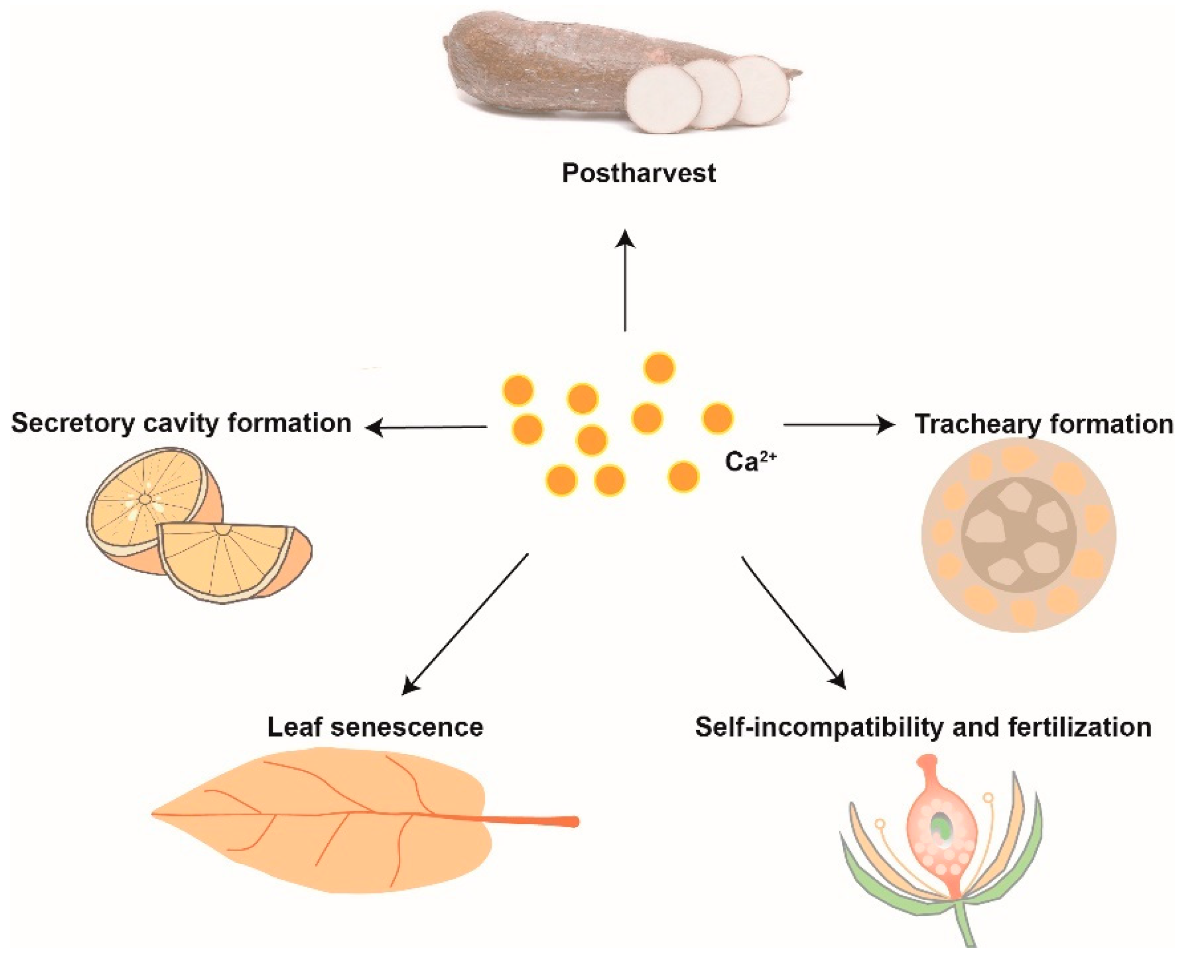
PCD is involved in several aspects of plant growth and development, such as tissue senescence, embryogenesis, self-incompatibility, and transition from bisexual to unisexual flowers [120]. Compared with abiotic-induced PCD, the molecular mechanisms of the Ca2+ signal in developmental PCD (dPCD) have only partially been explored. However, a few studies have demonstrated the crucial role of Ca2+ in dPCD processes, such as specific tissue formation, leaf senescence and fertilization. Previous research showed that tracheary element differentiation uses a specific mechanism coordinating secondary cell wall synthesis and PCD. Moreover, through pharmacological approaches (by using either EGTA to chelate Ca2+ or ruthenium red to inhibit Ca2+ influx), it has been established that the execution of cell death requires an influx of Ca2+ into the cells [121]. PPF1, a putative Ca2+ ion carrier, inhibited PCD in apical meristems of both G2 pea (Pisum sativum L.) and transgenic Arabidopsis plants by keeping the cytoplasmic Ca2+ concentration at a low level [122]. Previous reports showed that an increase in Ca2+ concentration in the nucleus may activate the PCD in secretory cavity cells, and that Ca2+ elevation improved the regulation of nuclear DNA degradation [123]. Subsequently, Bai et al. (2020) found that CgCaN, a Ca2+-dependent DNase, directly functioned in nuclear DNA degradation during the formation of secretory cavity by PCD in Citrus grandis fruit [124]. More recently, it was reported that CPK1 could control senescence-related PCD by phosphorylation of senescence master regulator ORE1 [125]. In another study on senescence-related cell death, it was found that WRKY transcription factor could be phosphorylated by CPK and then CPK-WSR1 (a WRKY regulating ROS and SA) modulated two well-defined inducers of leaf senescence, salicylic acid (SA) and reactive oxygen species (ROS), to control cell death and leaf senescence [126].
Double fertilization is a unique and significant process for flowering plant reproduction. Ca2+ plays crucial roles in pollen tube guidance and reception. During the process, it can lead to the PCD of the pollen tube and one synergid. It has been shown that the synergid controls sperm delivery through the FER signal transduction pathway to initiate and regulate their distinct Ca2+ signatures in response to the Ca2+ dynamics and growth behavior of the pollen tube [127]. Besides involvement in double fertilization, PCD is also induced by self-incompatibility (SI) in an S-specific manner incompatible pollen, which reveals a mechanism to prevent self-fertilization [128]. In Papaver rhoeas, S-protein, controlling the SI, interacts with incompatible pollen and triggers a Ca2+-dependent signature, leading to the inhibition of pollen tube growth [129,130]. In the development of the litchi flower, researchers found that the inner integument cells of male flowers underwent the PCD, which was triggered by distributional changes in Ca2+ [131].
Postharvest physiological deterioration (PPD) of cassava storage roots is a complex process, which involves ROS, Ca2+ signaling transduction, and PCD [132]. Owiti et al. (2011) showed that the expression of CaM proteins was significantly upregulated, which could be the result of an oxidative burst-induced rapid increase in Ca2+ during early PPD. During late PPD, the PCD pathway was activated due to an increase in cysteine proteases [133] (Figure 3).

2.4. Small Chemical Molecule

Many chemicals can induce PCD in plants, wherein the involvement of Ca2+ signaling has been demonstrated. An early research report showed that Ca2+ plays an important role in gallic acid-induced PCD which was effectively inhibited by a Ca2+ chelator BAPTA-AM [134]. Thaxtomin A (TXT) is a nitrated dipeptide phytotoxin produced by all plant-pathogenic Streptomyces species, and is necessary for the realization of PCD. It has been demonstrated that TXT induces the transient Ca2+ increase in cells, activates the anion channel and induces the accumulation of the defense gene PAL1, until PCD takes place. Further, Ca2+ inhibitors La3+, Gd3+, or BAPTA inhibited the TXT-induced PCD [134], showing an important role of Ca2+ in this process. In addition, it has also been demonstrated that Ca2+ is involved in Victorin C, a host-selective cyclic peptide toxin produced by Cochliobolus victoriae, that induced PCD in oats [135]. Chitosan, is a component of the cell wall of many fungi and has been widely used to mimic pathogen attack. Chitosan or oligochitosan induced PCD in soybean cells and tobacco suspension cells which was suppressed by Ca2+ channel inhibitors [136,137]. A study has shown that endopolygalacturonase (PG), a toxin produced by Sclerotinia sclerotiorum, induced a rapid increase in [Ca2+]cyt and triggered PCD in soybeans. These results were further confirmed by the observation that seedlings constitutively expressing a polygalacturonase-inhibiting protein (PGIP) did not undergo PG-induced PCD [138].

2.5. Metacaspases

Plant metacaspases (MCPs) are conserved cysteine proteases postulated as regulators of PCD. A study has reported that the expression of tomato type II metacaspase (LeMCA1) was rapidly upregulated in tomatoes during cell death induced by Botrytis cinerea, Similarly, in tobacco, the expression of NbMCA1 enhanced the resistance against Colletotrichum destructivum [139]. On the other hand, a decrease in the expression of the type II metacaspase asperata inhibited the PCD in the suspensor cells during embryogenesis in Picea [140].
Nine MCPs have been reported in Arabidopsis thaliana [141]. The in vitro catalytic activities of recombinant type II metacaspase subfamily members AtMC4 (AtMCP2d), AtMC5 and AtMC8 were found to be Ca2+-dependent while recombinant AtMC9 was active under mildly acidic conditions and not dependent on stimulation by Ca2+ [142]. As mentioned above, AtMC4 plays a positive regulatory role in both biotic and abiotic stress-induced PCD in Arabidopsis thaliana [47]. The residue Lys225 of AtMC4, a highly conserved residue among the six Arabidopsis type II MCPs, is critical for the catalytic activation by Ca2+, and essential for AtMC4-mediated activation of H2O2-induced cell death in yeast [142]. The recently resolved structure of AtMC4 revealed insights into its activation mechanism. The side chain of Lys225 in the linker domain blocks the active site by sitting directly between two catalytic residues. Activation of AtMC4 by Ca2+ and cleavage of its physiological substrate involves multiple cleavages in the linker domain [48]. MC5 was also found to mediate defense-related PCD in tobacco [143]. Another member AtMC8 regulates oxygen stress-induced PCD in Arabidopsis. The expression of AtMC8 was upregulated in UVC and H2O2 induced PCD, while the loss of AtMC8 inhibited the cell death [144]. To sum up, these results indicate that Ca2+ plays an important role in MCP-mediated PCD.

2.6. Crosstalk between Ca2+ and Other Signaling Molecules in PCD

PCD is a complex biological process. Many studies on PCD in plants have shown that PCD involves an intricate network of signaling pathways, including various molecular signals, such as Ca2+, ROS, NO and phytohormones [145]. By regulating various aspects of cellular signal transduction in plants, Ca2+ plays an essential role as a second messenger. Moreover, these different signals have a crosstalk with the Ca2+ signal and form a regulatory network for controlling PCD in plants in response to diverse stimuli. If Ca2+ is increased to the level as attained just before the onset of pathogen-induced HR in soybean, PCD would not occur. This indicates that the Ca2+ signal needs to coordinate with other signaling pathways to regulate PCD [146].
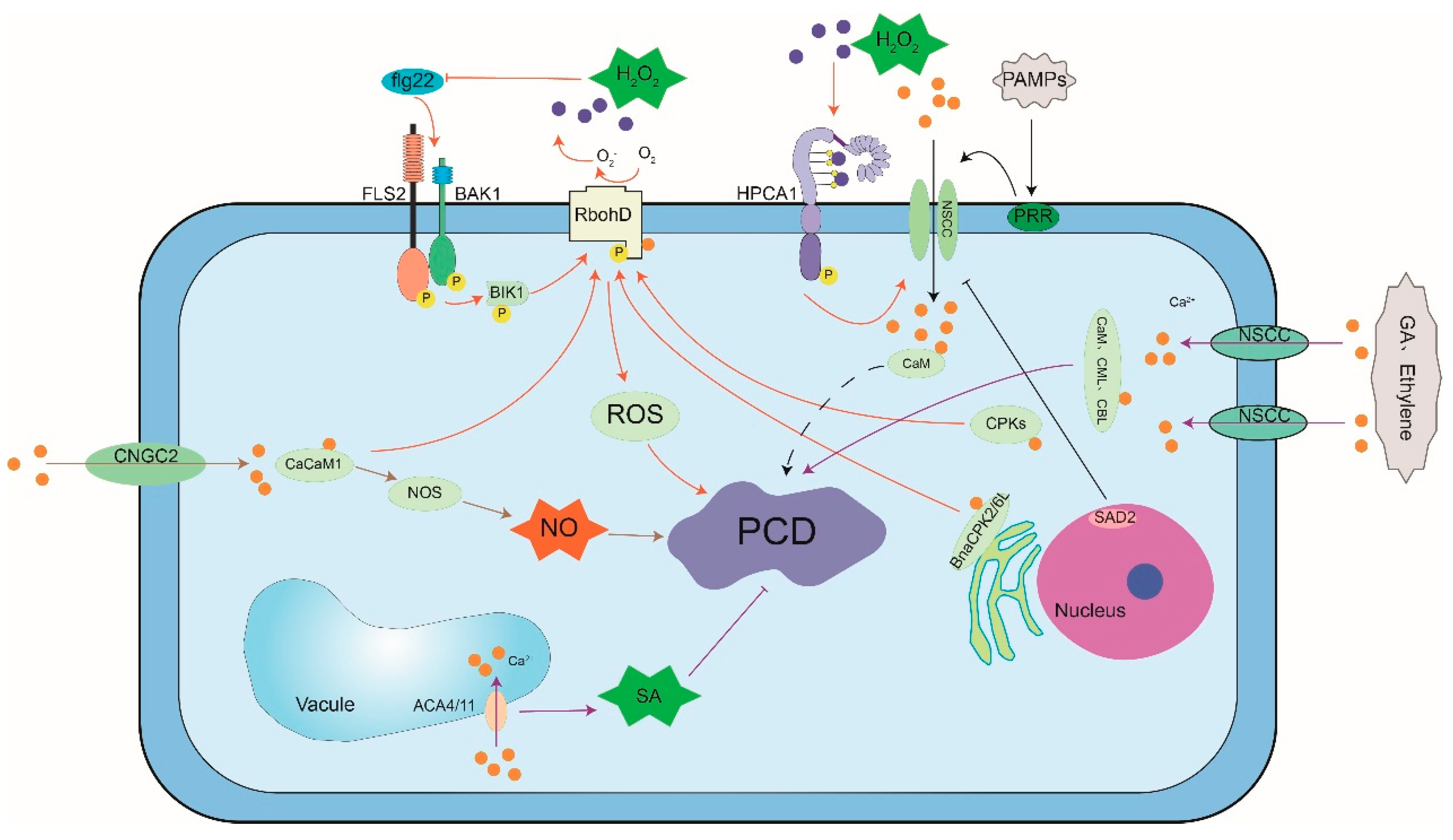
ROS signals play an important role in both biotic and abiotic stress-induced PCD. Activated in response to Ca2+ signal, CDPKs subsequently activate RBOH (respiratory burst oxidase homolog) to influence ROS in different plants. Thus, RBOH acts as a hub where Ca2+ and ROS signaling networks crosstalk [147,148,149,150]. It was reported that H2O2 stimulates a rapid influx of Ca2+ into soybean cells, which triggers physiological PCD [151]. In Arabidopsis, a mutation in the nuclear transporter SAD2 (sensitive to ABA and drought 2) is responsible for H2O2-induced cytosolic Ca2+ increase. Further research showed that SAD2 works downstream of FBR11 (fumonisin B1-resistant 11) and plays a role in Ca2+- and H2O2-mediated cell death [6]. Recently, H2O2 sensor LRR receptor kinase HPCA1 (hydrogen peroxide-induced Ca2+ increase 1) has been demonstrated to mediate H2O2-induced activation of Ca2+ channels in guard cells [152]. H2O2 may also regulate mitochondrial permeability transition by elevation of [Ca2+]cyt. Further analysis showed that the signaling pathway for [Ca2+]cyt-mediated mitochondrial permeability transition was associated with H2O2-induced in tobacco protoplasts [153]. In Arabidopsis, mechanical wounding triggered the activation of MPK8 which was dependent on two factors: its direct binding with calmodulins (CaMs) in a Ca2+-dependent manner, and phosphorylation and activation by a MAPKK MKK3. Once activated, MPK8 negatively regulates ROS accumulation by controlling the expression of the RbohD gene. These results suggest that MPK8 acts as converging point for Ca2+ and MAP kinase pathways for regulation of ROS dynamics [144,154]. BnaCPK6L/CPK2, located at the endoplasmic reticulum membrane, interact with RbohD and regulate its activity by phosphorylation. Transient expression of BnaCPK6L or overexpression of BnaCPK2 triggers ROS accumulation and HR-like cell death in Brassica napus L. [12,14].
Recent evidence indicates that NO acts as an important cellular mediator in PCD and defense responses. NO mobilizes intracellular Ca2+, while NO synthesis depends on upstream protein phosphorylation events and cytosolic free Ca2+ increase [155]. In pepper, a calmodulin gene, CaCaM1 plays important role in ROS and NO generation required for cell death and defense response [156]. In plant innate immune signaling cascades, Ca2+ increase and NO generation are crucial early steps and initiate HR to avirulent pathogens [22,157,158,159]. During this process, cytosolic Ca2+ rise could cause NO generation through CaM/CML, acting upstream of NO synthesis [22,159]. In Arabidopsis, CNGC2 mediates cyclic nucleotide monophosphate-dependent Ca2+ flux which leads to NO generation and HR. Further, the loss of function mutant of CNGC2 (DND1) did not exhibit HR in response to avirulent pathogens [22].
Plant hormones, like SA, GA, and ethylene induce Ca2+ signal and play key roles in PCD. It is reported that the double disruption of Arabidopsis vacuolar pumps ACA4 and ACA11 leads to a high frequency of apoptosis-like lesions, caused during SA-dependent PCD [22,38,160]. Therefore, these vacuolar pumps establish a link between vacuolar-mediated Ca2+ signal and PCD in plants [38]. Okadaic acid (OA), a protein phosphatase inhibitor, can completely inhibit the GA response which is induced by rapid changes in cytosolic Ca2+ through regulating the gene expression and accelerated cell death [161]. Gaseous phytohormone ethylene has been reported to be involved in cell death signaling in the aerenchyma formation in the root and stems of maize (Zea mays) [98] (Figure 4).

3. Conclusions and Perspective

In this review, we focused on the role of the Ca2+ signal in plant PCD. In recent years, various Ca2+ signaling components have been identified in the regulation of plant response to diverse stresses, including the sensors of biotic and abiotic stresses. We, hereby, reviewed their link with plant PCD. However, the upstream and downstream components of these pathways remain elusive. Moreover, how the plant senses heat, mechanical damage, and heavy metal stress and how the Ca2+ signal is regulated and transmitted to result in PCD during these stresses need further research. In addition, the crosstalk between Ca2+ and other signaling pathways is not yet clear and needs further exploration. It is also not clear whether other processes for the regulation of dPCD require the input of the Ca2+ signal. Future studies on these research gaps are expected to broaden our understanding on the role of Ca2+ signaling in PCD.

Author Contributions

H.R. wrote the first draft of abiotic-induced PCD and other parts of the manuscript and revised the manuscript. X.Z. wrote the first draft of hypersensitive response and worked with citations. W.L. drew the diagrams. J.H. proofread and revised the manuscript. G.Q. developed the concept and drew the diagrams, and acquired funding. S.L. develop the concept and revised the manuscript. All authors have read and agreed to the published version of the manuscript.

Funding

This work was supported by the Natural Science Foundation of Zhejiang Province (grant number: LY20C020001, LY18C020007), the Science and Technology Development Plan of Hangzhou (grant number: 20180432B10), the China postdoctoral Science Foundation (grant number: 2019M653803).

Institutional Review Board Statement

Not applicable.

Informed Consent Statement

Not applicable.

Data Availability Statement

Not applicable.

Conflicts of Interest

The authors declare that they have no competing interests. All authors read and approved the final manuscript.

Abbreviations

PCDProgrammed Cell Death
dPCDDevelopmental Programmed Cell Death
ePCDEnvironmental Programmed Cell Death
CNGCCyclic Nucleotide-Gated Channel
CaMCalmodulin
PPDPostharvest Physiological Deterioration
DHSD-Erythro-Sphinganine
MCPsMetacaspases
PGPolygalacturonase
MPTPMitochondrial Permeability Transition Pore
CBLCalcineurin B-Like Protein
CIPKCBL-Interacting Protein Kinase
CPKCa2+-Dependent Protein Kinase
PTIPattern-Triggered Immunity
ETIEffector-Triggered Immunity
PAMPPathogen-Associated Molecular Pattern
HRHypersensitive Response
EGTAEthylenebis (Oxyethylenenitrilo) Tetraacetic Acid
TPCsTwo-Pore Channels
CAXsCa2+/H+ exchangers
cAMP3′-5′-Cyclic Adenosine Monophosphate
cGMPCyclic Guanosine Monophosphate
PEPRsPep Receptors
DAMPsDamage-Associated Molecular Patterns
ETHEcdysis Triggering Hormone
CMLCaM-Like Protein
EFRElongation Factor Tu Receptor
ACAdenylate Cyclase
PDEPhosphodiesterase
PMPlasma Membrane
TvXTichoderma Viride Xylanase
MAPKMitogen-Activated Protein Kinase
BAPBiofilm Associated Protein
SASalicylic Acid
RBOHBRespiratory Burst Oxidase Homolog B
ROSReactive Oxygen Species
ETHEcdysis Triggering Hormone
GIPCsGlycosyl Inositol Phosphorylceramides
NOSNitric Oxide Synthase
KEAsPlastid K+ Exchange Antiporters
VPEVacuolar Processing Enzyme
PTPPermeability Transition Pore
BAPTA-AMBis-(O-Aminophenoxy)-N,N,N,N’-Tetraacetic Acid Acetoxymethyl Ester
PGIPPolygalacturonase-Inhibiting Protein
PGPyoderma Gangrenosum
HPCA1Hydrogen Peroxide Sensor
GLR Glutamate Receptors
PEPs Plant Elicitor Peptides
PEPRsExtracellular Pep Receptors
ER stressEndoplasmic Reticulum Stress
SERCAEr-Type Iia Ca2+ Pumps
PHSPhytosphingosine

References

  1. Petrov, V.; Hille, J.; Mueller-Roeber, B.; Gechev, T.S. ROS-mediated abiotic stress-induced programmed cell death in plants. Front. Plant Sci. 2015, 6, 69. [Google Scholar] [CrossRef] [PubMed] [Green Version]
  2. Mccabe, P.F.; Leaver, C.J. Programmed cell death in cell cultures. Plant Mol. Biol. 2000, 44, 359–368. [Google Scholar] [CrossRef]
  3. Sychta, K.; Tbomka, A.; Kuta, E. Insights into Plant Programmed Cell Death Induced by Heavy Metals-Discovering a Terra Incognita. Cells 2021, 10, 65. [Google Scholar] [CrossRef] [PubMed]
  4. Zhang, Q.F.; Li, J.; Bi, F.C.; Liu, Z.; Yao, N. Ceramide-Induced Cell Death Depends on Calcium and Caspase-Like Activity in Rice. Front. Plant Sci. 2020, 11, 145. [Google Scholar] [CrossRef] [Green Version]
  5. Tian, D.; Wang, J.; Zeng, X.; Gu, K.; Qiu, C.; Yang, X.; Zhou, Z.; Goh, M.; Luo, Y.; Murata-Hori, M.; et al. The Rice TAL Effecto’ Dependent Resistance Protein XA10 Triggers Cell Death and Calcium Depletion in the Endoplasmic Reticulum. Plant Cell 2014, 26, 497–515. [Google Scholar] [CrossRef] [PubMed] [Green Version]
  6. Zheng, Y.; Zhan, Q.D.; Shi, T.T.; Liu, J.; Zhao, K.J.; Gao, Y. The nuclear transporter SAD2 plays a role in calcium- and H2O2-mediated cell death in Arabidopsis. Plant J. 2020, 101, 324–333. [Google Scholar]
  7. Zhu, J.K. Abiotic stress signaling and responses in plants. Cell 2016, 167, 313–324. [Google Scholar] [CrossRef] [PubMed] [Green Version]
  8. Tian, W.; Wang, C.; Gao, Q.; Li, L.; Luan, S. Calcium spikes, waves and oscillations in plant development and biotic interactions. Nat. Plants 2020, 6, 1–10. [Google Scholar] [CrossRef] [PubMed]
  9. Orrenius, S.; Gogvadze, V.; Zhivotovsky, B. Calcium and mitochondria in the regulation of cell death. Biochem. Biophys. Res. Commun. 2015, 460, 72–81. [Google Scholar] [CrossRef]
  10. Jones, J.; Dangl, J. The plant immune system. Nature 2006, 444, 323–329. [Google Scholar] [CrossRef] [PubMed] [Green Version]
  11. Boller, T.; He, S.Y. Innate Immunity in Plants: An Arms Race Between Pattern Recognition Receptors in Plants and Effectors in Microbial Pathogens. Science 2009, 324, 742–744. [Google Scholar] [CrossRef] [PubMed] [Green Version]
  12. Pan, G.; Zhang, H.; Chen, B.; Gao, S.; Yang, B.; Jiang, Y. Rapeseed calcium-dependent protein kinase CPK6L modulates reactive oxygen species and cell death through interacting and phosphorylating RBOHD. Biochem. Biophys. Res. Commun. 2019, 518, 719–725. [Google Scholar] [CrossRef]
  13. Johansson, O.N.; Nilsson, A.K.; Gustavsson, M.B.; Backhaus, T.; Andersson, M.; Ellerström, M. A quick and robust method for quantification of the hypersensitive response in plants. Peer J. 2015, 3, e1469. [Google Scholar] [CrossRef] [PubMed] [Green Version]
  14. Wang, W.; Zhang, H.; Wei, X.; Yang, L.; Yang, B.; Zhang, L.; Li, J.; Jiang, Y. Functional characterization of calcium-dependent protein kinase (CPK) 2 gene from oilseed rape (Brassica napus L.) in regulating reactive oxygen species signaling and cell death control. Gene 2018, 651, 49–56. [Google Scholar] [CrossRef]
  15. Gao, X.; Cox, K.L.; He, P. Functions of Calcium-Dependent Protein Kinases in Plant Innate Immunity. Plants 2014, 3, 160–176. [Google Scholar] [CrossRef] [PubMed] [Green Version]
  16. Grant, M.; Brown, I.; Adams, S.; Knight, M.; Ainslie, A.; Mansfield, J. The RPM1 plant disease resistance gene facilitates a rapid and sustained increase in cytosolic calcium that is necessary for the oxidative burst and hypersensitive cell death. Plant J. 2000, 23, 441–450. [Google Scholar] [CrossRef] [PubMed]
  17. Flor, H.H. Current Status of the Gene-For-Gene Concept. Annu. Rev. Phytopathol. 1971, 9, 275–296. [Google Scholar] [CrossRef]
  18. Yang, Y.; Shah, J.; Klessig, D.F. Signal perception and transduction in plant defense responses. Genes Dev. 1997, 11, 1621–1639. [Google Scholar] [CrossRef] [Green Version]
  19. Rudd, J.J.; Franklin-Tong, V.E. Calcium signaling in plants. Cell Mol. Life Sci. 1999, 55, 214–232. [Google Scholar] [CrossRef] [PubMed]
  20. Dodd, A.N.; Kudla, J.; Sanders, D. The Language of Calcium Signalling. Annu. Rev. Plant Biol. 2010, 61, 593–620. [Google Scholar] [CrossRef] [PubMed]
  21. Wilkins, K.A.; Matthus, E.; Swarbreck, S.M.; Davies, J.M. Calcium-Mediated Abiotic Stress Signaling in Roots. Front. Plant Sci. 2016, 7, 1296. [Google Scholar] [CrossRef] [PubMed] [Green Version]
  22. Ma, W.; Smigel, A.; Tsai, Y.C.; Braam, J.; Berkowitz, G.A. Innate immunity signaling: Cytosolic Ca2+ elevation is linked to downstream nitric oxide generation through the action of calmodulin or a calmodulin-like protein. Plant Physiol. 2008, 148, 818–828. [Google Scholar] [CrossRef] [Green Version]
  23. Qi, Z.; Verma, R.; Gehring, C.; Yamaguchi, Y.; Zhao, Y.; Ryan, C.A.; Berkowita, G.A. Ca2+ signaling by plant Arabidopsis thaliana Pep peptides depends on AtPepR1, a receptor with guanylyl cyclase activity, and cGMP-activated Ca2+ channels. Proc. Natl. Acad. Sci. USA 2010, 107, 21193–21198. [Google Scholar] [CrossRef] [Green Version]
  24. Chin, K.; Defalco, T.A.; Moeder, W.; Yoshioka, K. The Arabidopsis cyclic nucleotide-gated ion channels AtCNGC2 and AtCNGC4 work in the same signaling pathway to regulate pathogen defense and floral transition. Plant Physiol. 2013, 163, 611–624. [Google Scholar] [CrossRef] [PubMed] [Green Version]
  25. Jurkowski, G.I.; Smith, R.K., Jr.; Yu, I.C.; Ham, J.H.; Sharma, S.B.; Klessig, D.F.; Fengler, K.A.; Bent, A. Arabidopsis DND2, a Second Cyclic Nucleotide-Gated Ion Channel Gene for Which Mutation Causes the “Defense, No Death” Phenotype. Mol. Plant Microbe. Interact. 2004, 17, 511–520. [Google Scholar] [CrossRef] [Green Version]
  26. Clough, S.J.; Fengler, K.A.; Yu, I.C.; Lippok, B.; Smith Jr, R.K.; Bent, A.F. The Arabidopsis dnd1 "defense, no death" gene encodes a mutated cyclic nucleotide-gated ion channel. Proc. Natl. Acad. Sci. USA 2000, 97, 9323–9328. [Google Scholar] [CrossRef] [PubMed] [Green Version]
  27. Tian, W.; Hou, C.; Ren, Z.; Wang, C.; Zhao, F.; Ahlbeck, D.D.; Hu, S.; Zhang, L.; Niu, Q.; Li, L.; et al. A calmodulin-gated calcium channel links pathogen patterns to plant immunity. Nature 2019, 572, 131–135. [Google Scholar] [CrossRef]
  28. Thuleau, P.; Aldon, D.; Cotelle, V.; Brière, C.; Ranty, B.; Galaud, J.; Mazars, C. Relationships between calcium and sphingolipid-dependent signalling pathways during the early steps of plant-pathogen interactions. Biochim. Biophys. Acta 2013, 1833, 1590–1594. [Google Scholar] [CrossRef] [PubMed] [Green Version]
  29. Abdel-Hamid, H.; Chin, K.; Moeder, W.; Yoshioka, K. High throughput chemical screening supports the involvement of Ca2+ in cyclic nucleotide-gated ion channel-mediated programmed cell death in Arabidopsis. Plant Signal Behav. 2011, 6, 1817–1819. [Google Scholar] [CrossRef] [PubMed] [Green Version]
  30. Zhang, Z.; Hou, C.; Tian, W.; Li, L.; Zhu, H.E. Electrophysiological Studies Revealed CaM1-Mediated Regulation of the Arabidopsis Calcium Channel CNGC12. Front. Plant Sci. 2019, 10, 1090. [Google Scholar] [CrossRef] [PubMed]
  31. Moeder, W.; Urquhart, W.; Ung, H.; Yoshioka, K. The Role of Cyclic Nucleotide-Gated Ion Channels in Plant Immunity. Mol. Plant 2011, 4, 442–452. [Google Scholar] [CrossRef] [PubMed]
  32. Yoshioka, K.; Moeder, W.; Kang, H.; Kachroo, P.; Masmoudi, K.; Berkowitz, G.; Klessig, D. The Chimeric Cyclic Nucleotide-Gated Ion Channel AtCNGC11/12 Activates Multiple Pathogen Resistance Responses. Plant Cell 2006, 18, 747–763. [Google Scholar] [CrossRef] [PubMed] [Green Version]
  33. Baxter, J.; Moeder, W.; Urquhart, W.; Shahinas, D.; Chin, K.; Christendat, D.; Kang, H.G.; Angelova, M.; Kato, N.; Yoshioka, K. Identification of a functionally essential amino acid for Arabidopsis cyclic nucleotide gated ion channels using the chimeric AtCNGC11/12 gene. Plant J. 2008, 56, 457–469. [Google Scholar] [CrossRef] [PubMed]
  34. Urquhart, W.; Gunawardena, A.; Moeder, W.; Ali, R.; Berkowitz, G.; Yoshioka, K. The chimeric cyclic nucleotide-gated ion channel ATCNGC11/12 constitutively induces programmed cell death in a Ca2+ dependent manner. Plant Mol. Biol. 2007, 65, 747–761. [Google Scholar] [CrossRef] [PubMed]
  35. Yu, X.; Xu, G.; Li, B.; Vespoli, L.; Liu, H.; Moeder, W.; Chen, S.; Oliveira, M.; Souza, S.; Shao, W.; et al. The Receptor Kinases BAK1/SERK4 Regulate Ca2+ Channel-Mediated Cellular Homeostasis for Cell Death Containment. Curr. Biol. 2019, 29, 3778–3790. [Google Scholar] [CrossRef]
  36. Hilleary, R.; Paez-Valencia, J.; Vens, C.; Toyota, M.; Palmgren, M.; Gilroy, S. Tonoplast-localized Ca2+ pumps regulate Ca2+ signals during pattern-triggered immunity in Arabidopsis thaliana. Proc. Natl. Acad. Sci. USA 2020, 117, 18849–18857. [Google Scholar] [CrossRef]
  37. Boursiac, Y.; Lee, S.M.; Romanowsky, S.M.; Blank, R.; Sladek, C.; Chung, W.; Harper, J. Disruption of the Vacuolar Calcium-ATPases in Arabidopsis Results in the Activation of a Salicylic Acid-Dependent Programmed Cell Death Pathway. Plant Physiol. 2010, 154, 1158–1171. [Google Scholar] [CrossRef] [Green Version]
  38. Kurusu, T.; Yagala, T.; Miyao, A.; Hirochika, H.; Kuchitsu, K. Identification of a putative voltage-gated Ca2+ channel as a key regulator of elicitor-induced hypersensitive cell death and mitogen-activated protein kinase activation in rice. Plant J. 2005, 42, 798–809. [Google Scholar] [CrossRef]
  39. Bjornson, M.; Pimprikar, P.; Nürnberger, T.; Zipfel, C. The transcriptional landscape of Arabidopsis thaliana pattern-triggered immunity. Nat. Plants 2021. [Google Scholar] [CrossRef] [PubMed]
  40. Zuppini, A.; Navazio, L.; Mariani, P. Endoplasmic reticulum stress-induced programmed cell death in soybean cells. J. Cell Sci. 2004, 117, 2591–2598. [Google Scholar] [CrossRef] [Green Version]
  41. Zhu, X.; Caplan, J.; Mamillapalli, P.; Czymmek, K.; Dinesh-Kumar, S. Function of endoplasmic reticulum calcium ATPase in innate immunity-mediated programmed cell death. EMBO J. 2010, 29, 1007–1018. [Google Scholar] [CrossRef] [Green Version]
  42. Ihara-Ohori, Y.; Nagano, M.; Muto, S.; Uchimiya, H.; Kawai-Yamada, M. Cell Death Suppressor Arabidopsis Bax Inhibitor-1 Is Associated with Calmodulin Binding and Ion Homeostasis. Plant Physiol. 2007, 143, 650–660. [Google Scholar] [CrossRef] [Green Version]
  43. Lemtiri-Chlieh, F.; Berkowitz, G.A. Cyclic Adenosine Monophosphate Regulates Calcium Channels in the Plasma Membrane of Arabidopsis Leaf Guard and Mesophyll Cells. J. Biol. Chem. 2004, 279, 35306–35312. [Google Scholar] [CrossRef] [PubMed] [Green Version]
  44. Volotovski, I.D.; Sokolovsky, S.G.; Molchan, O.V.; Knight, M.R. Second Messengers Mediate Increases in Cytosolic Calcium in Tobacco Protoplasts. Plant Physiol. 1998, 117, 1023–1030. [Google Scholar] [CrossRef] [Green Version]
  45. Ma, Y.; Zhao, Y.C.; Walker, R.K.; Berkowitz, G.A. Molecular Steps in the Immune Signaling Pathway Evoked by Plant Elicitor Peptides: Ca2+-Dependent Protein Kinases, Nitric Oxide, and Reactive Oxygen Species Are Downstream from the Early Ca2+ Signal. Plant Physiol. 2013, 163, 1459–1471. [Google Scholar] [CrossRef] [PubMed] [Green Version]
  46. Hander, T.; Fernández-Fernández, Á.D.; Kumpf, R.P.; Willems, P.; Schatowitz, H.; Rombaut, D.; Staes, A.; Nolf, J.; Pottie, R.; Yao, P.; et al. Damage on plants activates Ca2+-dependent metacaspases for release of immunomodulatory peptides. Science 2019, 363, eaar7486. [Google Scholar] [CrossRef] [Green Version]
  47. Zhu, P.; Yu, X.; Wang, C.; Zhang, Q.; Liu, W.; Mcsweeney, S.; Shanklin, J.; Lam, E.; Liu, Q. Structural Basis for Ca2+-Dependent Activation of a Plant Metacaspase. Nat. Commun. 2020, 11, 2249. [Google Scholar] [CrossRef] [PubMed]
  48. Zipfel, C.; Kunze, G.; Chinchilla, D.; Caniard, A.; Felix, G. Perception of the Bacterial PAMP EF-Tu by the Receptor EFR Restricts Agrobacterium-Mediated Transformation. Cell 2006, 125, 749–760. [Google Scholar] [CrossRef] [PubMed]
  49. Boller, T.; Felix, G. A Renaissance of Elicitors: Perception of Microbe-Associated Molecular Patterns and Danger Signals by Pattern-Recognition Receptors. Annu. Rev. Plant Biol. 2009, 60, 379–406. [Google Scholar] [CrossRef]
  50. Gómez-Gómez, L.; Boller, T. FLS2: An LRR receptor-like kinase involved in the perception of the bacterial elicitor flagellin in Arabidopsis. Mol. Cell 2000, 5, 1003–1011. [Google Scholar] [CrossRef]
  51. Lin, W.; Lu, D.; Gao, X.; Jiang, S.; Ma, X.; Wang, Z.; Mengiste, T.; He, P.; Shan, L. Inverse modulation of plant immune and brassinosteroid signaling pathways by the receptor-like cytoplasmic kinase BIK1. Proc. Natl. Acad. Sci. USA 2013, 110, 12114–12119. [Google Scholar] [CrossRef] [Green Version]
  52. Zhou, J.; Wang, P.; Claus, L.a.N.; Savatin, D.V.; Xu, G.; Wu, S.; Meng, X.; Russinova, E.; He, P.; Shan, L. Proteolytic Processing of SERK3/BAK1 Regulates Plant Immunity, Development, and Cell Death. Plant Physiol. 2019, 180, 543–558. [Google Scholar] [CrossRef] [PubMed] [Green Version]
  53. Liu, Y.; Huang, X.; Li, M.; He, P.; Zhang, Y. Loss-of-function of Arabidopsis receptor-like kinase BIR1 activates cell death and defense responses mediated by BAK1 and SOBIR1. New Phytol. 2016, 212, 637–645. [Google Scholar] [CrossRef] [PubMed] [Green Version]
  54. Kang, C.H.; Jung, W.Y.; Kang, Y.H.; Kim, J.Y.; Kim, D.G.; Jeong, J.C.; Baek, D.W.; Jin, J.B.; Lee, J.Y.; Kim, M.O.; et al. AtBAG6, a novel calmodulin-binding protein, induces programmed cell death in yeast and plants. Cell Death Differ. 2006, 13, 84–95. [Google Scholar] [CrossRef] [PubMed]
  55. Bouché, N.; Yellin, A.; Snedden, W.A.; Fromm, H. Plant-Specific Calmodulin-Binding Protens. Annu. Rev. Plant Biol. 2005, 56, 435–466. [Google Scholar] [CrossRef] [PubMed]
  56. Chiasson, D.; Ekengren, S.K.; Martin, G.B.; Dobney, S.L.; Snedden, W.A. Calmodulin-like Proteins from Arabidopsis and Tomato are Involved in Host Defense Against Pseudomonas syringae pv. tomato. Plant Mol. Biol. 2005, 58, 887–897. [Google Scholar] [CrossRef]
  57. Mei, J.; Ding, Y.; Li, Y.; Tong, C.; Du, H.; Yu, Y.; Wan, H.; Xiong, Q.; Yu, J.; Liu, S.; et al. Transcriptomic comparison between Brassica oleracea and rice (Oryza sativa) reveals diverse modulations on cell death in response to Sclerotinia sclerotiorum. Sci. Rep. 2016, 6, 33706. [Google Scholar] [CrossRef]
  58. Lachaud, C.; Prigent, E.; Thuleau, P.; Grat, S.; Silva, D.D.; Brière, C.; Mazars, C.; Cotelle, V. 14-3-3-Regulated Ca2+-dependent protein kinase CPK3 is required for sphingolipid-induced cell death in Arabidopsis. Cell Death Differ. 2013, 20, 209–217. [Google Scholar] [CrossRef] [Green Version]
  59. Wang, X.Y.; Zhang, N.; Zhang, L.; He, Y.X.; Chao, C.; Zhou, J.G.; Li, J.; Meng, X. Perception of the pathogen-induced peptide RGF7 by the receptor-like kinases RGI4 and RGI5 triggers innate immunity in Arabidopsis thaliana. New Phytol. 2021, 230, 1110–1125. [Google Scholar] [CrossRef] [PubMed]
  60. Torre, F.D.L.; Gutiérrez-Beltrán, E.; Pareja-Jaime, Y.; Chakravarthy, S.; Martin, G.B.; Pozo, O.D. The tomato calcium sensor Cbl10 and its interacting protein kinase Cipk6 define a signaling Pathway in plant immunity. Plant Cell 2013, 25, 2748–2764. [Google Scholar] [CrossRef] [Green Version]
  61. Yang, H.J.; Yang, S.H.; Li, Y.Q.; Hua, J. The Arabidopsis BAP1 and BAP2 genes are general inhibitors of programmed cell death. Plant Physiol. 2007, 145, 135–146. [Google Scholar] [CrossRef] [PubMed] [Green Version]
  62. Yuan, M.H.; Jiang, Z.Y.; Bi, G.; Nomura, K.; Liu, K.; He, M.; Zhou, J.; Xin, X.F. Pattern-recognition receptors are required for NLR-mediated plant immunity. Nature 2021, 592, 105–109. [Google Scholar]
  63. Ahmed, I.; Michael, R.; Nick, P. The jasmonate pathway mediates salt tolerance in grapevines. J. Exp. Bot. 2012, 63, 2127–2139. [Google Scholar]
  64. Emanuela, M.; Takashi, K.; Daniel, T.; Elisa, A.; Delphine, A.B.; Bernadette, B.; Joël, B.; Tomonori, K.; Stefano, M.; Franois, B. Deciphering early events involved in hyperosmotic stress-induced programmed cell death in tobacco BY-2 cells. J. Exp. Bot. 2014, 65, 1361–1375. [Google Scholar]
  65. Katsuhara, M.; Kawasaki, T. Salt stress induced nuclear and DNA degradation in meristematic cells of Barley Roots. Plant Cell Physiol. 1996, 37, 169–173. [Google Scholar] [CrossRef] [Green Version]
  66. Ismail, A.; El-Sharkawy, I.; Sherif, S. Salt Stress Signals on Demand: Cellular Events in the Right Context. Int. J. Mol. Sci. 2020, 21, 3918. [Google Scholar] [CrossRef]
  67. Lin, J.S.; Wang, Y.; Wang, G.X. Salt stress-induced programmed cell death via Ca2+-mediated mitochondrial permeability transition in tobacco protoplasts. Plant Growth Regul. 2005, 45, 243–250. [Google Scholar] [CrossRef]
  68. Li, J.Y.; Jiang, A.L.; Chen, H.Y.; Wang, Y.; Zhang, W. Lanthanum Prevents Salt Stress-induced Programmed Cell Death in Rice Root Tip Cells by Controlling Early Induction Events. J. Integr. Plant Biol. 2007, 49, 1024–1031. [Google Scholar] [CrossRef]
  69. Kim, Y.; Wang, M.Q.; Bai, Y.; Zeng, Z.H.; Guo, F.; Han, N.; Bian, H.W.; Wang, J.H.; Pan, J.W.; Zhu, M.Y. Bcl-2 suppresses activation of VPEs by inhibiting cytosolic Ca2+ level with elevated K+ efflux in NaCl-induced PCD in rice. Plant Physiol. Biochem. 2014, 80, 168–175. [Google Scholar] [CrossRef]
  70. Chaloupka, J.; Vinter, V. Programmed cell death in bacteria. Folia Microbiol. 1996, 41, 451–464. [Google Scholar] [CrossRef]
  71. Lee, D.Y.; Rhee, G.Y. Kinetics of cell death in the cyanobacterium anabaena flos-aquae and the production of dissolved organic carbon. J. Appl. Psychol. 2008, 33, 991–998. [Google Scholar]
  72. Ning, S.B.; Guo, H.L.; Wang, L.; Song, Y.C. Salt stress induces programmed cell death in prokaryotic organism Anabaena. J. Appl. Microbiol. 2002, 93, 15–28. [Google Scholar] [CrossRef]
  73. Jiang, Z.H.; Zhou, X.P.; Tao, M.; Yuan, F.; Liu, l.l.; Wu, F.H.; Wu, X.M.; Xiang, Y.; Niu, Y.; Liu, F.; et al. Plant cell-surface GIPC sphingolipids sense salt to trigger Ca2+ influx. Nature 2019, 572, 341–346. [Google Scholar] [CrossRef] [PubMed]
  74. Laohavisit, A.; Richards, S.l.; Shabala, l.; Chen, C.; Renato, D.D.R.C.; Swarbreck, S.M.; Shaw, E.; Dark, A.; Shabala, S.; Shang, Z. Salinity-Induced Calcium Signaling and Root Adaptation in Arabidopsis Require the Calcium Regulatory Protein Annexin1. Plant Physiol. 2013, 163, 253–262. [Google Scholar] [CrossRef] [PubMed] [Green Version]
  75. Yamada, N.; Theerawitaya, C.; Cha-Um, S.; Kirdmanee, C.; Takabe, T. Expression and functional analysis of putative vacuolar Ca2+-transporters (CAXs and ACAs) in roots of salt tolerant and sensitive rice cultivars. Protoplasma 2014, 251, 1067–1075. [Google Scholar] [CrossRef] [PubMed]
  76. Zuppini, A.; Bugno, V.; Baldan, B. Monitoring programmed cell death triggered by mild heat shock in soybean-cultured cells. Funct. Plant Biol. 2006, 33, 617–627. [Google Scholar] [CrossRef]
  77. Kratsch, H.; Wise, R.R. The ultrastructure of chilling stress. Plant Cell Environ. 2000, 23, 337–350. [Google Scholar] [CrossRef]
  78. Snedden, W.A.; Fromm, H. Calmodulin, calmodulin-related proteins and plant responses to the environment. Trends Plant Sci. 1998, 3, 299–304. [Google Scholar] [CrossRef]
  79. Liu, Q.B.; Ding, Y.L.; Shi, Y.T.; Ma, L.; Wang, Y.; Song, C.P.; Wilkins, K.A.; Davies, J.M.; Knight, H.; Knight, M.R.; et al. The calcium transporter ANNEXIN1 mediates cold-induced calcium signaling and freezing tolerance in plants. EMBO J. 2021, 40, 104559. [Google Scholar] [CrossRef] [PubMed]
  80. Nicotera, P.; Orrenius, S. The role of calcium in apoptosis. Cell Calcium 1998, 23, 173–180. [Google Scholar] [CrossRef]
  81. Chen, J.X.; Zhao, Y.Y.; Chen, X.H.; Peng, Y.; Hurr, B.M.; Mao, L.C. The Role of Ethylene and Calcium in Programmed Cell Death of Cold-Stored Cucumber Fruit. J. Food Biochem. 2013, 38, 337–344. [Google Scholar] [CrossRef]
  82. Nakagawa, Y.; Katagiri, T.; Shinozaki, K.; Qi, Z.; Tatsumi, H.; Furuichi, T.; Kishigami, A.; Sokabe, M.; Kojima, I.; Sato, S.; et al. Arabidopsis plasma membrane protein crucial for Ca2+ influx and touch sensing in roots. Proc. Natl. Acad. Sci. USA 2007, 104, 3639–3644. [Google Scholar] [CrossRef] [Green Version]
  83. Laohavisit, A.; Shang, Z.; Rubio, L.; Cuin, T.A.; Véry, A.A.; Wang, A.; Mortimer, J.C.; Macpherson, N.; Coxon, K.M.; Battey, N.H.; et al. Arabidopsis Annexin1 Mediates the Radical-Activated Plasma Membrane Ca2+ and K+ Permeable Conductance in Root Cells. Plant Cell 2012, 24, 1522–1533. [Google Scholar] [CrossRef] [Green Version]
  84. Ma, Y.; Dai, X.Y.; Xu, Y.Y.; Luo, W.; Zheng, X.M.; Zeng, D.; Pan, Y.J.; Lin, X.L.; Liu, H.H.; Zhang, D.J.; et al. COLD1 Confers Chilling Tolerance in Rice. Cell 2015, 160, 1209–1221. [Google Scholar] [CrossRef] [PubMed] [Green Version]
  85. Guo, X.Y.; Liu, D.F.; Chong, K. Cold signaling in plants: Insights into mechanisms and regulation. J. Integr. Plant Biol. 2018, 60, 745–756. [Google Scholar] [CrossRef] [PubMed] [Green Version]
  86. Wang, X.; Ma, X.L.; Wang, H.; Li, B.J.; Clark, G.; Guo, Y.; Roux, S.; Sun, D.; Tang, W.Q. Proteomic Study of Microsomal Proteins Reveals a Key Role for Arabidopsis Annexin 1 in Mediating Heat Stress-Induced Increase in Intracellular Calcium Levels. Mol. Cell Proteom. 2015, 14, 686–694. [Google Scholar] [CrossRef] [Green Version]
  87. Liao, C.; Zheng, Y.; Guo, Y. MYB30 transcription factor regulates oxidative and heat stress responses through ANNEXIN-mediated cytosolic calcium signaling in Arabidopsis. New Phytol. 2017, 216, 163–177. [Google Scholar] [CrossRef] [Green Version]
  88. Cui, Y.; Lu, S.; Li, Z.; Cheng, J.; Hu, P.; Zhu, T.; Wang, X.; Jin, M.; Wang, X.; Li, L.; et al. CYCLIC NUCLEOTIDE-GATED ION CHANNELs 14 and 16 Promote Tolerance to Heat and Chilling in Rice. Plant Physiol. 2020, 183, 1794–1808. [Google Scholar] [CrossRef]
  89. Finka, A.; Cuendet, A.; Maathuis, F.; Saidi, Y.; Goloubinoff, P. Plasma Membrane Cyclic Nucleotide Gated Calcium Channels Control Land Plant Thermal Sensing and Acquired Thermotolerance. Plant Cell 2012, 24, 3333–3348. [Google Scholar] [CrossRef] [PubMed] [Green Version]
  90. Gao, F.; Han, X.; Wu, J.; Zheng, S.; Shang, Z.; Sun, D.; Zhou, R.; Li, B. A heat-activated calcium-permeable channel-Arabidopsis cyclic nucleotide-gated ion channel 6-is involved in heat shock responses. Plant J. 2012, 70, 1056–1069. [Google Scholar] [CrossRef] [PubMed]
  91. Li, H.; Jiang, X.; Lv, X.; Ahammed, G.J.; Guo, Z.; Qi, Z.; Yu, J.; Zhou, Y. Tomato GLR3.3 and GLR3.5 mediate cold acclimation-induced chilling tolerance by regulating apoplastic H2O2 production and redox homeostasis. Plant Cell Environ. 2019, 42, 3326–3339. [Google Scholar] [CrossRef]
  92. Lenzoni, G.; Knight, M.R. Increases in Absolute Temperature Stimulate Free Calcium Concentration Elevations in the Chloroplast. Plant Cell Physiol. 2019, 60, 538–548. [Google Scholar] [CrossRef]
  93. D’angeli, S.; Altamura, M.M. Osmotin induces cold protection in olive trees by affecting programmed cell death and cytoskeleton organization. Planta 2007, 225, 1147–1163. [Google Scholar] [CrossRef] [PubMed]
  94. Yuan, F.; Yang, H.; Xue, Y.; Kong, D.; Ye, R.; Li, C.; Zhang, J.; Theprungsirikul, L.; Shrift, T.; Krichilsky, B.; et al. OSCA1 mediates osmotic-stress-evoked Ca2+ increases vital for osmosensing in Arabidopsis. Nature 2014, 514, 367–371. [Google Scholar] [CrossRef] [PubMed]
  95. Saidi, Y.; Peter, M.; Finka, A.; Cicekli, C.; Vigh, L.; Goloubinoff, P. Membrane lipid composition affects plant heat sensing and modulates Ca2+-dependent heat shock response. Plant Signal Behav. 2010, 5, 1530–1533. [Google Scholar] [CrossRef] [PubMed] [Green Version]
  96. Li, Z.; Yue, H.; Xing, D. MAP Kinase 6-mediated activation of vacuolar processing enzyme modulates heat shock-induced programmed cell death in Arabidopsis. New Phytol. 2012, 195, 85–96. [Google Scholar] [CrossRef] [PubMed]
  97. Drew, M.C.; He, C.J.; Morgan, P.W. Programmed cell death and aerenchyma formation in roots. Trends Plant Sci. 2000, 5, 123–127. [Google Scholar] [CrossRef]
  98. He, C.J.; Morgan, P.W.; Drew, M.C. Transduction of an Ethylene Signal Is Required for Cell Death and Lysis in the Root Cortex of Maize during Aerenchyma Formation Induced by Hypoxia. Plant Physiol. 1996, 112, 463–472. [Google Scholar] [CrossRef] [Green Version]
  99. Virolainen, E.; Blokhina, O.; Fagerstedt, K. Ca2+-induced high amplitude swelling and cytochrome c release from wheat (Triticum aestivum L.) mitochondria under anoxic stress. Ann. Bot. 2002, 90, 509–516. [Google Scholar]
  100. Huang, D.; Gong, X.; Liu, Y.; Zeng, G.; Lai, C.; Bashir, H.; Zhou, L.; Wang, D.; Xu, P.; Cheng, M. Effects of calcium at toxic concentrations of cadmium in plants. Planta 2017, 245, 863. [Google Scholar] [CrossRef]
  101. Dayod, M.; Tyerman, S.D.; Leigh, R.A.; Gilliham, M. Calcium storage in plants and the implications for calcium biofortification. Protoplasma 2010, 247, 215–231. [Google Scholar] [CrossRef]
  102. Huda, K.M.; Banu, M.S.; Tuteja, R.; Tuteja, N. Global calcium transducer P-type Ca2+-ATPases open new avenues for agriculture by regulating stress signalling. J. Exp. Bot. 2013, 64, 3099–3109. [Google Scholar] [CrossRef]
  103. Qiao, Z.; Tao, J.; Jin, Z.; Liang, Y.; Zhang, L.; Liu, Z. CDPKs enhance Cd tolerance through intensifying H2S signal in Arabidopsis thaliana. Plant Soil 2016, 398, 99–110. [Google Scholar] [CrossRef]
  104. Jiang, J.H.; Ge, G.; Gao, K.; Pang, Y.; Chai, R.C.; Jia, X.H.; Kong, J.G.; Yu, A.C.H. Calcium Signaling Involvement in Cadmium-Induced Astrocyte Cytotoxicity and Cell Death Through Activation of MAPK and PI3K/Akt Signaling Pathways. Neurochem. Res. 2015, 40, 1929–1944. [Google Scholar] [CrossRef] [PubMed]
  105. González, A.; Trebotich, J.; Vergara, E.; Medina, C.; Morales, B.; Moenne, A. Copper-induced calcium release from ER involves the activation of ryanodine-sensitive and IP3-sensitive channels in Ulva Compressa. Plant Signal. Behav. 2010, 5, 1647–1649. [Google Scholar] [CrossRef] [PubMed] [Green Version]
  106. Gonzalez, A.; Vera, J.; Castro, J.; Dennett, G.; Mellado, M.; Morales, B.; Correa, J.A.; Moenne, A. Co-occurring increases of calcium and organellar reactive oxygen species determine differential activation of antioxidant and defense enzymes in Ulva compressa (Chlorophyta) exposed to copper excess. Plant Cell Environ. 2010, 33, 1627–1640. [Google Scholar] [CrossRef]
  107. González, A.; Cabrera, M.L.; Henríquez, M.; Contreras, R.A.; Morales, B.; Moenne, A. Cross talk among calcium, hydrogen peroxide, and nitric oxide and activation of gene expression involving calmodulins and calcium-dependent protein kinases in ulva compressa exposed to copper excess. Plant Physiol. 2012, 158, 1451–1462. [Google Scholar] [CrossRef] [PubMed] [Green Version]
  108. He, H.; Huang, W.; Oo, T.L.; Gu, M.; He, L.F. Nitric oxide inhibits aluminum-induced programmed cell death in peanut (Arachis hypoganea L.) root tips. J. Hazard. Mater. 2017, 333, 285–292. [Google Scholar] [CrossRef]
  109. Huang, T.L.; Huang, H.J. ROS and CDPK-like kinase-mediated activation of MAP kinase in rice roots exposed to lead. Chemosphere 2008, 71, 1377–1385. [Google Scholar] [CrossRef]
  110. Ahmad, A.; Hadi, F.; Ali, N. Effective Phytoextraction of Cadmium (Cd) with Increasing Concentration of Total Phenolics and Free Proline in Cannabis sativa (L) Plant Under Various Treatments of Fertilizers, Plant Growth Regulators and Sodium Salt. Int. J. Phytoremediation 2015, 17, 56–65. [Google Scholar]
  111. Fang, H.; Tao, J.; Liu, Z.; Zhang, L.; Pei, Y. Hydrogen sulfide interacts with calcium signaling to enhance the chromium tolerance in Setaria italica. Cell Calcium 2014, 56, 472–481. [Google Scholar] [CrossRef] [PubMed]
  112. Huang, T.L.; Huang, L.Y.; Fu, S.F.; Trinh, N.N.; Huang, H.J. Genomic profiling of rice roots with short- and long-term chromium stress. Plant Mol. Biol. 2014, 86, 157–170. [Google Scholar] [CrossRef] [PubMed]
  113. Moreno, I.; Norambuena, L.; Maturana, D.; Toro, M.; Vergara, C.; Orellana, A.; Zurita-Silva, A.; Ordenes, V.R. AtHMA1 is a thapsigargin-sensitive Ca2+/heavy metal pump. J. Biol. Chem. 2008, 283, 9633–9641. [Google Scholar] [CrossRef] [PubMed] [Green Version]
  114. Mousavi, S.; Chauvin, A.; Pascaud, F.; Kellenberger, S.; Farmer, E.E. Glutamate Receptor-Like genes mediate leaf-to-leaf wound signalling. Nature 2013, 500, 422–426. [Google Scholar] [CrossRef] [PubMed] [Green Version]
  115. Yan, C.; Fan, M.; Yang, M.; Zhao, J.; Zhang, W.; Su, Y.; Xiao, L.; Deng, H.; Xie, D. Injury Activates Ca2+/Calmodulin-Dependent Phosphorylation of JAV1-JAZ8-WRKY51 Complex for Jasmonate Biosynthesis. Mol. Cell 2018, 70, 136–149. [Google Scholar] [CrossRef] [Green Version]
  116. Steinhorst, L.; Kudla, J. How plants perceive salt. Nature 2019, 572, 318–320. [Google Scholar] [CrossRef] [Green Version]
  117. Wang, C.; Teng, Y.; Zhu, S.; Zhang, L.; Liu, X. NaCl- and cold-induced stress activate different Ca2+-permeable channels in Arabidopsis thaliana. Plant Growth Regul. 2019, 87, 217–225. [Google Scholar] [CrossRef]
  118. Cao, X.Q.; Jiang, Z.H.; Yi, Y.Y.; Yang, Y.; Ke, L.P.; Pei, Z.M.; Shan, Z. Biotic and Abiotic Stresses Activate Different Ca2+ Permeable Channels in Arabidopsis. Front. Plant Sci. 2017, 8, 83. [Google Scholar] [CrossRef] [PubMed] [Green Version]
  119. Jiang, Z.; Zhu, S.; Ye, R.; Xue, Y.; Chen, A.; An, L.; Pei, Z.M. Relationship between NaCl- and H2O2-Induced Cytosolic Ca2+ Increases in Response to Stress in Arabidopsis. PLoS ONE 2013, 8, e76130. [Google Scholar] [CrossRef] [PubMed] [Green Version]
  120. Bosch, M.; Franklin-Tong, V.E. Self-incompatibility in Papaver: Signalling to trigger PCD in incompatible pollen. J. Exp. Bot. 2008, 146, 481–490. [Google Scholar] [CrossRef] [Green Version]
  121. Groover, A.; Jones, A.M. Tracheary element differentiation uses a novel mechanism coordinating programmed cell death and secondary cell wall synthesis. Plant Physiol. 1999, 119, 375–384. [Google Scholar] [CrossRef] [PubMed] [Green Version]
  122. Li, J.; Wang, D.Y.; Li, Q.; Xu, Y.J.; Cui, K.M.; Zhu, Y.X. PPF1 inhibits programmed cell death in apical meristems of both G2 pea and transgenic Arabidopsis plants possibly by delaying cytosolic Ca2+ elevation. Cell Calcium 2004, 35, 71–77. [Google Scholar] [CrossRef] [PubMed]
  123. Zheng, P.; Bai, M.; Chen, Y.; Liu, P.W.; Gao, L.; Liang, S.J.; Wu, H. Programmed cell death of secretory cavity cells of citrus fruits is associated with Ca2+ accumulation in the nucleus. Trees 2014, 28, 1137–1144. [Google Scholar] [CrossRef]
  124. Bai, M.; Liang, M.; Huai, B.; Gao, H.; Tong, P.; Shen, R.; He, H.; Wu, H. Ca2+-dependent nuclease is involved in DNA degradation during the programmed cell death of secretory cavity formation in fruit of Citrus grandis ‘Tomentosa’. J. Exp. Bot. 2020, 71, 4812–4827. [Google Scholar] [CrossRef] [PubMed]
  125. Durian, G.; Sedaghatmehr, M.; Matallana-Ramirez, L.P.; Schilling, S.M.; Schaepe, S.; Guerra, T.; Herde, M.T.; Witte, C.P.; Schulze, W.X.; Mueller-Roeber, B.; et al. Calcium-Dependent Protein Kinase CPK1 Controls Cell Death by In Vivo Phosphorylation of Senescence Master Regulator ORE1. Plant Cell 2020, 32, 1610–1625. [Google Scholar] [CrossRef] [PubMed] [Green Version]
  126. Cui, X.; Zhao, P.; Liang, W.; Cheng, Q.; Mu, B.; Niu, F.; Yan, J.; Liu, C.; Xie, H.; Kav, N.N.V.; et al. A rapeseed WRKY transcription factor phosphorylated by CPK modulates cell death and leaf senescence by regulating the expression of ROS and SA-synthesis-related genes. J. Agric. Food Chem. 2020, 68, 7348–7359. [Google Scholar] [CrossRef] [PubMed]
  127. Ngo, Q.; Vogler, H.; Lituiev, D.; Nestorova, A.; Grossniklaus, U. A calcium dialog mediated by the FERONIA signal transduction pathway controls plant sperm delivery. Dev. Cell 2014, 29, 491–500. [Google Scholar] [CrossRef] [PubMed] [Green Version]
  128. Thomas, S.G.; Franklin-Tong, V.E. Self-incompatibility triggers programmed cell death in Papaver pollen. Nature 2004, 429, 305–309. [Google Scholar] [CrossRef]
  129. Franklin-Tong, V.E.; Ride, J.P.; Read, N.D.; Trewavas, A.J.; Franklin, F.C.H. The self-incompatibility response in Papaver rhoeas is mediated 818by cytosolic free calcium. Plant J. 1993, 4, 163–177. [Google Scholar] [CrossRef]
  130. Jordan, N.D.; Franklin, F.C.H.; Franklin-Tong, V.E. Evidence for DNA fragmentation triggered in the selfincompatibility response in pollen of Papaver Rhoeas. Plant J. 2000, 23, 471–479. [Google Scholar] [CrossRef] [Green Version]
  131. Wang, X.P.; Li, X.S.U.; Centre, J.W. Distribution changes of calcium and programmed cell death in the pistil of litchi (Litchi chinensis Sonn.) flower during its development. J. Physiol. Mol. Biol. 2006, 32, 607. [Google Scholar]
  132. Djabou, A.S.M.; Carvalho, L.J.C.B.; Li, Q.X.; Niemenak, N.; Chen, S. Cassava postharvest physiological deterioration: A complex phenomenon involving calcium signaling, reactive oxygen species and programmed cell death. Acta Physiol. Plant 2017, 39, 91. [Google Scholar] [CrossRef] [PubMed] [Green Version]
  133. Owiti, J.; Grossmann, J.; Gehrig, P.; Dessimoz, C.; Laloi, C.; Hansen, M.B.; Gruissem, W.; Vanderschuren, H. iTRAQ-based analysis of changes in the cassava root proteome reveals pathways associated with post-harvest physiological deterioration. Plant J. 2011, 67, 145–156. [Google Scholar] [CrossRef] [PubMed]
  134. Sakaguchi, N.; Inoue, M.; Ogihara, Y. Reactive oxygen species and intracellular Ca2+, common signals for apoptosis induced by gallic acid. Biochem. Pharmacol. 1998, 55, 1973–1981. [Google Scholar] [CrossRef]
  135. Sun, J.; Zhang, C.L.; Deng, S.R.; Lu, C.F.; Shen, X.; Zhou, X.Y.; Zheng, X.J.; Hu, Z.M.; Chen, S.L. An ATP signalling pathway in plant cells: Extracellular ATP triggers programmed cell death in Populus euphratica. Plant Cell Environ. 2012, 35, 893–916. [Google Scholar] [CrossRef]
  136. Zuppini, A.; Baldan, B.; Millioni, R.; Favaron, F.; Navazio, L.; Mariani, P. Chitosan induces Ca2+-mediated programmed cell death in soybean cells. New Phytol. 2004, 161, 557–568. [Google Scholar] [CrossRef]
  137. Zhang, H.; Wang, W.; Yin, H.; Zhao, X.; Du, Y. Oligochitosan induces programmed cell death in tobacco suspension cells. Carbohydr. Polym. 2012, 87, 2270–2278. [Google Scholar] [CrossRef]
  138. Zuppini, A.; Navazio, L.; Sella, L.; Castiglioni, C.; Favaron, F.; Mariani, P. An endopolygalacturonase from Sclerotinia sclerotiorum induces calcium-mediated signaling and programmed cell death in soybean cells. Mol. Plant Microbe Interact. 2005, 18, 849–855. [Google Scholar] [CrossRef]
  139. Hao, L.; Goodwin, P.H.; Hsiang, T. Expression of a metacaspase gene of Nicotiana benthamiana after inoculation with Colletotrichum destructivum or Pseudomonas syringae pv. tomato, and the effect of silencing the gene on the host response. Plant Cell Rep. 2007, 26, 1879–1888. [Google Scholar] [CrossRef]
  140. Suarez, M.F.; Filonova, L.H.; Smertenko, A.; Savenkov, E.I.; Clapham, D.H.; Arnold, S.; Zhivotovsky, B.; Bozhkov, P.V. Metacaspase-dependent programmed cell death is essential for plant embryogenesis. Curr. Biol. 2004, 14, 339–340. [Google Scholar] [CrossRef] [Green Version]
  141. Tsiatsiani, L.; Breusegem, F.V.; Gallois, P.; Zavialov, A.; Bozhkov, P.V. Metacaspases. Cell Death Differ. 2011, 18, 1279–1288. [Google Scholar] [CrossRef] [PubMed]
  142. Zhang, Y.; Lam, E. Sheathing the swords of death: Post-translational modulation of plant metacaspases. Plant Signal. Behav. 2011, 6, 2051–2056. [Google Scholar] [CrossRef] [PubMed] [Green Version]
  143. Gong, P.; Riemann, M.; Dong, D.; Stoeffler, N.; Gross, B.; Markel, A.; Nick, P. Two grapevine metacaspase genes mediate ETI-like cell death in grapevine defence against infection of Plasmopara Viticola. Protoplasma 2019, 256, 951–969. [Google Scholar] [CrossRef] [PubMed]
  144. He, R.; Drury, G.E.; Rotari, V.I.; Gordon, A.; Willer, M.; Farzaneh, T.; Woltering, E.J.; Gallois, P. Metacaspase-8 modulates programmed cell death induced by ultraviolet light and H2O2 in Arabidopsis. J. Biol. Chem. 2008, 283, 774–783. [Google Scholar] [CrossRef] [Green Version]
  145. Locato, V.; De Gara, L. Programmed Cell Death in Plants: An Overview. Methods Mol. Biol. 2018, 1743, 1–8. [Google Scholar]
  146. Heath, M.C. Hypersensitive response-related death. Plant Mol. Biol. 2000, 44, 321–334. [Google Scholar] [CrossRef]
  147. Kobayashi, M.; Yoshioka, M.; Asai, S.; Nomura, H.; Kuchimura, K.; Mori, H.; Doke, N.; Yoshioka, H. StCDPK5 confers resistance to late blight pathogen but increases susceptibility to early blight pathogen in potato via reactive oxygen species burst. New Phytol. 2012, 196, 223–237. [Google Scholar] [CrossRef]
  148. Torres, M.A.; Dangl, J.L.; Jones, J. Arabidopsis gp91phox homologues AtrbohD and AtrbohF are required for accumulation of reactive oxygen intermediates in the plant defense response. Proc. Natl. Acad. Sci. USA 2002, 99, 517–522. [Google Scholar] [CrossRef] [Green Version]
  149. Asai, S.; Yoshioka, H. Nitric oxide as a partner of reactive oxygen species participates in disease resistance to necrotrophic pathogen botrytis cinerea in Nicotiana Benthamiana. Mol. Plant Microbe Interact. 2009, 22, 619–629. [Google Scholar] [CrossRef] [Green Version]
  150. Kwak, J.M.; Jones, J.; Pei, Z.M.; Torres, M.A.; Dangl, J.L.; Mori, I.C.; Leonhardt, N.; Bloom, R.E.; Bodde, S.; Schroeder, J.I. NADPH oxidase AtrbohD and AtrbohF genes function in ROS-dependent ABA signaling in Arabidopsis. EMBO J. 2003, 22, 2623–2633. [Google Scholar] [CrossRef]
  151. Levine, A.; Pennell, R.; Alvarez, M.E.; Palmer, R.; Lamb, C. Calcium-mediated apoptosis in a plant hypersensitive disease resistance response. Curr. Biol. 1996, 6, 427–437. [Google Scholar]
  152. Wu, F.; Chi, Y.; Jiang, Z.; Xu, Y.; Xie, L.; Huang, F.; Wan, D.; Ni, J.; Yuan, F.; Wu, X.; et al. Hydrogen peroxide sensor HPCA1 is an LRR receptor kinase in Arabidopsis. Nature 2020, 578, 577–581. [Google Scholar] [CrossRef] [PubMed]
  153. Wang, Y.; Lin, J.S.; Wang, G.X. Calcium-Mediated Mitochondrial Permeability Transition Involved in Hydrogen Peroxide-Induced Apoptosis in Tobacco Protoplasts. J. Integr. Plant Biol. 2006, 48, 433–439. [Google Scholar] [CrossRef]
  154. Takahashi, F.; Mizoguchi, T.; Yoshida, R.; Ichimura, K.; Shinozaki, K. Calmodulin-dependent activation of MAP kinase for ROS homeostasis in Arabidopsis. Mol. Cell 2011, 41, 649–660. [Google Scholar] [CrossRef]
  155. Lamotte, O.; Gould, K.; Lecourieux, D.; Sequeira-Legrand, A.; Lebrun-Garcia, A.; Durner, J.; Pugin, A.; Wendehenne, D. Analysis of nitric oxide signaling functions in tobacco cells challenged by the elicitor cryptogein. Plant Physiol. 2004, 135, 516–529. [Google Scholar] [CrossRef] [Green Version]
  156. Choi, H.W.; Lee, D.H.; Hwang, B.K. The pepper calmodulin gene CaCaM1 is involved in reactive oxygen species and nitric oxide generation required for cell death and the defense response. Mol. Plant Microbe Interact. 2009, 22, 1389–1400. [Google Scholar] [CrossRef] [Green Version]
  157. Delledonne, M. NO news is good news for plants. Curr. Opin. Plant Biol. 2005, 8, 390–396. [Google Scholar] [CrossRef]
  158. Lecourieux, D.; Ranjeva, R.; Pugin, A. Calcium in plant defence-signalling pathways. New Phytol. 2006, 171, 249–269. [Google Scholar] [CrossRef] [PubMed]
  159. Zeidler, D.; Zahringer, U.; Gerber, I.; Dubery, I.; Hartung, T.; Bors, W.; Hutzler, P.; Durner, J. Innate immunity in Arabidopsis thaliana: Lipopolysaccharides activate nitric oxide synthase (NOS) and induce defense genes. Proc. Natl. Acad. Sci. USA 2004, 101, 15811–15816. [Google Scholar] [CrossRef] [Green Version]
  160. Moeder, W.; Yoshioka, K. Lesion mimic mutants: A classical, yet still fundamental approach to study programmed cell death. Plant Signal. Behav. 2008, 3, 764–767. [Google Scholar] [CrossRef] [Green Version]
  161. Kuo, A.; Cappelluti, S.; Cervantes-Cervantes, M.; Bush, R. Okadaic acid, a protein phosphatase inhibitor, blocks calcium changes, gene expression, and cell death induced by gibberellin in wheat aleurone cells. Plant Cell 1996, 8, 259–269. [Google Scholar] [PubMed] [Green Version]
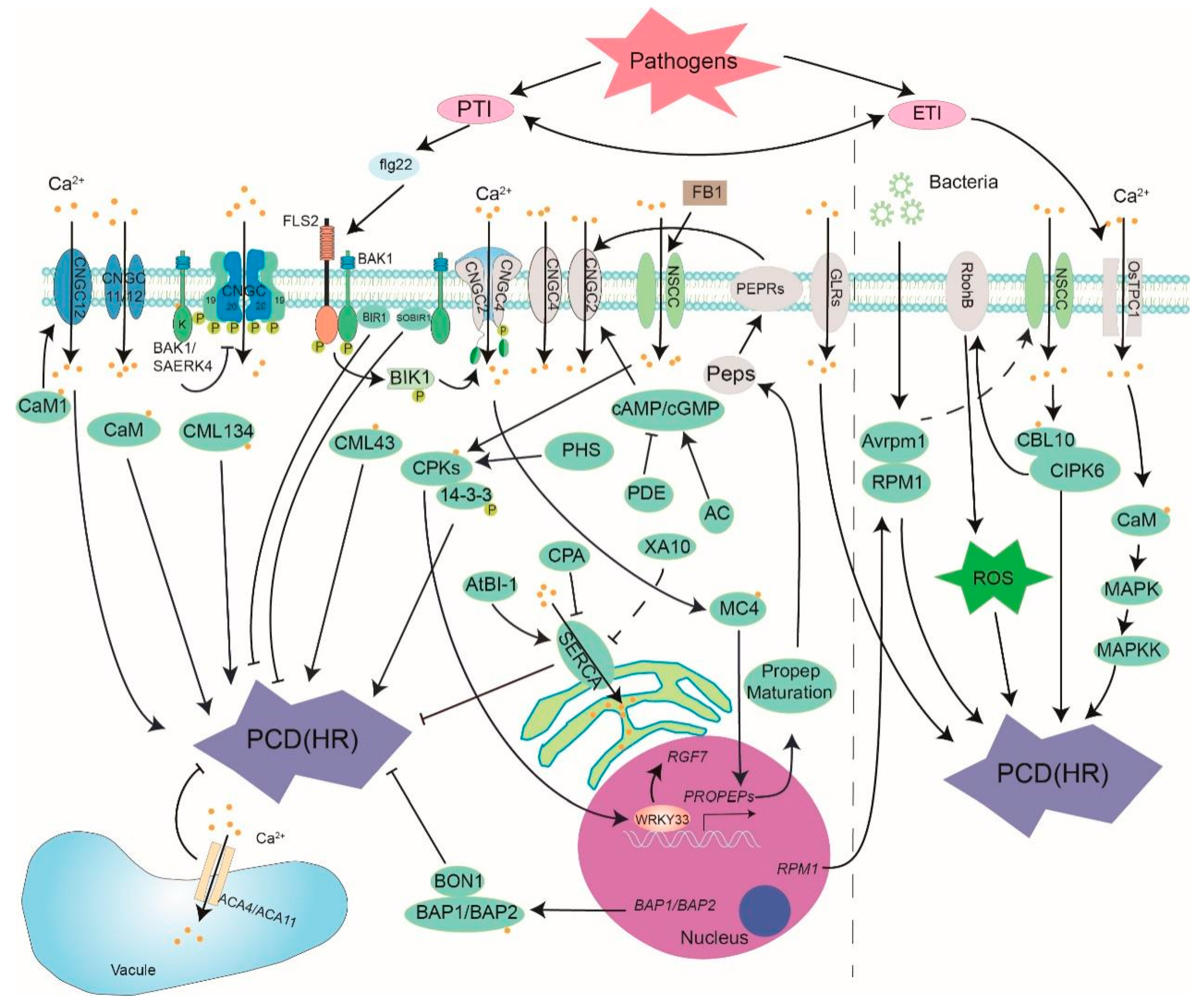
Figure 1. The role of calcium signal in biotic stress-induced PCD. Ca2+ channel, sensor and relative gene and protein are presented. PTI: pattern-triggered immunity; ETI: effector-triggered immunity; flg22: a 22 amino acid PAMP derived from bacterial flagellin; FB1: Fumonisins B1; FLS2: Flagellin-sensitive 2; CNGCs: Cyclic nucleotide gated channel; BAK1: brassinosteroid insensitive 1-associated receptor kinase 1; SERK4: Somatic embryogenesis receptor kinase 4; BIK1: botrytis-induced kinase 1; BIR1: BAK1-interacting receptor-like kinase 1; SOBIR1: suppressor of BIR1-1; Peps: plant elicitor peptide; PEPRs: extracellular Pep receptors; CaM: calmodulin; CML: CaM-like protein; CDPK(CPK): Ca2+-dependent protein kinase; CBL: calcineurin B-like protein; CIPK: CBL-interacting protein kinase; cAMP: 3’-5’-cyclic adenosine monophosphate; cGMP: cyclic guanosine monophosphate; AC: adenylate cyclase; PDE: phosphodiesterase; PHS: phytosphingosine; MC4: metacaspase 4; 14-3-3: 14-3-3 proteins; SERCA: sarco-endoplasmic reticulum Ca2+-ATPase; ACA: autoinhibited Ca2+-ATPase; RPM1: resistance to Pseudomonas syringae pv. Maculicola 1; AvrRpm1: Pseudomonas syringae type III effector; MAPK: Mitogen activated protein kinase (based on [10,11,12,13,14,15,16,17,18,19,20,21,22,23,24,25,26,27,28,29,30,31,32,33,34,35,36,37,38,39,40,41,42,43,44,45,46,47,48,49,50,51,52,53,54,55,56,57,58,59,60,61,62]).
Cells 10 01089 g001
Figure 2. The role of calcium signal in abiotic stress-induced PCD. Salt, temperature, anoxic, heavy metal and mechanic damage stresses are depicted. OSCA1: hyperosmolality-induced [Ca2+](i) increase 1; MCA1: mechanosensitive channel 1; GLRs: glutamate receptor-like channels; AtHMA1: heavy metal transporting ATPase 1; NSCC: nonselective cation channel; CAX: H+/Ca2+ antiporters; COLD1: chilling-tolerance divergence 1; AtANN1: Ca2+- permeable transporter ANNEXIN1; OST1: open stomata 1; RGA1: rice G-protein a subunit 1; VPE: vacuole processing enzymes; JJW: JAV1-JAZ8-WRKY51 complex; JA: jasmonic acid; GIPCs: glycosyl inositol phosphoryl ceramides (based on [63,64,65,66,67,68,69,70,71,72,73,74,75,76,77,78,79,80,81,82,83,84,85,86,87,88,89,90,91,92,93,94,95,96,97,98,99,100,101,102,103,104,105,106,107,108,109,110,111,112,113,114,115]).
Cells 10 01089 g002
Figure 3. Ca2+ participates in the PCD during plant development and postharvest.
Cells 10 01089 g003
Figure 4. Crosstalk between calcium signal and ROS-, NO-, phytohormone-induced PCD. HPCA1: hydrogen peroxide sensor; PAMPs: pathogen associated molecular pattern; PRR: pattern recognition receptor; RBOHD: respiratory burst oxidase homolog protein; SA: salicylic acid; GA: gibberellin. (based on [145,146,147,148,149,150,151,152,153,154,155,156,157,158,159,160,161]).
Cells 10 01089 g004
Table 1. The regulation factors of the calcium signal in plant PCD under biotic and abiotic stresses.
PCDReceptorCalcium ChannelRegulation Factor of Ca2+ ChannelCalcium SensorSubstrate
Biotic stressesPTIFLS2/BAK1CNGC2/4/11/12/19/20
GLR2.7/2.8/2.9
ACA4/11
SERCA
cAMP/cGMP
BAK1/BIK1
PEPR
CaM/CML
CPK3/5/6
RboHB
14-3-3
WRKY33
MC4
ETIOsTPC1CaM
SlCBL10
SlCIPK6
MPK
Abiotic stressesSaltGIPCANN1
CAX1
CaMOsVPE2/3
ColdCOLD1ANN1
SlGLR3.3/3.5
CNGC2/4
OsCNGC14/16
COLD1
OST1
CaMOsmotin
HeatANN1/4
OsCNGC14/16
CAS
MYB30CaMMPK6
γVPE
AnoxicCaMMPTP
Cytochrome C
Heavy metalHMA1CaM
CDPKs
MAPK8
DamageGLR3.3/3.6
MCA1
OSCA1.2
CaMJJW
MC4
Publisher’s Note: MDPI stays neutral with regard to jurisdictional claims in published maps and institutional affiliations.