Article

Detection and Quantification of Fusarium spp. (F. oxysporum, F. verticillioides, F. graminearum) and Magnaporthiopsis maydis in Maize Using Real-Time PCR Targeting the ITS Region

by
Maria Doroteia Campos
1,*,
Mariana Patanita
1,
Catarina Campos
1,
Patrick Materatski
1,
Carla M. R. Varanda
1,
Isabel Brito
2 and
Maria do Rosário Félix
3
1
ICAAM-Instituto de Ciências Agrárias e Ambientais Mediterrânicas, Instituto de Investigação e Formação Avançada, Universidade de Évora, Polo da Mitra, Ap. 94, 7006-554 Évora, Portugal
2
Departamento de Biologia, ICAAM-Instituto de Ciências Agrárias e Ambientais Mediterrânicas, Escola de Ciências e Tecnologia, Universidade de Évora, Polo da Mitra, Ap. 94, 7006-554 Évora, Portugal
3
Departamento de Fitotecnia, ICAAM-Instituto de Ciências Agrárias e Ambientais Mediterrânicas, Escola de Ciências e Tecnologia, Universidade de Évora, Polo da Mitra, Ap. 94, 7006-554 Évora, Portugal
*
Author to whom correspondence should be addressed.
Agronomy 2019, 9(2), 45; https://doi.org/10.3390/agronomy9020045
Submission received: 12 December 2018 / Revised: 10 January 2019 / Accepted: 18 January 2019 / Published: 22 January 2019
(This article belongs to the Special Issue Etiology and Control of Crop Diseases)

Abstract

:
Fusarium spp. and Magnaporthiopsis maydis are soil-inhabiting fungi and respectively the causal agents of fusarium ear rot and late wilt, two important diseases that can affect maize, one of the most important cereal crops worldwide. Here, we present two sensitive real-time PCR TaqMan MGB (Minor Groove Binder) assays that detect and discriminate several Fusarium spp. (F. oxysporum, F. verticillioides, and F. graminearum) from M. maydis. The method is based on selective real-time qPCR amplification of the internal transcribed spacer (ITS) region and allows the quantification of the fungi. The applicability of this newly developed TaqMan methodology was demonstrated in a field experiment through the screening of potentially infected maize roots, revealing a high specificity and proving to be a suitable tool to ascertain Fusarium spp. and M. maydis infection in maize. Its high sensitivity makes it very efficient for the early diagnosis of the diseases and also for certification purposes. Thus, qPCR through the use of TaqMan probes is here proposed as a promising tool for specific identification and quantification of these soil-borne fungal pathogens known to cause disease on a large number of crops.

1. Introduction

Maize (Zea mays L.) is one of the most important cereal crops worldwide [1] not only for food production but also in terms of its growing role in industry and energy resources. However, animal pests, weeds, and pathogens have an impact on its yield and quality [2]. Fusarium spp. are among the most important pathogenic fungal communities affecting crops, causing in maize the disease complexes fusarium ear rot (FER) [3]. These soil-borne fungi interfere with the water-conducting vessels of the plant and, as the infection spreads up into the stems and leaves, it restricts water flow, causing the foliage to wilt and turn yellow. Fusarium spp. fungi can induce serious plant diseases that can affect the yield and quality of maize grain, resulting in huge economic losses, and can also result in mycotoxin-contaminated grains that cause health-threatening issues [4,5]. Important causal species of FER in maize are Fusarium graminearum, F. verticillioides, F. proliferatum, and F. subglutinans [6,7]. F. verticillioides is the main causal agent of FER, although climatic conditions and crop management practices influence the occurrence and prevalence of other Fusarium species [8,9]. F. oxysporum and F. solani are also commonly found in maize seeds [10,11].
In recent years, late wilt caused by the soil-borne and seed-borne fungus Magnaporthiopsis maydis (Samra, Sabet & Hing.) Klaubauf, Lebrun & Crou (synonym Cephalosporium maydis Samra, Sabet & Hing.; Harpophora maydis (Samra, Sabet & Hing.) W. Gams) [12] has emerged as a worrying disease in maize in the Iberian Peninsula [13] and has also been reported in India, Israel, and Hungary as well [14]. This fungus is morphologically and molecularly closely related to Gaeumannomyces-Harpophora species complex [12,15,16]. The disease is characterized by relatively rapid wilting of maize plants due to clogging of the vascular system [17]. The most effective way of controlling late wilt is the use of tolerant maize varieties [4,5]. Although the complete absence of symptoms in tolerant maize until the end of the crop season is not frequent, material displaying moderate to high tolerance should be included within an integrated strategy for the control of late wilt of maize [18]. Because seeds can transport the pathogen and consequently spread the disease [19], molecular assays are important to rapidly select seeds free of M. maydis and, consequently, prevent its dissemination [20]. Additionally, in maize plants the rot of the base of the affected stem and the associated roots is often due to secondary infection by stalk rot-causing fungi [21]. Fusarium spp. are included in these secondary organisms. For instance, F. graminearum and F. verticillioides have been frequently found together with M. maydis in seed samples of maize in Egypt [22].
Given the increasing incidence of both Fusarium spp. and M. maydis and their negative economic impact, the importance of early detection arises, bringing the need of precise molecular identification. Together with information on plant disease severity, molecular quantification of fungi in plants offers an additional tool in the screening of plants resistant to fungal diseases. The present study reports for the first time a methodology for detection, quantification, and also discrimination between genomic DNA (gDNA) of Fusarium spp. and M. maydis in maize roots using real-time PCR (qPCR), through the development of two specific TaqMan MGB (Minor Groove Binder) assays: one targeting Fusarium species, and another that targets M. maydis. The assays are based on selective amplification of the internal transcribed spacer (ITS) region (ITS1, 5.8S rRNA, ITS2, large subunit ribosomal RNA gene). The use of qPCR instrumentation presents several advantages, since it requires considerably short hands-on time, and detection of amplified products is automated, simple, and reproducible [23]. qPCR combined with the chemistry of TaqMan MGB probes represents the most specific and sensitive detection system when low amounts of target DNA are present, as often occurs in the case of early interactions between plants and fungi. Finally, the applicability of the newly developed TaqMan methodology was exemplified in a field experiment through the screening of potentially infected maize roots with Fusarium spp. and M. maydis.

2. Materials and Methods

2.1. Study Site and Sample Collection

The sampling area, located in Ribatejo region (central Portugal) (39°20′11.74″ N, 8°32′59.98″ W), has been known to be infested with late wilt for many years. Maize was irrigated and grown under conventional management. The cultivars used in the experiment are considered tolerant and susceptible to M. maydis. Samples (biological replicates) consisted of maize roots, collected in bulked samples of six plants, at two (T0), four (T1), and six (T2) weeks after germination. The experimental design consisted of blocks of four replicates for each cultivar, for a total of 24 samples (2 cultivars x 3 time points x 4 replicates). Roots were transported to the laboratory and stored at 4 °C until further analysis, which occurred within 48 h. Root samples were used for both fungal isolation and gDNA extraction.

2.2. Fungal Isolates and Growth Conditions

Maize roots for fungal isolation were surface disinfected to suppress epiphytic microorganisms [24]. After disinfection, root pieces were dried in sterile Whatman paper, cut into sections, and placed on Petri dishes of 9 cm diameter containing Potato Dextrose Agar medium (PDA, Merck, Darmstadt, Germany) and incubated for 1–2 weeks at 23–25 °C. Morphologically different colonies were isolated by transferring an agar disk (about 5 mm2) of the growing fungi to fresh medium (PDA). From these colonies, 12 isolates of Fusarium spp. were firstly identified morphologically through characteristics such as rate of growth, mycelium color, texture, nature of the growing margin, and color of the reverse side. Shape of conidia was observed under an Olympus BX-50 compound microscope (1000x magnification). Additionally, for demonstration of the reliability of the TaqMan methodology, three morphologically different isolates of Fusarium spp. (obtained from maize plants) that belong to the collection of the Mycology Laboratory, Institute of Mediterranean Agricultural and Environmental Sciences (ICAAM), University of Évora, Portugal, were included in the experiment, and were grown on the same conditions as described above. In the experiment it was not possible to morphologically identify Magnaporthiopsis maydis and therefore a reference isolate (in PDA plates) was kindly provided by Dr. Ortiz-Bustos (Department of Crop Protection, Institute of Sustainable Agriculture, Córdoba, Spain) [25] (GenBank Acc number KP164518).

2.3. gDNA Extraction

Maize roots, Fusarium spp. mycelium (12 isolates from the experiment and 3 isolates from the laboratory collection), and M. maydis mycelium were ground in liquid nitrogen and stored at −80 °C until further analysis. gDNA was extracted using the CTAB (hexadecyltrimethylammonium bromide) method [24,26]. To quantify and assess gDNA purity, the absorbance was measured on a NanoDrop-2000C spectrophotometer (Thermo Scientific, Waltham, MA, USA). gDNA integrity was checked by 0.8% agarose gel electrophoresis and visualized using Gene Flash Bio Imaging system (Syngene, Cambridge, UK). Samples were diluted to a concentration of 20 ng μL−1.

2.4. Fungal Genetic Classification

The ribosomal internal transcribed spacer (ITS) region of nuclear rDNA from gDNA of the fungal isolates was amplified through PCR by using ITS1 and ITS4 primers [27]. Analysis of the ITS sequences was based on a ClustalW Multiple alignment using BioEdit software [28]. The search for homologous sequences was done using Basic Local Alignment Search Tool (BLAST) at the National Center for Biotechnology Information (NCBI) (http://www.ncbi.nlm.nih.gov/).

2.5. Specific TaqMan Assays and qPCR Conditions

Primers and probes were designed for detection of Fusarium spp. and M. maydis by using the Primer Express 3.0 software (Applied Biosystems, Foster City, CA, USA) for real-time PCR (Table 1), selecting the option MGB TaqMan probes, and using the default parameters of the software. ITS sequences isolated by ITS1 and ITS4 primers (as previously mentioned in Section 2.4) were used as targets for the design of the TaqMan assays. A single TaqMan assay was designed for the amplification of both of the Fusarium spp. that were identified in the experiment (Fusarium oxysporum, GenBank Acc number MH094661, and Fusarium verticillioides, GenBank Acc number MH094662) (Table 1). A bioinformatic approach was performed to assure the specificity of primers and probes, which included a BLASTn query at NCBI.
qPCRs were carried out on a 7500 Real Time PCR System (Applied Biosystems, Foster City, CA, USA) using 100 ng of gDNA as template, 2x NZY qPCR Probe Master Mix (Nzytech, Lisbon, Portugal), 400 nM of each primer, and 100 nM of probe (Nzytech, Lisbon, Portugal) in a total volume of 20 µL. The quantification cycle (Cq) values were acquired for each sample with the Applied Biosystems 7500 software v2.0.6 (Applied Biosystems, Foster City, CA, USA), with the following cycling conditions: 10 min at 95 °C for initial denaturation, an amplification program of 40 cycles at 95 °C for 15 s and 60 °C for 1 min. The fluorescence threshold was manually set above the background level. Four biological replicates and three technical replicates were considered for each sample. Fungi positive target controls and no template controls were included in all plates.
As a measure of sensitivity and the quantitative range of the developed qPCR procedure, the limit of detection was determined. A total of 16 standards were prepared by a two-fold serial dilution (2−1, 2−2, ……, 2−16) of the gDNA extracted from each target species, and the qPCR conditions were those described above.

2.6. DNA Calibrator Plasmids

In order to produce a set of calibrator plasmids to be used on qPCR, the target regions of Fusarium spp. and M. maydis were amplified using the ITS1 and ITS4 primers [27] referred to above for fungal genetic classification. Amplicons, with sizes of 545 bp for Fusarium spp. and 574 bp for M. maydis, were cloned into a pGem®-T Easy vector (Promega, Madison, WI, USA) and used to transform E. coli JM109 (Promega, Madison, WI, USA) competent cells, by standard procedures. Plasmids were extracted from putative recombinant clones using GeneJET Plasmid Miniprep Kit (Thermo Scientific, Waltham, MA, USA) and analyzed using EcoRI restriction enzyme (Thermo Scientific, Waltham, MA, USA ). The concentrations of purified plasmid DNA were determined in a NanoDrop-2000C spectrophotometer (Thermo Scientific, Waltham, MA, USA). Selected bacterial clones were sequenced in sense and antisense strands using T7 and SP6 universal primers (Macrogen, Inc., Madrid, Spain: www.macrogen.com). For each assay, standard curves were generated from ten-fold dilution series of plasmid DNA to draw a five-point calibration curve in the dynamic range chosen (8E1 to 8E5 target copies). A linear regression analysis set the correlation between measured Cq values and the logarithm of the concentration. qPCR conditions were as described above. Amplification efficiencies were calculated through the equation E = (10(−1/slope) − 1) × 100, as well as slope and linearity (coefficient of determination, R2). The method performed for absolute DNA quantification was based on the determination of the absolute number of target copies, TCN [29].

2.7. Statistical Analysis

Univariate and multivariate analyses were performed to the results obtained in the field experiment using the PRIMER v6 software [30] with the permutational analysis of variance (PERMANOVA) add-on package [31] in order to detect significant differences (p < 0.05) in target copy number between “cultivar” (tolerant and susceptible to M. maydis) and “time” (T0, T1, and T2) using 100 ng of gDNA (TCN100). PERMANOVA analyses were carried out with the following two-factor design: “cultivar”, tolerant and susceptible (two levels, fixed); and “time, T0, T1, and T2 (three levels, fixed). The data were square-root transformed in order to scale down the importance of high values of TCN. The PERMANOVA analysis was conducted on a Bray-Curtis similarity matrix [32]. If the number of permutations was lower than 150, the Monte Carlo permutation p-value was used. Whenever significant interaction effects were detected, these were examined using a posteriori pairwise comparisons, using 9999 permutations under a reduced model.

3. Results and Discussion

3.1. Fungus Isolates and TaqMan Assay Specificity

In this study, it was possible to identify 12 Fusarium isolates, belonging only to two different species: seven to F. oxysporum and five to F. verticillioides. The ITS sequences were identical within each Fusarium species, and two sequences were therefore published in GenBank (F. oxysporum Acc number MH094661, 99% identity; F. verticillioides Acc number MH094662, 99% identity). The ability of the ITS region to differentiate and provide accurate and rapid detection of fungi has been already reported at the species level [33], but recently the European and Mediterranean Plant Protection Organization (EPPO) suggested the multilocus sequence analysis as being more accurate for the identification of species [34]. F. verticillioides was already referred to as the most prevalent fungus associated with maize [10,11]. The analysis of the ITS sequences of other isolates of Fusarium spp. from the collection of the Mycology Laboratory allowed us to additionally identify the maize pathogenic species F. graminearum (GenBank Acc number MH094665, 100% identity), F. incarnatum (GenBank Acc number MH094664, 99% identity), and F. solani (GenBank Acc number MH094664, 99% identity), which were included in the Fusarium-specificity analysis.
The results obtained in the experiment led to the design of a single TaqMan Fusarium assay that specifically targeted both F. verticillioides and F oxysporum gDNA, the two Fusarium species that were identified in the sampling area. So far, the design of qPCR assays has been specific to individual Fusarium species [35,36,37], but in this work we intend to make the discrimination between the genomic gDNA of Fusarium spp. and M. maydis present in maize roots. In this sense, a single TaqMan Fusarium assay that targets both species was designed and used.
The specificity of the Fusarium spp. assay was firstly done in silico, performing searches against NCBI databases, including the Mycology Lab Fusarium collection. The results revealed the specificity of the Fusarium spp. probe to F. verticillioides and F. oxysporum, as expected, but also to F. graminearum, one of the most important causal species of FER in maize [11,38]. Secondly, the specificity of the assay was evaluated experimentally and, as revealed by in silico analysis, the isolates of F. verticillioides, F oxysporum, and F. graminearum were targeted by the Fusarium spp. assay, while F. incarnatum and F. solani species were not (Figure S1). The specificity of the Fusarium assay in relation to M. maydis was also confirmed, first in silico and then experimentally (Figure S1).
Regarding M. maydis, it was not possible its identification amongst the fungal isolates obtained in this experiment. Recovery of M. maydis, even from heavily infested material, was already referred to as difficult due to its slow growth on media and the relative abundance of other more rapidly growing fungi, most commonly Fusarium spp. [16]. Nevertheless, the sampling area where the experiment took place is known to be infected with this fungus. In this sense, a M. maydis probe was designed (GenBank Acc number KP164518) and specifically targeted its correspondent gDNA extracted from a known isolate. The similarity between Fusarium spp. and M. maydis ITS sequences (M. maydis GenBank Acc number KP164518 presented 88% identity with F. oxysporum, and 87% identity with F. verticillioides) made the design of specific probes difficult. Still, the specificity of the M. maydis assay in relation to Fusarium species was confirmed, first in silico and then experimentally (Figure S2).
Both, Fusarium spp. and M. maydis assays were also tested for cross-reactivity in gDNA of Alternaria tenuissima, Cladosporium cladosporioides, and Epicoccum nigrum belonging to the Mycology Laboratory collection, and no cross-reaction was found (Figures S1 and S2).

3.2. Calibration Curves and Probe Validation

A linear regression analysis set the correlation between measured Cq values and the logarithm of the concentration. The amplification efficiency, slope, and linearity were determined for each assay. RT-PCR assays presented an efficiency of 104% for M. maydis (slope = −3.228 and R2 = 0.99), and an efficiency of 105% for Fusarium spp. (slope = −3.207 and R2 = 0.98). All parameters fell within the acceptance criteria [39].

3.3. Sensitivity and Linearity

Dilution series of gDNA tested in PCR ranged from final concentration of 1.53 × 10⁻³ ng to 100 ng. Standard curves were automatically generated by the instrument software (Applied Biosystems, Foster City, CA, USA). Standard curves and amplification plots are indicated in Figure S3. Both species presented a linear correlation (R2 = 0.996 for Fusarium spp. and R2 = 0.960 for M. maydis) between Cq and template gDNA amount (Figure S3). These results confirm the reliability of the assays and suggest the absence of PCR inhibitors.
For each TaqMan assay, Cq values between 34 and 35 were taken as being indicative of trace amounts, while a Cq = 35 was considered the cut-off limit, defining no detection [29]. The method showed a high sensitivity for both Fusarium spp. and M. maydis assays (Table 2). The method allowed the detection of 6.10 × 10⁻³ ng of Fusarium spp. and 3.91 × 10⁻¹ ng of M. maydis (Table 2).

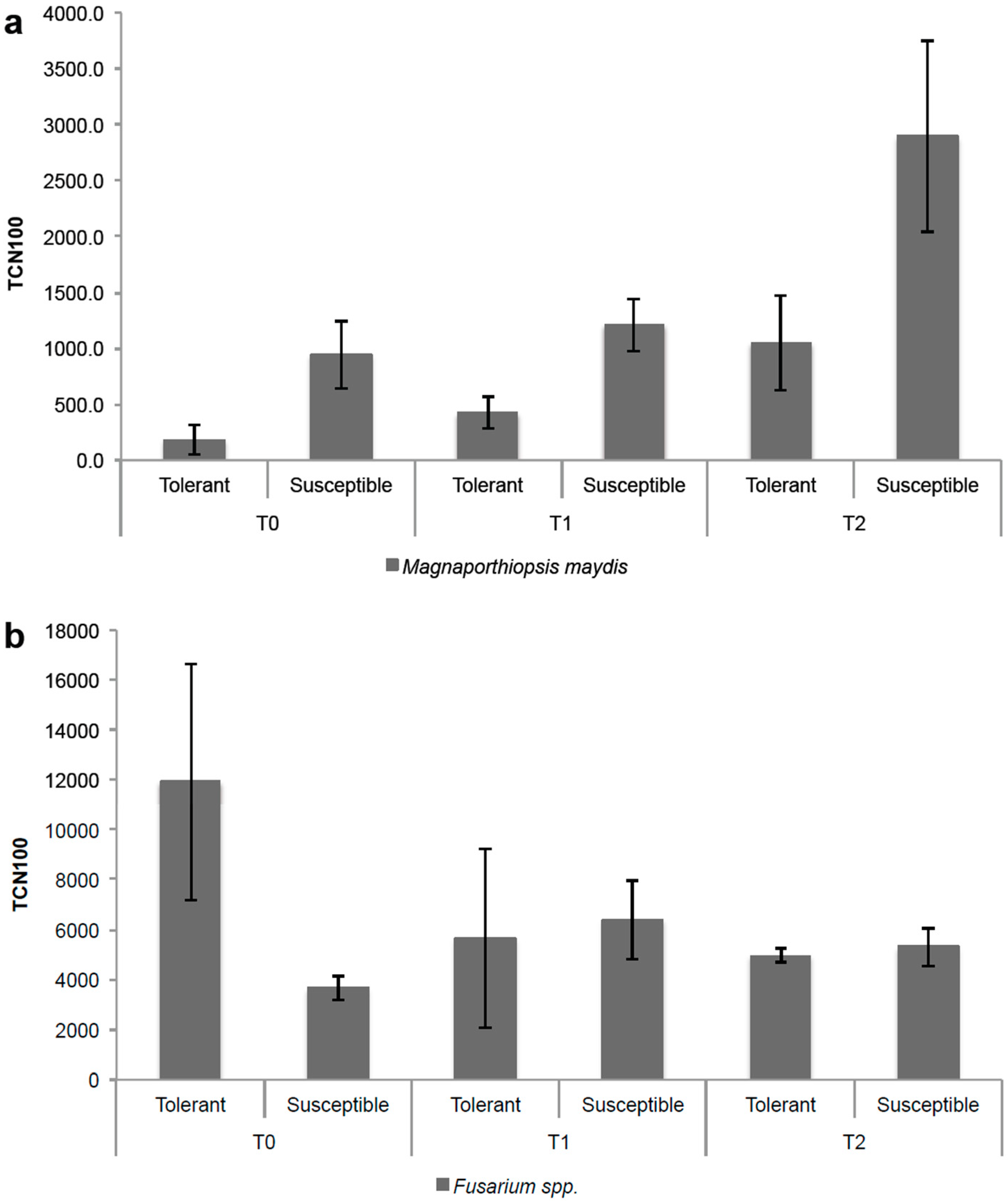
3.4. Applicability of the qPCR Assay in Maize Growing under Field Conditions

Since Fusarium spp. are often found together with M. maydis, a highly specific methodology that discriminates between these soil-borne fungal pathogens is extremely important. We therefore tested Fusarium spp. and M. maydis qPCR assays on samples from a field trial. This experiment was used as a proof-of-concept for the suitability of the technique. To evaluate target gDNA amount in terms of TCN using 100 ng of gDNA (TCN100), a total of 24 samples belonging to two cultivars (tolerant and susceptible) were analyzed at three time points (Figure 1). As expected, the M. maydis qPCR assay revealed that the susceptible cultivar consistently presented significantly higher values (p < 0.05) than the tolerant cultivar; this trend was observed for all of the maize growth times evaluated. Regarding the Fusarium spp. qPCR assay, it was possible to identify the presence of Fusarium spp. in both cultivars, although not following a specific trend (Figure 1). The results achieved demonstrate the suitability and applicability of the qPCR method based on ITS region to identify and discriminate between Fusarium spp. and M. maydis in maize roots under field conditions.
The degree of complexity of the etiology of late wilt in maize regarding the determination of the role of each fungal species in the development of the disease and/or the severity of symptoms should be studied by confirming the pathogenicity of the soil-borne fungi (including Fusarium spp.) [25]. The methodology presented here represents an important contribution towards establishing a better understanding of the interaction between both fungi.
To the best of our knowledge, this is the first time that a TaqMan probe assay has been developed to target M. maydis. Semi-quantitative analysis of DNA bands was previously applied for its detection [14], and recently a SYBR®Green qPCR assay was established for the detection of M. maydis DNA in maize growing under different conditions, including in an infested field [20], however, it was not tested whether the method used was specific to M. maydis, or if it also could target Fusarium spp. M. maydis is difficult to detect and isolate [16] and has the ability to become established and survive in stored maize seeds (even from apparently healthy parental plants) [14], which stresses the urgent need to develop new ways to monitor seed health and control disease spread.
Fusarium probes have been previously developed for target individual fungi species [35,36,37], but here we describe a methodology that targets both F. verticillioides and F. oxysporum, the Fusarium species that are prevalent in maize fields, but also targets F. graminearum, another important causal species of FER in maize.

4. Conclusions

Fusarium spp. and M. maydis TaqMan qPCR assays revealed a high specificity, with the Fusarium assay targeting F. oxysporum, F. verticillioides, and F. graminearum, important causal agents of FER in maize. This specificity includes even closely related species, and no false-positive results were observed. The specificity of the assay was validated in field conditions. The high sensitivity makes the methodology an efficient tool for the early diagnosis of the diseases as well as for sanitary certification purposes. Thus, qPCR through the use of TaqMan probes is proposed here as a promising tool for the discrimination and quantification of these soil-borne fungal pathogens known to cause disease on a large number of crops.

Supplementary Materials

The following are available online at https://www.mdpi.com/2073-4395/9/2/45/s1, Figure S1: Amplification plots to assess Fusarium spp. primers/probe specificity. a: Cross-reactivity in gDNA of different Fusarium species. Amplification of F. verticillioides, F. oxysporum, and F. graminearum and non-amplification of F. solani and F. incarnatum. b: Cross-reactivity in gDNA of Magnaporthiopsis maydis. Amplification of F. verticillioides. c: Cross-reactivity in gDNA of Alternaria tenuissima, Cladosporium cladosporioides, and Epicoccum nigrum. Amplification of F. verticillioides. Figure S2: Amplification plots to assess Magnaporthiopsis maydis primers/probe specificity. a: Cross-reactivity in gDNA of different Fusarium species (F. verticillioides, F. oxysporum, F. graminearum, F. solani, and F. incarnatum). Amplification of M. maydis. b: Cross-reactivity in gDNA of Alternaria tenuissima, Cladosporium cladosporioides, and Epicoccum nigrum. Amplification of M. maydis. Figure S3: Sensitivity and linearity of the TaqMan qPCR assays. Amplification plots and standard curves from serial dilutions of gDNA extracted from Magnaporthiopsis maydis (a) and Fusarium spp. (b) tested by the respective species-specific TaqMan assay.

Author Contributions

Conceptualization, M.D.C. and M.D.R.F.; Formal Analysis, M.D.C. and P.M.; Funding Acquisition, M.D.R.F. and I.B.; Investigation, M.D.C. and M.P.; Methodology, M.D.C. and C.C.; Resources, M.P., C.M.R.V. and P.M.; Resources, M.P., C.M.R.V. and P.M.; Supervision, M.D.R.F. and I.B.; Writing—Original Draft Preparation, M.D.C.; Writing—Review & Editing, M.D.C., M.P.; C.C., P.M., C.M.R.V., I.B., M.D.R.F.

Funding

This work was funded by National Funds through the Foundation for Science and Technology (FCT) under the Project UID/AGR/00115/2019. C. Campos (SFRH/BPD/108734/2015) and C. Varanda (SFRH/BPD/76194/2011) were supported by a post-doctoral fellowship from the Foundation for Science and Technology (FCT), funded by QREN-POPH-Typology 4.1 and co-funded by MES National Funding and The European Social Fund.

Acknowledgments

The authors wish to thank to Eng. João Coimbra and Eng. José da Luz for setting up the field trial, and Ortiz-Bustos (Department of Crop Protection, Institute of Sustainable Agriculture, Córdoba, Spain) for providing the fungus Magnaporthiopsis maydis.

Conflicts of Interest

The authors declare no conflict of interest.

References

  1. Czembor, E.; Stępień, Ł.; Waśkiewicz, A. Effect of environmental factors on Fusarium species and associated mycotoxins in maize grain grown in Poland. PLoS ONE 2015, 10, e0133644. [Google Scholar] [CrossRef] [PubMed]
  2. Oerke, E.C. Crop losses to pests. J. Agric. Sci. 2006, 144, 31–43. [Google Scholar] [CrossRef]
  3. Walder, F.; Schlaeppi, K.; Wittwer, R.; Held, A.Y.; Vogelgsang, S.; van der Heijden, M.G.A. Community Profiling of Fusarium in Combination with Other Plant-Associated Fungi in Different Crop Species Using SMRT Sequencing. Front. Plant Sci. 2017, 8, 2019. [Google Scholar] [CrossRef] [PubMed]
  4. Lanubile, A.; Maschietto, V.; Borrelli, V.M.; Stagnati, L.; Logrieco, A.F.; Marocco, A. Molecular Basis of Resistance to Fusarium Ear Rot in Maize. Front. Plant Sci. 2017, 8, 1774. [Google Scholar] [CrossRef] [PubMed]
  5. Maschietto, V.; Colombi, C.; Pirona, R.; Pea, G.; Strozzi, F.; Marocco, A.; Rossini, L.; Lanubile, A. QTL mapping and candidate genes for resistance to Fusarium ear rot and fumonisin contamination in maize. BMC Plant Biol. 2017, 17, 20. [Google Scholar] [CrossRef] [PubMed]
  6. Desjardins, A.E. Gibberella from A (venaceae) to Z (eae). Annu. Rev. Phytopathol. 2003, 41, 177–198. [Google Scholar] [CrossRef] [PubMed]
  7. Summerell, B.A.; Leslie, J.F. Fifty years of Fusarium: How could nine species have ever been enough? Fungal Divers. 2011, 50, 135–144. [Google Scholar] [CrossRef]
  8. Edwards, S.G. Influence of agricultural practices on fusarium infection of cereals and subsequent contamination of grain by trichothecene mycotoxins. Toxicol. Lett. 2004, 153, 29–35. [Google Scholar] [CrossRef]
  9. Xu, X.; Nicholson, P. Community Ecology of Fungal Pathogens Causing Wheat Head Blight. Annu. Rev. Phytopathol. 2009, 47, 83–103. [Google Scholar] [CrossRef]
  10. Aguín, O.; Cao, A.; Pintos, C.; Santiago, R.; Mansilla, P.; Butrón, A. Occurrence of Fusarium species in maize kernels grown in northwestern Spain. Plant Pathol. 2014, 63, 946–951. [Google Scholar] [CrossRef]
  11. Duan, C.; Qin, Z.; Yang, Z.; Li, W.; Sun, S.; Zhu, Z.; Wang, Z. Identification of pathogenic fusarium spp. causing maize ear rot and potential mycotoxin production in China. Toxins 2016, 8, 186. [Google Scholar] [CrossRef] [PubMed]
  12. Klaubauf, S.; Tharreau, D.; Fournier, E.; Groenewald, J.Z.; Crous, P.W.; de Vries, R.P.; Lebrun, M.-H. Resolving the polyphyletic nature of Pyricularia (Pyriculariaceae). Stud Mycol. 2014, 79, 85–120. [Google Scholar] [CrossRef] [PubMed] [Green Version]
  13. Molinero-Ruiz, M.L.; Melero-Vara, J.M.; Mateos, A. Cephalosporium maydis, the cause of late wilt in maize, a pathogen new to Portugal and Spain. Plant Dis. 2011, 94, 379. [Google Scholar] [CrossRef]
  14. Drori, R.; Sharon, A.; Goldberg, D.; Rabinovitz, O.; Levy, M.; Degani, O. Molecular diagnosis for Harpophora maydis, the cause of maize late wilt in Israel. Phytopathol Mediterr. 2013, 52, 16–29. [Google Scholar] [CrossRef]
  15. Gams, W. Phialophora and some similar morphologically little-differentiated anamorphs of divergent ascomycetes. Stud Mycol. 2000, 45, 187–199. [Google Scholar]
  16. Saleh, A.A.; Leslie, J.F. Cephalosporium maydis is a distinct species in the Gaeumannomyces-Harpophora species complex. Mycologia. 2004, 96, 1294–1305. [Google Scholar] [CrossRef] [PubMed]
  17. Abd El-Rahim, M.F.; Fahmy, G.M.; Fahmy, Z.M. Alterations in transpiration and stem vascular tissues of two maize cultivars under conditions of water stress and late wilt disease. Plant Pathol. 1998, 47, 216–223. [Google Scholar] [CrossRef] [Green Version]
  18. García-Carneros, A.B.; Girón, I.; Molinero-Ruiz, L. Aggressiveness of Cephalosporium maydis Causing Late Wilt of Maize in Spain. Commun. Agric. Appl. Biol. Sci. 2012, 77, 173–179. [Google Scholar] [PubMed]
  19. Michail, S.H.; Abou-Elseoud, M.S.; Nour Eldin, M.S. Seed health testing of corn for Cephalosporium maydis. Acta Phytopathol. Entomol. Hung. 1999, 34, 35–42. [Google Scholar]
  20. Degani, O.; Movshowitz, D.; Dor, S.; Meerson, A.; Goldblat, Y.; Rabinovitz, O. Evaluating azoxystrobin seed coating against maize late wilt disease using a sensitive qPCR-based method. Plant Dis. 2018. [Google Scholar] [CrossRef] [PubMed]
  21. Sabet, K.A.; Sanra, A.S.; Mansour, I.S. Interaction between Fusarium oxysporum F. vasinfectum and Cephalosporium maydis on cotton and maize. Ann. Appl. Biol. 1966, 58, 93–101. [Google Scholar] [CrossRef]
  22. Mohamed, H.A.; Ashour, W.E.; Sirry, A.R.; Fathi, S.M. Fungi carried by corn seed and their importance in causing corn diseases in the United Arab Republic. Plant Dis. Rep. 1967, 51, 53–56. [Google Scholar]
  23. Espy, M.; Uhl, J.; Sloan, L.; Buckwalter, S.; Jones, M.; Vetter, E.; Yao, J.D.C.; Wengenack, N.L.; Rosenblatt, J.E.; Cockerill, F.R., III; et al. Real-Time PCR in Clinical Microbiology: Applications for Routine Laboratory Testing Real-Time PCR in Clinical Microbiology: Applications for Routine Laboratory Testing. Clin. Microbiol. Rev. 2006, 19, 165–256. [Google Scholar] [CrossRef] [PubMed]
  24. Varanda, C.M.R.; Oliveira, M.; Materatski, P.; Landum, M.; Clara, M.I.E.; do Rosário Félixc, M. Fungal endophytic communities associated to the phyllosphere of grapevine cultivars under different types of management. Fungal Biol. 2016, 120, 1525–1536. [Google Scholar] [CrossRef] [PubMed] [Green Version]
  25. Ortiz-Bustos, C.M.; Testi, L.; García-Carneros, A.B.; Molinero-Ruiz, L. Geographic distribution and aggressiveness of Harpophora maydis in the Iberian peninsula, and thermal detection of maize late wilt. Eur J Plant Pathol. 2016, 144, 383–397. [Google Scholar] [CrossRef]
  26. Doyle, J.J.; Doyle, J.L. A rapid DNA isolation procedure for small quantities of fresh leaf tissue. Phytochem Bull. 1987, 19, 11–15. [Google Scholar]
  27. White, T.J.; Bruns, S.; Lee, S.; Taylor, J. Amplification and direct sequencing of fungal ribosomal RNA genes for phyologenetics. In PCR Protocols: A Guide to Methods and Applications; Academic Press: Cambridge, MA, USA, 1990; pp. 315–322. [Google Scholar] [CrossRef]
  28. Hall, T.A. BioEdit: A user-friendly biological sequence alignment editor and analysis program for Windows 95/98/NT. Nucleic Acids Symp. Ser. 1999, 41, 95–98. [Google Scholar]
  29. Campos, M.D.; Valadas, V.; Campos, C.; Morello, L.; Braglia, L.; Breviario, D.; Cardoso, H.H. A TaqMan real-time PCR method based on alternative oxidase genes for detection of plant species in animal feed samples. PLoS ONE 2018, 13, e0190668. [Google Scholar] [CrossRef]
  30. Clarke, K.R.; Warwick, R.M. Change in Marine Communities: An Approach to Statistical Analysis and Interpretation, 2nd ed.; PRIMER-E Ltd.: Plymouth, UK, 2001; 172p. [Google Scholar]
  31. Anderson, M.J.; Gorley, R.N.; Clarke, K.R. PERMANOVA+ for PRIMER: Guide to Software and Statistical Methods; PRIMER-E Ltd.: Plymouth, UK, 2008; pp. 1–214. [Google Scholar] [CrossRef]
  32. Clarke, K.R.; Green, R.H. Statistical design and analysis for a “biological effects” study. Mar. Ecol. Prog. Ser. 1988, 46, 213–226. [Google Scholar] [CrossRef]
  33. Zeng, X.; Kong, F.; Halliday, C.; Chen, S.; Lau, A.; Playford, G.; Sorrell, T.C. Reverse line blot hybridization assay for identification of medically important fungi from culture and clinical specimens. J. Clin. Microbiol. 2007, 45, 2872–2880. [Google Scholar] [CrossRef]
  34. Oepp, B.; Bulletin, E. PM 7/129 (1) DNA barcoding as an identification tool for a number of regulated pests. EPPO Bull. 2016. [Google Scholar] [CrossRef]
  35. Nicolaisen, M.; Suproniene, S.; Nielsen, L.K.; Lazzaro, I.; Spliid, N.H.; Justesen, A.F. Real-time PCR for quantification of eleven individual Fusarium species in cereals. J. Microbiol. Methods 2009, 76, 234–240. [Google Scholar] [CrossRef] [PubMed]
  36. Scauflaire, J.; Godet, M.; Gourgue, M.; Liénard, C.; Munaut, F. A multiplex real-time PCR method using hybridization probes for the detection and the quantification of Fusarium proliferatum, F. subglutinans, F. temperatum, and F. verticillioides. Fungal Biol. 2012, 116, 1073–1080. [Google Scholar] [CrossRef] [PubMed]
  37. Yli-Mattila, T.; Paavanen-Huhtala, S.; Jestoi, M.; Parikka, P.; Hietaniemi, V.; Gagkaeva, T.; Sarlin, T.; Haikara, A.; Laaksonen, S.; Rizzo, A. Real-time PCR detection and quantification of Fusarium poae, F. graminearum, F. sporotrichioides and F. langsethiae in cereal grains in Finland and Russia. Arch. Phytopathol. Plant Prot. 2008, 41, 243–260. [Google Scholar] [CrossRef]
  38. Dorn, B.; Forrer, H.R.; Jenny, E.; Wettstein, F.E.; Bucheli, T.D.; Vogelgsang, S. Fusarium species complex and mycotoxins in grain maize from maize hybrid trials and from grower’s fields. J. Appl. Microbiol. 2011, 111, 693–706. [Google Scholar] [CrossRef] [PubMed] [Green Version]
  39. Broeders, S.; Huber, I.; Grohmann, L.; Berben, G.; Taverniers, I.; Mazzara, M.; Roosens, N.; Morisset, D. Guidelines for validation of qualitative real-time PCR methods. Trends Food Sci. Technol. 2014, 37, 115–126. [Google Scholar] [CrossRef] [Green Version]
Figure 1. qPCR assay based on Magnaporthiopsis maydis (a) and Fusarium spp. (b) ITS region applied to a maize field trial. Cultivars were considered tolerant or susceptible to M. maydis. TCN100: target copy number using 100 ng of gDNA. T0: 2 weeks after germination; T1: 4 weeks after germination; T2: 6 weeks after germination.
Agronomy 09 00045 g001
Table 1. qPCR oligonucleotide primers and probes designed on internal transcribed spacer (ITS) gene region. AS: amplicon size.
SpeciesAccession IDPrimers (5’→3’)Probes (5’→3’)As(bp)
Fusarium oxysporum and Fusarium verticillioidesMH094661 and MH094662Fw: AAAACCCTCGTTACTGGTAATCGT
Rv: CCGAGGTCAACATTCAGAAGTTG
CGGCCACGCCGTT69
M. maydisKP164518Fw: TGCCTGTCCGAGCGTCAT
Rv: GGGTGCCCCAACACCAA
TCACCACTCAAGCCCAG54
Table 2. Sensitivity of the TaqMan qPCR assays. Serial dilutions of gDNA from Fusarium spp. and Magnaporthiopsis maydis using the corresponding TaqMan assay. The lowest amount of detection for both assays can be observed. Data are expressed as Cq values and target copy number. P.C.: positive control; TCN: target copy number; ND: not detected.
DilutiongDNA in PCR (ng)Fusarium spp.M. maydis
Cq Value (± SD)TCNCq Value (± SD)TCN
P.C.100.0020.86 (±0.23)11514448.525.16 (±0.71)46356.9
2−150.0021.76 (±0.15)6047961.528.42 (±0.53)4544.7
2−225.0023.06 (±0.10)2373007.729.84 (±0.09)1651.6
2−312.5023.46 (±0.20)1777009.531.30 (±0.19)582.2
2−46.2524.68 (±0.14)741004.632.01 (±0.25)350.3
2−53.1325.92 (±0.38)305129.632.77 (±0.32)203.4
2−61.5627.00 (±0.09)140048.333.23 (±0.36)147.1
2−77.81 × 10−127.68 (±0.23)85888.634.02 (±0.27)83.8
2−83.91 × 10−128.60 (±0.11)44230.934.95 (±0.11)42.9
2−91.95 × 10−129.72 (±0.11)19880.9NDND
2−109.77 × 10−230.81 (±0.20)9061.7NDND
2−114.88 × 10−231.28 (±0.10)6463.3NDND
2−122.44 × 10−232.50 (±0.03)2705.6NDND
2−131.22 × 10−233.46 (±0.15)1353.0NDND
2−146.10 × 10−334.24 (±0.34)772.3NDND
2−153.05 × 10⁻³NDNDNDND
2−161.53 × 10⁻³NDNDNDND

