Article

Halotolerant Bacterial Diversity Associated with Suaeda fruticosa (L.) Forssk. Improved Growth of Maize under Salinity Stress

Department of Microbiology and Molecular Genetics, University of the Punjab, Lahore-54590, Pakistan
*
Author to whom correspondence should be addressed.
Agronomy 2018, 8(8), 131; https://doi.org/10.3390/agronomy8080131
Submission received: 5 July 2018 / Revised: 20 July 2018 / Accepted: 26 July 2018 / Published: 28 July 2018
(This article belongs to the Special Issue Biotic and Abiotic Stress Responses in Crop Plants)

Abstract

:
Halotolerant bacterial strains associated with the rhizosphere and phytoplane of Suaeda fruticosa (L.) Forssk. growing in saline habitats were isolated to mitigate the salinity stress of Zea mays L. 16S rRNA gene sequencing confirmed the presence of strains that belong to Gracilibacillus, Staphylococcus, Virgibacillus, Salinicoccus, Bacillus, Zhihengliuella, Brevibacterium, Oceanobacillus, Exiguobacterium, Pseudomonas, Arthrobacter, and Halomonas genera. Strains were screened for auxin production, 1-aminocyclopropane-1-carboxylate (ACC)-deaminase, and biofilm formation. Bacterial auxin production ranged from 14 to 215 µg mL−1. Moreover, several bacterial isolates were also recorded as positive for ACC-deaminase activity, phosphate solubilization, and biofilm formation. In pot trials, bacterial strains significantly mitigated the salinity stress of Z. mays seedlings. For instance, at 200 and 400 mM NaCl, a significant increase of shoot and root length (up to onefold) was recorded for Staphylococcus jettensis F-11. At 200 mM, Zhihengliuella flava F-9 (45%) and Bacillus megaterium F-58 (42%) exhibited significant improvements for fresh weight. For dry weight, S. jettensis F-11 and S. arlettae F-71 recorded up to a threefold increase at 200 mM over the respective control. The results of this study suggest that natural plant settings of saline habitats are a good source for the isolation of beneficial salt-tolerant bacteria to grow crops under saline conditions.

1. Introduction

Soil salinity is one of the most severe environmental factors limiting agricultural productivity in arid or semiarid regions around the world. Salinity adversely affects the physical and chemical parameters of the soil as well as crop production to a significant extent [1]. Halophytes are the vegetation of saline habitats. Halophytes possess special morphological, anatomical, and physiological characteristics that are well suited to cope with saline habitats. Suaeda fruticosa (L.) Forssk. is a plant species that belongs to the family Amaranthaceae. It is a halophyte that occurs in arid and semiarid saline habitats or salt marches. S. fruticosa has a strong ability to accumulate and sequester NaCl [2]. It can be used for soil reclamation to reduce salinity and contamination of heavy metals. Halophytes have a number of adaptations to mitigate salinity stress, including salt discharge from roots, accumulation of organic acids, reduced stomatal conductance, lower water potential, and uptake of inorganic ions [3]. Moreover, specific types of amino acids, carbohydrates, and glycine betaine are accumulated as compatible solutes under abiotic stress [4].
High salt content in soil and irrigation water is a major threat to the sustainability of agriculture around the world. The presence of excess salts in the rhizosphere can cause severe injury to the root system followed by their gradual accumulation in other plant tissues. It can cause heavy damage to plant metabolism, which can lead to stunted plant growth and reduced yield [5]. Salinity can disrupt the water uptake and ion equilibrium of plants. It can lead to oxidative damage to plants due to the production of reactive oxygen species (ROS). This includes the production of superoxide anion (O2), hydrogen peroxide (H2O2), and hydroxyl radical (OH) etc. Halophytes have the ability to keep the level of these ROS at minimal levels due to the presence of an antioxidant system that consists of enzymes like catalase (CAT), peroxidase (POD), and superoxide dismutase (SOD) [6,7].
Rhizobacteria associated with the root system of halophytes can enhance the growth, development, and stress tolerance of plants growing in saline soils [8]. It has been demonstrated that beneficial microorganisms play a significant role in alleviating salt stress in plants, which results in increased crop yield. Plant-growth-promoting rhizobacteria (PGPR) are a group of microorganisms that colonize the root system of plants and trigger plant growth and development under stress conditions. PGPR can employ several direct or indirect mechanisms to enhance plant growth and productivity. Auxin production, 1-aminocyclopropane-1-carboxylate (ACC)-deaminase, siderophores production, and phosphate solubilization are well documented in the literature [9,10]. Indole-3-acetic acid (IAA) represents one of the most extensively studied auxins in plants. It mediates multidimensional developmental processes including apical dominance, tropic responses, cellular differentiation, and pattern formation [11]. IAA is also quantitatively the most abundant phytohormones secreted by rhizobacteria as secondary metabolites. Auxin production has been reported for several halotolerant bacterial genera including Bacillus, Enterobacter, Arthrocnemum, Agrobacterium, Ochrobactrum, Pseudomonas, and Pantoea in NaCl-amended medium [8,9]. Moreover, soil microorganisms possess the enzyme ACC-deaminase that converts ACC (precursor of ethylene) to α-ketobutyrate and ammonia. When plants are exposed to stress conditions, ethylene is synthesized, which results in retardation of root growth and senescence [12,13]. Although ethylene mediates growth responses in plants, it can inhibit plant growth at elevated levels (under stress), leading to physiological changes in plant tissues. Tolerance to biotic or abiotic stresses can be increased by treating plant roots with ACC-deaminase-producing rhizobacteria [14]. Halotolerant PGPR have been reported to confer upon plants the ability to mitigate environmental or abiotic stresses [7]. For instance, Bacillus aquimaris, Bacillus thuringiensis, Enterobacter cloacae, Enterobacter asburiae, Ochrobactrum anthropi, and Pseudomonas stutzeri significantly increased vegetative and yield parameters under saline conditions [9,10,15]. Maize (Zea mays L.) is the third most important cereal crop that is grown under a wide range of climatic conditions. Salt-tolerant PGPR isolated from halophytes have been reported to enhance the vegetative growth parameters of maize under induced salinity [16].
Soil salinity affects approximately 6.3 million hectors of irrigated land in Pakistan and has caused considerable losses to the agriculture sector [17]. There is a need to devise environmentally friendly strategies that can ameliorate soil salinity by the application of resident halotolerant bacteria. In the present study, salt-tolerant bacteria associated with halophytic plants have been evaluated to mitigate the salinity stress of maize. In Pakistan, S. fruticosa is distributed in coastal and inland saline habitats [6]. Little is known about the association of halotolerant bacterial communities with this plant. Therefore, the purpose of this study was to evaluate the bacterial diversity of the rhizosphere and phytoplane of S. fruticosa growing in saline habitats of the Photohar Plateau, a region of northeastern Pakistan. Bacterial communities associated with the surfaces of S. fruticosa may have naturally adapted to tolerate high salt concentrations. Therefore, screening of resident bacterial flora may indicate potential candidates for agricultural applications under saline habitats.

2. Materials and Methods

2.1. Isolation of Bacterial Strains

Suaeda fruticosa (L.) Forssk. is a halophytic plant that grows in the saline habitats of the Photohar Plateau, a region of northeastern Pakistan. Halotolerant bacterial strains were isolated from the rhizosphere and phytoplane of S. fruticosa. For rhizospheric samples, 1 g of soil was dissolved in 99 mL of sterilized distilled water. Then, the serial dilutions of the samples were prepared successively as described in Cappuccino and Sherman [18]. For endophytes, leaves were sterilized by soaking in 70% ethanol for 2–3 min and then washed with autoclaved distilled water. One gram of leaves was grounded to paste and transferred to a test tube containing 9 mL of autoclaved distilled water. About 50 µL from soil and leaf suspensions was plated on l-agar supplemented with 0.5 M and 1 M NaCl. Plates were incubated at 30 °C for 24 h. Discrete bacterial colonies were selected and purified by many rounds of streaking.

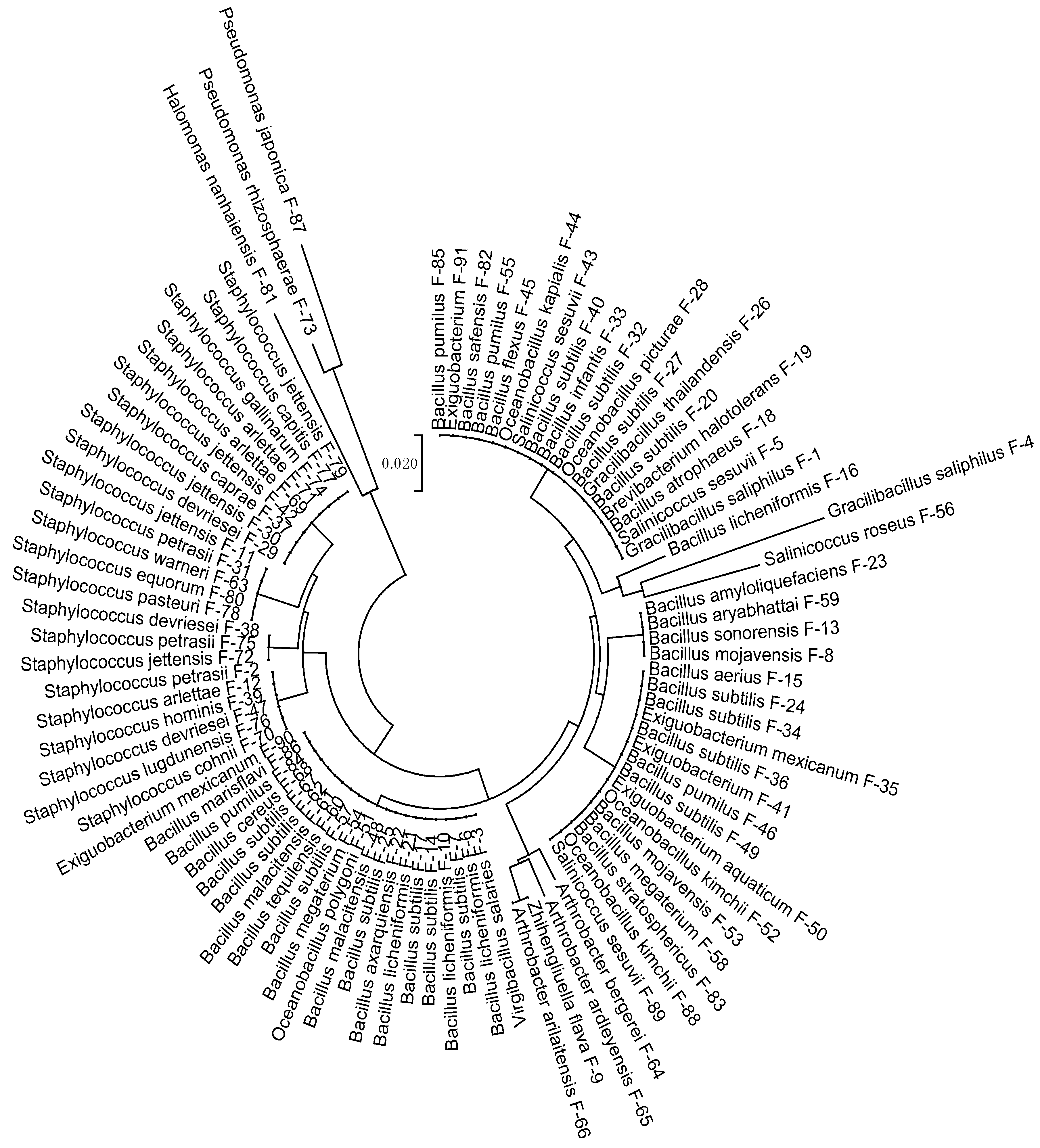
2.2. 16S rRNA Gene Sequencing

Bacterial strains were identified by 16S rRNA gene sequencing. Total genomic DNA from overnight grown bacterial cultures was extracted using the FavorPrepTM Tissue Genomic DNA Extraction Mini Kit (Favorgen Biotech Corporation, Ping-Tung, Taiwan). PCR amplification of 16S rRNA gene sequencing was accomplished by using forward primer 27f (5-AGAGTTTGATCCTGGCTCAG-3) and reverse primer 1522r (5-AAGGAGGTGATCCA (AG)CCGCA-3) [19]. PCR amplification was performed in 50 µL of Dream TaqTM Green PCR Master Mix (Fermentas, Waltham, MA, USA) with 0.5 µg of chromosomal DNA template and 0.5 µM of each primer. The reaction mixtures were incubated in a thermocycler Primus 96 (PeQLab, Erlangen, Germany) at 94 °C for 5 min and passed through 30 cycles: Denaturation for 20 s at 94 °C, primer annealing for 20 s at 50 °C, and extension at 72 °C for 2 min. The amplified products were purified by using the FavorPrepTM Gel Purification Mini Kit (Favorgen Biotech Corporation, Ping-Tung, Taiwan). Purified PCR products were sent to First Base Laboratories, Singapore for sequencing. Sequences were aligned with a multiple sequence alignment program (Clustal W) by using Molecular Evolutionary Genetics Analysis (MEGA) 6. software) [20]. The phylogenetic relationships among halotolerant bacterial strains were studied by constructing phylogenetic trees by the neighbor-joining method [21].

2.3. Halophility Assay

l-broth medium supplemented with 0, 0.5, 1, and 1.5 M NaCl was used to evaluate the salt tolerance of bacterial strains. After inoculation, strains were incubated overnight at 37 °C for 24 h at 130 rpm/min. For all treatments, three replicates were placed for comparison. The optical density of the cultures was recorded at 600 nm by a spectrophotometer (CECIL CE 7200, Cecil Instruments Limited, Cambridge, UK). Proline analysis of bacterial strains grown under salt stress was also accomplished by following the method of Cha-Um and Kirdmanee [22].

2.4. Plant-Growth-Promoting Traits

For auxin quantification, strains were inoculated in triplicate in l-broth supplemented with 1 M NaCl and 500 µg mL1 filter sterilized solution of l-tryptophan. After incubation at 37 °C for 72 h, cells were removed from stationary phase cultures by centrifugation. One milliliter of culture supernatant was mixed with 2 mL of Salkowski reagent [23]. Afterwards, samples were incubated in the dark for the development of pink color. Optical density was recorded at 535 nm with a spectrophotometer (CECIL CE 7200). The standard curve was constructed by using different concentrations of authentic auxins to calculate the auxin production by different bacterial strains. The phosphate solubilization potential of bacterial isolates was evaluated by streaking on Pikovskaya’s agar plates [24].
The 1-aminocyclopropane-1-carboxylate (ACC)-utilization ability of the strains was evaluated by following the method described in [25]. Bacterial strains were inoculated in 5 mL of l-broth in triplicate and incubated overnight at 28 °C on a shaker at 200 rpm/min. Two milliliters of each culture were harvested in a 2-mL Eppendorf tube by centrifugation at 8000× g for 5 min. The cell pellet was washed twice with 1 mL of liquid Dworkin-Foster(DF) medium. Afterwards, the pellet was suspended in 2 mL of DF-ACC medium in a 12-mL culture tube. The tubes were incubated at 28 °C on a shaker at 200 rpm/min for 24 h. A 2-mL sample of DF-ACC medium without inoculation was incubated in parallel. One milliliter of each culture was centrifuged in a 1.5 mL Eppendorf tube as mentioned above. One hundred microliters of each supernatant were diluted to 1 mL with liquid DF medium in a 1.5-mL Eppendorf tube. Sixty microliters of each tenfold-diluted supernatant was used for the 96-well PCR-plate ninhydrin-ACC assay and mixed with 120 µL of ninhydrin reagent. The PCR plate was then heated on a boiling water bath for 30 min. The DF medium was used as a blank. Each diluted supernatant was run in triplicate. After transfer of 100 µL of the remaining reaction solution, the absorbance of the samples was measured at 570 nm with a microplate spectrophotometer (Epoch, BioTek, Winooski, VT, USA).

2.5. Biofilm Formation

The biofilm-forming ability of bacterial strains was evaluated by following the method of [26]. Bacterial strains were inoculated in triplicate in Tryptic Soy Broth (TSB) supplemented with 0, 200, and 400 mM NaCl and incubated at 37 °C for 24 h. Following incubation, 20 µL of the cultures was transferred to the wells of a 96-well flat bottom microtiter plate that contained 180 µL of TSB supplemented with the abovementioned NaCl concentrations. Negative controls contained only 200 µL of the TSB medium and the assay was performed in triplicate. To promote biofilm formation, the plates were incubated on a shaker at 37 °C for 72 h. After incubation, the well contents were discarded and washed thrice with 250 µL of sterile distilled water to remove any nonadherent and weakly adherent cells. Plates were air dried for 30 min. The biofilm formed in the wells was fixed with 250 µL/well 98% methanol for 15 min. After air drying, the fixed bacterial cells were stained with 200 µL of 0.1% v/v crystal violet solution for 5 min. Excessive stain was removed by placing the plate under low-running tap water and the plate was air dried. Resolubilization of crystal violet with the adherent cells was done by adding 200 µL/well of 33% v/v glacial acetic acid. The optical density was measured at 570 nm using a microplate spectrophotometer (Epoch, BioTek, Winooski, VT, USA).

2.6. In Vitro Plant Bioassay

Seeds of maize (Zea mays L. Var. Sahiwal-2002) were obtained from Punjab Seed Corporation (Lahore, Pakistan). Seeds were surface sterilized by treatment with 0.1% HgCl2 for 23 min followed by repeated washing with sterilized distilled water. Rooting assay was performed with single or mixed bacterial cultures. Single bacterial cultures included F-9, F-11, F-12, F-35, F-37, F-58, F-71, F-72, F-81, F-83, F-84, and F-87. Mixed culture combinations C-1 (F-9, F-11, F-12, F-87), C-2 (F-35, F-71, F-81, F-83), and C-3 (F-37, F-58, F-72, F-84) were also included for seed inoculation. For inoculum preparation, strains were cultivated in L-broth for 24 h. Then, cultures were harvested by centrifugation and the cell pellet was washed with autoclaved distilled water. The optical densities of the cultures were adjusted to a final concentration of 107 colony forming units (CFU)/mL in sterilized distilled water. Sterilized seeds were soaked in a bacterial suspension for 30 min and placed in Petri plates containing two sterilized filter papers. Water-treated seeds were used for comparison. For each strain and combination, five seeds were inoculated in duplicate. Petri plates were incubated in an Environmental Test Chamber (MRL-350H; Sanyo, Osaka, Japan) at 25 °C. After 7 days, shoot length, root length, number of roots, and fresh and dry weights of seedlings were recorded.

2.7. Pot Trials with Salt Stress

Pot experiments were conducted to evaluate the growth-promoting effect of bacterial strains on maize under different salt-stress conditions. Seeds were surface sterilized as mentioned earlier and placed on moistened autoclaved double-filter paper in Petri plates. Healthy germinated seeds were selected for bacterization with single and mixed cultures of bacterial strains as mentioned above. Germinated seeds were sown in pots containing 100 g of autoclaved soil. Five seeds were sown in each pot and experiments were carried out in triplicate. Three salt-stress conditions, i.e., 0, 200, and 400 mM, were applied up to the field capacity of soil. Salt stress was applied twice—at the time of sowing and to 7-day-old seedlings. Water-treated seeds were used as a control. Pots were kept under conditions of 12 h of photoperiod and a temperature of 25 °C in the Environmental Test Chamber (MRL-350H; Sanyo, Osaka, Japan). After two weeks, plant-growth parameters such as shoot length, root length, number of roots, and fresh weight of seedlings were recorded. For dry weight, plants were incubated in an oven at 80 °C for 24 h.

2.8. Biochemical Analysis of Plants

The proline content of plants grown under 0, 200, and 400 mM NaCl was measured as described by Cha-Um and Kirdmanee [22]. Similarly, the method of Racusen and Foote [27] was used for the quantitative estimation of peroxidases from fresh leaves. Acid phosphatase was extracted from plant leaves following the method of Iqbal and Rafique [28].

2.9. Statistical Analysis

Data for bacterial auxin production and plant-growth parameters were subjected to statistical analysis by using SPSS 20 software (IBM Corporation, New York, NY, USA). The data were also subjected to analysis of variance (ANOVA). Finally, mean values were separated by using Duncan’s multiple range test (p ≤ 0.05). Correlation analysis between bacterial growth and NaCl concentrations was also performed (p = 0.05).

3. Results

3.1. Strains Identification

One hundred salt-tolerant bacterial strains were isolated from the rhizosphere and phytoplane of Suaeda fruticosa (L.) Forssk. fruticosa growing in saline habitats. 16S rRNA gene sequences were compared with already-deposited sequences in GenBank. The comparison indicated around 99% similarity with respective identified species. Analysis showed that the strains belong to the genera Gracilibacillus, Staphylococcus, Virgibacillus, Salinicoccus, Bacillus, Zhihengliuella, Brevibacterium, Oceanobacillus, Exiguobacterium, Pseudomonas, Arthrobacter, and Halomonas. Sequences were submitted to GenBank under accession numbers KT027652–KT027742. Rhizospheric soil samples recorded the presence of 45 salt-tolerant rhizobacteria and were represented by 9 bacterial genera (Table 1). Similarly, the phytoplane recorded the presence of 46 endophytic bacterial strains represented by 8 bacterial genera (Table 2). Figure 1 shows the phylogenetic relationships among different salt-tolerant bacteria isolated from the rhizosphere and phytoplane of S. fruticosa. Analysis showed that the majority of the Gram-positive strains (firmicutes) clustered in one major group. This included strains from the genera Bacillus and Staphylococcus. A few Gram-positive strains were also represented by the genera Oceanobacillus, Salinococcus, Gracilibacillus, Brevibacterium, and Exiguobacterium. In addition to that, Gram-positive Arthrobacter and Zhihengliuella clustered in a separate group. Gram-negative strains were represented by the genera Pseudomonas and Halomonas and occupied a separate cluster in the phylogenetic tree.

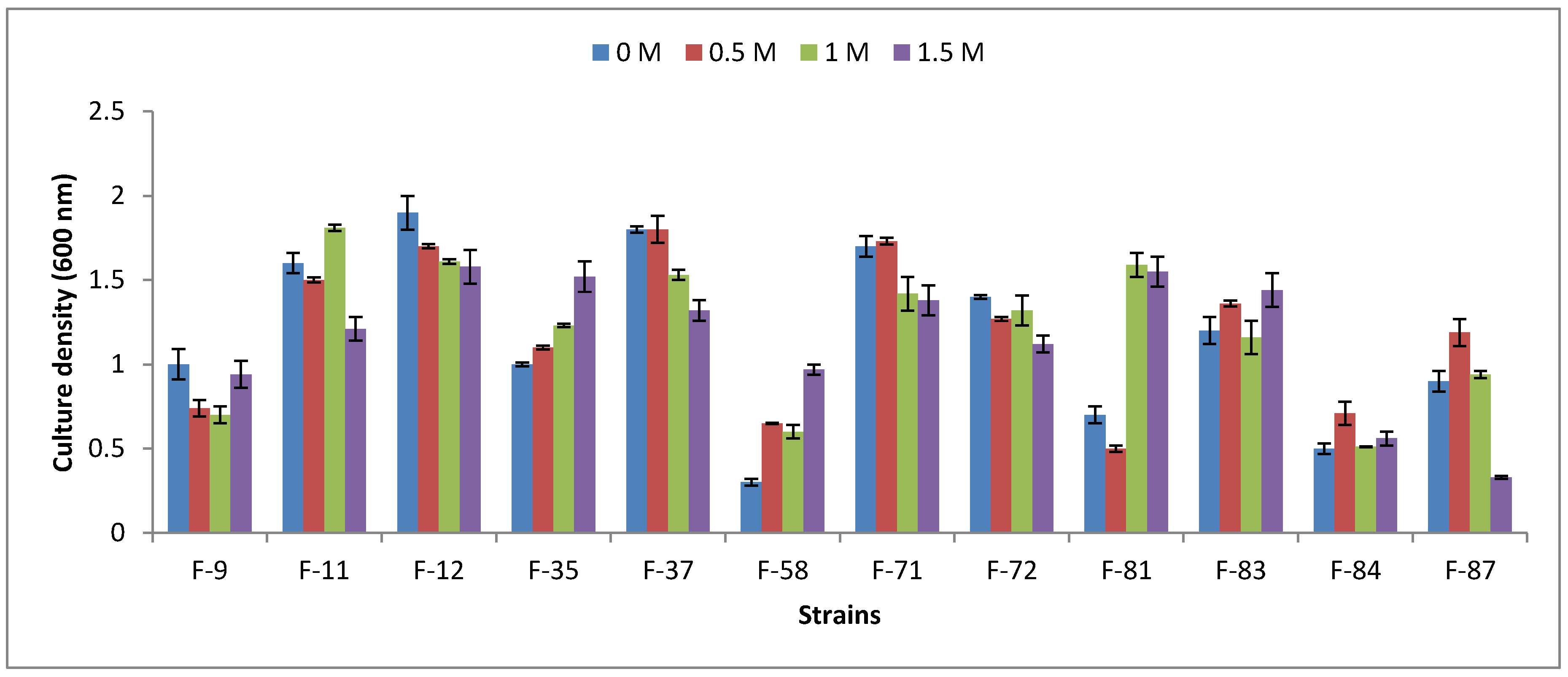
3.2. Halophility Assay

Bacterial isolates showed variable growth responses at different NaCl concentrations (Figure 2). The majority of the isolates that included S. jettensis F-11, S. arlettae F-12, S. caprae F-37, S. arlettae F-71, S. jettensis F-72, and B. safensis F-83 showed good growth up to 1.5 M NaCl. A few strains recorded sensitivity against increasing levels of NaCl. For example, Z. flava F-9, B. megaterium F-58, and B. pumilus F-84 recorded poor growth as compared to other halotolerant bacterial strains. Proline is produced as an osmoprotectant under osmotic stress induced at high salinity. It accumulates as a compatible solute in living cells to counter the effects of NaCl toxicity. Bacterial proline content did not show a significant difference with the increasing salt content from 0 to 1.5 M NaCl. The highest concentration of proline was observed with S. jettensis F-11 (228 µg g−1) and S. jettensis F-72 (216 µg g−1) at 1.5 M NaCl (Figure 3).

3.3. Plant-Growth-Promoting Traits

One hundred bacterial strains were screened for in vitro auxin production by colorimetric analysis. Screening indicated significant auxin production by 30 bacterial strains (Table 3). In the absence of l-tryptophan, bacterial auxin production ranged from 5.70 to 87.90 µg mL−1. Bacterial strains produced significant levels of auxin in culture supernatant in the presence of 500 µg mL−1 l-tryptophan as compared to the nonamended control. For instance, S. petrasii F-2, V. salarius F-3, Z. flava F-9, S. jettensis F-11, S. arlettae F-12, and S. arlettae F-71 recorded 10-, 2-, 5-, 3-, 3-, and 2-fold increases in auxin production over the control. Overall, auxin production ranged from 14 to 215 µg mL−1 in l-tryptophan-amended medium. On the other hand, 14 isolates were recorded as positive for phosphate solubilization. S. jettensis F-11, S. arlettae F-12, and S. jettensis F-72 were the most efficient as compared to other strains.
Bacterial isolates showed variable potential for ACC-deaminase activity. Highly significant activity was observed for S. caprae F-37, S. arlettae F-71, B. subtilis F-14, E. mexicanum F-35, and O. kapialis F-44. For biofilm formation, S. caprae F-37 and B. pumilus F-84 recorded good production at 200 mM NaCl. However, at higher NaCl levels, poor biofilm formation was recorded for the majority of the strains (Figure 4).

3.4. In Vitro Plant Bioassay

Bacterial strains as single or mixed cultures significantly enhanced the growth of Z. mays under in vitro conditions. Maximum increases in shoot length over the water-treated control were observed for S. jettensis F-72 (61%), S. arlettae F-71 (48%), and S. arlettae F-12 (48%). Similarly, for root length, S. arlettae F-12, S. jettensis F-72, and B. safensis F-83 showed 92%, 85%, and 78% increases, respectively, over the control. A statistically significant increase in the number of roots per plant was recorded for S. jettensis F-72 and mixed culture C-2. For fresh and dry weights, S. arlettae F-12 and mixed culture combination C-1 were the most promising (Table 4).

3.5. Pot Trials under Salt Stress

Halotolerant bacterial strains were evaluated to mitigate the effects of salinity on the plants of Z. mays. Bacterial strains were used as single or mixed cultures as mentioned above. Bacterization of seeds significantly enhanced growth parameters at 0 mM NaCl. A highly significant response for shoot and root length was shown by S. arlettae F-12 and S. arlettae F-71 over the water-treated control. For fresh weight, Z. flava F-9 was the most effective. For dry weight, S. jettensis F-11, S. caprae F-37, and H. nanhaiensis F-81 recorded up to threefold increases. Increases in salt content (0–400 mM NaCl) negatively affected the growth of plants in control treatments (Table 5). However, inoculations by halotolerant rhizobacteria significantly improved different growth parameters under saline conditions. At 200 mM NaCl, significant increases in shoot length were observed with S. jettensis F-11 (59%), S. arlettae F-12 (55%), and S. arlettae F-71 (50%) over the respective control. At higher stress (400 mM NaCl), S. jettensis F-11, S. arlettae F-12, and B. marisflavi F-87 recorded around a onefold increase for shoot elongation over the respective control. For root length, S. jettensis F-11, S. arlettae F-12, S. arlettae F-71, and H. nanhaiensis F-81 gave a promising response at 200 mM NaCl. Similarly, at 400 mM salt stress, the highest response for root elongation was observed with S. jettensis F-11 (85%) and S. arlettae F-12 (69%). For number of roots, S. jettensis F-72 (98%) and E. mexicanum F-35 (52%) recorded significant improvements over the control. At 400 mM stress, a 46% increase in root number was observed with S. arlettae F-12. Seed bacterization also significantly influenced plant weight under saline conditions. For instance, at 200 mM salt stress, Z. flava F-9 (45%) and B. megaterium F-58 (42%) demonstrated statistically significant improvements for fresh weight. Similarly, at higher NaCl content (400 mM), S. jettensis F-11 (44%) was the most effective (Table 5). On the other hand, for dry weight, S. jettensis F-11 and S. arlettae F-71 recorded two- to threefold increases over the 200 mM control. At 400 mM salt stress, the mixed culture C-1 gave good results for dry weight.

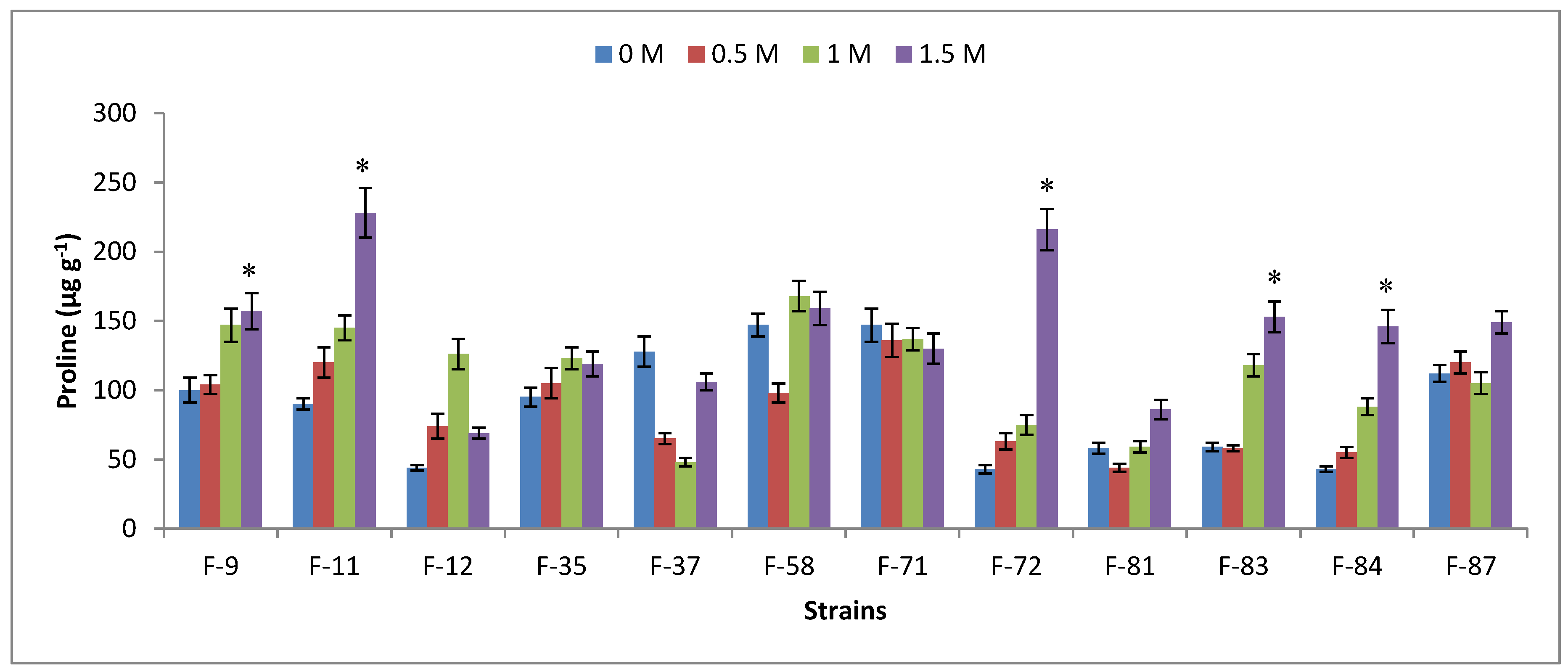
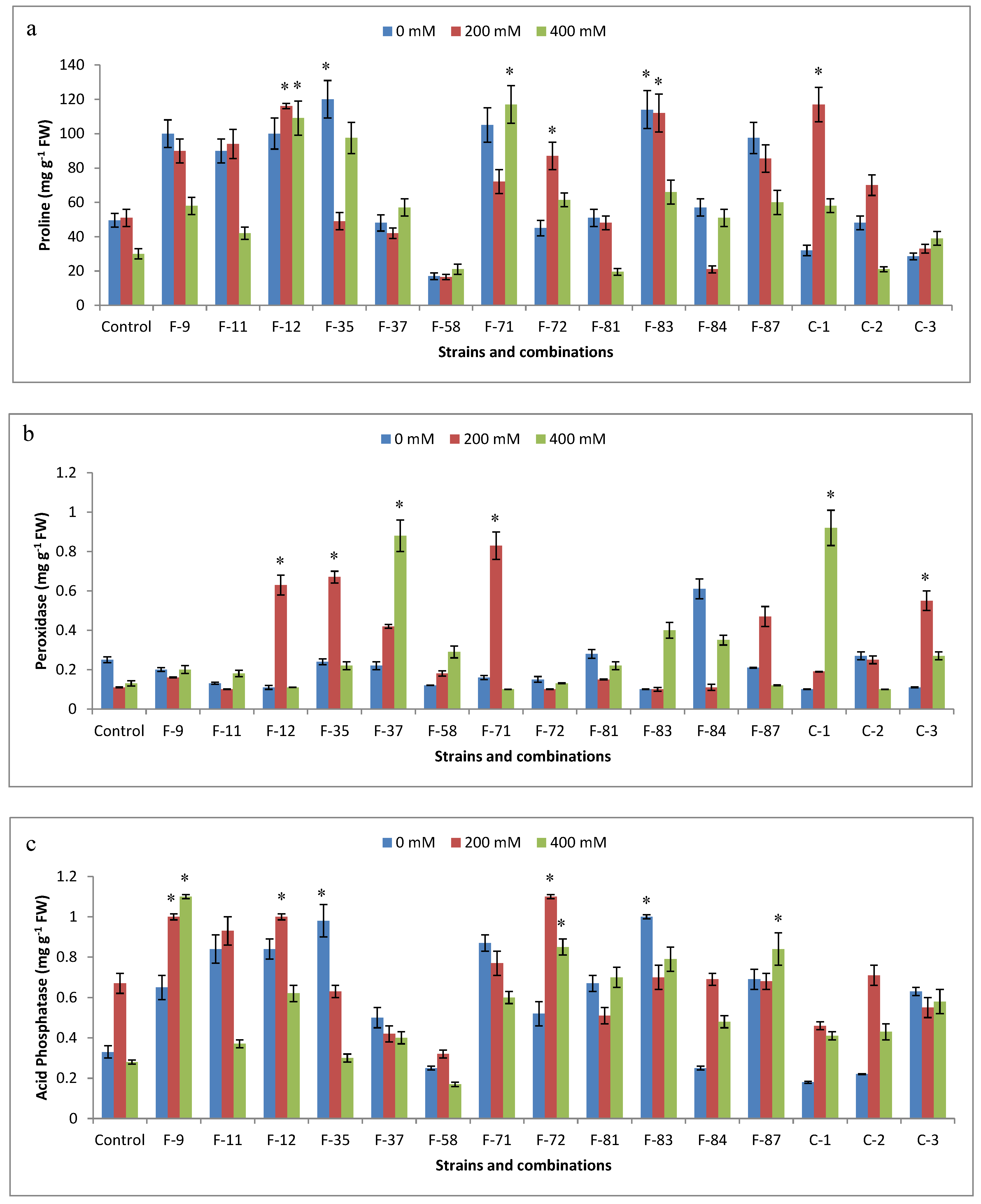
3.6. Antioxidant Analysis

Bacterial inoculations recorded high proline accumulation in plants. At 200 mM NaCl, S. arlettae F-12, B. safensis F-83, and C-1 (mixed culture) recorded the highest accumulation of proline. Similarly, at 400 mM salt stress, S. arlettae F-12 and S. arlettae F-71 resulted in considerable increases in proline concentrations over water-treated control plants (Figure 5a). Regarding peroxidases, plants produced a very low content in water-treated seeds. However, with increasing salinity (200–400 mM), considerable enhancements in peroxidase activity was observed (Figure 5b). Acid phosphatase production significantly increased at 200 mM NaCl with Z. flava F-9, S. arlettae F-12, and S. jettensis F-72. At 400 mM salt stress, Z. flava F-9, S. jettensis F-72, and B. safensis F-83 recorded up to twofold increases for acid phosphatase content over the control (Figure 5c).

4. Discussion

Soil salinity severely affects crop productivity and resident soil microbial communities. In Pakistan, soil salinity results in approximately 26% losses for the agroeconomy because it affects the germination, growth, and respiration of seeds [17]. The present study was carried out to examine the bacterial diversity of S. fruticosa growing in saline habitats of northeastern Pakistan. Extreme habitats have been reported to be colonized by a variety of microorganisms that are well adapted to these environments [29,30,31]. Natural plant settings growing in these localities may harbor beneficial microbes for agricultural applications under saline conditions [9]. S. fruticosa is a halophytic plant that mainly colonizes saline habitats. Therefore, we targeted the isolation of plant-growth-promoting rhizobacteria (PGPR) associated with the surfaces and rhizosphere of this plant. Analysis of 16S rRNA gene sequences confirmed the presence of several strains of halotolerant bacteria. Since little is known about the bacterial diversity associated with S. fruticosa, we therefore reported in detail the diversity and agricultural significance of bacterial communities colonizing the surfaces of this halophytic plant.
Bacterial strains showed good potential for salt tolerance and production of proline up to 1.5 M NaCl stress. Especially, E. mexicanum F-35 recorded a significant positive correlation (r = 0.967; p = 0.05) with increasing NaCl concentration in culture media. Inoculation of Z. mays by halotolerant rhizobacteria also influenced the production of antioxidant enzymes. For instance, plants grown under salinity stress exhibited the accumulation of proline as a compatible solute to retain water that otherwise might be lost due the surrounding hypertonic environment. Similarly, bacterial inoculations also influenced the peroxidase and acid phosphatase contents of plants. Peroxidases and acid phosphatases are considered very important enzymes to mitigate salinity-induced stresses. Exposure of plants to environmental stresses can lead to the production of reactive oxygen species (ROS) that can damage plant macromolecules. In addition to plants, microbes can augment the supply of enzymatic and nonenzymatic metabolites to detoxify the impact of ROS [32,33]. Previously, bioassays for the antioxidant system have also been reported with different plants and microorganisms [34,35].
In the present study, halotolerant rhizobacteria also showed good potential to produce a variety of plant-growth-promoting attributes. For example, strains recorded up to a 10-fold increase in auxin content in the presence of l-tryptophan over the unamended control. Under in vitro conditions, strains of genus Staphylococcus (F-12, F-71, F-72) were the most promising to enhance the rooting or shooting of inoculated maize plants. In pot trials, strains belonging to Bacillus, Staphylococcus, Zhihengliuella, and Halomonas genera were very effective at enhancing plant growth in salt-amended soils. However, strains of S. arlettae (F-12, F-71) recorded consistent results under both sets of experiments. Auxin-producing PGPR have been reported to improve plant growth under different sets of conditions. Charcoal-based formulations of rhizobacteria with auxin-production ability have been shown to enhance the vegetative and yield parameters of wheat [36]. Previously, we also reported the growth and yield improvement of plants grown under different salinity and water stresses by auxin-producing rhizobacteria [9,33]. Defez et al. [37] demonstrated the role of bacterial auxin in the upregulation of nitrogenase activity of inoculated rice plants. Inoculation of maize seeds with auxin-producing B. megaterium and Azotobacter chroococcum significantly enhanced the rooting and shooting compared to control seedlings [38]. Bacterial auxin has also been shown to alleviate the salt stress of plants grown under saline conditions. For instance, halotolerant bacteria from saline habitats stimulated plant growth by mitigating osmotic stress [9,39]. Similarly, salt-tolerant rhizobacteria exhibiting multiple plant-growth-promoting traits stimulated the growth of wheat in salt-affected soils [40]. In another study, strains of Bacillus facilitated the growth of maize under water-stress conditions [41].
High salinity levels in soil or in irrigation water are a major threat to the sustainability of agricultural production. The accumulation of salts in the vicinity of a plant’s rhizosphere can cause severe injury to the root system. High concentrations of salts can result in the production of a gaseous hormone ethylene after seed germination. Elevated levels of ethylene can suppress the root system, which can result in compromised plant growth and development. Salt-tolerant rhizobacteria have the ability to produce the enzyme ACC-deaminase, which can degrade the substrate (ACC) of ethylene into α-ketobutyrate and ammonia [13,32]. Hence, the ACC-deaminase activity of rhizobacteria mitigates deleterious levels of stress-induced ethylene. In the present study, highly significant ACC-deaminase activity was exhibited by S. caprae F-37, S. arlettae F-71, and B. subtilis F-14. PGPR with ACC-deaminase activity can protect inoculated plants from the deleterious effects of abiotic stressors. Orhan et al. [42] reported the salt-stress alleviation of wheat by halotolerant and halophilic PGPR that exhibited ACC-deaminase activity. Halotolerant Klebsiella with ACC-deaminase activity has also been shown to promote the vegetative growth parameters and chlorophyll content of plants under salt stress [43]. Similarly, Tank and Saraf [44] reported the positive effects of ACC-deaminase-containing bacteria on plant growth against salinity stress.
Biofilm formation is another important trait of PGPR that can help plants tolerate abiotic stress. It is a complex association of bacterial cells attached to the plant’s root system and plays a very critical role in retaining moisture and protecting against different biotic or abiotic stresses. In this study, S. caprae F-37 and B. pumilus F-84 demonstrated good production of biofilm at 200 mM NaCl. The biofilm-forming ability of halotolerant rhizobacteria has been shown to enhance plant biomass under saline conditions [45]. Inoculation of barley plants with biofilm-forming B. amyloliquefaciens ameliorated salt stress and also stimulated different plant-growth parameters [46].

5. Conclusions

Finally, it can be concluded that S. fruticosa growing in saline areas harbor a range of beneficial plant-growth-promoting rhizobacteria. Bacterial strains showed a good ability to grow in up to 1.5 M NaCl. Halotolerant bacterial strains showed good auxin-production potential, ACC-deaminase activity, and biofilm formation. In pot trials, strains recorded good results for the amelioration of salt stress of maize plants. Especially, S. jettensis F-11, S. arlettae F-12, B. marisflavi F-87, Z. flava F-9, and H. nanhaiensis F-81 were shown to exhibit multiple plant-growth-promoting traits. In pot trials, strains were also very effective at mitigating the salinity stress of maize plants. For instance, S. jettensis F-11, F-12, S. arlettae F-71, B. marisflavi F-87, H. nanhaiensis F-81, Z. flava F-9, and E. mexicanum F-35 enhanced plant-growth parameters under salt stress. The data presented in this work are very encouraging for the use halotolerant rhizobacteria to enhance plant growth under salinity stress. The results of this study also indicated that the natural plant settings of a saline habitat are a good source for isolating beneficial PGPR to grow crops under saline conditions.

Author Contributions

F.A. conducted the lab experiments related to the isolation, identification of bacterial strains and pot trials. B.A. developed the experimental layout, performed statistical analysis and wrote this manuscript.

Funding

This research work was supported by the grant (No. 104-P-50/Budget) received form University of the Punjab, Lahore, Pakistan.

Conflicts of Interest

The authors declare no conflict of interest.

References

  1. Singh, K. Microbial and enzyme activities of saline and sodic Soils. Land Degrad. Dev. 2016, 27, 706–718. [Google Scholar] [CrossRef]
  2. Labidi, N.; Ammari, M.; Mssedi, D.; Benzerti, M.; Snoussi, S.; Abdelly, C. Salt excretion in Suaeda fruticosa. Acta Biol. Hung. 2010, 61, 299–312. [Google Scholar] [CrossRef] [PubMed]
  3. Khan, M.A.; Aziz, I. Salinity tolerance in some mangrove species from Pakistan. Wetl. Ecol. Manag. 2001, 9, 229–233. [Google Scholar] [CrossRef]
  4. Mahajan, S.; Tuteja, N. Cold, salinity and drought stresses: An overview. Arch. Biochem. Biophys. 2005, 444, 139–158. [Google Scholar] [CrossRef] [PubMed]
  5. Shrivastava, P.; Kumar, R. Soil salinity: A serious environmental issue and plant growth promoting bacteria as one of the tools for its alleviation. Saudi J. Biol. Sci. 2015, 22, 123–131. [Google Scholar] [CrossRef] [PubMed]
  6. Hameed, A.; Hussain, T.; Gulzar, S.; Aziz, I.; Gul, B.; Khan, M.A. Salt tolerance of a cash crop halophyte Suaeda fruticosa: Biochemical response to salt and exogenous chemical treatments. Acta Physiol. Plant. 2012, 34, 2331–2340. [Google Scholar] [CrossRef]
  7. Ngumbi, E.; Kloepper, J. Bacterial-mediated drought tolerance: Current and future prospects. Appl. Soil Ecol. 2016, 105, 109–125. [Google Scholar] [CrossRef]
  8. Sharma, S.; Kulkarni, J.; Jha, B. Halotolerant rhizobacteria promote growth and enhance salinity tolerance in peanut. Front. Microbiol. 2016, 7, 1600. [Google Scholar] [CrossRef] [PubMed]
  9. Raheem, A.; Ali, B. Halotolerant rhizobacteria: Beneficial plant metabolites and growth enhancement of Triticum aestivum L. in salt amended soils. Arch. Agron. Soil Sci. 2015, 61, 1691–1705. [Google Scholar] [CrossRef]
  10. Bhise, K.K.; Bhagwat, P.K.; Dandge, P.B. Plant growth-promoting characteristics of salt tolerant Enterobacter cloacae strain KBPD and its efficacy in amelioration of salt stress in Vigna radiata L. J. Plant Growth Regul. 2017, 36, 215–226. [Google Scholar] [CrossRef]
  11. Woodward, A.W.; Bartel, B. Auxin: Regulation, action and interaction. Ann. Bot. 2005, 95, 707–735. [Google Scholar] [CrossRef] [PubMed]
  12. Nadeem, S.M.; Zahir, Z.A.; Naveed, M.; Asghar, H.N.; Arshad, M. Rhizobacteria capable of producing ACC-deaminase may mitigate salt stress in wheat. Soil Sci. Soc. Am. J. 2010, 74, 533–542. [Google Scholar] [CrossRef]
  13. Glick, B.R. Bacteria with ACC deaminase can promote plant growth and help to feed the world. Microbiol. Res. 2014, 169, 30–39. [Google Scholar] [CrossRef] [PubMed]
  14. Zhang, F.; Zhang, J.; Chen, L.; Shi, X.; Lui, Z.; Li, C. Heterologous expression of ACC deaminase from Trichoderma asperellum improves the growth performance of Arabidopsis thaliana under normal and salt stress conditions. Plant Physiol. Biochem. 2015, 94, 41–47. [Google Scholar] [CrossRef] [PubMed]
  15. Li, H.Q.; Jiang, Z.W. Inoculation with plant growth-promoting bacteria (PGPB) improves salt tolerance of maize seedling. Russ. J. Plant Physiol. 2017, 64, 235–241. [Google Scholar] [CrossRef]
  16. Ullah, S.; Bano, A. Isolation of plant-growth-promoting rhizobacteria from rhizospheric soil of halophytes and their impact on maize (Zea mays L.) under induced salinity. Can. J. Microbiol. 2015, 61, 307–313. [Google Scholar] [CrossRef] [PubMed]
  17. Murtaza, G.; Murtaza, B.; Usman, H.M.; Ghafoor, A. Amelioration of saline-sodic soil using gypsum and low quality water in following sorghum-berseem crop rotation. Int. J. Agric. Biol. 2013, 15, 640–648. [Google Scholar]
  18. Cappuccino, J.G.; Sherman, N. Microbiology: A Laboratory. In Microbial Populations in Soil: Enumeration, 6th ed.; Pearson Education: Singapore, 2002; pp. 349–354. [Google Scholar]
  19. Johnson, J.L. Methods for general and molecular bacteriology. In Similarity Analysis of rRNAs; Gerhardt, P., Murray, R.G.E., Wood, W.A., Krieg, N.R., Eds.; American Society for Microbiology: Washington, DC, USA, 1994; pp. 625–700. [Google Scholar]
  20. Tamura, K.; Stecher, G.; Peterson, D.; Filipski, A.; Kumar, S. MEGA6: Molecular evolutionary genetics analysis version 6.0. Mol. Biol. Evol. 2013, 30, 2725–2729. [Google Scholar] [CrossRef] [PubMed]
  21. Saitou, N.; Nei, M. The neighbor-joining method: A new method for reconstructing phylogenetic trees. Mol. Biol. Evol. 1987, 4, 406–425. [Google Scholar] [PubMed]
  22. Cha-Um, S.; Kirdmanee, C. Effect of salt stress on proline accumulation, photosynthetic ability and growth characters in two maize cultivars. Pak. J. Bot. 2009, 41, 87–98. [Google Scholar]
  23. Tang, W.; Borner, J. Enzymes involved in synthesis and breakdown of indoleacetic acid. Mod. Method Plant Anal. 1979, 7, 238–241. [Google Scholar]
  24. Pikovskaya, R.I. Mobilization of phosphorus in soil in connection with viral activity of some microbial species. Mikrobiologiya 1948, 17, 362–370. [Google Scholar]
  25. Li, Z.; Chang, S.; Lin, L.; Li, Y.; An, Q. A colorimetric assay of 1-aminocyclopropane-1-carboxylate (ACC) based on ninhydrin reaction for rapid screening of bacteria containing ACC deaminase. Lett. Appl. Microbiol. 2011, 53, 178–185. [Google Scholar] [CrossRef] [PubMed] [Green Version]
  26. Christensen, G.D.; Simpson, W.; Younger, J.; Baddour, L.; Barrett, F.; Melton, D.; Beachey, E. Adherence of coagulase-negative staphylococci to plastic tissue culture plates: A quantitative model for the adherence of staphylococci to medical devices. J. Clin. Microbiol. 1985, 22, 996–1006. [Google Scholar] [PubMed]
  27. Racusen, D.; Foote, M. Protein synthesis in dark-grown bean leaves. Can. J. Bot. 1965, 43, 817–824. [Google Scholar] [CrossRef]
  28. Iqbal, J.; Rafique, N. Toxic effects of BaCl2 on germination, early seedling growth, soluble-proteins and acid-phosphatases in Zea mays L. Pak. J. Bot. 1987, 19, 1–8. [Google Scholar]
  29. Demergasso, C.; Escudero, L.; Casamayor, E.O.; Chong, G.; Balagué, V.; Pedrós-Alió, C. Novelty and spatio-temporal heterogeneity in the bacterial diversity of hypersaline lake Tebenquiche (Salar de Atacama). Extremophiles 2008, 12, 491–504. [Google Scholar] [CrossRef] [PubMed]
  30. Roohi, A.; Ahmed, I.; Iqbal, M.; Jamil, M. Preliminary isolation and characterization of halotolerant and halophilic bacteria from salt mines of Karak, Pakistan. Pak. J. Bot. 2012, 44, 365–370. [Google Scholar]
  31. Goswami, D.; Dhandhukia, P.; Patel, P.; Thakker, J.N. Screening of PGPR from saline desert of Kutch: Growth promotion in Arachis hypogea by Bacillus licheniformis A2. Microbiol. Res. 2014, 169, 66–75. [Google Scholar] [CrossRef] [PubMed]
  32. Forni, C.; Duca, D.; Glick, B.R. Mechanisms of plant response to salt and drought stress and their alteration by rhizobacteria. Plant Soil 2017, 410, 335–356. [Google Scholar] [CrossRef]
  33. Raheem, A.; Shaposhnikov, A.; Belimov, A.A.; Dodd, I.C.; Ali, B. Auxin production by rhizobacteria was associated with improved yield of wheat (Triticum aestivum L.) under drought stress. Arch. Agron. Soil Sci. 2018, 64, 574–587. [Google Scholar] [CrossRef]
  34. Chakraborty, U.; Chakraborty, B.; Chakraborty, A.; Dey, P. Water stress amelioration and plant growth promotion in wheat plants by osmotic stress tolerant bacteria. World J. Microbiol. Biotechnol. 2013, 29, 789–803. [Google Scholar] [CrossRef] [PubMed]
  35. Estrada, B.; Aroca, R.; Barea, J.M.; Ruiz-Lozano, J.M. Native arbuscular mycorrhizal fungi isolated from a saline habitat improved maize antioxidant systems and plant tolerance to salinity. Plant Sci. 2013, 201, 42–51. [Google Scholar] [CrossRef] [PubMed]
  36. Aslam, F.; Ali, B. Efficacy of charcoal based formulations of Bacillus and Escherichia coli to enhance the growth and yield of Triticum aestivum L. Res. J. Biotechnol. 2015, 10, 81–88. [Google Scholar]
  37. Defez, R.; Andreozzi, A.; Bianco, C. The overproduction of indole-3-acetic acid (IAA) in endophytes upregulates nitrogen fixation in both bacterial cultures and inoculated rice plants. Microb. Ecol. 2017, 74, 441–452. [Google Scholar] [CrossRef] [PubMed]
  38. Đorđević, S.; Stanojević, D.; Vidović, M.; Mandić, V.; Trajković, I. The use of bacterial indole-3-acetic acid (IAA) for reduce of chemical fertilizers doses. Hem. Ind. 2017, 71, 195–200. [Google Scholar]
  39. Kim, K.; Jang, Y.; Lee, S.; Oh, B.; Chae, J.; Lee, K. Alleviation of salt stress by Enterobacter sp. EJ01 in tomato and Arabidopsis is accompanied by up-regulation of conserved salinity responsive factors in plants. Mol. Cells 2014, 37, 109–117. [Google Scholar] [CrossRef] [PubMed]
  40. Tiwari, S.; Singh, P.; Tiwari, R.; Meena, K.K.; Yandigeri, M.; Singh, D.P.; Arora, D.K. Salt-tolerant rhizobacteria-mediated induced tolerance in wheat (Triticum aestivum) and chemical diversity in rhizosphere enhance plant growth. Biol. Fertil. Soils 2011, 47, 907. [Google Scholar] [CrossRef]
  41. Sajid, M.; Raheem, A.; Ali, B. Phylogenetic diversity of drought tolerant Bacillus spp. and their growth stimulation of Zea mays L. under different water regimes. Res. J. Biotechnol. 2017, 12, 38–46. [Google Scholar]
  42. Orhan, F. Alleviation of salt stress by halotolerant and halophilic plant growth-promotingbacteria in wheat (Triticum aestivum). Braz. J. Microbiol. 2016, 47, 621–627. [Google Scholar] [CrossRef] [PubMed]
  43. Singh, R.P.; Jha, P.; Jha, P.N. The plant-growth-promoting bacterium Klebsiella sp. SBP-8 confers induced systemic tolerance in wheat (Triticum aestivum) under salt stress. J. Plant Physiol. 2015, 184, 57–67. [Google Scholar] [CrossRef] [PubMed]
  44. Tank, N.; Saraf, M. Salinity-resistant plant growth promoting rhizobacteria ameliorates sodium chloride stress on tomato plants. J. Plant Interact. 2010, 5, 51–58. [Google Scholar] [CrossRef] [Green Version]
  45. Qurashi, A.W.; Sabri, A.N. Bacterial exopolysaccharide and biofilm formation stimulate chickpea growth and soil aggregation under salt stress. Braz. J. Microbiol. 2012, 43, 1183–1191. [Google Scholar] [CrossRef] [PubMed] [Green Version]
  46. Kasim, W.A.; Gaafar, R.M.; Abou-Ali, R.M.; Omar, M.N.; Hewait, H. Effect of biofilm forming growth promoting rhizobacteria on salinity tolerance in barley. Ann. Agric. Sci. 2016, 61, 217–227. [Google Scholar] [CrossRef]
Figure 1. Phylogenetic tree showing the relationships among different halotolerant bacterial strains isolated from rhizosphere and phytoplane of S. fruticosa. (L.) Forssk. Scale bar represents mutations per nucleotide position.
Agronomy 08 00131 g001
Figure 2. Halophility assay of salt-tolerant rhizobacteria in the presence of different concentrations of NaCl. Bar represents mean ± SE of three replicates.
Agronomy 08 00131 g002
Figure 3. Effect of NaCl concentrations on proline content of halotolerant bacteria. Bars represent mean ± SE of three replicates. Treatments followed by * indicate significant difference over control using Duncan’s multiple range test (p ≤ 0.05).
Agronomy 08 00131 g003
Figure 4. Effect of different NaCl concentrations on biofilm-forming ability of bacterial strains. Bars represent mean ± SE of three replicates. Treatments followed by * indicate significant difference over control using Duncan’s multiple range test (p ≤ 0.05).
Agronomy 08 00131 g004
Figure 5. Effect of single and mixed bacterial inoculations and different salinity levels on the activities of antioxidant metabolites or enzymes: (a) Proline; (b) Peroxidase; (c) Acid phosphatase. Bars represent mean ± SE of three replicates. Treatments followed by * indicate significant difference over respective control using Duncan’s multiple range test (p ≤ 0.05).
Agronomy 08 00131 g005
Table 1. 16S rRNA gene sequencing of halotolerant bacterial strains isolated from the rhizosphere of Suaeda fruticose (L.) Forssk.
S. No.StrainsIdentified asAccessions
1F-1Gracilibacillus saliphilus F-1KT027652
2F-2Staphylococcus petrasii F-2KT027653
3F-3Virgibacillus salarius F-3KT027654
4F-4G. saliphilus F-4KT027655
5F-5Salinicoccus sesuvii F-5KT027656
6F-6Bacillus licheniformis F-6KT027657
7F-7B. subtilis F-7KT027658
8F-8B. mojavensis F-8KT027659
9F-9Zhihengliuella flava F-9KT027660
10F-10B. licheniformis F-10KT027661
11F-11S. jettensis F-11KT027662
12F-12S. arlettae F-12KT027663
13F-13B. sonorensis F-13KT027664
14F-14B. subtilis F-14KT027665
15F-15B. aerius F-15KT027666
16F-16B. licheniformis F-16KT027667
17F-17B. subtilis F-17KT027668
18F-18B. atrophaeus F-18KT027669
19F-19Brevibacterium halotolerans F-19KT027670
20F-20B. subtilis F-20KT027671
21F-21B. licheniformis F-21KT027672
22F-22B. axarquiensis F-22KT027673
23F-23B. amyloliquefaciens F-23KT027674
24F-24B. subtilis F-24KT027675
25F-25B. subtilis F-25KT027676
26F-26G. thailandensis F-26KT027677
27F-27B. subtilis F-27KT027678
28F-28Oceanobacillus picturae F-28KT027679
29F-29S. devriesei F-29KT027680
30F-30S. jettensis F-30KT027681
31F-31S. petrasii F-31KT027682
32F-32B. subtilis F-32KT027683
33F-33B. infantis F-33KT027684
34F-34B. subtilis F-32KT027685
35F-35Exiguobacterium mexicanum F-35KT027686
36F-36B. subtilis F-36KT027687
37F-37S. caprae F-37KT027688
38F-38S. devriesei F-38KT027689
39F-39S. hominis F-39KT027690
40F-83B. stratosphericus F-83KT027734
41F-84B. pumilus F-84KT027735
42F-85B. pumilus F-85KT027736
43F-86B. marisflavi F-86KT027737
44F-87Pseudomonas japonica F-87KT027738
45F-89Sal. sesuvii F-89KT027740
Table 2. 16S rRNA gene sequencing of halotolerant bacterial strains isolated from the phytoplane of S. fruticosa.
S. No.StrainsIdentified asAccessions
1F-40Bacillus subtilis F-40KT027691
2F-41Exiguobacterium sp. F-41KT027692
3F-42Staphylococcus jettensis F-42KT027693
4F-43Salinicoccus sesuvii F-43KT027694
5F-44Oceanobacillus kapialis F-44KT027695
6F-45B. flexus F-45KT027696
7F-46B. pumilus F-46KT027697
8F-47S. devriesei F-47KT027698
9F-48B. malacitensis F-48KT027699
10F-49B. malacitensis F-49KT027700
11F-50E. aquaticum F-50KT027701
12F-51O. polygoni F-51KT027702
13F-52O. kimchii F-52KT027703
14F-53B. mojavensis F-53KT027704
15F-54B. megaterium F-54KT027705
16F-55B. pumilus F-55KT027706
17F-56Sal. roseus F-56 KT027707
18F-57B. subtilis F-57KT027708
19F-58B. megaterium F-58KT027709
20F-59B. aryabhattai F-59KT027710
21F-60B. tequilensis F-60 KT027711
22F-61B. malacitensis F-61KT027712
23F-62B. subtilis F-62KT027713
24F-63S. warneri F-63KT027714
25F-64Arthrobacter bergerei F-64KT027715
26F-65A. ardleyensis F-65KT027716
27F-66A. arilaitensis F-66KT027717
28F-67B. subtilis F-67KT027718
29F-68B. cereus F-68KT027719
30F-69S. arlettae F-69KT027720
31F-70S. cohnii F-70KT027721
32F-71S. arlettae F-71KT027722
33F-72S. jettensis F-72KT027723
34F-73Pseudomonas rhizosphaerae F-73KT027724
35F-74S. gallinarum F-74KT027725
36F-75S. petrasii F-75KT027726
37F-76S. lugdunensis F-76KT027727
38F-77S. capitis F-77KT027728
39F-78S. pasteuri F-78KT027729
40F-79S. jettensis F-79KT027730
41F-80S. equorum F-80KT027731
42F-81Halomonas nanhaiensis F-81KT027732
43F-82B. safensis F-82KT027733
44F-88O. kimchii F-88KT027739
45F-90E. mexicanum F-90KT027741
46F-91Exiguobacterium sp. F-91KT027742
Table 3. l-tryptophan-dependent bacterial auxin production in the presence of 1 M NaCl.
S. No.Strainsl-tryptophan (µg mL−1)
0500
Auxin (µg mL−1)
1Gracilibacillus saliphilus F-16.20 (a)15.70 (a)
2Staphylococcus petrasii F-29.30 (a)113.50 (o)
3Virgibacillus salarius F-387.90 (i)150.00 (p)
4Zhihengliuella flava F-944.70 (d–h)215.00 (r)
5S. jettensis F-1163.20 (g–i)185.70 (qr)
6S. arlettae F-1251.40 (e–i)174.30 (pq)
7Exiguobacterium sp. F-416.40 (a)43.50 (a–f)
8S. jettensis F-4233.60 (d–g)78.70 (l–o)
9Salinicoccus sesuvii F-4316.50 (abc)28.40 (abc)
10S. devriesei F-4778.30 (hi)28.00 (abc)
11Bacillus megaterium F-5820.80 (abc)48.70 (a–g)
12B. aryabhattai F-5925.90 (d–g)61.20 (i–n)
13S. warneri F-636.40 (a)14.00 (a)
14Arthrobacter bergerei F-6413.40 (ab)25.50 (abc)
15A. ardleyensis F-659.10 (a)29.60 (abc)
16S. arlettae F-6914.30 (abc)54.20 (h–n)
17S. cohnii F-7015.70 (abc)42.10 (a–f)
18S. arlettae F-7153.60 (f–i)88.60 (no)
19S. jettensis F-7230.60 (d–g)59.80 (i–n)
20Pseudomonas rhizosphaerae F-7336.80 (d–g)67.70 (g–n)
21S. gallinarum F-7415.40 (abc)23.00 (abc)
22S. pasteuri F-7810.40 (a)36.90 (a–c)
23S. jettensis F-795.70 (a)24.50 (abc)
24S. equorum F-807.60 (a)32.20 (a–d)
25Halomonas nanhaiensis F-8121.10 (abc)83.30 (mno)
26B. safensis F-829.50 (a)43.90 (a–f)
27B. stratosphericus F-8323.60 (def)72.70 (k–n)
28B. pumilus F-847.90 (a)18.10 (ab)
29B. pumilus F-8532.60 (a–d)59.80 (i–n)
30B. marisflavi F-8622.60 (abc)38.60 (a–e)
Mean of three replicates. Different letters followed by numerical values within same column indicate significant difference between treatments using Duncan’s multiple range test (p ≤ 0.05).
Table 4. Effect of bacterial strains on growth parameters of Zea mays under in vitro conditions.
StrainsShoot Length (cm)Root Length (cm)Roots/Plant Fresh Weight (g)Dry Weight (g)
Control23 (a)13 (b)6.4 (de)4.6 (a)0.7 (ab)
F-928 (bcd)20 (cd)4.8 (a)5.3 (ab)0.9 (abc)
F-1132 (defg)20 (cde)5.6 (abcd)6.8 (a–e)1.1 (bc)
F-1234 (fgh)25 (f)6.2 (bcde)8.8 (ef)1.3 (c)
F-3531 (c–g)21 (de)5.7 (abcd)5.7 (abc)0.8 (ab)
F-3729 (cd)20 (cde)5.5 (abcd)10.2 (f)0.9 (abc)
F-5833 (efgh)20 (cde)6.0 (bcd)7.4 (b–f)0.9 (abc)
F-7134 (gh)22 (def)5.7 (abcd)8.1 (cdef)1.1 (bc)
F-7237 (h)24 (f)7.1 (e)7.8 (b–f)1.0 (abc)
F-8130 (cdef)20 (cd)5.0 (ab)7.5 (b–f)1.3 (c)
F-8329 (cde)23 (ef)6.2 (cde)4.2 (a)0.6 (a)
F-8431 (c–g)22 (def)5.1 (abc)6.0 (abcd)1 (abc)
F-87 28 (bc)19 (cd)5.4 (abcd)8.6 (def)1.1 (bc)
* C-122 (a)10 (a)5.6 (abcd)9.1 (ef)1.1 (bc)
* C-224 (ab)14 (b)6.5 (de)7.8 (b–f)1.3 (c)
* C-323 (a)17 (c)5.6 (abcd)10.1 (f)1.4 (ab)
Mean of 10 plants. Different letters within same column indicate significant difference between treatments using Duncan’s multiple range rest (p ≤ 0.05); * Mixed culture combinations: C-1 (F-9, F-11, F-12, F-87), C-2 (F-35, F-71, F-81, F-83), C-3 (F-37, F-58, F-72, F-84).
Table 5. Effect of halotolerant bacterial strains on the growth of Z. mays in salt amended soils.
StrainsNaCl (mM)Shoot Length (cm)Root Length (cm)Roots/Plant Fresh Weight (g)Dry Weight (g)
Control022 (b–g)13 (abc)5.0 (jklm)0.9 (abcd)0.1 (a)
20022 (bcde)16 (bcde)4.2 (i–m)1.2 (c–j)0.1 (abcd)
40014 (a)13 (abc)4.3 (a–e)1.1 (c–g)0.1 (ab)
F-9029 (i–o)19 (e–k)5.0 (b–j)2.0 (r)0.3 (e–l)
20029 (i–p)21 (g–n)5.2 (b–l)1.7 (pqr)0.2 (abcd)
40025 (d–i)21 (f–m)4.6 (a–f)1.6 (l–q)0.2 (c–h)
F-11032 (n–s)20 (e–k)6.0 (e–l)1.8 (qr)0.4 (nop)
20035 (rs)25 (m)6.0 (f–l)1.6 (m–r)0.4 (op)
40032 (n–r)24 (klmn)5.0 (b–k)1.6 (n–r)0.2 (e–j)
F-12034 (pqrs)25 (mn)6.2 (h–m)1.6 (n–r)0.3 (j–p)
20034 (pqrs)23 (j–n)6.0 (f–m)1.4 (g–n)0.3 (d–j)
40030 (k–r)22 (h–n)6.3 (jklm)1.3 (f–l)0.3 (e–k)
F-35031 (l–r)21 (g–n)6.0 (f–l)1.5 (j–p)0.2 (c–h)
20026 (f–l)21 (g–n)6.4 (klm)1.2 (c–i)0.2 (c–g)
40025 (e–i)21 (g–m)5.0 (b–j)1.0 (b–f)0.2 (e–j)
F-37029 (i–o)20 (f–l)5.5 (e–l)1.0 (a–e)0.4 (k–p)
20027 (g–m)21 (g–n)5.3 (b–l)0.7 (ab)0.1 (abc)
40023 (b–g)16 (bcde)5.1 (b–k)1.4 (g–n)0.3 (i–o)
F-58033 (o–s)21 (f–m)6.0 (g–m)1.2 (d–k)0.3 (e–j)
20029 (i–o)21 (f–m)5.0 (b–i)1.7 (opqr)0.2 (c–i)
40023 (b–g)21 (f–m)3.4 (a)1.1 (c–g)0.4 (l–p)
F-71034 (qrs)22 (h–n)6.0 (f–l)1.4 (h–p)0.3 (f–m)
20033 (o–s)23 (j–n)6.0 (g–m)1.5 (l–q)0.3 (e–k)
40023 (c–h)18 (d–i)5.0 (b–i)1.5 (k–p)0.3 (g–m)
F-72037 (s)24 (lmn)7.1 (m)1.7 (opqr)0.3 (f–m)
20032 (n–r)18 (d–h)8.6 (n)1.6 (m–r)0.2 (c–h)
40025 (e–i)21 (g–n)5.0 (b–h)1.3 (g–m)0.3 (i–o)
F-81030 (i–q)19 (e–j)5.0 (b–k)1.4 (i–p)0.4 (p)
20028 (i–o)20 (f–l)5.0 (b–g)1.5 (j–p)0.2 (bcde)
40020 (bcd)16 (cdef)4.6 (a–g)1.0 (b–f)0.1 (abc)
F-83029 (i–o)23 (i–n)6.2 (i–m)1.0 (a–f)0.2 (c–g)
20029 (i–o)22 (g–n)5.5 (e–l)1.0 (abcd)0.2 (c–h)
40021 (bcde)19 (e–k)4.6 (a–f)0.7 (a)0.2 (b–f)
F-84031 (m–r)22 (h–n)5.1 (b–k)1.1 (c–g)0.2 (d–j)
20028 (i–n)18 (d–h)5.0 (b–i)1.1 (c–g)0.2 (b–f)
40020 (bcd)18 (d–h)4.1 (abcd)1.4 (g–o)0.3 (h–n)
F-87028 (i–n)19 (e–j)5.4 (d–l)1.2 (c–h)0.2 (d–j)
20032 (n–s)22 (g–n)6.0 (f–m)1.2 (e–l)0.3 (e–l)
40028 (h–n)20 (f–l)5.0 (b–k)1.4 (g–n)0.3 (e–j)
* C-1022 (b–f)10 (a)6.0 (e–l)1.3 (f–l)0.2 (c–h)
20019 (bc)12 (ab)5.2 (b–l)1.4 (g–n)0.2 (b–f)
40025 (e–i)18 (d–h)4.0 (ab)1.6 (m–r)0.4 (mnop)
* C-2024 (d–i)14 (abcd)6.5 (lm)1.4 (g–n)0.2 (c–h)
20019 (b)15 (bcde)5.4 (c–l)1.4 (g–n)0.2 (c–g)
40026 (e–k)15 (bcde)4.6 (a–f)1.4 (g–o)0.2 (c–h)
* C-3023 (c–h)17 (defg)5.6 (e–l)1.1 (c–g)0.2 (abcd)
20022 (bcde)18 (d–h)4.6 (a–g)1.0 (abc)0.2 (c–g)
40019 (bc)21 (f–m)4.0 (abc)1.2 (c–i)0.3 (i–o)
Mean of 15 plants. Different letters within same column indicates significant difference between treatments using Duncan’s multiple range rest (p ≤ 0.05); * Mixed culture combinations.

