Article

Intracellular Polyamines and Released Endochitinase EP3-like Proteins as Indicators of Embryogenic Potential of Musa spp. cvs. ‘Grande Naine’ (AAA) and ‘FHIA-18’ (AAAB) Cell Suspensions


by
Carlos Noceda
1,2,3,4,*,†,
Mayra Rodríguez
5,
Rafael Gómez Kosky
6,
Maritza Reyes Vega
6,
Ricardo Hernández
7,8,
Elio Jiménez González
6,
Roberto Rodríguez
3 and
María Jesús Cañal
3
1
Biotecnología Celular y Molecular de Plantas/Biotecnología Industrial, Departamento de Ciencias de la Vida y de la Agricultura, Universidad de las Fuerzas Armadas ESPE, Av. General Rumiñahui s/n, P.O. Box 171-5-231B, Sangolquí 171103, Ecuador
2
Departamento de Investigación-Facultad de Ingeniería, Universidad Estatal de Milagro, Milagro 091050, Ecuador
3
Área de Fisiología Vegetal, Departamento Biología de Organismos y Sistemas, Universidad de Oviedo, c/Catedrático Rodrigo Uría s/n, 33071 Oviedo, Spain
4
Prometeo Researcher (Senescyt), Centro de Investigaciones Biotecnológicas del Ecuador (CIBE), Escuela Superior Politécnica del Litoral (ESPOL), Vía Perimetral Km. 30.5, Apdo. 09-01-5863, Guayaquil 090112, Ecuador
5
Laboratorio de Genómica Funcional de Plantas, Centro de Ingeniería Genética y Biotecnología, Ave 31 e/158 y 190, Havana 10600, Cuba
6
Instituto de Biotecnología de Las Plantas, Universidad Central “Marta Abreu” de Las Villas, Carretera a Camajuaní Km. 5, Santa Clara 54830, Cuba
7
Tropical Research & Education Center, Institute of Food and Agricultural Sciences, University of Florida, 18905 SW 280th St, Homestead, FL 33031, USA
8
National Technological Institute of Mexico/Technological Institute of Zacatepec, Zacatepec de Hidalgo 62780, Mexico
*
Author to whom correspondence should be addressed.
Current address: Departamento de Ciencias de la Vida, Universidad de las Fuerzas Armadas ESPE, Av. General Rumiñahui, Sangolquí 171103, Ecuador.
Agronomy 2026, 16(7), 736; https://doi.org/10.3390/agronomy16070736
Submission received: 13 March 2025 / Revised: 6 May 2025 / Accepted: 28 May 2025 / Published: 31 March 2026
(This article belongs to the Section Plant-Crop Biology and Biochemistry)

Abstract

The molecular characterization of embryogenic cultures helps us to understand the physiology of somatic embryogenesis and the events related to the occurring cell reprogramming and then to optimize protocols for this process. The present work seeks to contribute to these aims by biochemically defining embryogenic and non-embryogenic cell suspensions of two commercial bananas: Musa spp. cvs. ‘Grande Naine’ (AAA) and “FHIA-18” (AAAB). Thus, two types of putative biochemical indicators of embryogenic potential were studied: (i) major intracellular polyamines—1,3-diaminopropane (DAP), putrescine (Put), spermidine (Spd), and spermine (Spm)—taking into account their type of linkage with other molecules, and (ii) released endochitinase EP3-like proteins. Polyamine profiles of embryogenic and non-embryogenic cultures were analyzed using high-performance liquid chromatography (HPLC) with fluorescence detection, whereas immunoanalytical techniques (dot and Western blot) allowed the evaluation of the association of EP3-like proteins with different stages of somatic embryogenesis. The results indicated that polyamine contents and ratios discriminate the capacity for somatic embryogenesis and differentiation/proliferation status in cell suspensions. For example, the absence of Spm in the insoluble conjugate fraction of polyamines, lower Put contents (less than 1000 nmol per gram of fresh mass), and a lower free Put/Spm ratio (less than 1) were indicative of embryogenic cell suspensions when compared with those that were non-embryogenic and more proliferative. Furthermore, EP3-like proteins of banana were mainly released in highly embryogenic cultures. This supports the important role of EP3-like proteins in the viability of plants, since these enzymes are found to be related to somatic embryogenesis in a wide range of plant species.

1. Introduction

The high degree of sterility and polyploidy of edible varieties of banana and plantain (Musa spp.) makes traditional crossing methods difficult [1]. The use of in vitro regeneration systems, especially those based on somatic embryogenesis, facilitates the genetic improvement and large-scale propagation of cultivars of interest. Embryogenic cell suspensions of Musa have been established via calli from bases of leaf sheaths or rhizome fragments of in vitro plants, shoot tips, immature zygotic embryos, and young female flowers, being most commonly initiated from immature scalps or from young male flowers [2,3,4,5]. In the present study, embryogenic and non-embryogenic cell suspensions from two commercially important banana cultivars, “Grande Naine” (triploid AAA) (GN) and “FHIA-18” (tetraploid AAAB) (F18), both with identical well-established somatic embryogenesis in cell suspension cultures, were biochemically characterized. F18 is a hybrid obtained in the breeding program of the Honduran Agricultural Research Foundation, and its additional plantain chromosomic set (B) confers it resistance to several fungal diseases common in bananas (Panama disease, black Sigatoka) [6,7], without detriment to fruit organoleptic and nutritional characteristics regarding GN.
Embryogenic cells of Musa are isodiametric, smaller than 30 μm, and with prominent nuclei, a single nucleolus, and dense cytoplasm [8]. Cell suspensions can contain a variable number of cells without the capacity to develop somatic embryos, which determines the efficiency of the technical process. The embryogenic potential of cell suspensions can also be characterized according to molecular and biochemical criteria, which helps to understand the physiology of somatic embryogenesis, and thus to optimize protocols for the process. Although knowledge on biochemical and molecular characteristics of embryogenic cultures of a few species—mainly Arabidopsis thaliana and carrot—is continuously increasing [9], information is relatively limited for most cultivated species. Polyamine (PA) production and some secreted proteins have been proposed to be associated with somatic embryogenesis, and such is the basis of the research presented here.
In this work, we study the role of four major polyamines in the induction of somatic embryogenesis: 1,3-diaminopropane (DAP), putrescine—1,4-diaminobutane—(Put), spermidine—N′-(3-aminopropil)butane-1,4-diamine—(Spd), and spermine—N′-(3-aminopropil)butane-1,4-diamine—(Spm). Put, a short-chain diamine, derives from the amino acid arginine, whereas the long-chain polyamines—the triamine Spd and the tetra-amine Spm—are biosynthesized from Put, although an inverse pathway also occurs ([10] and references therein). The diamine DAP, the shortest polyamine studied here, is a catabolic product of the other mentioned polyamines (here named anabolic polyamines). Furthermore, polyamines can be conjugated with distinct compounds for different physiological purposes. The classical literature on the topic indicates the existence of a role of PA biosynthesis [11,12,13,14,15] and/or contents [11,14,15,16,17,18,19,20] in the embryogenic capacity of calli or tissues. The biosynthesis of long-chain PAs has long been postulated to directly favor somatic embryogenesis [21]. Nevertheless, a number of results have shown that cell re-determination to develop somatic embryogenesis is associated with decreases in PA contents [22,23,24,25,26,27,28]. On the basis of such results, more recent studies, especially those regarding polyamine supplementation, indicate a role of Put in the proliferation of competent structures and/or of Spm in somatic embryo differentiation, accompanied by decreases in total PAs and frequently in the Put/(Spd+Spm) ratio between both phases [29,30,31,32,33,34,35,36,37,38,39,40,41]. In fact, these differences between proliferation and differentiation have been used as biochemical indicators for the selection of cell lines with embryogenic potential in Gymnosperms [7,42]. At this point, it is worth emphasizing the role of the long-chain polyamines in chromatin plasticity and consequently in cell reprogramming and gene expression changes [43].
Several proteins exert a regulatory action on cell expansion and morphogenesis, especially those released to the medium, as demonstrated in carrot [44]. Among these proteins are found the so-called EP proteins, which include different enzymes with hydrolase or oxide-reductase activity. EP3 glycoproteins are 32 kD hydrolases, concretely endochitinases (1,4-beta-poly-N-acetylglucosaminidase; EC 3.2.1.14), found to be secreted in carrot cell suspensions. These enzymes promote somatic embryogenesis to occur in recalcitrant structures [45] or under adverse conditions [46]. The presence of proteins serologically analogous to EP3, as well as 32 kD, and also positively related with somatic embryogenesis has been confirmed in suspension cultures of the dicots Coffea spp. [47] and Arabidopsis thaliana (L.) Heynh [48], the monocots Saccharum sp.—proteins PE32—[49] and Dactylis glomerata L. [50], as well as the dicot Glycine max L. embryogenic cultures in semi-solid medium [51]. The expression of EP3 genes in carrot and Arabidopsis happens in cells surrounding the developing somatic embryo, a fact that led EP3 proteins to be proposed as involved in regulating programmed cell death [48,52]. On the other hand, endochitinases can split arabinogalactan proteins [52], thus releasing small molecules such as oligosaccharides, which may be signals for embryo development. Furthermore, several chitinase-like proteins have been found to be related to somatic embryogenesis in dicots [53,54], monocots [55,56] and Gymnosperms [57,58].
Given the connections between both endogenous PAs and released endochytinases with the embryogenic capacity of plant cell cultures, the present work seeks to biochemically characterize cell suspensions with and without embryogenic competency, and thereby to delve into understanding the underlying physiological mechanisms of plant cell reprogramming towards morphogenetic pathways. Furthermore, specific immunodetection allowed the evaluation of the association of EP3-like proteins with different stages of somatic embryogenesis. With those goals, accumulation profiles for intracellular PAs and released EP3-like proteins (in this case proteins serologically related to PE32) were obtained for Musa embryogenic and non-embryogenic cell suspensions. This basic research may help in the development of more efficient protocols for somatic embryogenesis, especially in bananas, for example, on the basis of supplementation of certain molecules.

2. Materials and Methods

2.1. Plant Material and Culture Conditions

Inflorescences of Musa cvs. ‘Grande Naine’ (AAA) (GN) and ‘FHIA-18’ (AAAB) (F18) were collected in the production areas of “La Cuba” company (Ciego de Ávila, Cuba), at the moment of the opening of the 10th bract counted from the more distal male flower. Somatic embryogenesis was induced by culturing male flower nodules on semi-solid medium, according to Escalant et al. [2], in dark conditions. After 6 months of culture, 250 mg fresh mass of somatic embryos were taken from the generated calli, according to GómezKosky et al. [3], to initiate cell suspensions for PA analyses. After 2 weeks, cell suspensions with fine structures were filtered through 500 μm pores. This was considered as time 0 of the multiplication phase for all cell suspensions.
Both initiation and multiplication of cell suspensions were carried out in liquid medium [59] composed of salts of Murashige and Skoog [60], 4.5 μM 2,4-dichlorophenoxyacetic acid, 2 μM biotin, 0.7 mM L-glutamine, 132 mM sucrose, and 100 mg L−1 malt extract. During multiplication of cell suspensions, every 2 weeks, culture medium was renewed by 25% and progressively increased: 10, 15 and 30 mL in 25, 100 and 250 mL Erlenmeyer flasks, respectively, at weeks 2, 4 and 6, respectively. At week 4, two types of cell suspensions were defined, as follows:
-
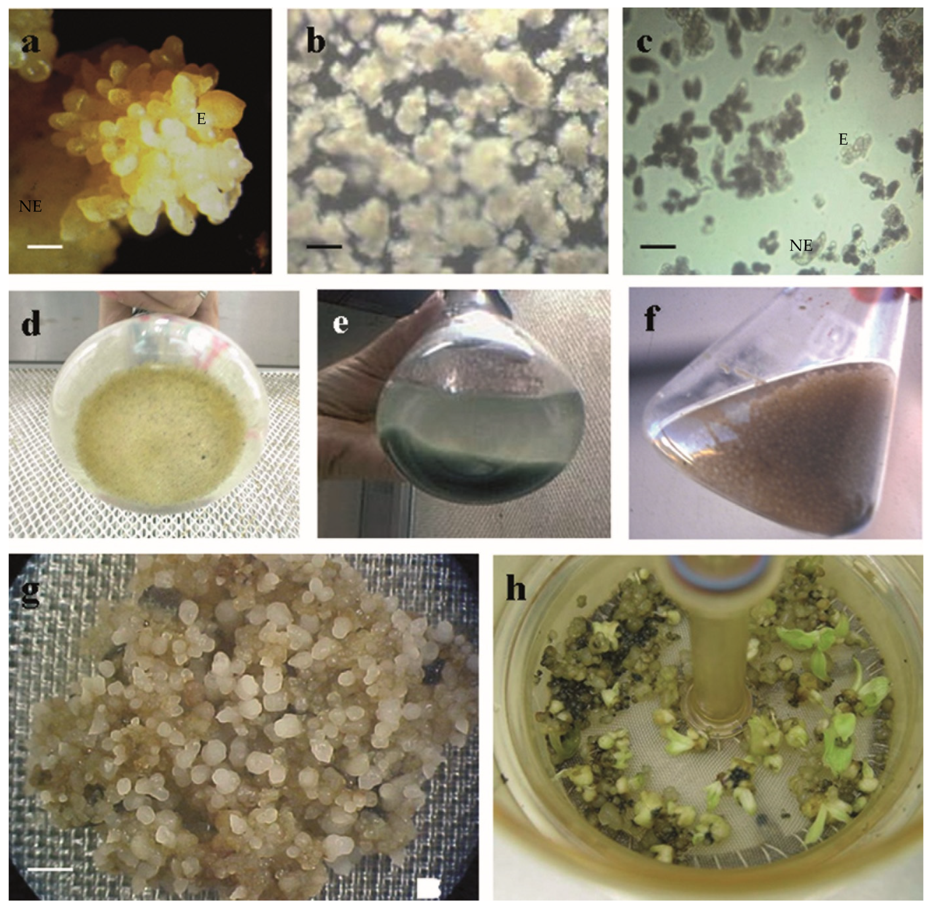
Embryogenic cell suspensions (E), when more than 90% of cells were of embryogenic nature (Figure 1b), i.e., isodiametric, smaller than 30 μm, and with prominent nuclei, a single nucleolus and dense cytoplasm [8]. These suspensions were frequently originated from callus-whitish somatic embryo masses (Figure 1a), which are very friable structures mainly constituted by somatic embryos in different developmental stages.
-
Non-embryogenic cell suspensions (NE), when more than 90% of cells were not of embryogenic nature, but manifested a greater propensity for cell division (Figure 1c). Cells of this type possessed parenchymatic appearance. These suspensions were always formed from not so friable, yellowish structures on nodular tissue (Figure 1a), constituted partially by cells in transition to proembryogenic stages.
From week 8 of multiplication onwards, subcultures were carried out every 2 weeks at least, whenever the ratio of packed cell to suspension volume was greater than 1:15.
After 14 weeks of multiplication, F18 embryogenic cell suspensions were transferred to the embryo formation phase (Figure 1d–f) by dilution 1:15 in modified [59] SH [61] medium. Culture medium was renewed every 2 weeks. Liquid cultures from establishment until the end of embryo formation phase were placed into darkness, on an orbital shaker at 100 rpm, at 27 ± 2 °C.
After 6 weeks in embryo formation phase, 0.7 g of fresh somatic embryos (Figure 1g) were transferred and cultured into “automatized temporal immersion recipients” (RITA®, Sartorius©, CIRAD, Paris, France) according to [3], where they germinated (Figure 1h).
Figure 1. Different stages of somatic embryogenesis process in Musa. (a) Friable embryogenic masses (E) and yellowish non-embryogenic cells (NE) in callus (bar = 2.50 mm). (b) Aggregates from embryogenic cell suspension (bar = 0.40 mm). (c) Portion of non-embryogenic cell suspension, showing non-embryogenic (NE) and embryogenic (E) cells (bar = 0.045 mm). (df) Somatic embryos at weeks 2, 4 and 6, respectively, of the embryo formation phase. (g) Somatic embryos ready to be transferred to the germination phase (bar = 3 mm). (h) Germinating embryos in RITA®.
Agronomy 16 00736 g001

2.2. Quantification of Polyamines

Analysis of PAs (DAP, Put, Spd and Spm) was carried out from three biological replicates (25 mg dry mass/sample) in each of the following three fractions: fraction of free, not covalently linked PAs (S); fraction of PAs liberated from perchloric acid-soluble conjugates (SH), and fraction of PAs liberated from perchloric acid-insoluble conjugates (PH). After 4 weeks of multiplication, the sediment of cell suspensions was placed on filter paper, then rapidly frozen in liquid nitrogen, triturated in porcelain mortars and lyophilized for 48 h. Obtained samples were stored in vacuum-sealed containers at −20 ± 2 °C until analysis. Extraction, purification, and quantification of PAs were carried out according to the methodology described by Fraga et al. [62]. Briefly, polyamine extraction was performed with 5% cold perchloric acid (PCA), and then, samples were centrifuged at 27,000× g for 20 min. The supernatant contained S and SH fraction, and the pellet, with PH fraction, was re-suspended in 1 mL of 1 M NaOH. Conjugated polyamines were released by hydrolysis with 12 M HCl using a proportion 1/1 (v/v) at 100 °C for 20 h. Filtered and dried material was re-suspended in 5% PCA and all fractions frozen until dansylation. Dansylated PAs were extracted in toluene; the obtained organic phase was dried, re-dissolved in acetonitrile (ACN), and separated by high-performance liquid chromatography (HPLC). This separation was carried out in a Waters Chromatography S.A. (Barcelona, Spain) equipment. The used column was a C18 Kromasil of 5 µm and 250 mm × 4 mm. ACN and H2O Milli-Q (Millipore Ibérica SA, Madrid, Spain) were used as solvents, and the different PAs were separated with the following gradient: 68% ACN for 4 min, lineal gradient for 1 min until 100% ACN. This was maintained for 4 min to return to the initial conditions (68% ACN) using another lineal gradient for 1 min. Analysis time was 15 min, and flux rate 1.5 mL min−1. Eluted samples were excited at 350 nm, and fluorescence emission was monitored at 500 nm. Two technical samples were measured.

2.3. Immunodetection of Proteins Serologically Analogous to PE32

For immunochemical analyses of extracellular proteins, liquid media from embryogenic (E) and non-embryogenic (NE) cell suspensions obtained from embryogenic and non-embryogenic calli, respectively, were filtered through double gauze, centrifuged at 12,000× g at 4 ± 2 °C for 10 min, and preserved in Eppendorf tubes at –20 ± 2 °C until use.
To compare immunochemical responses depending on ability for somatic embryogenesis, E and NE of two weeks of multiplication were employed for dot blot immunodetection. Furthermore, two E lines of each cultivar (L1 and L2 of GN, and L3 and L4 of F18) were also monitored at weeks 4, 8, and 12 of multiplication. Simultaneously, NE of both cultivars were monitored as well. Liquid media were also taken every 3 days during the first 4 weeks of embryo formation phase (line L7 from F18) and at weeks 2, 4 and 6 in this phase, as well as at week 3 of germination culture in RITAs (line L3 from F18). All dot blots were performed by applying 20 µL of each sample onto Hybond PVDF membrane previously treated with ethanol for 5 min. A mixture of extracellular Saccharum sp. proteins (20 µg) was used as positive control for the immunochemical reaction.
To determine antibody specificity and molecular mass of the detected proteins, a Western blot was carried out with proteins corresponding to more advanced stages of multiplication (week 18) in E of GN; as a negative control, a non-embryogenic cell suspension of this cultivar was used. Separation of extracellular proteins was carried out by electrophoresis in a discontinuous gel system [63] with 2.5% SDS, at 120 V and 20 mA. The fractionated proteins were then electrophoretically transferred by semi-dry blotting to PVDF membranes at 0.8 mA cm−2 for 1 h. These membranes were incubated with a polyclonal antibody related to sugar cane proteins PE32 (α-PE32) [64]. Antigen–antibody complexes were detected with anti-rabbit alkaline phosphatase conjugate (SIGMA).
Nitro blue tetrazolium chloride (NBT) and 5-bromo-4-chloro-3-indolyl phosphate (BCIP) constituted the substrate reaction mixture. Molecular weight indicators were visualized with 0.1% Coomassie R (PhastGel Blue R, Pharmacia Biotech, Uppsala, Sweden) after electro-transfer.

2.4. Statistical Analysis

Differences in PA contents and ratios of cell aggregates between cultivars (GE and F18) and between cell suspensions with different competency for somatic embryogenesis (NE and E) were studied using two-way ANOVA with Holm–Sidak post hoc tests (α = 0.01). Pairwise comparison procedures were performed for factors (cultivars and embryogenic competencies), and, when interactions were detected, for factors inside each level: for cultivar inside both E and NE, and for embryogenic competence inside both GN and F18. Proper value transformations were used when data did not meet ANOVA criteria. SigmaStat 3.5 was employed for these analyses.

3. Results

3.1. Polyamines

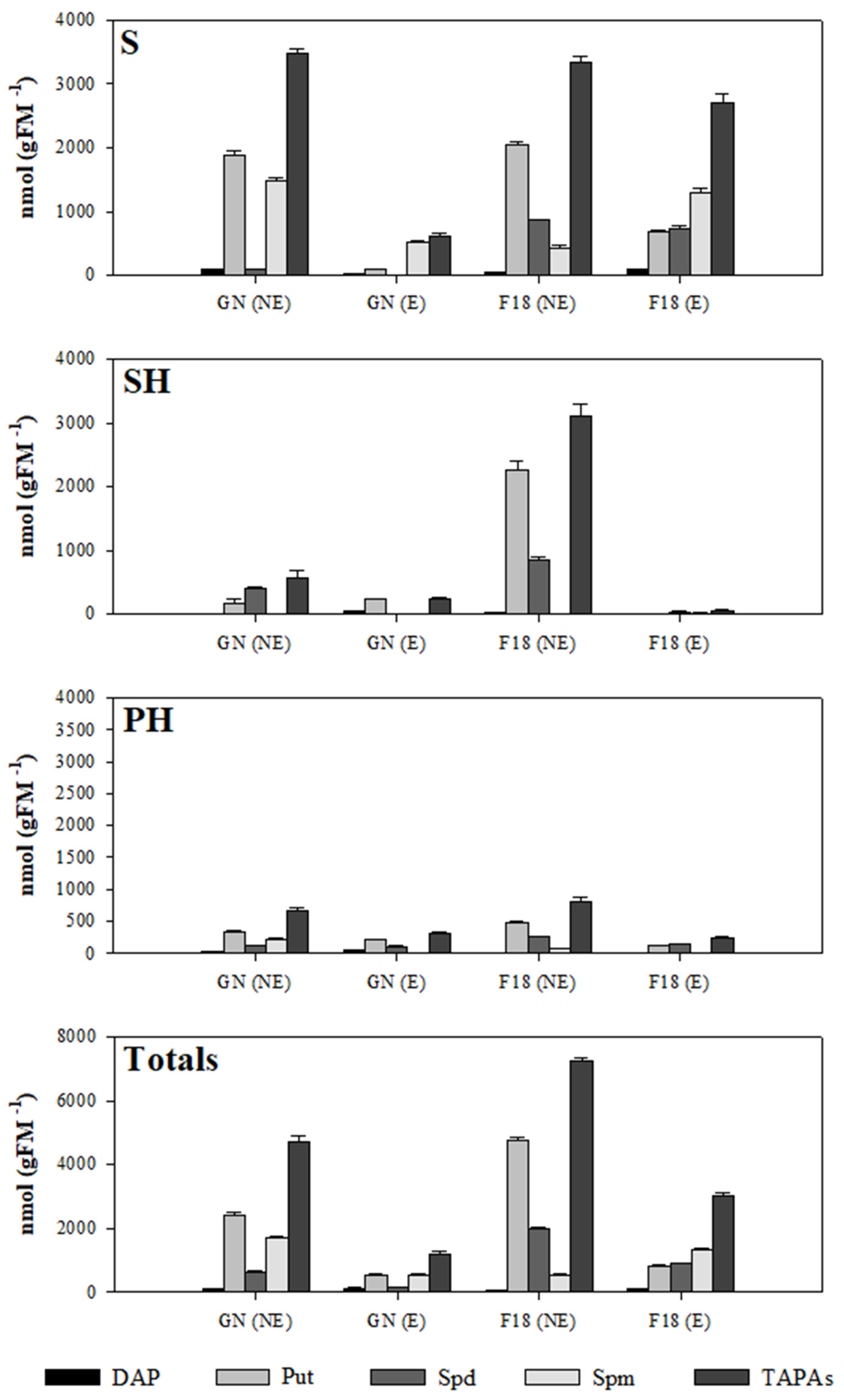
PA profiles are presented in Figure 2. Significant interactions between both studied factors (cultivar, embryogenic capacity) were always detected when individual PA contents per fraction were analyzed (Table 1).
In both studied Musa cultivars (GN and F18), many of the individual anabolic PAs –the diamine Put, the tri-amine Spd and the tetra-amine Spm– contents per fraction were significantly higher in NE than in E (Table 1): Put (S and PH), Spd (S and SH) and Spm (PH), which is completely absent in E (Figure 2). As a consequence of these differential contributions, the ratio Put/Spm (S, affecting total of fractions) resulted significantly higher in NE (Table 2). The diamine DAP –a catabolite of the long-chain PAs Spd and Spm– quantified in PH, is the unique PA per fraction which contents were found to be significantly higher in E in both analyzed cultivars but, in any case, this molecule possessed always a low relative contribution with respect to the total contents of anabolic PAs (Figure 2).
In F18, Spd (S) contents are significantly higher than in GN, independently of the analyzed type of cell suspension (NE, E) (Table 2). The inverse situation occurred in the case of DAP (PH), where the contents are thus significantly higher in GN aggregates since they are absent in those from F18.
After statistical analyses, some indicators of embryogenic potential are proposed (Table 3).

3.2. Extracellular Proteins Serologically Analogous to PE32

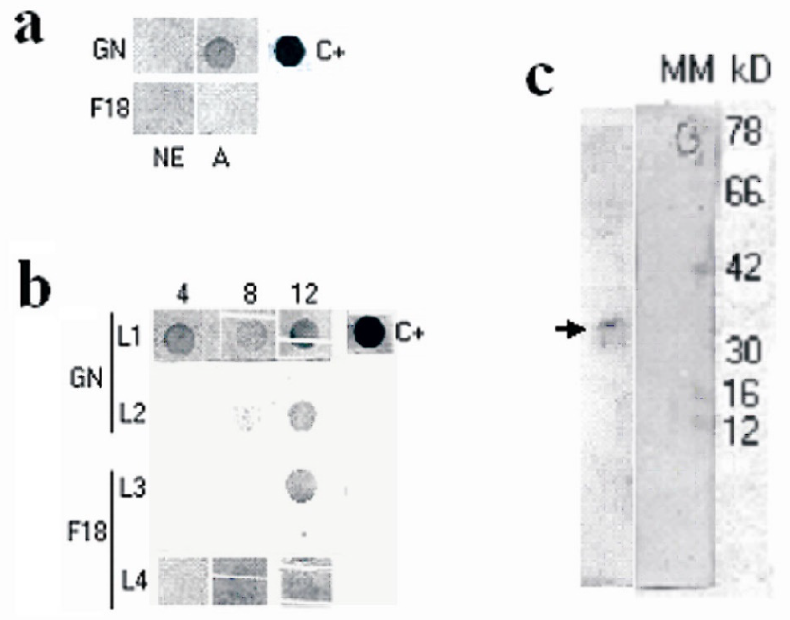
At week 2 of multiplication phase, a strong immunodetection signal appeared only in one of the GN (E) lines, the line L1 (Figure 3a,b). The signal intensity fluctuated in more advanced multiplication subcultures of this line (weeks 8 and 12 of multiplication) (Figure 3b), but at week 12 of multiplication, signals for the entire analyzed E, belonging to both GN and F18, are visible. On the contrary, the dot blot from time-equivalent NE, also from both GN and F18, did not show immunodetection signals. Western blot analysis revealed a 32 kD proteins in GN (E) at 18 weeks of multiplication (Figure 3c). No bands were observed in a GN (NE) of the same time.
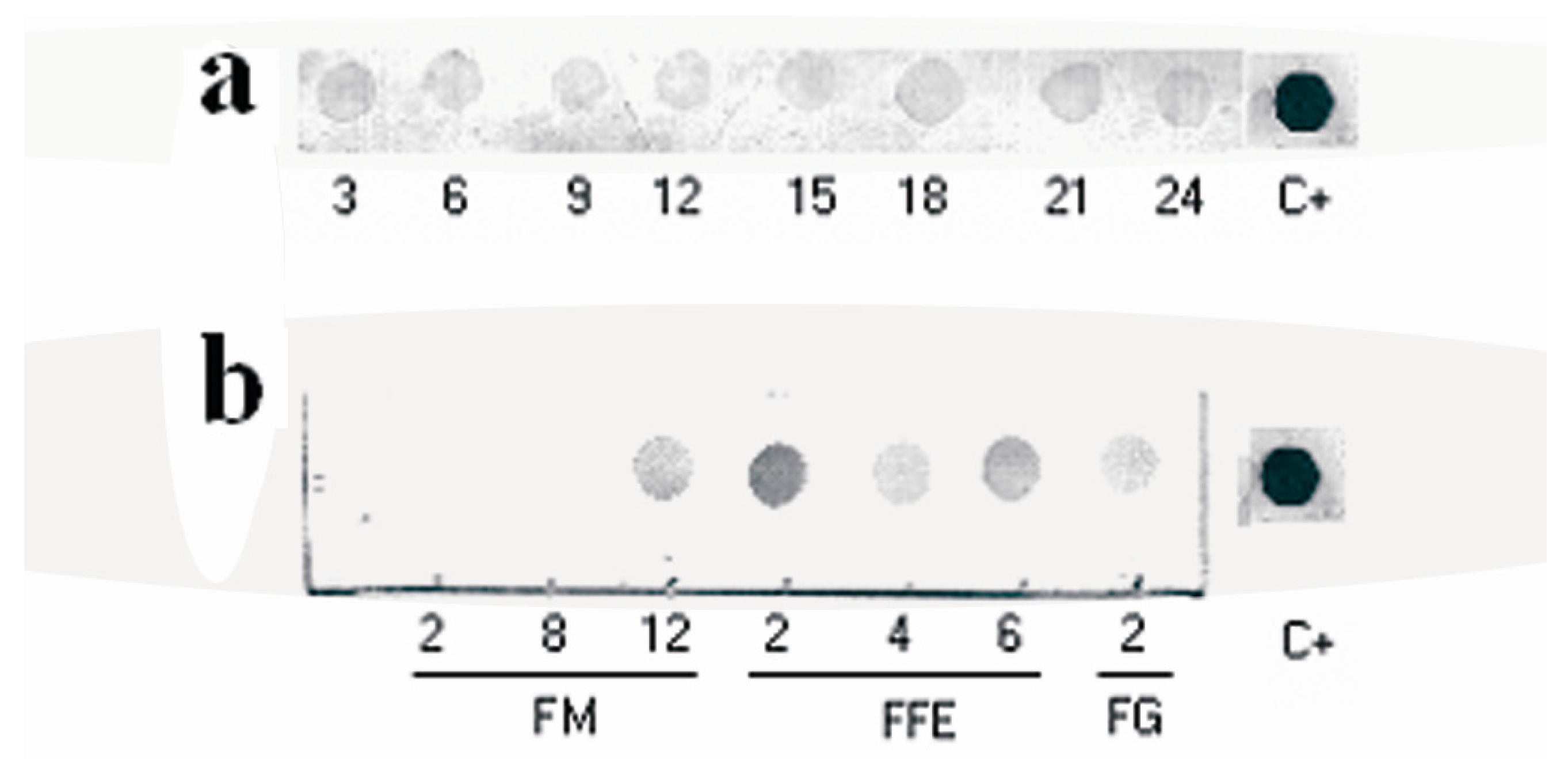
In the embryo formation phase, which was analyzed for F18, intensity of immunodetection signals is slightly fluctuant during the first 24 days for line L7 (Figure 4a). For line L3, belonging to the same cultivar, the highest signal intensity of the whole embryogenic process was observed at week 2 in such a phase (Figure 4b), but a decrease in signal intensity occurred at week 4, corresponding to the differentiation of most of somatic embryos from cell aggregates. At week 6, a certain recovering of the signal intensity was observed. After 2 weeks of germination in RITA®, slight detection of PE32-like proteins still continued.

4. Discussion

4.1. Polyamines

Since NE were observed to be more proliferative than E, a direct relationship between proliferation and contents of major anabolic PAs can be concluded, agreeing with results of other studies. Thus, high contents of free PAs were correlated with cell proliferation in, for example, cotton tissues [65], and the necessity of a rise in free PA contents in dividing cells is well documented in many plant systems for some time [66,67]. More recently, differential contents of specific PAs in concrete molecular fractions have been examined between highly proliferating and pre-differentiating calli or suspension cells. Such differences in PA contents, like those found in the present work between NE and E, could be explained on the basis of structural or regulatory roles of those molecules [43,68]. For example, PA interactions could constitute a system of preservation of protein accumulation necessary for cell proliferation: stabilization of RNAs, transcriptional and post-transcriptional control of protein synthesis, and post-translational interactions, including inhibitory binding to proteases [43,68,69,70]. In particular, proliferative embryogenesis-inductive stages in many culture systems are concomitant with increases in free or/and conjugated PA contents, mainly of Put [12,17,71], but also of only long-chain PAs [14,18]. In this last case [14], such increases were associated with an enhancement of the expression of most of the genes involved in PA biosynthesis. Furthermore, embryogenic capability of Citrus sinensis calli [28] and Pinus nigra masses from distinct genotypes [29] resulted positively correlated with the levels of endogenous polyamines.
Nevertheless, cell re-programming that coincides with embryogenic re-determination seems to be frequently associated with a nearly previous, concomitant or nearly further fall in all or specific PA contents [22,23,24,25,26,27,28]. Furthermore, Litz and Schaffer [22] found lower endogenous concentrations of PAs in embryogenic than in non-embryogenic calli, suggesting that stress of culture conditions could shoot PA biosynthesis in tissues. In our case study, NE could remain at an initial highly proliferative stage, and then over-accumulate PAs, avoiding cell changes and thus allowing a further cell re-programming towards morphogenetic pathways, a hypothesis which is compatible with ideas proposed by Meijer and Simmonds [23]. The blocking of ion channels by PAs [72,73] may be involved in such an effect, since at least potassium channels seem to be essential for carrot somatic embryogenesis [74,75].
Specifically, the higher Spd contents found in proliferating NE lines of the present study are also in agreement with the occurrence in high cell proliferation processes in which cells possess high mitotic activity without somatic embryo differentiation [76,77]. Spd conjugation led to a higher contribution of the SH fraction in NE with respect to E, a fact that could represent a way for phenol inactivation, since cell division and accumulation of phenolic compounds are inversely related at least in some plant systems ([17] and cites therein). On the other hand, bound Spd was the mainly enhanced PA form in photoinhibition of somatic embryogenesis in Araujia sericifera petals [78]. Furthermore, there have been experiments in which exogenous Spd could have had an inhibitory effect on the somatic embryogenesis inductive proliferation phase [22,79]. In spite of these facts, many experiments, including assays on PA biosynthesis inhibition/complementation and supplementation, support a possible direct role of Spd in triggering somatic embryogenesis [14,16,17,18,19,20,25,80,81,82]. Furthermore, the accumulation of this tri-amine in Picea abies calli [25], as well as in embryogenic cultures of carrot [21,24], was found to be positively correlated with the correct progress of further somatic embryo differentiation. All those apparently contradictory results could be indicative that there is a window of concrete PA contents and/or ratios that is related to the re-programming towards somatic embryogenesis differentiation.
Indeed, in our experiments, the higher free Put contents quantified in NE when compared with E caused a higher ratio Put/Spm (S) in those lines. The inverse relationship of this parameter with the embryogenic capacity, also caused by important differential contents of Put, is also evident in Citrus sinensis calli [14], cultures/tissues of several grass species [83], and masses of different Pinus nigra genotypes [29]. These facts suggest a direct relationship of relative contribution of free Put—a pre-requisite for cell proliferation ([84] and references therein)—with dividing cells and a relative contribution of free Spm with a greater differentiation capacity and less cell division.
There are many studies showing a relationship of relatively high proportional contribution of long-chain PAs (Spd and/or Spm) with the proliferative and/or inductive phases previous to somatic embryo formation [11,14,18,23,27,83,85,86]. Nevertheless, frequently, callogenic media supplementation with only long-chain polyamines negatively affects later somatic embryogenesis [21,22,79,87], although the increased biosynthesis of PAs may be accompanied by embryogenesis [13,88,89]. These facts could be explained on the basis of a positive role of the biosynthesis process of long-chain PAs in the induction of somatic embryogenesis, rather than as a direct effect of their accumulation. In this way, an intracellular decrease in precursors of long-chain PAs may negatively affect other biochemical routes, such as the inhibition of DNA-methyl transferase, thus affecting gene expression patterns and embryogenic cell reprogramming [29], or as the biosynthesis of ethylene [90], an inhibitor of somatic embryogenesis in some species [91].
Regarding this last effect, Patil et al. [92] inferred that the diamine Put, the precursor of the long-chain PAs, also plays a key role in regulating the biosynthesis of ethylene, and consequently somatic embryogenesis. The tetra-amine (Spm) biosynthesis makes these effects stronger than the tri-amine (Spd) biosynthesis, since the first synthesis consumes more common precursors with ethylene. This would explain the observation in the current study of the relative contribution of Spm and the lack of contribution of Spd, which is associated with the embryogenic capacity, a hypothesis supported by results obtained on calli of Hevea brasiliensis [93]. This biosynthetic activity in E could generate a re-distribution of PAs among fractions which could contribute to explain their lower contents in Spd (SH), as SH fraction might act as a regulating pull of free PAs [94].
Both PA biosynthesis and differential conjugation could also be associated with the higher accumulation of the long-chain PA degradation product DAP in E when compared with NE. This fact could be indicative of a more active metabolic replacement of PAs during the acquisition of embryogenic competence once post-mitosis re-programming occurs, since the tetra-amine Spm is formed from the tri-amine Spd, which is formed from the diamine Put. Increases in Spd catabolism on Picea abies calli—observed in results of Santanen and Simola [95]—are related to the correct progress of somatic embryogenesis. Furthermore, the higher DAP contents of E exclusively in PH, which is the fraction corresponding to cell wall components and proteins, support the idea of an excess of this residual product in E. Robie and Minocha [21] suggested that increased biosynthesis and fast turnover of long-chain PAs, rather than high PA contents, could be critical for somatic embryogenesis to proceed normally. Thus, recycling of long-chain PAs contributes to high consumption of S-adenosylmethyonine, a methyl donor in common with DNA methylation pathway, thereby stabilizing the epigenetic status of the cell [96], which in the present case would be re-programmed towards morphogenenetic pathways.
Apart from the role of this turnover, the involvement of Put in cell proliferation concomitant with the acquisition of competence of somatic embryogenesis and the implication of long-chain polyamines in the differentiation are both supported by several recent works [29,31,32,33,34,36,37,39,40,41].
Taking into account the multiple suggestions about association of PA metabolism and embryogenic potential, we propose the following hypothesis for banana embryogenic cell suspensions during the multiplication phase:
-
NE is characterized by higher Put/Spm (S) ratio, higher anabolic PA contents, and lower DAP contents. All these features can indicate over-accumulation of Put due to its low rates of degradation or/and conversion to long-chain PAs, whose levels anyway increased because of their low degradation to DAP. This situation could be concomitant with a cell stage that blocks any signal for triggering somatic embryogenesis.
-
E is characterized by opposite characteristics, i.e., lower Put/Spm (S) ratio, lower anabolic PA contents, and higher DAP(PH) contents. This case could involve a high conversion rate of Put into long-chain PAs, which would not suffer over-accumulation due to its conversion to DAP, excess of which can be absorbed by conjugation to macromolecules. In such a situation of continuous biosynthesis of long-chain PAs, all the commented biochemical mechanisms would lead to a cell status of competency for somatic embryogenesis.
Thus, we propose some previously mentioned polyamine variables as indicators of embryogenic character (Table 3): low contents of specific individual PAs in specific fractions, even absence of Spm (PH), and low Put/Spm (S) ratio. All these indicators are valid for cell suspensions of both banana cultivars, and are consistent with the results obtained in other cases, such as cell suspensions of the dicot Coffea canephora P. [97] and calli of the Gymnosperm Pinus nigra Arnold [98]. It is therefore plausible that the proposed indicators of embryogenic character suit a wide range of Phanerogams. However, the involvement of several endogenous factors in influencing somatic embryogenesis implies that the association of PAs with somatic embryogenesis initiation steps may not be universal and could be much more system-specific. Differences in PA profiles between cultivars were slight. The lower Spd (S) and higher DAP (PH) contents in GN aggregates could be indicative of higher free Spd catabolism and production of DAP conjugates in GN lines. This fact can be associated with the higher embryogenic competency manifested by this cultivar in semi-solid medium (data not shown). Genotypic differences in PA contents or ratios constitute indicators of embryogenic potential in both Angiosperm and Gymnosperm species [13,16,27,82].

4.2. Extracellular Proteins Serologically Analogous to PE32

With the available data, it is not possible to establish a direct relationship between the possible effect of polyamine biosynthesis on epigenetic changes and the expression of specific proteins. In any case, a correlation was found between Spd accumulation and releasing of PE32-like proteins. The obtained immunoanalytical results constitute a first approximation of the utilization of PE32-like proteins as markers of embryogenic potential in cell suspensions of Musa, since such proteins were detected in E but not in NE. The beginning of the release of PE32-like proteins depended on the lines. This could be associated with a differential embryogenic capacity of established cell suspensions, even belonging to the same cultivar. This different capacity could influence the time needed for the manifestation of embryogenic potential, the degree of differentiation and the conformity of somatic embryos. Consequently, PE32-like proteins could be related to the efficiency of the embryogenic process in banana.
The obtained GN Western blot profile shows detection in the multiplication phase of a 32 kD molecule, such as the PE32 proteins of sugarcane (Saccharum spp. hybrid). This result suggests a structural homology between these proteins and those detected in Musa. Consequently, given the functional, serological and physical homology between sugarcane PE32 and carrot EP3, the detected PE32-like proteins can be considered as banana EP3-like proteins.
The concentration of released PE32-like (EP3-like) proteins is the highest at the initial stages of the differentiation to globular somatic embryos from cell aggregates, week 2 of embryo formation phase. The relative maximum of signal intensity at the end of this phase could be explained on the basis of a repetitive somatic embryogenesis due to the increase in density of non-developed globular somatic embryos at this stage. EP3-like proteins were also found to be associated with initial stages of somatic embryogenesis in the monocot Saccharum spp. hybrid [49] and the dicots Coffea canephora P. and C. arabica L. [47], Daucus carota L. [52], Cucurbita pepo L. [99], and Arabidopsis thaliana (L.) Heynh [48]. In this last case, the expression of an orthologue of the carrot gene codifying two isoendochitinases EP3 was restricted to cells close to the developing embryos. Moreover, two 32 kDa chitinase isoforms were shown to accumulate in the medium of embryo cultures to a much higher level than in the medium of a non-embryogenic Cichorium cultivar [53]. Petrovska et al. [54] described lower molecular mass chitinases as potential markers in Linum usitatissimum L. embryogenic cultures. Over-accumulation of a possible EP3 transcript orthologous may also be related to the somatic embryogenesis potential in Glycine max L. Merrill [51].
With respect to monocots other than Musa, results on chitinases are abundant regarding somatic embryogenesis of Gramineae. Further development of somatic embryos of sugarcane was conditioned by PE32 at initial stages of somatic embryogenesis [64], and chitinase-like 32 kDa acidic serologically homologous proteins to EP3 have been identified in the medium of embryogenic suspension cultures of the Gramineae Dactylis glomerata L., and persist during the entire somatic embryogenesis process [50]. Extracellular chitinases related with somatic embryogenesis induction have been detected in Hordeum vulgare L. [55] and Zea mays L. [56]. Additionally, a soluble 32 kD protein was present in extracts of embryogenic and non-embryogenic calli derived from immature embryos of Triticum aestivum [100].
Regarding Gymnosperms, chitinases were also detected along somatic embryogenesis of conifers. Thus, chitinases were also found to be related to induction and development during somatic embryogenesis in the genus Picea [57,58]. Nevertheless, although the importance of chitinases for cell growth and development of somatic embryos was recognized for Abies hybrids and Pinus nigra, results reject such enzymes as markers of embryogenesis [101].
Given the apparent universality of 32 kD proteins related with somatic embryogenesis and the serological and molecular mass homology between PE32 and EP3 chitinases, as well as the relationship of chitinases with somatic embryogenesis, a chitinase role of the banana PE32/EP3-analogous proteins could be expected. It may be suggested that these proteins possess a highly conserved structure in Angiosperms, and possibly in all Phanerogams. These proteins might be evolutionarily conserved due to their important function during zygotic embryo development. This function also seems to be essential during initial stages of somatic embryogenesis [45]. On these bases, a ‘nursing role’ of EP3 proteins during zygotic embryogenesis was proposed, possibly exerting their effect through hydrolytic activity on endogenous arabinogalactan proteins, some of which control somatic embryogenesis [46,52]. More experimental support on the participation of chitinases in somatic embryogenesis was obtained from Pinus caribaea embryogenic tissues containing ionically bound cell wall chitinase-like protein that was able to act releasing sugars on embryogenic tissue-specific arabinogalactan proteins [102].
On the basis of all these collective factors, we propose the use of PE32/EP3-like proteins as an indicator of initial phases of somatic embryogenesis not only in the genus Musa, but also for a possible application in a wide range of plant species.

5. Conclusions

Both endogenous polyamine profiles and PE32/EP3-like protein release were showed to characterize the potential for somatic embryogenesis of banana cell suspensions. Thus, low contents of specific individual PAs in specific fractions, even absence of Spm (PH), and low Put/Spm (S) ratio are proposed as indicators of potential for somatic embryogenesis valid for cell suspensions of GN and F18. Furthermore, PE32-like proteins were shown to be indicative of initial stages of somatic embryogenesis in such embryogenic cell suspensions. The proposed indicators of embryogenic character are supported by physiological bases and by results from other works involving a wide range of Phanerogams. Nevertheless, the involvement of several endogenous factors in influencing somatic embryogenesis implies that the association of endogenous PAs and released PE32/EP3-like proteins with somatic embryogenesis initiation steps could be more species- and/or culture method-specific. The distinction between suspensions with more proliferative behavior or already induced for differentiation would allow for improving the selection of aggregates in protocols for somatic embryogenesis. Future molecular research with polyamine and endochitinase supplementation experiments would throw more light on the underlying reprogramming processes leading to the induction of the somatic embryogenesis.

Author Contributions

C.N.: data curation; formal analysis; funding acquisition; investigation; visualization; writing—original draft. M.R.: investigation; methodology; resources; supervision; writing—original draft. R.G.K.: methodology; project administration; resources; supervision; visualization; writing—original draft. M.R.V.: investigation; visualization; writing—original draft. R.H.: investigation; methodology. E.J.G.: formal analysis; resources; writing—original draft. R.R.: resources; writing—review and editing. M.J.C.: methodology; project administration; resources; supervision; writing—original draft; writing—review and editing. All authors have read and agreed to the published version of the manuscript.

Funding

This work was partially financed by the Agencia Española de Cooperación Internacional (Government of Spain), by means of a MUTIS scholarship. Prometeo Project from the Secretariat for Higher Education, Science, Technology and Innovation (SENESCYT) of the Republic of Ecuador is acknowledged for allowing Carlos Noceda the partial writing of this article. These sponsors have not participated in the scientific development of the work.

Data Availability Statement

The data presented in this study are available on request from the corresponding author (the data are not publicly available due to privacy).

Acknowledgments

We thank Tomás Moreira (CNIC, La Habana, Cuba) for his help in the lyophilization of the samples used for polyamine analyses. Special thanks are given to Nadine Fladd, from the Writing and Communication Centre at the University of Waterloo, who helped us with the grammar of part of the draft.

Conflicts of Interest

The authors declare no conflicts of interest.

Abbreviations

DAP1,3-diaminopropane
Eembryogenic cell suspensions
F18‘cultivar FHIA-18’
GNcultivar ‘Grande Naine’
NEnon-embryogenic cell suspensions
PA(s)polyamine(s)
PCVpacked cell volume
PHfraction containing polyamines liberated from perchloric acid-insoluble conjugates
PutPutrescine
Sfraction of free polyamines
SHfraction containing polyamines liberated from perchloric acid-soluble conjugates
SpdSpermidine
Spmspermine

References

  1. Ganry, J. Les Recherches Sur Les Bananiers et Plantains à l’Institut de Recherches Sur Les Fruits et Agrumes: Situation Actuelle et Environnement International. Fruits 1990, special edition, 5–22. [Google Scholar]
  2. Escalant, J.V.; Teisson, C.; Cote, F. Amplified Somatic Embryogenesis from Male Flowers of Triploid Banana and Plantain Cultivars (Musa spp.). In Vitro Cell. Dev. Biol.—Plant 1994, 30, 181–186. [Google Scholar] [CrossRef]
  3. Gómez Kosky, R.; De Feria Silva, M.; Posada Pérez, L.; Gilliard, T.; Bernal Martínez, F.; Reyes Vega, M.; Chávez Milian, M.; Quiala Mendoza, E. Somatic Embryogenesis of the Banana Hybrid Cultivar FHIA-18 (AAAB) in Liquid Medium and Scaled-up in a Bioreactor. Plant Cell Tissue Organ Cult. 2002, 68, 21–26. [Google Scholar] [CrossRef]
  4. Strosse, H.; Domergue, R.; Panis, B.; Escalant, J.-V.; Côte, F. Banana and Plantain Embryogenic Cell Suspensions; Technical Guidelines; INIBAP: Montpellier, France, 2003. [Google Scholar]
  5. Sidha, M.; Suprasanna, P.; Bapat, V.; Kulkarni, U.G.; Shinde, B.N. Developing Somatic Embryogenic Culture System and Plant Regeneration in Banana. Bhabha At. Ressearch Cent. (BARC) Newsl. 2007, 285, 12–14. [Google Scholar]
  6. Mintoff, S.J.L.; Nguyen, T.V.; Kelly, C.; Cullen, S.; Hearnden, M.; Williams, R.; Daniells, J.W.; Tran-Nguyen, L.T.T. Banana Cultivar Field Screening for Resistance to Fusarium oxysporum f.sp. cubense Tropical Race 4 in the Northern Territory. J. Fungi 2021, 7, 627. [Google Scholar] [CrossRef] [PubMed]
  7. Soares, J.M.S.; Rocha, A.J.; Nascimento, F.S.; Santos, A.S.; Miller, R.N.G.; Ferreira, C.F.; Haddad, F.; Amorim, V.B.O.; Amorim, E.P. Genetic Improvement for Resistance to Black Sigatoka in Bananas: A Systematic Review. Front. Plant Sci. 2021, 12, 657916. [Google Scholar] [CrossRef] [PubMed]
  8. Grapin, A.; Ortíz, J.L.; Lescot, T.; Ferrière, N.; Côte, F.X. Recovery and Regeneration of Embryogenic Cultures from Female Flowers of False Horn Plantain. Plant Cell Tissue Organ Cult. 2000, 61, 237–244. [Google Scholar] [CrossRef]
  9. Yang, X.; Zhang, X. Regulation of Somatic Embryogenesis in Higher Plants. CRC Crit. Rev. Plant Sci. 2010, 29, 36–57. [Google Scholar] [CrossRef]
  10. Pál, M.; Szalai, G.; Gondor, O.K.; Janda, T. Unfinished Story of Polyamines: Role of Conjugation, Transport and Light-Related Regulation in the Polyamine Metabolism in Plants. Plant Sci. 2021, 308, 110923. [Google Scholar] [CrossRef]
  11. Santos, M.; Claparols, I.; Torné, J.M. Effect of Exogenous Arginine, Ornithine, Methionine and γ-Amino Butyric Acid on Maize (Zea mays L.) Embryogenesis, and Polyamine Content. J. Plant Physiol. 1993, 142, 74–80. [Google Scholar] [CrossRef]
  12. Koetje, D.S.; Kononowicz, H.; Hodges, T.K. Polyamine Metabolism Associated with Growth and Embryogenic Potential of Rice. J. Plant Physiol. 1993, 141, 215–221. [Google Scholar] [CrossRef]
  13. Loukanina, N.; Thorpe, T.A. Arginine and Ornithine Decarboxylases in Embryogenic and Non-Embryogenic Carrot Cell Suspensions. In Vitro Cell. Dev. Biol.—Plant 2008, 44, 59–64. [Google Scholar] [CrossRef]
  14. Wu, X.B.; Wang, J.; Liu, J.H.; Deng, X.X. Involvement of Polyamine Biosynthesis in Somatic Embryogenesis of Valencia Sweet Orange (Citrus sinensis) Induced by Glycerol. J. Plant Physiol. 2009, 166, 52–62. [Google Scholar] [CrossRef]
  15. Rajesh, M.K.; Radha, E.; Sajini, K.K.; Karun, A. Polyamine-Induced Somatic Embryogenesis and Plantlet Regeneration in Vitro from Plumular Explants of Dwarf Cultivars of Coconut (Cocos nucifera). Indian J. Agric. Sci. 2014, 84, 527. [Google Scholar] [CrossRef]
  16. Shoeb, F.; Yadav, J.S.; Bajaj, S.; Rajam, M.V. Polyamines as Biomarkers for Plant Regeneration Capacity: Improvement of Regeneration by Modulation of Polyamine Metabolism in Different Genotypes of Indica Rice. Plant Sci. 2001, 160, 1229–1235. [Google Scholar] [CrossRef]
  17. Cvikrová, M.; Binarová, P.; Cenklová, V.; Eder, J.; Macháčková, I. Reinitiation of Cell Division and Polyamine and Aromatic Monoamine Levels in Alfalfa Explants during the Induction of Somatic Embryogenesis. Physiol. Plant. 1999, 105, 330–336. [Google Scholar] [CrossRef]
  18. Huang, X.L.; Li, X.J.; Li, Y.; Huang, L.Z. The Effect of AOA on Ethylene and Polyamine Metabolism during Early Phases of Somatic Embryogenesis in Medicago Sativa. Physiol. Plant. 2001, 113, 424–429. [Google Scholar] [CrossRef]
  19. Monteiro, M.; Kevers, C.; Dommes, J.; Gaspar, T. A Specific Role for Spermidine in the Initiation Phase of Somatic Embryogenesis in Panax ginseng CA Meyer. Plant Cell Tissue Organ Cult. 2002, 68, 225–232. [Google Scholar] [CrossRef]
  20. Mal, J.; Cvikrov, M.; Mchov, P.; Gemperlov, L. Role of Polyamines in Efficiency of Norway Spruce (Hurst Ecotype) Somatic Embryogenesis. In Embryogenesis; IntechOpen: London, UK, 2012. [Google Scholar]
  21. Robie, C.A.; Minocha, S.C. Polyamines and Somatic Embryogenesis in Carrot. I. The Effects of Difluoromethylornithine and Difluoromethylarginine. Plant Sci. 1989, 65, 45–54. [Google Scholar] [CrossRef]
  22. Litz, R.E.; Schaffer, B. Polyamines in Adventitious and Somatic Embryogenesis in Mango (Mangifera indica L.). J. Plant Physiol. 1987, 128, 251–258. [Google Scholar] [CrossRef]
  23. Meijer, E.G.M.; Simmonds, J. Polyamine Levels in Relation to Growth and Somatic Embryogenesis in Tissue of Medicago sativa L. J. Exp. Bot. 1988, 39, 787–794. [Google Scholar] [CrossRef]
  24. Mengoli, M.; Bagni, N.; Luccarini, G.; Ronchi, V.N.; Serafini-Fracassini, D. Daucus Carota Cell Cultures: Polyamines and Effect of Polyamine Biosynthesis Inhibitors in the Preembryogenic Phase and Different Embryo Stages. J. Plant Physiol. 1989, 134, 389–394. [Google Scholar] [CrossRef]
  25. Santanen, A.; Simola, L.K. Changes in Polyamine Metabolism during Somatic Embryogenesis in Picea abies. J. Plant Physiol. 1992, 140, 475–480. [Google Scholar] [CrossRef]
  26. Garin, E.; De March, G.G.; Grenier, E.; Martin-Tanguy, J. Polyamine Changes during the Development of Zygotic Embryos in Prunus avium—Effects of a 2,4-D and Kinetin Treatment on Growth and Polyamine Levels in Cotyledons Cultivated in Vitro. Plant Growth Regul. 1995, 16, 279–286. [Google Scholar] [CrossRef]
  27. Yadav, J.S.; Rajam, M.V. Temporal Regulation of Somatic Embryogenesis by Adjusting Cellular Polyamine Content in Eggplant. Plant Physiol. 1998, 116, 617–625. [Google Scholar] [CrossRef]
  28. Liu, H.Y.; Xiao, L.T.; Lu, X.D.; Hu, J.J.; Wu, S.; He, C.Z.; Deng, X.X. Changes in Polyamine Levels in Citrus sinensis Osb. cv. Valencia Callus during Somatic Embryogenesis. J. Plant Physiol. Mol. Biol. 2005, 31, 275–280. [Google Scholar]
  29. Noceda, C.; Salaj, T.; Pérez, M.; Viejo, M.; Cañal, M.J.; Salaj, J.; Rodriguez, R. DNA Demethylation and Decrease on Free Polyamines Is Associated with the Embryogenic Capacity of Pinus nigra Arn. Cell Culture. Trees—Struct. Funct. 2009, 23, 1285–1293. [Google Scholar] [CrossRef]
  30. Thiruvengadam, M.; Rekha, K.T.; Jayabalan, N.; Praveen, N.P.; Kim, E.H.; Chung, I.M. Effect of Exogenous Polyamines Enhances Somatic Embryogenesis via Suspension Cultures of Spine Gourd (Momordica dioica Roxb. Ex. Willd.). Aust. J. Crop Sci. 2013, 7, 446–453. [Google Scholar]
  31. Rakesh, B.; Sudheer, W.N.; Nagella, P. Role of Polyamines in Plant Tissue Culture: An Overview. Plant Cell Tissue Organ Cult. 2021, 145, 487–506. [Google Scholar] [CrossRef]
  32. Xiong, L.; Liu, C.; Liu, D.; Yan, Z.; Yang, X.; Feng, G. Optimization of an Indirect Regeneration System for Common Bean (Phaseolus vulgaris L.). Plant Biotechnol. Rep. 2023, 17, 821–833. [Google Scholar] [CrossRef]
  33. Liu, Q.; Xu, L.; Li, Y.; Xu, W.; Vetukuri, R.R.; Xu, X. Overexpression of an Autophagy-Related Gene DiATG3 from Davidia involucrata Improves Plant Thermotolerance by Enhancing the Accumulation of Polyamines and Regulating Genes in Calcium and MAPK Signaling Pathways. Environ. Exp. Bot. 2023, 208, 105235. [Google Scholar] [CrossRef]
  34. Farias-Soares, F.L.; Steiner, N.; Schmidt, É.C.; Pereira, M.L.T.; Rogge-Renner, G.D.; Bouzon, Z.L.; Floh, E.S.I.; Guerra, M.P. The Transition of Proembryogenic Masses to Somatic Embryos in Araucaria angustifolia (Bertol.) Kuntze Is Related to the Endogenous Contents of IAA, ABA and Polyamines. Acta Physiol. Plant. 2014, 36, 1853–1865. [Google Scholar] [CrossRef]
  35. Vondráková, Z.; Eliášová, K.; Vágner, M.; Martincová, O.; Cvikrová, M. Exogenous Putrescine Affects Endogenous Polyamine Levels and the Development of Picea abies Somatic Embryos. Plant Growth Regul. 2015, 75, 405–414. [Google Scholar] [CrossRef]
  36. Salo, H.M.; Sarjala, T.; Jokela, A.; Häggman, H.; Vuosku, J. Moderate Stress Responses and Specific Changes in Polyamine Metabolism Characterize Scots Pine Somatic Embryogenesis. Tree Physiol. 2016, 36, 392–402. [Google Scholar] [CrossRef]
  37. Aydin, M.; Hossein Pour, A.; Haliloğlu, K.; Tosun, M. Effect of Polyamines on Somatic Embryogenesis via Mature Embryo in Wheat. Turk. J. Biol. 2016, 40, 1178–1184. [Google Scholar] [CrossRef]
  38. El-Dawayati, M.; Ghazzawy, H.; Munir, M. Somatic Embryogenesis Enhancement of Date Palm Cultivar Sewi Using Different Types of Polyamines and Glutamine Amino Acid Concentration under In-Vitro Solid and Liquid Media Conditions. Int. J. Biosci. 2018, 12, 149–159. [Google Scholar] [CrossRef]
  39. Sathish, D.; Theboral, J.; Vasudevan, V.; Pavan, G.; Ajithan, C.; Appunu, C.; Manickavasagam, M. Exogenous Polyamines Enhance Somatic Embryogenesis and Agrobacterium Tumefaciens-Mediated Transformation Efficiency in Sugarcane (Saccharum Spp. Hybrid). In Vitro Cell. Dev. Biol.—Plant 2020, 56, 29–40. [Google Scholar] [CrossRef]
  40. Sundararajan, S.; Sivakumar, H.P.; Nayeem, S.; Rajendran, V.; Subiramani, S.; Ramalingam, S. Influence of Exogenous Polyamines on Somatic Embryogenesis and Regeneration of Fresh and Long-Term Cultures of Three Elite Indica Rice Cultivars. Cereal Res. Commun. 2021, 49, 245–253. [Google Scholar] [CrossRef]
  41. do Nascimento, A.M.M.; Polesi, L.G.; Back, F.P.; Steiner, N.; Guerra, M.P.; Castander-Olarieta, A.; Moncaleán, P.; Montalbán, I.A. The Chemical Environment at Maturation Stage in Pinus spp. Somatic Embryogenesis: Implications in the Polyamine Profile of Somatic Embryos and Morphological Characteristics of the Developed Plantlets. Front. Plant Sci. 2021, 12, 771464. [Google Scholar] [CrossRef]
  42. Jo, L.; Dos Santos, A.L.W.; Bueno, C.A.; Barbosa, H.R.; Floh, E.I.S.; Sederoff, R. Proteomic Analysis and Polyamines, Ethylene and Reactive Oxygen Species Levels of Araucaria Angustifolia (Brazilian Pine) Embryogenic Cultures with Different Embryogenic Potential. Tree Physiol. 2014, 34, 94–104. [Google Scholar] [CrossRef]
  43. Emmons-Bell, M.; Forsyth, G.; Sundquist, A.; Oldeman, S.; Gardikioti, A.; de Souza, R.; Coene, J.; Kamel, M.H.; Ayyapan, S.; Fuchs, H.A.; et al. Polyamines regulate cell fate by altering the activity of histone-modifying enzymes. bioRxiv 2024, preprint. [Google Scholar] [CrossRef]
  44. Van Engelen, F.A.; De Vries, S.C. Extracellular Proteins in Plant Embryogenesis. Trends Genet. 1992, 8, 66–70. [Google Scholar] [CrossRef] [PubMed]
  45. De Jong, A.J.; Cordewener, J.; Lo Schiavo, F.; Terzi, M.; Vandekerckhove, J.; Van Kammen, A.; De Vries, S.C. A Carrot Somatic Embryo Mutant Is Rescued by Chitinase. Plant Cell 1992, 4, 425–433. [Google Scholar] [CrossRef] [PubMed]
  46. Van Hengel, A.J.; Tadesse, Z.; Immerzeel, P.; Schols, H.; Van Kammen, A.; De Vries, S.C. N-Acetylglucosamine and Glucosamine-Containing Arabinogalactan Proteins Control Somatic Embryogenesis. Plant Physiol. 2001, 125, 1880–1890. [Google Scholar] [CrossRef]
  47. Rodríguez, M.; Cevallos, A.; Montes, S. Proteinas Extracelulares Marcadoras Del Potencial Embriogenico En Suspensiones Celulares de Coffea spp. Cultiv. Trop. 2000, 21, 11–15. [Google Scholar]
  48. Passarinho, P.A.; Van Hengel, A.J.; Fransz, P.F.; De Vries, S.C. Expression Pattern of the Arabidopsis Thaliana AtEP3/AtchitIV Endochitinase Gene. Planta 2001, 212, 556–567. [Google Scholar] [CrossRef] [PubMed]
  49. Rodríguez, M.; Niubó, E.; Maribona, R.H. Análisis Inmunoquímico de La Embriogénesis Somática En Suspensiones Celulares de La Caña de Azúcar. Rev. CENIC Cienc. Biológicas 1999, 30, 2. [Google Scholar]
  50. Tchorbadjieva, M.I.; Pantchev, I.Y. Secretion of a Chitinase-like Protein in Embryogenic Suspension Cultures of Dactylis glomerata L. Biol. Plant. 2006, 50, 142–145. [Google Scholar] [CrossRef]
  51. Zheng, Q.; Perry, S.E. Alterations in the Transcriptome of Soybean in Response to Enhanced Somatic Embryogenesis Promoted by Orthologs of AGAMOUS-Like15 and AGAMOUS-Like18. Plant Physiol. 2014, 164, 1365–1377. [Google Scholar] [CrossRef]
  52. Van Hengel, A.J.; Guzzo, F.; Van Kammen, A.; De Vries, S.C. Expression Pattern of the Carrot EP3 Endochitinase Genes in Suspension Cultures and in Developing Seeds. Plant Physiol. 1998, 117, 43–53. [Google Scholar] [CrossRef]
  53. Helleboid, S.; Hendriks, T.; Bauw, G.; Inzé, D.; Vasseur, J.; Hilbert, J.L. Three Major Somatic Embryogenesis Related Proteins in Cichorium Identified as PR Proteins. J. Exp. Bot. 2000, 51, 1189–1200. [Google Scholar] [CrossRef] [PubMed]
  54. Petrovská, B.; Salaj, T.; Moravčíková, J.; Libantová, J.; Salaj, J. Development of Embryo-like Structures in the Suspension Cultures of Flax Coincides with Secretion of Chitinase-like Proteins. Acta Physiol. Plant. 2010, 32, 651–656. [Google Scholar] [CrossRef]
  55. Matthias Kragh, K.; Jacobsen, S.; Dalgaard Mikkelsen, J.; Nielsen, K.A. Purification and Characterization of Three Chitinases and One β-1,3-Glucanase Accumulating in the Medium of Cell Suspension Cultures of Barley (Hordeum vulgare L.). Plant Sci. 1991, 76, 65–77. [Google Scholar] [CrossRef]
  56. Borderies, G.; le Béchec, M.; Rossignol, M.; Lafitte, C.; Le Deunff, E.; Beckert, M.; Dumas, C.; Matthys-Rochon, E. Characterization of Proteins Secreted during Maize Microspore Culture: Arabinogalactan Proteins (AGPs) Stimulate Embryo Development. Eur. J. Cell Biol. 2004, 83, 205–212. [Google Scholar] [CrossRef]
  57. Dong, J.Z.; Dunstan, D.I. Endochitinase and β-1,3-Glucanase Genes Are Developmentally Regulated during Somatic Embryogenesis in Picea Glauca. Planta 1997, 201, 189–194. [Google Scholar] [CrossRef]
  58. Wiweger, M.; Farbos, I.; Ingouff, M.; Lagercrantz, U.; Von Arnold, S. Expression of Chia4-Pa Chitinase Genes during Somatic and Zygotic Embryo Development in Norway Spruce (Picea abies): Similarities and Differences between Gymnosperm and Angiosperm Class IV Chitinases. J. Exp. Bot. 2003, 54, 2691–2699. [Google Scholar] [CrossRef] [PubMed]
  59. Bieberach, C. Embriogénesis Somática y Regeneración de Plantas En Cultivares de Musa sp. Master’s Thesis, CATIE, Turrialba, Costa Rica, 1995. [Google Scholar]
  60. Murashige, T.; Skoog, F. A Revised Medium for Rapid Growth and Bio Assays with Tobacco Tissue Cultures. Physiol. Plant. 1962, 15, 473–497. [Google Scholar] [CrossRef]
  61. Schenk, R.U.; Hildebrandt, A.C. Medium and Techniques for Induction and Growth of Monocotyledonous and Dicotyledonous Plant Cell Cultures. Can. J. Bot. 1972, 50, 199–204. [Google Scholar] [CrossRef]
  62. Fraga, M.F.; Cañal, M.J.; Rodríguez, R. Phase-Change Related Epigenetic and Physiological Changes in Pinus Radiata D. Don. Planta 2002, 215, 672–678. [Google Scholar] [CrossRef]
  63. Laemmli, U.K. Cleavage of Structural Proteins during the Assembly of the Head of Bacteriophage T4. Nature 1970, 227, 680–685. [Google Scholar] [CrossRef]
  64. Rodríguez, M.; Lukse, E.; Maribona, R.H.; Ramos Leal, M. Análisis Inmunoquímico de La Embriogénesis Somática de La Caña de Azúcar. In Proceedings of the X Seminario Científico, INCA, Havana, Cuba, 4–8 June 1996; p. 100. [Google Scholar]
  65. Davidonis, G. Changes in Polyamine Distribution during Cotton Fiber AndSeed Development. J. Plant Physiol. 1995, 145, 108–112. [Google Scholar] [CrossRef]
  66. Bagni, N.; Serafini-Fracassini, D.; Torrigiani, P. Polyamines and Cellular Growth Processes in Higher Plants. In Plant Growth Substances; Wareing, P.E., Ed.; Academic Press: London, UK, 1982; pp. 473–487. [Google Scholar]
  67. Walker, M.A.; Roberts, D.R.; Shih, C.Y.; Dumbroff, E.B. A Requirement for Polyamines during the Cell Division Phase of Radicle Emergence in Seeds of Acer Saccharum. Plant Cell Physiol. 1985, 26, 967–971. [Google Scholar]
  68. Kakkar, R.K.; Sawhney, V.K. Polyamine Research in Plants—A Changing Perspective. Physiol. Plant. 2002, 116, 281–292. [Google Scholar] [CrossRef]
  69. Gallardo, M.; Matilla, A.; Muñoz De Rueda, P.; Sánchez-Calle, I.M. Papel de Las Poliaminas En El Crecimiento y Desarrollo. ARS Pharm. 1996, 37, 17–27. [Google Scholar]
  70. Wallace, H.M.; Fraser, A.V.; Hughes, A. A Perspective of Polyamine Metabolism. Biochem. J. 2003, 376, 1–14. [Google Scholar] [CrossRef]
  71. Steiner, N.; Santa-Catarina, C.; Silveira, V.; Floh, E.I.S.; Guerra, M.P. Polyamine Effects on Growth and Endogenous Hormones Levels in Araucaria Angustifolia Embryogenic Cultures. Plant Cell Tissue Organ Cult. 2007, 89, 55–62. [Google Scholar] [CrossRef]
  72. Liu, K.; Fu, H.; Bei, Q.; Luan, S. Inward Potassium Channel in Guard Cells as a Target for Polyamine Regulation of Stomatal Movements. Plant Physiol. 2000, 124, 1315–1326. [Google Scholar] [CrossRef]
  73. Zhao, F.; Song, C.P.; He, J.; Zhu, H. Polyamines Improve K+/Na+ Homeostasis in Barley Seedlings by Regulating Root Ion Channel Activities. Plant Physiol. 2007, 145, 1061–1072. [Google Scholar] [CrossRef] [PubMed]
  74. Formentin, E.; Naso, A.; Varotto, S.; Picco, C.; Gambale, F.; Lo Schiavo, F. KDC2, a Functional Homomeric Potassium Channel Expressed during Carrot Embryogenesis. FEBS Lett. 2006, 580, 5009–5015. [Google Scholar] [CrossRef]
  75. Bregante, M.; Yang, Y.; Formentin, E.; Carpaneto, A.; Schroeder, J.I.; Gambale, F.; Lo Schiavo, F.; Costa, A. KDC1, a Carrot Shaker-like Potassium Channel, Reveals Its Role as a Silent Regulatory Subunit When Expressed in Plant Cells. Plant Mol. Biol. 2008, 66, 61–72. [Google Scholar] [CrossRef]
  76. Sethi, U.; Basu, A.; Guha-Mukherjee, S. Control of Cell Proliferation and Differentiation by Regulating Polyamine Biosynthesis in Cultures of Brassica and Its Correlation with Glyoxalase-I Activity. Plant Sci. 1988, 56, 167–175. [Google Scholar] [CrossRef]
  77. Klimaszewska, K.; Noceda, C.; Pelletier, G.; Label, P.; Rodriguez, R.; Lelu-Walter, M.A. Biological Characterization of Young and Aged Embryogenic Cultures of Pinus Pinaster (Ait. ). In Vitro Cell. Dev. Biol.—Plant 2009, 45, 20–33. [Google Scholar] [CrossRef]
  78. Torné, J.M.; Moysset, L.; Claparols, I.; Simón, E. Photocontrol of Somatic Embryogenesis and Polyamine Content in Araujia Sericifera Petals. Physiol. Plant. 1996, 98, 413–418. [Google Scholar] [CrossRef]
  79. Akhtar, N. Endogenous Polyamines: A Temporal Cellular Modulator of Somatic Embryogenesis in Guava (Psidium guajava L.) cv. Allahabad Safeda. Res. Plant Sci. 2013, 1, 4–14. [Google Scholar]
  80. El Hadrami, I.; D’Auzac, J. Effects of Polyamine Biosynthetic Inhibitors on Somatic Embryogenesis and Cellular Polyamines in Hevea Brasiliensis. J. Plant Physiol. 1992, 140, 33–36. [Google Scholar] [CrossRef]
  81. Feirer, R.P. The Biochemistry of Conifer Embryo Development: Amino Acids, Polyamines and Storage Proteins. In Somatic Embryogenesis in Woody Plants; Kluwer Academic Publishers: Dordrecht, The Netherlands, 1995; pp. 317–336. [Google Scholar]
  82. Minocha, R.; Smith, D.R.; Reeves, C.; Steele, K.D.; Minocha, S.C. Polyamine Levels during the Development of Zygotic and Somatic Embryos of Pinus Radiata. Physiol. Plant. 1999, 105, 155–164. [Google Scholar] [CrossRef]
  83. Kackar, A.; Shekhawat, N.S. Plant Regeneration through Somatic Embryogenesis and Polyamine Levels in Cultures of Grasses of Thar Desert. J. Cell Mol. Biol. 2007, 6, 121–128. [Google Scholar]
  84. Paul, A.; Mitter, K.; Raychaudhuri, S. Sen Effect of Polyamines on in Vitro Somatic Embryogenesis in Momordica charantia L. Plant Cell Tissue Organ Cult. 2009, 97, 303–311. [Google Scholar] [CrossRef]
  85. Nabha, S.; Lamblin, F.; Gillet, F.; Laurain, D.; Fliniaux, M.; David, A.; Jacquin, A. Polyamine Content and Somatic Embryogenesis in Papaver Somniferum Cells Transformed with Sam-1 Gene. J. Plant Physiol. 1999, 154, 729–734. [Google Scholar] [CrossRef]
  86. Li, Z.; Burritt, D.J. Changes in Endogenous Polyamines during the Formation of Somatic Embryos from Isogenic Lines of Dactylis glomerata L. with Different Regenerative Capacities. Plant Growth Regul. 2003, 40, 65–74. [Google Scholar] [CrossRef]
  87. Kevers, C.; Gaspar, T.; Dommes, J. The Beneficial Role of Different Auxins and Polyamines at Successive Stages of Somatic Embryo Formation and Development of Panax Ginseng in Vitro. Plant Cell Tissue Organ Cult. 2002, 70, 181–188. [Google Scholar] [CrossRef]
  88. Montague, M.J.; Koppenbrink, J.W.; Jaworski, E.G. Polyamine Metabolism in Embryogenic Cells of Daucus Carota. Plant Physiol. 1978, 62, 430–433. [Google Scholar] [CrossRef] [PubMed]
  89. Minocha, S.C.; Minocha, R. Role of Polyamines in Somatic Embryogenesis. In Biotechnology in Agriculture and Forestry 30, Somatic Embryogenesis and Synthetic Seed; Bajaj, Y.P.S., Ed.; Springer: Berlin/Heidelberg, Germany, 1995; pp. 53–70. ISBN 978-3-662-03091-2. [Google Scholar]
  90. Kushad, M.M.; Dumbroff, E.B. Metabolic and physiological relationships between the polyamine and ethylene biosynthetic pathways. In Biochemistry and Physiology of Polyamines in Plants; CRC Press: Boca Raton, FL, USA, 2024; pp. 77–92. [Google Scholar] [CrossRef]
  91. Biddington, N.L. The Influence of Ethylene in Plant Tissue Culture. Plant Growth Regul. 1992, 11, 173–187. [Google Scholar] [CrossRef]
  92. Patil, P.P.; Chandra, R.; Khetarpal, S.S. Influence of Polyamines and Ethylene Inhibitors on Somatic Embryo Induction in Chickpea (Cicer arietinum L.). Ind. J. Plant Physiol 1998, 3, 26–31. [Google Scholar]
  93. El Hadrami, I.; Michaux-Ferriere, N.; Carron, M.-P.; D’Auzac, J. Les Polyamine, Facteur Limitant Possible de l’embryogénèse Somatique Chez l’Hevea Brasiliensis. Comptes Rendus L’Academie Sci. Serie III 1989, 308, 205–211. [Google Scholar]
  94. Mader, J.C.; Hanke, D.E. Polyamine Sparing May Be Involved in the Prolongation of Cell Division Due to Inhibition of Phenylpropanoid Synthesis in Cytokinin-Starved Soybean Cells. J. Plant Growth Regul. 1997, 16, 89–93. [Google Scholar] [CrossRef]
  95. Santanen, A.; Simola, L.K. Catabolism of Putrescine and Spermidine in Embryogenic and Non-embryogenic Callus Lines of Picea abies. Physiol. Plant. 1994, 90, 125–129. [Google Scholar] [CrossRef]
  96. Karouzakis, E.; Gay, R.E.; Gay, S.; Neidhart, M. Increased Recycling of Polyamines Is Associated with Global DNA Hypomethylation in Rheumatoid Arthritis Synovial Fibroblasts. Arthritis Rheum. 2012, 64, 1809–1817. [Google Scholar] [CrossRef]
  97. Noceda, C.; Quiala, E.; Jiménez, E.; Rodríguez, R.; Gómez, R.; Barbón, R.; Capote, A.; Pérez, N.; de Feria, M.; Chávez, M. Proporciones Diferenciales de Poliaminas (PAs) En Explantes Foliares y Suspensiones Celulares Embriogénicas y No Embriogénicas de Coffea Arabica Var. “Caturra Rojo”. Café Cacao 2002, 3, 39–41. [Google Scholar]
  98. Rodríguez, R.; Fraga, M.F.; Diego, L.; Berdasco, M.; Hasbun, R.; Noceda, C.; Mendivil, C.; Salajová, T.; Radojevic, L.; Escalona, M.; et al. Micropropagation and Physiological Aspects. In Proceedings of the International Scientific Congress BIOFOR’02–Sustainable Forestry, Wood Products and Biotechnology, Vitoria, Spain, 11–14 November 2002; VI-552/03. pp. 15–30. [Google Scholar]
  99. Crnković, A.; Garić, R.; Leljak-Levanić, D.; Mihaljević, S. Reduced Protein Secretion and Glycosylation Induced by Ammonium Stress Inhibits Somatic Embryo Development in Pumpkin. Biol. Plant. 2014, 58, 209–217. [Google Scholar] [CrossRef]
  100. Nato, A.; Mirshahi, A.; Tichtinsky, G.; Mirshahi, M.; Faure, J.P.; Lavergne, D.; De Buyser, J.; Jean, C.; Ducreux, G.; Henry, Y. Immunological Detection of Potential Signal-Transduction Proteins Expressed during Wheat Somatic Tissue Culture. Plant Physiol. 1997, 113, 801–807. [Google Scholar] [CrossRef] [PubMed]
  101. Fráterová, L.; Salaj, T.; Matušíková, I.; Salaj, J. The Role of Chitinases and Glucanases in Somatic Embryogenesis of Black Pine and Hybrid Firs. Cent. Eur. J. Biol. 2013, 8, 1172–1182. [Google Scholar] [CrossRef]
  102. Domon, J.M.; Neutelings, G.; Roger, D.; David, A.; David, H. A Basic Chitinase-like Protein Secreted by Embryogenic Tissues of Pinus Caribaea Acts on Arabinogalactan Proteins Extracted from the Same Cell Lines. J. Plant Physiol. 2000, 156, 33–39. [Google Scholar] [CrossRef]
Figure 2. Polyamine contents in cell aggregates of non-embryogenic (NE) and embryogenic (E) cell suspension cultures of cvs. ‘Grande Naine’ (GN) and ‘FHIA-18’ (F18) in free polyamine fraction (S), and in perchloric acid-soluble (SH) and insoluble (PH) polyamine conjugates fractions, and total anabolic polyamines (TAPAs). Means ± SE (n = 6) are presented. gFM: grams of fresh mass.
Agronomy 16 00736 g002
Figure 3. Immunoblots with extracellular proteins from cell suspensions of Musa using α-PE32 antibody. C+: positive control with sugarcane proteins. (a): Dot blot after 2 weeks of multiplication of cvs. ‘Grande Naine’ (GN) and ‘FHIA-18’ (F18) embryogenic (E) and non-embryogenic (NE) cell suspensions. (b): Dot blot after the indicated weeks in multiplication phase of cvs. Grande Naine (lines L1 and L2) and FHIA-18 (lines L3 and L4) embryogenic cell lines; non-embryogenic lines, which have no signal, are not shown. (c): Western blot (arrow, at 32 kD) after 18 weeks of multiplication of cv. ‘Grande Naine’ embryogenic line; a contemporary non-embryogenic cell suspension, which has no signal, is not shown; MM: molecular marker (kD).
Agronomy 16 00736 g003
Figure 4. Immunoblot with α-PE32 with extracellular proteins from Musa cv. FHIA-18 embryogenic cell lines after different times. (a): line L7 (numbers represent days in embryo formation phase). (b): line L3 (numbers represent weeks in each phase). FM: Multiplication Phase. FFE: Embryo Formation Phase. FG: Germination Phase. C+: positive control with sugarcane proteins.
Agronomy 16 00736 g004
Table 1. Cell suspension type and cultivar for which the contents of the indicated polyamines were significantly higher for both studied cultivars or for both suspension types, respectively. Significant interactions between embryogenic competence and cultivar were detected for the contents of all studied polyamines. E and NE: embryogenic and non-embryogenic cell suspensions, respectively. GN: ‘Grande Naine’. F18: ‘FHIA 18’. n.s.: non-significant at least in one level (E and NE, GN and F18) in both studied factors (embryogenic competence and cultivar). S: Free polyamine fraction. SH: and PH: perchloric acid-soluble and insoluble polyamine conjugates fractions, respectively. DAP: 1,3-diaminopropane. Put: putrescine. Spd: spermidine. Spm: spermine.
FractionDAPPutSpdSpm
Sn.s.NENE, F18n.s.
SHn.s.n.s.NEn.s.
PHE, GNNEn.s.NE
Table 2. Polyamine ratios (means ± SE in grams of fresh mass; n = 6) in different types of cell aggregates. *: cell suspensions type –embryogenic (E) and non-embryogenic (NE) for which the showed ratios were significantly higher in both cultivars, and cultivar –‘Grande Naine’ (GN) and ‘FHIA 18’ (F18)– for which the showed ratios were significantly higher in both E and NE. Significant interactions between embryogenic competence and cultivar were detected for all showed ratios, except when indicated (n.i.). n.s.: non-significant at least in one level (E and NE, GN and F18) in both studied factors (embryogenic competence and cultivar). TAPAs: Total anabolic polyamines (Put, Spd, Spm). Rest of abbreviations: like in Table 1.
Fraction and RatioGN (NE)GN (E)F18 (NE)F18 (E)*
SPut/Spd19.34 ± 1.0910.14 ± 1.162.33 ± 0.050.93 ± 0.01GN, NE, n.i.
Put/Spm1.26 ± 0.040.17 ± 0.025.23 ± 0.770.53 ± 0.01F18, NE, n.i.
PHPut/Spm1.66 ± 0.0764.70 ± 13.255.51 ± 0.35>100n.s.
Put/Spd3.94 ± 0.134.11 ± 0.062.39 ± 0.020.90 ± 0.02GN
TAPAsPut/Spm1.41 ± 0.041.00 ± 0.0210.13 ± 1.540.61 ± 0.01NE, F18, no int
Spd/Spm0.36 ± 0.020.24 ± 0.014.24 ± 0.670.69 ± 0.01n.s.
S/SHSpd0.25 ± 0.020.80 ± 0.211.04 ± 0.06>100n.s.
Spm>100>100>100>100n.s.
S/PHPut5.69 ± 0.470.42 ± 0.064.54 ± 0.386.10 ± 0.32n.s.
TAPAs5.40 ± 0.451.93 ± 0.084.16 ± 0.2411.03 ± 0.82n.s.
SH/PHTAPAs0. 84 ± 0.100.75 ± 0.053.92 ± 0.410.20 ± 0.11n.s.
Table 3. Indicators of embryogenic potential in cvs. Grande Naine and FHIA-18 cell suspensions. ↑: High contents. ↓: Low contents. +: Presence. -: Absence.
IndicatorCELL Suspensions
Non-EmbryogenicEmbryogenic
DAP (PH)
Put (S and PH)
Spd (S and SH)
Spm (PH)+-
Put/Spm (S)>1<1
Disclaimer/Publisher’s Note: The statements, opinions and data contained in all publications are solely those of the individual author(s) and contributor(s) and not of MDPI and/or the editor(s). MDPI and/or the editor(s) disclaim responsibility for any injury to people or property resulting from any ideas, methods, instructions or products referred to in the content.