
Article

Optimization of Cultivation System and Transcriptome Analysis: From Unpollinated Chinese Chive Ovules to Plant Formation

Institute of Horticulture, Guizhou Academy of Agricultural Sciences/Horticultural Engineering Technology Research Center of Guizhou, Guiyang 550006, China
*
Author to whom correspondence should be addressed.
These authors contributed equally to this work and should be considered co-first authors.
Agronomy 2026, 16(6), 627; https://doi.org/10.3390/agronomy16060627
Submission received: 27 January 2026 / Revised: 6 March 2026 / Accepted: 11 March 2026 / Published: 16 March 2026
(This article belongs to the Special Issue Application of In Vitro Culture for Horticultural Crops)

Abstract

Unpollinated ovary culture is an effective approach for generating haploid and doubled haploid lines, but its application in Chinese chive (Allium tuberosum) breeding has been constrained by strong genotype dependence and low regeneration efficiency. This study evaluated an efficient gynogenesis induction system and analyzed transcriptomic changes associated with embryogenesis. Among 20 evaluated genotypes, the commercial cultivar ‘21-CJ46’ showed the strongest response. The optimized induction conditions utilized ovaries collected 1 day before anthesis, cultured on Murashige and Skoog medium supplemented with 90 g/L sucrose, 1.0 mg/L 6-BA, and 0.2 mg/L 2,4-D at 25 °C. Under this system, ‘21-CJ46’ achieved a maximum embryo induction rate of 86.67%. Flow cytometry and chromosome counting indicated frequent spontaneous chromosome doubling, with regenerants mainly distributed as diploids and tetraploids. RNA-seq analysis comparing pre-induction (0 d) and 14 d ovaries showed extensive transcriptional reprogramming, including significant enrichment of phenylpropanoid biosynthesis, plant–pathogen interaction, and plant hormone signal transduction pathways. Differential expression analysis demonstrated that key embryogenesis regulators, such as BBM2, WUSCHEL9, LEC, PLT2, and ABI3, were regulated at 14 d. These results provide an induction protocol and molecular indications relevant to accelerating Chinese chive breeding.

1. Introduction

Allium tuberosum Rottler ex Spr., commonly known as Chinese chive, is a perennial herbaceous plant in the Amaryllidaceae [1]. It is widely cultivated in East and Central Asia as an important vegetable crop, valued for its distinct flavor and nutritional composition, including vitamins, dietary fiber, and carotenoids [2,3,4]. Beyond culinary use, A. tuberosum has been reported to exhibit antimicrobial, antithrombotic, and lipid-lowering activities [5,6,7]. Despite its economic value, genetic improvement of Chinese chive remains challenging. The species is highly heterozygous, in part due to asynchronous maturation of androecium and gynoecium, which promotes outcrossing and hinders trait fixation through conventional inbreeding [1,8,9,10,11]. Traditional breeding therefore requires multiple generations to approach homozygosity, which is time-consuming and labor-intensive [12,13,14,15]. Haploid induction technology offers a route to generate homozygous DH lines within a single generation, facilitating rapid fixation of alleles and development of uniform varieties [16,17,18,19,20].
In vitro gynogenesis, including culture of unpollinated ovaries or ovules, is a key haploid induction route, especially for species with low androgenic responsiveness [14,21,22,23]. This process redirects the female gametophyte from a gametophytic toward a sporophytic pathway, enabling embryo formation without fertilization [24]. Within Allium, gynogenesis has been used for haploid production in onion, leek, and Chinese chive [21,25]. For A. tuberosum, protocols using Murashige and Skoog (MS) medium supplemented with plant growth regulators such as 2,4-D and zeatin have been reported [21,26]. Embryos may originate from the egg cell (parthenogenesis) or from apogamic antipodal cells [11]. However, practical use remains limited by unstable induction rates and strong dependence on genotype, explant stage, and culture conditions [14,27,28]. In many systems, stress treatments (e.g., temperature shock) can contribute to reprogramming of gametophytic cells [29,30], but requirements are species- and protocol-dependent. Molecular mechanisms underlying the transition from female gametophyte to embryo in A. tuberosum remain insufficiently characterized.
Embryogenic reprogramming involves complex transcriptional networks. In model species, embryogenesis initiation is regulated by transcription factors such as BABY BOOM (BBM) in the AP2/ERF family [31,32,33]. Ectopic BBM expression can trigger somatic embryogenesis and has been linked to parthenogenesis induction in rice and dicots [34,35]. BBM acts within regulatory modules including LEAFY COTYLEDON genes (LEC1, LEC2) and FUSCA3 (FUS3), which regulate embryo identity and maturation [36,37]. Epigenetic repressors, including PICKLE and Polycomb group proteins, also influence the embryogenic transition [38,39]. Transcriptomic analyses in cucumber ovary culture indicate that embryogenic switching involves stress response genes, hormone signaling, and embryogenesis-related transcription factors, including BBM and AGL15 [24]. Determining whether similar modules operate in Chinese chive is relevant for protocol optimization and mechanistic understanding.
Therefore, this study screened key factors affecting embryo induction in unpollinated ovary culture of Chinese chive, including genotype, explant stage, temperature treatments, and medium composition. In parallel, transcriptome profiling was conducted before and during embryo formation to identify pathways and candidate regulators associated with gynogenesis initiation.

2. Materials and Methods

2.1. Plant Material

Twenty Chinese chive genotypes were used, including five broad-leaf chive accessions (Allium hookeri; 20-CJ1, 20-CJ2, 20-CJ17, 20-CJ26, 20-CJ27), eight regional farmer-cultivated Chinese chive genotypes (20-CJ9, 20-CJ10, 20-CJ24, 20-CJ28, 20-CJ29, 20-CJ31, 20-CJ35, 20-CJ38), and seven commercial cultivars of A. tuberosum (21-CJ1, 21-CJ24, 21-CJ26, 21-CJ32, 21-CJ38, 21-CJ46, 21-CJ47) (Table 1). All materials were introduced and maintained for 2–3 years at the experimental base of the Institute of Horticulture, Guizhou Academy of Agricultural Sciences.

2.2. Screening of High-Frequency Embryoid Induction Genotypes

Based on a preliminary induction formulation, unpollinated ovary culture was performed for the 20 genotypes listed in Table 1. Flower buds collected 1 day before anthesis were used as explants. Buds were surface sterilized by immersion in 75% (v/v) ethanol for 30 s, rinsed twice with sterile water, then treated with 2% sodium hypochlorite for 15 min, followed by three rinses with sterile water under gentle agitation. After drying on sterile filter paper, petals and anthers were removed using fine forceps. Pedicel-attached ovaries were inoculated on induction medium (MS + 30 g/L sucrose + 0.1 mg/L 2,4-D + 0.5 mg/L 6-BA + 9 g/L agar; pH 5.9). For the preliminary screening of high-frequency embryoid induction genotypes, a baseline induction medium containing a fixed sucrose concentration of 30 g/L (3%) was utilized. For each genotype, five culture vessels were used (100 ovaries per vessel), with three independent biological replicates. The basic culture regime was: 4 °C pretreatment for 1 d, followed by 14 d at 25 °C in darkness, and then transfer to a growth chamber at 25 °C under a 16 h light/8 h dark photoperiod (3500 lx).

2.3. Selection of Sampling Period and Explant Stage

Using ‘21-CJ46’ as the test genotype, two external factors were assessed in a completely randomized design: (i) flowering phase (early flowering, full bloom, and late bloom), and (ii) ovary developmental stage (0 d at anthesis, −1 d, −2 d, −3 d, and −4 d relative to anthesis). Flowers at 0 d were bagged before opening to prevent pollination. Culture conditions followed Section 2.2.

2.4. Screening of Culture Conditions

Using ‘21-CJ46’, embryo induction rates were compared under three temperature regimes: heat stress (33 °C), cold stress (4 °C), and control culture (25 °C). Heat and cold treatments were applied for 1, 2, 3, or 4 days; after treatment, cultures were incubated following the regime described in Section 2.2. For the 25 °C control, two light regimes were compared: (i) 14 days in darkness followed by transfer to the lighted growth room, and (ii) direct culture in the lighted growth room from the start. All other conditions were identical to Section 2.2.

2.5. Screening of Induction Media

MS medium (9 g/L agar) was used as the basal medium. Sucrose was fixed at 30 g/L unless otherwise specified. Hormone compositions for induction media are shown in Table 2. Other culture conditions followed Section 2.2. After 35 days, the gynogenic response rate was recorded to identify an optimal hormone type and level.

2.6. Screening of Proliferation Media

Adventitious buds obtained after induction were transferred to media containing different hormone combinations. Five ovaries were cultured per vessel; each treatment included five vessels and three independent replicates. After 30 d, the number of proliferated shoots was recorded. The proliferation coefficient was calculated as the total number of newly proliferated adventitious shoots divided by the initial number of inoculated explants per culture vessel. Hormone combinations are listed in Table 3.

2.7. Rooting, Acclimation, and Transplanting

Rooting. Excessively long leaves were trimmed, and single plantlets were transferred to three rooting media: M (MS + 30 g/L sucrose + 9 g/L agar), M1 (MS + 0.2 mg/L NAA + 30 g/L sucrose + 9 g/L agar), and M2 (1/2 MS + 0.2 mg/L NAA + 30 g/L sucrose + 9 g/L agar). Each treatment included five vessels (five plantlets per vessel) with three replicates. Rooting initiation time and rooting performance were recorded.
Acclimation. After rooting, vessels were opened and kept in a greenhouse for 2 days. Medium residues were removed with sterile water before transplanting. Plantlets were maintained in a growth chamber (12 h photoperiod, 3500 lx, day/night temperature 25 °C/18 °C) for 14 d, then transplanted into different substrate mixtures and managed under routine irrigation, fertilization, and pest control.
Transplanting. Plantlets were transplanted into five substrate mixtures (peat: coconut coir: cattle manure: perlite, v/v): T1 = 6:0:1:1; T2 = 4:2:1:1; T3 = 3:3:1:1; T4 = 2:4:1:1; T5 = 0:6:1:1. After 1 month, survival rate and growth traits were measured. Plantlets were grown in round plastic pots (12.5 cm diameter, 7 cm height); each pot contained 10 clumps, with four plantlets per clump. Each treatment included three pots and three replicates.

2.8. Ploidy Identification by Flow Cytometry and Chromosome Counting

Flow cytometry. Approximately 0.50–1.00 cm2 of young leaf tissue was chopped in 400 μL of pre-chilled nuclei extraction buffer on ice using a sharp razor blade. The homogenate was supplemented with propidium iodide (PI) staining solution, gently mixed, filtered through a 400-mesh nylon filter, and incubated at 4 °C in the dark for 1 min before measurement using a CyFlow Space flow cytometer (Sysmex Partec GmbH, Münster, Germany). For each sample, 5000–10,000 nuclei were collected at low flow rate.
Chromosome counting. Actively growing root tips (1.0–1.5 cm) were collected from regenerants. To arrest cells at metaphase, root tips were pretreated in 0.002 M 8-hydroxyquinoline at 4 °C for 4 h, rinsed with distilled water, and fixed in Carnoy’s solution (absolute ethanol: glacial acetic acid = 3:1, v/v) at 4 °C for ≥24 h. Samples were stored in 70% ethanol at 4 °C until use. Before slide preparation, root tips were rinsed, hydrolyzed in 1 N HCl at 60 °C for 8–10 min, rinsed again, and stained with modified carbol fuchsin (Solarbio, Beijing, China) for 15–20 min. Meristematic regions were squashed on slides to spread chromosomes. Chromosomes were observed and imaged under a microscope (BX53, Olympus, Tokyo, Japan) using a 100× oil objective with a CCD camera (Sony Corp., Tokyo, Japan).

2.9. Transcriptome Sequencing and Bioinformatic Analysis

Ovary tissues from genotype ‘21-CJ46’ were collected at 0 d (control) and 14 d (induction stage), each with three biological replicates. Total RNA was extracted using the RNeasy Plant Mini Kit (Qiagen, Hilden, Germany). RNA degradation and contamination were examined by 1% agarose gel electrophoresis, and RNA integrity was assessed using an Agilent 2100 Bioanalyzer (Agilent Technologies, Santa Clara, CA, USA). Sequencing libraries were prepared using the NEBNext® Ultra™ RNA Library Prep Kit for Illumina® (NEB, Ipswich, MA, USA). Libraries were sequenced by Novogene Co., Ltd. (Beijing, China) on the Illumina NovaSeq 6000 platform to generate 150 bp paired-end reads.
Raw reads were evaluated using FastQC (v0.12.0) and summarized with MultiQC (v1.26). Adapters and low-quality reads were removed using fastp v0.24.0 to generate clean reads. Because a reference genome was not used, de novo assembly was performed using Trinity (v2.15.2) on the pooled clean reads from all samples. Redundant transcripts were clustered and removed with CD-HIT-EST (CD-HIT v4.8.1) to obtain a representative unigene set. Assembly consistency was evaluated by mapping clean reads back to the assembled transcripts and calculating mapping rates, and assembly completeness was assessed using BUSCO (v5.7.1) against conserved single-copy orthologs. Transcript abundance was quantified with Salmon (v1.10.2) (quasi-mapping mode) to obtain TPM values and raw counts. Differential expression analysis between 0 d and 14 d was conducted using DESeq2 (v1.42.0). p-values were adjusted using the Benjamini–Hochberg method to control false discovery rate (FDR). Genes were defined as differentially expressed genes (DEGs) based on the dual thresholds of an p < 0.05 (FDR controlled) and an absolute |log_2 (Fold Change)| > 1. GO and KEGG enrichment analyses were performed using clusterProfiler (v4.10.0); terms/pathways with adjusted p (or q) < 0.05 were considered significantly enriched.

2.10. qRT-PCR Validation

To validate RNA-seq results, six embryogenesis-related candidate genes (BBM2, WUSCHEL9, LEC, PLT2, ABI3, ABI4) were assessed by qRT-PCR. Gene-specific primers were designed with Primer Premier (v6.25) based on assembled coding sequences and synthesized by Sangon Biotech (Shanghai, China). Primer specificity is ensured by aligning all primer sequences against a fully assembled transcriptome database to prevent non-specific binding. Primer sequences are provided in Table 4. First-strand cDNA was synthesized from 1 μg total RNA using PrimeScript™ RT Reagent Kit with gDNA Eraser (Takara, Dalian, China). qRT-PCR was performed using PowerUp™ SYBR™ Green Master Mix (Applied Biosystems, Foster City, CA, USA) on a CFX96 Touch™ system (Bio-Rad, Hercules, CA, USA). Each 20 μL reaction contained 10 μL 2× master mix, 0.4 μL each primer (10 μM), 2 μL diluted cDNA, and 7.2 μL nuclease-free water. Cycling conditions were: 95 °C for 2 min; 40 cycles of 95 °C for 15 s and 60 °C for 1 min. Melting curve analysis (65–95 °C) was performed to verify specificity. Expression was normalized to Allium actin and calculated using the 2−ΔΔCT method. Each sample included three biological and three technical replicates.

2.11. Statistical Analysis

All experiments were conducted in a completely randomized design with at least three independent biological replicates. The entire statistical workflow was programmatic and executed using the Python programming language (Version 3.11.2). Prior to parametric testing, data were evaluated for normality using the Shapiro–Wilk test (scipy.stats.shapiro) and for homogeneity of variances using Levene’s test (scipy.stats.levene). Data satisfying these fundamental assumptions were subjected to one-way Analysis of Variance (ANOVA) using the scipy.stats.f_oneway module. Post hoc mean separations were performed using Duncan’s multiple range test (implemented via custom Python scripts integrating with the statsmodels library) at a strict significance level of p < 0.05.

3. Results

3.1. Key Factors Affecting Embryoid Induction in Unpollinated Ovary Culture

Genotype was the primary limiting factor for embryoid induction in unpollinated ovary culture. Embryo induction rates differed significantly among genotypes (Figure 1a). The five broad-leaf chive genotypes (20-CJ1, 20-CJ2, 20-CJ17, 20-CJ26, 20-CJ27) were non-responsive, with no detectable embryogenesis (0.00%). In contrast, cultivated Chinese chive genotypes showed variable responses. Among the eight farmer-cultivated genotypes, ‘20-CJ31’ and ‘20-CJ28’ showed relatively higher induction rates, whereas ‘20-CJ24’ showed the lowest. Among the seven commercial cultivars, ‘21-CJ46’ exhibited the strongest response, reaching ~80% induction. On a combined basis, ‘21-CJ46’ showed the highest regeneration frequency and was selected for subsequent optimization experiments.
Sampling stage and physical culture conditions also influenced induction efficiency. Flowering-phase screening (Figure 1c) indicated that early flowering and full bloom were suitable collection periods, with no significant difference when ovaries were at an optimal developmental stage. In contrast, sampling at late bloom substantially reduced induction, consistent with reduced embryogenic competence during inflorescence senescence. Across flowering phases, ovaries collected 1 day before anthesis (−1 d) showed the highest induction rates, significantly exceeding those collected at −3 to −4 d or at anthesis (0 d).
Regarding physical conditions (Figure 1b), direct culture at 25 °C under the lighted growth room regime produced the highest induction. Cold pretreatment at 4 °C for 1–2 days did not differ from the 25 °C control, whereas 3–4 days of cold stress significantly reduced induction. Dark culture and heat shock at 33 °C both suppressed induction; heat shock for 1–4 days consistently reduced embryoid formation compared with the control.
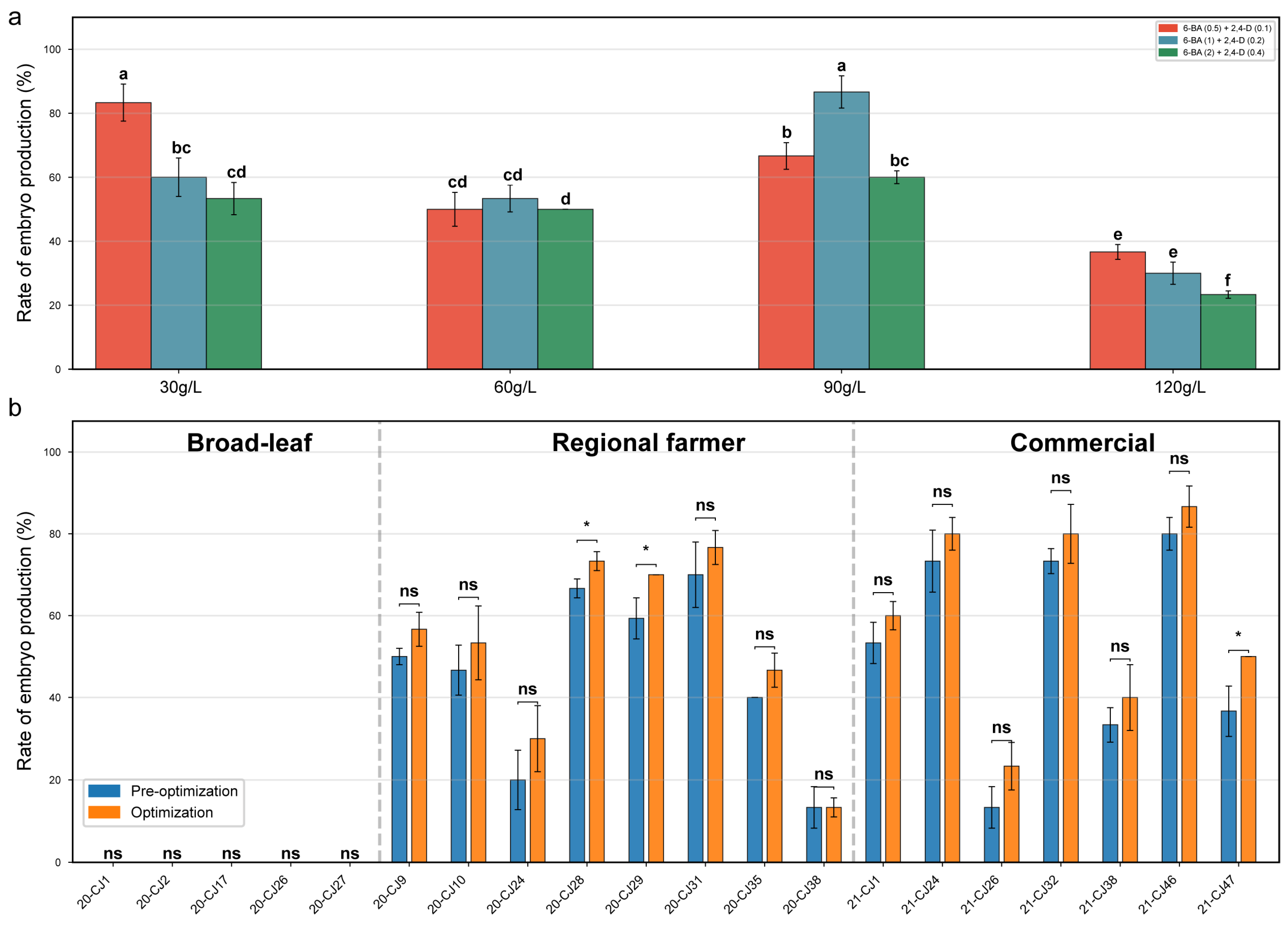
Sucrose concentration interacted with hormone composition to influence induction (Figure 2a). A sucrose level of 9% was generally optimal across hormone combinations and outperformed most treatments at 3%, 6%, or 12%. At 12% sucrose, induction decreased, consistent with inhibitory effects of excessive osmotic pressure on cell division. Under 9% sucrose, the combination of 1.0 mg/L 6-BA and 0.2 mg/L 2,4-D produced the highest mean induction rate (86.67%) and was selected as the optimized formulation.
To evaluate applicability of the optimized system (25 °C, −1 d ovaries, 90 g/L sucrose + 1.0 mg/L 6-BA + 0.2 mg/L 2,4-D), all 20 genotypes were retested (Figure 2b). The optimized conditions improved induction in some genotypes (e.g., ‘20-CJ28’, ‘20-CJ29’, and ‘21-CJ47’) with statistically significant increases. For highly responsive genotypes such as ‘21-CJ46’, ‘21-CJ24’, and ‘21-CJ32’, mean values increased numerically, although variance limited statistical significance. Broad-leaf chive genotypes remained non-responsive, indicating that medium and culture optimization did not overcome their genetic constraints.

3.2. Morphological Observations During Embryo Development

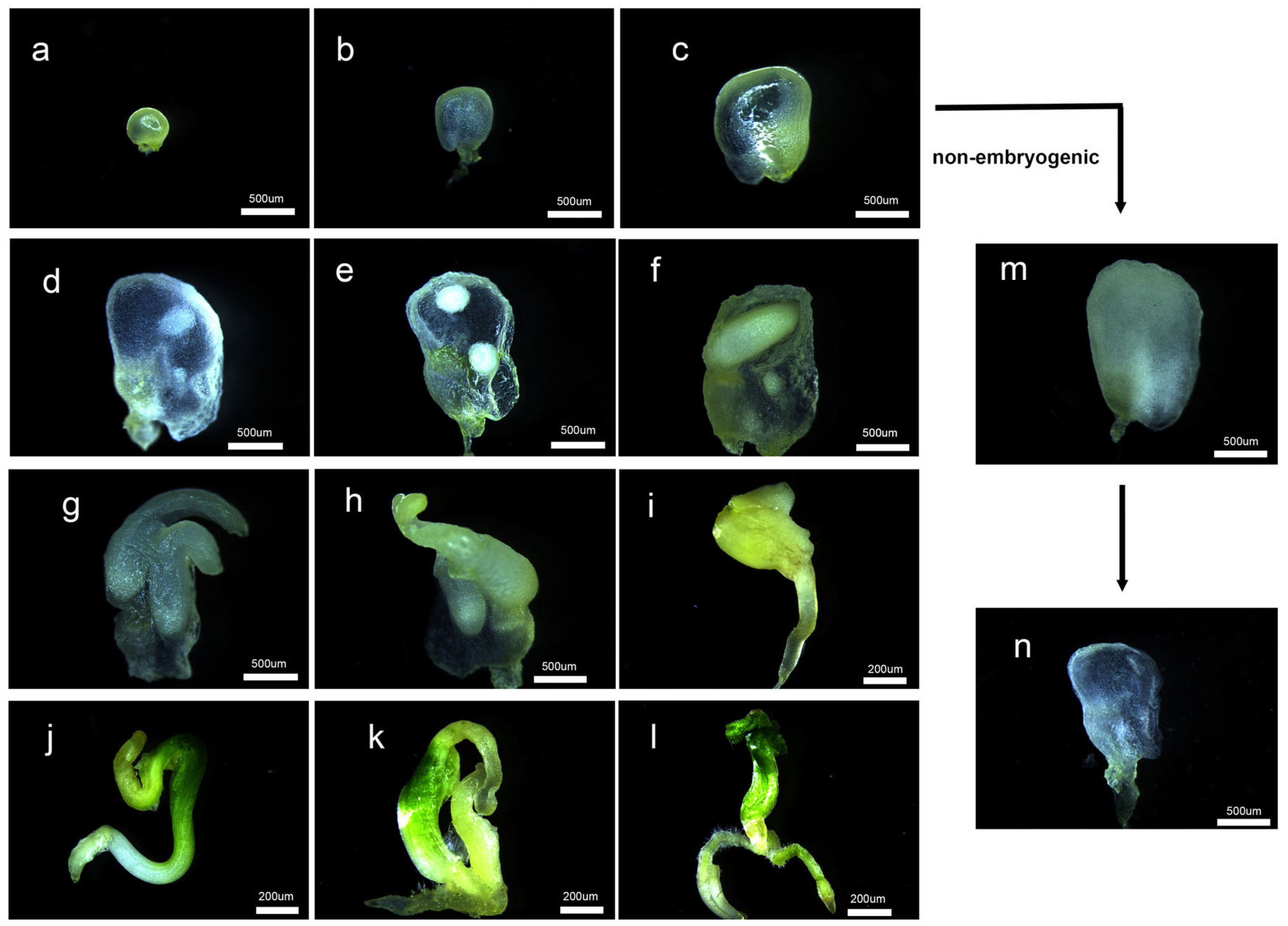
To trace developmental progression during gynogenesis, ovary morphology was monitored under the optimized conditions (Figure 3). At the initial stage, ovaries remained green and compact (Figure 3a). After ~14 days, successful induction was characterized by appearance of distinct white spherical structures within ovules, accompanied by localized tissue translucency (Figure 3c–e). Non-induced ovaries showed partial translucency but did not form such structures (Figure 3m,n). As culture progressed, spherical embryos penetrated the ovary wall (Figure 3f,g), underwent polarization, and developed into irregular bipolar structures (Figure 3h,i). These structures subsequently greened and differentiated into complete plantlets with visible shoots and roots (Figure 3j–l).

3.3. Chromosome Ploidy Identification

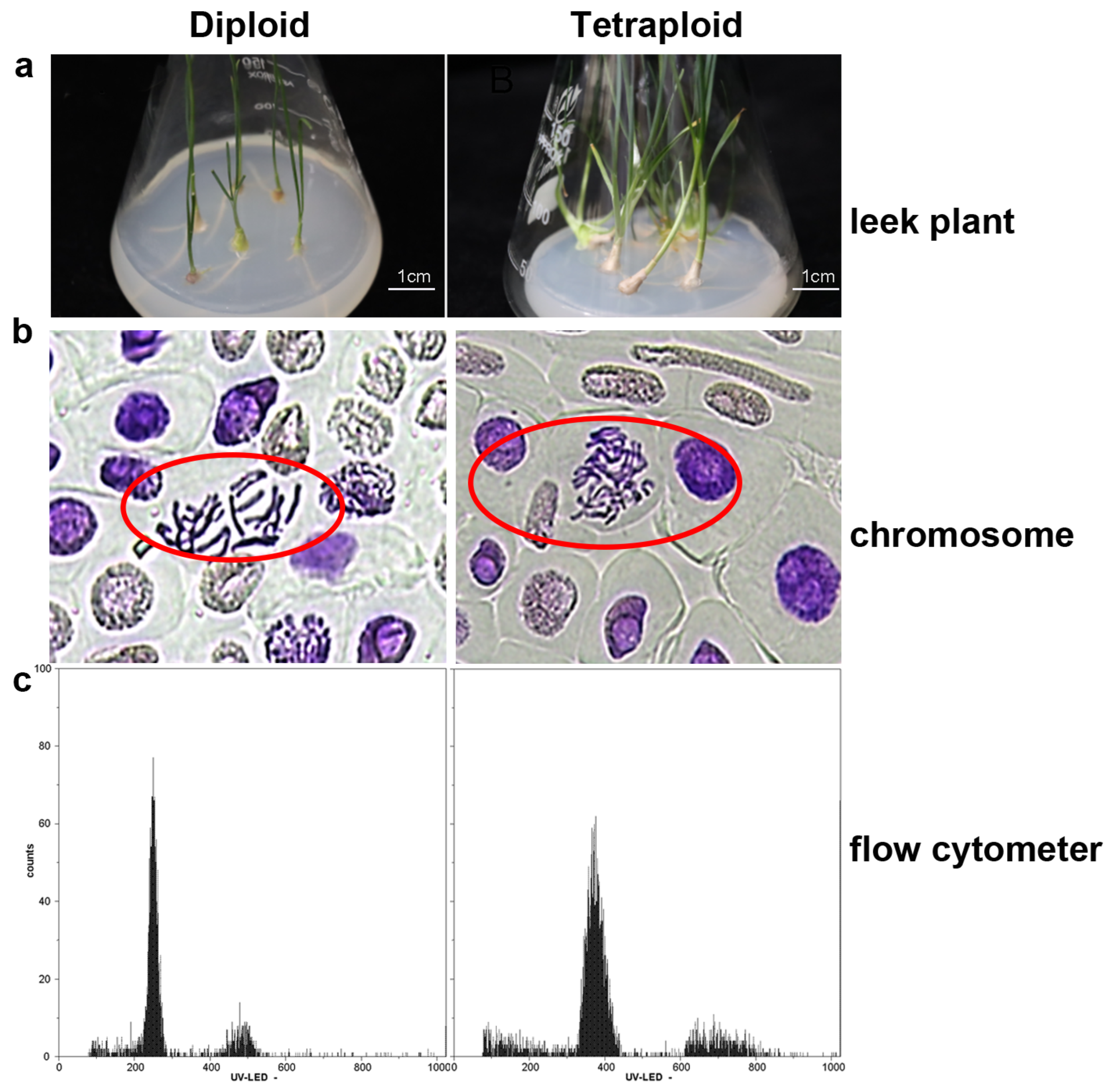
Flow cytometry and root-tip chromosome counting were used to assess ploidy in 100 randomly selected regenerants (Figure 4). Two characteristic DNA content peaks were observed: a diploid (2n) peak at ~200 fluorescence units and a tetraploid (4n) peak at ~400 (Figure 4c). Chromosome counting confirmed corresponding chromosome sets (Figure 4b). Among the 100 regenerants, tetraploids (4n) accounted for 28% and diploids (2n) for 72%, indicating frequent spontaneous chromosome doubling in this system. Given that the donor cultivar ‘21-CJ46’ is an autotetraploid (2n = 4x = 32), the regenerants exhibiting a diploid DNA content (2n = 2x = 16, peak at ~200 fluorescence units) represent the expected reduced-ploidy (haploid) products originating from gametophytic cells. The regenerants display a tetraploid profile (4n = 4x = 32, peak at ~400 fluorescence units) reflect populations that underwent spontaneous chromosomal doubling during the in vitro culture phase.

3.4. Impact of Hormone Ratios on Adventitious Bud Proliferation

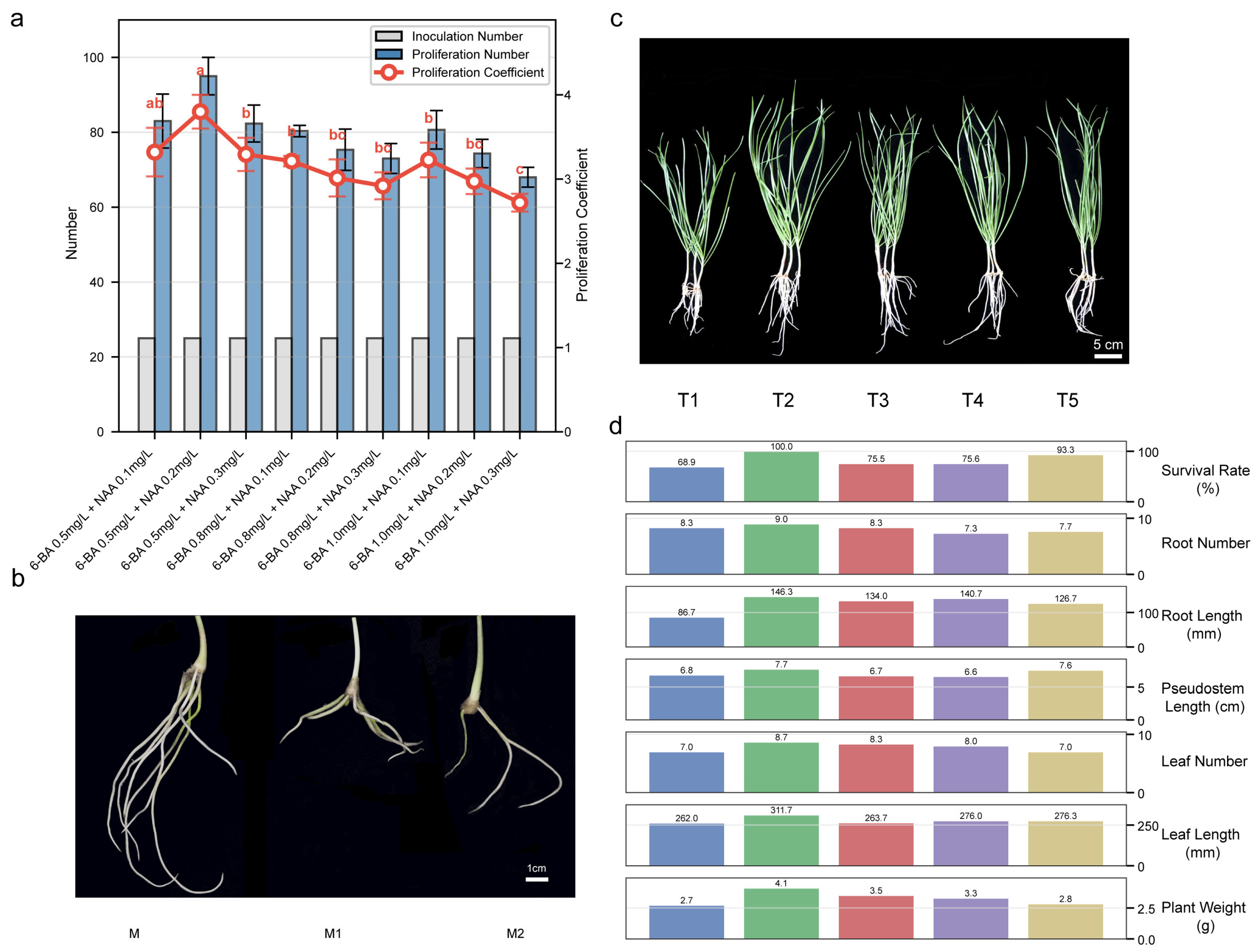
After regeneration, the proliferation system was optimized using a hormone gradient. The medium containing 0.5 mg/L 6-BA and 0.2 mg/L NAA produced the highest multiplication coefficient (~3.8) and supported stable shoot growth (Figure 5a). This treatment did not differ significantly from 0.5 mg/L 6-BA + 0.1 mg/L NAA, but it outperformed higher 6-BA treatments (0.8 and 1.0 mg/L). These results indicate that relatively low cytokinin, combined with low NAA, is sufficient for efficient shoot proliferation and may reduce risks of cytokinin-associated growth inhibition or vitrification.
During rooting, significant differences among media were observed (Figure 5b). The hormone-free MS medium (MS + 30 g/L sucrose + 9 g/L agar) gave the best rooting performance. Roots initiated at ~5 days after transfer, with 8–11 roots per plantlet on average; roots were thick and supported abundant root hairs. In contrast, NAA-containing media delayed rooting to 7–9 days, reduced root number, and produced thinner roots. Overall, hormone-free MS with sucrose and agar was the preferred rooting medium under these conditions.
Among five substrate mixtures, T2 (peat: coconut coir: cattle manure: perlite = 4:2:1:1) performed best (Figure 5c,d), with higher values for root number, root length, pseudo stem length, leaf number, and fresh weight compared with other treatments, and 100% survival after transplanting. T3–T5 showed no significant differences in growth traits, with survival rates ranging from 75.5% to 93.3%. T1 (without coconut coir) produced the lowest survival (68.89%) and weaker growth. These results suggest that incorporating coconut coir at an appropriate proportion improves substrate physical properties and supports establishment and growth.

3.5. Transcriptome Analysis During Gynogenesis Induction

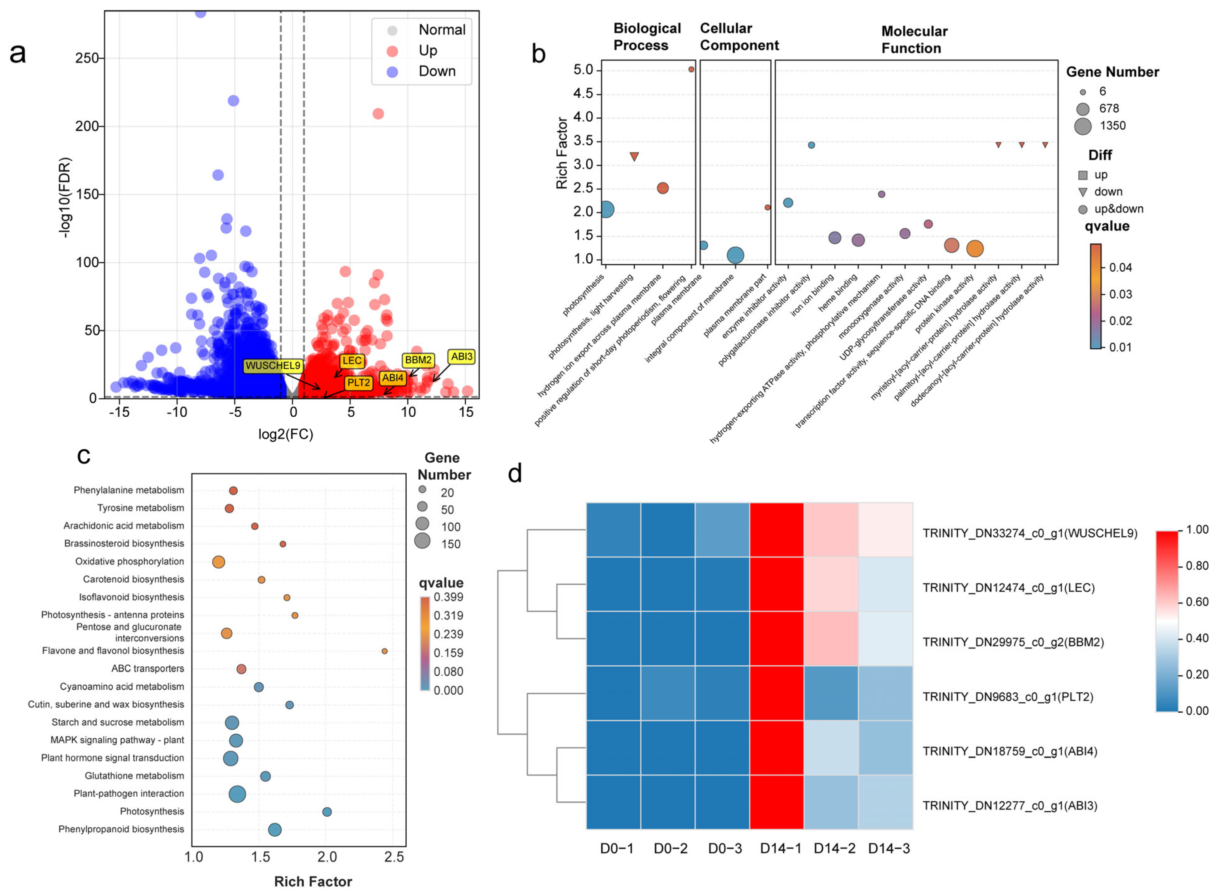
To explore molecular changes associated with the morphological transition observed in Section 3.2, RNA-seq was conducted for ovaries at 0 d and 14 d. De novo assembly generated a total of 165,807 unigenes. 8730 DEGs, accounting for ~5.27% of all unigenes. Volcano-plot analysis indicated that, at 14 d, DEGs 4120 were upregulated and 4610 were downregulated relative to 0 d (Figure 6a). GO enrichment analysis showed marked cellular reprogramming during embryogenesis initiation. In biological processes, pathways related to photosynthesis were significantly downregulated (Figure 6b). For cellular components, plasma membrane and integral component of membrane were strongly enriched, indicating broad expression changes in membrane-associated genes. At the molecular function level, enrichment of transcription factor activity and protein kinase activity indicated substantial reconfiguration of transcriptional regulation and signaling.
KEGG enrichment analysis highlighted pathways associated with gynogenesis initiation (Figure 6c). Phenylpropanoid biosynthesis showed strong enrichment, consistent with active cell wall remodeling. Plant–pathogen interaction and glutathione metabolism were also highly enriched, indicating stress adaptation and redox regulation during early culture. In signaling, plant hormone signal transduction and MAPK signaling (plant) were significantly enriched, consistent with activation of intracellular signaling cascades in response to exogenous 2,4-D and 6-BA. Enrichment of starch and sucrose metabolism aligned with the requirement for high sucrose (90 g/L) in the optimized medium. Based on DEG profiles, embryogenesis-related regulators were prioritized. Members of the BBM and WUSCHEL families, LEC, PLT, and ABI gene families showed minimal expression at 0 d but strong induction at 14 d, supporting their association with embryogenic fate acquisition under unpollinated ovary culture (Figure 6d).
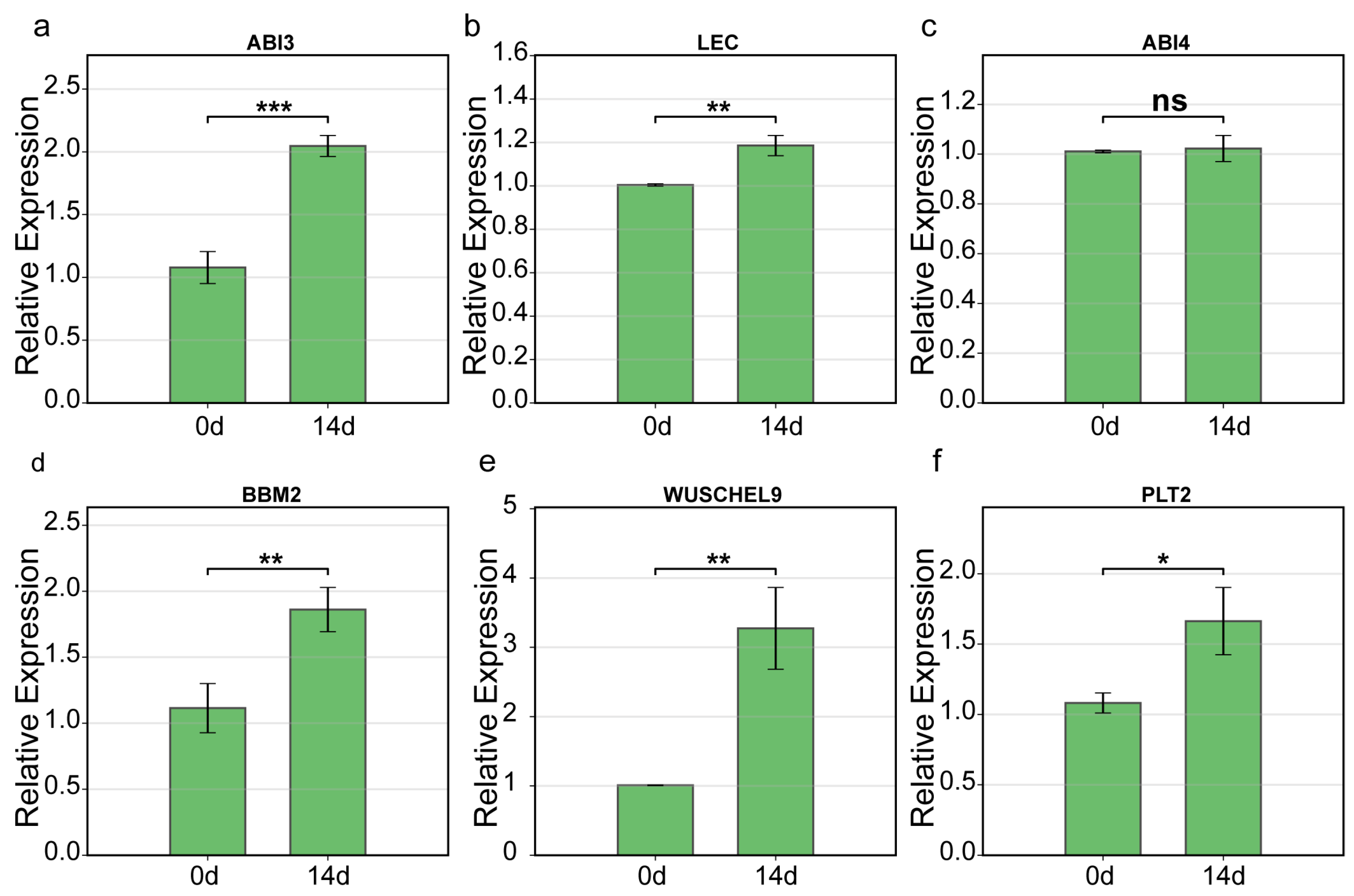
qRT-PCR generally matched RNA-seq trends (Figure 6d). BBM2, WUSCHEL9, LEC, PLT2, and ABI3 showed marked upregulation at 14 d (Figure 7a–f). BBM2 and WUSCHEL9 displayed the largest increases, consistent with potential roles as major regulators. In contrast, ABI4 differed between methods: RNA-seq suggested increased transcript abundance at 14 d, whereas qRT-PCR detected no significant difference. This discrepancy may reflect methodological sensitivity differences or primer selectivity for specific splice variants.

4. Discussion

Genotype dependence is a common feature of in vitro embryogenesis and callus induction across plant species. Among the 20 genotypes tested, embryoid induction rates differed substantially, with ‘21-CJ46’ showing the strongest induction, whereas all broad-leaf chive (A. hookeri) genotypes failed to produce embryoids. This agrees with previous reports in Chinese chive and other Allium species that gynogenic responsiveness is strongly dependent on donor genetic background [21,25]. Severe genotype dependency is a pervasive and fundamental characteristic of in vitro embryogenesis and callus induction across diverse plant species. For instance, the A188 inbred line in maize (Zea mays) is classically renowned for its exceptionally high competence for in vitro embryogenesis and callus formation compared to recalcitrant lines, and similarly strong genotype dependencies have been explicitly noted in dicot models such as cucumber ovary culture [27,40]. The recalcitrance of broad-leaf chive in this study may reflect intrinsic genetic or regulatory constraints. Accordingly, identifying highly responsive genotypes is a prerequisite for establishing robust induction systems.
In the present system, temperature stress was not required to initiate embryogenesis. Direct culture at 25 °C yielded the highest induction, whereas heat shock caused severe browning and reduced induction. This differs from some species where heat or cold stress is commonly used to interrupt gametophytic development and promote sporophytic reprogramming. For example, cucumber unpollinated ovary culture often employs 33–35 °C heat shock to trigger embryogenesis [41]. By contrast, successful Chinese chive ovary culture has been reported under moderate temperatures, consistent with the temperature sensitivity observed here [11]. The optimized sucrose level (90 g/L) also agrees with prior Chinese chive reports [11]. Beyond serving as a carbon source, elevated sucrose likely contributes to an osmotic component that can constrain non-embryogenic growth and promote embryogenic division, similar to osmotic effects described in other systems [42].
Flow cytometry and chromosome counting indicated frequent spontaneous chromosome doubling among regenerants. Spontaneous doubling has been reported in Allium gynogenesis [11,21] and can reduce reliance on chemical chromosome doubling procedures [14]. However, interpretation of 2n and 4n regenerants depends on the donor ploidy background and should be considered in that context.
Transcriptome comparisons between 0 d and 14 d reveal a highly coordinated cellular reprogramming sequence during embryogenesis initiation, and likewise indicated extensive metabolic reprogramming and activation of signaling pathways during this process. The significant enrichment of ‘phenylpropanoid biosynthesis’, ‘plant–pathogen interaction’, and ‘glutathione metabolism’ pathways suggests that the excised ovaries perceive the in vitro environment—specifically the high osmoticum (90 g/L sucrose) and tissue excision—as a distinct physiological stressor; correspondingly, enrichment of phenylpropanoid biosynthesis and plant–pathogen interaction suggests that explants were in a stress-responsive state early in culture. Stress-response activation has been observed in cucumber ovary culture and is often associated with embryogenesis onset [24]. Concurrently, the pronounced enrichment of ‘MAPK signaling’ and ‘plant hormone signal transduction’ pathways serves as the mechanistic bridge translating exogenous hormonal cues (2,4-D and 6-BA) into intrinsic transcriptional cascades. We propose a regulatory model wherein this integration of stress adaptation and exogenous hormone signaling dismantles the gametophytic program and directly drives the robust activation of cellular totipotency. Specifically, this stress-hormone signaling axis culminates in the near-zero to high-expression transition of BBM2, a central AP2/ERF regulator initiating embryogenic development, and notably key embryogenesis regulators (BBM2, WUSCHEL9, LEC, PLT2, and ABI3) were strongly induced at 14 d, consistent with activation of conserved transcriptional programs. BBM is a central regulator of totipotency and can induce embryogenic development when ectopically expressed [31,35]. Downstream of these primary signals, the orchestrated upregulation of WUSCHEL9 and PLT2 executes the de novo establishment of stem-cell niches and structural patterning, while upregulation of WUS and PLT suggests de novo establishment of stem-cell-like niches and embryonic patterning [43,44]. In parallel, the robust engagement of the LAFL network (LEC, ABI3)—which is fundamentally intertwined with the aforementioned hormone signal transduction pathways—solidifies embryo identity and maturation, and activation of LAFL network members (including LEC and ABI3) further supports engagement of an embryo-development program [37,45]. While these key regulators demonstrated high analytical concordance, ABI4 exhibited a notable discrepancy, showing transcript accumulation in RNA-seq but no significant change in targeted qRT-PCR; given that ABI4 is a core ABA-response transcriptional regulator with established roles in embryo-associated reserve mobilization and in the repression of germination programs, reflecting its capacity to couple hormonal status to developmental state [46], accurately capturing its induction dynamics and isoform usage is particularly important when interpreting embryogenesis initiation. This divergence is rationally explained by the methodological dichotomy between the global quantification of all assembled transcript isoforms in RNA-seq versus the strict amplification of a single coding domain in qRT-PCR, potentially compounded by the narrow, transient expression bursts characteristic of stress-responsive genes.

5. Conclusions

This study established a highly efficient in vitro gynogenesis system for Chinese chive, achieving a maximum embryo induction rate of 86.67% in the highly responsive cultivar ‘21-CJ46’. The optimal parameters required ovaries collected one day before anthesis, cultured at 25 °C on MS medium supplemented with 90 g/L sucrose, 1.0 mg/L 6-BA, and 0.2 mg/L 2,4-D, notably eliminating the requirement for severe thermal stress. The critical developmental milestone occurred at approximately 14 days of culture, characterized by distinct morphological transformations including localized ovarian tissue translucency and the emergence of macroscopic globular embryoids. RNA-seq analysis revealed that this 14 d morphological switch is underpinned by extensive transcriptomic reprogramming. The in vitro environment acts as a distinct physiological stressor, activating stress-response pathways (e.g., phenylpropanoid biosynthesis) and MAPK/hormone signal transduction, which collectively dismantle the gametophytic program and drive the de novo activation of essential totipotency and embryogenesis regulators, specifically BBM2, WUSCHEL9, PLT2, LEC, and ABI3. Practically, the high frequency of spontaneous chromosomal doubling observed in these regenerants significantly reduces the reliance on chemical doubling procedures. This optimized protocol, combined with the mapped transcriptomic regulatory network, provides a direct and robust framework for generating homozygous lines, thereby accelerating advanced breeding strategies in Chinese chive.

Author Contributions

Q.-Q.W. and B.-H.Z. designed the experiments and performed the majority of the laboratory work. L.-F.Z., N.A. and L.T. conducted the culture optimization experiments. B.T., C.-Y.M., W.Y. and W.-Y.F. participated in data analysis and validation. Y.D. conceived the project, supervised the study, and wrote the manuscript. All authors have read and agreed to the published version of the manuscript.

Funding

This research was funded by Guizhou Provincial Science and Technology Department through the Guizhou Provincial Science and Technology Support Program, grant number Qian Ke He Zhi Cheng [2022] Yi Ban 085 (Breeding and Introduction Screening of New Chinese Chive Varieties in Guizhou) and the Guizhou Academy of Agricultural Sciences via Guizhou Academy of Agricultural Sciences “15th Five-Year Plan” Outstanding Team Project on Mountain Vegetable Biological Breeding and Key Core Technology Innovation and Application (Qiannongke Outstanding Team [2026] No. 07). The APC was funded by Qian Ke He Zhi Cheng [2022] Yi Ban 085.

Data Availability Statement

The raw sequencing data generated in this study have been deposited in the CNCB database (National Genomics Data Center, China National Center for Bioinformation) under the accession number PRJCA056819. The assembled transcriptome sequences and annotation files are available from the corresponding author upon reasonable request.

Conflicts of Interest

The authors declare no conflicts of interest.

Abbreviations

The following abbreviations are used in this manuscript:
ABIABA INSENSITIVE
AFLPAmplified Fragment Length Polymorphism
AGLAGAMOUS-like
ANP1Arabidopsis Nucleoside Diphosphate-associated 1
AP2/ERFAPETALA2/Ethylene Responsive Factor
BBMBABY BOOM
BUSCOBenchmarking Universal Single-Copy Orthologs
CD-HIT-ESTClustering at high identity with tolerance for errors
CKControl
CC-BYCreative Commons Attribution
cDNAComplementary DNA
DAPI4’,6-diamidino-2-phenylindole
DEGsDifferentially Expressed Genes
DHDoubled Haploid
dday
dpadays post anthesis
FDRFalse Discovery Rate
FUS3FUSCA3
GOGene Ontology
KEGGKyoto Encyclopedia of Genes and Genomes
LAFLLEC/ABI/FUS3/LEC2 network
LECLEAFY COTYLEDON
MAPKMitogen-Activated Protein Kinase
MSMurashige and Skoog medium
NAANaphthaleneacetic acid
ODOptical Density
PProbability level
PCRPolymerase Chain Reaction
PICPolymorphic Information Content
PLTPLETHORA
qRT-PCRQuantitative Real-Time Polymerase Chain Reaction
RNARibonucleic Acid
RNA-seqRNA sequencing
SDStandard Deviation
TPMTranscripts Per Million
UPGMAUnweighted Pair Group Method with Arithmetic Mean
2,4-D2,4-dichlorophenoxyacetic acid
6-BA6-benzylaminopurine

References

  1. Deng, W.; Li, Q.-G.; Tian, H.-Q. Dimorphism of synergids in Allium tuberosum (Amaryllidaceae). Ann. Bot. Fenn. 2016, 53, 267–275. [Google Scholar] [CrossRef]
  2. Fritsch, R.M.; Friesen, N. Evolution, domestication and taxonomy. In Allium Crop Science: Recent Advances; CABI: Wallingford, UK, 2002; pp. 5–30. [Google Scholar] [CrossRef]
  3. Jiang, K.; Kuang, Y.; Feng, L.; Liu, Y.; Wang, S.; Du, H.; Shen, W. Molecular hydrogen maintains the storage quality of Chinese chive through improving antioxidant capacity. Plants 2021, 10, 1095. [Google Scholar] [CrossRef]
  4. Yamashita, K.; Nakazawa, Y.; Namai, K.; Amagai, M.; Tsukazaki, H.; Wako, T.; Kojima, A. Modes of inheritance of two apomixis components, diplospory and parthenogenesis, in Chinese chive (Allium ramosum) revealed by analysis of the segregating population generated by back-crossing between amphimictic and apomictic diploids. Breed. Sci. 2012, 62, 160–169. [Google Scholar] [CrossRef]
  5. Abdelrahman, M.; Ariyanti, N.A.; Sawada, Y.; Tsuji, F.; Hirata, S.; Hang, T.T.M.; Okamoto, M.; Yamada, Y.; Tsugawa, H.; Hirai, M.Y.; et al. Metabolome-based discrimination analysis of shallot landraces and bulb onion cultivars associated with differences in the amino acid and flavonoid profiles. Molecules 2020, 25, 5300. [Google Scholar] [CrossRef]
  6. Gao, Q.; Li, X.-B.; Sun, J.; Xia, E.D.; Tang, F.; Cao, H.Q.; Xun, H. Isolation and identification of new chemical constituents from Chinese chive (Allium tuberosum) and toxicological evaluation of raw and cooked Chinese chive. Food Chem. Toxicol. 2018, 112, 400–411. [Google Scholar] [CrossRef]
  7. Kamenetsky, R.; Rabinowitch, H.D. Physiology of domesticated alliums: Onions, garlic, leek, and minor crops. In Encyclopedia of Applied Plant Sciences; Elsevier: Amsterdam, The Netherlands, 2017; pp. 255–261. [Google Scholar] [CrossRef]
  8. Tian, H.Q.; Yang, H.Y. Ultrastructural observations on parthenogenesis and antipodal apogamy of Allium tuberosum Roxb. Acta Bot. Sin. 1991, 33, 819–824. [Google Scholar]
  9. Tian, H.; Yang, H. Embryo sac development and embryogeny in Allium tuberosum. Plant Sci. J. 1991, 9, 5–10. [Google Scholar]
  10. Lin, Y.H.; Lin, M.Z.; Chen, Y.Q.; Tian, H.Q. Isolation of male and female gametes, zygotes and proembryos of leek (Allium tuberosum Roxb). Zygote 2020, 28, 278–285. [Google Scholar] [CrossRef]
  11. Yang, S.J.; Peng, L.; Tian, H.Q. Parthenogenesis and apogamy in unpollinated ovary culture of leek (Allium tuberosum Roxb.). J. Plant Biochem. Biotechnol. 2021, 30, 522–531. [Google Scholar] [CrossRef]
  12. Germanà, M.A. Doubled haploid production in fruit crops. Plant Cell Tissue Organ Cult. 2006, 86, 131–146. [Google Scholar] [CrossRef]
  13. Khan, H.; Bhardwaj, S.C.; Gangwar, O.P.; Prasad, P.; Rathore, R. Efficiency of double haploid production in wheat through wide hybridization and embryo rescue. Indian J. Genet. Plant Breed. 2017, 77, 428. [Google Scholar] [CrossRef]
  14. Niazian, M.; Shariatpanahi, M.E. In vitro-based doubled haploid production: Recent improvements. Euphytica 2020, 216, 69. [Google Scholar] [CrossRef]
  15. Zou, T.; Su, H.-N.; Wu, Q.; Sun, X.-W. Haploid induction via unfertilized ovary culture in watermelon. Plant Cell Tissue Organ Cult. 2018, 135, 179–187. [Google Scholar] [CrossRef]
  16. Dunwell, J.M. Haploids in flowering plants: Origins and exploitation. Plant Biotechnol. J. 2010, 8, 377–424. [Google Scholar] [CrossRef]
  17. Dwivedi, S.L.; Britt, A.B.; Tripathi, L.; Sharma, S.; Upadhyaya, H.D.; Ortiz, R. Haploids: Constraints and opportunities in plant breeding. Biotechnol. Adv. 2015, 33, 812–829. [Google Scholar] [CrossRef]
  18. Gilles, L.M.; Khaled, A.; Laffaire, J.; Chaignon, S.; Gendrot, G.; Laplaige, J.; Bergès, H.; Beydon, G.; Bayle, V.; Barret, P.; et al. Loss of pollen-specific phospholipase NOT LIKE DAD triggers gynogenesis in maize. EMBO J. 2017, 36, 707–717. [Google Scholar] [CrossRef] [PubMed]
  19. Li, T.; Wang, C.; Pan, J.; Tabusam, J.; Li, Y.; Yao, J.; Chen, W.; Wang, Y.; Gao, W.; Rong, J.; et al. Exploring potential strategies for haploid induction based on double fertilization in plants. Plant Biotechnol. J. 2025, 23, 4000–4016. [Google Scholar] [CrossRef]
  20. Vijverberg, K.; Ozias-Akins, P.; Schranz, M.E. Identifying and engineering genes for parthenogenesis in plants. Front. Plant Sci. 2019, 10, 128. [Google Scholar] [CrossRef] [PubMed]
  21. Alan, A.R.; Celebi Toprak, F.; Kaska, A. Production and evaluation of gynogenic leek (Allium ampeloprasum L.) plants. Plant Cell Tissue Organ Cult. 2016, 125, 249–259. [Google Scholar] [CrossRef]
  22. Keller, E.R.J.; Korzun, L. Haploidy in onion and other Allium species. In In Vitro Haploid Production in Higher Plants; Jain, S.M., Sopory, S.K., Veilleux, R.E., Eds.; Springer: Dordrecht, The Netherlands, 1996; Volume 3, pp. 51–75. [Google Scholar] [CrossRef]
  23. Sharma, S.; Satardekar, K.V.; Barve, S.S. Genetic improvement of medicinal and aromatic plants through haploid and DH development. In Biotechnological Approaches for Medicinal and Aromatic Plants; Springer: Singapore, 2018; pp. 523–556. [Google Scholar] [CrossRef]
  24. Deng, Y.; Fu, W.; Tang, B.; Tao, L.; Zhang, L.; Zhou, X.; Wang, Q.; Li, J.; Chen, J. Transcriptome analysis of ovary culture-induced embryogenesis in cucumber (Cucumis sativus L.). PeerJ 2021, 9, e12145. [Google Scholar] [CrossRef]
  25. Chen, J.-F.; Cui, L.; Malik, A.A.; Mbira, K.G. In vitro haploid and dihaploid production via unfertilized ovule culture. Plant Cell Tissue Organ Cult. 2011, 104, 311–319. [Google Scholar] [CrossRef]
  26. Wang, L.; Wang, Z.; Ma, P.; Zhang, W.; Liu, W.; Zhang, Y.; Li, D. Study on regeneration of plants from macrospore induction culture of Chinese chive and transplantation techniques. Agric. Sci. Technol. Newsl. 2017, 10, 157–158. [Google Scholar]
  27. Diao, W.-P.; Jia, Y.-Y.; Song, H.; Zhang, X.-Q.; Lou, Q.-F.; Chen, J.-F. Efficient embryo induction in cucumber ovary culture and homozygous identification of the regenerants using SSR markers. Sci. Hortic. 2009, 119, 246–251. [Google Scholar] [CrossRef]
  28. Shalaby, T.A. Factors affecting haploid induction through in vitro gynogenesis in summer squash. Sci. Hortic. 2007, 115, 1–6. [Google Scholar] [CrossRef]
  29. Shariatpanahi, M.E.; Belogradova, K.; Hessamvaziri, L.; Heberle-Bors, E.; Touraev, A. Efficient embryogenesis and regeneration in cultured wheat microspores without stress pretreatment. Plant Cell Rep. 2006, 25, 1294–1299. [Google Scholar] [CrossRef] [PubMed]
  30. Touraev, A.; Vicente, O.; Heberle-Bors, E. Initiation of microspore embryogenesis by stress. Trends Plant Sci. 1997, 2, 297–302. [Google Scholar] [CrossRef]
  31. Boutilier, K.; Offringa, R.; Sharma, V.K.; Kieft, H.; Ouellet, T.; Zhang, L.; Hattori, J.; Liu, C.M.; van Lammeren, A.A.; Miki, B.L.; et al. Ectopic expression of BABY BOOM triggers a conversion from vegetative to embryonic growth. Plant Cell 2002, 14, 1737–1749. [Google Scholar] [CrossRef]
  32. Shi, C.; Zhao, Z.; Zhong, Y.; Qiao, Y.; Zhang, L.; Yang, F.; Li, S.; Li, L.; Huang, L.; Tang, X.; et al. Reprogramming of microspore fate via BBM-BAR1 for highly efficient in vivo haploid induction. Cell 2025, 188, 6109–6120.e15. [Google Scholar] [CrossRef]
  33. Soriano, M.; Li, H.; Boutilier, K. Microspore embryogenesis: Establishment of embryo identity and pattern in culture. Plant Reprod. 2013, 26, 181–196. [Google Scholar] [CrossRef]
  34. Chen, B.; Maas, L.; Figueiredo, D.; Zhong, Y.; Reis, R.; Li, M.; Horstman, A.; Riksen, T.; Weemen, M.; Liu, H.; et al. BABY BOOM regulates early embryo and endosperm development. Proc. Natl. Acad. Sci. USA 2022, 119, e2201761119. [Google Scholar] [CrossRef] [PubMed]
  35. Khanday, I.; Skinner, D.; Yang, B.; Mercier, R.; Sundaresan, V. A male-expressed rice embryogenic trigger redirected for asexual propagation through seeds. Nature 2019, 565, 91–95. [Google Scholar] [CrossRef]
  36. Braybrook, S.; Harada, J. LECs go crazy in embryo development. Trends Plant Sci. 2008, 13, 624–630. [Google Scholar] [CrossRef]
  37. Stone, S.L.; Braybrook, S.A.; Paula, S.L.; Kwong, L.W.; Meuser, J.; Pelletier, J.; Hsieh, T.F.; Fischer, R.L.; Goldberg, R.B.; Harada, J.J. Arabidopsis LEAFY COTYLEDON2 induces maturation traits and auxin activity. Proc. Natl. Acad. Sci. USA 2008, 105, 3151–3156. [Google Scholar] [CrossRef]
  38. Suzuki, M.; Wang, H.H.-Y.; McCarty, D.R. Repression of the LEC1/B3 network by VP1/ABI3-like genes. Plant Physiol. 2007, 143, 902–911. [Google Scholar] [CrossRef]
  39. Zhang, H.; Bishop, B.; Ringenberg, W.; Muir, W.M.; Ogas, J. The CHD3 remodeler PICKLE associates with genes enriched for H3K27me3. Plant Physiol. 2012, 159, 418–432. [Google Scholar] [CrossRef]
  40. Ge, F.; Qu, J.; Liu, P.; Pan, L.; Zou, C.; Yuan, G.; Yang, C.; Pan, G.; Huang, J.; Ma, L.; et al. Genome assembly of the maize inbred line A188 provides a new reference genome for functional genomics. Crop J. 2022, 10, 47–55. [Google Scholar] [CrossRef]
  41. Kurtar, E.S.; Balkaya, A.; Kandemir, D. Evaluation of haploidization efficiency in winter squash and pumpkin through anther culture. Plant Cell Tissue Organ Cult. 2016, 127, 497–511. [Google Scholar] [CrossRef]
  42. Ahmadi, B.; Shariatpanahi, M.E. Proline and chitosan enhanced efficiency of microspore embryogenesis induction and plantlet regeneration in Brassica napus L. Plant Cell Tissue Organ Cult. 2015, 123, 57–65. [Google Scholar] [CrossRef]
  43. Aida, M.; Beis, D.; Heidstra, R.; Willemsen, V.; Blilou, I.; Galinha, C.; Nussaume, L.; Noh, Y.S.; Amasino, R.; Scheres, B. The PLETHORA genes mediate patterning of the Arabidopsis root stem cell niche. Cell 2004, 119, 109–120. [Google Scholar] [CrossRef]
  44. Zuo, J.; Niu, Q.-W.; Frugis, G.; Chua, N.-H. The WUSCHEL gene promotes vegetative-to-embryonic transition in Arabidopsis. Plant J. 2002, 30, 349–359. [Google Scholar] [CrossRef] [PubMed]
  45. Lotan, T.; Ohto, M.; Yee, K.M.; West, M.A.; Lo, R.; Kwong, R.W.; Yamagishi, K.; Fischer, R.L.; Goldberg, R.B.; Harada, J.J. Arabidopsis LEAFY COTYLEDON1 is sufficient to induce embryo development in vegetative cells. Cell 1998, 93, 1195–1205. [Google Scholar] [CrossRef] [PubMed]
  46. Penfield, S.; Li, Y.; Gilday, A.D.; Graham, S.; Graham, I.A. Arabidopsis ABA INSENSITIVE4 regulates lipid mobilization in the embryo and reveals repression of seed germination by the endosperm. Plant Cell 2006, 18, 1887–1899. [Google Scholar] [CrossRef] [PubMed]
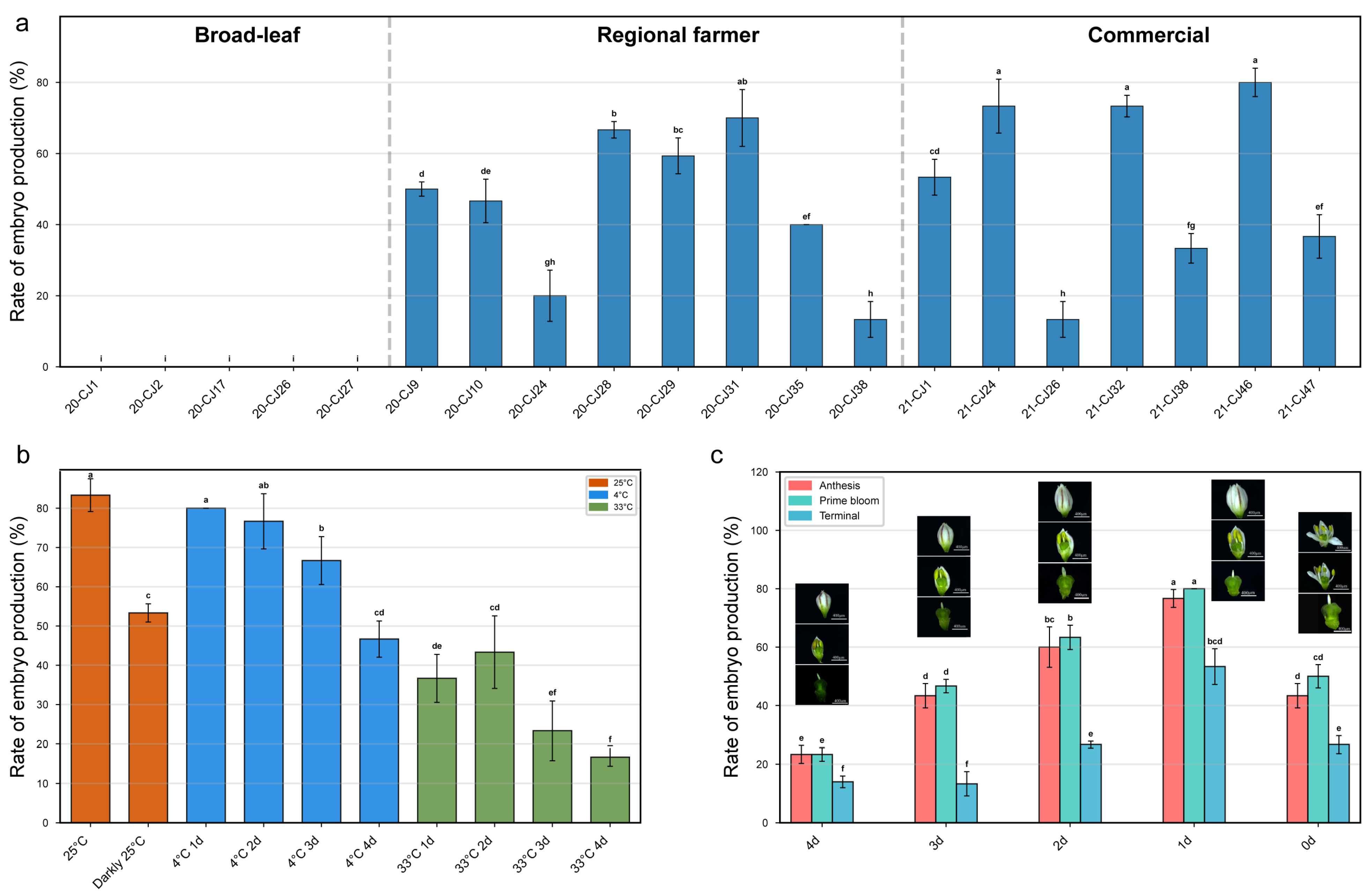
Figure 1. Screening and optimization of biological and physical factors affecting gynogenesis in Allium tuberosum unpollinated ovary culture. (a) Effect of donor genotype on embryo induction rate. Twenty genotypes were screened, including broad-leaf chives (A. hookeri), regional farmer landraces, and commercial cultivars. (b) Effect of physical culture conditions. Ovaries were subjected to different temperature treatments: constant 25 °C, cold stress (4 °C for 1–4 d), and heat stress (33 °C for 1–4 d). (c) Effect of maternal flowering phase and ovarian developmental stage. Ovaries were collected from plants at early (Anthesis), prime, or late (Terminal) bloom stages, and at different temporal stages relative to anthesis (0 d to −4 d). Data represent the mean ± SD of three independent biological replicates. Different lowercase letters indicate statistically significant differences according to Duncan’s multiple range test (p < 0.05).
Agronomy 16 00627 g001
Figure 2. Optimization of culture medium composition and systematic validation of the induction protocol. (a) Interaction effects of sucrose concentration gradients and exogenous hormone combinations (6-BA and 2,4-D) on the embryoid induction rate of cultivar ‘21-CJ46’. (b) Comparative validation of induction efficiency between the baseline pre-optimization system and the final optimized culture system across all 20 evaluated genotypes. Data represent the mean ± SD of three independent biological replicates. In panel (a), different lowercase letters indicate significant differences (p < 0.05). In panel (b), asterisks indicate statistically significant differences between the pre-optimization and optimized conditions within the same genotype (* p < 0.05); ns indicates not significant.
Agronomy 16 00627 g002
Figure 3. Morphological and developmental characteristics of gynogenesis in Allium tuberosum unpollinated ovary culture. (a) Freshly inoculated ovary at 0 d, appearing green and compact. (b) Ovary during early culture. (ce) Induction phase (approx. 14 d): Appearance of white globular structures (embryoids) within the ovule and transparency of the surrounding ovarian tissue. (f,g) Globular embryos breaking through the ovary wall. (h,i) Polarization stage: Embryos developing into irregular bipolar structures. (jl) Regeneration stage: Differentiation into complete plantlets with distinct shoots and roots. (m,n) Induction failure phase (approximately 14 days).
Agronomy 16 00627 g003
Figure 4. Ploidy identification of regenerated Allium tuberosum plants. (a) Regenerated plantlets growing in culture bottles. (b) Chromosome counting of root tip cells showing metaphase chromosomes (indicated by red circles) (100×). (c) Flow cytometry histograms of nuclei are isolated from regenerated leaves. Left: Peak at ~200 fluorescence intensity corresponds to diploid (2n) plants. Right: Peak at ~400 fluorescence intensity corresponds to tetraploid (4n) plants. The population exhibited spontaneous chromosomal doubling. Left: Diploid cell (2n); Right: Tetraploid cell (4n).
Agronomy 16 00627 g004
Figure 5. Optimization of proliferation, rooting, and transplanting systems for regenerated Allium tuberosum plantlets. (a) Effect of different hormone combinations (6-BA and NAA) on the proliferation coefficient and number of adventitious buds. (b) Rooting morphology of plantlets cultured on different media: M (hormone-free), M1 (1/2 MS + NAA), and M2 (MS + NAA). Note the robust root system in M medium. (c) Morphological comparison of plantlets transplanted into five different substrate mixtures (T1–T5) after one month. (d) Statistical analysis of survival rate, root number, root length, pseudostem length, leaf number, leaf length, and plant fresh weight across the five substrate treatments.
Agronomy 16 00627 g005
Figure 6. Global transcriptomic landscape and expression profiling during gynogenesis initiation. (a) Volcano plot of differentially expressed genes (DEGs) between control (0 d) and induced ovaries (14 d). Red dots represent significantly up-regulated genes, and blue dots represent significantly down-regulated genes. (b) Gene Ontology (GO) enrichment analysis of DEGs categorized by biological processes, cellular components, and molecular functions. (c) KEGG pathway enrichment analysis highlighting the top enriched metabolic and signaling pathways, including phenylpropanoid biosynthesis and plant hormone signal transduction. (d) Expression heatmap showing the transcript abundance patterns (based on TPM values) of key somatic embryogenesis-related regulatory genes (WUSCHEL9, LEC, BBM2, PLT2, ABI4, and ABI3) at 0 d and 14 d.
Agronomy 16 00627 g006
Figure 7. Targeted qRT-PCR validation of core embryogenesis-associated regulatory genes. (af) Relative transcript expression levels of BBM2, PLT2, ABI3, LEC, ABI4, and WUSCHEL9 measured by quantitative real-time PCR at 0 d and 14 d. Expression data were normalized to the Allium actin reference gene. Data represent the mean ± SD of three independent biological replicates (n = 3). Asterisks indicate significant differences (* p < 0.05, ** p < 0.01, *** p < 0.0001, ns: not significant).
Agronomy 16 00627 g007
Table 1. Unpollinated ovary culture materials.
No.GenotypeVariety
120-CJ1broad-leaf chive (A. hookeri)
220-CJ2broad-leaf chive (A. hookeri)
320-CJ17broad-leaf chive (A. hookeri)
420-CJ26broad-leaf chive (A. hookeri)
520-CJ27broad-leaf chive (A. hookeri)
620-CJ9Local cultivated chive (A. tuberosum) of Guizhou Province
720-CJ10Local cultivated chive (A. tuberosum) of Guizhou Province
820-CJ24Local cultivated chive (A. tuberosum) of Guizhou Province
920-CJ28Local cultivated chive (A. tuberosum) of Guizhou Province
1020-CJ29Local cultivated chive (A. tuberosum) of Guizhou Province
1120-CJ31Local cultivated chive (A. tuberosum) of Guizhou Province
1220-CJ35Local cultivated chive (A. tuberosum) of Guizhou from Fusheng Company, Tongren, China
1320-CJ38Local cultivated chive (A. tuberosum) of Guizhou Province
1421-CJ1commercial cultivar (A. tuberosum) from Fusheng Company
1521-CJ24commercial cultivar (A. tuberosum) from Fusheng Company
1621-CJ26commercial cultivar (A. tuberosum) from Shandong Province
1721-CJ32commercial cultivar (A. tuberosum) from Fusheng Company
1821-CJ38commercial cultivar (A. tuberosum) from Pinglong Chive Industry Company, Pengzhou, China
1921-CJ46commercial cultivar (A. tuberosum) from Henan Province
2021-CJ47commercial cultivar (A. tuberosum) from Pinglong Chive Industry Company
Table 2. Induction medium formulations (sucrose × hormone combinations).
Sucrose Concentration (g/L)6-BA 0.5 mg/L + 2,4-D 0.1 mg/L6-BA 1.0 mg/L + 2,4-D 0.2 mg/L6-BA 2.0 mg/L + 2,4-D 0.4 mg/L
30(3-1)(3-2)(3-3)
60(6-1)(6-2)(6-3)
90(9-1)(9-2)(9-3)
120(12-1)(12-2)(12-3)
Table 3. Proliferation medium formulations.
Treatment6-BA (mg/L)NAA (mg/L)
10.50.1
20.50.2
30.50.3
40.80.1
50.80.2
60.80.3
71.00.1
81.00.2
91.00.3
Note: The basal medium utilized was MS medium. A constant sample size of 75 proliferated adventitious buds was inoculated per treatment group (comprising 3 independent biological replicates, 5 vessels per replicate, and 5 explants per vessel). Abbreviations: 6-BA, 6-benzylaminopurine; NAA, 1-Naphthaleneacetic acid.
Table 4. Primers used for qRT-PCR.
Gene (Trinity ID)Forward Primer (5′–3′)Reverse Primer (5′–3′)Amplicon Size (bp)
ABI3 (TRINITY_DN12277_c0_g1)ACCAAACTGCCCTCCGTGGCTTGAGAACGGGTGGCT133
LEC (TRINITY_DN12474_c0_g1)TCATGGCCCAAAGCACGTCATCACGAGCGAGGCCAA80
ABI4 (TRINITY_DN18759_c0_g1)AGCGGTTGTTCCAGCTCCAGGAGGCGGTCTAGGCAA105
BBM2 (TRINITY_DN29975_c0_g2)AGCAACCTCCCAATCGGCTTGTAGGCCTGGGCTTGC146
WUSCHEL9 (TRINITY_DN33274_c0_g1)AGCCCAACCGACACAAGGCCACTGACTGCTGCTGCT101
PLT2 (TRINITY_DN9683_c0_g1)TGCCCGAAGCTTGCAAACTGTGGGTCTGGGGCCATA149
Polyubiquitin (TRINITY_DN554_c1_g2)(reference gene primers)CGAGCAAGCTTCGAGGCTAAAGCATCCGACCACCCG104
Disclaimer/Publisher’s Note: The statements, opinions and data contained in all publications are solely those of the individual author(s) and contributor(s) and not of MDPI and/or the editor(s). MDPI and/or the editor(s) disclaim responsibility for any injury to people or property resulting from any ideas, methods, instructions or products referred to in the content.