Article

Genome Duplication Reshapes Leaf Structure and Trait Coordination in Mangoes (Mangifera indica L.)


by
Marcos Adrián Ruiz-Medina
1,*,
Águeda M. González-Rodríguez
1,
Noé Jesús Liria-Martín
1 and
María José Grajal-Martín
2
1
Departamento de Botánica, Ecología y Fisiología Vegetal, Universidad de La Laguna, 38206 La Laguna, Spain
2
Departamento de Producción Vegetal en Zonas Tropicales y Subtropicales, Instituto Canario de Investigaciones Agrarias (ICIA), 38270 La Laguna, Spain
*
Author to whom correspondence should be addressed.
Agronomy 2026, 16(13), 1226; https://doi.org/10.3390/agronomy16131226
Submission received: 11 March 2026 / Revised: 18 June 2026 / Accepted: 22 June 2026 / Published: 24 June 2026

Abstract

Polyploidy is increasingly recognized as a mechanism enhancing physiological resilience in woody fruit crops, yet its functional consequences remain poorly understood in mangoes (Mangifera indica L.), a major tropical species expanding into water-limited environments. Because leaf structure underpins plant water relations and gas exchange, this study evaluated how genome duplication alters foliar traits by comparing diploid and autotetraploid individuals of three polyembryonic cultivars (Gomera-1, Gomera-3, and Kensington Pride). Morphological and anatomical analyses revealed consistent ploidy-related modifications. Autotetraploids exhibited enlarged stomatal guard cells, increased leaf thickness, and changes in mesophyll organization, indicating greater structural investment in leaf tissues. These features are commonly associated with structural strategies that may contribute to water retention and hydraulic regulation, although their direct physiological consequences were not evaluated in the present study. Overall, our results indicate that genome duplication substantially modifies leaf structural traits in mangoes, although the magnitude and direction of these responses were cultivar-dependent. This study provides new insights into how polyploidy reshapes leaf morphology and anatomy in mangoes and advances our understanding of polyploid-induced structural variation in perennial fruit crops.

1. Introduction

Mangifera indica L. (mango) is an evergreen fruit tree of the family Anacardiaceae, domesticated in South and Southeast Asia and cultivated for thousands of years, resulting in a wide diversity of landraces and cultivars. Today, mangoes are one of the most important tropical fruit crops worldwide, ranking second in global production among tropical fruits after banana [1]. Beyond their economic relevance as a fresh fruit, mangoes also hold nutritional and nutraceutical value due to their richness in bioactive compounds such as phenolics, flavonoids, carotenoids, and vitamins, which contribute to antioxidant and anti-inflammatory properties [2,3].
As mango cultivation expands into subtropical and Mediterranean regions, orchards are increasingly exposed to high temperatures, intense solar radiation, and seasonal water deficits [4,5]. Under these environmental constraints, plant performance depends strongly on leaf structural and functional traits that regulate transpiration, photosynthesis, and hydraulic efficiency. Morphological and anatomical features such as leaf thickness, cuticle development, stomatal characteristics, and vascular organization are therefore central to plant adaptation to drought and heat stress [6,7,8].
Polyploidy is recognized as an important driver of structural and physiological variation in plants and has been associated with enhanced tolerance to abiotic stresses in both woody and herbaceous species [9]. Genome duplication frequently results in increased cell size and modifications in tissue organization, which can affect leaf thickness, stomatal dimensions, and vascular structure, ultimately influencing plant water relations and gas exchange [10,11]. Such changes may alter transpiration dynamics, leaf elasticity, and hydraulic conductance, providing a potential structural basis for improved drought performance.
Although most commercial mango cultivars are diploid (2n = 40) [12], spontaneous autotetraploid genotypes have been identified and selected. Previous studies on mangoes indicate that autotetraploids may exhibit greater leaf water retention, increased proline accumulation, and a more conservative water-use behavior under water deficit compared with diploid plants [13,14,15]. These functional differences have been linked to structural traits such as larger cells, thicker leaves, and modified vascular elements, yet the anatomical mechanisms underlying these responses remain insufficiently characterized in this species.
Understanding how genome duplication modifies leaf structure is particularly relevant for mango cultivation in Mediterranean-type climates, where water availability is a major limiting factor for productivity and long-term orchard sustainability. Therefore, this study compares selected diploid and autotetraploid mango cultivars to determine how polyploidy influences leaf morphological, anatomical, and vascular traits. By integrating these parameters, we aim to identify structural mechanisms that may contribute to improved water-use efficiency and drought resilience in autopolyploid mango genotypes.

2. Materials and Methods

2.1. Study Site, Plant Material and Collection

The study was conducted at the experimental farm of the Canary Islands Agricultural Research Institute–ICIA-, Finca Cueva del Polvo (Guía de Isora, Tenerife, Canary Islands, Spain; 28.229161 N, −16.834213 W; ~100 m a.s.l.). According to the agroclimatic classification of southern Tenerife, the site corresponds to the lower arid belt, characterized by scarce rainfall and high solar radiation. Meteorological data, averaged over a five-year period (2019–2024), indicated a mean air temperature of 21.0 °C, with maximum and minimum absolute temperatures of 30.0 °C and 13.0 °C, respectively. Average relative humidity was 71.8%, with absolute maxima and minima of 99.6% and 34.5%. Cumulative precipitation averaged 89.6 mm, while mean wind speed was 0.66 m/s and maximum wind speed reached 4.38 m/s. Total daily radiation at the site averaged 5503.3 Wh/m2, reflecting the high solar exposure typical of this coastal arid belt [16].
Three polyembryonic mango (Mangifera indica L.) cultivars were included in the study: Gomera 1 (also known as Manga Blanca), Kensington Pride and Gomera 3 (also known as ‘Turpentine’). For each cultivar, both diploid (2n) and autotetraploid (4n) genotypes were available and evaluated. Diploid and autotetraploid adult trees were compared as paired genotypes within each cultivar in order to specifically assess the effects of ploidy level while minimizing genetic background variation.
The tetraploid materials used in this study were obtained by selection from seedling populations of the corresponding diploid polyembryonic cultivars rather than by artificial polyploidy induction. Putative tetraploid seedlings were identified based on morphological traits associated with polyploidy, mainly leaf thickness and darker green coloration, and were subsequently confirmed by flow cytometry. In some populations, all seedlings were screened directly by flow cytometry. Chromosome counts were performed only during the initial characterization stage, whereas subsequent ploidy verification relied on flow cytometry [13]. Additionally, molecular characterization by microsatellite analyses was performed, and no difference between the diploid and the autotetraploid material was observed (Table S1).
Three adult, well-irrigated trees per cultivar and ploidy level were selected for fruit sampling. All trees were grown under the same agronomic management conditions. From each cultivar × ploidy combination, ten mature leaves were randomly collected from each of three trees (n = 30 leaves per treatment). Selected leaves were positioned third from the apex to the base of a branch without vegetative growth or floral development. Leaves were placed in pre-moistened, airtight bags and kept in a portable cooler (Campingaz®, Lyon, France) until their arrival at the laboratory.

2.2. Macro-Morphometric Measurements

Leaf morphometric traits were measured on a total of 30 mature leaves per cultivar and ploidy level (n = 30) using a digital caliper and included petiole length (cm), petiole thickness (mm), lamina length (cm), leaf length (cm), leaf width (cm), lamina thickness (mm), and midrib thickness (mm). Fresh weight (g), saturated weight (g), and dry weight (g) were determined using a digital balance (Sartorius AG, Göttingen, Germany). Leaf area (cm2) was calculated from digital photographs analyzed using ImageJ 1.5 software (Wayne Rasband, National Institutes of Health, Bethesda, MD, USA).
Leaf mass per area (LMA, g/m2) and relative water content (RWC, %) were calculated using the measured parameters. LMA was determined as:
LMA = Leaf Dry Mass/Leaf Area
where leaf mass was obtained after drying leaves in an oven at constant temperature until reaching a stable weight, and leaf area was measured or calculated as described above.
Relative water content (RWC) was calculated as:
RWC = (Fresh Weight − Dry Weight)/(Saturated Weight − Dry Weight) × 100

2.3. Leaf Venation Analysis

Three mature leaves per cultivar and ploidy level were used for venation analysis. Leaf clearing followed a modified alkaline digestion method to remove pigments and mesophyll contents. Samples were immersed in 5% (w/v) NaOH at ambient temperature until translucent (24–72 h), rinsed thoroughly with distilled water, neutralized in 1% acetic acid, and bleached, when necessary, in diluted sodium hypochlorite. Cleared leaves were stained with 0.1% safranin solution, dehydrated through an ethanol series, and preserved in glycerol for observation. Venation patterns were photographed under a Leica stereomicroscope (Leica M205 C, Leica Microsystems, Wetzlar, Germany) equipped with a digital camera (Leica DFC450, Leica Microsystems, Wetzlar, Germany). Vein length and leaf area were quantified from digital images using ImageJ software after scale calibration and image skeletonization. For each cultivar and ploidy level, a total of 30 vein measurements were recorded (n = 30), corresponding to 10 measurements taken from each of the three sampled leaves, to determine vein length per unit area (VLA, mm mm−2), calculated as the ratio between total vein length and lamina area.

2.4. Histological Preparation, Staining and Microscopic Measurements

A transverse segment from the central region of three mature leaves per cultivar and ploidy level was carefully excised using a sterile scalpel. The selected leaf segments were fixed in FAA for 24 h at room temperature. After fixation, samples were dehydrated through a graded ethanol series, cleared in xylene, and subsequently embedded in paraffin wax following standard histological procedures. Paraffin-embedded samples were sectioned into 10 µm-thick slices using a rotary microtome (Leica RM2235, Leica Microsystems, Wetzlar, Germany). The resulting sections were mounted on glass slides, deparaffinized, rehydrated through a descending ethanol series, and stained with safranin–fast green according to standard protocols. Finally, the stained sections were dehydrated, cleared, and coverslipped for microscopic examination.
Anatomical measurements were divided into two groups according to the analyzed leaf region: lamina traits and midrib traits. For each cultivar and ploidy level, a total of 50 measurements (n = 50) were randomly recorded across the transverse sections obtained from the three sampled leaves for each anatomical group.
Leaf lamina traits included: adaxial and abaxial cuticle thickness (µm), adaxial and abaxial epidermis cell maximum length (µm), palisade parenchyma cell maximum length (µm), spongy parenchyma cell maximum length (µm).
Leaf midrib traits included: adaxial and abaxial cuticle thickness (µm), adaxial and abaxial epidermis cell maximum length (µm), sclerenchyma cell maximum length (µm), parenchyma cell maximum length (µm), midrib area (mm2), xylem area (mm2), phloem area (mm2), and large xylem cell maximum length (µm).
Histological sections were photographed using a digital slide scanner (Leica Aperio CS2, Leica Biosystems, Germany), and all morphometric measurements were performed with the image analysis software ImageJ. Representative cells and tissues were selected from intact regions, while avoiding distorted or artifacted areas, to ensure accuracy and reproducibility.

2.5. Stomatal Density Analysis

Stomatal traits were analyzed using both light microscopy and scanning electron microscopy (SEM). For light microscopy, epidermal replicas of the abaxial leaf surface were obtained using the nail polish impression method. A thin layer of transparent nail polish was evenly applied to the lamina, allowed to dry for 10–15 min, and carefully peeled off using clear adhesive tape. The resulting imprint was mounted on a glass slide and examined using a compound light microscope (Motic BA310, Motic, Hong Kong, China) equipped with a digital camera (Moticam 1080, Motic, Hong Kong, China).
For SEM observations, additional leaf fragments (approximately 5 × 5 mm) were fixed in 2.5% glutaraldehyde in 0.1 M phosphate buffer (pH 7.2) for 24 h at 4 °C, rinsed in the same buffer, and post-fixed in 1% osmium tetroxide for 1 h. Samples were dehydrated through a graded ethanol series (30–100%), critical-point dried using CO2, mounted on aluminum stubs, and observed using a scanning electron microscope (JEOL JSM-IT300, JEOL Ltd., Tokyo, Japan) operated at 10 kV.
For each cultivar and ploidy level, five mature leaves were analyzed. From each leaf, ten stomatal measurements were randomly recorded, yielding a total of 50 observations per treatment (n = 50). The following parameters were determined from calibrated digital images using ImageJ: stomatal density (number mm−2), guard cell length (µm), subsidiary cell maximum length (µm), and substomatal chamber area (µm2).

2.6. Statistical Analysis

All statistical analyses were performed using IBM SPSS Statistics v.28.0 (IBM Corp., Armonk, NY, USA). Data were first tested for normality and homogeneity of variances using the Shapiro–Wilk and Levene tests, respectively. When these assumptions were met, comparisons between ploidy levels (diploid vs. autotetraploid) within each cultivar were conducted using Student’s t-test for independent samples. In cases where assumptions were not satisfied, the Mann–Whitney U test was applied as a non-parametric alternative. Descriptive statistics (mean ± standard error) were calculated for all quantitative variables. Statistical significance was established at p < 0.05.
To evaluate the overall effects of ploidy level, cultivar, and their interaction on the set of leaf traits, a multivariate analysis of variance (MANOVA) was performed using leaf thickness, leaf mass per area (LMA), palisade cell maximum length, spongy mesophyll cell maximum length, stomatal area, stomatal density, vein length per area (VLA), relative water content (RWC), and xylem cell maximum length as dependent variables.
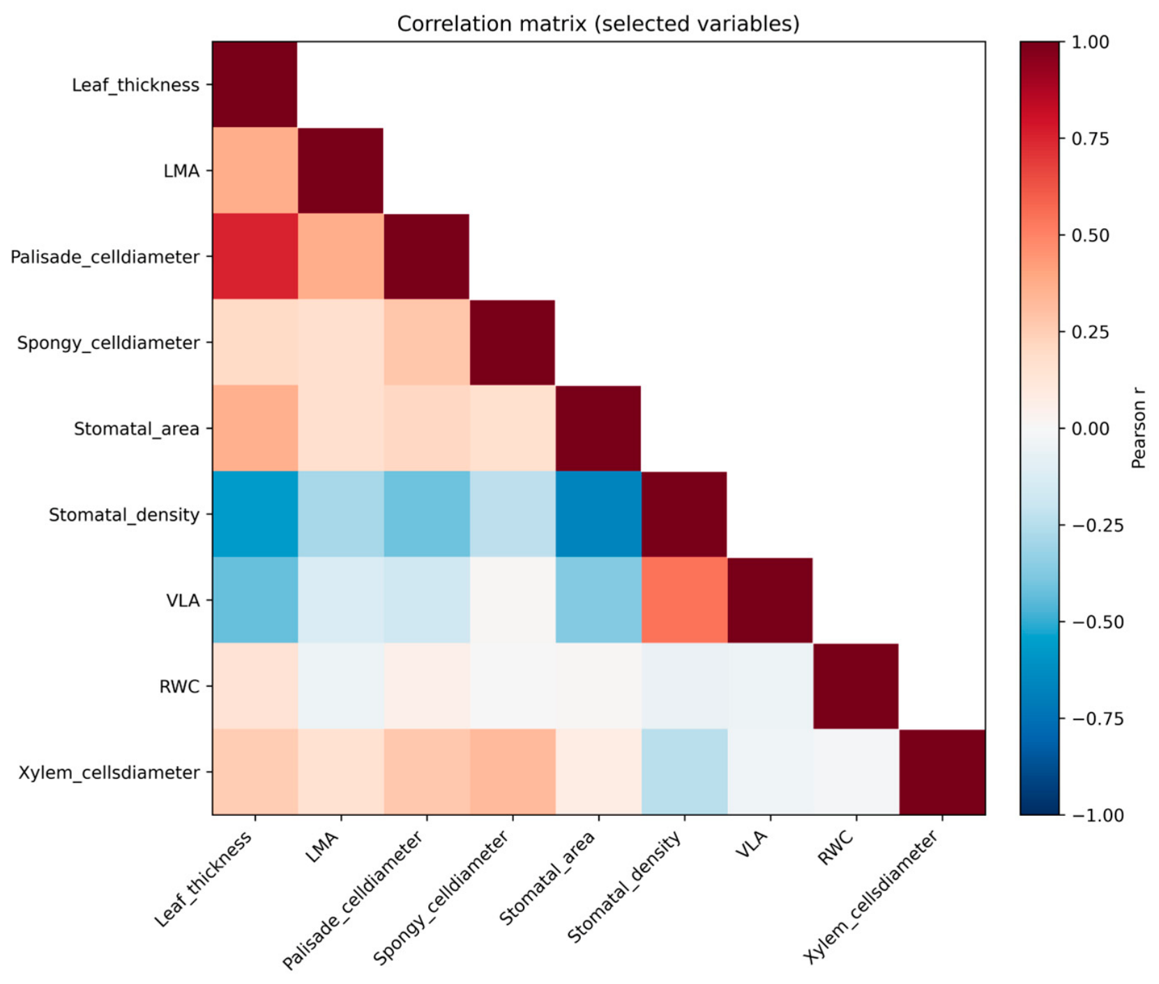
To explore relationships among variables, Pearson correlation coefficients were calculated using the same set of traits, and results were visualized using a correlation matrix.
In addition, a principal component analysis (PCA) was conducted to investigate multivariate patterns of trait variation. Prior to analysis, all variables were standardized (Z-scores) to remove scale effects. The PCA was performed using the correlation matrix, and mean values for each cultivar × ploidy combination were used for visualization purposes.
ChatGPT (version GPT-5.3) developed by OpenAI was used exclusively to improve the clarity and linguistic quality of the manuscript. Specifically, these tools were employed to refine the English language, including grammar, spelling, and overall readability, without modifying the scientific content, interpretation of results, or conclusions of the study. All analyses, data interpretation, and scientific decisions were carried out solely by the authors, who take full responsibility for the accuracy and integrity of the work.

3. Results

3.1. Multivariate Effects of Ploidy and Cultivar

A multivariate analysis of variance (MANOVA) revealed a significant effect of ploidy level on the overall set of leaf traits (Wilks’ λ = 0.110, F = 148.89, p < 0.001). Cultivar also showed a significant effect (Wilks’ λ = 0.186, F = 24.32, p < 0.001). A significant interaction between ploidy level and cultivar was also detected (Wilks’ λ = 0.097, F = 40.87, p < 0.001).

3.2. General Morphological Leaf Traits

The leaf morphological characteristics according to ploidy level and cultivar are shown in Table 1.
In ‘Gomera-1’, no significant differences were found in petiole length or thickness between ploidy levels. However, autotetraploids exhibited wider but shorter leaves than diploids. Leaf lamina thickness increased from 0.189 ± 0.005 mm in diploids to 0.238 ± 0.004 mm in autotetraploids, while midrib thickness increased from 1.22 ± 0.02 mm to 1.37 ± 0.02 mm. Leaf area was also significantly greater in autotetraploids (133 ± 8 cm2) than in diploids (104 ± 4 cm2). No significant differences were detected in LMA or RWC.
In ‘Kensington Pride’, autotetraploids showed longer petioles and substantially thicker leaf laminas and midribs than diploids. Although leaf and lamina lengths were reduced in autotetraploids, leaf width increased significantly. In addition, autotetraploids displayed higher LMA values (187 ± 3 g/m2) than diploids (151 ± 4 g/m2), indicating greater structural investment per unit leaf area.
In ‘Gomera-3’, autotetraploid individuals presented longer and thicker petioles, as well as significantly greater lamina and midrib thickness. Leaf width increased markedly, and leaf area expanded from 84.3 ± 5 cm2 in diploids to 107 ± 3 cm2 in autotetraploids. Tetraploids also exhibited higher fresh, saturated, and dry leaf weights. Furthermore, both LMA and RWC were significantly higher in autotetraploids, with LMA increasing from 168 ± 5 g/m2 to 190 ± 5 g/m2 and RWC rising from 96.8 ± 0.3% to 98.1 ± 0.4%.
Overall, autotetraploid plants showed higher values for several morphological traits, although differences varied among cultivars.

3.3. Vein Length per Unit Area (VLA)

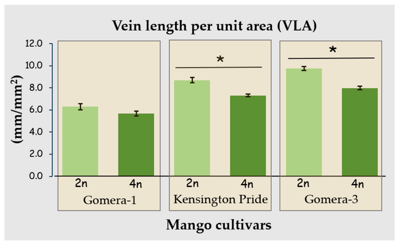
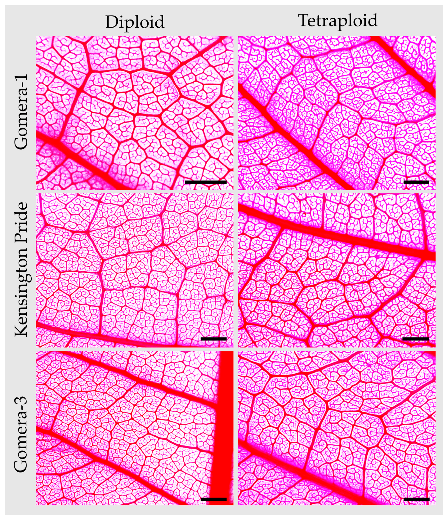
Differences in vein length per unit area were observed between diploid and autotetraploid leaves within each cultivar (Figure 1 and Figure 2). In ‘Gomera-1’, no significant differences were found between tetraploids and diploids. In ‘Kensington Pride’, VLA decreased significantly in tetraploids (7.3 ± 0.1 mm/mm2) relative to diploids (8.7 ± 0.22 mm/mm2). Similarly, in ‘Gomera-3’, VLA values were significantly lower in tetraploids (8 ± 0.2 mm/mm2) compared with diploids (9.8 ± 0.2 mm/mm2), indicating a marked reduction in vein density between ploidy levels. Differences in VLA between ploidy levels were cultivar-dependent.

3.4. Leaf Lamina Anatomical Traits

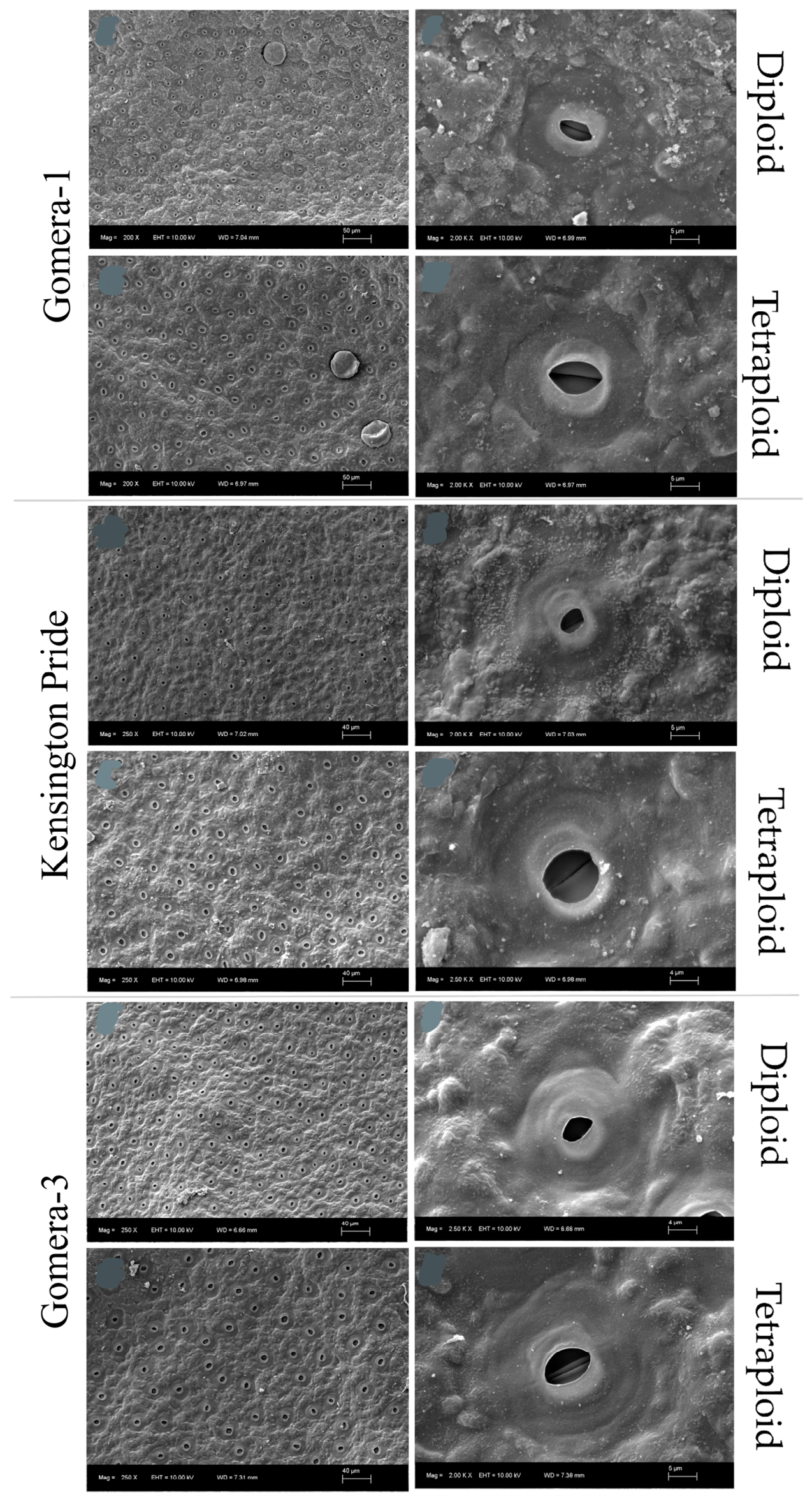
The leaf lamina anatomical characteristics according to ploidy level and cultivar are shown in Table 2. Representative scanning electron micrographs illustrating stomatal morphology in diploid and autotetraploid leaves are presented in Figure 3.
In ‘Gomera-1’, autotetraploids exhibited thicker cuticles on both leaf surfaces, larger adaxial and abaxial epidermal cells, and increased palisade and spongy parenchyma cell lengths compared with diploids. Stomatal density decreased markedly from 760 ± 13 to 438 ± 6 stomata mm−2, whereas guard and subsidiary cell lengths increased. Substomatal chamber area was also greater in autotetraploids.
In ‘Kensington Pride’, autotetraploids showed thicker adaxial cuticles, larger epidermal cells, and increased palisade and spongy parenchyma cell lengths. As observed in ‘Gomera-1’, stomatal density was significantly lower in autotetraploids, while guard and subsidiary cell lengths were greater. However, substomatal chamber area was slightly reduced in autotetraploids compared with diploids.
In ‘Gomera-3’, autotetraploids exhibited thicker adaxial cuticles and larger epidermal cells than diploids. The most pronounced difference was observed in palisade parenchyma cell length, which increased from 40.7 ± 0.6 µm in diploids to 97.6 ± 3.3 µm in autotetraploids. Spongy parenchyma cells were also larger. Stomatal density was markedly lower in autotetraploids (431 ± 9 stomata mm−2) than in diploids (673 ± 18 stomata mm−2), whereas guard and subsidiary cell lengths increased. Substomatal chamber area also expanded in autotetraploids.
Autotetraploids showed consistent differences in epidermal, mesophyll, and stomatal traits compared with diploids.

3.5. Leaf Midrib Anatomical Traits

The leaf midrib anatomical characteristics according to ploidy level and cultivar are shown in Table 3.
In ‘Gomera-1’, autotetraploids exhibited thicker abaxial cuticles, larger adaxial epidermal and sclerenchyma cells, and expanded midrib and phloem areas relative to diploids. Large and small xylem cells were also larger in autotetraploids, increasing from 39.5 ± 1.8 µm to 54.6 ± 2.6 µm and from 5.9 ± 0.3 µm to 9.0 ± 0.4 µm, respectively.
In ‘Kensington Pride’, autotetraploids showed larger adaxial epidermal and sclerenchyma cells, together with increased midrib, xylem, and phloem areas. Both large and small xylem cells were also significantly larger in autotetraploids than in diploids.
In ‘Gomera-3’, autotetraploids exhibited thicker cuticles on both leaf surfaces and larger adaxial epidermal cells. Sclerenchyma and parenchyma cell lengths increased markedly, while midrib, xylem, and phloem areas were substantially greater than in diploids. Large xylem cell length increased from 26.1 ± 1.2 µm in diploids to 43.9 ± 1.8 µm in autotetraploids.
Differences between ploidy levels were observed in vascular and supporting tissues across all cultivars.

3.6. Multivariate Analysis of Leaf Traits

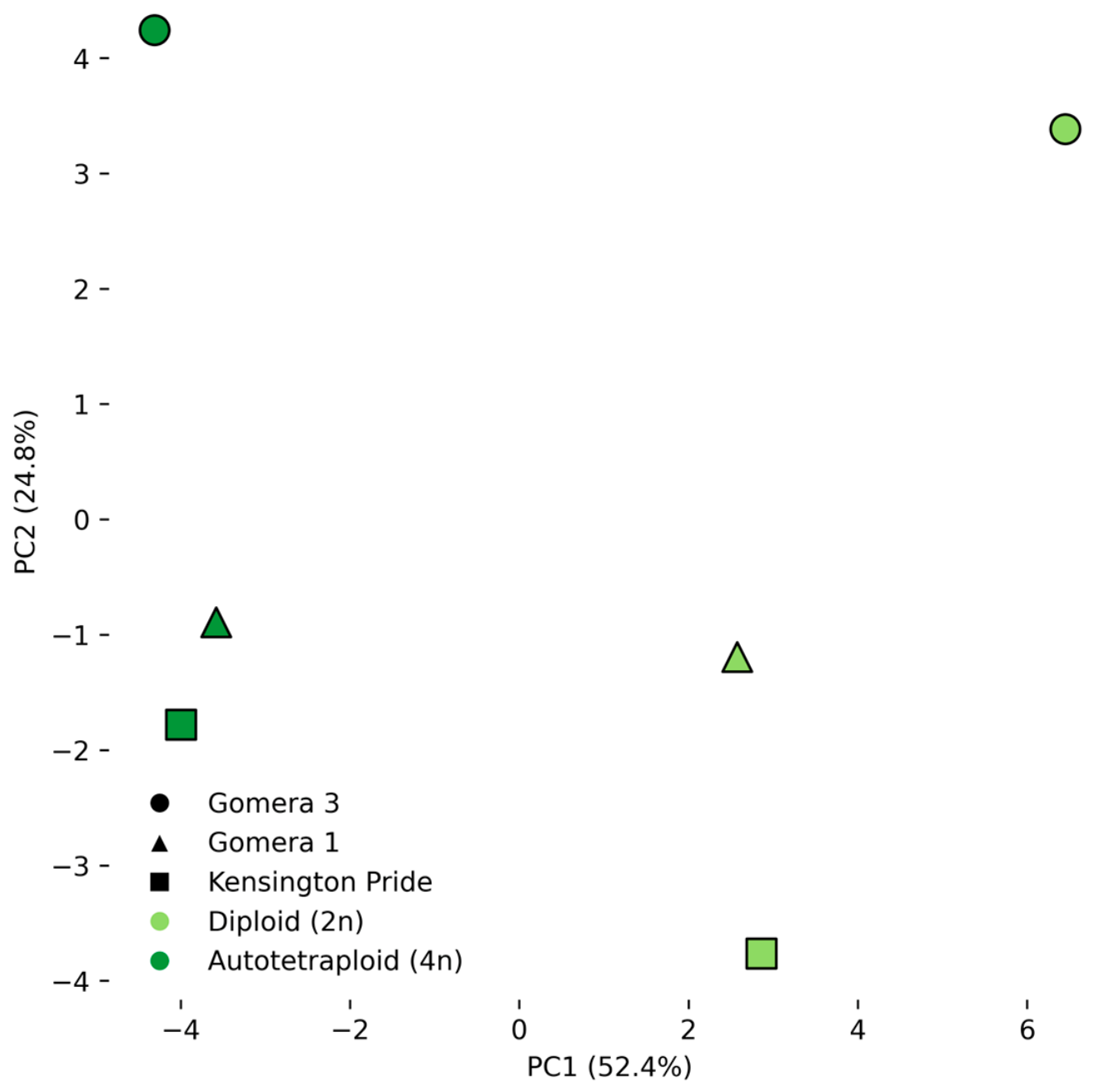
Principal component analysis (PCA) showed that the first two components accounted for the main sources of variation in the dataset (Figure 4). The first principal component (PC1) was mainly associated with variables related to leaf structural traits, including leaf thickness, palisade cell maximum length, spongy mesophyll cell maximum length, LMA, and xylem cell maximum length. The second principal component (PC2) was primarily associated with stomatal traits and relative water content (RWC). A separation between diploid and autotetraploid genotypes was observed along PC1. However, partial overlap among cultivars was also detected, indicating variability in the distribution of samples across both components.

3.7. Trait Correlations

Pearson correlation analysis showed significant relationships among leaf traits (Figure 5). Leaf thickness was positively correlated with LMA, palisade cell maximum length, spongy mesophyll cell maximum length, and xylem cell maximum length. Positive correlations were also observed among mesophyll-related traits and vascular variables. Stomatal traits showed contrasting relationships, with stomatal size negatively correlated with stomatal density. Vein length per area (VLA) showed negative correlations with several structural variables, including leaf thickness and mesophyll traits. Relative water content (RWC) showed moderate positive correlations with some structural variables and weak or non-significant relationships with others.

4. Discussion

This study aimed to determine how genome duplication modifies leaf structure in mangoes, and the results reveal clear and consistent differences between diploid and autotetraploid plants. Across all cultivars examined, autotetraploid mango trees displayed thicker laminas and broader midribs, confirming that genome duplication induces a structural upscaling of foliar tissues. Similar increases in leaf dimensions have previously been reported in autotetraploid mangoes [13] and in other woody polyploid species, including Citrus [17,18], Malus [19], Pyrus [20], and Vitis vinifera [21]. However, the response was not uniform among cultivars. While autotetraploids generally exhibited greater leaf width, leaf length tended to be reduced in some cultivars, and increases in total leaf area were cultivar-dependent. These results indicate that polyploidy consistently modifies leaf morphology in mangoes, although the magnitude and direction of specific responses depend on genetic background [22].
Autotetraploid leaves exhibited substantial enlargement of epidermal and mesophyll cells together with increased cuticle thickness. In particular, palisade parenchyma cell length increased by approximately 13–41% in ‘Gomera-1’ and ‘Kensington Pride’, and by more than 100% in ‘Gomera-3’, indicating a strong effect of genome duplication on tissue organization. Similar increases in cell size have previously been reported in mangoes [14,23] and other polyploid species [24]. Studies in Citrus have reported associations between thicker cuticles, enlarged epidermal cells, and responses to water deficit conditions [25,26,27,28]. However, because physiological traits were not directly evaluated in the present study, these anatomical features should be interpreted as structural characteristics that have been associated with drought-related responses in previous studies rather than as direct evidence of enhanced drought tolerance in the studied genotypes.
Tetraploid leaves exhibited the characteristic “polyploid stomatal syndrome”, a widely reported pattern in which stomata become larger while stomatal density decreases following genome duplication [29,30,31]. In the present study, stomatal density decreased by approximately 33–42% across cultivars, whereas guard cell length increased by 8–33%. Similar trends have been reported in mangoes [14,23], Citrus [17], and Malus [19]. Previous studies have reported associations between this anatomical configuration and differences in stomatal conductance and transpiration dynamics [32]. Nevertheless, because gas-exchange measurements were not performed, the functional implications of the stomatal changes observed here remain speculative and should be evaluated experimentally in future studies.
Leaf mass per area (LMA) increased significantly in autotetraploid ‘Gomera-3’ and ‘Kensington Pride’, consistent with previous observations in mangoes [14]. The magnitude of this increase ranged from approximately 13% to 24%, which falls within the range reported for several polyploid woody species [33,34]. Increased LMA reflects a greater structural investment per unit leaf area and is commonly associated with thicker or denser leaves. In the present study, higher LMA values were accompanied by increased leaf thickness and enlarged mesophyll cells, supporting the view that genome duplication promotes substantial modifications in leaf structural organization.
The midrib of tetraploid leaves exhibited larger xylem vessels and expanded phloem areas relative to diploids. Larger conduits are often associated with lower theoretical hydraulic resistance [23]. However, larger vessel diameters may also increase vulnerability to cavitation depending on species-specific vascular architecture [35]. Therefore, the functional consequences of the vascular modifications observed here cannot be inferred directly from anatomy alone. Nevertheless, the observed enlargement of vascular tissues is consistent with previous reports in mangoes [14,23] and indicates that polyploidy substantially alters vascular organization. Future studies combining anatomical and hydraulic measurements will be necessary to determine the physiological consequences of these structural changes.
Tetraploids consistently exhibited lower vein length per area (VLA) than diploids, a pattern expected from the geometric consequences of larger cells and thicker mesophyll tissues. Reduced vein density has been associated with both lower hydraulic investment and lower maximum water transport capacity [36,37]. At the same time, some studies have suggested that variation in vein density may influence hydraulic safety–efficiency relationships [38]. Consequently, VLA should be interpreted as a trait that may involve functional trade-offs rather than a unidirectional indicator of hydraulic performance. The lower VLA observed in tetraploid mangoes therefore reflects an important structural modification associated with genome duplication, although its physiological significance remains to be determined experimentally.
Although the observed differences were generally associated with ploidy level, the tetraploid materials analyzed here originated from seedling populations of polyembryonic cultivars rather than from experimentally induced chromosome duplication. Microsatellite analyses did not detect polymorphisms between diploid and autotetraploid materials within each cultivar. Nevertheless, a residual degree of genetic variation cannot be completely excluded and may have contributed to some of the cultivar-specific responses revealed by the significant ploidy × cultivar interaction. Therefore, the observed patterns should be interpreted primarily as ploidy-associated responses while acknowledging the potential contribution of residual genetic variation.

5. Conclusions

Genome duplication in mangoes induces consistent anatomical and structural modifications in leaves, including increased lamina thickness, enlargement of epidermal and mesophyll cells, expansion of vascular tissues, and reduced stomatal density. These changes were observed across all studied cultivars, although their magnitude varied depending on the genetic background. The combination of univariate and multivariate analyses indicates that polyploidy affects multiple leaf traits simultaneously and promotes coordinated variation among structural and physiological characteristics. Overall, these results demonstrate that genome duplication is associated with systematic changes in leaf architecture in mangoes and highlight the importance of considering genotype-dependent responses when evaluating the effects of polyploidy in this species.

Supplementary Materials

The following supporting information can be downloaded at https://www.mdpi.com/article/10.3390/agronomy16131226/s1. Table S1: Characterization of DNA fragments of different Mangifera indica cultivars using a sequencer (AB 3500) by capillary electrophoresis with an internal size standard (GS 600 LIZ v.2.0) at the Genomics Service of the General Research Support Services of the University of La Laguna (SEGAI). Ref. [39] is cited in the Supplementary Materials.; Figure S1: Transverse sections of the leaf lamina in diploid (2n) and tetraploid (4n) mango cultivars (Gomera-1, Kensington Pride and Gomera-3). Scale bar = 100 µm; Figure S2: Transverse sections of the leaf lamina in diploid (2n) and tetraploid (4n) mango cultivars (Gomera-1, Kensington Pride and Gomera-3). Scale bar = 100 µm.

Author Contributions

Conceptualization, M.A.R.-M., Á.M.G.-R. and M.J.G.-M.; methodology, M.A.R.-M., Á.M.G.-R., N.J.L.-M. and M.J.G.-M.; software, M.A.R.-M.; validation, M.A.R.-M. and M.J.G.-M.; formal analysis, M.A.R.-M.; investigation, M.A.R.-M., Á.M.G.-R., N.J.L.-M. and M.J.G.-M.; resources, M.J.G.-M.; data curation, M.A.R.-M. and M.J.G.-M.; writing—original draft preparation, M.A.R.-M.; writing—review and editing, M.A.R.-M., Á.M.G.-R., N.J.L.-M. and M.J.G.-M.; visualization, M.A.R.-M.; supervision, M.A.R.-M., Á.M.G.-R. and M.J.G.-M.; project administration, M.J.G.-M.; funding acquisition, M.J.G.-M. All authors have read and agreed to the published version of the manuscript.

Funding

This research was supported by “Proyectos de investigación del Consejo Asesor de Investigaciones Agrarias de la Consejería de Agricultura, Ganadería, Pesca y Soberanía Alimentaria, Gobierno de Canarias, CAIA 006 Selección y Mejora de Frutales tropicales y Subtropicales”.

Data Availability Statement

The data supporting the findings of this study are available from the corresponding author, Marcos Adrián Ruiz-Medina, upon request.

Acknowledgments

We would like to sincerely thank the staff of the Cueva del Polvo Experimental Farm of the Instituto Canario de Investigaciones Agrarias (ICIA) for their valuable support throughout this research work. The authors used ChatGPT (version GPT-5.3) developed by OpenAI exclusively to improve the clarity of the English language, grammar, and spelling of the manuscript.

Conflicts of Interest

The authors declare no conflicts of interest.

Abbreviations

The following abbreviations are used in this manuscript:
VLAVein length per unit area
LMALeaf mass per area
RWCRelative water content

References

  1. Jedele, S.; Hau, A.M.; von Oppen, M. An Analysis of the World Market for Mangos and Its Importance for Developing Countries. In Proceedings of the Conference on International Agricultural Research for Development, Göttingen, Germany, 8–10 October 2003. [Google Scholar]
  2. Maldonado-Celis, M.E.; Yahia, E.M.; Bedoya, R.; Landázuri, P.; Loango, N.; Aguillón, J.; Restrepo, B.; Guerrero Ospina, J.C. Chemical Composition of Mango (Mangifera indica L.) Fruit: Nutritional and Phytochemical Compounds. Front. Plant Sci. 2019, 10, 1073. [Google Scholar] [CrossRef] [PubMed]
  3. Marcillo-Parra, V.; Anaguano, M.; Molina, M.; Tupuna-Yerovi, D.S.; Ruales, J. Characterization and Quantification of Bioactive Compounds and Antioxidant Activity in Three Different Varieties of Mango (Mangifera indica L.) Peel from the Ecuadorian Region Using HPLC-UV/VIS and UPLC-PDA. NFS J. 2021, 23, 1–7. [Google Scholar] [CrossRef]
  4. Durán Zuazo, V.H.; Rodríguez Pleguezuelo, C.R.; Gálvez Ruiz, B.; Gutiérrez Gordillo, S.; García-Tejero, I.F. Water Use and Fruit Yield of Mango (Mangifera indica L.) Grown in a Subtropical Mediterranean Climate. Int. J. Fruit Sci. 2019, 19, 136–150. [Google Scholar] [CrossRef]
  5. Sillero-Medina, J.A.; González-Pérez, J.; Hueso-González, P.; González-Fernández, J.J.; Hormaza-Urroz, J.I.; Ruiz-Sinoga, J.D. Effect of Different Deficit Irrigation Regimens on Soil Moisture, Production Parameters of Mango (Mangifera indica L.), and Spectral Vegetation Indices in the Mediterranean Region of Southern Spain. Remote Sens. Appl. Soc. Environ. 2025, 37, 101415. [Google Scholar] [CrossRef]
  6. Léchaudel, M.; Lopez-Lauri, F.; Vidal, V.; Sallanon, H.; Joas, J. Response of the Physiological Parameters of Mango Fruit (Transpiration, Water Relations and Antioxidant System) to Its Light and Temperature Environment. J. Plant Physiol. 2013, 170, 567–576. [Google Scholar] [CrossRef] [PubMed]
  7. Nardini, A. Hard and Tough: The Coordination between Leaf Mechanical Resistance and Drought Tolerance. Flora 2022, 288, 152023. [Google Scholar] [CrossRef]
  8. Heredia, A.; González-Moreno, A.; Benítez, J.J.; Domínguez, E. Plant Cuticles Exhibit Significant Mid-Infrared Emissivity in the Atmospheric Windows. Int. J. Mol. Sci. 2025, 26, 9917. [Google Scholar] [CrossRef] [PubMed]
  9. Tamayo-Ordóñez, M.C.; Espinosa-Barrera, L.A.; Tamayo-Ordóñez, Y.J.; Ayil-Gutiérrez, B.; Sánchez-Teyer, L.F. Advances and Perspectives in the Generation of Polyploid Plant Species. Euphytica 2016, 209, 1–22. [Google Scholar] [CrossRef]
  10. Beaulieu, J.M.; Leitch, I.J.; Patel, S.; Pendharkar, A.; Knight, C.A. Genome Size Is a Strong Predictor of Cell Size and Stomatal Density in Angiosperms. New Phytol. 2008, 179, 975–986. [Google Scholar] [CrossRef] [PubMed]
  11. Maherali, H.; Walden, A.E.; Husband, B.C. Genome Duplication and the Evolution of Physiological Responses to Water Stress. New Phytol. 2009, 184, 721–731. [Google Scholar] [CrossRef] [PubMed]
  12. Arumuganathan, K.; Earle, E.D. Nuclear DNA Content of Some Important Plant Species. Plant Mol. Biol. Rep. 1991, 9, 208–218. [Google Scholar] [CrossRef]
  13. Galán-Saúco, V.; Grajal-Martín, M.J.; Galván, D.F.; Torres, A.C.; Juárez, J.; Navarro, L. Occurrence of spontaneous tetraploid nucellar mango plants. HortScience 2001, 36, 755–757. [Google Scholar] [CrossRef]
  14. Barceló-Anguiano, M.; Holbrook, N.M.; Hormaza, J.I.; Losada, J.M. Changes in Ploidy Affect Vascular Allometry and Hydraulic Function in Mangifera indica Trees. Plant J. 2021, 108, 541–554. [Google Scholar] [CrossRef] [PubMed]
  15. Perera-Castro, A.V.; Hernández, B.; Grajal-Martín, M.J.; González-Rodríguez, Á.M. Assessment of Drought Stress Tolerance of Mangifera indica L. Autotetraploids. Agronomy 2023, 13, 277. [Google Scholar] [CrossRef]
  16. AgroCabildo. Available online: https://www.agrocabildo.org/agrometeorologia_estaciones_detalle.asp?id=44 (accessed on 30 November 2025).
  17. Romero-Aranda, R.; Bondada, B.R.; Syvertsen, J.P.; Grosser, J.W. Leaf Characteristics and Net Gas Exchange of Diploid and Autotetraploid Citrus. Ann. Bot. 1997, 79, 153–160. [Google Scholar] [CrossRef]
  18. Guerra, D.; Schifno Wittmann, M.T.; Schwarz, S.F.; de Souza Dutra, P.V.D.; Gonzatto, M.P.; Weiler, R.L. Comparação Entre Porta-Enxertos Cítricos Diploides e Tetraploides: Caracterização Morfológica e Avaliação Do Crescimento. Bragantia 2014, 73, 1–7. [Google Scholar] [CrossRef]
  19. Xue, H.; Zhang, B.; Tian, J.R.; Chen, M.M.; Zhang, Y.Y.; Zhang, Z.H.; Ma, Y. Comparison of the Morphology, Growth and Development of Diploid and Autotetraploid ‘Hanfu’ Apple Trees. Sci. Hortic. 2017, 225, 277–285. [Google Scholar] [CrossRef]
  20. Liu, R.; Gao, C.; Jin, J.; Wang, Y.; Jia, X.; Ma, H.; Zhang, Y.; Zhang, H.; Qi, B.; Xu, J. Induction and Identification of Tetraploids of Pear Plants (Pyrus bretschneideri and Pyrus betulaefolia). Sci. Hortic. 2022, 304, 111322. [Google Scholar] [CrossRef]
  21. Catalano, C.; Abbate, L.; Motisi, A.; Crucitti, D.; Cangelosi, V.; Pisciotta, A.; di Lorenzo, R.; Carimi, F.; Carra, A. Autotetraploid Emergence via Somatic Embryogenesis in Vitis vinifera Induces Marked Morphological Changes in Shoots, Mature Leaves, and Stomata. Cells 2021, 10, 1336. [Google Scholar] [CrossRef] [PubMed]
  22. Madlung, A. Polyploidy and Its Effect on Evolutionary Success: Old Questions Revisited with New Tools. Heredity 2013, 110, 99–104. [Google Scholar] [CrossRef] [PubMed]
  23. Fonollá, A.; Hormaza, J.I.; Losada, J.M. Foliar Pectins and Physiology of Diploid and Autotetraploid Mango Genotypes under Water Stress. Plants 2023, 12, 3738. [Google Scholar] [CrossRef] [PubMed]
  24. Li, M.; Zhang, C.; Hou, L.; Liu, X.; Zhao, H.; Pang, X.; Bo, W.; Li, Y. Differences in Leaf Cuticular Wax Induced by Whole-Genome Duplication in Autotetraploid Sour Jujube. Hortic. Plant J. 2024, 10, 66–76. [Google Scholar] [CrossRef]
  25. Allario, T.; Brumos, J.; Colmenero-Flores, J.M.; Iglesias, D.J.; Pina, J.A.; Navarro, L.; Talon, M.; Ollitrault, P.; Morillon, R. Tetraploid Rangpur Lime Rootstock Increases Drought Tolerance via Enhanced Constitutive Root Abscisic Acid Production. Plant Cell Environ. 2013, 36, 856–868. [Google Scholar] [CrossRef] [PubMed]
  26. Oliveira, T.M.; Yahmed, J.B.; Dutra, J.; Maserti, B.E.; Talon, M.; Navarro, L.; Ollitraut, P.; da S. Gesteira, A.; Morillon, R. Better Tolerance to Water Deficit in Doubled Diploid ‘Carrizo Citrange’ Compared to Diploid Seedlings Is Associated with More Limited Water Consumption. Acta Physiol. Plant. 2017, 39, 204. [Google Scholar] [CrossRef]
  27. Wei, T.; Wang, Y.; Xie, Z.; Guo, D.; Chen, C.; Fan, Q.; Deng, X.; Liu, J.H. Enhanced ROS Scavenging and Sugar Accumulation Contribute to Drought Tolerance of Naturally Occurring Autotetraploids in Poncirus trifoliata. Plant Biotechnol. J. 2019, 17, 1394–1407. [Google Scholar] [CrossRef] [PubMed]
  28. Hussain, S.; Sohail, H.; Noor, I.; Ahmad, S.; Ejaz, S.; Ali, M.A.; Khalid, M.F. Physiological and Biochemical Determinants of Drought Tolerance in Tetraploid vs. Diploid Sour Orange Citrus Rootstock. J. Hortic. Sci. Biotechnol. 2023, 98, 772–785. [Google Scholar] [CrossRef]
  29. Normasiwi, S.; Efendi, M.; Rahman, W.; Hafiizh, E.A.; Ermayanti, T.M.; Lelono, R.A.; Yunarto, N. Growth, Stomata and Trichome Characteristics of Diploid and Tetraploid Artemisia annua L. Plants. IOP Conf. Ser. Earth Environ. Sci. 2021, 762, 012022. [Google Scholar] [CrossRef]
  30. Yao, P.Q.; Chen, J.H.; Ma, P.F.; Xie, L.H.; Cheng, S.P. Stomata Variation in the Process of Polyploidization in Chinese Chive (Allium tuberosum). BMC Plant Biol. 2023, 23, 595. [Google Scholar] [CrossRef] [PubMed]
  31. Kramer, L.J.; Bamberg, J. Comparing Methods of Ploidy Estimation in Potato (Solanum) Species. Am. J. Potato Res. 2019, 96, 419–426. [Google Scholar] [CrossRef]
  32. Koziara-Ciupa, M.; Trojak-Goluch, A. The Effect of Polyploidisation on the Physiological Parameters, Biochemical Profile, and Tolerance to Abiotic and Biotic Stresses of Plants. Agronomy 2025, 15, 1918. [Google Scholar] [CrossRef]
  33. Wright, I.J.; Reich, P.B.; Westoby, M.; Ackerly, D.D.; Baruch, Z.; Bongers, F.; Cavender-Bares, J.; Chapin, T.; Cornelissen, J.H.C.; Diemer, M.; et al. The Worldwide Leaf Economics Spectrum. Nature 2004, 428, 821–827. [Google Scholar] [CrossRef] [PubMed]
  34. Poorter, H.; Niinemets, Ü.; Poorter, L.; Wright, I.J.; Villar, R. Causes and Consequences of Variation in Leaf Mass per Area (LMA): A Meta-Analysis. New Phytol. 2009, 182, 565–588. [Google Scholar] [CrossRef] [PubMed]
  35. Hacke, U.G.; Sperry, J.S.; Pockman, W.T.; Davis, S.D.; McCulloh, K.A. Trends in Wood Density and Structure Are Linked to Prevention of Xylem Implosion by Negative Pressure. Oecologia 2001, 126, 457–461. [Google Scholar] [CrossRef] [PubMed]
  36. Wójcik, D.; Marat, M.; Marasek-Ciołakowska, A.; Klamkowski, K.; Buler, Z.; Podwyszyńska, M.; Tomczyk, P.P.; Wójcik, K.; Treder, W.; Filipczak, J. Apple Autotetraploids—Phenotypic Characterisation and Response to Drought Stress. Agronomy 2022, 12, 161. [Google Scholar] [CrossRef]
  37. Losada, J.M.; Blanco-Moure, N.; Fonollá, A.; Martínez-Ferrí, E.; Hormaza, J.I. Hydraulic Tradeoffs Underlie Enhanced Performance of Polyploid Trees under Soil Water Deficit. Plant Physiol. 2023, 192, 1821–1835. [Google Scholar] [CrossRef] [PubMed]
  38. Sack, L.; Scoffoni, C. Leaf Venation: Structure, Function, Development, Evolution, Ecology and Applications in the Past, Present and Future. New Phytol. 2013, 198, 983–1000. [Google Scholar] [CrossRef] [PubMed]
  39. Viruel, M.A.; Escribano, P.; Barbieri, M.; Ferri, M.; Hormaza, J.I. Fingerprinting, embryo type and geographic differentiation in mango (Mangifera indica L., Anacardiaceae) with microsatellites. Mol. Breed. 2005, 15, 383–393. [Google Scholar] [CrossRef]
Figure 1. Vein length per unit area (VLA) in diploid (2n) and autotetraploid (4n) plants of three mango cultivars (‘Gomera-1’, ‘Kensington Pride’, and ‘Gomera-3’). Values represent mean ± SE based on 30 vein measurements per cultivar and ploidy level (n = 30; 10 measurements from each of three leaves). Asterisks indicate significant differences between ploidy levels within each cultivar (p < 0.05).
Agronomy 16 01226 g001
Figure 2. Leaf venation patterns of diploid (2n) and autotetraploid (4n) mango cultivars (‘Gomera-1’, ‘Kensington Pride’, and ‘Gomera-3’) stained with safranin. Images were adjusted for brightness and contrast to enhance vein visibility. Scale bar = 2 mm.
Agronomy 16 01226 g002
Figure 3. Scanning electron micrographs showing stomatal density and morphology on the abaxial leaf surface of diploid (2n) and autotetraploid (4n) mango cultivars (‘Gomera-1’, ‘Kensington Pride’, and ‘Gomera-3’).
Agronomy 16 01226 g003
Figure 4. Principal component analysis (PCA) of leaf morpho-anatomical and physiological traits in diploid (2n) and autotetraploid (4n) mango cultivars. Each point represents the mean value of each cultivar × ploidy combination. Symbols indicate cultivars and colors represent ploidy levels. The first principal component (PC1) is mainly associated with leaf structural traits, including leaf thickness, LMA, mesophyll cell dimensions, and xylem cell maximum length, whereas the second principal component (PC2) is primarily associated with stomatal traits and relative water content (RWC).
Agronomy 16 01226 g004
Figure 5. Pearson correlation matrix among selected leaf morpho-anatomical and physiological traits in mangoes. Color intensity represents the strength and direction of correlations (r values), ranging from −1 (blue) to +1 (red). Only the lower triangle of the matrix is shown for clarity. Variables included leaf thickness, leaf mass per area (LMA), palisade cell maximum length, spongy mesophyll cell maximum length, stomatal area, stomatal density, vein length per area (VLA), relative water content (RWC), and xylem cell maximum length.
Agronomy 16 01226 g005
Table 1. Mean ± SE values of morphological leaf traits in diploid (2n) and tetraploid (4n) plants of three mango cultivars (‘Gomera-1’, ‘Kensington Pride’, and ‘Gomera-3’). Values are based on 30 leaf measurements per cultivar and ploidy level (n = 30).
General TraitsGomera-1 2nGomera-1 4nKensington Pride 2nKensington Pride 4nGomera-3 2nGomera-3 4n
Petiole length (cm)5.06 ± 0.304.42 ± 0.223.98 ± 0.315.02 ± 0.30 *3.15 ± 0.153.70 ± 0.12 *
Petiole thickness (mm)2.94 ± 0.073.13 ± 0.092.85 ± 0.072.99 ± 0.082.45 ± 0.042.84 ± 0.04 *
Lamina length (cm)24.6 ± 0.4 *21.6 ± 0.628.6 ± 0.9 *24.9 ± 0.521.3 ± 0.621.6 ± 0.5
Leaf length (cm)29.5 ± 0.6 *26.1 ± 0.732.5 ± 0.1 *29.9 ± 0.624.3 ± 0.725.2 ± 0.5
Leaf width (cm)6.40 ± 0.148.30 ± 0.24 *7.17 ± 0.177.83 ± 0.15 *5.98 ± 0.147.60 ± 0.13 *
Leaf lamina thickness (mm)0.19 ± 0.010.24 ± 0.00 *0.16 ± 0.000.22 ± 0.00 *0.18 ± 0.000.30 ± 0.00 *
Leaf midrib thickness (mm)1.22 ± 0.021.37 ± 0.02 *1.13 ± 0.011.63 ± 0.02 *1.10 ± 0.011.93 ± 0.06 *
Area (cm2)104 ± 4133 ± 8 *138 ± 7128 ± 584.3 ± 5107 ± 3 *
LMA (g/m2)175 ± 5179 ± 6151 ± 4187 ± 3 *168 ± 5190 ± 5 *
RWC (%)97.6 ± 0.297.8 ± 0.396.1 ± 1.297.0 ± 0.296.8 ± 0.398.1 ± 0.4 *
* Asterisks indicate significant differences between ploidy levels within each cultivar (p < 0.05).
Table 2. Mean ± SE values of leaf lamina anatomical traits in diploid (2n) and autotetraploid (4n) mango cultivars (‘Gomera-1’, ‘Kensington Pride’, and ‘Gomera-3’). Values are based on 50 anatomical measurements per cultivar and ploidy level (n = 50), obtained from transverse sections of three sampled leaves.
Leaf Lamina TraitsGomera-1
2n
Gomera-1
4n
Kensington Pride 2n Kensington Pride 4n Gomera-3 2nGomera-3 4n
Adaxial cuticle thickness (µm)1.72 ± 0.042.60 ± 0.13 *1.82 ± 0.072.68 ± 0.14 *2.05 ± 0.083.84 ± 0.27 *
Abaxial cuticle thickness (µm)1.21 ± 0.011.54 ± 0.06 *1.89 ± 0.11.93 ± 0.092.07 ± 0.122.40 ± 0.11
Adaxial epidermis cell maximum length (µm)14.6 ± 0.418.5 ± 0.7 *12.6 ± 0.514.8 ± 0.4 *14.7 ± 0.420.8 ± 0.7 *
Abaxial epidermis cell maximum length (µm)10.3 ± 0.212.8 ± 0.4 *8.89 ± 0.211.3 ± 0.3 *8.04 ± 0.212.7 ± 0.4 *
Palisade parenchyma cell maximum length (µm)63.2 ± 1.171.7 ± 2.0 *48.1 ± 2.267.9 ± 2.7 *40.7 ± 0.697.6 ± 3.3 *
Spongy parenchyma cell maximum length (µm)20.2 ± 0.723.3 ± 0.9 *16.7 ± 0.726.3 ± 1 *16.8 ± 0.920.3 ± 0.9 *
Stomatal density (stomata/mm2)760 ± 13 *438 ± 6631 ± 15 *415 ± 12673 ± 18 *431 ± 9
Guard cells length (µm)12.3 ± 0.313.3 ± 0.2 *9.7 ± 0.312.9 ± 0.2 *9.9 ± 0.213.2 ± 0.3 *
Subsidiary cells maximum length (µm)12.6 ± 0.215.0 ± 0.2 *12.4 ± 0.415.0 ± 0.3 *10.9 ± 0.214.0 ± 0.4 *
Substomatal chamber area (µm2)255 ± 9299 ± 5 *169 ± 1 *145 ± 1170 ± 2205 ± 8 *
* Asterisks indicate significant differences between ploidy levels within each cultivar (p < 0.05).
Table 3. Mean ± SE values of leaf midrib anatomical traits in diploid (2n) and autotetraploid (4n) mango cultivars (‘Gomera-1’, ‘Kensington Pride’, and ‘Gomera-3’). Values are based on 50 anatomical measurements per cultivar and ploidy level (n = 50), obtained from transverse sections of three sampled leaves.
Leaf Midrib TraitsGomera-1 2nGomera-1 4nKensington Pride 2n Kensington Pride 4n Gomera-3 2nGomera-3 4n
Adaxial cuticle thickness (µm)4.91 ± 0.154.82 ± 0.156.25 ± 0.26.67 ± 0.23.77 ± 0.115.15 ± 0.21 *
Abaxial cuticle thickness (µm)3.35 ± 0.114.28 ± 0.17 *4.55 ± 0.134.80 ± 0.143.64 ± 0.164.64 ± 0.24 *
Adaxial epidermis cell maximum length (µm)13.2 ± 0.315.7 ± 0.4 *9.1 ± 0.212.6 ± 0.4 *11.4 ± 0.215.2 ± 0.4 *
Abaxial epidermis cell maximum length (µm)9.3 ± 0.39.5 ± 0.27.5 ± 0.37.7 ± 0.210.0 ± 0.510.4 ± 0.3
Sclerenchyma cell maximum length (µm)15.0 ± 0.719.1 ± 0.7 *15.3 ± 0.619.6 ± 0.8 *14.8 ± 0.620.5 ± 0.8 *
Parenchyma cell maximum length (µm)15.3 ± 0.616.4 ± 0.717.3 ± 0.719.2 ± 0.918.6 ± 0.927.2 ± 1.2 *
Midrib area (mm2)0.99 ± 0.061.30 ± 0.05 *1.12 ± 0.051.68 ± 0.08 *1.10 ± 0.042.04 ± 0.13 *
Xylem area (mm2)0.34 ± 0.030.33 ± 0.030.34 ± 0.020.54 ± 0.04 *0.22 ± 0.010.49 ± 0.04 *
Phloem area (mm2)0.17 ± 0.010.38 ± 0.02 *0.23 ± 0.020.32 ± 0.02 *0.40 ± 0.020.66 ± 0.05 *
Large xylem cell maximum length (µm)39.5 ± 1.854.6 ± 2.6 *43.0 ± 1.654.2 ± 1.6 *26.1 ± 1.243.9 ± 1.8 *
Small xylem cell maximum length (µm)5.9 ± 0.39.0 ± 0.4 *6.9 ± 0.313.1 ± 0.7 *11.4 ± 0.711.2 ± 0.5
Phloem cells maximum length (µm)10.34 ± 0.410.77 ± 0.3210.91 ± 0.3410.41 ± 0.397.99 ± 0.3611.04 ± 0.46 *
* Asterisks indicate significant differences between ploidy levels within each cultivar (p < 0.05).
Disclaimer/Publisher’s Note: The statements, opinions and data contained in all publications are solely those of the individual author(s) and contributor(s) and not of MDPI and/or the editor(s). MDPI and/or the editor(s) disclaim responsibility for any injury to people or property resulting from any ideas, methods, instructions or products referred to in the content.