Article

Identification of Monilinia yunnanensis Causing Brown Rot in Korla Fragrant Pear and Evaluation of Bacillus siamensis PL55 as a Biocontrol Agent

1
College of Horticulture and Forestry, Tarim University, 705 Hongqiao South Road, Alar 843300, China
2
National-Local Joint Engineering Laboratory for High-Efficiency Cultivation and Deep Processing of Characteristic Fruit Trees in Southern Xinjiang, 1490 East Tarim Avenue, Alar 843300, China
3
Key Laboratory of Integrated Pest Management in Southern Xinjiang, 1490 East Tarim Avenue, Alar 843300, China
*
Authors to whom correspondence should be addressed.
Agronomy 2025, 15(12), 2678; https://doi.org/10.3390/agronomy15122678
Submission received: 12 October 2025 / Revised: 12 November 2025 / Accepted: 20 November 2025 / Published: 21 November 2025
(This article belongs to the Section Pest and Disease Management)

Abstract

To identify the primary pathogen responsible for brown rot in mature Korla fragrant pears and to screen for effective biocontrol bacterial strains, a research team collected 30 samples of brown rot-infected fruits and 84 samples from healthy trees (including branches, leaves, and fruits) from three Korla pear cultivation areas in Korla, Aksu, and Kashgar between September and October 2024. Investigation into the severity of the disease revealed the local field incidence rate of brown rot ranged from 17% to 31%. From the 30 disease samples, 23 morphologically identical fungal strains were isolated, with 13 strains isolated from Korla City (56.5%), 6 from Aksu City (26.1%), and 4 from Tumushuk (17.4%), all detected in infected fruits. Pathogenicity tests were conducted using both inoculation by wounding with mycelium and spray inoculation experiments to study the pathogens’ effect on brown rot in Korla fragrant pears. From the 84 samples of healthy trees, 55 bacterial strains were isolated, and the antagonistic bacteria’s inhibitory effect on the isolated pathogens was determined using the dual-culture method. The pathogen was identified by morphological characteristics and phylogenetic analysis based on multi-locus sequencing (ITS-TUB2-Lcc2). Antagonistic bacterial strains were identified through morphological observation and 16S rDNA sequencing. The results demonstrated that the pathogen isolated from the diseased Korla fragrant pear tissues was identified as Monilinia yunnanensis. Among the biocontrol antagonistic bacteria isolated from the branches, bark, and leaves of healthy Korla fragrant pear trees, Bacillus siamensis exhibited significant inhibitory effects against the pathogen, with an in vitro inhibition rate of (88.18 ± 3.43)%.

1. Introduction

Korla Fragrant Pear (Pyrus sinkiangensis Yü), belonging to the subtribe Pomindeae of the family Rosaceae and the genus Pyrus, is a highly region-specific premium fruit [1]. Due to its unique fragrance and outstanding quality, it has become a renowned geographical indicator product in China [2]. It is found exclusively in the southern region of Xinjiang, with its main production area located in Korla City of Bayingolin Mongol Autonomous Prefecture [3]. The pear is named for its strong aroma. However, in recent years, climate change and altered precipitation patterns have led to an increased incidence of brown rot disease in Korla Fragrant Pears, severely affecting fruit quality and posing significant challenges to the sustainable development of the pear industry [4].
According to existing literature, three primary pathogens causing fruit brown rot in China have been identified: Monilia yunnanensis, Monilinia fructicola, and Monilia mumecola [5,6]. These pathogens widely infect various fruit trees, including apples, peaches, nectarines, pears, apricots, and cherries [7,8]. Although different brown rot fungi can infect fruit trees and cause brown rot, they display certain differences in geographical distribution and host preference [9]. Among them, Hu et al. [10] conducted phylogenetic analysis and morphological observations of isolates from diseased peaches and identified three Monilinia species—M. fructicola, M. mumecola, and a newly described species named Monilinia yunnanensis. Similarly, Zhu et al. [11] characterized 247 Monilinia isolates from major Chinese apple and pear regions by morphology and ITS, TUB and lcc2 sequencing, identifying Monilia yunnanensis (77%), M. polystroma (20%) and M. fructicola (3%). Additionally, various studies have documented the identification of brown rot pathogens across different fruit species and regions. For example, Lei et al. [12] identified M. fructicola as a pathogen for plum brown rot, and Jiang et al. [13] reported M. yunnanensis as the pathogen for apple brown rot in Gansu’s Jingning and Yuzhong counties.
Brown rot is a globally distributed fungal disease, causing severe yield losses and significantly reducing the quality of stone fruits [14]. It typically affects nearly mature fruits, first appearing as light brown, circular water-soaked spots on the fruit surface, which rapidly expand into soft rot that results in complete fruit decay within about 10 days. Concentric bands of gray-white to gray-brown fuzzy mold clusters (2–3 mm in size) form around the central lesion. Infected fruit flesh becomes spongy and elastic, eventually dehydrating to form black mummified fruit [15]. Currently, chemical control remains the primary management strategy for peach brown rot, but its long-term use poses risks to ecological safety and accelerates pathogen resistance [16,17]. This highlights the urgent need for efficient, environmentally friendly, and sustainable control measures, particularly for brown rot affecting Korla Fragrant Pears.
The causal agent of brown rot varies significantly depending on geographical location, host type, and climate conditions. While M. fructicola and M. laxa dominate in temperate regions, Hu et al. [5] identified M. yunnanensis as a new species primarily distributed across southern China, but which also affects apple and pear orchards in northern provinces, including Xinjiang. Limited studies have investigated the genetic diversity of M. yunnanensis populations and explored effective biocontrol agents against this species, particularly in pear-growing regions. M. yunnanensis presents unique challenges compared to other Monilinia species due to its distinct pathogenicity and ecological adaptation to host tissues. In light of this, biological control methods—which are pollution-free and environmentally friendly—are receiving increasing attention as sustainable alternatives for plant disease management.
Among biocontrol agents, Bacillus strains are considered ideal due to their simple nutritional requirements, efficient growth, production of diverse secondary metabolites with antimicrobial activity (e.g., lipopeptides, volatile organic compounds), and ability to form stress-resistant spores that enhance survival in hostile agricultural environments [18]. Furthermore, endophytic Bacillus strains, directly isolated from host plants, offer advantages such as superior colonization and compatibility, which improve their potential for long-term biocontrol efficacy. Zhou et al. [19] demonstrated that volatile organic compounds (e.g., benzothiazole) from Bacillus subtilis CF-3 effectively suppressed Monilinia fructicola, enhancing peach resistance by inhibiting fungal enzymes and inducing plant defense responses. Similarly, Meltem Avan’s study indicated significant effects of Bacillus subtilis TV-6F, in biocontrol formulation, against M. laxa, including an 84.06% control efficacy on peach blossoms [20]. Despite these promising advancements, research on antagonistic bacterial screening against M. yunnanensis, particularly for regional hosts like Korla Fragrant Pears, remains scarce. Further studies that expand our understanding of Monilinia–host–microbe interactions and develop tailored biological control strategies for this pathogen are urgently needed.
This study isolated and purified pathogens from infected Korla Fragrant Pear fruits, assessed their pathogenicity, and identified the species through morphological and molecular biological methods. To screen for more efficient antagonistic strains against brown rot in Korla Fragrant Pears, we targeted isolated brown rot pathogens, screening for microbiological strains from branches, bark, and leaves that exhibit strong inhibition of the disease. The aim is to determine their biocontrol potential and provide resources for the biological management of brown rot in Korla Fragrant Pears.

2. Materials and Methods

2.1. Collection of Diseased Samples and Isolation, Purification and Pathogenicity Tests of Pathogens

From September to October 2024, diseased Korla fragrant pear (Pyrus sinkiangensis) fruits exhibiting brown rot were collected from orchards in three major Korla pear production areas of Xinjiang Uygur Autonomous Region: Korla City (86°05′39″ E, 41°40′45″ N), Aral City, Aksu Prefecture (81°17′22″ E, 40°32′53″ N) and Tumushuke City, Third Division, Kashgar (79°08′24″ E, 39°49′34″ N). A preliminary survey indicated that brown rot incidence in the sampled orchards ranged from 17% to 31%. A total of 30 diseased fruits were collected. Using the tissue isolation method [21], tissue fragments measuring 3 mm × 3 mm × 3 mm were cut from the junction between healthy and diseased areas of the fruits. These fragments were surface sterilized with 1% sodium hypochlorite for 15 s, followed by 75% ethanol for 2 min, and rinsed three times with sterile water. The treated tissues were inoculated onto sterile PDA plates and incubated in the dark at 26 °C. Due to the difficulty of brown rot fungi sporulation on PDA, the growing colonies were streak-purified repeatedly for three rounds until single colonies were obtained [22]. Ultimately, from the 30 disease samples, 23 morphologically identical fungal strains were isolated, with 13 strains isolated from Korla City (56.5%), 6 from Aksu City (26.1%), and 4 from Tumushuk (17.4%). Pathogenicity verification was conducted using the wound-mycelium inoculation method [23]. After incubating the isolates on PDA plates at 26 °C for 8 days, 5 mm agar plugs were taken from the colony edges using a sterile puncher and kept for use. The surfaces of the pear fruits were disinfected, and 10 mL sterile syringes were used to wound the fruit surfaces. The fungal agar plugs were then placed on the wounds, while sterile PDA plugs served as controls. The inoculated fruits were placed in disposable sterile fresh-keeping boxes to maintain humidity and incubated in the dark at 26 °C. Disease symptoms were observed, and lesion diameters were measured at 3, 4, and 5 days post-inoculation. From the symptomatic fruits, conidia were collected and suspended in sterile water to prepare a conidial suspension (1 × 10⁶ spores/mL). Non-wounded in vitro pathogenicity tests were performed using the spray inoculation method [24]. Korla fragrant pear fruits were collected from the pear orchard at Tarim University, surface sterilized, and used in the tests. Fruits in the treatment group were sprayed with the conidial suspension, while fruits in the control group were sprayed with sterile water. Both groups of fruits were placed in sterile fresh-keeping boxes to maintain humidity and were incubated in the dark at 26 °C. Each treatment group included 5 fruits, with three replicates per group. Disease symptoms were observed daily and photographed, and measurements of incidence rates were recorded for each treatment at 4, 5, and 6 days post-inoculation. Pathogens were re-isolated from the symptomatic fruits and compared with the original isolates to verify Koch’s postulates. One strain was selected as the representative strain and designated as strain YY4 for further research.

2.2. Morphological Identification of Pathogens

Morphological identification of the representative strain YY4 following the morphological identification method described by Lane [25] and Wei Jingchao’s Manual of Fungal Identification [26], a detailed observation and identification of the representative strain YY4 were conducted. A small amount of mycelium was taken from the edge of the purified strain YY4 and placed in the center of a PDA plate. This was incubated at a constant temperature of 26 °C in the dark for 3 days. Subsequently, using a puncher (Φ = 5 mm), a section of the colony edge was taken and transferred to the center of a new PDA plate for further incubation at 26 °C in darkness. The growth rate of the colony was recorded daily. Every other day, observations were made on the shape of the colony on PDA medium, changes in color from the front and reverse sides of the colony, and whether conidiophores formed on the surface of the medium during the later stages of growth. If conidiophores were produced, their time of appearance, size, shape, and arrangement were documented. Microscopic morphological characteristics of the hyphae, spores, and other structures were examined using a digital biological microscope (E 200) [27]. The size of spores was measured with a digital microscope, and Excel was used to calculate the averages and standard deviations. This detailed morphological analysis provided crucial information for the accurate identification of strain YY4.

2.3. Molecular Identification of Pathogens

After activating the test strain culture for 3 days, a puncher (Φ = 5 mm) was used to take agar plugs from the edge of the fungal colonies. These plugs were then transferred to PDA plates pre-laid with cellophane and incubated at a constant temperature of 26 °C in the dark for 5 days. Mycelia cultured on the cellophane surface for 5 days were collected into 2 mL centrifuge tubes [28]. Genomic DNA was extracted using the method described by Paterson Et Al. [29]. The strain YY4 underwent PCR amplification using three sets of primers: ITS primers (ITS1/ITS4) [30], TUB2 primers (Bt2a/Bt2b) [31], and Lcc2 primers LacaF (5′-GCATCTGCATCTGCTATTCCAGCT-3′) and LacaR (5′-CTTACCGCCACCAACGCAGTT-3′) [23]. The PCR amplified product (55 μL) was then subjected to 1% agarose gel electrophoresis (U: 110V) for detection. Subsequently, the PCR products were sent to Shanghai Bioengineering Co., Ltd. (Shanghai, China) for sequencing. The obtained gene sequences were analyzed using BLAST (https://blast.ncbi.nlm.nih.gov/Blast.cgi?PROGRAM=blastn&PAGE_TYPE=BlastSearch&LINK_LOC=blasthome, accessed on 16 July 2025) in the NCBI database. Reference sequences of the same genus were downloaded from the GenBank database, and a multi-gene phylogenetic tree (ITS-TUB2-Lcc2) was constructed using the neighbor-joining (NJ) method in PhyloSuite. This approach provided detailed genetic insights and helped confirm the molecular identity of the isolated strain YY4.

2.4. Isolation and Screening of Biocontrol Strains

Branches, barks, and leaves of the Korla fragrant pear tree were used for extracting endophytic biocontrol bacteria, as detailed in Table 1. The Korla fragrant pear fruit samples were collected from the fragrant pear orchard at Tarim University (coordinates: 81°17′23″ E, 40°32′53″ N) to conduct pathogenicity testing of brown rot disease in Korla fragrant pears.
Using the dilution isolation method, the collected samples were washed with clean water and cut into pieces of approximately 0.5 cm2 in size. Those were then disinfected with 75% alcohol for 1 min and 1% sodium hypochlorite for 3 min. The samples were rinsed three times with sterile distilled water, and then thoroughly dried using sterile absorbent paper. The final rinse with sterile distilled water was plated to test the effectiveness of the disinfection. The disinfected samples were ground into a paste using a sterile mortar, and diluted with sterile distilled water to obtain a homogenous tissue solution. This solution was further diluted in four parts to concentrations of 10−1, 10−2, 10−3, and 10−4, which were then spread evenly on LB medium plates using a sterile spreader, with four replicates for each concentration. The plates were incubated in an incubator at 28 °C for 3 days. Two rounds of streak purification were performed using colony color and morphology for testing antifungal activities. The isolated strains were stored at −80 °C for further use. Quantitative pathogenic bacteria were placed in Erlenmeyer flasks containing Luria–Bertani liquid medium and incubated on a shaker at 28 °C and 220 rpm for 24 h. Following incubation, the bacterial suspension was sprayed onto the surface of the fruit, and the condition of the fruit was observed daily. Plant and host safety tests were conducted to verify the safety and compatibility of the isolated bacteria. Using M. yunnanensis as the target strain, the antagonistic ability of isolated bacteria was determined through the plate confrontation method to identify potential biocontrol strains. M. yunnanensis was inoculated at the center of a PDA plate, and the endophytic bacteria were inoculated 2.5 cm away from the edge of the fungal disk. The plate inoculated only with M. yunnanensis served as a control. Those were incubated in darkness at 25 °C for 3 days. Each treatment was replicated four times. Once the control colonies grew fully, their diameters were measured using the cross-streak method to calculate the inhibition rate. Hyphae from the edge of M. yunnanensis observed in the confrontation test, compared with normally growing edge hyphae of M. yunnanensis, were observed under an optical microscope to assess their morphological changes.

2.5. Identification of Biocontrol Strains

Molecular Biology Identification: The DNA of the biocontrol strain was extracted using a bacterial DNA extraction kit. PCR amplification was performed using universal bacterial primers 27F (5′-AGAGTTTGATCCTGGCTCAG-3′) and 1492R (5′-GGTTACCTTGTTACGACTT-3′). The PCR reaction system was set as follows: initial denaturation at 95 °C for 3 min, 30 cycles of denaturation at 95 °C for 45 s, annealing at 55 °C for 45 s, and extension at 72 °C for 90 s; followed by a final extension at 72 °C for 5 min. The amplified products were detected by 1% agarose gel electrophoresis, excised from the gel, purified, and then sent to Sangon Biotech Co., Ltd., (Shanghai, China) for sequencing. The sequencing results were compared for homology with the GenBank and NCBI databases, and finally used to construct a phylogenetic tree in the MEGA 11 software.

2.6. Statistical Analysis

All experiments were conducted with a minimum of three replicates. Statistical analyses were performed using SPSS 27.0 software. The experimental design involved a completely randomized design (CRD) to evaluate the treatment effects. Prior to applying Duncan’s new multiple range test for assessing the significance of differences between groups, key statistical assumptions were verified, including homogeneity of variances and normality of data distribution. Homogeneity of variances was tested using Levene’s test, while normality was assessed using the Shapiro–Wilk test. Data that did not meet these assumptions underwent appropriate transformations to ensure validity of the analyses. For all statistical tests, differences were considered significant at a level of p < 0.05.

3. Results

3.1. Symptoms Characterization of Brown Rot in Korla Fragrant Pears

During the fruit maturation period, under conditions of high humidity, the initial symptoms of brown rot in Korla fragrant pears appear centered around the infection sites as light brown to brown patches on the fruit surface. The lesions rapidly expand, causing the fruit tissue to soften and rot (Figure 1A). At the lesion site, clusters of concentric ring-shaped disease characteristics are formed, consisting of conidiophores and conidia of the pathogen, often accompanied by a fermented odor, rendering the fruit inedible and significantly affecting fruit yield and quality. Pathogenicity tests conducted on 30 fungal isolates from the diseased tissue of Korla fragrant pears showed the following results: Mycelium inoculation: After incubation in a humidified environment for 3 days, the inoculated fruits developed yellow-brown lesions and sparse mycelial masses (Figure 1B). By the fourth day, the lesions expanded, and mycelial masses increased (Figure 1C). After 5 days, the lesions expanded to cover half of the fruit, with the mycelial masses forming a ring pattern (Figure 1D). The fruits in the control group showed no signs of disease (Figure 1E). From Table 2, it can be observed that the lesion area increased over time. Conidial suspension inoculation: After humidified incubation at 26 °C for 4 days, multiple yellow-brown lesions appeared on the surface of all inoculated fruits (Figure 2A). By the fifth day, the lesions expanded further and white mycelium began to appear (Figure 2B). After 6 days, the lesions covered half of the fruit’s surface, with mycelium forming concentric ring patterns (Figure 2C). The control group continued to show no signs of disease (Figure 2D), On the seventh day, the disease incidence of fruits inoculated with spore suspension reached 86.67%. Infected fruit tissues at the boundary between diseased and healthy areas were subjected to the same tissue isolation method to re-isolate the pathogen strains. The colony morphology, The hyphal structure, and spore characteristics of the re-isolated strains were found to be identical to those of the original inoculated strains. Based on Koch’s postulates, it was confirmed that the 30 isolated fungal strains were the causal pathogens of brown rot in Korla fragrant pears. One strain was selected as the representative strain and designated as strain YY4 for further research.

3.2. Identification of Monilinia yunnanensis as the Causal Agent

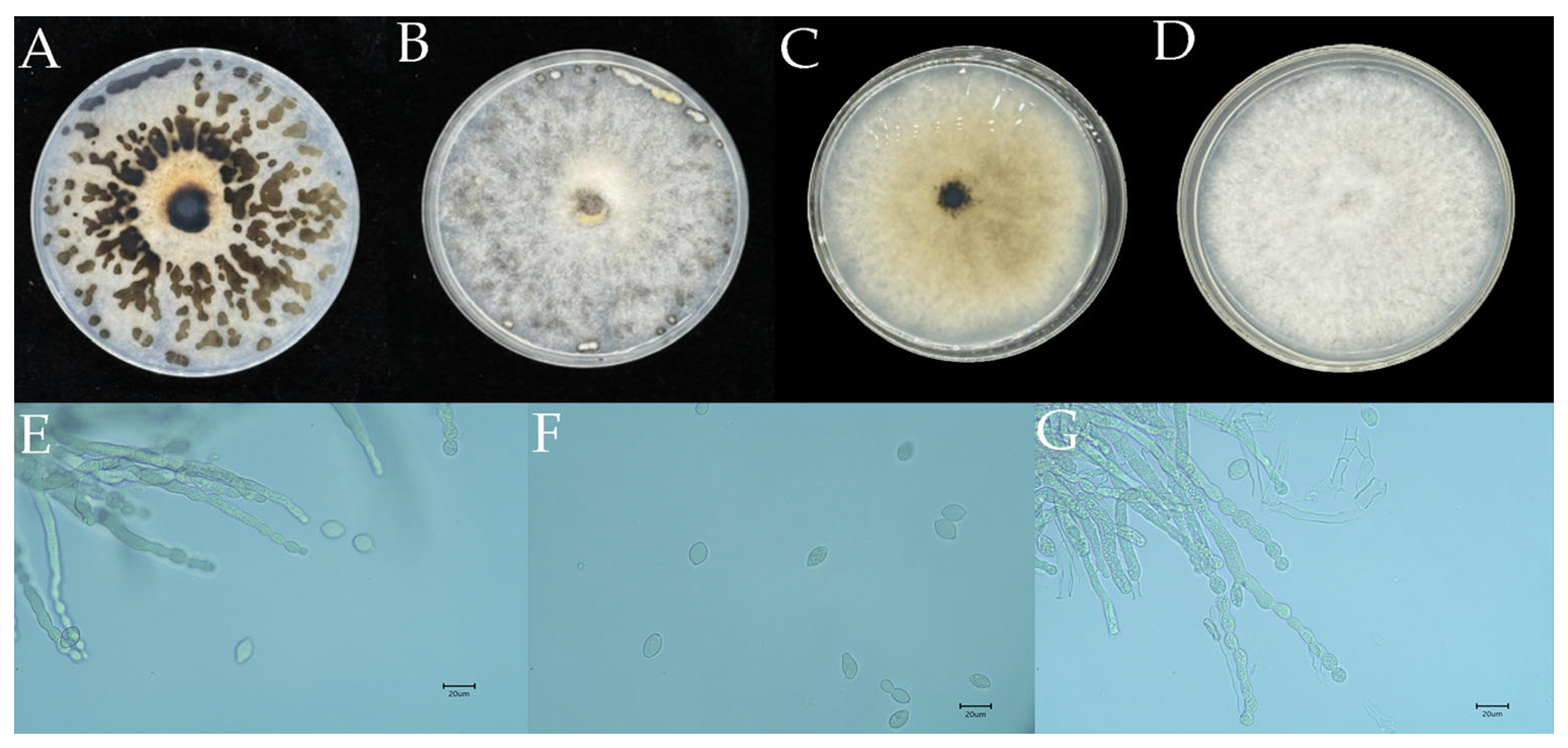
Strain YY4 was cultured on PDA medium at 26 °C in the dark. After 8 days, the colony fully covered a 9 cm Petri dish. The colony edges were neat, with grayish-white hyphae, and the aerial mycelia were closely attached to the surface of the dish. After 10 days, the reverse side of the colony turned dark brown, while the edges remained grayish-white (Figure 3A,B). By 15 days, the colony began to produce deep brown substrates of varying sizes and shapes (Figure 3C,D). Morphological analysis of the pathogen was conducted using a light microscope to observe the features of the spore suspension. The hyphae were colorless, transparent, and septate. The conidiophores originated from the hyphae and were bead-like in appearance, with either unbranched or dichotomous branching, and the branches exhibited acute angles (Figure 3E,F). The conidia were colorless and unicellular, arranged in a bead-like pattern, and were ovoid or lemon-shaped. The conidia ranged in size from 7.81 μm to 10.65 μm × 12.39 μm to 19.88 μm (Figure 3G). The morphological characteristics of strain YY4 were highly consistent with the described features of Monilia yunnanensis [32].

3.3. Confirmation of Monilinia yunnanensis Through Molecular Phylogenetics

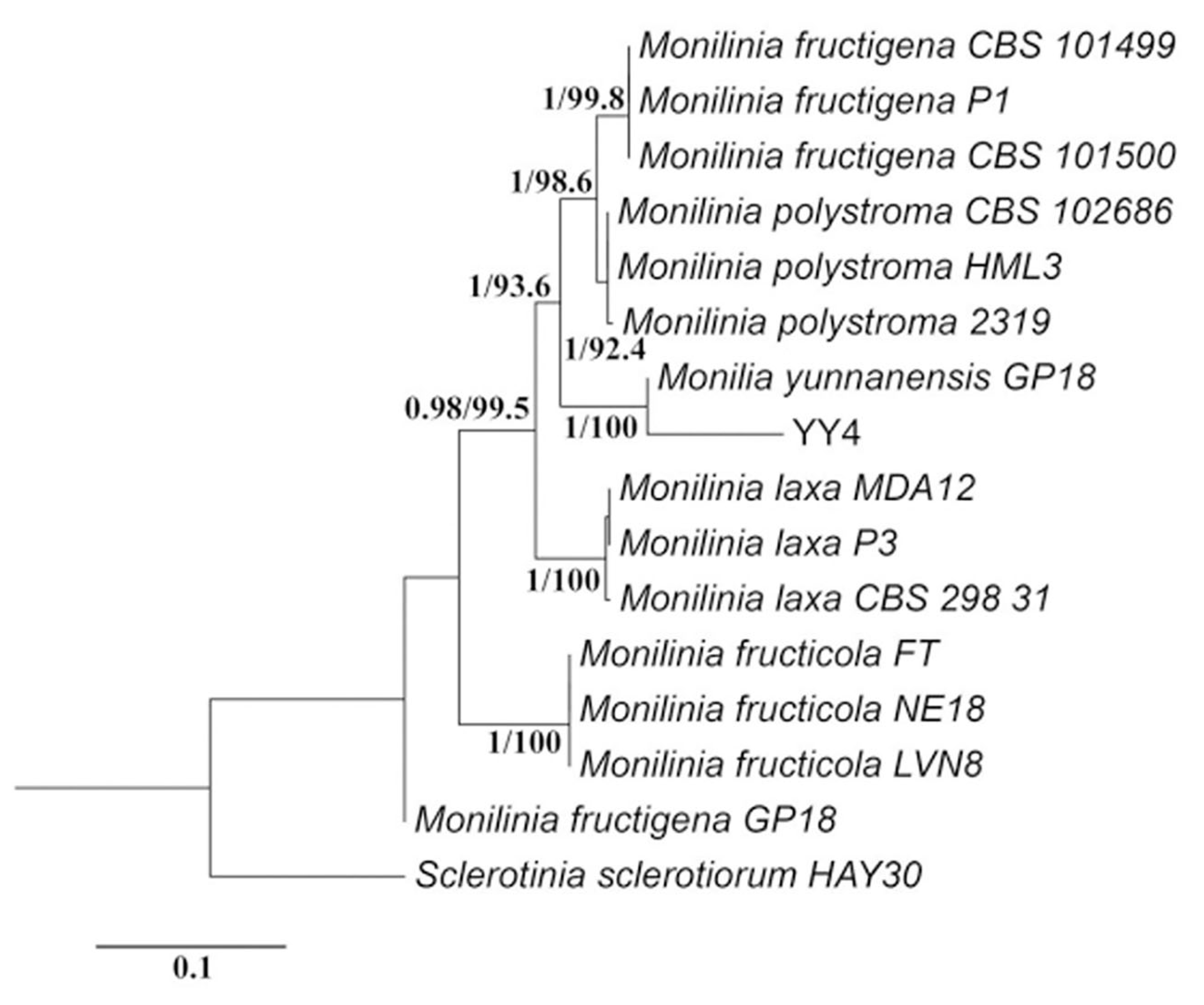
Using three pairs of primers (ITS, TUB2, and Lcc2), the DNA sequences of strain YY4 were amplified and subjected to bidirectional sequencing. The lengths of the ITS, TUB2, and Lcc2 sequences for YY4 were determined to be 517 bp, 474 bp, and 817 bp, respectively, with GenBank accession numbers PX128871, PX130397, and PX130396. In the combined ITS-TUB2-Lcc2 phylogenetic tree, strain YY4 clustered closely with the Monilia yunnanensis GP18 strain, thereby confirming the identification of this pathogen as Monilia yunnanensis. This molecular evidence firmly establishes strain YY4 as the causal agent responsible for brown rot in Korla fragrant pears (Figure 4).

3.4. Selection and Isolation of Effective Biocontrol Strain PL55

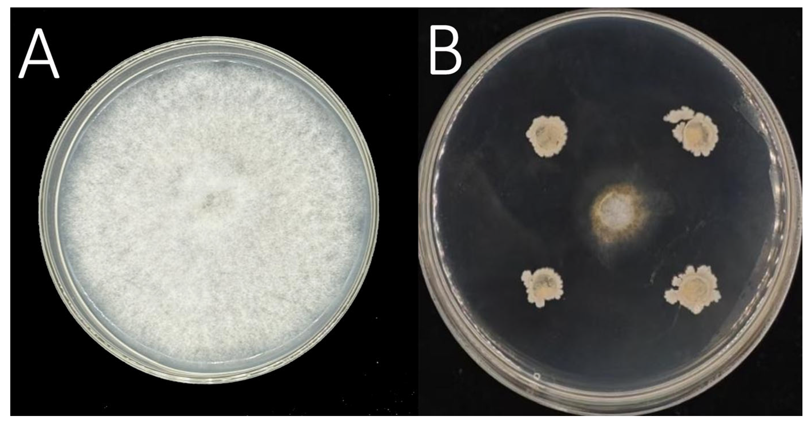
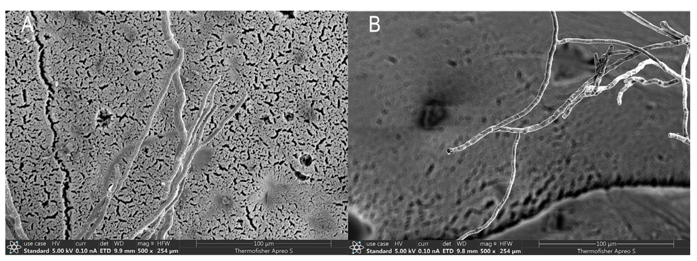
Using the dilution isolation method, a total of 55 strains were isolated from 84 Korla fragrant pear fruit tissue samples. By screening the strains using the plate confrontation method, it was found that 26 of these strains exhibited varying degrees of antagonistic effects against the brown rot pathogen M. yunnanensis (Table 3). The PL55 strain, which showed the most significant inhibitory effect, was selected for further study with an inhibition rate as high as 88.18% (Figure 5). Microscopic observation revealed that the hyphae of the pathogen treated with the PL55 strain had increased branching, swelling, and shortened internodes (Figure 6). These observations may suggest possible interference in the normal growth and development of the pathogen’s hyphae by the PL55 strain, although further investigations are needed to elucidate the exact mechanisms behind these effects. Additionally, plant and host safety tests were performed and confirmed that the PL55 strain did not cause any adverse effects on the host plant, indicating its safety and compatibility for potential biocontrol applications.

3.5. Molecular Confirmation of Bacillus siamensis Strain PL55

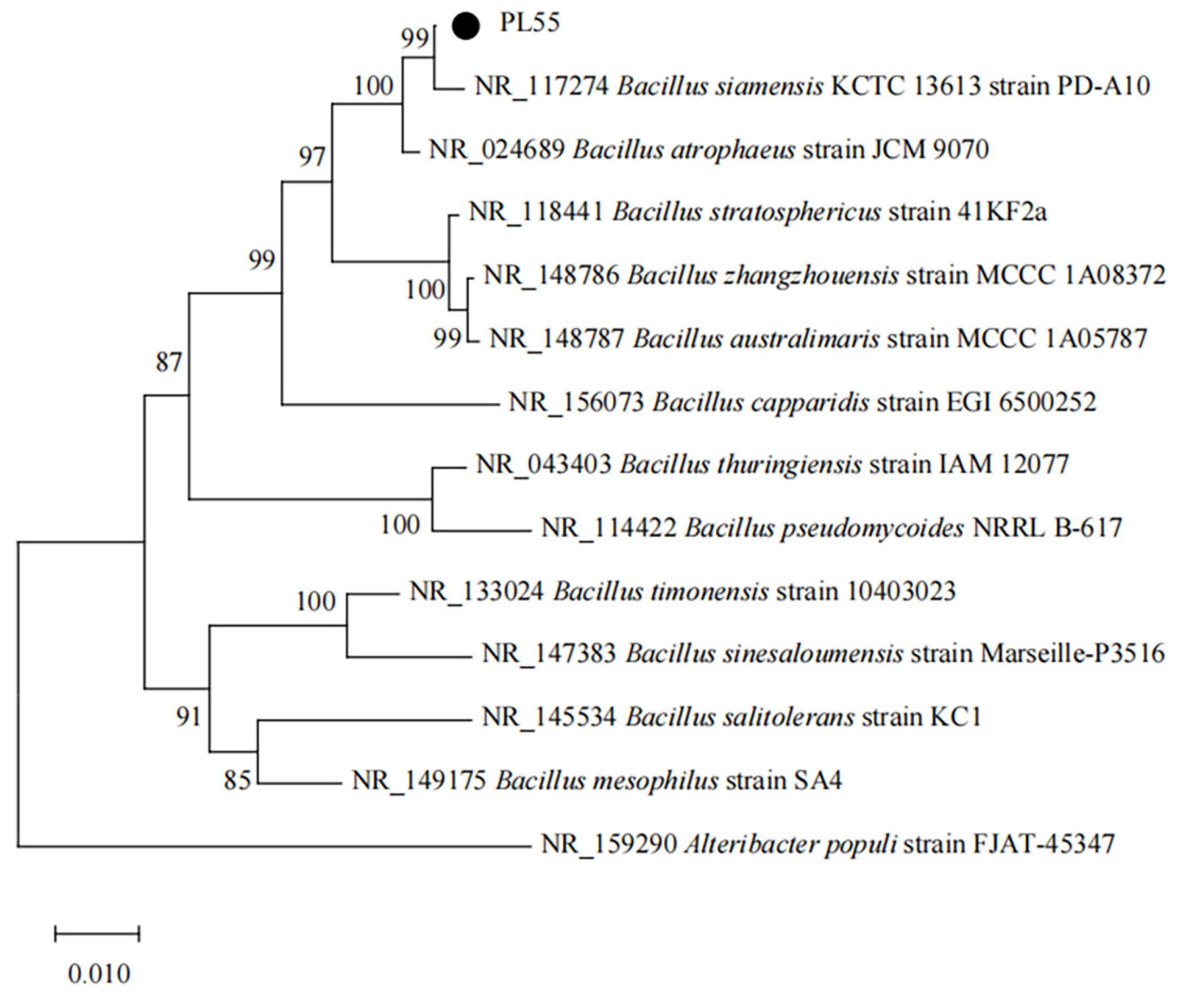
Based on the 16S rDNA gene sequence analysis of the PL55 strain, a phylogenetic tree was constructed using the Alteribacter populi strain FJAT-45347 (GenBank accession: NR_159290) as the outgroup (Figure 7). In this phylogenetic tree, the PL55 strain clustered in the same highly supported branch as the Bacillus siamensis KCTC 13613 strain PD-A10 (GenBank accession: NR_117274), with the 16S rDNA sequence similarity between them being higher than 99%. The above results indicate that the PL55 strain has a very close phylogenetic relationship with Bacillus siamensis. Taking into account its morphological characteristics and physiological biochemical analysis results, the PL55 strain was identified as Bacillus siamensis. Note that the 16S rDNA sequence of this strain has already been submitted to the NCBI GenBank database, with the accession number PV751088.1.

4. Discussion

This study provides novel insights into the pathogen causing brown rot in Korla fragrant pears by confirming Monilinia yunnanensis as the predominant causal agent through comprehensive morphological and molecular analyses (ITS, TUB2, Lcc2 genes with phylogenetic tree construction), which aligns with previous studies on the diversity of Monilinia spp. [11]. However, discrepancies with earlier reports from Zhou Fang [33] and Niu et al. [21], which identified M. fructigena as the major pathogen in Shanxi Province and some regions of Xinjiang, highlight the importance of assessing how ecological conditions and host factors contribute to the distribution of Monilinia species. Such differences underscore M. yunnanensis’ likely regional adaptation and the necessity of tailored research on its specific ecological and pathogenic traits.
The genus Bacillus offers promising opportunities for biological control in agricultural disease management owing to its efficient inhibition of plant pathogens, diverse secondary metabolites, including volatile organic compounds (VOCs), and its ability to survive adverse environmental conditions by forming stress-resistant spores [34]. Extensive prior research has identified various biocontrol Bacillus strains (e.g., Bacillus amyloliquefaciens, Bacillus tequilensis) antagonistic to M. fructicola [35] and Bacillus velezensis inhibiting M. fructigena [36], demonstrating the broad efficacy of Bacillus species against Monilinia spp.
Our identification of Bacillus siamensis strain PL55 as an antagonist of M. yunnanensis, exhibiting a significant mycelial inhibition rate of 88.18 ± 3.43%, highlights its potential as a biocontrol agent specifically for M. yunnanensis. While this result provides a critical first step, it aligns with previous findings suggesting that Bacillus VOCs may play important roles in plant pathogen suppression through diverse mechanisms. However, this study did not investigate VOCs from the PL55 strain, and therefore, no conclusions can be drawn regarding the involvement of VOCs or specific mechanisms in this process. For example, Song et al. [37] demonstrated that VOCs from Bacillus subtilis KRS015 exert inhibitory effects against Verticillium dahliae by activating the pathogen’s reactive oxygen species metabolism genes. Similarly, VOCs from Bacillus subtilis Y8 significantly reduced the spore germination rate of Curvularia lunata while destroying the ultrastructure of its mycelia, thereby reducing its virulence [38]. Furthermore, Huang et al. [39] showed that Bacillus atrophaeus XW2, isolated from healthy poplar leaves, could inhibit Colletotrichum gloeosporioides spore germination through VOC production, achieving an inhibition rate of 60.2%. Consistent with these examples, B. siamensis PL55 induced morphological abnormalities in M. yunnanensis hyphae, including increased branching, swelling, and internode shrinkage.
Although our findings demonstrate the biocontrol potential of B. siamensis against M. yunnanensis, it is essential to recognize the limitations of this study. Firstly, in vitro assays alone cannot fully replicate the complexity of field environments; the real-world efficacy of PL55 needs further validation through in planta and field experiments. We propose conducting controlled field trials in Korla fragrant pear orchards with randomized block designs, where parameters such as lesion diameter, disease incidence, and fruit quality will be assessed under different treatment applications. These trials will also involve biosafety testing to evaluate the effects of PL55 on non-target organisms and natural ecosystems. Secondly, utilizing a single pathogen isolate and a single biocontrol strain limits our ability to generalize these findings to other pathogen populations and geographic regions. Future research should focus on exploring the population diversity of M. yunnanensis and conducting multi-strain, multi-location trials.
Another critical focus lies in elucidating the biocontrol mechanisms of PL55. This includes investigating its capacity to produce secondary metabolites such as VOCs, lipopeptides (e.g., surfactin, iturin, fengycin), and extracellular enzymes that may contribute to its antagonistic efficacy. Furthermore, advanced methodologies like LC-MS/MS or thin-layer chromatography could be employed to identify specific antimicrobial compounds, while functional assays and transcriptomic analyses could shed light on the interaction between VOCs and M. yunnanensis. Exploring the dynamics of these mechanisms under varying environmental conditions is essential for developing effective and sustainable biocontrol strategies. In summary, this study takes crucial steps toward characterizing M. yunnanensis and identifying biological agents like Bacillus siamensis PL55 for its control. However, much work remains in terms of validation, safety assessments, and mechanistic understanding before the practical implementation of these findings in agricultural production systems.

5. Conclusions

This study identified Monilinia yunnanensis as the causal agent of brown rot in Korla fragrant pears and demonstrated that Bacillus siamensis strain PL55, isolated from healthy pear phyllosphere, exhibited potent antagonistic activity (88.18 ± 3.43% inhibition) against this pathogen in vitro. Importantly, this work establishes a novel biocontrol resource specifically adapted to the unique ecological conditions of Korla pear orchards, offering a foundation for developing region-specific, sustainable disease management strategies that could reduce reliance on chemical fungicides. The integration of such biocontrol agents into commercial pear production systems could contribute to more sustainable agricultural practices while maintaining fruit quality and marketability. This research thus provides both immediate practical value and establishes a framework for future investigations into microbiome-based disease management in fruit crop systems.

Author Contributions

Conceptualization, Q.X., Y.T., Z.L. and J.Y.; methodology, Q.X., Z.W., J.Y. and Z.L.; software, Q.X., Z.L., Y.T. and L.W.; validation, L.W., H.F. and Q.X.; formal analysis, Q.X.; investigation, Q.X.; resources, Q.X.; data curation, Q.X. and Y.T.; writing—original draft preparation, Q.X., L.W., H.F. and Z.W.; writing—review and editing, Q.X., Y.T., L.W. and H.F.; visualization, Q.X., Z.L. and J.Y.; project administration, Q.X.; funding acquisition, L.W., H.F. and Z.W. All authors have read and agreed to the published version of the manuscript.

Funding

This research was funded by the Joint Funds of the National Natural Science Foundation of China (U1903206); the Guided Science and Technology Program of Xinjiang Production and Construction Corps (2024ZD078); and the Scientific Research and Innovation Project for Postgraduates of Tarim University (TDGRI2024022).

Data Availability Statement

The data are available upon reasonable request to the corresponding author.

Conflicts of Interest

The authors declare no conflicts of interest.

References

  1. Zhao, X.M.; Yang, Y.Y.; Li, J.K.; Wang, Y.; Zhang, P.; Liu, H.X. Effects of 1-MCP treatment on postharvest waxy changes of the peel of Korla fragrant pear. Food Sci. 2015, 36, 262–266. [Google Scholar]
  2. Jiang, W.; Yan, P.; Zheng, Q.; Wang, Z.; Chen, Q.; Wang, Y. Changes in the metabolome and nutritional quality of pulp from three types of Korla fragrant Pears with different appearances as revealed by widely targeted metabolomics. Plants 2023, 12, 3981. [Google Scholar] [CrossRef]
  3. Yu, S.; Tang, Y.; Lan, H.; Li, X.; Zhang, H.; Zeng, Y.; Niu, H.; Jin, X.; Liu, Y. Construction method of quantitative evaluation model for the maturity of Korla fragrant pear. Int. J. Agric. Biol. Eng. 2022, 15, 243–250. [Google Scholar] [CrossRef]
  4. Rao, Y.Y.; Wang, H.Y. Current status, problems, and countermeasures of the fragrant pear industry development in Korla City. Shanxi Agric. Econ. 2024, 192–194. [Google Scholar]
  5. Yin, L.F.; Cai, M.L.; Du, S.F.; Luo, C.X. Identification of two Monilia species from apricot in China. J. Integr. Agric. 2017, 16, 2496–2503. [Google Scholar] [CrossRef]
  6. Yin, L.F.; Chen, S.N.; Chen, G.K.; Schnabel, G.; Du, S.F.; Chen, C.; Li, G.Q.; Luo, C.X. Identification and characterization of three Monilinia species from plum in China. Plant Dis. 2015, 99, 1775–1783. [Google Scholar] [CrossRef] [PubMed]
  7. Morca, A.U.; Teksür, P.K.; Eğerci, Y. Morphological and molecular identification of Monilinia spp. causing blossom blight and fruit rot on sweet cherries in the Aegean region of Turkey. Phytoparasitica 2022, 50, 527–542. [Google Scholar] [CrossRef]
  8. Zhu, X.; Duan, W.; Hu, M.; Guo, L. Research progress on the pathogens of brown rot in stone and pome fruits. Acta Mycol. 2022, 41, 331–348. [Google Scholar]
  9. Zhou, Y.Z.; Chen, Y.; Yi, T.Y. Research progress on peach brown rot. J. Biol. Disaster Sci. 2025, 48, 286–293. [Google Scholar]
  10. Hu, M.J.; Cox, K.D.; Schnabel, G.; Luo, C.X. Monilinia species causing brown rot of peach in China. PLoS ONE 2011, 6, e24990. [Google Scholar] [CrossRef]
  11. Zhu, X.Q.; Niu, C.W.; Chen, X.Y.; Guo, L.Y. Monilinia species associated with brown rot of cultivated apple and pear fruit in China. Plant Dis. 2016, 100, 2240–2250. [Google Scholar] [CrossRef] [PubMed]
  12. Lei, J.Q.; Shi, Y.N.; Huang, M.L.; Zhang, Y.; Wang, X.Y.; Li, Y.P. Identification of brown rot pathogens and selection of control agents for green crisp plum. J. Shanxi Agric. Univ. (Nat. Sci. Ed.) 2024, 44, 88–94. [Google Scholar]
  13. Jiang, J.J.; Zhou, Z.X.; Du, H.; Lü, Z.L.; Li, J.P.; Wang, Y. Isolation and identification of brown rot pathogens in apple from some regions of Gansu, and screening of antagonistic bacteria. Biotechnol. Bull. 2023, 39, 209–218. [Google Scholar]
  14. Luo, C.X.; Schnabel, G.; Hu, M.; De Cal, A. Global distribution and management of peach diseases. Phytopathol. Res. 2022, 4, 30. [Google Scholar] [CrossRef]
  15. Grabke, A.; Hu, M.J.; Luo, C.X.; Bryson, P.K.; Schnabel, G. First report of brown rot of apple caused by Monilinia fructicola in Germany. Plant Dis. 2011, 95, 772. [Google Scholar] [CrossRef]
  16. Li, Z.W.; Wang, S.L.; Liu, B.Y.; Du, J.F.; Wang, Y.Z. Population identification and pathogenicity analysis of peach brown rot pathogens in Shandong province. J. Fruit Sci. 2023, 40, 327–339. (In Chinese) [Google Scholar]
  17. Rungjindamai, N.; Jeffries, P.; Xu, X.M. Epidemiology and management of brown rot on stone fruit caused by Monilinia laxa. Eur. J. Plant Pathol. 2014, 140, 1–17. [Google Scholar] [CrossRef]
  18. Sidorova, T.M.; Asaturova, A.M.; Homyak, A.I. Biologically active metabolites of Bacillus subtilis and their role in the control of phytopathogenic microorganisms. Agric. Biol. 2018, 53, 29–37. [Google Scholar] [CrossRef]
  19. Zhou, M.; Li, P.; Wu, S.; Zhao, P.; Gao, H. Bacillus subtilis CF-3 volatile organic compounds inhibit Monilinia fructicola growth in peach fruit. Front. Microbiol. 2019, 10, 1804. [Google Scholar] [CrossRef]
  20. Avan, M.; Kotan, R.; Albastawisi, E.M.; Erarslan, G. Biological control of blossom blight and brown rot caused by Monilinia laxa by using a Bacillus subtilis strain TV-6F. Erwerbs-Obstbau 2023, 65, 2399–2405. [Google Scholar] [CrossRef]
  21. Sun, G.Y.; Zong, Z.F. Plant Pathology Experimental Techniques; China Agricultural Press: Beijing, China, 2002. [Google Scholar]
  22. Lu, M.; Liu, J.F.; Zheng, X.F.; Liu, Y.L.; Hu, H.T. Isolation of Anoectochilus roxburghii stalk rot pathogens and a preliminary screening of its antagonistic bacteria. J. Zhejiang Agric. Sci. 2022, 63, 2354–2358, (In Chinese with English Abstract). [Google Scholar]
  23. Lü, Z.L.; Jiang, J.J.; Li, J.P.; Zhai, Y.Q.; Hui, N.N.; Wang, L.; Ma, S.B.; Zheng, G.; Qi, Y.H.; Wang, S.S.; et al. Isolation and identification of brown rot pathogens in apple fruits. Northwest Agric. J. 2023, 32, 488–494. [Google Scholar]
  24. He, Y.Y.; Wang, L.N.; Yang, G.Z.; Zhang, W.; Shu, Q.; Su, J.; Yunnan Academy of Agricultural Sciences Horticultural Crops Research Institute. Identification of the pathogen of anthracnose on Yunnan Caiyunhong pear. Plant Pathol. J. 2024, 54, 1248–1251. [Google Scholar]
  25. Lane, C.R. A synoptic key for differentiation of Monilinia fructicola, M. fructigena and M. laxa, based on examination of cultural characters. EPPO Bull. 2002, 32, 489–493. [Google Scholar] [CrossRef]
  26. Wei, J.C. Handbook of Fungal Identification; Shanghai Scientific & Technical Press: Shanghai, China, 1979. [Google Scholar]
  27. Niu, C.W.; Wang, J.R.; Zhu, X.Q.; Li, J.; Zhao, W.S.; Li, B.H. Species of brown rot pathogens in wild fruit forests in Xinjiang. Acta Mycol. 2016, 35, 1514–1525. [Google Scholar]
  28. Zhang, Y.; Wang, Z.; Zhang, Z.; Wang, L.; Feng, H. Study on Pathogenesis of Cytospora pyri in Korla Fragrant Pear Trees (Pyrus sinkiangensis). J. Fungi 2025, 11, 257. [Google Scholar] [CrossRef]
  29. Paterson, A.H.; Brubaker, C.L.; Wendel, J.F. A rapid method for extraction of cotton (Gossypium spp.) genomic DNA Suitable for RFLP or PCR analysis. Plant Mol. Biol. Rep. 1993, 11, 122–127. [Google Scholar] [CrossRef]
  30. White, T.J.; Bruns, T.; Lee, S.J.; Taylor, J. Amplification and direct sequencing of fungal ribosomal RNA genes for phylogenetics. In PCR Protocols: A Guide to Methods and Applications; Academic Press: New York, NY, USA, 1990; Volume 18, pp. 315–322. [Google Scholar]
  31. Glass, N.L.; Donaldson, G.C. Development of primer sets designed for use with the PCR to amplify conserved genes from filamentous ascomycetes. Appl. Environ. Microbiol. 1995, 61, 1323–1330. [Google Scholar] [CrossRef]
  32. Zhao, B.Y.; Ji, B.Z.; Yin, W.X.; Luo, C.X. First Report of brown rot caused by Monilinia yunnanensis on sweet cherry in China. Plant Dis. 2024, 108, 226. [Google Scholar] [CrossRef]
  33. Zhou, F. Population Structure and Pathogenicity of Monilinia in Shanxi Province. Master’s Thesis, Shanxi Agricultural University, Jinzhong, China, 2015. [Google Scholar]
  34. Kim, S.Y.; Lee, S.Y.; Weon, H.Y.; Sang, M.K.; Song, J. Complete genome sequence of Bacillus velezensis M75, a biocontrol agent against fungal plant pathogens, isolated from cotton waste. J. Biotechnol. 2017, 241, 112–115. [Google Scholar] [CrossRef]
  35. Xi, L.Q.; Zhang, S.K.; Chen, Y.S.; Zhou, Y.N.; Zhang, Q.Y.; Xin, L.; Zhang, Q.; Wu, P. Isolation of ‘Mei Zao’ sweet cherry brown rot pathogens and identification of endogenous antagonistic bacteria. Food Ferment. Ind. 2020, 46, 85–91. [Google Scholar]
  36. Guo, J.X.; Zhang, Y.M.; Zhu, H.L.; Ren, L.; Zhang, B.J. Antifungal activity of volatile organic compounds from Bacillus amyloliquefaciens XJ5 and control effect against Monilinia fructigena. Chin. J. Biol. Control. 2020, 36, 575–580. [Google Scholar]
  37. Song, J.; Wang, D.; Han, D.; Zhang, D.D.; Li, R.; Kong, Z.Q.; Dai, X.F.; Subbarao, K.V.; Chen, J.Y. Characterization of the endophytic Bacillus subtilis KRS015 strain for its biocontrol efficacy against Verticillium dahliae. Phytopathology 2024, 114, 61–72. [Google Scholar] [CrossRef]
  38. Liu, J. Antifungal Activities Od Volatile Compounds and Component Analysis of Volatile Compounds Produced by Endophytic Bacillus subtilis Y8 Against Curvularia lunata. Master’s Thesis, Anhui Agricultural University, Hefei, China, 2019. [Google Scholar]
  39. Huang, H.; Wu, Z.; Tian, C.; Liang, Y.; You, C.; Chen, L. Identification and characterization of the endophytic bacterium Bacillus atrophaeus XW2, antagonistic towards Colletotrichum gloeosporioides. Ann. Microbiol. 2015, 65, 1361–1371. [Google Scholar] [CrossRef]
Figure 1. The field symptom and the inoculation symptoms of brown rot on fruit of Korla fragrant pear using the wound-mycelium inoculation method. (A): Brown rot on fruit of Korla fragrant pear in the field; (BD): Symptoms at 3, 4, and 5 days of inoculation with wounded; (E): Control inoculated with PDA.
Agronomy 15 02678 g001
Figure 2. The inoculation symptoms of brown rot on fruit of Korla fragrant pear using a spray inoculation method. (AC): Symptoms at 4, 5, and 6 d of inoculation; (D): Control inoculated with sterile water.
Agronomy 15 02678 g002
Figure 3. The pathogen morphological characteristics of Korla fragrant pear brown rot. (A,B): Colony of Monilia yunnanensis on PDA (10 d); (C,D): Colony of M. yunnanensis on PDA (15 d); (E,F): Conidiophore of M. yunnanensis; (G): Conidia of M. yunnanensis.
Agronomy 15 02678 g003
Figure 4. Phylogram of M. yunnanensis based on the following combination (ITS; TUB2; Lcc2). Sclerotinia sclerotiorum HAY30 was selected as the outgroup. The tree was constructed using Bayesian Inference (BI) and Maximum Likelihood (ML) methods, based on the concatenated ITS-TUB2-Lcc2 sequences. BI posterior probability (PP ≥ 0.8) and Iqtree bootstrapping support rate (ML ≥ 80%) were marked on nodes (PP/ML).
Agronomy 15 02678 g004
Figure 5. Screening of biocontrol endophytic strains inhibiting M. yunnanensis. (A): CK (Control Group). (B): Inhibitory Effect of PL55 Strain on M. yunnanensis.
Agronomy 15 02678 g005
Figure 6. Scanning electron microscopy observation of the effect of PL55 strain on the hyphal morphology of M. yunnanensis. (A): Hyphal morphology of M. yunnanensis under normal growth conditions. (B): Hyphal morphology of M. yunnanensis after treatment with PL55 strain. (Scale bar = 100 μm).
Agronomy 15 02678 g006
Figure 7. Phylogenetic tree of strain PL55 and related strains based on 16S rDNA sequences using the neighbor-joining method.
Agronomy 15 02678 g007
Table 1. Distribution points of collected Korla fragrant pear fruit samples.
Collection TimeCollection Location and CoordinatesNumber of Collected Specimens
2024.05Tamen Town, Aral City, First Division 80°39′51″, 40°37′20″11
2024.05Tarim University, Aral City, First Division 81°17′22″, 40°32′53″15
2024.05Xinjingzi Town, Aral City, First Division 81°37′26″, 40°40′40″13
2024.08Korla City, Bayingolin Mongol Autonomous Prefecture 86°0′31″, 41°36′50″16
2024.08Korla City, Bayingolin Mongol Autonomous Prefecture 86°5′39″, 41°40′45″11
2024.08Tumxuk City, Third Division 79°8′24″, 39°49′34″18
Table 2. Lesion Area Record Table from Inoculation Trial.
Inoculation Days (d)345
Lesion Area (cm2)4.56 ± 1.15 c16.88 ± 0.83 b33.04 ± 0.82 a
Data are mean ± SD. Different lowercase letters in the same column indicate significant difference by Duncan’s new multiple range test (p < 0.05).
Table 3. Inhibition Rate of Biocontrol Strains on M. yunnanensis.
Strain NameInhibition Rate (%)Strain NameInhibition Rate (%)
PL5588.18 ± 3.43 aPL2273.82 ± 1.38 b
PL1268.31 ± 1.15 cdPL1762.43 ± 1.73 e
PL3639.46 ± 2.34 jPL2944.24 ± 1.96 i
VP1529.55 ± 2.71 lVP6131.54 ± 1.66 l
VP1273.96 ± 3.15 bVP6246.95 ± 1.41 i
VP1158.08 ± 2.37 fgXL6331.36 ± 1.19 l
XL1263.15 ± 1.26 eXL6471.03 ± 2.36 bc
LH1256.13 ± 2.03 ghLH1859.74 ± 00.41 ef
LH2461.33 ± 2.21 efLH3373.46 ± 1.42 b
ZT1339.11 ± 1.09 jkZT2159.66 ± 2.43 ef
ZT1261.15 ± 1.29 efZT3167.02 ± 1.38 d
ZT1967.59 ± 0.96 dXT1554.62 ± 1.53 h
XT1369.42 ± 1.16 cdXT2236.12 ± 2.33 k
Data are mean ± SD. Different lowercase letters in the same column indicate significant difference by Duncan’s new multiple range test (p < 0.05).
Disclaimer/Publisher’s Note: The statements, opinions and data contained in all publications are solely those of the individual author(s) and contributor(s) and not of MDPI and/or the editor(s). MDPI and/or the editor(s) disclaim responsibility for any injury to people or property resulting from any ideas, methods, instructions or products referred to in the content.