Article

The Identification, Characterization, and Fungicide Sensitivity of Leptosphaerulina trifolii Causing Didymellaceae Leaf Spot of Elymus Plants in China

1
Grassland Research Center of National Forestry and Grassland Administration, Chinese Academy of Forestry, Beijing 100091, China
2
State Key Laboratory of Herbage Improvement and Grassland Agro-Ecosystems, Key Laboratory of Grassland Livestock Industry Innovation, Ministry of Agriculture and Rural Affairs, Engineering Research Center of Grassland Industry, Ministry of Education, Gansu Tech Innovation Center of Western China Grassland Industry, College of Pastoral Agriculture Science and Technology, Lanzhou University, Lanzhou 730020, China
3
Institute of Agricultural Resources and Environment, Sichuan Academy of Agricultural Sciences, Chengdu 610066, China
*
Authors to whom correspondence should be addressed.
Agronomy 2025, 15(11), 2502; https://doi.org/10.3390/agronomy15112502
Submission received: 8 September 2025 / Revised: 21 October 2025 / Accepted: 23 October 2025 / Published: 28 October 2025
(This article belongs to the Section Pest and Disease Management)

Abstract

Leptosphaerulina trifolii (Didymellaceae) is a widespread phytopathogen commonly associated with leaf spot diseases on legumes. However, its occurrence on Poaceae hosts has rarely been documented. In this study, leaf spot symptoms on Elymus plants were observed in Gansu and Qinghai Provinces, China. Morphological characterization, combined with multi-locus phylogenetic analyses (ITS, LSU, and RPB2) and pathogenicity assays, confirmed L. trifolii as the causal agent. Phylogenetic reconstruction demonstrated that newly obtained isolates clustered with ex-type and reference strains of L. trifolii with high support, while inoculation trials reproduced typical field symptoms and fulfilled Koch’s postulates. Growth condition assays further revealed that the fungus exhibited optimal proliferation at 20 °C, with KNO3 and D-maltose as the most favorable nitrogen and carbon sources, respectively, and under either continuous darkness or a 12 h light/12 h dark regime. To our knowledge, this is the first report of L. trifolii causing leaf spot on Elymus spp. in China. This study provides the first evidence of L. trifolii on Elymus species, thereby expanding its known host range. Identification was confirmed through field surveys, morphological and molecular analyses, pathogenicity tests, and fungicide sensitivity, supporting the validity of this host record.

1. Introduction

Elymus plants, a genus within the family Poaceae, are among the principal constructive or dominant species in the alpine meadows of the Tibetan Plateau [1]. Elymus plants constitute a key grass group in alpine meadows, valued in both livestock production and modern ecological restoration projects for their rich nutritional content and remarkable resilience to stresses like drought, cold, intense radiation, and high elevation and their ability to complete growth cycles in a short season [2]. Owing to these traits, they are extensively employed in restoring degraded grasslands and establishing artificial pastures on the Tibetan Plateau and are also utilized in urban greening and highway slope stabilization [3]. Among them, E. nutans and E. sibiricus are the most representative and widely distributed species in the region [1,4,5].
Emerging fungal diseases are increasingly recognized as major threats to plant populations and ecosystem stability, as they can cause extensive host mortality and alter biodiversity patterns in both natural and managed ecosystems [6]. Based on our previous research, pathogenic fungi such as Bipolaris sorokiniana, Didymella boeremae, D. pomorum, Leptosphaerulina miscanthi, Neoascochyta cylindrispora, and N. europaea can cause leaf spot diseases in Elymus species, which may directly impair their yield and quality [2,7]. Therefore, as an important vector for the transmission of many pathogens, Elymus plants may indirectly influence the biodiversity and productivity of grassland systems [6,8]. However, research on Elymus diseases remains limited, and to date, only 17 fungal diseases have been recorded worldwide, mainly including rust, smut, powdery mildew, ergot, choke, and leaf spot diseases, with many pathogens lacking systematic identification [2,9].
Through successive disease surveys conducted by our team in 2021–2022, we identified leaf spot disease as a common affliction of Elymus plants [2,10]. As one of the largest fungal families, Didymellaceae encompasses numerous plant pathogens that cause diseases in leaves across a wide range of hosts [11]. The sexual genus Leptosphaerulina was included in the study of Didymellaceae by Zhang et al. (2009) [12]. Within this family, Leptosphaerulina is a genus that includes several important plant pathogens [13,14]. Field surveys have reported incidences of Leptosphaerulina as high as 78.3% [15]. Pathogens of Leptosphaerulina typically cause leaf spot diseases on leguminous plants, such as L. trifolii on Medicago sativa [10,16] and Trifolium repens [17] and L. arachidicola on Arachis hypogaea [18]. Furthermore, this pathogen has been found to infect other plants, including Elymus spp. [2], Colocasia esculenta [19], and Lilium longiflorum [20]. In addition, L. australis was recently reported to cause maize leaf spot in Yunnan, China [21], further indicating that members of this genus are capable of infecting a wide variety of plant hosts beyond legumes. Despite these findings, there is still a lack of comprehensive research on Leptosphaerulina, which hinders a full understanding of its pathosystem and underscores the need for further investigation, particularly on Elymus plants. Both morphological and molecular phylogenetic analyses are fundamental to contemporary fungal research, with molecular phylogeny being a crucial requirement for species-level identification of Didymellaceae [4]. Phylogenetic analyses combining ITS, LSU, and RPB2 sequences with morphological traits have proven effective for species-level identification of Didymellaceae [4,22,23,24].
Chemical control plays a secondary and supportive role in the management of forage crop diseases, and the appropriate use of fungicides can reduce the severity of pathogen infection [25]. However, since L. trifolii has not previously been reported to infect Elymus species, its sensitivity to fungicides commonly used in grassland disease control remains unknown. This research aims to identify the pathogen responsible for Didymellaceae leaf spot in Elymus plants and to propose management strategies based on insights from previous studies.

2. Materials and Methods

2.1. Pathogen Isolation

Elymus spp. leaves showing leaf spot symptoms were collected from Gansu and Qinghai of China in 2023 and July 2024 (Table 1). Leaf tissues were surface-sterilized by immersion in 70% ethanol for 1 min, followed by treatment with 5% commercial bleach (NaClO, available chlorine ≥ 5.5%; NaOH 5.0 to 6.0%) for 3 min. After being rinsed three times with sterile distilled water and air-dried, the samples were placed on potato dextrose agar (PDA) amended with 50 mg/L ampicillin sodium salt and incubated at 25 °C for 2 to 7 days [26]. Emerging fungal hyphae were promptly transferred to fresh PDA, and actively growing mycelia from colony margins were subsequently subcultured in a 20 °C incubator. Isolates were preliminarily identified based on ITS sequence similarity in NCBI and their cultural characteristics. For short-term preservation, fresh mycelia were stored on PDA slants in 5-mL centrifuge tubes at 4 °C, while pure cultures were obtained using the single-hyphal-tip isolation method [27].

2.2. Morphological Identification

Mycelial plugs (7 mm in diameter) were excised from the actively growing margins of 7-day-old pure cultures and transferred to potato dextrose agar (PDA; 200 g potato, 20 g dextrose, 20 g agar per liter of distilled water). Plates were incubated at 20 °C ± 1 °C in complete darkness. Colony characteristics, including pigmentation, texture, and growth rate, were recorded daily from 4 to 10 days after inoculation. For sporulation induction, fresh mycelial plugs were transferred onto PDA, potato carrot agar (PCA; 20 g carrot, 20 g potato, 20 g agar per liter of distilled water), malt extract agar (MEA; 10 g malt extract, 4 g glucose, 4 g yeast extract, 20 g agar per liter of distilled water), and oatmeal agar (OA; 30 g oatmeal, 20 g agar per liter of distilled water). Cultures were incubated at 20 °C ± 1 °C in darkness for 5 days, then exposed to near-ultraviolet (NUV) light (12 h light/12 h dark photoperiod, approximately 360 nm wavelength) for 5–15 days to stimulate sporulation. Micromorphological structures were observed and photographed using a fluorescence microscope (BX63; Olympus, Tokyo, Japan). At least 30 ascospores per isolate were measured using cellSens imaging software to assess morphological variation.

2.3. DNA Extraction, PCR Amplification and Sequencing, and Phylogenetic Analyses

Strains were cultured on PDA at room temperature for 5 days, and total genomic DNA was extracted from fresh mycelia using a Fungal DNA Extraction Kit (Omega, Guangzhou, China). The ITS region of representative isolates was amplified using the primer pair ITS1/ITS4 [28]. Additionally, the LSU and RPB2 genes of 6 representative strains (Table 1) were amplified with primer pairs LSU1Fd/LR5 and RPB2-5f2/fRPB2-7cr, respectively [29].
PCR reactions were performed in a total volume of 25 µL containing 1 µL of genomic DNA, 1 µL of each primer (10 µM), 12.5 µL of I-5 2 × High-Fidelity Master Mix (MCLAB, South San Francisco, CA, USA), and 9.5 µL of sterile water, using a T100 thermal cycler (Bio-Rad, Hercules, CA, USA). The cycling program consisted of an initial denaturation at 98 °C for 5 min; 34 cycles of 98 °C for 40 s, annealing at gene-specific temperatures (54 °C for ITS and LSU, 60 °C for RPB2) for 40 s, and extension at 72 °C for 40 s; followed by a final elongation at 72 °C for 10 min. PCR products were visualized on 1% agarose gels, purified, and bidirectionally sequenced by Tsingke Biological Technology Co. (Xi’an, China).
For phylogenetic analyses, sequences were initially compared against NCBI using BLAST to identify closely related species. Representative sequences from GenBank were collected for alignment. Individual gene regions were aligned using MAFFT v.7 [30] and manually refined in MEGA v.6 [31]. Multi-locus datasets were concatenated using SequenceMatrix v.1.8 [32]. Phylogenetic trees were reconstructed from the combined ITS, LSU, and RPB2 dataset using Bayesian inference [33] (MrBayes v.3.2.6) and maximum likelihood [34] (RAxML via raxmlGUI v.2.0) approaches. Phylogenetic inference with RAxML was conducted under the K2 + G + I substitution model. For Bayesian inference, the optimal model for each locus was selected using MrModeltest v.2.3 [35] through MrMTgui v.1.0 [36], yielding SYM for ITS, HKY for LSU, and GTR + I for RPB2. The topology presented in this study is based on the RAxML analysis, with node support indicated by RAxML bootstrap values (1000 replicates) and Bayesian posterior probabilities (2,000,000 generations); only values ≥ 70% (RAxML) and ≥0.90 (BI) are shown.

2.4. Cultural Characteristics of Isolates In Vitro

Temperature, nitrogen (N) source, carbon (C) source, and light conditions were tested for the growth of L. trifolii isolates (EPJC39, EPJC116, EPJC126, EPJC130, EPJC134, and EPJC141). Fungal plugs (7 mm in diameter) were inoculated at the center of fresh PDA plates and incubated under each experimental condition, with five replicates per treatment. Temperature experiments were conducted at 5, 10, 15, 20, 25, and 30 °C on PDA in the dark. Nitrogen sources (KNO3, NH4CL, (NH4)2SO4, urea, glycine, L-arginine, D-leucine) and carbon sources (soluble starch, D-glucose, D-fructose, D-maltose, α-lactose, D-galactose, sucrose) were tested on Czapek agar (CA; 30 g sucrose, 2 g NaNO3, 1 g K2HPO4, 0.5 g MgSO4·7H2O, 0.5 g KCl, 0.01 g FeSO4·7H2O, 20 g agar per liter of distilled water). Light conditions included continuous light, continuous darkness, and 12 h light/12 h dark cycles at 25 °C. The colony diameters were measured using the intersecting method after 6 days of incubation, and the growth rates were subsequently calculated based on the diameter measurements. The results from each treatment are presented as the mean of five replicates.

2.5. Pathogenicity Tests

After culturing on PDA at 25 °C in the dark for 5 to 10 days, actively growing mycelia from five plates were gently scraped using a sterile glass slide and suspended in 50 mL of sterile distilled water. The resulting suspension was homogenized using a magnetic stirrer for 3 to 5 min to ensure uniform dispersal of mycelial fragments. The concentration of the suspension was determined with a hemocytometer and adjusted to 2.1 to 5.0 (mean 4.0) × 104 CFU/mL using sterile water containing 0.01% Tween 80 to improve adhesion and uniform coverage on leaf surfaces.
Seeds of E. nutans (‘Qingmu No. 2’) and E. sibiricus (‘Qingmu No. 1’) were pre-germinated under low-temperature conditions and then sown in sterilized soil at eight seeds per pot (16 cm diameter, 0.02 m2 surface area). The plants were maintained in a greenhouse under controlled conditions, with daytime temperatures of 22–25 °C and approximately 16 h of light, and nighttime temperatures of 15–18 °C with about 8 h of darkness. Water and nutrient solution were supplied every 4–6 days, and inoculation experiments were conducted after seven weeks of growth. Each representative isolate was inoculated in 5 pots, resulting in a total of 35 pots, including the control. Following the spray-inoculation procedure described by Xue et al. (2023) [27], the leaves of Elymus plants were evenly sprayed with the prepared mycelial suspension using a handheld atomizer (approximately 5 mL per plant). Control plants were sprayed with sterile water.
Immediately after inoculation, plants were covered with transparent polyethylene bags lined with black cloth to maintain high humidity and darkness for 48 h, simulating natural dew formation conditions that favor infection. The plants were then uncovered and transferred to a growth chamber maintained at 25 ± 1 °C with a 12 h light/12 h dark photoperiod. After 5 days, the bags were removed, and symptom development was monitored.

2.6. In Vitro Evaluation of Fungicide Effects on L. trifolii

Six commercial fungicides were selected to evaluate their inhibitory effects on L. trifolii. The tested fungicides, formulations, and manufacturers are summarized in Table 2. The tested fungicides were weighed according to their active ingredient content and diluted with sterile water to prepare stock solutions at the desired concentrations. For each fungicide, 8 gradient concentrations were established, with five replicates per treatment (Table 3). Fungicide efficacy was evaluated using a mycelial growth inhibition assay on PDA plates [37]. Fresh mycelial plugs (7 mm diameter) were placed at the center of each plate. Four equidistant wells (2.5 cm from the center) were created and filled with 100 μL of fungicide solution, while sterile water served as the control. Plates were incubated at 25 °C for 6 days, after which colony diameters were measured using the cross method. The inhibition rate was calculated using the formula:
Inhibition   rate   ( % ) = D c D t D c × 100
where Dc and Dt represent the colony diameters of the control and treatment, respectively. Regression equations between the log-transformed fungicide concentrations (x) and the corresponding probit values of inhibition (y) were then established to determine correlation coefficients and EC50 values.

3. Results

3.1. Disease Symptoms

Between 2023 and 2024, we conducted field surveys of leaf spot diseases affecting Elymus species in Gansu and Qinghai Provinces, China. A total of 45 fresh leaf spot samples were collected, from which 25 fungal isolates were preliminarily obtained. Based on the identified by the identity of ITS sequence in NCBI, six representative isolates were identified as L. trifolii: EPJC39 (8 isolates), EPJC116 (4 isolates), EPJC125 (5 isolates), EPJC130 (2 isolates), EPJC134 (2 isolates), and EPJC141 (4 isolates) (Table 1). The symptoms observed on the leaves of diseased Elymus plants were irregular, elongated spots, ranging in color from dark black to brown, with gray centers as the infection progressed. In the later stages of infection, the lesions gradually spread across the entire leaf and extend to neighboring healthy leaves (Figure 1).

3.2. Morphological Characterization of the Strains

L. trifolii was cultivated on PDA medium at 25 °C for 12 days. On PDA, the colony was cotton-like, with a light yellow center surrounded by mycelium ranging from light yellow to brown, and the underside was also light yellow to brown (Figure 2a,b). After inoculation in MEA (malt extract agar) at 25 °C under NUV light, colonies of L. trifolii were successfully induced to produce ascospores (Figure 2c–g). The ascospores were fusoid, muriform, hyaline to dark brown, with 3 to 5 transverse septa and 1 to 2 longitudinal septa, measuring 8.68 to 35.10 × 5.02 to 17.09 µm (avg. 19.33 × 10.23 µm, n = 35). Based on morphological characteristics, the 6 isolates were tentatively identified as L. trifolii [10,16,17,38].

3.3. Molecular Phylogeny

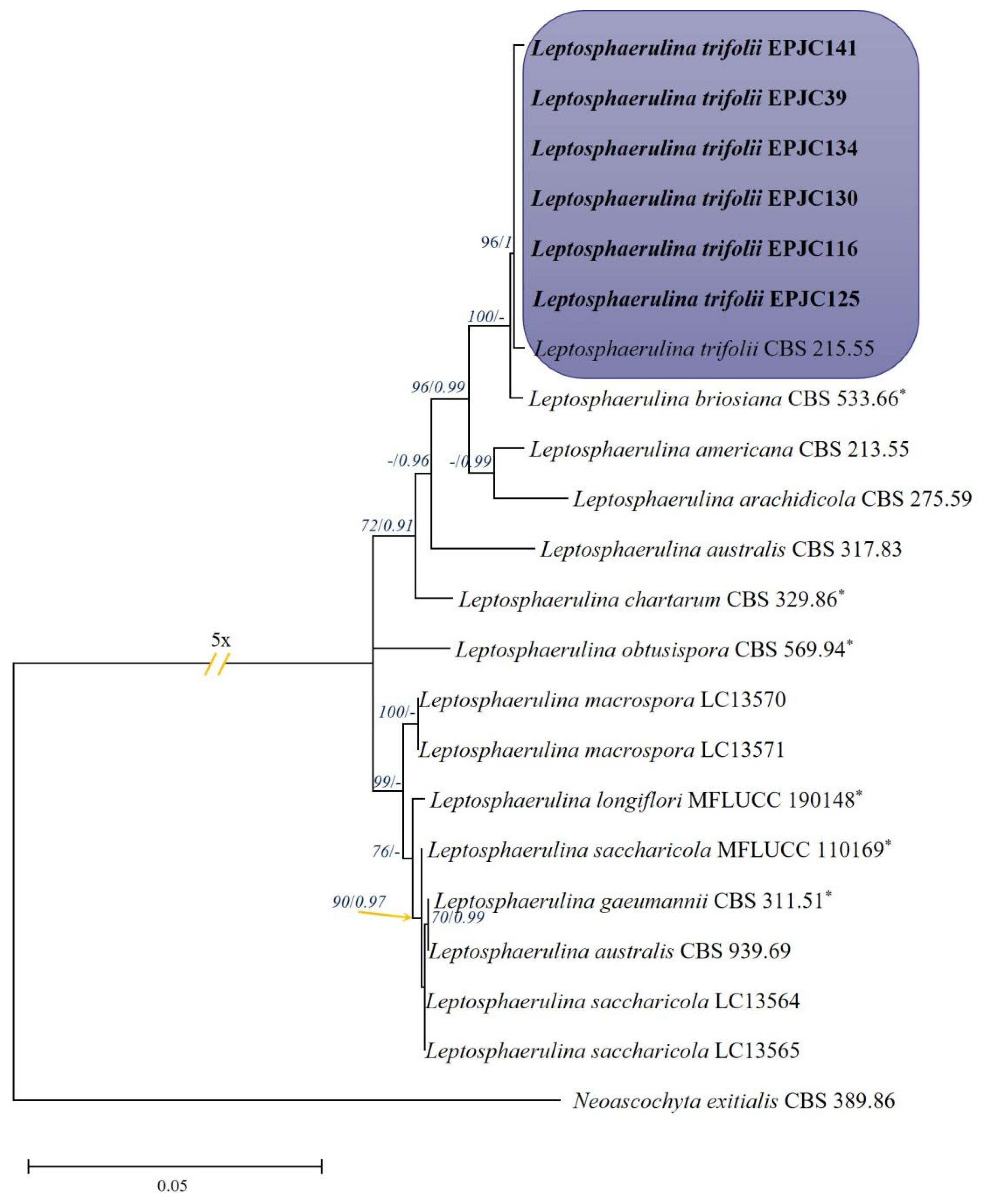
Sequences of three loci (ITS, LSU, and RPB2) from 14 published strains and the ex-type strain CBS 389.86 of Neoascochyta exitialis (used as outgroup; Table S1) were retrieved from GenBank, while those of six representative cultures were newly generated in this study (Table 1). Phylogenetic analyses (Figure 3) revealed that these six isolates consistently clustered into well-supported subclades (RAxML/BP = –/0.99) with the corresponding ex-type or other reference strains. Integrating molecular evidence with morphological and cultural characteristics, the six cultures were conclusively identified as L. trifolii.

3.4. Biological Characteristics of the Fungus

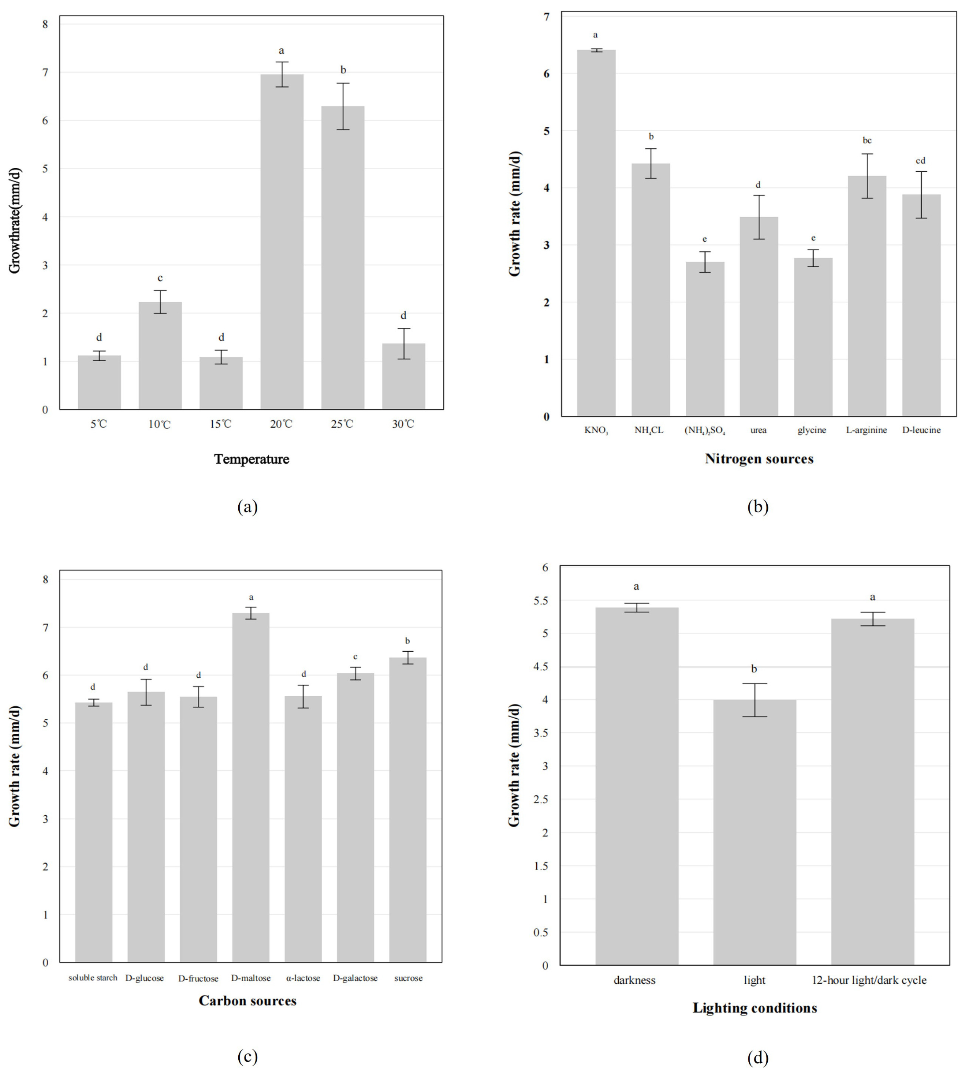
Colonies grew within a temperature range of 5 °C to 30 °C, with optimal growth occurring at 20 °C. At 20 °C, the colony growth rate was fastest, reaching 6.95 ± 0.24 mm/d, significantly higher than at other temperatures (p < 0.05) (Figure 4a).
The growth rate of colonies was assessed under different nitrogen sources, with the fastest growth observed in the KNO3 medium, reaching 6.40 ± 0.02 mm/d, significantly higher than other nitrogen sources (p < 0.05) (Figure 4b). Similarly, when tested with various carbon sources, the colonies showed the most rapid growth in D-maltose medium, achieving a growth rate of 7.30 ± 0.11 mm/d, which was significantly greater than the other carbon sources (p < 0.05) (Figure 4c).
As shown in Figure 4d, the rates of mycelial growth of these strains with different lighting conditions were obviously different. Under continuous darkness and 12 h light/12 h dark cycles, the colony growth rates were measured at 5.39 ± 0.06 mm/d and 5.22 ± 0.09 mm/d, respectively, both significantly higher than that observed under continuous light conditions (p < 0.05).
These physiological characteristics indicate that the fungus is well adapted to the cool, nutrient-limited, and shaded environments typical of alpine grasslands. Its preference for moderate temperatures, KNO3, and D-maltose may enhance its survival and colonization capacity in these ecosystems. Together, these findings provide ecological insight into the environmental tolerance and adaptive potential of the fungus in its native habitat.

3.5. The Result of Pathogenicity Tests

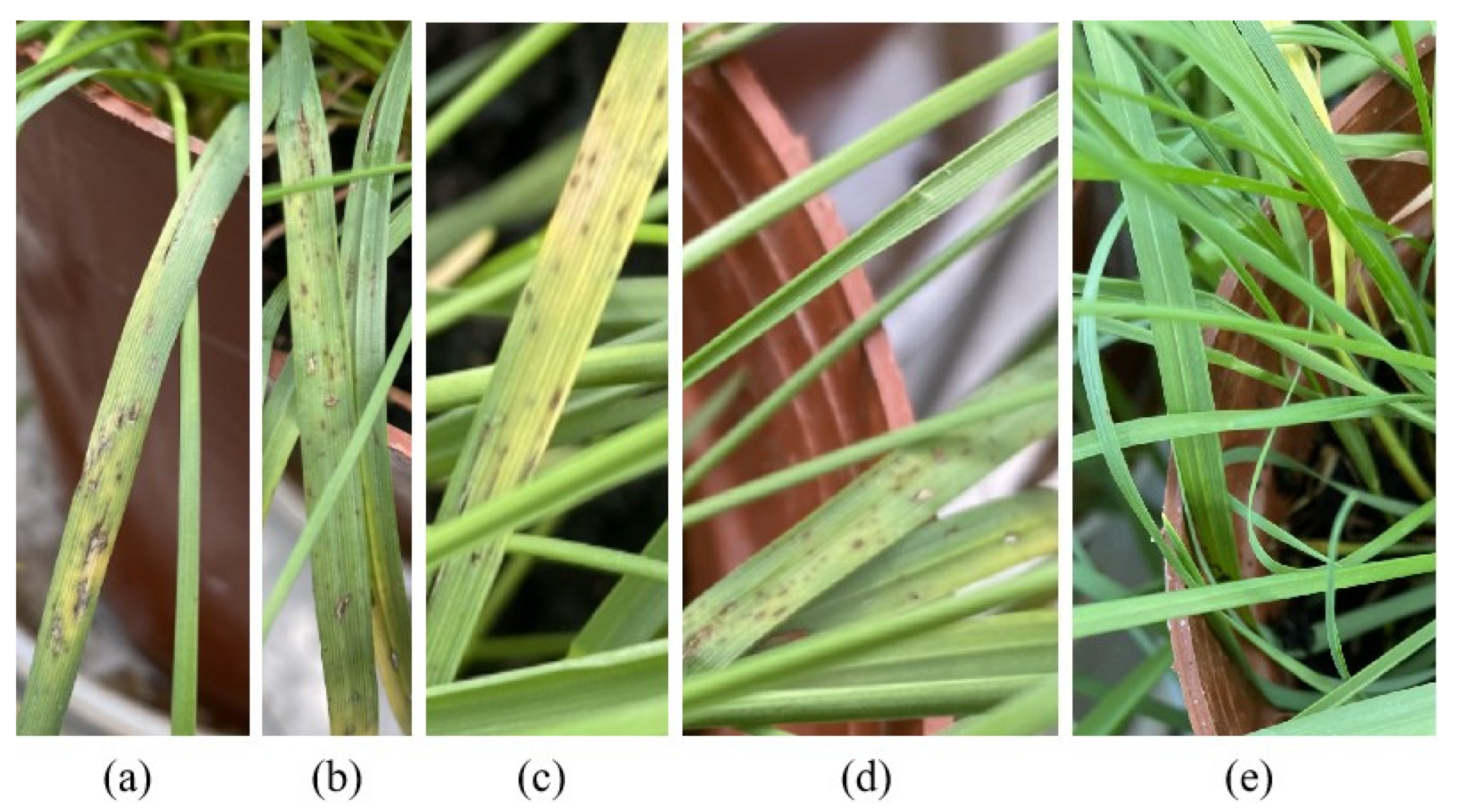
14 days after inoculation on whole plants, all tested strains in the dark treatment (using black material bags) induced leaf lesions to varying degrees, while no symptoms were observed on the negative control plants (Figure 5a). The symptoms induced by L. trifolii are characterized by irregular spots, varying from brown to black, with some lesions displaying gray centers (Figure 5b–d). These lesions resembled those seen on field leaves. In contrast, control leaves remained free of symptoms (Figure 5e). The re-isolation of the pathogen from infected lesions yielded L. trifolii, consistent with Koch’s postulates.

3.6. In Vitro Fungicide Sensitivity of L. trifolii

The inhibitory effects of six fungicides on the mycelial growth of L. trifolii were evaluated across eight concentration levels, and the corresponding EC50 values were determined for each compound (Table 4). Six days post-treatment, 30% Benzalconazole exhibited the most potent antifungal activity, with an EC50 of 11.76 mg/L, indicating strong efficacy at relatively low concentrations. Both 40% Isoprothiolane and 70% Propineb also showed considerable bioactivity against L. trifolii, with EC50 values of 51.29 and 87.10 mg/L, respectively, demonstrating moderate inhibitory effects. In contrast, the antifungal activities of 47% Spring Thunder King Copper and 50% Carbendazim were comparatively weak, with EC50 values exceeding 100 mg/L (177.83–223.87 mg/L), suggesting that substantially higher doses are required to achieve 50% inhibition of mycelial growth. Overall, the data indicate a clear hierarchy of antifungal potency among the tested compounds, with 30% Benzalconazole emerging as the most effective, followed by 40% Isoprothiolane and 70% Propineb, and finally 47% Spring Thunder King Copper and 50% Carbendazim. These findings provide a quantitative basis for selecting efficacious fungicides for the management of L. trifolii in agricultural applications.

4. Discussion

In this study, leaf spot symptoms associated with Leptosphaerulina trifolii were observed on Elymus plants in Gansu and Qinghai Provinces of China. The identification of the pathogen was corroborated by field surveys, morphological characteristics, multigene phylogenetic analyses, and pathogenicity assays. L. trifolii is commonly reported on legumes, particularly Medicago spp. and Trifolium repens [10,11,16,17,39], while infections on grasses have rarely been documented. This study represents the first confirmed report of L. trifolii causing leaf spot disease on Elymus plants in China. The occurrence of L. trifolii on Elymus species may reflect the ecological flexibility and host-range dynamics that are characteristic of the Didymellaceae. Members of this family exhibit remarkable adaptability, with several genera capable of infecting taxonomically diverse hosts or shifting among phylogenetically distant plant lineages [13]. This ecological adaptability and cross-host potential may facilitate occasional colonization of non-leguminous hosts, particularly in natural grassland ecosystems where legumes and grasses coexist. In these habitats, fungal pathogens are readily dispersed by wind or rain-splash, enabling contact with novel hosts [40,41]. L. trifolii likewise produces wind-dispersed ascospores, a strategy similar to that of L. maculans, which enhances its potential for cross-host infection [42].
Traditional morphological identification of Didymellaceae fungi largely relies on the characteristics of fruiting bodies and spores, with spore morphology considered a key criterion for distinguishing genera such as Phoma and Ascochyta [43,44,45,46]. L. trifolii, a member of Didymellaceae, exhibits features consistent with previous reports for the genus, such as globose or subglobose asci and ellipsoid, brown, multicellular ascospores measuring 31.0–41.6 × 75.0–87.5 µm [15]. Moreover, under standard culture conditions, Didymellaceae fungi are often difficult to induce to sporulate [11,14]. In this study, OA media combined with ultraviolet light induction facilitated the sporulation of L. trifolii, providing a useful reference for future morphological characterization of Didymellaceae fungi.
Among molecular markers, RPB2 provides the highest resolution at species and genus levels and is considered the most reliable locus for L. trifolii [11,29,43,47], while ITS and LSU are also commonly employed [11]. In this study, the combined analyses of ITS, LSU, and RPB2 sequences greatly enhanced the accuracy of species identification and clarified the phylogenetic relationships among closely related taxa [48].
Our results confirm that L. trifolii can grow in vitro between 5 and 35 °C, with optimal growth at 20 °C [10,49]. Studies of its other traits, including carbon and nitrogen sources and light conditions, further enrich knowledge of this species. Fungicides continue to be the most direct and effective means in combating plant diseases [50,51]. Previous studies reported that Propineb effectively controls L. trifolii under field conditions [52]. Consistently, in our study, 70% Propineb exhibited notable inhibitory activity with an EC50 of 87.10 mg/L. Furthermore, we identified 30% Benzalconazole as a more potent fungicide, with an EC50 of 11.76 mg/L.

5. Conclusions

This study reports the first isolation of the pathogen from Elymus plants affected by leaf spot disease in China. Investigations of temperature, nitrogen, carbon, and light regimes on the growth of L. trifolii showed that the fungus proliferates most rapidly at 20 °C, with KNO3 as the preferred nitrogen source, D-maltose as the optimal carbon source, and either continuous darkness or a 12 h light/12 h dark cycle providing the most favorable light conditions. Moreover, fungicides such as 30% Benzalconazole (EC50 = 11.76 mg/L), 40% Isoprothiolane (EC50 = 51.29 mg/L), and 70% Propineb (EC50 = 87.10 mg/L) have been found to effectively inhibit the growth and development of this pathogen. The findings of this study may provide a useful reference for the development of management strategies against Didymellaceae leaf spot diseases in different regions.

Supplementary Materials

The following supporting information can be downloaded at: https://www.mdpi.com/article/10.3390/agronomy15112502/s1. Table S1: Collection details and GenBank accession numbers of Leptosphaerulina species included in this study. Table S2: List of primers used in this study [53,54,55,56,57,58,59].

Author Contributions

Conceptualization, J.L. and L.X.; methodology, J.L.; software, J.L.; validation, J.L., L.X. and C.L.; formal analysis, J.L.; investigation, J.L.; resources, L.X.; data curation, J.L.; writing–original draft preparation, J.L.; writing–re-view and editing, J.L. and L.X.; visualization, J.L.; supervision, C.L.; project administration, C.L.; funding acquisition, C.L. All authors have read and agreed to the published version of the manuscript.

Funding

This research was funded by National Key R & D Program of China (grant number: 2022YFD1401101), Special Fund for Basic Scientific Research of Chinese Academy of Forestry (grant number: CAFYBB2021ZD001) and Lanzhou University “Double First-Class” Guiding Special Project (grant number: 561120201). The APC was funded by the same grant.

Data Availability Statement

The original contributions presented in this study are included in the article/Supplementary Material. Further in-quiries can be directed to the corresponding authors.

Conflicts of Interest

The authors declare no conflicts of interest.

References

  1. Li, M.Q.; Yang, J.; Wag, X.; Li, D.X.; Zhang, C.B.; Tian, Z.H.; You, M.H.; Bai, S.Q.; Lin, H.H. Transcriptome profiles identify the common responsive genes to drought stress in two Elymus species. J. Plant Physiol. 2020, 250, 153–183. [Google Scholar] [CrossRef] [PubMed]
  2. Liu, J.; Xue, L.; Li, C. Characterization of Didymellaceae leaf spot associated with Elymus plants in the Tibetan Plateau of China. Plant Dis. 2025. [Google Scholar] [CrossRef] [PubMed]
  3. Song, J.; Yang, H.; Yu, X.; Chen, Y.; Yang, C.; He, Y.; Wang, H. Effects of combined application of nitrogen, phosphorus, and potassium fertilizers on seed yield, se0ed quality and economic returns of Elymus nutans in alpine region. BMC Plant Biol. 2025, 25, 130. [Google Scholar] [CrossRef]
  4. Hou, C.Y.; Wei, X.; Zhou, L.; Ma, J.H.; Ren, X.; Wang, Y.Y.; Wu, P.F. Dynamics of soil nematode community in perennial Gramineae artificial grasslands in Northwest Sichuan. Acta Ecol. Sin. 2023, 43, 10104–10118. [Google Scholar] [CrossRef]
  5. Quan, X.; Qiao, Y.; Chen, M.; Duan, Z.; Shi, H. Comprehensive evaluation of the allelopathic potential of Elymus nutans. Ecol. Evol. 2021, 11, 12389–12400. [Google Scholar] [CrossRef] [PubMed]
  6. Fisher, M.C.; Henk, D.A.; Briggs, C.J.; Brownstein, J.S.; Madoff, L.C.; McCraw, S.L.; Gurr, S.J. Emerging fungal threats to ani-mal, plant and ecosystem health. Nature 2012, 484, 186–194. [Google Scholar] [CrossRef]
  7. Liu, J.Q.; Bao, G.S.; Li, C.J. First report of brown leaf spot of Elymus nutans caused by Bipolaris sorokiniana in northwestern China. Plant Dis. 2023, 107, 941. [Google Scholar] [CrossRef]
  8. Liu, Y.; Duan, D.; Jiang, F.; Tian, Z.; Feng, X.; Wu, N.; Hou, F.; Nan, Z.; Chen, T. Long-term heavy grazing increases community-level foliar fungal diseases by shifting plant composition. J. Appl. Ecol. 2022, 59, 791–800. [Google Scholar] [CrossRef]
  9. Xue, L.H.; Liu, J.Q.; Li, C.J. Progress in research on fungal diseases of the genus Elymus. Acta Prataculturae Sin. 2024, 33, 226–241. (In Chinese). Available online: http://cyxb.magtech.com.cn/CN/10.11686/cyxb2023132 (accessed on 7 September 2025).
  10. Huo, H.; Huangfu, J.; Song, P.; Zhang, D.; Shi, Z.; Zhao, L.; Li, Z.; Zhou, H. Isolation, identification and characterization of Leptosphaerulina trifolii, the causative agent of alfalfa Leptosphaerulina Leaf spot in Inner Mongolia, China. Agronomy 2024, 14, 1156. [Google Scholar] [CrossRef]
  11. Hou, L.W.; Groenewald, J.Z.; Pfenning, L.H.; Yarden, O.; Crous, P.W.; Cai, L. The phoma-like dilemma. Stud. Mycol. 2020, 96, 309–396. [Google Scholar] [CrossRef]
  12. Zhang, Y.; Schoch, C.L.; Fournier, J.; Crous, P.W.; de Gruyter, J.; Woudenberg, J.H.C.; Hirayama, K.; Tanaka, K.; Pointing, S.B.; Spatafora, J.W.; et al. Multi-locus phylogeny of Pleosporales: A taxonomic, ecological and evolutionary re-evaluation. Stud. Mycol. 2009, 64, 85–102. [Google Scholar] [CrossRef]
  13. Chen, Q.; Hou, L.W.; Duan, W.J.; Crous, P.W.; Cai, L. Didymellaceae revisited. Stud. Mycol. 2017, 87, 105–159. [Google Scholar] [CrossRef]
  14. Valenzuela-Lopez, N.; Cano-Lira, J.F.; Guarro, J.; Sutton, D.A.; Wiederhold, N.; Crous, P.W.; Stchigel, A.M. Coelomycetous Dothideomycetes with emphasis on the families Cucurbitariaceae and Didymellaceae. Stud. Mycol. 2018, 90, 177–301. [Google Scholar] [CrossRef]
  15. Zhang, L.L.; Li, Y.Z. First report of alfalfa leaf spot caused by Leptosphaerulina australis in China. Plant Dis. 2021, 105, 2254. [Google Scholar] [CrossRef]
  16. Liu, X.P.; Jing, X.M.; Yan, H.X.; Li, G.L.; Luo, Y.H. First report of Leptosphaerulina leaf spot caused by Leptosphaerulina trifolii on alfalfa in Heilongjiang Province, China. Plant Dis. 2019, 103, 2673. [Google Scholar] [CrossRef]
  17. Victoria, A.A.D.; Furtado, B.E.; Holz, M.T.; Romero-Arenas, O.; Dallagnol, L.J. First report of leptosphaerulina leaf spot caused by Leptosphaerulina trifolii on trifolium repens in brazil. Plant Dis. 2020, 104, 972. [Google Scholar] [CrossRef]
  18. Sheng, Y.T.; Yu, X.L.; Mao, T.T.; Zhang, J.; Guo, X.T.; Song, Z.Z.; Zhang, H.X. Genome sequence data of Leptosphaerulina arachidicola, a causal agent of peanut scorch spot in China. Plant Dis. 2022, 106, 748–750. [Google Scholar] [CrossRef]
  19. Phookamsak, R.; Liu, J.K.; Chukeatirote, E.; McKenzie, E.H.; Hyde, K.D. Phylogeny and morphology of Leptosphaerulina saccharicola sp. nov. and Pleosphaerulina oryzae and relationships with Pithomyces. Cryptogam. Mycol. 2013, 34, 303–319. [Google Scholar] [CrossRef]
  20. Tennakoon, D.S.; Thambugala, K.M.; De Silva, N.I.; Kuo, C.H.; Hyde, K.D. Leaf litter saprobic Didymellaceae (Dothideomycetes): Leptosphaerulina longiflori sp. nov. and Didymella sinensis, a new record from Roystonea regia. Asian J. Mycol. 2019, 2, 87–100. [Google Scholar] [CrossRef]
  21. Liu, Y.; Wu, S.; Li, X.; Yang, M.; Sun, L.; He, S.; Wu, D.; Fu, Y. Occurrence of maize leaf spot disease caused by Leptosphaerulina australis in Yunnan, China. Plant Dis. 2024, 108, 3407. [Google Scholar] [CrossRef]
  22. Chen, Q.; Bakhshi, M.; Balci, Y.; Broders, K.D.; Cheewangkoon, R.; Chen, S.F.; Fan, X.L.; Gramaje, D.; Halleen, F.; Jung, M.H.; et al. Genera of phytopathogenic fungi: GOPHY 4. Stud. Mycol. 2022, 101, 417–564. [Google Scholar] [CrossRef]
  23. Keirnan, E.C.; Tan, Y.P.; Laurence, M.H.; Mertin, A.A.; Liew, E.C.; Summerell, B.A.; Shivas, R.G. Cryptic diversity found in Didymellaceae from Australian native legumes. MycoKeys 2021, 78, 1–20. [Google Scholar] [CrossRef] [PubMed]
  24. Wang, Y.C.; Tu, Y.Y.; Chen, X.L.; Jiang, H.; Ren, H.Z.; Lu, Q.H.; Wei, C.L.; Lv, W.Y. Didymellaceae species associated with tea plant (Camellia sinensis) in China. MycoKeys 2024, 105, 217–251. [Google Scholar] [CrossRef]
  25. Nan, Z.B. Establishing sustainable management system for diseases of pasture crops in China. Acta Prataculturae Sin. 2000, 2, 1–9. (In Chinese). Available online: https://kns.cnki.net/kcms2/article/abstract?v=o8vMLOX1CKtXTwECOybpHkymflmFDoPnHyE5VwGYgrO95lkBMExMYbIaluPy1hOi7TbWGq1UgtQ0YIs5PggZPsHcASrGz8JZ7JDPOSM6Mm3iPhxrXTEaMFHPsyhdd-pz_0bt6_Rt5kiSmzaB10S9gymn687yhc0mlHYfwNH87XVhJxXel_6gvA==&uniplatform=NZKPT&language=CHS (accessed on 7 September 2025).
  26. Xue, L.; Liu, Y.; Zhou, S.; White, J.F.; Li, C. Characterization of Pyrenophora species causing brown leaf spot on Italian ryegrass (Lolium multiflorum) in southwestern China. Plant Dis. 2020, 104, 1900–1907. [Google Scholar] [CrossRef]
  27. Xue, L.H.; Xu, Z.T.; Liu, J.Q.; Chen, H.; White, J.F.; Malik, K.l.; Li, C.J. Differences in the characteristics and pathogenicity of Pyrenophora species associated with seeds of Italian ryegrass. Plant Dis. 2023, 107, 758–770. [Google Scholar] [CrossRef]
  28. White, T.J.; Bruns, T.; Lee, S.J.W.T.; Taylor, J. Amplification and direct sequencing of fungal ribosomal RNA genes for phylogenetics. PCR Protoc. A Guide Methods Appl. 1990, 18, 315–322. [Google Scholar]
  29. Chen, Q.; Jiang, J.R.; Zhang, G.Z.; Cai, L.; Crous, P.W. Resolving the Phoma enigma. Stud. Mycol. 2015, 82, 137–217. [Google Scholar] [CrossRef]
  30. Katoh, K.; Standley, D.M. MAFFT multiple sequence alignment software version 7: Improvements in performance and usability. Mol. Biol. Evol. 2013, 30, 772–780. [Google Scholar] [CrossRef]
  31. Tamura, K.; Stecher, G.; Peterson, D.; Filipski, A.; Kumar, S. MEGA6: Molecular evolutionary genetics analysis version 6.0. Mol. Biol. Evol. 2013, 30, 2725–2729. [Google Scholar] [CrossRef]
  32. Vaidya, G.; Lohman, D.J.; Meier, R. SequenceMatrix: Concatenation software for the fast assembly of multi-gene datasets with character set and codon information. Cladistics 2011, 27, 171–180. [Google Scholar] [CrossRef]
  33. Ronquist, F.; Teslenko, M.; Van Der Mark, P.; Ayres, D.L.; Darling, A.; Höhna, S.; Bret, L.; Liu, L.; Suchard, M.A.; Huelsenbeck, J.P. MrBayes 3.2: Efficient bayesian phylogenetic inference and model choice across a large model space. Syst. Biol. 2012, 61, 539–542. [Google Scholar] [CrossRef] [PubMed]
  34. Edler, D.; Klein, J.; Antonelli, A.; Silvestro, D. raxmlGUI 2.0: A graphical interface and toolkit for phylogenetic analyses using RAxML. Methods Ecol. Evol. 2021, 12, 373–377. [Google Scholar] [CrossRef]
  35. Nylander, J.A.A. MrModeltest v2. Program distributed by the author. Evolutionary Biology Centre, Uppsala University.ampignons de l’Équateur (Pugillus IV). Bull. L’Herb. Boissier 2004, 3, 53–74. [Google Scholar]
  36. Nuin, P. MrMTgui: Cross-Platform Interface for ModelTest and Mr Modeltest 2008. Available online: https://mrmtgui.software.informer.com/ (accessed on 7 September 2025).
  37. Snow, M.J.; Manley-Harris, M. On the nature of non-peroxide antibacterial activity in New Zealand manuka honey. Food Chem. 2004, 84, 145–147. [Google Scholar] [CrossRef]
  38. Liang, J.; Li, G.; Hou, L.; Zhao, M.; Cai, L. Leptosphaerulina species isolated from golf turfgrass in China, with description of L. macrospora, sp. nov. Mycologia 2021, 113, 956–967. [Google Scholar] [CrossRef]
  39. Barbetti, M.J. Resistance in annual Medicago spp. to Phoma medicaginis and Leptosphaerulina trifolii and its relationship to induced production of a phytoestrogen. Plant Dis. 2007, 91, 239–244. [Google Scholar] [CrossRef]
  40. Karisto, P.; Suffert, F.; Mikaberidze, A. Measuring splash dispersal of a major wheat pathogen in the field. PhytoFrontiers™ 2022, 2, 30–40. [Google Scholar] [CrossRef]
  41. Penet, L.; Guyader, S.; Pétro, D.; Salles, M.; Bussière, F. Direct splash dispersal prevails over indirect and subsequent spread during rains in Colletotrichum gloeosporioides infecting yams. PLoS ONE 2014, 9, e115757. [Google Scholar] [CrossRef]
  42. Fitt, B.D.; Brun, H.; Barbetti, M.J.; Rimmer, S.R. World-wide importance of Phoma stem canker (Leptosphaeria maculans and L. biglobosa) on oilseed rape (Brassica napus). Eur. J. Plant Pathol. 2006, 114, 3–15. [Google Scholar] [CrossRef]
  43. Aveskamp, M.M.; de Gruyter, J.; Woudenberg, J.H.C.; Verkley, G.J.M.; Crous, P.W. Highlights of the Didymellaceae: A polyphasic approach to characterise Phoma and related pleosporalean genera. Stud. Mycol. 2010, 65, 1–60. [Google Scholar] [CrossRef] [PubMed]
  44. Boerema, G.A.; Bollen, G.J. Conidiogenesis and conidial septation as differentiating criteria between Phoma and Ascochyta. Persoonia-Mol. Phylogeny Evol. Fungi 1975, 8, 111–144. Available online: https://repository.naturalis.nl/pub/531868/ (accessed on 7 September 2025).
  45. De Gruyter, J.; Aveskamp, M.M.; Woudenberg, J.H.; Verkley, G.J.; Groenewald, J.Z.; Crous, P.W. Molecular phylogeny of Phoma and allied anamorph genera: Towards a reclassification of the Phoma complex. Mycol. Res. 2009, 113, 508–519. [Google Scholar] [CrossRef]
  46. Morgan-Jones, G. Concerning some species of Microsphaeropsis. Can. J. Bot. 1974, 52, 2575–2579. [Google Scholar] [CrossRef]
  47. Aveskamp, M.M.; Verkley, G.J.; de Gruyter, J.; Murace, M.A.; Perello, A.; Woudenberg, J.H.; Groenewald, J.Z.; Crous, P.W. DNA phylogeny reveals polyphyly of Phoma section Peyronellaea and multiple taxonomic novelties. Mycologia 2009, 101, 363–382. [Google Scholar] [CrossRef]
  48. Woudenberg, J.H.; De Gruyter, J.; Crous, P.W.; Zwiers, L.H. Analysis of the mating-type loci of co-occurring and phylogenetically related species of Ascochyta and Phoma. Mol. Plant Pathol. 2012, 13, 350–362. [Google Scholar] [CrossRef]
  49. Olanya, O.M.; Campbell, C.L. Isolate characteristics and epidemic components of Leptosphaerulina leaf spots on alfalfa and white clover. Phytopathology 1990, 80, 1278–1282. [Google Scholar] [CrossRef]
  50. Karim, M.M.; Usman, H.M.; Tan, Q.; Hu, J.J.; Fan, F.; Hussain, R.; Luo, C.X. Fungicide resistance in Colletotrichum fructicola and Colletotrichum siamense causing peach anthracnose in China. Pestic. Biochem. Physiol. 2024, 203, 106006. [Google Scholar] [CrossRef]
  51. Ahonsi, M.O.; Ames, K.A.; Gray, M.E.; Bradley, C.A. Biomass reducing potential and prospective fungicide control of a new leaf blight of Miscanthus× giganteus caused by Leptosphaerulina chartarum. BioEnergy Res. 2013, 6, 737–745. [Google Scholar] [CrossRef]
  52. Lowe, K.F.; Langdon, P.W.; Bowdler, T.M. Combined effects of leaf spot diseases caused by Stemphylium vesicarium (Wallr.) Simmons and Leptosphaerulina trifolii (Rostr.) Petr. on lucerne cultivars, and the efficacy of some fungicidal control methods. Aust. J. Exp. Agric. 1987, 27, 59–65. [Google Scholar] [CrossRef]
  53. Crous, P.W.; Schoch, C.L.; Hyde, K.D.; Wood, A.R.; Gueidan, C.; De Hoog, G.S.; Groenewald, J.Z. Phylogenetic lineages in the Capnodiales. Stud. Mycol. 2009, 64, 17–47. [Google Scholar] [CrossRef]
  54. Gardes, M.; Bruns, T.D. ITS primers with enhanced specificity for basidiomycetes-application to the identification of mycorrhizae and rusts. Mol. Ecol. 1993, 2, 113–118. [Google Scholar] [CrossRef]
  55. Liu, Y.J.; Whelen, S.; Hall, B.D. Phylogenetic relationships among ascomycetes: Evidence from an RNA polymerse II subunit. Mol. Biol. Evol. 1999, 16, 1799–1808. [Google Scholar] [CrossRef]
  56. Schoch, C.L.; Seifert, K.A.; Huhndorf, S.; Robert, V.; Spouge, J.L.; Levesque, C.A.; Levesque, A.; Chen, W.; Fungal Barcoding Consortium. Nuclear ribosomal internal transcribed spacer (ITS) region as a universal DNA barcode marker for Fungi. Proc. Natl. Acad. Sci. USA 2012, 109, 6241–6246. [Google Scholar] [CrossRef] [PubMed]
  57. Simon, U.K.; Groenewald, J.Z.; Crous, P.W. Cymadothea trifolii, an obligate biotrophic leaf parasite of Trifolium, belongs to Mycosphaerellaceae as shown by nuclear ribosomal DNA analyses. Persoonia-Mol. Phylogeny Evol. Fungi 2009, 22, 49–55. [Google Scholar] [CrossRef] [PubMed]
  58. Sung, G.H.; Sung, J.M.; Hywel-Jones, N.L.; Spatafora, J.W. A multi-gene phylogeny of Clavicipitaceae (Ascomycota, Fungi): Identification of localized incongruence using a combinational bootstrap approach. Mol. Phylogenetics Evol. 2007, 44, 1204–1223. [Google Scholar] [CrossRef] [PubMed]
  59. Vu, D.; Groenewald, M.; De Vries, M.; Gehrmann, T.; Stielow, B.; Eberhardt, U.; Al-Hatmi, A.; Groenewald, J.Z.; Cardinali, G.; Houbraken, J.; et al. Large-scale generation and analysis of filamentous fungal DNA barcodes boosts coverage for kingdom fungi and reveals thresholds for fungal species and higher taxon delimitation. Stud. Mycol. 2019, 92, 135–154. [Google Scholar] [CrossRef]
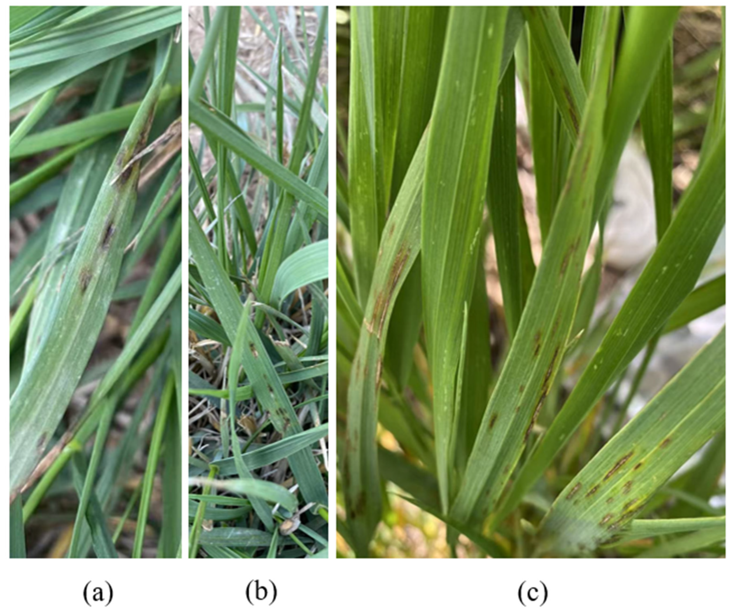
Figure 1. Field symptoms of leaf spot disease on Elymus spp. (a) Early-stage symptoms in natural grassland and (b) cultivated grassland and (c) late-stage symptoms with disease extend to neighboring leaves.
Agronomy 15 02502 g001
Figure 2. Morphological and pathological characteristics of Leptosphaerulina trifolii: (a,b) colony on PDA (front and reverse) at 20 °C on day 12; (c,e) ascospores of L. trifolii on MEA; (d) ascomata (f,g) sporangiophores.
Agronomy 15 02502 g002
Figure 3. Maximum likelihood (RAxML) phylogenetic tree based on the combined ITS, LSU and RPB2 alignment of Leptosphaerulina species. Bootstrap support values/Bayesian pp scores above 70%/0.70 are shown at the nodes (BS/PP). The three-locus datasets were analyzed by maximum likelihood using RAxML as implemented in raxmlGUI 2.0.8; the GTRGAMMA substitution model for the nucleotide partitions and the default setting for binary (indel) data were used, and rapid bootstrap analyses with 1000 replications were conducted for branch support. For Bayesian Inference (BI) analysis, the best-fit nucleotide substitution models were selected: SYM for ITS (453 bp); HKY for LSU (769 bp); GTR + I for RPB2 (595 bp). Leptosphaerulina trifolii are delimited in coloured boxes. Strains isolated in this study are shown in bold. Neoascochyta exitialis (CBS 389.86) is used as an outgroup. Ex-type or holotype specimens are emphasized with an asterisk.
Agronomy 15 02502 g003
Figure 4. Effects of environmental factors on the mycelial growth of the tested isolates. Growth rates were measured under different (a) temperatures, (b) nitrogen sources, (c) carbon sources, and (d) lighting conditions. Different letters indicate that there are statistically significant differences (p < 0.05).
Agronomy 15 02502 g004
Figure 5. Leaf spot symptoms caused by Leptosphaerulina trifolii on Elymus nutans and E. sibiricus. (a,b) L. trifolii on E. nutans. (c,d) L. trifolii on E. sibiricus. (e) Plants treated with sterile distilled water as a control.
Agronomy 15 02502 g005
Table 1. GenBank and culture collection accession numbers of the isolates (L. trifolii) in this study.
StrainsNumber of
Isolated
Strain (n)
HostLocationGenBank Accession
ITSLSURPB2
EPJC398Elymus nutansMaqu, Gannan, GansuPP343084PX099127PV837467
EPJC1164E. nutansMaqu, Gannan, GansuPX099222PX099128PV837468
EPJC1255E. sibiricusHaiyan, Haibei, QinghaiPX099223PX099129PV837469
EPJC1302E. nutansMaqu, Gannan, GansuPX108633PX108636PX111706
EPJC1342E. nutansMaqu, Gannan, GansuPX108634PX108637PX111707
EPJC1414E. sibiricusHaiyan, Haibei, QinghaiPX108635PX108638PX111708
Table 2. Tested fungicide information.
FungicideFormulationManufacturer
30% BenzalconazoleSuspo-emulsionSichuan Guoguang Agrochemical Co., Ltd., Chengdu, China
70% PropinebWettable powderBayer AG, North Rhine-Westphalia, Germany
47% Spring Thunder King CopperWettable powderBeijing Green Agricultural Science and Technology Group Co., Ltd., Beijing, China
40% IsoprothiolaneEmulsifiable concentrateBeijing Green Agricultural Science and Technology Group Co., Ltd., Beijing, China
50% CarbendazimWettable powderHebei Dewoduo Biotechnology Co., Ltd., Hengshui, China
Table 3. The fungicide concentration gradient used for testing.
FungicideConcentration Gradient of Active Constituent (mg/L)
30% Benzalconazole0.15, 0.75, 1.5, 3, 6, 30, 150, 300
70% Propineb0.35, 1.75, 3.5, 7, 14, 70, 350, 850
47% Spring Thunder King Copper0.235, 1.175, 2.35, 4.7, 9.4, 47, 470, 940
40% Isoprothiolane0.25, 1.25, 2.5, 5, 10, 50, 250, 500
50% Carbendazim0.2, 1, 2, 4, 8, 40, 500, 1000
Table 4. The in vitro inhibitory effects of various fungicides on L. trifolii.
FungicidesRegression EquationEC50 (mg/L)Correlation Index (r2)95% Confidence Intervals
30% BenzalconazoleY = 26.46X + 21.8011.760.9721.80–31.11
70% PropinebY = 24.72X + 2.18287.100.8815.72–33.72
47% Spring Thunder King CopperY = 20.07X + 4.866177.830.9113.72–26.42
40% IsoprothiolaneY = 21.92X + 12.5851.290.9516.81–27.02
50% CarbendazimY = 17.31X + 9.372223.870.9513.49–21.13
Disclaimer/Publisher’s Note: The statements, opinions and data contained in all publications are solely those of the individual author(s) and contributor(s) and not of MDPI and/or the editor(s). MDPI and/or the editor(s) disclaim responsibility for any injury to people or property resulting from any ideas, methods, instructions or products referred to in the content.