Article

Foliar Biofortification with Sodium Selenate Enhances Selenium Content in Ocimum basilicum L. Cultivars in a Totally Controlled Environment System

1
Department of Agricultural, Forest, and Food Sciences, Inhortosanitas Lab, University of Turin, 10095 Grugliasco, TO, Italy
2
Department of Health Sciences, School of Medicine, University of Piemonte Orientale, 28100 Novara, NO, Italy
3
Department of Horticultural Science, University of Zanjan, Zanjan 45371-38791, Iran
4
Department of Agronomy and Plant Breeding, Faculty of Agriculture, University of Azarbaijan Shahid Madani, Tabriz 53714-161, Iran
*
Authors to whom correspondence should be addressed.
Agronomy 2025, 15(10), 2368; https://doi.org/10.3390/agronomy15102368
Submission received: 7 September 2025 / Revised: 3 October 2025 / Accepted: 8 October 2025 / Published: 10 October 2025

Abstract

Selenium (Se) is an essential micronutrient for human health, yet its dietary intake is insufficient in many populations worldwide. Agronomic biofortification represents an effective strategy to enrich crops with Se, and Totally Controlled Environment Agriculture (TCEA) provides a reliable platform to evaluate cultivar-specific responses under standardized conditions. This study evaluated the effects of foliar sodium selenate doses of 0, 5, 10, and 15 µM on two basil (Ocimum basilicum L.) cultivars, ‘Fine Verde’ (FV) and ‘Red Rubin’ (RR), in a micro-TCEA system. The yield was not significantly different at 5–10 µM but declined by 21% at 15 µM, particularly for FV. RR out-yielded FV (+14%), whereas FV produced taller shoots. The 5 µM Se concentration did not affect the total chlorophyll content and quantum yield of photosystem II under control conditions. The highest Se dose (15 µM) decreased the chlorophyll content and electron transport rate by 18% and 12%, respectively, while increasing the stomatal conductance (50%) and transpiration rate (120%). The total phenolics content in RR was double that in FV and increased with Se, whereas the NO3 concentration in RR decreased by 9% at 10 µM. Principal component analysis separated treatments by Se dose (PC1 = 44.5%) and cultivar (PC2 = 42.7%), showing RR’s stronger connection of RR to biomass and antioxidant accumulation under moderate Se. Overall, a single foliar application of 5 µM sodium selenate appears optimal to achieve effective Se enrichment while maintaining productivity and quality. These findings support basil as a promising candidate for Se biofortification in TCEA systems, with potential contributions to dietary Se intake.

1. Introduction

Selenium (Se) is an essential micronutrient for human and animal health, serving critical biological functions in selenoproteins, particularly in antioxidant processes [1]. Se deficiency in human diet is associated with several pathological conditions [2,3]. While Se’s essentiality in plants is debated, it benefits growth at low concentrations (5–10 µM) [4,5]. Crop biofortification can address dietary Se deficiency [6,7] by increasing micronutrient concentrations in plant tissues through genetic or agronomic methods [8]. Agronomic biofortification using Se-containing fertilizers effectively enhances Se intake in populations [9]. Selenate (Se6+) is preferred for biofortification due to efficient plant absorption [10,11,12]. Sodium selenate (Na2SeO4) has proven effective in various crops, including Lactuca sativa L. [13], Anethum graveolens L. [14], Brassica oleracea L. ‘Italica’ [15], Mentha × Piperita L., and O. basilicum L. [16].
Recognising that target element accumulation depends on nutrient application and genotypic variability is critical for biofortification programs [17,18]. Cultivars of the same species have different abilities to absorb micronutrients in edible tissues due to genetic characteristics controlling element acquisition [17,19]. Selecting cultivars with high Se concentrations optimizes biofortification [17,20]. Studies show Se concentration variations among cultivars in Brassica oleracea L. [21,22], and Zea mays L. [23]. For O. basilicum L., biofortification response varies by cultivar, affecting mineral concentration [24,25].
To accurately examine genotypic variability, Totally Controlled Environment Agriculture (TCEA) is crucial [26]. TCEA provides significant benefits compared to traditional open-field and greenhouse farming, including precise environmental regulation, improved resource use efficiency (water, nutrients, and energy), reduced reliance on pesticides, allowed year-round crop production, and consistently high-quality yields [27]. The scalability and adaptability of these systems have facilitated their successful implementation across diverse crop species, such as basil (O. basilicum L.), which is a suitable candidate because of its high economic value, rapid growth cycle, and favourable response to controlled environments [13,16]. This aromatic plant is of great interest and is appreciated for its culinary use and rich phytochemical profile [28]. It is an important source of bioactive compounds, including essential oils, flavonoids, and phenolic acids [29].
Different basil cultivars may vary in their secondary metabolite content and profiles. For example, studies on the relative species, O. tenuiflorum L., in TCEA have shown significant differences in the total phenol, flavonoid, and anthocyanin contents among different cultivars [30]. Recent research has demonstrated that green and purple basil cultivars exhibit distinct physiological and nutraceutical responses to UV-A wavelengths in the TCEA system [31]. Similarly to the Se biofortification effect, Ciriello et al. (2022) [24] investigated Zn biofortification in two basil cultivars (‘Aroma 2’ and ‘Eleonora’) grown hydroponically with four Zn doses and found differentiation results between cultivars and a significant impact on the mineral concentration and antioxidant compounds. Genotypic variability in response to biofortification is well documented across various crops, as highlighted in the review by Hossain et al. (2021) [7]. This includes species like Triticum aestivum L. (wheat), Hordeum vulgare L. (barley), Avena sativa L. (oats), and Oryza sativa L. (rice), as well as legumes such as Cicer arietinum L. (chickpeas), Lens culinaris Medik. (lentils), Vigna radiata (L.) R. Wilczek (mung beans), and Glycine max (L.) Merr (soybeans). Limited research has been conducted on Se biofortification in commonly grown basil cultivars under controlled cultivation conditions, particularly regarding the effects of different Se doses [32,33].
This study tested the hypothesis that cultivar-specific differences influence the efficiency and physiological tolerance of basil to Se biofortification under Totally Controlled Environment Agriculture (TCEA) conditions. The specific objectives were: (i) to compare the response of two contrasting basil cultivars (‘Fine Verde’ and ‘Red Rubin’) to foliar sodium selenate application at different doses; (ii) to evaluate the effects on Se concentrations, biomass production, photosynthetic performance, and nutritional quality traits; and (iii) to identify the dose that maximizes Se enrichment while minimizing adverse effects on growth and physiology, providing a basis for future protocols in controlled environment agriculture.

2. Materials and Methods

2.1. Experimental Design

The study was conducted from 7 September to 9 October 2023 at the Department of Agricultural, Forest and Food Sciences (DISAFA), University of Turin, Grugliasco, Italy (45°03′59.73″ N; 7°35′24.72″ E; 310 m a.s.l.). Basil plants (Ocimum basilicum L. ‘Fine Verde’ (FV) and ‘Red Rubin’ (RR)) were grown in a floating system (FS) installed inside a micro-TCEA modular unit named “Radix” (Fujian Sananbio Technology Co., Ltd., Xiamen, China). The Radix module consisted of four stacked cultivation layers (total cultivable area = 4.2 m2; internal volume = 2.62 m3). Radix was equipped with an LED lighting array (Sananbio “Vegmax”) delivering 255 µmol m−2 s−1 PPFD (61% red, 12% blue, 15% green, 11% far-red; red:blue ratio = 5.08) with a 14 h photoperiod (06:00–20:00). Radix is equipped with a NIDO ONE V1 fertiliser supplier and hydroponic controller (Nido Srl, Carpineti, RE, Italy) that modulates the nutrient solution to the plants and monitors and records air temperature, relative humidity (RH), vapour pressure deficit (VPD), pH, and electrical conductivity (EC) every 15 min. During the cultivation period, the environmental conditions inside the module were maintained constant: air temperature of 24.2 °C, RH = 70%; VPD = 0.85 kPa. Each FS layer was filled with 30 L of NS with a constant EC of 1.84 dS m−1 and pH 5.5–6.0. The macronutrient formulation was (in mM): 12 N (NO3:NH4+ = 60:40), 2 P (PO4), 6 K (KNO3), 2 Mg (MgSO4·7H2O), 2.5 Ca (Ca(OH)2). Micronutrients were (in µM): 140 B, 50 Cu, 110 Fe, 220 Mn, 2 Mo, and 140 Zn. Lysodin® Multimix (Intrachem Production Srl, Bergamo, Italy) was added at 0.30 g L−1.
A randomised complete block design (RCBD) with two blocks was adopted. Treatments were in factorial combination of two basil cultivars, ‘Fine Verde’ (FV) and ‘Red Rubin’ (RR), and four foliar Se doses, i.e., 0, 5, 10, and 15 µM (Se_0, Se_5, Se_10, and Se_15), for a total of eight treatments (FV_0, FV_5, FV_10, FV_15, RR_0, RR_5, RR_10, and RR_15). Each treatment was replicated once per block (n = 2).
Basil seeds were supplied by “Bertolino Garden” (Moncalieri, TO, Italy). On 7 September basil seeds were sown in seven 150-cell plastic trays (four seeds cell−1, 600 seeds tray−1) using rockwool plugs (Grodan® SBS 36/77; Grodan, Roermond, The Netherlands). The trays were incubated for three days in the dark (23 °C; 95% RH) for germination. On 10 September the trays were transferred to the Radix under the LED regime described above and irrigated twice per day for 1 min per event with tap water until transplantation. The distance between the lamp and canopy was maintained at 30 cm. After seven days (13 September), 960 uniform seedlings (cotyledons fully expanded and two true leaves) were transplanted into the Radix FS trays. Each Radix FS tray contained eight cultivation units composed of 30 rockwool plugs (0.25 × 0.30 m; plant density = 1600 plants m−2).
Twelve days after transplanting (2 October), Se biofortification foliar spray was delivered. The foliar application was administered once, uniformly across all leaves, two hours after the daily photoperiod began (08:00–10:00) and when plants reached a height of 20 cm. The amount of Se solution for each basil experimental unit was 37 mL applied uniformly, leading to the amount of sodium selenate solution used per plant of ca 0.308 mL plant−1 of Na2SeO4 solution, providing 0.000, 0.291, 0.582, and 0.873 µg Se plant−1 for Se_0, Se_5, Se_10, and Se_15, respectively. Se biofortification was performed using sodium selenate (Na2SeO4, purity ≥ 99.8%; Thermo Scientific GmbH, Darmstadt, Germany). The Se solution was prepared in 1 L of distilled water, whereas Se_0 plants received only distilled water. Plants were harvested 33 days after sowing (9 October) and 7 days post-Se biofortification, when the canopy height averaged 30 cm. The 32-day single-cut cultivation cycle was intentionally chosen to standardize plant growth under TCEA conditions and to capture the early physiological responses to Se biofortification, rather than cumulative multi-cut effects.

Plant Growth Measures

At harvest, ten plants were randomly selected from each replicate and treatment group. Plant height (cm) was measured as the vertical distance from the plant collar to the tip of the tallest leaf. The canopy from each replicate was washed and dried with blotting paper, and the yield (FW) was recorded in g m−2. A subsample of leaves (10 g) was immediately frozen in liquid nitrogen and stored at −80 °C for further physiological and biochemical analyses. To determine the dry weight (DW), fresh samples were oven-dried at 60 °C (Binder ED56, Binder GmbH, Tuttlingen, Germany) until a constant weight was reached. The dry matter (DM) was calculated as follows: DM = (DW/FW) × 100.

2.2. Selenium Content

Dried leaves (1.5 g per replicate) were prepared by grinding and drying, followed by the following mineralisation procedure. The digestion process included the addition of 7 mL of concentrated nitric acid (70% v/v HNO3) and 1.5 mL of hydrogen peroxide (30% v/v H2O2) to facilitate complete sample digestion. Mineralisation was performed using an Ethos 1 microwave digestion system (Milestone Inc., 25 Controls Drive, Shelton, CT 06484, USA), which ensured thorough and consistent sample preparation.
Elemental analysis of Se was performed using an inductively coupled plasma mass spectrometer (ICP-MS) Xseries II (Thermo Scientific Inc., Bremen, Germany), following the methodological approach described by Squadrone (2016, 2017) [34,35]. To ensure analytical reliability, the method’s recovery was verified using standard reference material (SRM 1573a, Tomato Leaves, National Institute of Standards and Technology, Gaithersburg, MD, USA)) and certified reference material (BCR®-668, Joint Research Centre, Institute for Reference Materials and Measurements, Geel, Belgium). The limit of quantification (LOQ) was set at 0.010 mg kg−1 for trace elements and 0.001 mg kg−1 for rare earth elements (REEs) [35].

2.3. Content of Nitrogen, Carbon and Sulphur

Using an UNICUBE elemental analyser instrument (Elementar Analysensystem GmbH, Langenselbold, Germany), 60 mg of dried leaves, per replicate, were weighed and placed in 90 mg tungsten capsules. The capsules were sealed and inserted into the instrument after air was removed. The analysis involved several steps. First, the sample was burned at 1150 °C. Oxygen was added in two steps: 30 mL L−1 for 30 s, and then 100 mL L−1 for 120 s. The sample passed through a tube with CuO and Pt catalysts, converting all parts into gases, such as nitrogen oxides (NOx), carbon dioxide (CO2), and sulphur dioxide (SO2). Helium gas was used to transport these gases to the detector. The detector uses a method to separate and identify the gases. NOx was detected first, whereas CO2 and SO2 were retained on dedicated heated columns. The CO2 column was heated to 230 °C to release and detect CO2. The SO2 column was heated to 210 °C to detect SO2. The amounts were measured using a calibration curve with sulphanilamide (N = 16.23%, S = 18.61%, and C = 41.62%).

2.4. Physiological Parameters

Physiological measurements were conducted at 09:00 a.m., three hours after the hydroponic lighting system was activated to ensure steady-state photosynthetic conditions. From each experimental unit, one fully expanded leaf from each of three independent plants per experimental unit was selected, and three technical measurements were performed on each leaf. The Leaf greenness Index was measured using a Minolta SPAD-502 Plus (Konica Minolta, Inc., Marunouchi, Chiyoda-ku, Tokyo, Japan). The sensor was applied to the adaxial surface, and three readings were recorded for each leaf. Gas exchange and chlorophyll fluorescence measurements were conducted with the same portable LI-COR LI-600 system (LI-COR Biosciences, Lincoln, NE, USA), set to 150 µmol s−1 airflow, 400 ppm CO2, 25 °C, 60 % relative humidity, and a match frequency of 500 Hz. For each leaf, three readings of transpiration rate (E) and stomatal conductance (gsw) were collected; immediately thereafter, without any dark adaptation, a saturation flash of 7000 µmol m−2 s−1 was applied to measure Fm′, Fs, and ΦPSII (kinetics recorded at a PAR of 6995 µmol m−2 s−1). Predefined leaf absorptance and PSII fraction coefficients in the LI-COR LI-660/N software (version 3.0.1; LI-COR Biosciences, Lincoln, NE, USA) were used to calculate the electron transport rate (ETR). The mean of the three technical replicates per plant was calculated and used for statistical analysis of the treatment effects.

2.5. Chlorophyll Content

Chlorophyll content (Chl) was analysed by grinding fresh leaves (300 mg per replicate) in liquid nitrogen to ensure complete cellular disruption. The ground tissue was mixed with 15 mL of high-purity ethanol (96% v/v) and carefully stored in complete darkness at 4 °C for a precisely controlled 24 h extraction period. The extracts were filtered and analysed spectrophotometrically using a Cary spectrophotometer (Agilent Technologies, Santa Clara, CA, USA). Absorbance was recorded at 649 nm and 665 nm, and chlorophyll a (Chl a) and chlorophyll b (Chl b) concentrations were calculated according to Lichtenthaler and Wellburn (1983) [36], using both absorbance values in the following equations: Chl a = 13.95 × A665 − 6.88 × A649; Chl b = 24.96 × A649 − 7.32 × A665. The sum of chlorophyll a and b is expressed as mg g−1 FW.

2.6. Extraction and Measurement of Total Phenolic Content

Fresh leaves (1 g per replicate) were crushed in a mortar and extracted with methanol (1:3, w/v) and liquid nitrogen. The extracts were kept in an ice bath for 30 min and centrifuged at 5000× g for 30 min at 4 °C. The supernatant was stored at −20 °C until analysis. The total phenol content (TPC) was measured using a modified version of the method described by Arnaldos et al. (2002) [37]. The phenolic extract (100 µL), 2% sodium carbonate (Na2CO3; 1 mL), and Folin-Ciocalteau reagent (75 µL; Sigma-Aldrich, 3050 Spruce Street, St. Louis, MO 63103, USA) were added to the phenolic extract. After incubation for 15 min at 25 °C in the dark, the absorbance was measured spectrophotometrically at 725 nm. Gallic acid was used as the standard [38].

2.7. Nitrate Content

Nitrate content (NO3), from 0.5 g of leaves, per replicate, was determined using the salicyl sulphuric acid method [39]. Extraction samples (20 µL) were added to 80 µL of 5% (w/v) salicylic acid dissolved in concentrated H2SO4 (96–98%) and subsequently diluted with 3 mL of 1.5 M NaOH. The samples were cooled at RT for 15 min, and spectrophotometric readings were obtained at 410 nm. The NO3 content was calculated using a KNO3 standard calibration curve (0, 1, 2.5, 5, 7.5, and 10 mM KNO3).

2.8. Statistical Analysis

Two-way analysis of variance (ANOVA) was employed to analyse the data, evaluate the interaction between basil cultivar and Se biofortification treatments, and identify potential differences between the two basil cultivars and the Se biofortification treatments. The trials employed a randomised complete block design (RCBD) with two replicates. Mean comparisons were conducted using the Bonferroni post hoc test (p ≤ 0.05), and the results are presented as mean ± standard error (SE). Additionally, a Heatmap using Pearson’s two-tailed correlation test was used to examine the relationships between the variables. Principal component analysis (PCA) was conducted. ANOVA, Pearson’s two-tailed correlation test, and PCA were analysed using R Studio (version 2023.12.1+402; Posit Software PBC, Boston, MA, USA).

3. Results

3.1. Plant Yield and Morphological Traits

FW, DW, and shoot height were significantly influenced by the interaction between cultivar and Se biofortification (Table 1). In the FV cultivar, FW decreased progressively with increasing Se doses, ranging from the highest value at FV_0 (3047.66 g m−2) to the lowest at FV_15 (2170.60 g m−2). FV also displayed a marked increase in shoot height at FV_5 (31.50 cm) compared to that in the control (27.25 cm). In contrast, the RR cultivar maintained consistently high FW across treatments, with a maximum at RR_10 (3281.60 g m−2), whereas shoot height remained stable between 25.00 and 27.17 cm. These results indicate that moderate Se application (5–10 µM) did not influence biomass or shoot elongation, particularly in the FV cultivar. In contrast, high Se concentrations (15 µM) adversely affect plant growth and development.
Regarding the main effects, the cultivar significantly affected FW, DW, and shoot height. FV produced lower FW and DW (2695.21 and 137.18 g m−2, respectively) than RR (3068.66 and 156.53 g m−2, respectively) but exhibited taller plants (28.40 cm vs. 26.25 cm). Se biofortification also significantly influenced FW and DW, with the highest yields recorded in the control (3101.73 and 157.96 g m−2, respectively), followed by Se_10 and Se_5 treatments. The lowest values were observed at Se_15, where FW decreased by 21% relative to that of the control. No significant effects were observed for DM.

3.2. Content of Selenium, Nitrogen, Carbon and Sulphur

Leaf Se concentration was significantly affected by Se biofortification, while no significant interactions between cultivar and Se biofortification, nor effects on N, C, or S content were detected (Table 2). Selenium concentration increased steadily with increasing Se biofortification, ranging from 0.05 mg kg−1 DW in the control to 37.48 mg kg−1 DW in Se_15. Intermediate doses resulted in 24.36 mg kg−1 DW (Se_5) and 30.55 mg kg−1 DW (Se_10) of Se. No significant differences were observed between the two cultivars, although FV showed a slightly higher tendency to accumulate Se than RR.

3.3. Leaf Greenness Index Values and Total Chlorophyll Content

SPAD values were significantly affected by the cultivar, while no significant effects of Se biofortification or interaction were detected (Table 3). Across treatments, FV consistently exhibited lower SPAD values (29.97) than RR (32.50).
Chl content was significantly influenced by the interaction, as well as by both main factors (Table 3). In the FV cultivar, Chl content reached the highest levels at FV_0 and FV_5 (1.33 and 1.49 mg g−1 FW, respectively), but declined with increasing Se biofortification, reaching its lowest value at FV_15 (1.17 mg g−1 FW). A similar response was observed in RR, with higher Chl content at RR_0 and RR_5 (1.31 and 1.12 mg g−1 FW, respectively) and a progressive decline to the minimum at RR_15 (0.98 mg g−1 FW). Considering the cultivar main factor, FV accumulated significantly more Chl than RR, averaging 1.31 vs. 1.10 mg g−1 FW, respectively. Regarding Se biofortification main factor, the highest Chl content was recorded at Se_5 (1.30 mg g−1 FW) and Se_0 (1.32 mg g−1 FW), while Se_10 and Se_15 resulted in lower values (1.14 and 1.07 mg g−1 FW, respectively).

3.4. Gas Exchange and Photosynthetic Efficiency

gsw, E, and ΦPSII were significantly affected by cultivar and Se biofortification without showing interaction effects. In contrast, ETR was significantly influenced by the interaction, cultivar, and Se biofortification (Table 3).
For the cultivar effect, FV consistently showed higher values than RR for gsw (0.22 vs. 0.18 mol m−2 s−1), ΦPSII (0.72 vs. 0.67), and ETR (63.24 vs. 55.63 µmol m−2 s−1). Regarding Se biofortification, gsw and E increased progressively with Se dosage, reaching maximum values at Se_15 (0.24 mol m−2 s−1 and 3.52 mmol m−2 s−1, respectively). ΦPSII remained relatively stable across the treatments, with minimal variation.
The interaction for ETR revealed that in FV, the highest values were recorded at FV_5 (67.78 µmol m−2 s−1) and FV_0 (66.53 µmol m−2 s−1), while lower values occurred at FV_10 (54.54 µmol m−2 s−1) and FV_15 (64.11 µmol m−2 s−1). In RR, ETR peaked at RR_0 (69.01 µmol m−2 s−1) and declined substantially at RR_5, RR_10, and RR_15 (50.68, 51.09, and 51.75 µmol m−2 s−1, respectively). Se biofortification was the main factor that caused a decrease in ETR, from 67.77 µmol m−2 s−1 at Se_0 to 52.82 µmol m−2 s−1 at Se_10.

3.5. Total Phenolics and Nitrate Content

The TPC was significantly influenced by the interaction between the cultivar and Se biofortification and by the cultivar main factor (Table 4). The cultivar × Se biofortification interaction revealed distinct varietal response patterns to Se biofortification. In the FV cultivar, the TPC increased progressively from the control level (FV_0) at 5.15 mg GAE g−1 FW to the highest level (FV_15) at 6.19 mg GAE g−1 FW, demonstrating a positive dose–response relationship. Similarly, in the RR cultivar, TCP started significantly at high values for all doses, although much higher than in the FV; the TPC increased from RR_0 (10.59 mg GAE g−1 FW) to RR_10 (11.29 mg GAE g−1 FW), but at RR_15, it decreased back to the same level as the control group (10.75 mg GAE g−1 FW). However, these differences were not statistically significant for cultivar, indicating a lack of a clear dose–response relationship (Table 4). The results for species showed that FV displayed consistently lower content than RR. FV accumulated 5.49 mg GAE g−1 FW, while RR reached 10.91 mg GAE g−1 FW, representing an approximately two-fold difference between the cultivars.
The NO3 content was significantly affected by the interaction between the cultivar and Se biofortification and by the main factor of Se biofortification (Table 4). Cultivar factors were not statistically significant. For the interaction effect, the FV cultivar demonstrated a relatively stable NO3 content across treatments, whereas RR showed a decline in NO3 content with increasing Se doses. Specifically, the NO3 concentration peaked slightly at FV_15 (2049.30 mg NO3 kg−1 FW), whereas in the RR group, it declined steadily from RR_0 (2020.97 mg NO3 kg−1 FW) to RR_15 (1797.09 mg NO3 kg−1 FW). These data reflect the differential responses of NO3 metabolism between the two cultivars under Se exposure. The lowest NO3 level was observed at Se_10 (1854.87 mg NO3 kg−1 FW) compared to the control (2031.05 mg NO3 kg−1 FW), suggesting a potential influence of Se on NO3 reduction.

3.6. Cross-Associations of the Treatments and Studied Parameters

Figure 1 illustrates Pearson’s correlation matrices for the FV and RR cultivars, highlighting the trait-to-trait associations under Se biofortification. FV (Figure 1a), FW and DW were strongly and positively correlated (r = 0.84, p < 0.01). FW showed pronounced negative correlations with TPC (r = −0.86, p < 0.01) and E (r = −0.87, p < 0.01). FW was also negatively correlated with Se concentration (r = −0.76, p < 0.01) and with gsw (r = −0.66, p < 0.05). By contrast, correlations of SPAD with Chl content (r = 0.54), Chl with ΦPSII (r = 0.52), and Se with ΦPSII (r = −0.48) or TPC (r = 0.59) did not reach significance. Chl content was positively correlated with ETR (r = 0.73, p < 0.05), while Se concentration was significantly associated with S (r = −0.65, p < 0.05) and with E (r = 0.85, p < 0.01). The correlation between Se and gsw (r = 0.62) was not statistically supported. Collectively, these results indicate that only strong associations, particularly those linking FW with DW, TPC, E, and Se, remained significant.
In RR (Figure 1b), FW and DW were highly and positively correlated (r = 0.94, p < 0.01). Both traits correlated with leaf N (r = 0.79 and 0.76, p < 0.01), while the correlation with C was weaker and significant only for FW–C (r = 0.66, p < 0.05), not for DW–C (r = 0.55). FW and DW were negatively correlated with gsw (r = −0.67 and –0.64, p < 0.05). Se concentration was negatively correlated with SPAD (r = −0.70, p < 0.05) and Chl content (r = −0.91, p < 0.01), and positively correlated with gsw (r = 0.87, p < 0.01). By contrast, its associations with ETR (r = −0.75) and E (r = 0.71) did not reach significance. gsw was negatively correlated with Chl content (r = −0.68, p < 0.05), whereas its correlations with SPAD (r = −0.60) and ETR (r = −0.53) were not statistically significant.
Together, these matrices confirm that although the FW–DW relationship is conserved, only the strongest correlations (|r| ≥ 0.76) remained significant, especially those linking biomass with N, Se with Chl content, and Se with gsw.

3.7. Principal Component Analysis

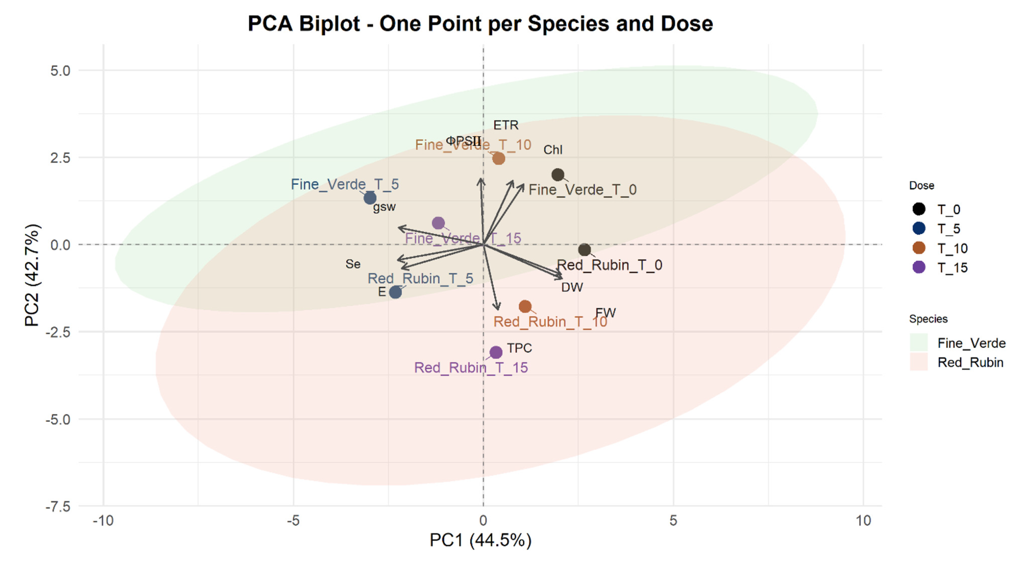
Principal Component Analysis (PCA) was performed to explore the multivariate distribution of morphological and physiological traits in both basil cultivars under Se biofortification. DM, plant height, SPAD, C, and S were not included in the PCA because, after evaluating sampling adequacy with the Kaiser-Meyer-Olkin (KMO) test, each of these variables yielded a value < 0.50, indicating that they were not suitable for reliably explaining the variance between the FV and RR.
The PCA biplot (Figure 2) illustrates the relationships between the treatments and measured variables across PC1 (44.5%) and PC2 (42.7%), which together explain 87.2% of the total variance in the data. FV_Se_15 was projected in the upper left quadrant in association with gsw, indicating a strong stomatal response to high Se exposure but without alignment with productivity-related vectors. FV_Se_5 and FV_Se_10 were positioned on the upper half of the plot near ΦPSII and ETR, suggesting that moderate Se application promoted photochemical and light-use efficiencies. FV_Se_0 was closely aligned with the Chl content, indicating high pigment accumulation under untreated conditions. The RR cultivar showed a distinct pattern, with RR_5 and RR_10 grouped in the upper portion of the biplot near the DW, FW, and TPC vectors. This clustering suggests that moderate Se doses simultaneously stimulated FW and TPC production in the RR cultivar. RR_Se_15 was in the lower left quadrant, near Se and E, and Se uptake occurred under high-Se conditions. In contrast, RR_Se_0 was aligned with Chl content and ETR, suggesting a balanced photochemical status in the absence of Se treatment. FV samples tended to be distributed more broadly, reflecting the variation in photosynthetic traits under Se influence, whereas RR showed a tighter association between intermediate Se doses and FW antioxidant accumulation.

4. Discussion

Se biofortification is promoted as this trace element is essential for human health yet scarce in diets globally, affecting one billion people and driving efforts to enrich plants with safe doses of Se [40,41]. The application presents challenges due to the narrow margin between benefits and toxicity. At optimal micromolar levels, Se enhances plant growth and antioxidant capacity [42]. However, excess Se causes phytotoxicity, including chlorosis, oxidative stress, and growth inhibition [42]. Significant genotypic variations exist among species in Se uptake and tolerance [43], with closely related cultivars showing different responses to Se supplementation to stress [44]. Therefore, Se application must be optimized for each cultivar to achieve effective biofortification without compromising plant growth.
Dose-dependent growth responses. Our study demonstrated a dose-dependent response to Se in plant growth parameters, with notable varietal differences between the two basil cultivars. Low to moderate concentrations of Se_5 and Se_10 generally promoted biomass accumulation in both cultivars, with the RR cultivar showing a particularly strong positive response [4,45]. In contrast, Se_15 treatment resulted in significant growth inhibition, with the FV cultivar demonstrating greater sensitivity to Se toxicity than the RR cultivar did. This differential response was evidenced by marked reductions in both FW and DW, indicating that the threshold between beneficial and toxic Se levels varies substantially among cultivars. This growth enhancement at optimal Se levels is consistent with previous findings that suggest that Se can be beneficial at low concentrations, possibly due to its antioxidant properties and role in stress tolerance mechanisms [46,47]. Similar growth reductions following Se supplementation have been documented in various species, including Spinacia oleracea L. [48] and Vicia faba L. [49], Brassica juncea L. [50], Stevia rebaudiana Bertoni [51], Ocimum basilicum L., and Mentha × piperita L. [16]. The underlying mechanisms governing the dual effects of Se on plant growth likely involve complex interactions between the biochemical properties of Se and cellular metabolism [52]. At low concentrations, Se may enhance antioxidant enzyme activity, improve photosynthetic efficiency, and confer protection against various abiotic stresses [5]. Conversely, excessive Se accumulation can disrupt protein synthesis through the non-specific incorporation of selenoamino acids, generate reactive oxygen species, and interfere with sulphur metabolism, ultimately leading to cellular dysfunction and growth inhibition [42]. These observations indicate that the varying responses of different basil cultivars, along with the lack of a significant yield penalty at moderate Se doses, may highlight a delicate balance between the stimulating effects of Se and the early signs of metabolic stress in plants. This balance may contribute to the overall resilience of basil plants during Se biofortification [53].
Se concentrations and nutritional implications. In agreement with these findings, the Se concentration in basil leaves increased progressively in response to Se biofortification, with the highest values observed at Se_15. This finding is consistent with several studies indicating that applying Se, whether in the culture medium or through foliar spraying, results in a dose-dependent increase in Se concentration in plant tissue [11,54]. The average Se concentration in dry tissue was 24.17 mg kg−1 DW in FV and 22.05 mg kg−1 DW in RR cultivars. These data align with those of previous studies [16,44], in which the Se concentration in basil plants was 10–30 mg kg−1 DW. The Se concentrations found in our study, with a maximum of approximately 38 mg kg−1 DW, confirm that basil plants fall within the typical range of non-hyperaccumulating plants (<100 mg Se kg−1 DW) [55]. The slight difference in Se concentration between the cultivars, FV and RR, may have been influenced by genetic variability. This observation is supported by Mezeyová et al. (2019) [56], who reported varying Se concentrations (at the same Se biofortification dose) in three O. basilicum L. cultivars: ‘Purple Ruffles’, ‘Red Rubin’ and ‘Dark Green’ (2.34 to 14.38 mg Se kg−1 DW).
From a nutritional perspective, dietary supplements derived from Se-enriched aromatic plants can effectively meet the daily Se requirement [14,16,57]. The recommended daily intake of Se for adults ranges from 55–70 μg per day, depending on age and gender [58]. Based on our findings, consuming approximately 2–3 g (DW) of Se-biofortified basil could potentially contribute to meeting daily Se requirements [44]. Overall, the Se concentrations detected in both FV and RR cultivars confirmed the effective uptake and biofortification potential of basil under controlled Se supplementation conditions. These results highlight the suitability of basil as a functional crop for dietary Se enrichment without exceeding toxicity thresholds, making it a promising candidate for addressing Se deficiency in populations with inadequate Se intake [13,44].
Effects on chlorophyll and photosynthetic performance. When considering the critical physiological parameters that define plant responses to Se biofortification, SPAD values and Chl content serve as key indicators of photosynthetic capacity and overall plant health [59]. In this study, SPAD values remained relatively stable and were not significantly affected by Se treatment across all concentrations tested. Chl content exhibited a dose-dependent reduction pattern. The reduction was minimal from the control group to the Se_5 dose but significantly decreased with higher Se biofortification doses (Se_10 and Se_15). Se may interfere with sulfhydryl group-containing enzymes, such as porphobilinogen synthetase (PBG-synthase), a metal-sensitive enzyme crucial for chlorophyll synthesis regulation [48,60]. Additionally, high Se doses can induce oxidative stress by promoting reactive oxygen species (ROS) accumulation, including hydrogen peroxide (H2O2), superoxide anion (O2), and hydroxyl radicals (OH•), which directly damage chloroplast membranes and the photosynthetic machinery [61]. This effect could lead to a degradation of Chl content and reduced photosynthetic efficiency. Our findings align with previous research demonstrating the dose-dependent effects of Se on photosynthetic parameters, as Sali et al. (2018) [62] examined the impact of Se on Chl content in Zea mays L. plants. The findings indicated that the lowest Se dose of 1.3 mg L−1, which is comparable to our Se_5 dose, did not have a negative effect. However, increasing Se doses reduced the Chl content. The detrimental effects of increasing Se doses on Chl have been consistently documented across multiple plant species, confirming the universal nature of this physiological response included in O. basilicum L. [44]. Basil plants treated with different concentrations of Se (0, 4, 8, and 12 mg Se L−1) showed a reduction in Chl content compared to the control and the group with the lowest Se concentration. This pattern closely mirrors our findings, where Chl content degradation became significant only at Se_10 and Se_15 concentrations in the present study. Similar results in Chl content reduction were observed in Valerianella locusta L. [55] and Solanum lycopersicum L. [63]. Both the literature and our data concur that only low doses of Se (approximately 5 µM) are physiologically tolerated. In contrast, exposure to higher concentrations quickly destabilizes Chl content, confirming that Chl content serves as an early biomarker of Se biofortification stress.
The Se_15 dose reduced the Chl content in basil leaves [48], which provides a mechanistic explanation for the concurrent decline in key photosynthetic performance parameters, such as ETR and ΦPSII. The fundamental relationship between Chl content and photosynthetic efficiency stems from the central role of chlorophyll as the primary light-harvesting pigment, where absorbed photons drive photochemical reactions within PSII, ultimately determining the rate of ETR and overall photosynthetic carbon fixation [64,65]. In our study, ETR and ΦPSII were also reduced by increasing Se doses and were found to be strongly correlated with Chl content in relation to Se concentration. These findings align with previous research on O. tenuiflorum, which found a significant positive correlation between net photosynthesis (Pn) and the maximum PSII efficiency, photochemical efficiency of ΦPSII, and ETR [66]. This concordance across different basil species indicates that the tight coupling between Chl content and photosynthetic ETR represents a conserved physiological relationship within the genus Ocimum. Another study by Vrakas et al. (2021) [67] has also reported that a lower Chl content in the leaves of O. basilicum L., Salvia officinalis L., and Mentha × piperita L. corresponded to a decrease in Pn. Consequently, the studies conducted by Vrakas et al. (2021) [67] and Chutimanukul et al. (2022b) [30] demonstrated a significant positive correlation between Chl content and Pn in basil. This finding underscores that Pn is a direct result of photosynthetic efficiency and is closely linked to the effectiveness of light-dependent reactions, such as those assessed by the ETR and PSII efficiency.
Despite the reduction in Chl content, ΦPSII, and ETR due to Se application in our study, gsw and E moved in the opposite direction. According to the literature, the effects of Se are complex and sometimes contradictory, as both basil cultivars and dosage strongly modulate the response [51,68]. In the present study, the two basil cultivars displayed a comparable increase in gsw and E with increasing Se concentrations. In line with the findings of Song et al. (2024) [63] and Sepehri & Gharehbaghli (2019) [69] on Solanum lycopersicum L. and Allium sativum L., respectively, these parameters were higher under Se biofortification than under control conditions. Their concomitant increase is further supported by the positive correlations (Figure 1a,b) recorded between gsw and E [70] and the Se concentration in the leaves. However, unlike the above-mentioned studies, gsw and E in our plants continued to increase even at the Se_10 and Se_15 doses, whereas ΦPSII and ETR decreased in our study. This divergence is most likely a consequence of dose-dependent Se phytotoxicity [48,68]. Consistent with our results, previous studies have reported that low Se doses do not induce toxicity or nutrient imbalance in plants [7]. These studies demonstrate that foliar or nutrient solution applications of ≈1 mg L−1 Se enhance photosynthetic performance by increasing Chl [7,48], ΦPSII, ETR, gsw, and E [64,71,72], which is what was observed in our Se_5 treatment.
Impact on secondary metabolism: phenolic compounds. Elevated Se supply, applied here as a stepwise foliar biofortification, coincided with a gradual rise in TPC in leaves, an adjustment that plants commonly deploy when confronted with abiotic challenges. This physiological adjustment involves increasing the activity of the shikimate and phenylpropanoid pathways, which are the main biosynthetic routes for producing phenolic compounds under stressful conditions [16]. Although the mean TPC of Se-enriched basil did not differ significantly from that of the control, the numerical increase aligns with trends observed in Cucumis sativus L., as reported by Amerian et al. (2024) [73], when given 0–10 mg L−1 sodium selenate. In accordance with these findings, Profico et al. (2025) [16] observed a similar situation for Mentha × piperita L. and O. basilicum L. when treated with 0–10 mg L−1 sodium selenate. Similar enhancements, approximately 17% in total phenolics and flavonoids, were described in Solanum lycopersicum L. exposed to Se [74]. Studies on Zea mays L. and Anethum graveolens L. have demonstrated that Se treatments can enhance the antioxidant phenolic content while simultaneously mitigating stress-induced cellular damage, even under challenging conditions, such as salinity stress [73]. Collectively, the literature indicates that Se enrichment can improve the biochemical quality of crops by stimulating secondary metabolite synthesis, thereby reinforcing stress tolerance [75]. Regarding cultivar-specific responses, the consistently higher TPC observed in the red-pigmented cultivar ‘Red Rubin’ compared to the green ‘Fine Verde’ reflects the fundamental differences in phenolic content. These differences stem from variations in the activity of the phenylpropanoid pathway between cultivars, where anthocyanin biosynthesis in red-pigmented cultivars requires increased production of phenolic precursors [76,77].

5. Conclusions

This study demonstrates that foliar biofortification with sodium selenate can enrich O. basilicum L. with nutritionally useful Se while maintaining its agronomic performance. Taken together, these results indicate that a single foliar dose of 5 µM represents the optimal compromise between biofortification and plant health, providing enough Se to cover the human daily requirement with a modest serving of fresh herbs while avoiding phytotoxicity. Integrating such a protocol into commercial hydroponic basil production would provide an easy, plant-based Se source for consumers, without compromising grower productivity. Future studies should refine the timing and frequency of low-dose foliar applications, screen additional cultivars for Se tolerance, evaluate promising Se doses in other horticultural species cultivated under TCEA systems, and further investigate the speciation of organic Se compounds and their relationship with plant physiological responses. These findings could ultimately be validated through field experiments on a larger scale to assess their applicability under commercial multi-harvest conditions.

Author Contributions

Conceptualisation, C.M.P. and S.N.; methodology, C.M.P., S.F.S.K. and S.H.; software, C.M.P. and S.F.S.K.; validation, S.N. and V.R.; formal analysis, S.N. and S.H.; investigation, C.M.P. and S.F.S.K.; data curation, C.M.P.; writing—original draft preparation, C.M.P. and S.F.S.K.; writing—review and editing, C.M.P., S.H., V.R. and S.N.; Supervision, S.N.; project administration, S.N.; funding acquisition, S.N. All authors have read and agreed to the published version of the manuscript.

Funding

This work was supported by the Italian Ministry of Education and Research (MUR), within the call for Research Projects of National Interest (PRIN), within the project “VFARM—Sustainable Vertical Farming” (Project code: 2020ELWM82, CUP: J33C20002350001).

Data Availability Statement

The raw data supporting the conclusions of this article will be made available by the authors on request.

Conflicts of Interest

The authors declare no conflicts of interest.

References

  1. Sarwar, N.; Akhtar, M.; Kamran, M.A.; Imran, M.; Riaz, M.A.; Kamran, K.; Hussain, S. Selenium Biofortification in Food Crops: Key Mechanisms and Future Perspectives. J. Food Compos. Anal. 2020, 93, 103615. [Google Scholar] [CrossRef]
  2. Barchielli, G.; Capperucci, A.; Tanini, D. The Role of Selenium in Pathologies: An Updated Review. Antioxidants 2022, 11, 251. [Google Scholar] [CrossRef] [PubMed]
  3. Rayman, M.P. The Argument for Increasing Selenium Intake. Proc. Nutr. Soc. 2002, 61, 203–215. [Google Scholar] [CrossRef] [PubMed]
  4. Boldrin, P.F.; Faquin, V.; Clemente, A.D.C.S.; De Andrade, T.; Guilherme, L.R.G. Genotypic Variation and Biofortification with Selenium in Brazilian Wheat Cultivars. J. Environ. Qual. 2018, 47, 1371–1379. [Google Scholar] [CrossRef] [PubMed]
  5. Lara, T.S.; de Lima Lessa, J.H.; de Souza, K.R.D.; Corguinha, A.P.B.; Martins, F.A.D.; Lopes, G.; Guilherme, L.R.G. Selenium Biofortification of Wheat Grain via Foliar Application and Its Effect on Plant Metabolism. J. Food Compos. Anal. 2019, 81, 10–18. [Google Scholar] [CrossRef]
  6. Alam, A.; Bibi, F.; Deshwal, K.; Sahariya, A.; Bhardwaj, C.; Emmanuel, I. Biofortification of Staple Crops to Eradicate Hidden Hunger: A Review. Nat. Resour. Hum. Health 2021, 2, 91–99. [Google Scholar] [CrossRef]
  7. Hossain, A.; Skalicky, M.; Brestic, M.; Maitra, S.; Sarkar, S.; Ahmad, Z.; Vemuri, H.; Garai, S.; Mondal, M.; Bhatt, R.; et al. Selenium Biofortification: Roles, Mechanisms, Responses and Prospects. Molecules 2021, 26, 881. [Google Scholar] [CrossRef]
  8. Carucci, F.; Moreno-Martín, G.; Madrid-Albarrán, Y.; Gatta, G.; De Vita, P.; Giuliani, M.M. Selenium Agronomic Biofortification of Durum Wheat Fertilized with Organic Products: Se Content and Speciation in Grain. Agronomy 2022, 12, 2492. [Google Scholar] [CrossRef]
  9. Ramos, D.P.; Tavares, T.C.D.O.; Sousa, S.A.D.; Nascimento, V.L.; Martinez, R.A.S.; Chagas Junior, A.F.; Fidelis, R.R. Agronomic Biofortification of Cowpea with Selenium by Foliar Fertilization: Effect of Doses in Three Cultivars. J. Plant Nutr. 2020, 43, 538–547. [Google Scholar] [CrossRef]
  10. Tavan, M.; Wee, B.; Fuentes, S.; Pang, A.; Brodie, G.; Viejo, C.G.; Gupta, D. Biofortification of Kale Microgreens with Selenate-Selenium Using Two Delivery Methods: Selenium-Rich Soilless Medium and Foliar Application. Sci. Hortic. 2024, 323, 112522. [Google Scholar] [CrossRef]
  11. Viltres-Portales, M.; Sánchez-Martín, M.-J.; Llugany, M.; Boada, R.; Valiente, M. Selenium Biofortification of Microgreens: Influence on Phytochemicals, Pigments and Nutrients. Plant Physiol. Biochem. 2024, 206, 108283. [Google Scholar] [CrossRef] [PubMed]
  12. Wang, M.; Zhou, F.; Cheng, N.; Chen, P.; Ma, Y.; Zhai, H.; Qi, M.; Liu, N.; Liu, Y.; Meng, L.; et al. Soil and Foliar Selenium Application: Impact on Accumulation, Speciation, and Bioaccessibility of Selenium in Wheat (Triticum aestivum L.). Front. Plant Sci. 2022, 13, 988627. [Google Scholar] [CrossRef] [PubMed]
  13. Puccinelli, M.; Malorgio, F.; Incrocci, L.; Rosellini, I.; Pezzarossa, B. Effects of Individual and Simultaneous Selenium and Iodine Biofortification of Baby-Leaf Lettuce Plants Grown in Two Different Hydroponic Systems. Horticulturae 2021, 7, 590. [Google Scholar] [CrossRef]
  14. Sheikhi, H.; Nicola, S.; Delshad, M.; Bulgari, R. Sodium Selenate Biofortification, through Seed Priming, on Dill Microgreens Grown in Two Different Cultivation Systems. Front. Plant Sci. 2024, 15, 1474420. [Google Scholar] [CrossRef]
  15. Poblaciones, M.J.; García-Latorre, C.; Velazquez, R.; Broadley, M.R. Effects of Selenate Application on Growth, Nutrient Bioaccumulation, and Bioactive Compounds in Broccoli (Brassica oleracea var. italica L.). Horticulturae 2024, 10, 808. [Google Scholar] [CrossRef]
  16. Profico, C.M.; Hassanpour, M.; Hazrati, S.; Ertani, A.; Mollaei, S.; Nicola, S. Sodium Selenate Biofortification of Basil (Ocimum basilicum L.) and Peppermint (Mentha × piperita L.) Plants Grown in a Floating System under Salinity Stress. J. Agric Food Res. 2025, 21, 101842. [Google Scholar] [CrossRef]
  17. Santos, E.F.; Filho, E.C.; Fontes, L.E.M.F.; Silva, M.A.P.; Silva, G.N.; Oliveira, A.A.; De Moura Rocha, M.; Silva, V.M.; Reis, A.R. Selenium Agronomic Biofortification and Genotypic Variability in Physiological Responses of Cowpea Plants under Field Conditions. Acta Physiol. Plant. 2025, 47, 18. [Google Scholar] [CrossRef]
  18. Souza, G.A.; Hart, J.J.; Carvalho, J.G.; Rutzke, M.A.; Albrecht, J.C.; Guilherme, L.R.G.; Kochian, L.V.; Li, L. Genotypic Variation of Zinc and Selenium Concentration in Grains of Brazilian Wheat Lines. Plant Sci. 2014, 224, 27–35. [Google Scholar] [CrossRef]
  19. Banerjee, S.; Roy, P.; Nandi, S.; Roy, S. Advanced Biotechnological Strategies towards the Development of Crops with Enhanced Micronutrient Content. Plant Growth Regul. 2023, 100, 355–371. [Google Scholar] [CrossRef]
  20. Silva, V.M.; Nardeli, A.J.; Mendes, N.A.C.; Gabriel Filho, L.R.A.; Gabriel, C.P.C.; Reis, A.R.D. Does Selenium Application Increase the Yield of Cowpea Plants? Evidence from 29 Genotypes on Ureides and Sugar Index Affecting the Yield. J. Soil Sci. Plant Nutr. 2023, 23, 5899–5908. [Google Scholar] [CrossRef]
  21. Ramos, S.J.; Yuan, Y.; Faquin, V.; Guilherme, L.R.G.; Li, L. Evaluation of Genotypic Variation of Broccoli (Brassica oleracea var. italic) in Response to Selenium Treatment. J. Agric. Food Chem. 2011, 59, 3657–3665. [Google Scholar] [CrossRef]
  22. Šindelářová, K.; Száková, J.; Tremlová, J.; Mestek, O.; Praus, L.; Kaňa, A.; Najmanová, J.; Tlustoš, P. The Response of Broccoli (Brassica oleracea convar. italica) Varieties on Foliar Application of Selenium: Uptake, Translocation, and Speciation. Food Addit. Contam. Part A 2015, 32, 150928143022009. [Google Scholar] [CrossRef]
  23. Xue, Y.-F.; Li, X.-J.; Yan, W.; Miao, Q.; Zhang, C.-Y.; Huang, M.; Sun, J.-B.; Qi, S.-J.; Ding, Z.-H.; Cui, Z.-L. Biofortification of Different Maize Cultivars with Zinc, Iron and Selenium by Foliar Fertilizer Applications. Front. Plant Sci. 2023, 14, 1144514. [Google Scholar] [CrossRef]
  24. Ciriello, M.; Formisano, L.; Kyriacou, M.; Soteriou, G.A.; Graziani, G.; De Pascale, S.; Rouphael, Y. Zinc Biofortification of Hydroponically Grown Basil: Stress Physiological Responses and Impact on Antioxidant Secondary Metabolites of Genotypic Variants. Front. Plant Sci. 2022, 13, 1049004. [Google Scholar] [CrossRef] [PubMed]
  25. Ciriello, M.; Formisano, L.; Zarrelli, A.; Corrado, G.; Kyriacou, M.C.; De Pascale, S.; Rouphael, Y. Zinc Biofortification of Genovese Basil: Influence on Mineral Profile and Estimated Daily Intake in Adults and Children. Food Res. Int. 2023, 164, 112374. [Google Scholar] [CrossRef] [PubMed]
  26. Kozai, T.; Niu, G. Role of the Plant Factory with Artificial Lighting (PFAL) in Urban Areas. In Plant Factory; Elsevier: Amsterdam, The Netherlands, 2020; pp. 7–34. ISBN 978-0-12-816691-8. [Google Scholar]
  27. Hayashi, E. Advances in Plant Factories: New Technologies in Indoor Vertical Farming; Kozai, T., Ed.; Burleigh Dodds Series in Agricultural Science; Burleigh Dodds Science Publishing: Cambridge, UK, 2023; ISBN 978-1-80146-316-4. [Google Scholar]
  28. Azizah, N.S.; Irawan, B.; Kusmoro, J.; Safriansyah, W.; Farabi, K.; Oktavia, D.; Doni, F.; Miranti, M. Sweet Basil (Ocimum basilicum L.)―A Review of Its Botany, Phytochemistry, Pharmacological Activities, and Biotechnological Development. Plants 2023, 12, 4148. [Google Scholar] [CrossRef] [PubMed]
  29. Romano, R.; De Luca, L.; Aiello, A.; Pagano, R.; Di Pierro, P.; Pizzolongo, F.; Masi, P. Basil (Ocimum basilicum L.) Leaves as a Source of Bioactive Compounds. Foods 2022, 11, 3212. [Google Scholar] [CrossRef]
  30. Chutimanukul, P.; Wanichananan, P.; Janta, S.; Toojinda, T.; Darwell, C.T.; Mosaleeyanon, K. The Influence of Different Light Spectra on Physiological Responses, Antioxidant Capacity and Chemical Compositions in Two Holy Basil Cultivars. Sci. Rep. 2022, 12, 588. [Google Scholar] [CrossRef]
  31. Viršilė, A.; Laužikė, K.; Sutulienė, R.; Brazaitytė, A.; Kudirka, G.; Samuolienė, G. Distinct Impacts of UV-A Light Wavelengths on Nutraceutical and Mineral Contents in Green and Purple Basil Cultivated in a Controlled Environment. Horticulturae 2023, 9, 1168. [Google Scholar] [CrossRef]
  32. Chutimanukul, P.; Jindamol, H.; Thongtip, A.; Korinsak, S.; Romyanon, K.; Toojinda, T.; Darwell, C.T.; Wanichananan, P.; Panya, A.; Kaewsri, W.; et al. Physiological Responses and Variation in Secondary Metabolite Content among Thai Holy Basil Cultivars (Ocimum tenuiflorum L.) Grown under Controlled Environmental Conditions in a Plant Factory. Front. Plant Sci. 2022, 13, 1008917. [Google Scholar] [CrossRef]
  33. Dou, H.; Niu, G.; Gu, M. Pre-Harvest UV-B Radiation and Photosynthetic Photon Flux Density Interactively Affect Plant Photosynthesis, Growth, and Secondary Metabolites Accumulation in Basil (Ocimum basilicum) Plants. Agronomy 2019, 9, 434. [Google Scholar] [CrossRef]
  34. Squadrone, S.; Brizio, P.; Stella, C.; Prearo, M.; Pastorino, P.; Serracca, L.; Ercolini, C.; Abete, M.C. Presence of Trace Metals in Aquaculture Marine Ecosystems of the Northwestern Mediterranean Sea (Italy). Environ. Pollut. 2016, 215, 77–83. [Google Scholar] [CrossRef] [PubMed]
  35. Squadrone, S.; Brizio, P.; Battuello, M.; Nurra, N.; Sartor, R.M.; Benedetto, A.; Pessani, D.; Abete, M.C. A First Report Of Rare Earth Elements in Northwestern Mediterranean Seaweeds. Mar. Pollut. Bull. 2017, 122, 236–242. [Google Scholar] [CrossRef] [PubMed]
  36. Lichtenthaler, H.K.; Wellburn, A.R. Determinations of Total Carotenoids and Chlorophylls a and b of Leaf Extracts in Different Solvents. Biochem. Soc. Trans. 1983, 11, 591–592. [Google Scholar] [CrossRef]
  37. Arnaldos, T.L.; Ferrer, M.A.; García, A.A.C.; Muńoz, R. Changes in Peroxidase Activity and Isoperoxidase Pattern during Strawberry (Fragaria × ananassa) Callus Development. J. Plant Physiol. 2002, 159, 429–435. [Google Scholar] [CrossRef]
  38. Meenakshi, S.; Gnanambigai, D.M. Total Flavanoid and In Vitro Antioxidant Activity of Two Seaweeds of Rameshwaram Coast. Glob. J. Pharmacol. 2009, 3, 59–62. [Google Scholar]
  39. Cataldo, D.A.; Maroon, M.; Schrader, L.E.; Youngs, V.L. Rapid Colorimetric Determination of Nitrate in Plant Tissue by Nitration of Salicylic Acid. Commun. Soil Sci. Plant Anal. 1975, 6, 71–80. [Google Scholar] [CrossRef]
  40. Danso, O.P.; Asante-Badu, B.; Zhang, Z.; Song, J.; Wang, Z.; Yin, X.; Zhu, R. Selenium Biofortification: Strategies, Progress and Challenges. Agriculture 2023, 13, 416. [Google Scholar] [CrossRef]
  41. Cruz, F.J.R. Selenium in Plants: Biofortification, Toxicity, and Redox State Balance. In The Power of Antioxidants-Unleashing Nature’s Defense Against Oxidative Stress; Novo Barros, A., Cristina Santos Abraão, A., Eds.; IntechOpen: London, UK, 2025; Volume 60, ISBN 978-0-85466-206-7. [Google Scholar]
  42. Somagattu, P.; Chinnannan, K.; Yammanuru, H.; Reddy, U.K.; Nimmakayala, P. Selenium Dynamics in Plants: Uptake, Transport, Toxicity, and Sustainable Management Strategies. Sci. Total Environ. 2024, 949, 175033. [Google Scholar] [CrossRef]
  43. Sunic, K.; Spanic, V. Genetic Biofortification of Winter Wheat with Selenium (Se). Plants 2024, 13, 1816. [Google Scholar] [CrossRef]
  44. Puccinelli, M.; Pezzarossa, B.; Rosellini, I.; Malorgio, F. Selenium Enrichment Enhances the Quality and Shelf Life of Basil Leaves. Plants 2020, 9, 801. [Google Scholar] [CrossRef]
  45. Feng, R.; Wei, C.; Tu, S. The Roles of Selenium in Protecting Plants against Abiotic Stresses. Environ. Exp. Bot. 2013, 87, 58–68. [Google Scholar] [CrossRef]
  46. Bian, Z.; Lei, B.; Cheng, R.; Wang, Y.; Li, T.; Yang, Q. Selenium Distribution and Nitrate Metabolism in Hydroponic Lettuce (Lactuca sativa L.): Effects of Selenium forms and Light Spectra. J. Integr. Agric. 2020, 19, 133–144. [Google Scholar] [CrossRef]
  47. Hawrylak-Nowak, B. Selenite is More Efficient than Selenate in Alleviation of Salt Stress in Lettuce Plants. Acta Biol. Cracoviensia. Ser. Bot. 2015, 57, 49–54. [Google Scholar] [CrossRef]
  48. Saffaryazdi, A.; Lahouti, M.; Ganjeali, A.; Bayat, H. Impact of Selenium Supplementation on Growth and Selenium Accumulation on Spinach (Spinacia oleracea L.) Plants. Not. Sci. Biol. 2012, 4, 95–100. [Google Scholar] [CrossRef]
  49. Mroczek-Zdyrska, M.; Strubińska, J.; Hanaka, A. Selenium Improves Physiological Parameters and Alleviates Oxidative Stress in Shoots of Lead-Exposed Vicia faba L. Minor Plants Grown Under Phosphorus-Deficient Conditions. J. Plant Growth Regul. 2017, 36, 186–199. [Google Scholar] [CrossRef]
  50. Golubkina, N.; Kekina, H.; Caruso, G. Yield, Quality and Antioxidant Properties of Indian Mustard (Brassica juncea L.) in Response to Foliar Biofortification with Selenium and Iodine. Plants 2018, 7, 80. [Google Scholar] [CrossRef]
  51. Borbély, P.; Molnár, Á.; Valyon, E.; Ördög, A.; Horváth-Boros, K.; Csupor, D.; Fehér, A.; Kolbert, Z. The Effect of Foliar Selenium (Se) Treatment on Growth, Photosynthesis, and Oxidative-Nitrosative Signalling of Stevia rebaudiana Leaves. Antioxidants 2021, 10, 72. [Google Scholar] [CrossRef]
  52. Gupta, M.; Gupta, S. An Overview of Selenium Uptake, Metabolism, and Toxicity in Plants. Front. Plant Sci. 2017, 7, 2074. [Google Scholar] [CrossRef]
  53. Wang, Q.; Hu, J.; Hu, H.; Li, Y.; Xiang, M.; Wang, D. Integrated Eco-Physiological, Biochemical, and Molecular Biological Analyses of Selenium Fortification Mechanism in Alfalfa. Planta 2022, 256, 114. [Google Scholar] [CrossRef]
  54. Brito, C.; Andrade, S.; Ferreira, H.; Matos, C.; Martins, S.; Moutinho-Pereira, J. The Synergetic Effect of Light Spectra and Selenium Supplementation on Eruca sativa Mill. Growth and Physiological and Metabolic Responses. Horticulturae 2024, 10, 511. [Google Scholar] [CrossRef]
  55. Skrypnik, L.; Styran, T.; Savina, T.; Golubkina, N. Effect of Selenium Application and Growth Stage at Harvest on Hydrophilic and Lipophilic Antioxidants in Lamb’s Lettuce (Valerianella locusta L. Laterr.). Plants 2021, 10, 2733. [Google Scholar] [CrossRef]
  56. Mezeyová, I.; Hegedűsová, A.; Hegedűs, O.; Farkaš, J.; Šlosár, M.; Mezey, J. Effect of Foliar Application of Selenium on Its Uptake and Yields in Basils. AGRFOR Int. J. 2019, 4, 111–119. [Google Scholar] [CrossRef]
  57. Kiran, A.; Wakeel, A.; Mahmood, K.; Mubaraka, R.; Hafsa; Haefele, S.M. Biofortification of Staple Crops to Alleviate Human Malnutrition: Contributions and Potential in Developing Countries. Agronomy 2022, 12, 452. [Google Scholar] [CrossRef]
  58. Schiavon, M.; Nardi, S.; dalla Vecchia, F.; Ertani, A. Selenium Biofortification in the 21st Century: Status and Challenges for Healthy Human Nutrition. Plant Soil 2020, 453, 245–270. [Google Scholar] [CrossRef] [PubMed]
  59. Wei, L.; Lu, L.; Shang, Y.; Ran, X.; Liu, Y.; Fang, Y. Can SPAD Values and CIE L*a*b* Scales Predict Chlorophyll and Carotenoid Concentrations in Leaves and Diagnose the Growth Potential of Trees? An Empirical Study of Four Tree Species. Horticulturae 2024, 10, 548. [Google Scholar] [CrossRef]
  60. Lin, L.; Sun, J.; Cui, T.; Zhou, X.; Liao, M.; Huan, Y.; Yang, L.; Wu, C.; Xia, X.; Wang, Y.; et al. Selenium Accumulation Characteristics of Cyphomandra betacea (Solanum betaceum) Seedlings. Physiol. Mol. Biol. Plants 2020, 26, 1375–1383. [Google Scholar] [CrossRef]
  61. Trippe, R.C.; Pilon-Smits, E.A.H. Selenium Transport and Metabolism in Plants: Phytoremediation and Biofortification Implications. J. Hazard. Mater. 2021, 404, 124178. [Google Scholar] [CrossRef]
  62. Sali, A.; Zeka, D.; Fetahu, S.; Rusinovci, I.; Kaul, H.-P. Selenium Supply Affects Chlorophyll Concentration and Biomass Production of Maize (Zea mays L.). J. Land Manag. Food Environ. 2018, 69, 249–255. [Google Scholar] [CrossRef]
  63. Song, J.; Xin, L.; Gao, F.; Liu, H.; Wang, X. Effects of Foliar Selenium Application on Oxidative Damage and Photosynthetic Properties of Greenhouse Tomato under Drought Stress. Plants 2024, 13, 302. [Google Scholar] [CrossRef]
  64. Su, L.; Xie, Y.; He, Z.; Zhou, X.; Liu, Y.; Zhang, R.; Li, C. Selenium Mitigates Cd-Induced Oxidative Stress and Photosynthesis Inhibition in Two Cherry Tomato Cultivars. J. Soil Sci. Plant Nutr. 2022, 22, 3212–3227. [Google Scholar] [CrossRef]
  65. Zou, Z.; Li, M.; Jia, R.; Zhao, H.; He, P.; Zhang, Y.; Guo, A. Genes Encoding Light-Harvesting Chlorophyll a/b-Binding Proteins in Papaya (Carica papaya L.) and Insight into Lineage-Specific Evolution in Brassicaceae. Gene 2020, 748, 144685. [Google Scholar] [CrossRef] [PubMed]
  66. Ashenafi, E.L.; Nyman, M.C.; Holley, J.M.; Mattson, N.S.; Rangarajan, A. Phenotypic Plasticity and Nutritional Quality of Three Kale Cultivars (Brassica oleracea L. var. acephala) under Field, Greenhouse, and Growth Chamber Environments. Environ. Exp. Bot. 2022, 199, 104895. [Google Scholar] [CrossRef]
  67. Vrakas, K.; Florou, E.; Koulopoulos, A.; Zervoudakis, G. Physiological Responses of Ocimum basilicum, Salvia officinalis, and Mentha piperita to Leaf Wounding. Plants 2021, 10, 1019. [Google Scholar] [CrossRef]
  68. Khan, Z.; Thounaojam, T.C.; Chowdhury, D.; Upadhyaya, H. The Role of Selenium and Nano Selenium on Physiological Responses in Plant: A Review. Plant Growth Regul. 2023, 100, 409–433. [Google Scholar] [CrossRef]
  69. Sepehri, A.; Gharehbaghli, N. Selenium Alleviate Cadmium Toxicity by Improving Nutrient Uptake, Antioxidative and Photosynthetic Responses of Garlic. Russ. J. Plant Physiol. 2019, 66, 152–159. [Google Scholar] [CrossRef]
  70. Qi, Z.; Huang, X.; Peng, Y.; Wu, H.; Xu, Z.; Tan, B.; Zhong, Y.; Zhu, P.; Gong, W.; Chen, G.; et al. Identifying Superior Growth and Photosynthetic Traits in Eighteen Oak Varieties for Southwest China. Forests 2024, 15, 2006. [Google Scholar] [CrossRef]
  71. Sharifi, P.; Amirnia, R.; Torkian, M.; Bidabadi, S.S. Protective Role of Exogenous Selenium on Salinity-Stressed Stachys Byzantine Plants. J. Soil Sci. Plant Nutr. 2021, 21, 2660–2672. [Google Scholar] [CrossRef]
  72. Yadav, S.; Sharma, S.; Sharma, K.D.; Dhansu, P.; Devi, S.; Preet, K.; Ahlawat, P.; Kamboj, P.; Rani, P.; Rani, B.; et al. Selenium Mediated Alterations in Physiology of Wheat under Different Soil Moisture Levels. Sustainability 2023, 15, 1771. [Google Scholar] [CrossRef]
  73. Amerian, M.; Palangi, A.; Gohari, G.; Ntatsi, G. Enhancing Salinity Tolerance in Cucumber through Selenium Biofortification and Grafting. BMC Plant Biol. 2024, 24, 24. [Google Scholar] [CrossRef]
  74. Morales-Espinoza, M.C.; Cadenas-Pliego, G.; Pérez-Alvarez, M.; Hernández-Fuentes, A.D.; Cabrera De La Fuente, M.; Benavides-Mendoza, A.; Valdés-Reyna, J.; Juárez-Maldonado, A. Se Nanoparticles Induce Changes in the Growth, Antioxidant Responses, and Fruit Quality of Tomato Developed under NaCl Stress. Molecules 2019, 24, 3030. [Google Scholar] [CrossRef]
  75. Santander, C.; Vidal, G.; Ruiz, A.; Vidal, C.; Cornejo, P. Salinity Eustress Increases the Biosynthesis and Accumulation of Phenolic Compounds That Improve the Functional and Antioxidant Quality of Red Lettuce. Agronomy 2022, 12, 598. [Google Scholar] [CrossRef]
  76. Flanigan, P.M.; Niemeyer, E.D. Effect of Cultivar on Phenolic Levels, Anthocyanin Composition, and Antioxidant Properties in Purple Basil (Ocimum basilicum L.). Food Chem. 2014, 164, 518–526. [Google Scholar] [CrossRef]
  77. Thongtip, A.; Mosaleeyanon, K.; Janta, S.; Wanichananan, P.; Chutimanukul, P.; Thepsilvisut, O.; Chutimanukul, P. Assessing Light Spectrum Impact on Growth and Antioxidant Properties of Basil Family Microgreens. Sci. Rep. 2024, 14, 27875. [Google Scholar] [CrossRef]
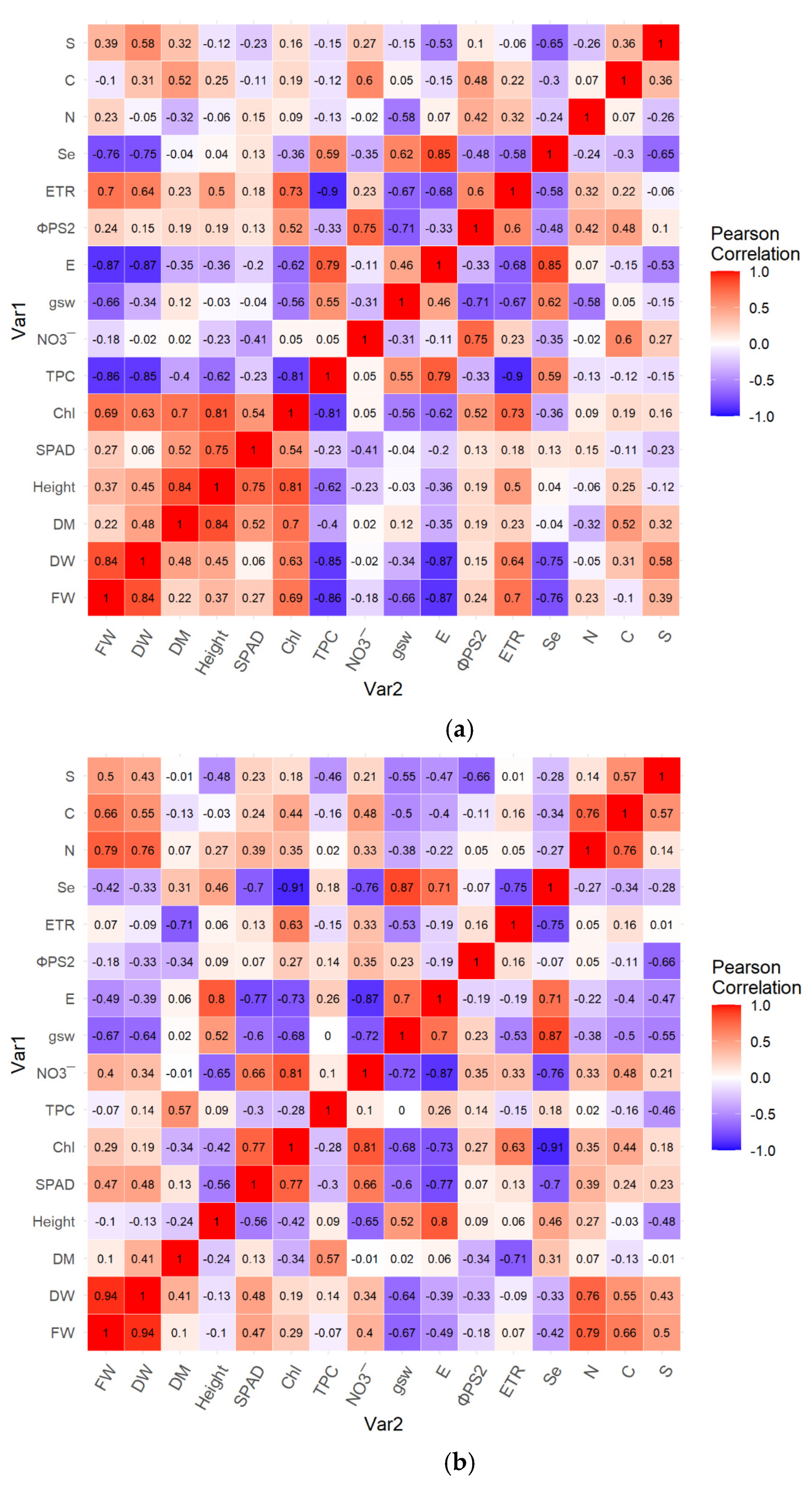
Figure 1. Pearson’s correlation coefficients among measured traits in basil subjected to Se biofortification: ‘Fine Verde’ (a) and ‘Red Rubin’ (b). The colour scale ranges from −1 (strong negative correlation, blue) to +1 (strong positive correlation, red). Traits: yield (FW), dry weight, dry matter percentage, plant height, SPAD index (SPAD), chlorophyll (Chl), total phenol content (TPC), nitrate (NO3), stomatal conductance (gsw), transpiration rate (E), effective quantum yield of PSII (ΦPSII), electron transport rate (ETR), selenium (Se), nitrogen (N), carbon (C), and sulphur (S). Correlations were based on n = 10 observations (df = 8).
Agronomy 15 02368 g001
Figure 2. Principal component biplot of basil traits under Se biofortification (Se 0, Se 5, Se 10, Se 15) in ‘Fine verde’ and ‘Red rubin’. cultivars: yield (FW), dry weight (DW), chlorophyll (Chl), Total Phenol content (TPC), stomatal conductance (gsw), transpiration (E), quantum yield of ΦPSII, electron transport rate (ETR), and selenium (Se).
Agronomy 15 02368 g002
Table 1. Effects of Se biofortification on yield (FW), dry weight (DW), dry matter percentage (DM), and shoot height of two basil cultivars.
FWDWDMShoot Height
g m−2g m−2%cm
Basil cultivar
FV2695.21 ± 130.90 b137.18 ± 7.62 b4.90 ± 0.17 a28.40 ± 0.76 a
RR3068.66 ± 97.59 a156.53 ± 4.80 a5.24 ± 0.09 a26.25 ± 0.39 b
Se Biofortification
Se_03101.73 ± 184.23 a157.96 ± 5.58 a4.88 ± 0.48 a26.56 ± 0.87 a
Se_52975.50 ± 240.25 ab155.06 ± 7.50 a5.44 ± 0.16 a28.25 ± 3.84 a
Se_103011.06 ± 317.78 ab151.66 ± 23.95 a5.15 ± 0.43 a27.67 ± 1.10 a
Se_152439.46 ± 362.79 b122.73 ± 17.13 b4.81 ± 0.34 a26.83 ± 0.94 a
Interaction
FV_03047.66 ± 129.00 a157.66 ± 5.53 ab4.74 ± 0.53 a27.25 ± 0.25 b
FV_52822.06 ± 129.13 ab150.86 ± 6.06 ab5.50 ± 0.10 a31.50 ± 0.90 a
FV_102740.53 ± 57.20 ab132.00 ± 9.33 bc4.81 ± 0.15 a28.38 ± 0.88 ab
FV_152170.60 ± 160.06 b108.20 ± 0.46 c4.54 ± 0.09 a26.50 ± 0.50 b
RR_03155.80 ± 168.60 a158.26 ± 4.00 ab5.02 ± 0.14 a25.87 ± 0.37 b
RR_53128.93 ± 151.06 a159.26 ± 3.53 ab5.37 ± 0.15 a25.00 ± 0.50 b
RR_103281.60 ± 42.80 a171.33 ± 0.40 a5.50 ± 0.14 a26.97 ± 0.27 b
RR_152708.33 ± 165.00 ab137.26 ± 4.19 bc5.07 ± 0.15 a27.17 ± 0.92 b
Basil Cultivar*****ns**
Se Biofortification*****nsns
Cultivar × Se biof.**ns**
Different letters within columns indicate statistically significant differences (p ≤ 0.05) among Se treatments: Se_0 (0 µM), Se_5 (5 µM), Se_10 (10 µM), and Se_15 (15 µM). Data are presented as mean ± standard error (SE). Significance levels: *** = p ≤ 0.001, ** = p ≤ 0.01, * = p ≤ 0.05, and ns = not significant.
Table 2. Leaf concentrations of selenium (Se), nitrogen (N), carbon (C), and sulphur (S) were affected by Se biofortification in two basil cultivars.
SeNCS
mg kg−1 (DW)%
Basil Cultivar
FV24.17 ± 5.52 a6.32 ± 0.05 a35.20 ± 0.36 a0.46 ± 0.04 a
RR22.05 ± 5.19 a6.04 ± 0.10 a33.81 ± 0.69 a0.42 ± 0.02 a
Se Biofortification
Se_00.05 ± 0.02 d6.24 ± 0.20 a34.94 ± 1.01 a0.51 ± 0.17 a
Se_524.36 ± 2.07 c6.17 ± 0.24 a35.48 ± 0.85 a0.45 ± 0.04 a
Se_1030.55 ± 3.60 b6.28 ± 0.09 a34.00 ± 1.16 a0.43 ± 0.06 a
Se_1537.48 ± 1.35 a6.01 ± 0.42 a33.61 ± 2.82 a0.37 ± 0.02 a
Interaction
FV_00.07 ± 0.00 a6.37 ± 0.18 a35.58 ± 0.80 a0.59 ± 0.17 a
FV_525.96 ± 0.60 a6.32 ± 0.19 a35.97 ± 0.74 a0.42 ± 0.01 a
FV_1032.75 ± 2.76 a6.31 ± 0.10 a34.09 ± 0.02 a0.44 ± 0.01 a
FV_1537.91 ± 0.42 a6.27 ± 0.01 a35.18 ± 0.79 a0.37 ± 0.02 a
RR_00.04 ± 0.01 a6.12 ± 0.04 a34.30 ± 0.25 a0.43 ± 0.06 a
RR_522.75 ± 0.97 a6.02 ± 0.09 a35.00 ± 0.28 a0.48 ± 0.03 a
RR_1028.35 ± 1.49 a6.26 ± 0.02 a33.92 ± 1.42 a0.42 ± 0.08 a
RR_1537.05 ± 1.48 a5.76 ± 0.38 a32.05 ± 2.54 a0.36 ± 0.00 a
Basil Cultivarnsnsnsns
Se Biofortification***nsnsns
Cultivar × Se biof.nsnsnsns
Different letters within columns indicate statistically significant differences (p ≤ 0.05) among Se treatments: Se_0 (0 µM), Se_5 (5 µM), Se_10 (10 µM), and Se_15 (15 µM). Data are presented as mean ± standard error (SE). Significance levels: *** = p ≤ 0.001 and ns = not significant.
Table 3. Effect of Se biofortification on SPAD index, total chlorophyll content (Chl), stomatal conductance (gsw), transpiration rate (E), effective quantum yield of PSII (ΦPSII), and electron transport rate (ETR) in two basil cultivars.
Leaf Greenness IndexChla+b ContentgswEΦPSIIETR
SPADmg g−1 FWM m−2 s−1mM m−2 s−1 µM m−2 s−1
Basil cultivar
FV29.97 ± 0.80 b1.31 ± 0.04 a0.22 ± 0.01 a2.39 ± 0.29 a0.72 ± 0.01 a64.07 ± 1.37 a
RR32.50 ± 0.77 a1.10 ± 0.05 b0.18 ± 0.01 b2.57 ± 0.35 a0.67 ± 0.01 b57.47 ± 1.82 b
Se Biofortification
Se_031.65 ± 3.51 a1.32 ± 0.02 a0.16 ± 0.03 b1.60 ± 0.48 c0.71 ± 0.03 a65.61 ± 3.06 a
Se_532.20 ± 1.55 a1.30 ± 0.21 a0.19 ± 0.02 ab1.89 ± 0.63 bc0.71 ± 0.05 a60.89 ± 8.30 ab
Se_1031.89 ± 2.85 a1.14 ± 0.17 b0.20 ± 0.03 ab2.93 ± 0.47 ab0.66 ± 0.05 a57.65 ± 4.66 b
Se_1529.22 ± 1.24 a1.07 ± 0.14 b0.24 ± 0.03 a3.52 ± 0.20 a0.71 ± 0.02 a58.93 ± 2.60 ab
Interaction
FV_028.66 ± 0.23 a1.33 ± 0.00 ab0.19 ± 0.02 a1.43 ± 0.07 a0.74 ± 0.00 a66.53 ± 3.33 ab
FV_531.50 ± 0.90 a1.49 ± 0.04 a0.21 ± 0.00 a2.14 ± 0.68 a0.74 ± 0.00 a67.78 ± 1.35 a
FV_1031.36 ± 2.86 a1.27 ± 0.08 abc0.23 ± 0.02 a2.66 ± 0.16 a0.68 ± 0.03 a60.88 ± 0.57 ab
FV_1528.38 ± 0.88 a1.17 ± 0.08 abc0.23 ± 0.03 a3.35 ± 0.00 a0.72 ± 0.01 a61.11 ± 0.79 ab
RR_034.63 ± 0.79 a1.31 ± 0.02 abc0.13 ± 0.00 a1.77 ± 0.53 a0.69 ± 0.01 a64.68 ± 1.10 ab
RR_532.91 ± 1.35 a1.12 ± 0.03 bc0.17 ± 0.00 a1.64 ± 0.05 a0.68 ± 0.04 a54.01 ± 2.62 b
RR_1032.41 ± 1.85 a1.00 ± 0.03 bc0.18 ± 0.02 a3.19 ± 0.41 a0.64 ± 0.05 a54.43 ± 3.39 b
RR_1530.06 ± 0.36 a0.98 ± 0.06 c0.25 ± 0.01 a3.69 ± 0.03 a0.69 ± 0.01 a56.75 ± 0.20 ab
Basil Cultivar****ns****
Se Biofortificationns*****ns***
Cultivar × Se biof.ns*nsnsns**
Different letters within columns indicate statistically significant differences (p ≤ 0.05) among Se treatments: Se_0 (0 µM), Se_5 (5 µM), Se_10 (10 µM), and Se_15 (15 µM). Data are presented as mean ± standard error (SE). Significance levels: *** = p ≤ 0.001, ** = p ≤ 0.01, * = p ≤ 0.05, and ns = not significant.
Table 4. Effect of Se biofortification on total phenolics content (TPC), nitrate (NO3) in two basil cultivars.
TPCNO3
mg GE g−1 FWmg kg−1 FW
Basil cultivar
FV5.49 ± 0.20 b1983.82 ± 30.97 a
RR10.91 ± 0.24 a1922.01 ± 39.52 a
Se Biofortification
Se_07.87 ± 3.23 a2031.05 ± 47.39 a
Se_57.97 ± 3.54 a2002.53 ± 31.94 a
Se_108.50 ± 3.22 a1854.87 ± 43.83 b
Se_158.47 ± 2.66 a1923.20 ± 146.81 ab
Interaction
FV_05.15 ± 0.27 b2041.12 ± 36.50 a
FV_54.93 ± 0.25 b1989.24 ± 10.70 ab
FV_105.71 ± 0.10 b1855.60 ± 32.60 ab
FV_156.19 ± 0.17 b2049.30 ± 17.42 a
RR_010.59 ± 0.92 a2020.97 ± 42.82 a
RR_511.01 ± 0.49 a2015.82 ± 32.60 a
RR_1011.29 ± 0.17 a1854.14 ± 42.63 ab
RR_1510.75 ± 0.45 a1797.09 ± 14.99 b
Basil Cultivar***ns
Se Biofortificationns**
Cultivar × Se biof.****
Different letters within columns indicate statistically significant differences (p ≤ 0.05) among Se treatments: Se_0 (0 µM), Se_5 (5 µM), Se_10 (10 µM), and Se_15 (15 µM). Data are presented as mean ± standard error (SE). Significance levels: *** = p ≤ 0.001, ** = p ≤ 0.01, and ns = not significant.
Disclaimer/Publisher’s Note: The statements, opinions and data contained in all publications are solely those of the individual author(s) and contributor(s) and not of MDPI and/or the editor(s). MDPI and/or the editor(s) disclaim responsibility for any injury to people or property resulting from any ideas, methods, instructions or products referred to in the content.